notes: ch. 8 blood & serology
TRANSCRIPT
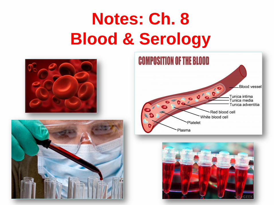
Notes: Ch. 8
Blood & Serology

• Why we need blood types
https://www.youtube.com/watch?v=xfZhb6lmxjk
• Crash course true blood part 1
https://www.youtube.com/watch?v=HQWlcSp9Sls
• Bozeman science blood
https://www.youtube.com/watch?v=KXTF7WehgM8
• ABO simplified http://study.com/academy/lesson/blood-types-abo-
system-red-blood-cell-antigens-blood-groups.html
• FF blood trails https://www.youtube.com/watch?v=ZQVAutGZEdM
• Catching killers
http://www.smithsonianchannel.com/shows/catching-killers/blood-
spatter/1003122/3375551
• http://learn.genetics.utah.edu/content/inheritance/blood/
• http://www.nobelprize.org/educational/medicine/landstei
ner/readmore.html
2

I. Forensic Serology
A. The study of blood, other body fluids, & its
application to the law
– Blood
– Saliva
– Urine
– Semen

B. Blood Clotting and Forensics
• 1. The clotting process begins 3-15 minutes from injury. – 1st forms dark, shiny, jelly-like mass
– 2nd begins to contract and separate from serum.
• 2. How long ago did the victim begin bleeding? – A few minutes: Blood is still liquid
– Less than an hour: Blood is shiny, gelantinous, in a pool.
– Several hours: Blood has separated into clot and serum

II. What is Blood? 1. a liquid tissue
2. 7% of body weight
• Females 4-5 liters (about 1-1.3 gallons)
• males have between 5-6 liters (1.3-1.6
gallons).
3. Blood = cells (45%) & plasma (55%)
a. Plasma – yellow- cells, gases (O2 & CO
2)
and molecules such as proteins, fats and
carbohydrates (sugars).

6
Composition of Blood

Hematocrit
• A blood hematocrit is normally 45% cells and 55% plasma.
Hematocrit means the
percentage of red blood cells
in blood.
Scientists use a centrifuge to
separate the parts of blood
based upon density of the
parts.

4. 3 kinds of cells :
RBCs = erythrocytes.
• Have hemoglobin
• Iron-containing protein
• Carry oxygen /CO2
• No nucleus
WBCs = leukocytes
• immune system cell
• Fight disease/ produce
antibodies
• Contain DNA
• most common types =
lymphocytes and
neutrophils
Platelets = thrombocytes • small cell fragments
• assist in blood clotting

Forensic Science:
Fundamentals &
Investigations, Chapter 8 9
Types of Blood Cells

There are about one billion red blood cells in two
to three drops of blood. For every 600 red blood
cells, there are about 40 platelets and one white
cell.

RBC’s= No DNA, no nucleus
WBC’s = DNA in nucleus
***Blood is Individualized evidence (due to )
but class if just the type is known
DNA

Blood
typing is
considered
class
evidence
and is good
to rule out
suspects
DNA profiling from blood is
considered individual
evidence and can help
pinpoint a suspect

III. Crime Scene Investigator Tasks
A. Is fluid actually blood?
B. Is blood human or animal?
C. Whose is it? Type, alcohol/drugs
D. Perform blood pattern analysis to det.
chain of events, type of injury, etc.

A. Light Source
• high-intensity light or UV lights help
find traces of latent blood and other
bodily fluids.
IV. Is it blood? CSI Tests….
B. Blood Reagent Tests
• based upon hemoglobin properties
• referred to as presumptive tests
“Reaction”

Presumptive Tests for Blood
Determination • 1. Kastle-Meyer test most common-
phenolphthalein reacts with hemoglobin causes deep
pink color
• 2. Hematest® tablet - reacts with the heme (iron)
group in blood causing a blue-green color
• 3. Luminol test (most sensitive)- Solution sprayed
and if “positive” blue glowing light is produced.
– Same chemical used in “glowsticks”
15 Luminol Reaction

Phenolphthalein
• pink color
HemaStix is a strip that
has been coated with
tetramethylbenzidine
(TMB) and will produce
a shade of green or green
spots w/ the presence of
hemoglobin.
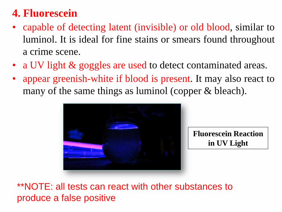
4. Fluorescein
• capable of detecting latent (invisible) or old blood, similar to
luminol. It is ideal for fine stains or smears found throughout
a crime scene.
• a UV light & goggles are used to detect contaminated areas.
• appear greenish-white if blood is present. It may also react to
many of the same things as luminol (copper & bleach).
Fluorescein Reaction
in UV Light
**NOTE: all tests can react with other substances to
produce a false positive

What do you see with luminol?
It glows a bright blue in the dark, when it comes in contact with blood
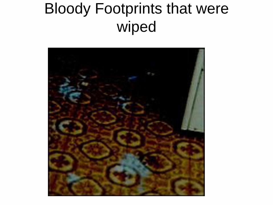
Bloody Footprints that were
wiped

Human vs. Animal Blood

V. It’s blood. Human or Animal?
A. Human
• Red
• Round cells,
depressed donuts
• RBC’s - no nucleus
B. Animals
• Poss. diff. color-
green, blue, yellow, etc.
• diff. shapes, sizes
• Birds/reptiles-
DO have nucleus

Human vs Animal Blood
Animal Blood – red blood cells have larger nuclei
Frog Blood

Microscopic
Views
Bird Blood
Cat Blood
Dog Blood
Fish Blood
Frog Blood
Snake Blood Human Blood
Horse Blood

Human vs. Animal Blood (cont.)
24
C. Precipitin test - very sensitive and
requires only a small amount of blood.
•Human blood injected in rabbit
•Rabbit produces antibodies
•Antibodies extracted as antiserium
•Antiserum exposed to blood
•If it clumps, it’s human blood.

Precipitin Test
(Human blood)
(Human antiserum
made in rabbits)

Blood Types

VI. Blood Terminology
A. Antigen – a protein that can stimulate the body to
make antibodies.
– Certain antigens on rbc account for blood type.
27

Blood Terminology
• B. Antibody – a substance that reacts with an
antigen
• C. Agglutination – clumping of red blood cells;
results if blood types with different antigens are
mixed.
28

VII. Blood Types
A. Blood type established in vitro. (womb)
1. inherit one gene from mom and one from
dad
2.genes cause presence or absence of
proteins (called AGGLUTINOGENS) on
the surface of red blood cells.

• 3. 1901 - Karl Landsteiner discovered three blood types
based on antigens on red blood cells. (A, B, O)
• 4. 1940 Landsteiner reported the discovery of the Rh
factor by studying the blood of the Rhesus monkey.
– 82% of Caucasians, 93% of Hispanic and African
Americans and 99% of all Asians are Rh positive.
30
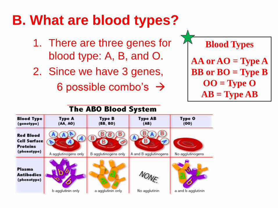
B. What are blood types?
1. There are three genes for
blood type: A, B, and O.
2. Since we have 3 genes,
6 possible combo’s
Blood Types
AA or AO = Type A
BB or BO = Type B
OO = Type O
AB = Type AB

C. Blood Groups ** You can memorize this table or learn how the antigens
react with the antibodies. I suggest the second option!!!
32
Type Antigen
Antibody (will
agglutinate
with)
Can Give
Blood To
Can Get
Blood From
A
B
AB
O
A
B
A and B
Neither
A nor B
B
A
Neither
A nor B
A and B
A, AB O, A
B, AB O , B
AB
A, B, O, AB
A, B, O, AB
O
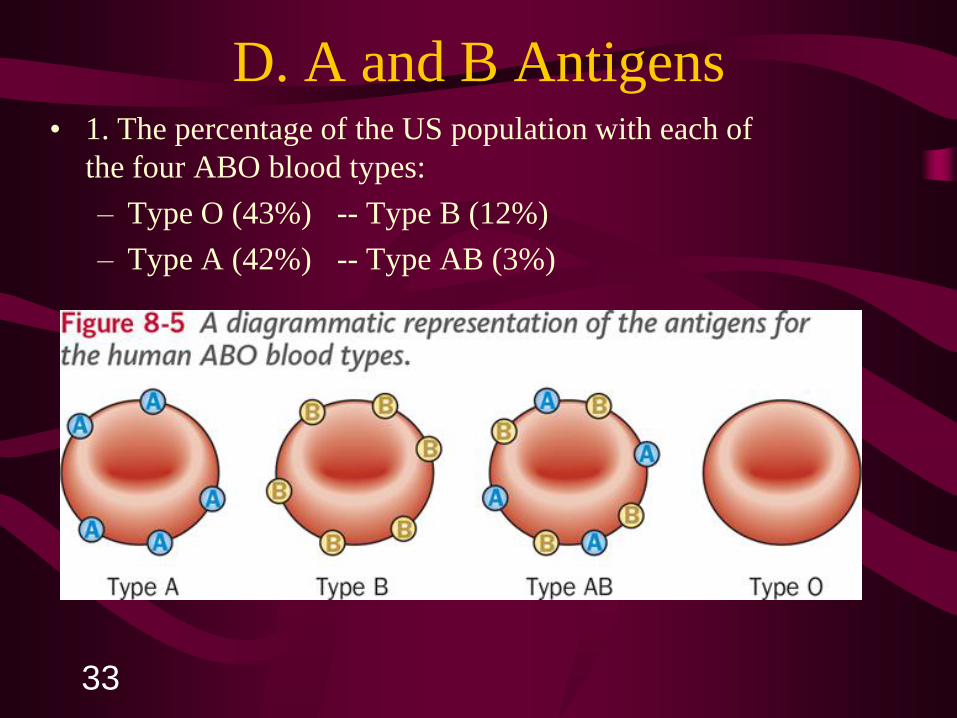
D. A and B Antigens
33
• 1. The percentage of the US population with each of
the four ABO blood types:
– Type O (43%) -- Type B (12%)
– Type A (42%) -- Type AB (3%)
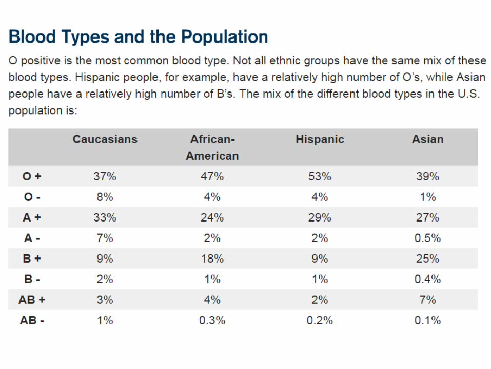

How common is your blood type?
46.1%
38.8%
11.1%
3.9%

E. Rh Factors • 1. Eighty-five percent of the human population has a
protein called Rh factor on their red blood cells.
• 2. Found from studying Rhesus monkey.
• 3. The presence of a specific protein, or lack of it, is referred to as the Rh (for Rhesus) factor.
• Yes protein = Rh positive (Rh+)
• No protein = Rh negative (Rh-).
A+ A-
B+ B-
AB + AB-
O + O-

F. Blood Transfusions (donations) 1. blood transfusion is a procedure in which blood is given to a patient
through an intravenous (IV) line in one of the blood vessels. Blood
transfusions are done to replace blood lost during surgery or a serious
injury. A transfusion also may be done if a person’s body can't make blood
properly because of an illness.
• 2. Who can give you blood?
• Type 0 Universal Donors, can GIVE
blood to any blood type.
• TYPE AB Universal Recipients,
• can RECEIVE any blood type.
Rh+ Can receive + or -
Rh- Can only receive -
Universal Donor
Universal Recipient

3. Antigen-Antibody Response
• a. occurs when white blood cells recognize a
substance as foreign and try to destroy it.
38

4. Agglutination
• a. If clumping occurs within the circulatory
system blood could cease to flow.
• b. Without blood circulation, a person dies.
39

E. Test for determining Blood Type
• 3 serums called: anti-A, anti-B, and anti-Rh
• Mix one drop of blood w/ each serum respectively.
• If mixture coagulates (looks spotted/chunky) then
the person has that protein.
A B + or -


• Genotype: describes the allelic pair
combination
• Phenotype: describe the trait/characteristic
expressed

Genetic Blood Typing (Genotypes)
Mother
Father
A B O
A AA AB AO
B AB BB BO
O AO BO OO

Transmission of our traits
Allele Combinations
Blood Type
(phenotype)
AA + AO A
BB +BO B
AB AB
OO OO

VIII. Bloodstain Pattern
Analysis

A. Blood Splatter
o 1. 1939—splatter patterns first analyzed
o 2. Blood may splatter when a wound is inflicted
o 3. Blood splatter pattern—a grouping of blood stains
o 4. Patterns help to reconstruct the events surrounding a shooting, stabbing, or beating

B. Analysis of a spatter pattern can help to determine:
1. direction blood traveled
2. angle of impact
3. point of origin of the blood
4. velocity of the blood
5. manner of death
Bloodstain pattern analysis can also help one evaluate the credibility of statements provided by a witness, a victim, or a suspect.

C. Cohesion
1. Blood sticks together as it falls maintaining a
round shape.
2. Blood also resists flattening out when it falls on
a flat surface. Cohesion and surface tension
help it to maintain a curved shape.
D. The shape of an individual drop of blood can provide important clues
• 1. When blood falls from a height, or at a high velocity, it forms satellite droplets, small secondary droplets around the main drop (parent drop)
• 2. When blood falls onto a less-than-smooth surface, the edges may have spikes or extensions

E. An elongated blood drop indicates blood was traveling
from a different direction when it landed.
1. The point of impact may appear darker and wider
with a tail pointing in the direction of the blood’s
movement.
a. Smaller, secondary droplets may break off and will
land in front of the moving droplet of blood,
allowing scientists to determine direction of
spatter.

Forensic Science:
Fundamentals &
Investigations, Chapter 8 50

F. When there are two or more blood spatters a scientist can draw LINES OF CONVERGENCE that can pinpoint the location of the blood source.

Forensic Science:
Fundamentals &
Investigations, Chapter 8 52
G. Blood Splatter Analysis
—Six Patterns
Describe each of these:
a) Passive drops
b) Arterial gushes
c) Splashes
d) Smears
e) Trails
f) Pools

Passive drops 6. Arterial
gushes Splashes
Smears Trails Pools
2. Splashes help show position of victim
3. Smears – bleeding victim touching walls or furniture 4. Blood Trails – victim moving from one place to another 5. Blood Pools – victim bleeds heavily
1. Passive fall (90o angle to floor)– circular drops w/secondary satellites

1. Smear patterns from a large volume of blood, at least 0.5 ml, are often
distorted so much that further classification is not possible.
2. However, Transfer Patterns occur when a wet bloody surface contacts a
second unstained surface creating recognizable mirror image or at least
a recognizable portion of the original surface.
a. Swipe Pattern - the transfer of blood onto a surface not already
contaminated with blood. One side is usually feathered which
indicates the direction of travel.
i. One common pattern at scenes is a hair swipe - a long thin
fine line transfer.
b.Wipe Pattern - created when an object moves through blood that
has not completely dried and moves, removes, or otherwise alters
it.

1.Arterial spurting usually occurs when an artery is damaged and the blood spurts or gushes from the wound in large volume pulses. It continues spurting as long as the heart continues beating.
2.Large drops striking a vertical surface decelerate from air resistance and produce a pattern without spines. The drops strike the surface and then characteristically drip or run downward due to their large volume.

H. Speed and Velocity also impact blood spatter.
High Velocity Medium Velocity Low Velocity
Example:
Gunshot wound
Beating,
stabbing
Blunt object
impact Size of blood
droplets: Less than 1 mm 1-4 mm 4-6 mm

1. The movement and the number of swings can often be documented by examining the cast-off pattern.
2. During a beating with an instrument which produces the bleeding, blood will not normally collect on the surface of the instrument from the first strike.
3. On subsequent strikes at the same location, blood will adhere to the instrument since it now strikes a blood source. When the instrument is raised or swung backward, its movement allows small drops of blood to be released from its surface.
Some of these small drops will strike a surface,
often a ceiling, at a 90-degree impact angle.

The field of bloodstain pattern analysis requires knowledge of
math, physics, biology and chemistry. Students in Criminology
and Criminal Justice learn about bloodstain pattern analysis in
forensic science classes or classes specifically on blood spatter.
But most analysts begin as law-enforcement officers who learn
on the job, acquire certifications and take courses, workshops
and seminars. Many train in bloodstain pattern (or blood
spatter) analysis through the International Association of
Bloodstain Pattern Analysis (IABPA). The IABPA developed
criteria for the Basic Bloodstain Pattern Analysis Course, an
introductory 40-hour course on the subject. Other organizations,
such as the International Association for Identification (IAP),
offer workshops and seminars as well as advanced courses
which lead to certification in blood spatter analysis.

IX. What is BPA?
(Bloodstain Pattern Analysis)
a. Analysis of bloodstain patterns left by
falling, projected or smeared blood.
b. Observation/measurement of position and
shape of bloodstains give lots of info:
1. direction of travel
2. height above target
3. angle of impact
4. speed or velocity
c. Provide a determination of the physical
events responsible for their deposition

Quote:
“Through the examination of
bloodstains and bloodstain
patterns, in association with
knowledge of the underpinning
sciences, they provide a
determination of the physical
events responsible for their
deposition.”
RCMP - Royal Canadian Mounted Police

d. Why is it important in Forensics?
1. can prove or refute a suspect’s account
of what happened
2. can possibly be used to reconstruct a
crime
3. can tell us the “how” of a crime.

X. Surface/Height & shape of blood drops
A. Porous B. Nonporous
Glass
Linoleum
Concrete
Wood

Impact Types &
Angle of Impact

*Diameter of bloodstains ↑ = Height ↑

A. Low impact
• blood under the influence of gravity
– it just falls.
• dripping
XI. 3 Types of Impact

B. Medium Impact
• Medium impact occurs when a force, such
as a bat, is applied.

C. High Impact • fine mist of droplets
• gunshot, heavy machinery

• “The tail tells the tale”
• The tail points the direction the blood is going
XII. Direction of Travel

XIII. Calculating Angle of Impact 1. Measure WIDTH (in cm.)
- may be up and down, do not include spines
2. Measure LENGTH
- do not include spines or tails
3. Formula: Angle = sin-1 (width/length)
- divide smaller # by larger #

WS: Angle of Impact Practice
1.) Width = mm.
Length = mm.
Angle of Impact ° = Sin -1 x
A.O.I.° = °
Width
Length ( )
Smaller #
Larger #
1.0
1.4
Sin -1 x = ( ) 1.0
1.4
Sin -1 x = (0.714)
**MAKE SURE YOUR CALCULATOR IS IN DEGREES!!!
