notes: ch 48 neurons, synapses, and signaling. a nervous system has three overlapping functions: 1)...
TRANSCRIPT
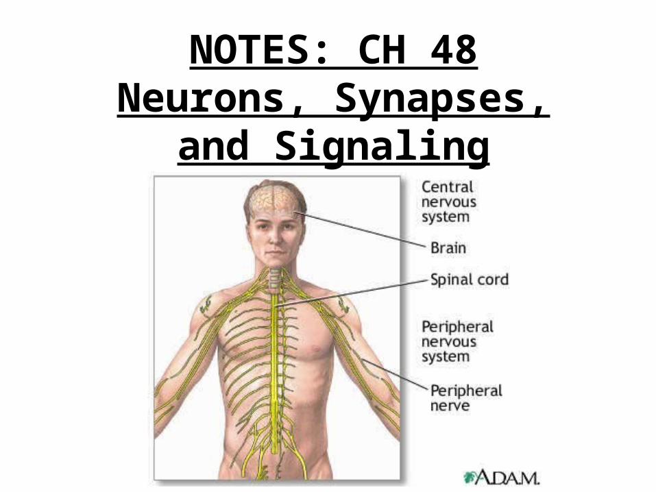
NOTES: CH 48Neurons, Synapses, and
Signaling

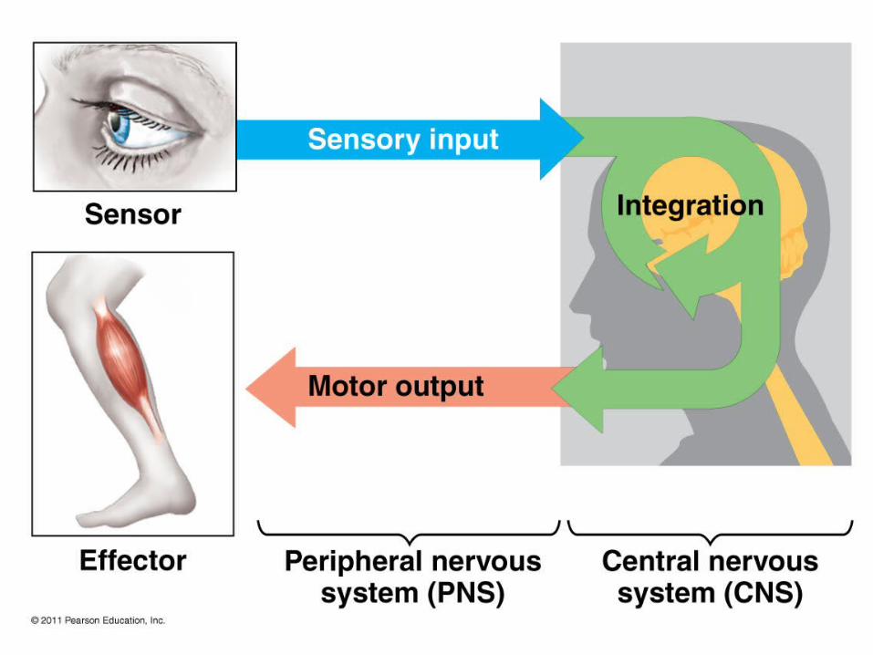
A nervous system has three overlapping functions:
1) SENSORY INPUT: signals from sensory receptors to integration centers
2) INTEGRATION: information from sensory receptors is interpreted and associated with appropriate responses
3) MOTOR OUTPUT: conduction of signals
from the integration center to effector cells (muscle cells or gland cells)

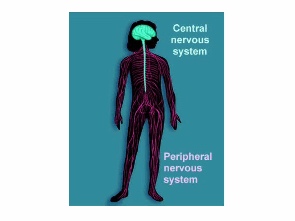
*CENTRAL NERVOUS SYSTEM (CNS)
integration center
brain and spinal cord

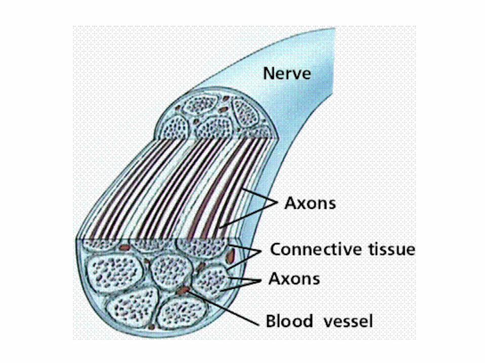
*PERIPHERAL NERVOUS
SYSTEM (PNS)
made up of nerves
(ropelike bundles
of neurons)
nerves communicate
motor and sensory
signals to and from CNS
and rest of body
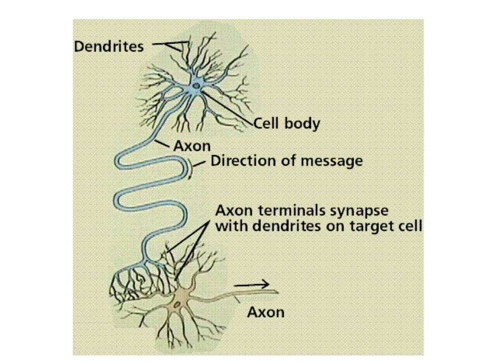
Two Main Classes of Cells:1) NEURONS:
functional unit of the nervous system
transmits signals from one location to another
made up of: cell body, dendrites, axon
many axons are enclosed by an insulating layer called the MYELIN SHEATH
include: sensory neurons,
interneurons,
motor neurons


2) GLIAL CELLS (“GLIA”) - SUPPORTING CELLS
10 to 50 times more numerous than neurons
provide structure; protect, insulate, assist neurons
example: Schwann cells and oligodendrocytes form myelin sheaths in the PNS and CNS, respectively
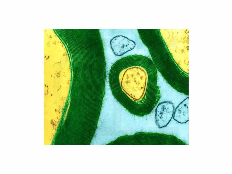
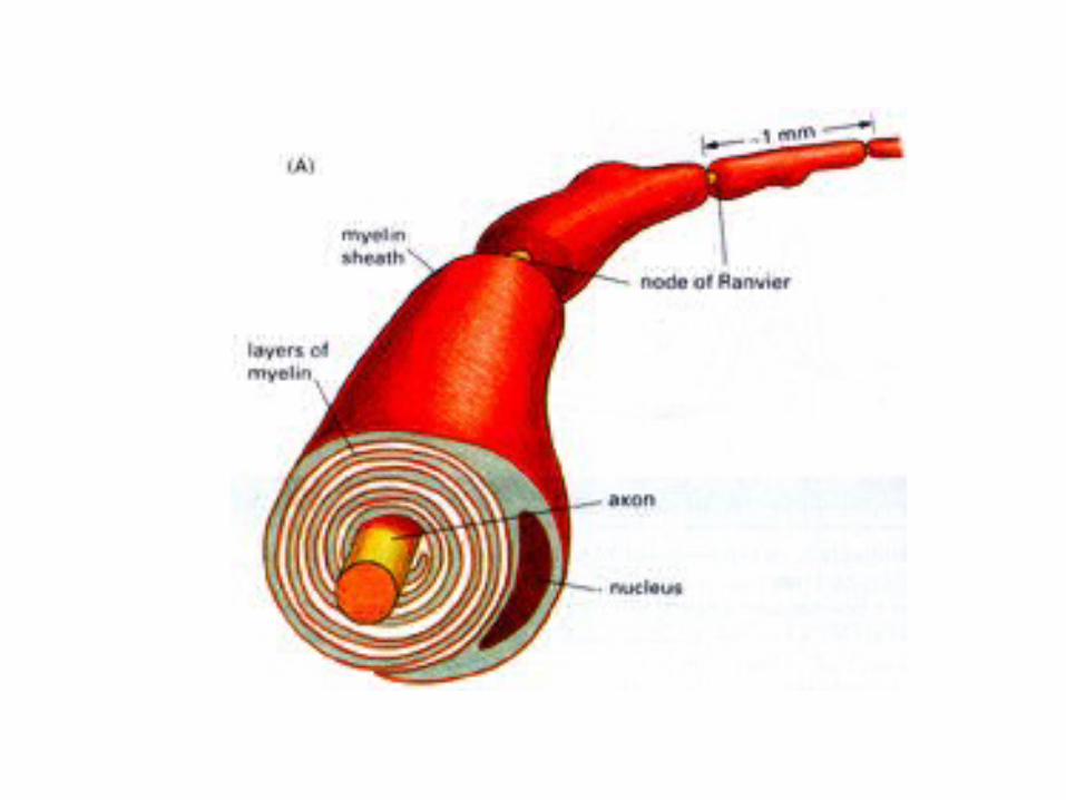

MYELIN SHEATH:
produced by Schwann cells in the peripheral nervous system;
gaps between successive Schwann cells are called NODES OF RANVIER….
***the #10 term!!!
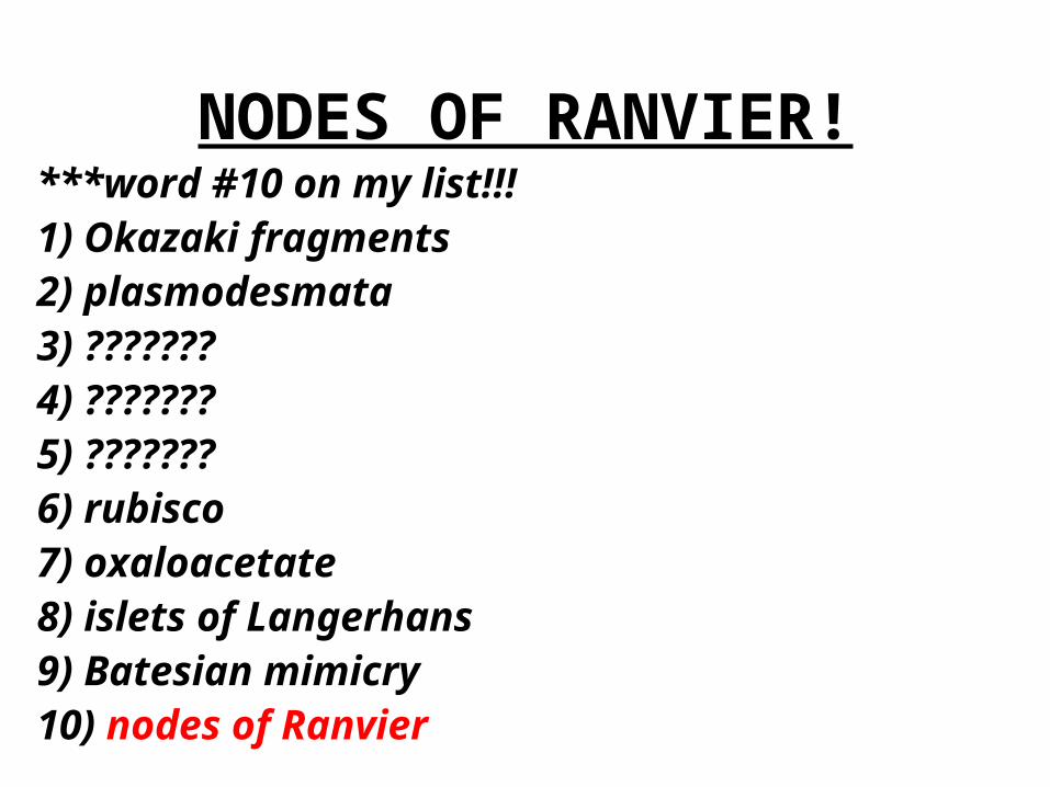
NODES OF RANVIER!***word #10 on my list!!!1) Okazaki fragments2) plasmodesmata3) ???????4) ???????5) ???????6) rubisco7) oxaloacetate8) islets of Langerhans9) Batesian mimicry10) nodes of Ranvier
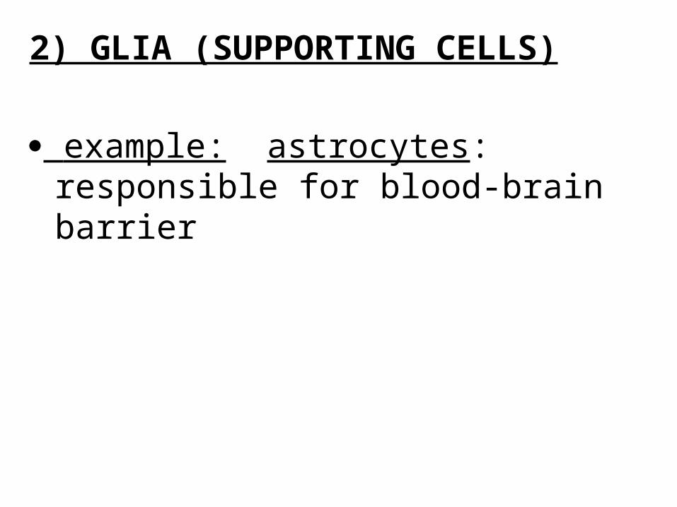
2) GLIA (SUPPORTING CELLS)
example: astrocytes: responsible for blood-brain barrier
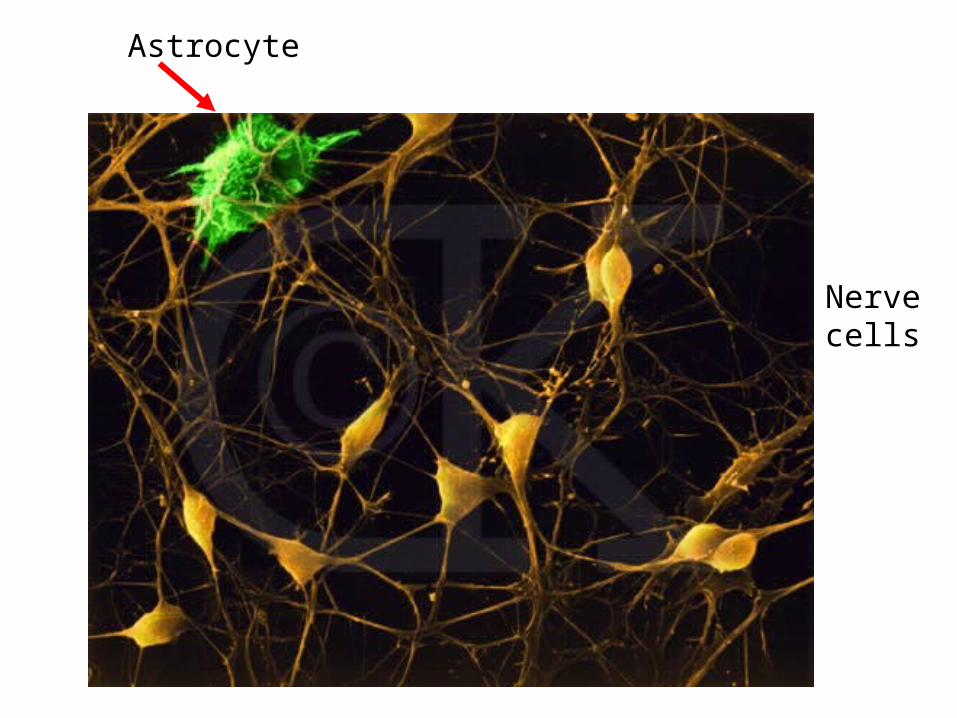
Nerve cells
Astrocyte
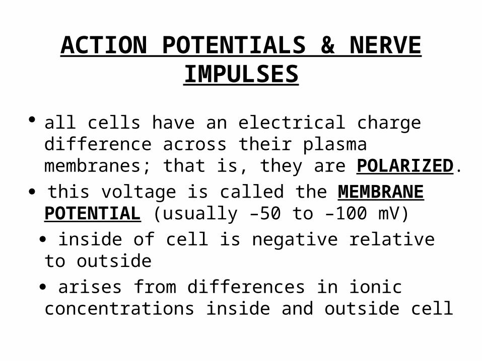
ACTION POTENTIALS & NERVE IMPULSES
all cells have an electrical charge difference across their plasma membranes; that is, they are POLARIZED.
this voltage is called the MEMBRANE POTENTIAL (usually –50 to –100 mV)
inside of cell is negative relative to outside
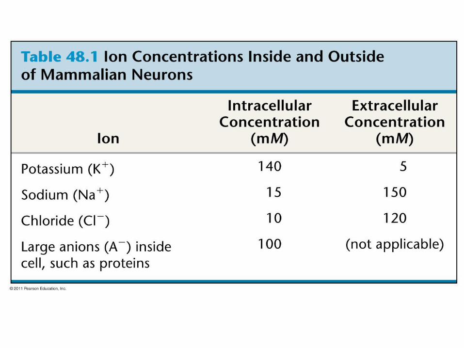
arises from differences in ionic concentrations inside and outside cell
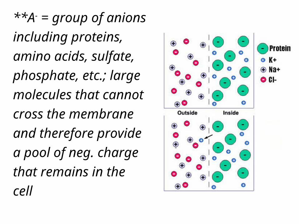
**A- = group of anions
including proteins,
amino acids, sulfate,
phosphate, etc.; large
molecules that cannot
cross the membrane
and therefore provide
a pool of neg. charge
that remains in the
cell
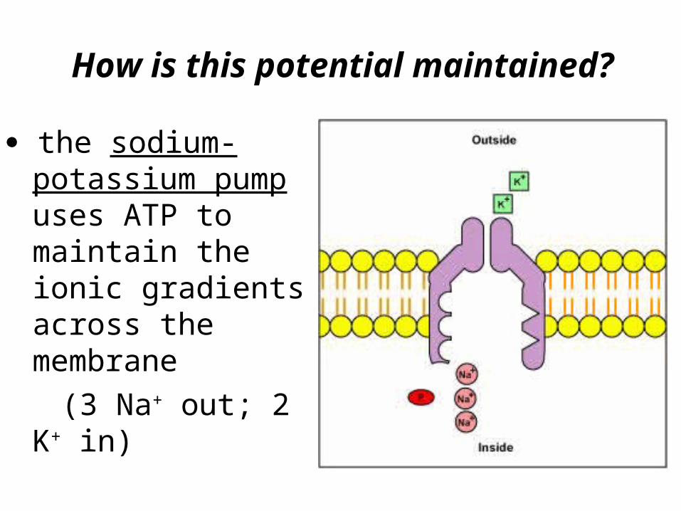
the sodium-potassium pump uses ATP to maintain the ionic gradients across the membrane
(3 Na+ out; 2 K+ in)
How is this potential maintained?

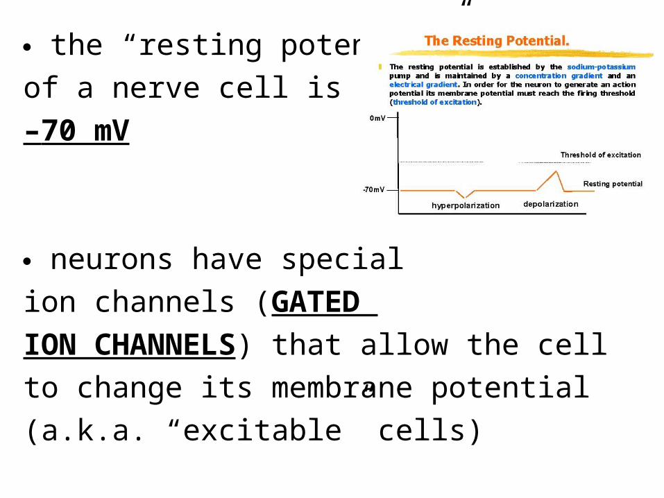
the “resting potential”
of a nerve cell is approx.
–70 mV
neurons have special
ion channels (GATED
ION CHANNELS) that allow the cell
to change its membrane potential
(a.k.a. “excitable” cells)

when a stimulus reaches a neuron, it causes the opening of gated ion channels
(e.g.: light photoreceptors in the eye; sound waves/vibrations hair cells in inner ear)

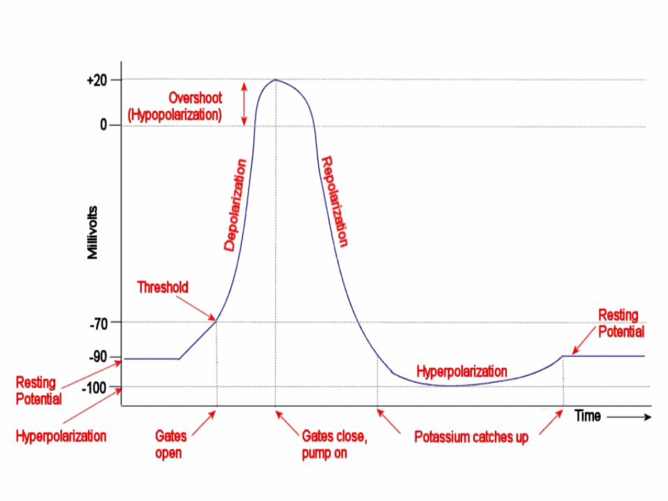
HYPERPOLARIZATION: membrane potential becomes more negative (K+ channel opens; increased outflow of K+)
DEPOLARIZATION: membrane potential becomes less negative
(Na+ channel opens; increased inflow
of Na+)
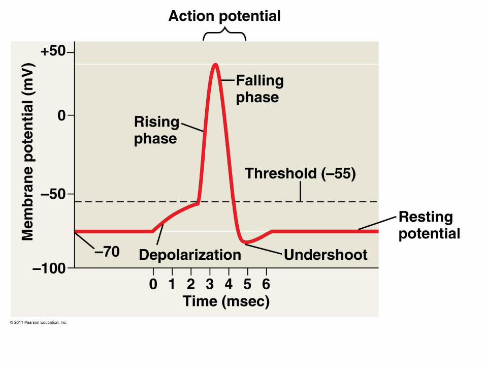
**If the level of depolarization reaches the THRESHOLD POTENTIAL, an ACTION POTENTIAL is triggered.
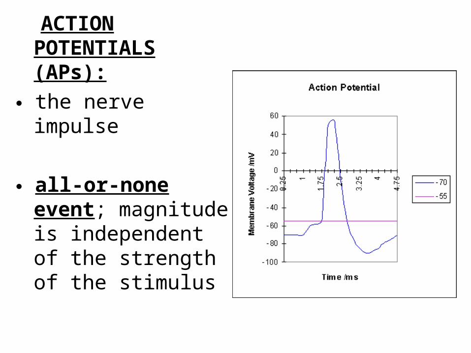
ACTION POTENTIALS (APs):
the nerve impulse
all-or-none event; magnitude is independent of the strength of the stimulus
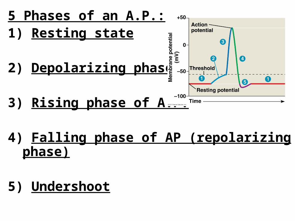
5 Phases of an A.P.:1) Resting state 2) Depolarizing phase
3) Rising phase of A.P.
4) Falling phase of AP (repolarizing phase)
5) Undershoot
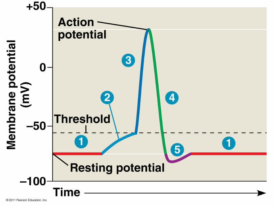

Phase of A.P.
State of Voltage-Gated Sodium (Na+) Channel
State of Voltage-Gated Potassium (K+) channelActivation
gateInact. Gate
Entire channel
1) Resting closed
open closed closed
2 & 3) Depolari-
zation
open open open closed
4) Repolar-ization
open closed closed open
5) Undersho
ot
closed
closed closed open


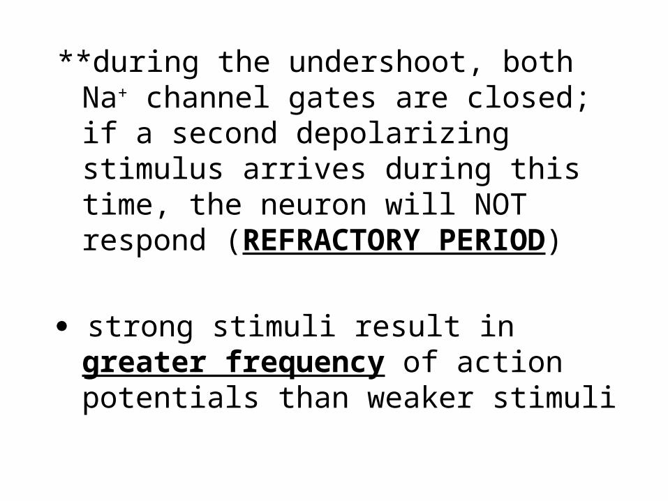
**during the undershoot, both Na+ channel gates are closed; if a second depolarizing stimulus arrives during this time, the neuron will NOT respond (REFRACTORY PERIOD)
strong stimuli result in greater frequency of action potentials than weaker stimuli


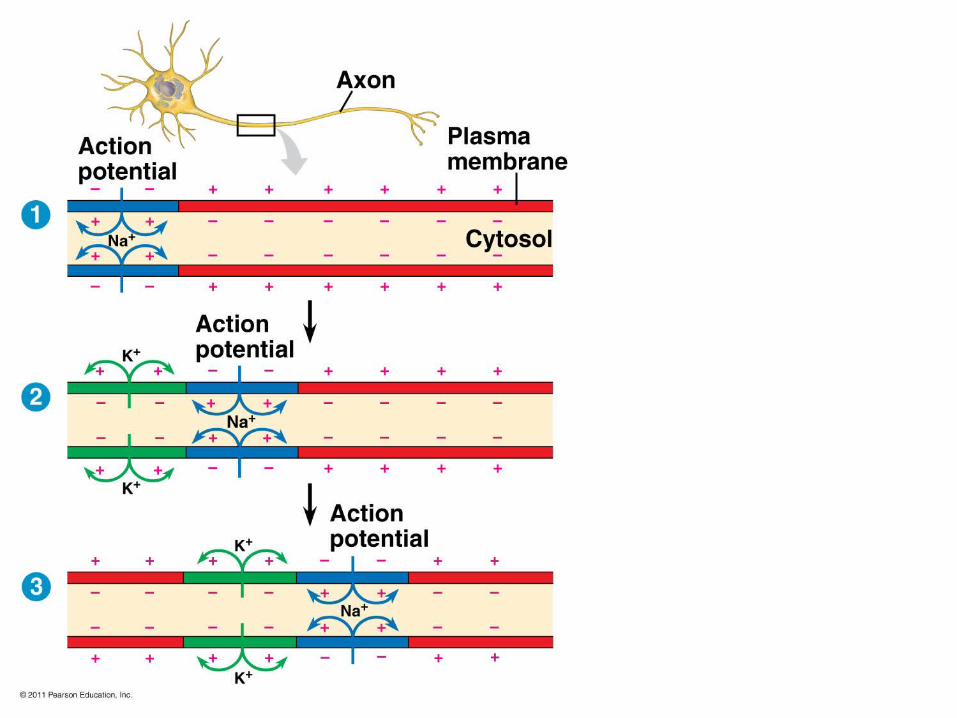
How do action potentials “travel” along an axon?
the strong depolarization of one action potential assures that the neighboring region of the neuron will be depolarized above threshold, triggering a new action potential, and so on…


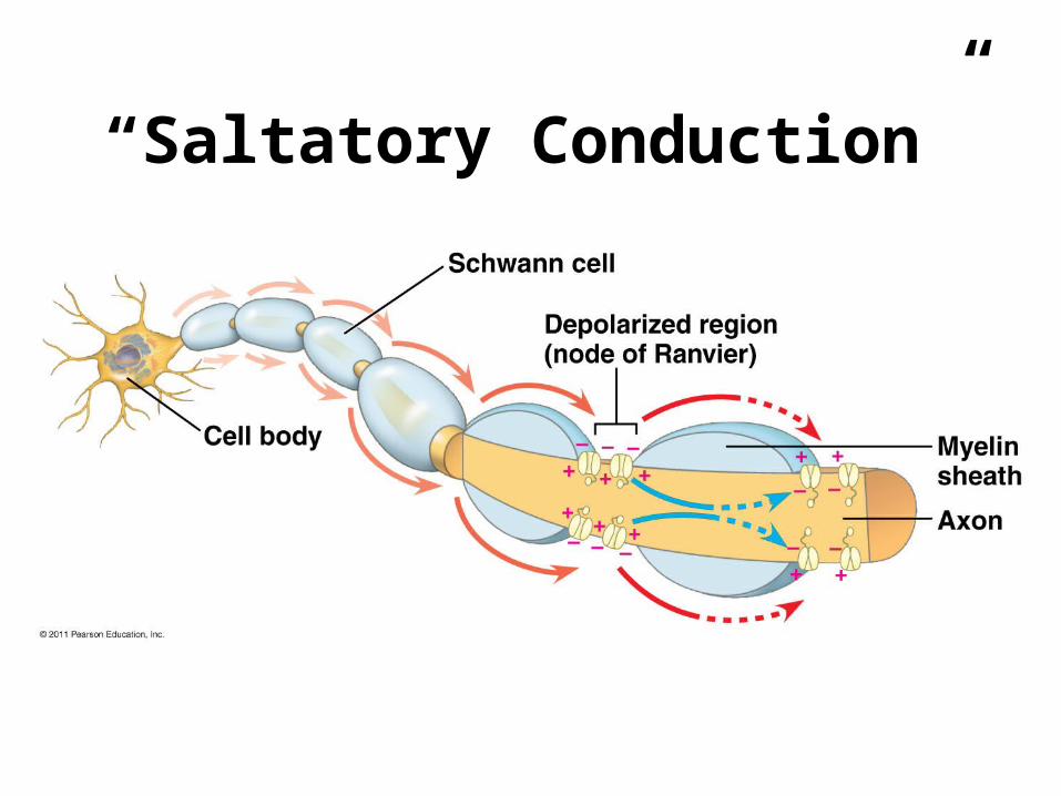
“Saltatory Conduction”
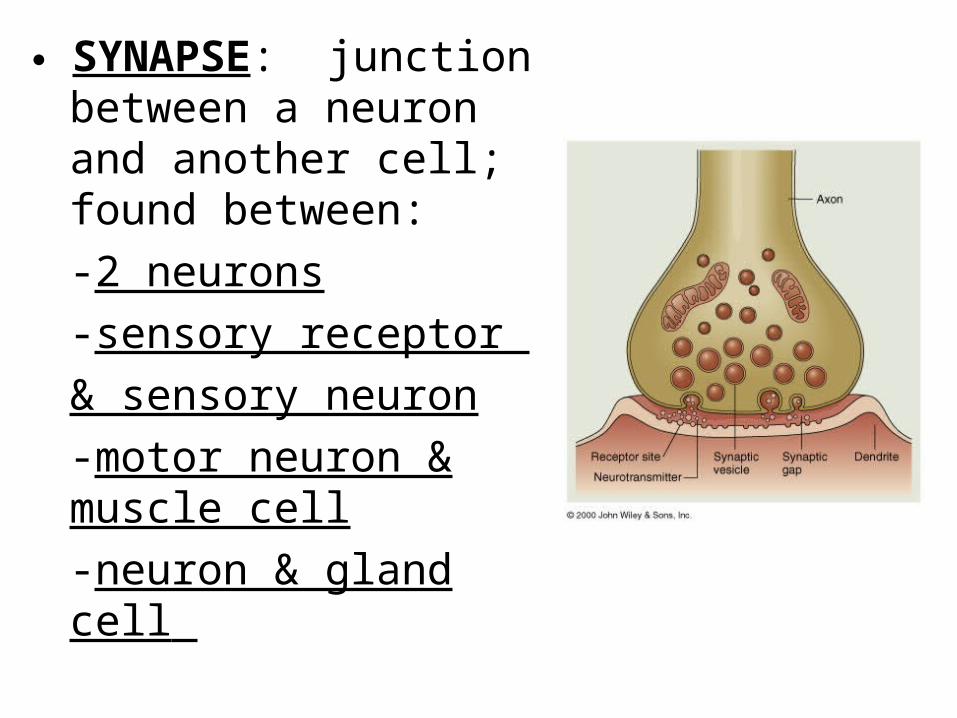
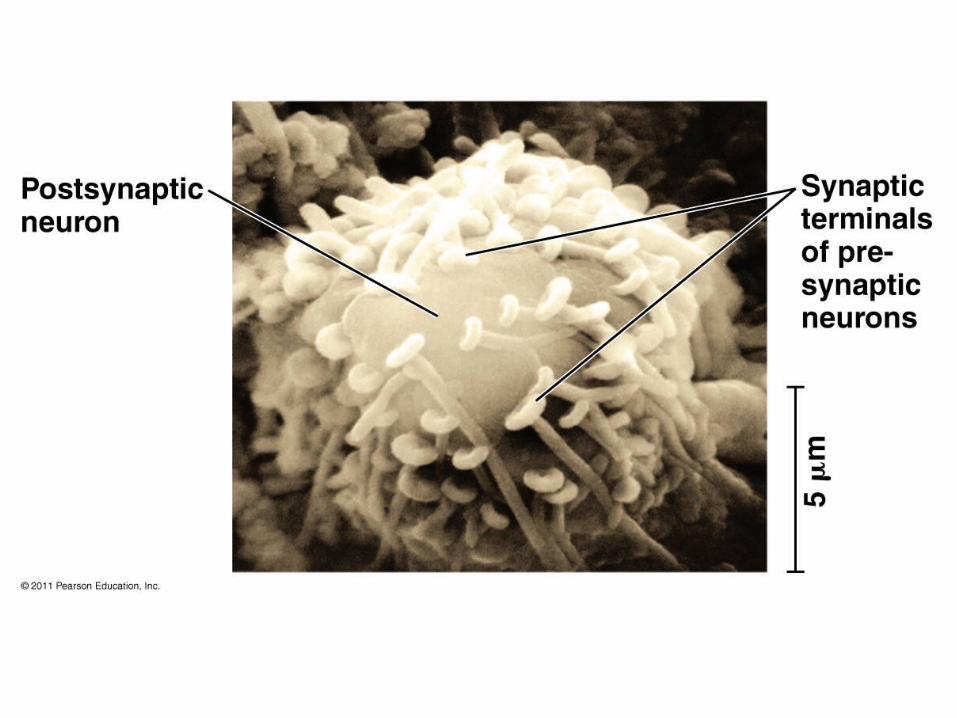
SYNAPSE: junction between a neuron and another cell; found between:
-2 neurons
-sensory receptor
& sensory neuron
-motor neuron & muscle cell
-neuron & gland cell
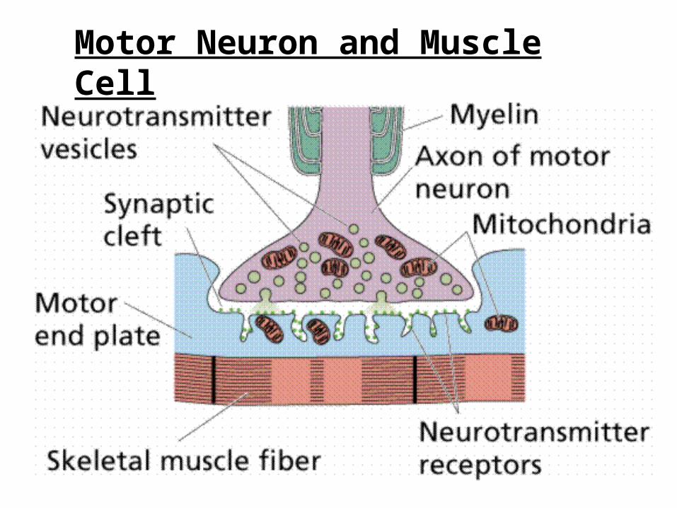
Motor Neuron and Muscle Cell
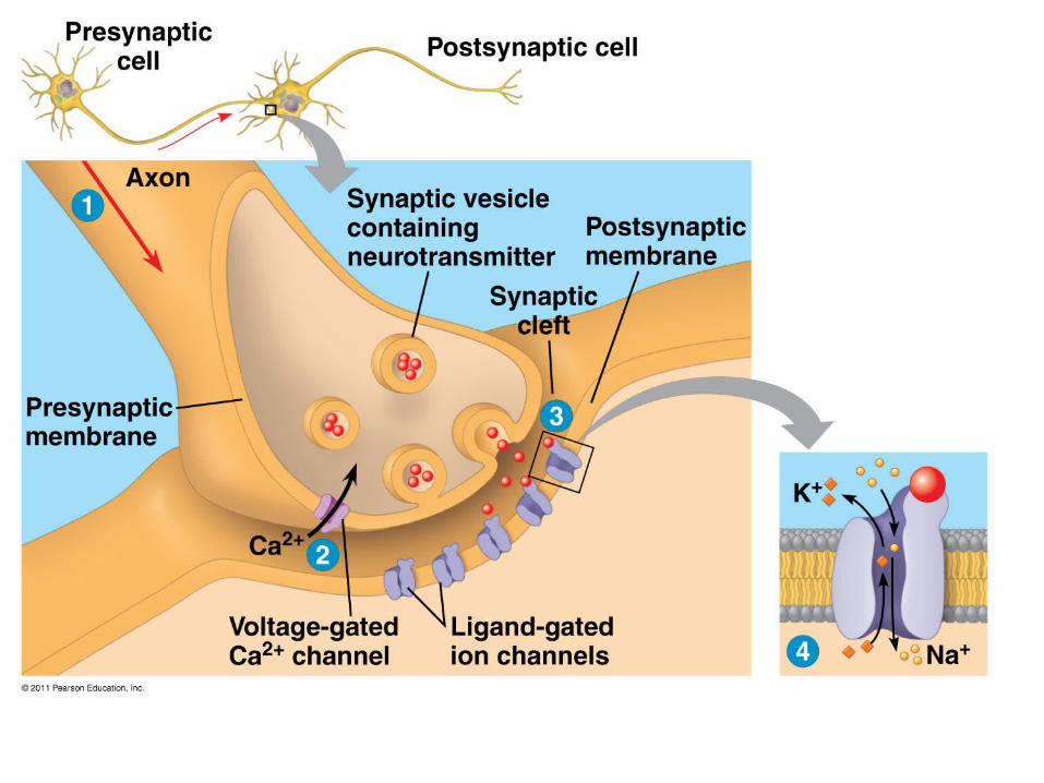

Presynaptic cell = transmitting cell
Postsynaptic cell = receiving cell

Electrical Synapses: allow action potentials to spread directly from pre- to postsynaptic cell
*connected by gap junctions (intercellular channels that allow local ion currents)
**Most synapses are…
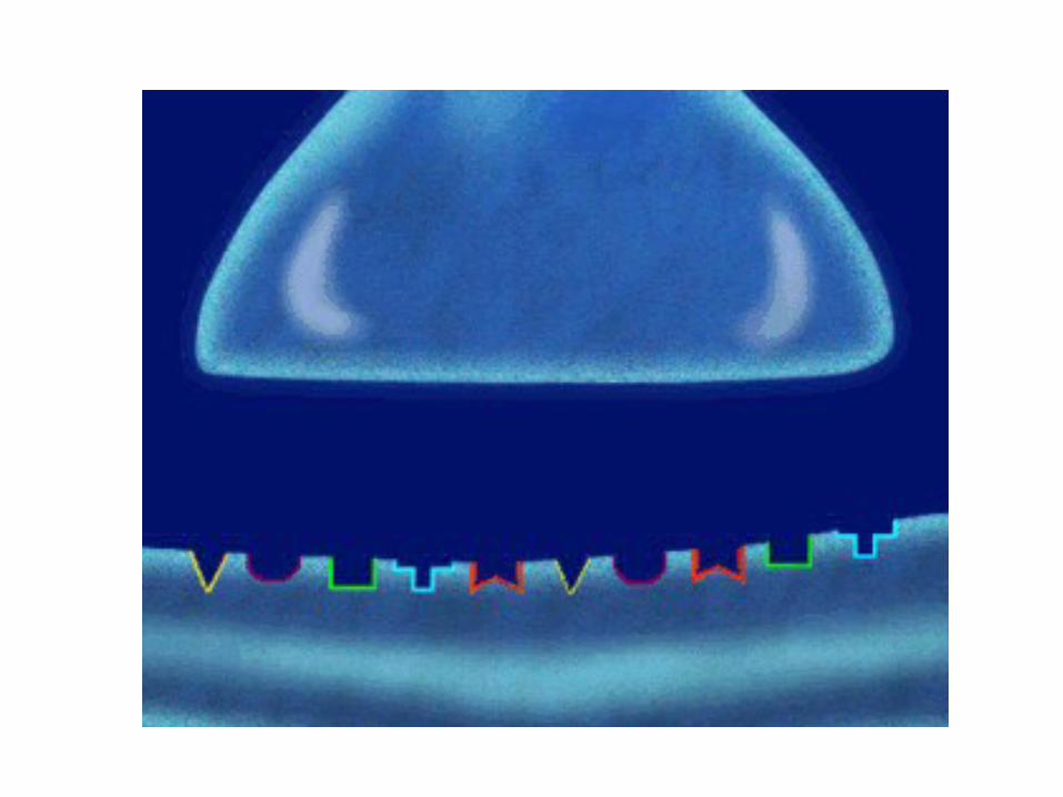
Chemical Synapses: cells are separated by a synaptic cleft, so cells are not electrically coupled; a series of events converts:
elec. signal chem.signal elec.signal
HOW???...

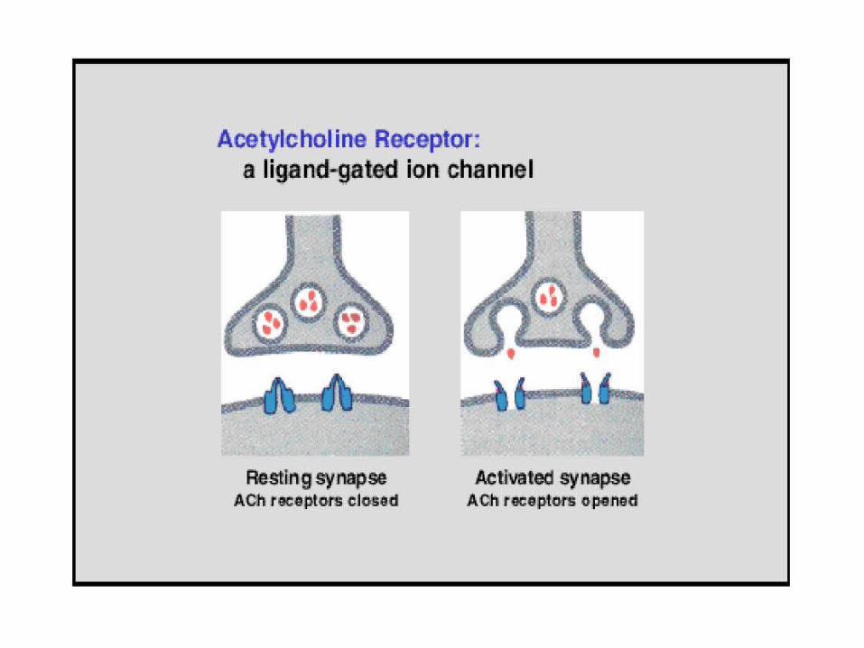
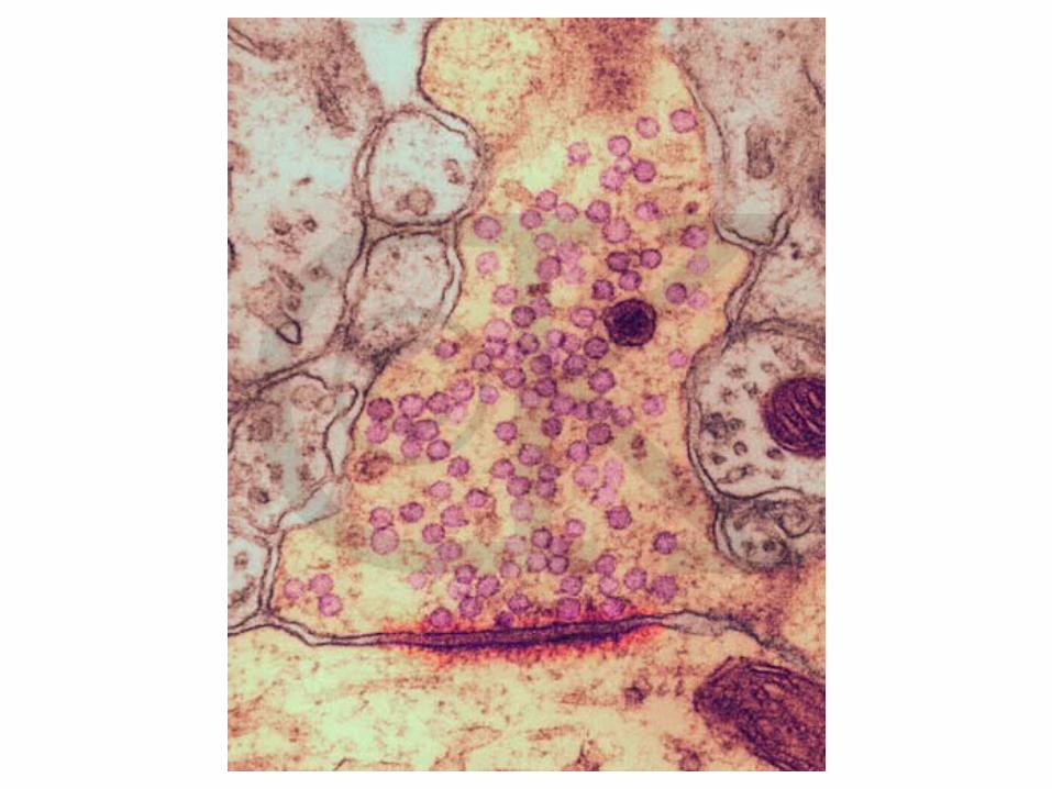
NEUROTRANSMITTERS: intercellular messengers; released into synaptic cleft when synaptic vesicles fuse with presynaptic membrane
specific receptors for neurotransmitters project from postsynaptic membrane; most receptors are coupled with ion channels
neurotransmitters are quickly broken down by enzymes so that the stimulus ends


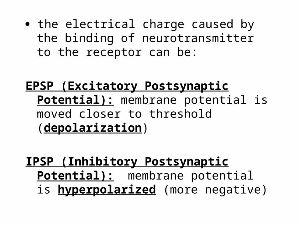
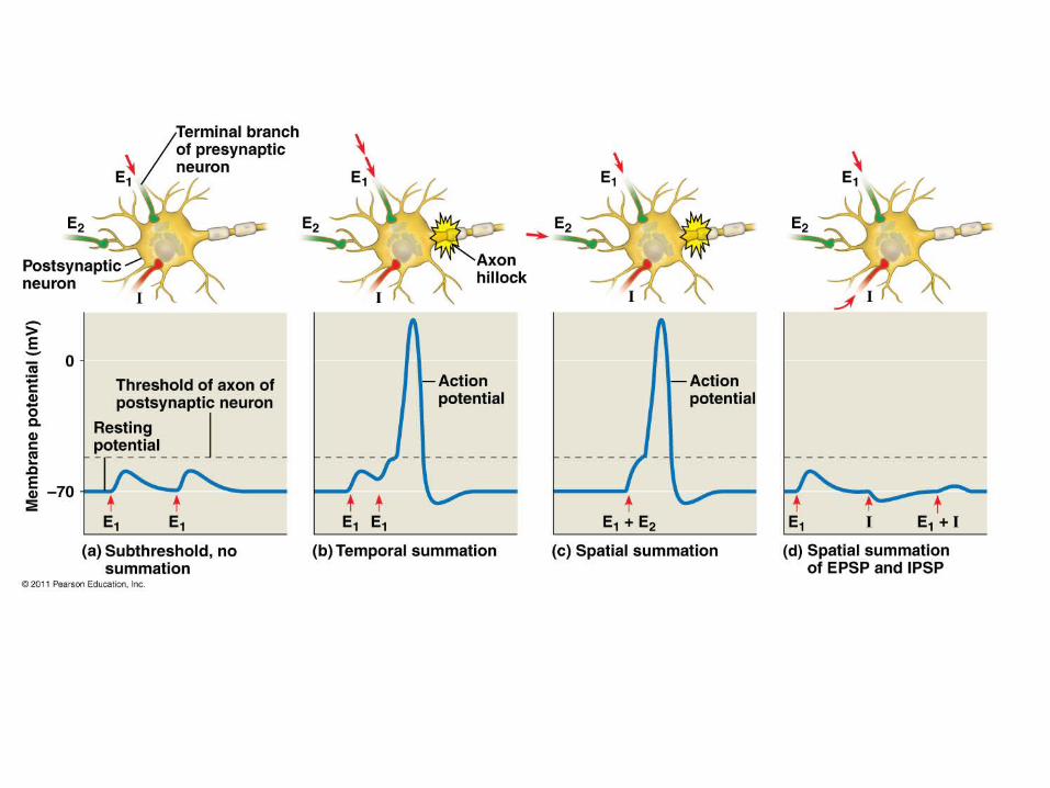
the electrical charge caused by the binding of neurotransmitter to the receptor can be:
EPSP (Excitatory Postsynaptic Potential): membrane potential is moved closer to threshold (depolarization)
IPSP (Inhibitory Postsynaptic Potential): membrane potential is hyperpolarized (more negative)

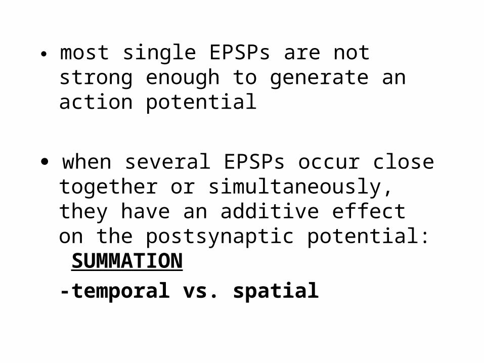
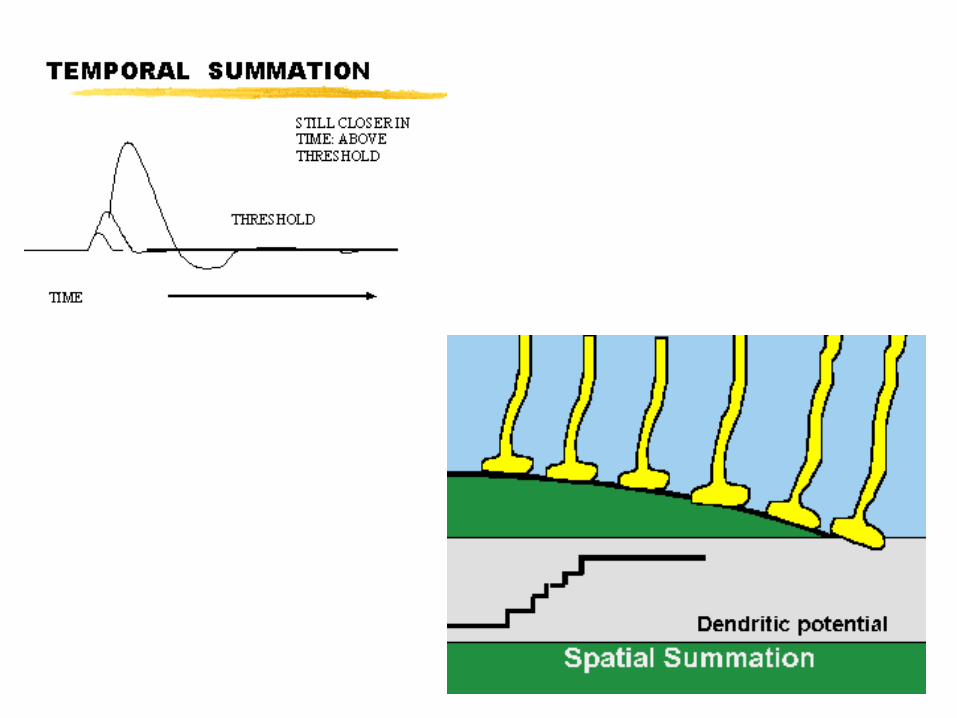
most single EPSPs are not strong enough to generate an action potential
when several EPSPs occur close together or simultaneously, they have an additive effect on the postsynaptic potential: SUMMATION
-temporal vs. spatial

Examples of neurotransmitters: **acetylcholine
Neuromuscular junction; can be inhibitory or excitatory
epinephrine
norepinephrine
dopamine
serotonin
endorphins
Decrease perception of pain by CNS; (heroin & morphine mimic endorphins)
dop. & ser. both affect sleep, mood,attention, learning; LSD & mescaline bind to ser. & dop. receptors
epin. & norep. also function ashormones; “fight or flightresponse”

Neurotransmitters: Ach
ACETYLCHOLINE: triggers skeletal muscle fibers to contract…
so, how does a muscle contraction stop???
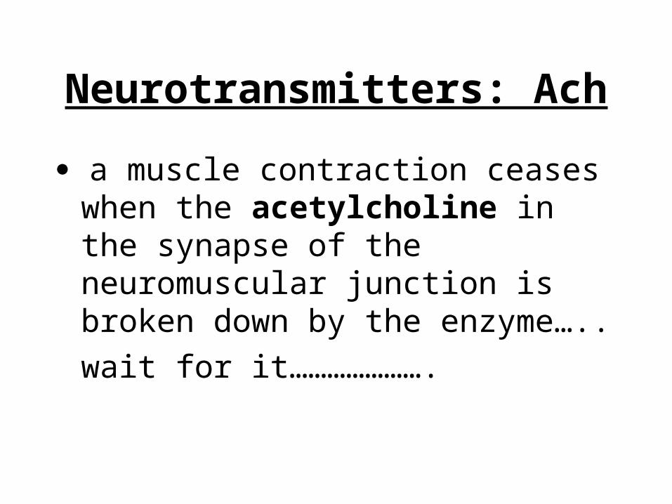
Neurotransmitters: Ach
a muscle contraction ceases when the acetylcholine in the synapse of the neuromuscular junction is broken down by the enzyme…..
wait for it………………….
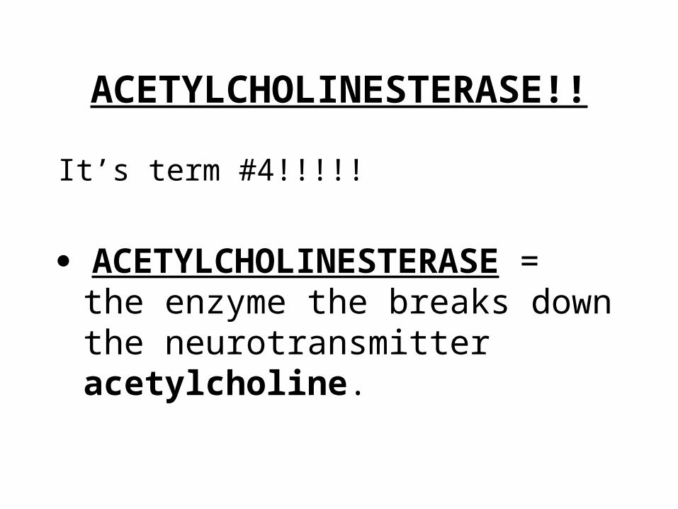
ACETYLCHOLINESTERASE!!
It’s term #4!!!!!
ACETYLCHOLINESTERASE = the enzyme the breaks down the neurotransmitter acetylcholine.
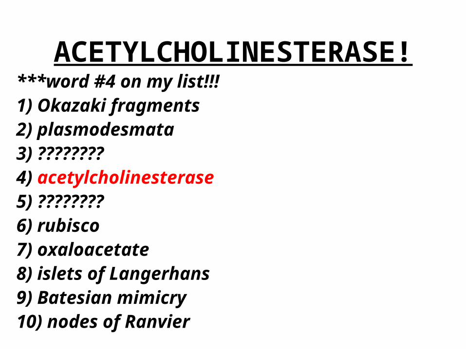
ACETYLCHOLINESTERASE!***word #4 on my list!!!1) Okazaki fragments2) plasmodesmata3) ????????4) acetylcholinesterase5) ????????6) rubisco7) oxaloacetate8) islets of Langerhans9) Batesian mimicry10) nodes of Ranvier