northumbria fdr xair system trial
TRANSCRIPT
Author: Jessica Brealey –Trial Team Lead and Radiographer
NORTHUMBRIA HEALTHCARE NHS FOUNDATION TRUST
NORTHUMBRIA
FDR XAIR SYSTEM
TRIAL
Phase 3 Report
Project Lead & Trust Lead Radiographer– Deborah Henderson
Jessica Brealey (RTF) NHCT
Trial Team Leader & Radiographer
INTRODUCTION
This final phase of the trial aimed to consolidate the learning from previous phases and
integrate this into a safe and effective multi-disciplinary service within the pre-hospital
acute setting (Please read with Phase 1 & 2 reports). A partnership with the Emergency
Department (ED) and the North East Ambulance Service (NEAS) was established, creating a
multidisciplinary team that would provide a pre-hospital assessment service, including plain
film imaging.
In 2019 NHS England produced the Planning to Safely Reduce Avoidable Conveyance
document with the aim of safely and sustainably reducing avoidable conveyance in England
by 2023. Avoidable conveyance is when a patient has to be conveyed to a hospital
emergency department unnecessarily. The plan encourages multidisciplinary working and
initiatives that allow a patient needs to be met in the community. There are many successful
examples of this such as falls cars and schemes that have put in place emergency care
practitioners (ECP) or specialist paramedic roles (SP). These manage emergency calls from
care homes and make an assessment on site which will often make it possible to put into
place a temporary care package rather than transport the patient to ED.
The Fujifilm FDR Xair unit, is a very portable x-ray unit which allows delivery of a radiologic
diagnosis outside of the hospital, improving pre-hospital care. By providing on-scene
imaging, more informed clinical decisions can be made about ongoing care. The provision of
this service could allow patient transfer directly to a ward where appropriate, bypassing the
ED and reducing operational pressures in this area.
The team includes a radiographer, a junior doctor and a paramedic, with an ED consultant
providing additional support from within the hospital site. The team can respond to 999 and
111 calls from care homes, sheltered living accommodations and private residences. The
service provides paramedic assessment with the addition of off-site plain film imaging
before the decision is made by the ED Consultant and junior doctor, for either a safe patient
discharge at the scene or to transport to the ED, or ward depending on the patient’s
condition and medical needs.
Jessica Brealey (RTF) NHCT
Trial Team Leader & Radiographer
KEY AIMS
To evaluate the FDR Xair portable x-ray system for emergency pre-hospital use.
To avoid unnecessary time spent in the ED for elderly, infirm or vulnerable patients,
where appropriate.
To decrease avoidable conveyance numbers within the ambulance service.
To evaluate the time-efficiency of the service compared to an ED attendance.
To improve the patient’s experience in the pre-hospital setting as well as in-hospital
care.
METHODOLOGY
The third and final phase began upon completion of Phase 1 data analysis; this indicated an
appropriate use of the Fujifilm FDR Xair system for pre-hospital emergency imaging. Both
NEAS and ED teams were approached to discuss the concept and the potential to test this
for Phase 3.
A partnership between Radiology, NEAS and ED was established, and steps towards a pre-
hospital imaging pilot were put in place through an initial stakeholder meeting. The trial had
the support of the Regional lead for Diagnostics and the National Imaging Transformation
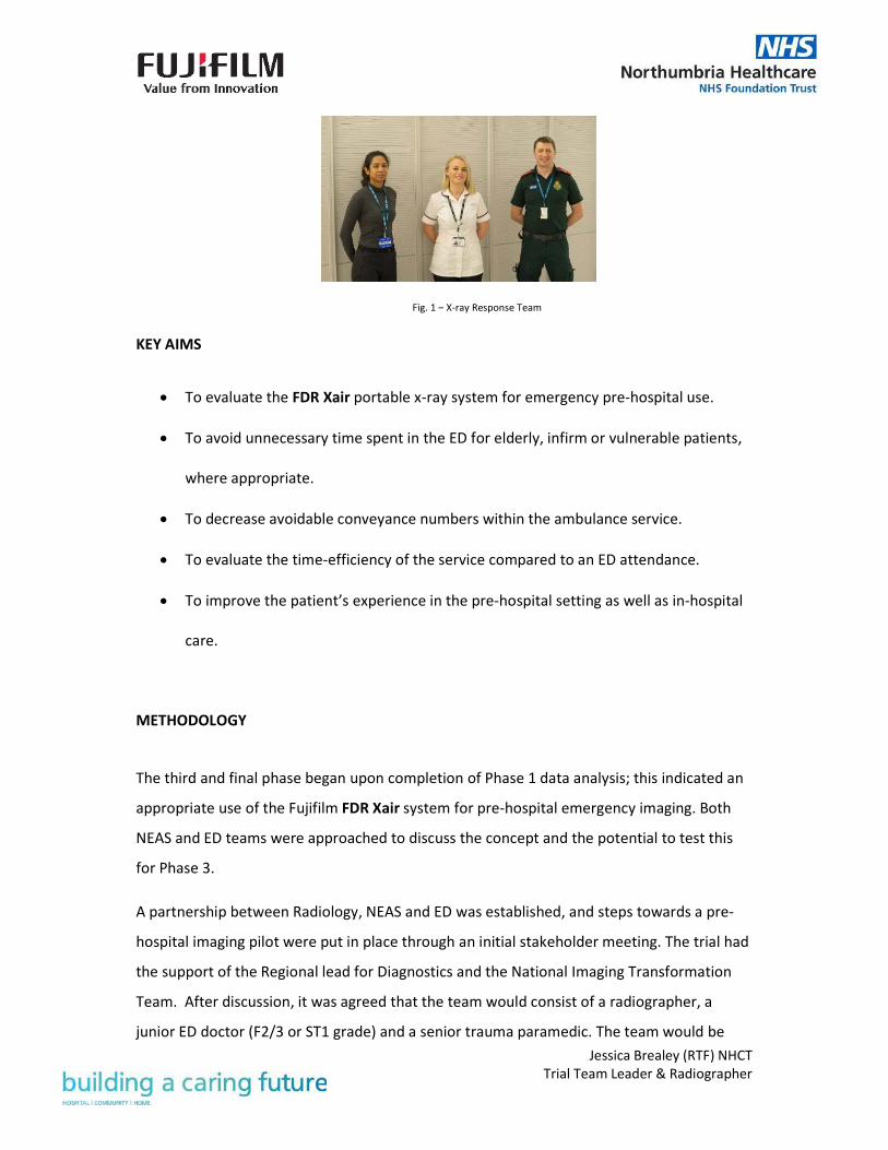
Team. After discussion, it was agreed that the team would consist of a radiographer, a
junior ED doctor (F2/3 or ST1 grade) and a senior trauma paramedic. The team would be
Fig. 1 – X-ray Response Team
Jessica Brealey (RTF) NHCT
Trial Team Leader & Radiographer
overseen by an ED/PHEM (Pre-Hospital Emergency Medicine) consultant who was on-call
for the service. The team would travel in the trauma car, provided by NEAS, and use the
Terrafix system in order to be designated a patient job by the control room.
An inclusion and exclusion criteria for the imaging that could be performed were created by
the Radiology Project Lead and Team Lead which guided the control room staff and duty
manager as to what would be appropriate for the team to attend. This was based upon the
data and knowledge gained from Phases 1 and 2. A decision was made by the strategic team
to begin imaging the pelvis within this phase; a plan to complete an AP pelvis was made and
that further horizontal beam hip imaging (HBL) could be completed in the Radiology
department if hospital transfer was necessary.
The service was named ‘X-ray Response Team’ (XRT) and NHS approved logos were
produced for copyrighting of documentation.
A ‘dry run’ shift was organised in order to discuss the logistics; this involved developing a
clinical governance strategy and organising the workflow of the day, which included:
Shift time/length
Job allocation from control room
Image requesting process
Locations covered by XRT
Handover procedure to relevant services
From previous feedback, it was established that for the service to be used to its optimum
potential it required consistency of approach, reliability, uniformity and regularity. Following
this a monthly rota was created by the team lead to organise availability and establish a
reliable system for staffing, as well as create a reliable service schedule for the NEAS Duty
Manager and Primary Care.
Jessica Brealey (RTF) NHCT
Trial Team Leader & Radiographer
RESULTS
Quantitative Data
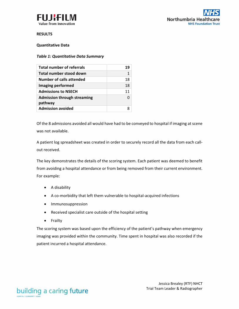
Table 1: Quantitative Data Summary
Of the 8 admissions avoided all would have had to be conveyed to hospital if imaging at scene
was not available.
A patient log spreadsheet was created in order to securely record all the data from each call-
out received.
The key demonstrates the details of the scoring system. Each patient was deemed to benefit
from avoiding a hospital attendance or from being removed from their current environment.
For example:
A disability
A co-morbidity that left them vulnerable to hospital-acquired infections
Immunosuppression
Received specialist care outside of the hospital setting
Frailty
The scoring system was based upon the efficiency of the patient’s pathway when emergency
imaging was provided within the community. Time spent in hospital was also recorded if the
patient incurred a hospital attendance.
Total number of referrals 19
Total number stood down 1
Number of calls attended 18
Imaging performed 18
Admissions to NSECH 11
Admission through streaming
pathway
0
Admission avoided 8

Jessica Brealey (RTF) NHCT
Trial Team Leader & Radiographer
Table 2: KPI Data Summary
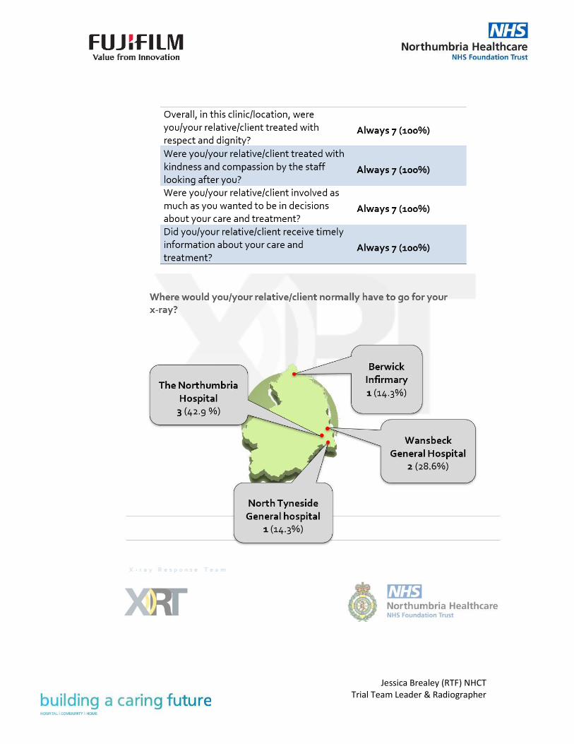
Qualitative Data
The qualitative data collected during Phase 3 mainly comprised of patient feedback. With the input
of the patient experience team an online survey was developed, which could be completed by
patients and / or their carers. This aided the collection of appropriate data to determine the value
and impact of pre-hospital imaging.
The data summary can be found in Appendix 2.
PATIENT
CASE
NUMBER
OUTCOME
SCORE
TOTAL TIME
WITH XRT
(MINS)
TIME TO DISCHARGE
FROM EMERGENCY
DEPARTMENT (HRS)
1 2 98 189
2 2 172 260
3 2 90 234
4 5 83 0
5 5 153 0
6 5 67 0
7 5 135 0
8 2 45
9 5 101 0
10 2 118 317
11 2 53 224
12 2 36 124
13 1 29 0
14 5 35 0
15 2 49 231
16 2 103 331
17 2 43 217
18 5 60 0
19 2 65 968

Jessica Brealey (RTF) NHCT
Trial Team Leader & Radiographer
DISCUSSION
Image Quality
With the support of the Fujifilm Applications Specialist some image processing was adjusted
to align the image processing preferences of the Trust’s reporting radiographers and
radiologists. This allowed the team to start imaging of the pelvis (AP) as operator confidence
and image quality improved.
Storage
After clinical testing of the previous storage cases and feedback to Fujifilm it was thought
necessary to provide a more lightweight solution for storage and transportation. This was to
allow the FDR Xair system to be used to its maximum potential as the unit itself was so
lightweight, however the storage was too bulky to be fit for purpose.
The cases used for Phase 3 consisted of a small hard case for the FDR Xair device tube to
protect from damage. The other components of the unit were stored in a compact rucksack
for easy transportability. No cases were provided for the mounting stands; this was not was
not thought to be necessary service delivery, as the stands are stowed securely in a car.
Connectivity
Since completion of Phase 2, end-to-end connectivity was achieved allowing access to CRIS
(Radiology Information System), PACS (Picture Archiving Communication System), requesting
systems and AI (Artificial Intelligence) software. This has improved the efficiency of the
imaging process and allows for immediate access to imaging from within the Trust. This has
been especially useful when liaising with the on-call PHEM consultant as well as for organising
orthopaedic and care of the elderly (COTE) referrals.
The team has been attending callouts throughout North Tyneside and Northumberland. Due
to the vast areas covered by XRT, connectivity has been an issue in some remote areas due to
lack of signal. This was highlighted as an issue to the Fujifilm team, who have worked on a
multi-network solution to provide greater signal coverage and help to resolve the issue.
Patient Experience
Feedback from this phase was overwhelmingly positive and has truly demonstrated the value
in improving patient experience by providing an acute bedside imaging service.

Jessica Brealey (RTF) NHCT
Trial Team Leader & Radiographer
It should be noted that some responses recorded were negative, but after correlating these
with written feedback on the survey we have determined that these questions may have been
misunderstood. As the surveys are anonymised as per the Caldicott approval, it is not possible
to clarify whether these questions were understood correctly. The team lead is working with
the Patient Experience team to make these questions clearer to ensure more accurate
responses.
Staff Satisfaction
Working with this kind of x-ray machine was vastly different to an in-hospital portable unit
and adaptation to accommodate different scenarios can be challenging. However, after
some adjustments to our usual approach, the team has successfully implemented the unit
into the pre-hospital setting.
Amongst the strategic team and after consultation with the Medical Physics Team and the
HSE, it was deemed necessary to have a small core team of experienced radiographers that
had received dedicated training on the FDR Xair system in order to run a pre-hospital
service to its full potential. Risk Assessments (RA) have to be completed on scene to ensure
radiation safety and it is important to establish if not suitable the examination will not be
performed. Therefore, all XRT radiographers are either radiation protection supervisors
(RPS) or have completed training on RA.
The team also highlighted the need for a training guide offering on-demand help during a
shift. This was produced by the team lead radiographer. This decision has also improved
staff confidence levels when working on this service.
The team also share debrief notes after each callout so learning can be shared and aid
future examinations.
Patient Pathway Development
The data collected during this phase allowed the team to develop some alternative patient
pathways to avoid having to convey the patient to ED after an XRT pre-hospital assessment
with imaging. Once developed, this will allow patients attended by XRT to be conveyed
straight to an appropriate ward when they require additional hospital interventions.

Jessica Brealey (RTF) NHCT
Trial Team Leader & Radiographer
There are several different pathways being looked at such as a neck of femur fracture
pathway which will look at streamlining the patients preassessment with the aim to reduce
time in the ED department and ultimately at direct ward admission although there are many
factors that would need to be in place.
Follow up fracture appointments is another aspect the team are looking at for more
vulnerable patients where the clinic appointment maybe virtual but need imaging.
Other potential pathways being looked at are with COTE which would be valuable for
patients that may not have a fracture but needed further support and evaluation in a
hospital setting.
RECOMMENDATIONS FOR THE FUTURE
The Fujifilm FDR Xair unit has many applications, both clinical and non-clinical. Our work has
highlighted some key areas where the portability and lightweight features may be
beneficial:
Work with primary care networks to provide routine imaging to vulnerable and frail
patients
Provide Fascia iliaca Compartment Block (FICB) out in the community for patients
with fractured neck of femurs.
Create orthopaedic and COTE pathways to streamline patients imaged acutely to get
them to the right place first time rather than traditional route through ED.
Due to the need for adaptation of technique and the carrying out of a RA in an acute
setting this service needs a core team of experienced and suitable trained
radiographers and would not be suitable for junior radiographers or Assistant
Practitioners. However, in a more elective stream where there would not be a doctor
or paramedic these members of staff could support the core team radiograph.
Provide imaging within mental health and rehabilitation facilities where conveyance
to hospital often requires multiple escorts and disruption to the patient’s current
environment.
Jessica Brealey (RTF) NHCT
Trial Team Leader & Radiographer
CONCLUSION
The Fujifilm FDR Xair system is an innovation that releases radiology from technological and
geographical restraints by providing on-demand access to patient imaging wherever it is
required. It has the potential to be applied to a vast array of clinical and non-clinical
settings.
The pilot only ran 2 days a week as it was unfunded and relied on voluntary work; therefore,
was not promoted. This accounts for the small patient numbers, which meant that financial
analysis was limited and the decision was made to evaluate this in a longer trial. However,
cost of emergency transfer is £236 one way and if a hospital stay is required this cost can be
substantial so if numbers imaged in the community were increased we believe significant
cost savings could be made. It is important to look at the cost saving as a whole healthcare
pathway rather than breaking down to departmental costs as clearly there would not be a
cost saving to radiology departments. We believe that the implementation of this x-ray unit
into healthcare systems has the potential to create significant economic benefit. This is due
to the creation of more efficient patient pathways such as the pathways that were
developed during this phase.
The trial has proved a success in evidencing the value and dexterity of such a portable
system in providing resilience to radiology services. Phase 3 has demonstrated how
radiology can be applied to pre-hospital medicine and aid in reducing ED and ambulance
service pressures.

APPENDIX
1. Pilot Timeline
Date Action
14/09/2020 Meeting with ED and PHEM Consultant team to discuss potential
for off-site imaging in the emergency setting -? Phase 3 project.
05/11/2020 Pre-Hospital Stakeholder Meeting with Radiology, NEAS & ED.
14/11/2020 Phase 2 complete – End-to-end connectivity.
21/11/2020 First XRT shift – Dry Run.
09/12/2020 Patient Experience Contacted.
16/12/2020 –
17/12/2020
Paramedic XRT Training Days – a demonstration of the
equipment and discussion of the service to increase awareness
within NEAS.
18/12/2020 XRT Team Job Delegation Meeting
08/01/2021 E-Learning Portal Development Meeting
20/01/2021 Learning Guide Completed
31/01/2021 Phase 3 Complete

Jessica Brealey (RTF) NHCT
Trial Team Leader & Radiographer
2. Patient Survey Data
Jessica Brealey (RTF) NHCT
Trial Team Leader & Radiographer

Jessica Brealey (RTF) NHCT
Trial Team Leader & Radiographer