normal brain scan. spect images are in all the three ... · the left cortex, involving the frontal...
TRANSCRIPT

www.nuclearmd.com
Normal Brain Scan.
SPECT images are
acquired up to an hour
after the administration
of 15-30 mCi of Tc99m
HMPAO/ neurolite. The
images are reconstructed
in all the three orthogonal
planes. The transaxial
images on the left show
normal distribution of the
tracer in the cortex,
subcortical structures and
the cerebellum.

www.nuclearmd.com
Transaxial images from a
brain SPECT, showing a
large perfusion defect in
the left cortex, involving
the frontal lobe and
extending to the parietal
and temporal lobes, from
a recent infarct.
Decreased activity seen
in the right cerebellum is
likely from crossed
cerebellar diaschisis.
www.nuclearmd.com
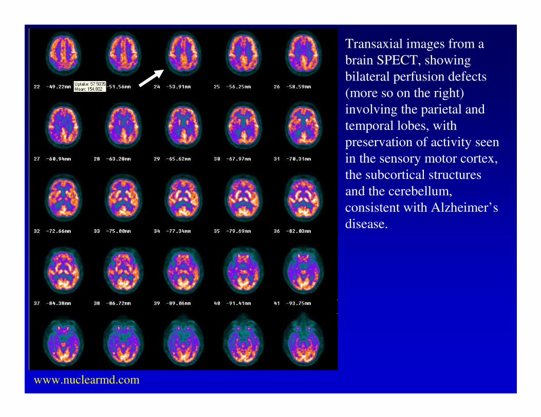
Transaxial images from a
brain SPECT, showing
bilateral perfusion defects
(more so on the right)
involving the parietal and
temporal lobes, with
preservation of activity seen
in the sensory motor cortex,
the subcortical structures
and the cerebellum,
consistent with Alzheimer’s
disease.

www.nuclearmd.com
Transaxial images from a brain SPECT, showing a bilateral frontal perfusion
defects, likely to be consistent with Pick’s disease.

www.nuclearmd.com
Transaxial images from a brain FDG-PET, showing marginal hypermetabolism in
the post surgical cavity in the right parietal-temporal cortex, likely to be
consistent with tumor recurrence.

www.nuclearmd.com
Transaxial images from a brain FDG-PET, showing marginal hypermetabolism in
the post surgical cavity, in the left frontal-parietal cortex, likely to be consistent
with tumor recurrence.

www.nuclearmd.com
Transaxial and coronal images from a brain interictal FDG-PET, showing
hypometabolism in the right anterior temporal lobe, likely to be consistent with
presence of seizure focus.

www.nuclearmd.com
Transaxial and coronal images from a brain interictal FDG-PET, showing
hypometabolism in the left anterior temporal lobe, likely to be consistent with
presence of seizure focus.

www.nuclearmd.com
Negative Brain Death Study. Blood flow is seen in the
brain and the static views show intra-cerebral activity.

www.nuclearmd.com
Positive Brain Death Study. No activity is seen
in the brain on flow or the static images.

www.nuclearmd.com
Positive Thallium Brain SPECT. The ring enhancing lesion in this
AIDS patient is likely to be lymphoma rather than toxoplasmosis.

www.nuclearmd.com
Positive Diamox
Brain SPECT. The
top row of transaxial
images are images at
rest. The bottom row
of axial images were
obtained after the
administration of 1gm
of IV diamox. The
images show a
predominantly
reversible cerebral
perfusion defect in
the left cortex,
suggestive of diamox
induced ischemia.