non-melanoma skin cancer
TRANSCRIPT
Non-melanoma skin cancer
BY
OSAMA ELZAAFARANY
ASSISTANT LECTURER OF CLINICAL ONCOLOGY
MEDICAL RESEARCH INSTITUTE-ALEXANDRIA UNIVERSITY
MAY 2015

Epidemiology: Non-melanoma skin cancer is the most commonly occurring cancer in
the United States.
BCC is the more common type of the two non-melanoma types.
It was estimated that 2,152,500 persons were treated for non-melanoma
skin cancers in 2006; [ Rogers HW, Weinstock MA, Harris AR, et al.: Incidence estimate of nonmelanoma skin
cancer in the United States, 2006. Arch Dermatol 146 (3): 2837, 2010 ].
Although the two types of non-melanoma skin cancer are the most
common of all malignancies, they account for less than 0.1% of patient
deaths caused by cancer.
Risk Factors Epidemiologic evidence suggests that exposure to ultraviolet (UV) radiation
and the sensitivity of an individual’s skin to UV radiation are risk factors for skin cancer.
Skin cancer are more likely to occur in individuals of light complexion who have had substantial exposure to sunlight.
Skin cancers are more common in the southern latitudes of the Northern hemisphere.
The immune system may play a role in pathogenesis of skin cancers; Organ transplant recipients receiving immunosuppressive drugs are at an elevated risk of skin cancers, particularly SCC.
Arsenic exposure also increases the risk of cutaneous SCC.
Serologic evidence from a population based case-control study has shown a possible association between infection with the human papilloma virus (HPV) genus beta-species 1 and SCC: Patel AS, Karagas MR, Perry AE, et al.: Exposure profiles and human
papillomavirus infection in skin cancer: an analysis of 25 genus betatypes in a populationbased study. J Invest Dermatol 128 (12): 288893, 2008.
Other types of malignant disease of the skin include the
following:
Cutaneous T-cell lymphomas (e.g., mycosis fungoides).
Kaposi sarcoma.
Extra-mammary Paget disease.
Apocrine carcinoma of the skin.
Metastatic malignancies from various primary sites

Basal Cell Carcinoma
About three times more common than SCC in non-
immunocompromised patients.
It usually occurs on sun exposed areas of skin, and the nose is
the most frequent site.
the most characteristic clinical presentation is the asymptomatic
nodular or nodular ulcerative lesion that is elevated from the
surrounding skin, has a pearly quality, and contains
telangiectatic vessels.
Has a tendency to be locally destructive.
Hig-hrisk areas for tumor recurrence after initial treatment
include the central face (e.g., periorbital region, eyelids,
nasolabial fold, or nosecheek angle), postauricular region,
pinna, ear canal, forehead, and scalp.

Morpheaform sub-type: specific subtype
of BCC, this subtype typically appears as
a scar-like, firm plaque. Because of indistinct clinical tumor margins, the
morpheaform type is difficult to treat
adequately with traditional treatments.
BCC is slow growing and rarely metastasize.
Pathology: BCCs are composed of non-
keratinizing cells derived from the basal
cell layer of the epidermis.
Molecular biology: BCC often have a
characteristic mutation in the patched 1
tumor suppressor gene (PTCH1).

Squamous Cell Carcinoma Also tend to occur on sun-exposed portions of the skin, such as the
ears, lower lip, and dorsa of the hands.
SCC that arise in areas of non sun-exposed skin or that originate de
novo on areas of sun-exposed skin are prognostically worse
because they have a greater tendency to metastasize than those
that occur on sun-exposed skin that develop from actinic keratosis.
More aggressive than BCCs and have a range of growth, invasive,
and metastatic potential.
Composed of keratinizing cells.
Predisposing factors:
Chronic sun damage.
Sites of prior burns.
Arsenic exposure.
Chronic cutaneous inflammation as long standing skin ulcers. Sites of previous x-ray therapy.
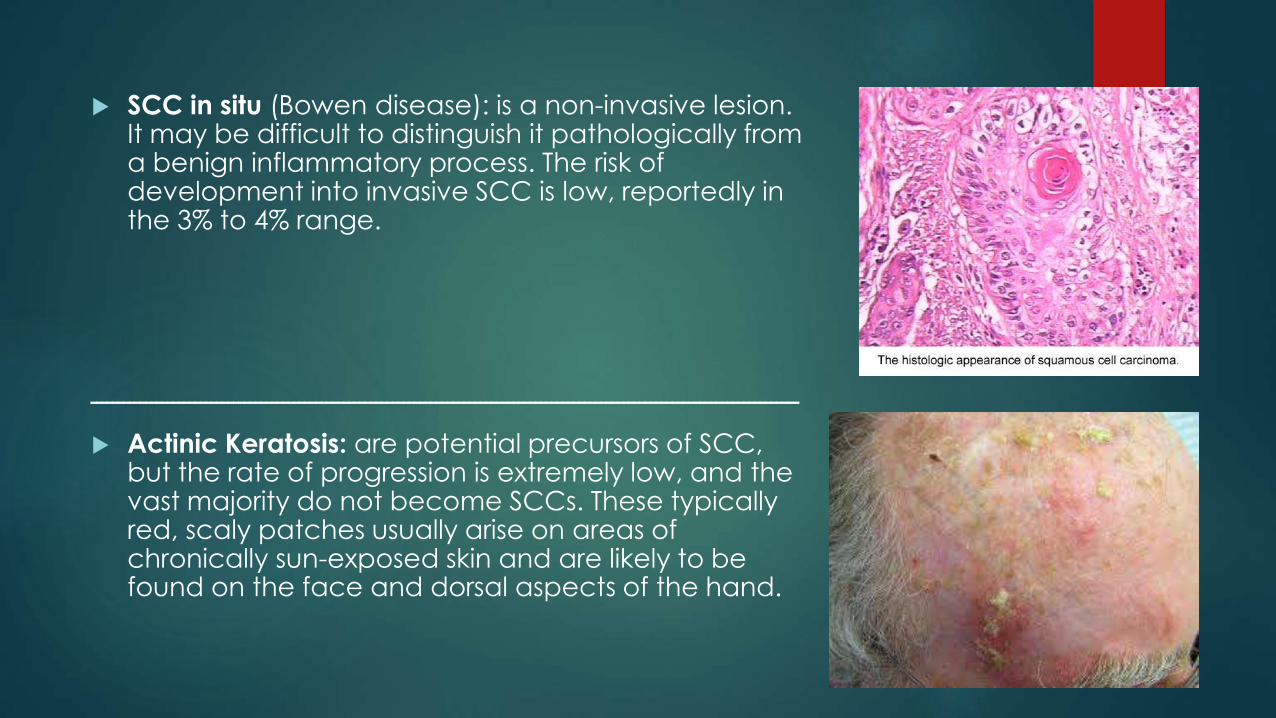
SCC in situ (Bowen disease): is a non-invasive lesion. It may be difficult to distinguish it pathologically from a benign inflammatory process. The risk of development into invasive SCC is low, reportedly in the 3% to 4% range.
ـــــــــــــــــــــــــــــــــــــــــــــــــــــــــــــــــــــــــــــــــــــــــــــــــــــــــــــــــــــــــــــــــ
Actinic Keratosis: are potential precursors of SCC, but the rate of progression is extremely low, and the vast majority do not become SCCs. These typically red, scaly patches usually arise on areas of chronically sun-exposed skin and are likely to be found on the face and dorsal aspects of the hand.

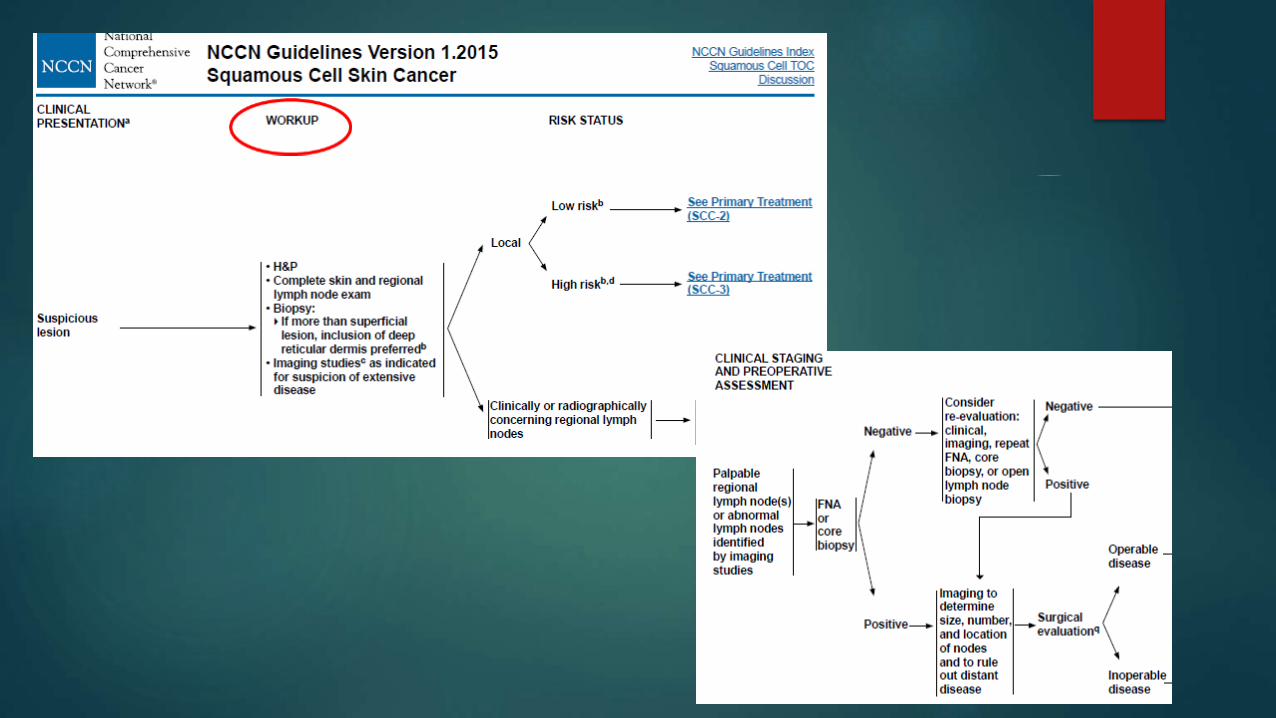
Diagnostic workup: Basal cell carcinoma (BCC) rarely metastasizes, thus, a metastatic
workup is usually not necessary.
Regional lymph nodes should be routinely examined in all cases of
SCC, especially for high-risk tumors appearing on the lips, ears,
perianal and perigenital regions, or high-risk areas of the hand.
In addition, regional lymph nodes should be examined with
particular care in cases of SCCs arising in sites of chronic ulceration
or inflammation, burn scars, or sites of previous radiation therapy treatment.

Staging: There are separate staging systems in the 7th edition of the
American Joint Committee on Cancer’s (AJCC) AJCC Cancer
Staging Manual for carcinomas of the eyelid versus other skin
surfaces.
The staging system for non-eyelid skin cancers is primarily designed
for squamous cell carcinomas (SCCs).
The staging system for carcinoma of the eyelid addresses carcinomas of all histologies.
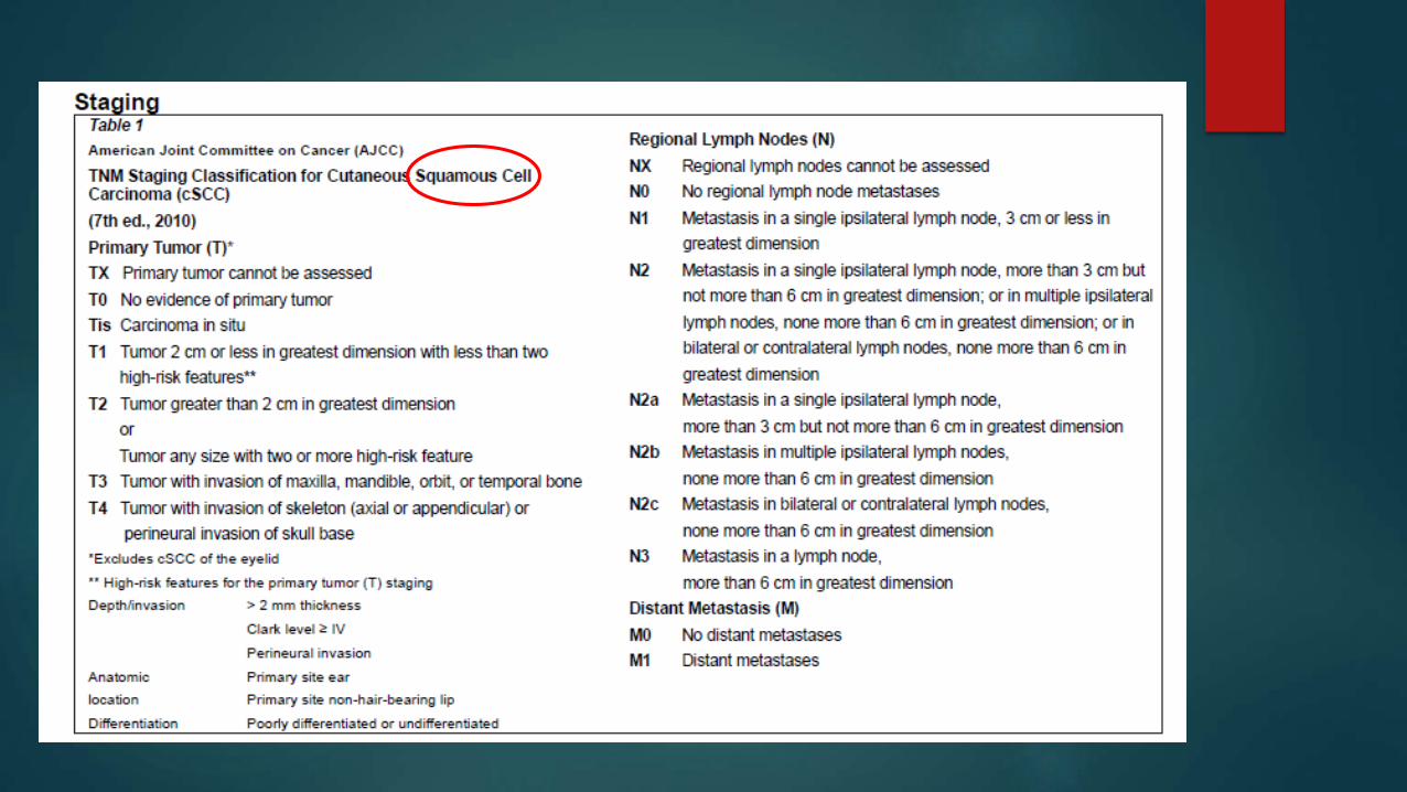

Risk features that should be evaluated for non-eyelidcarcinomas
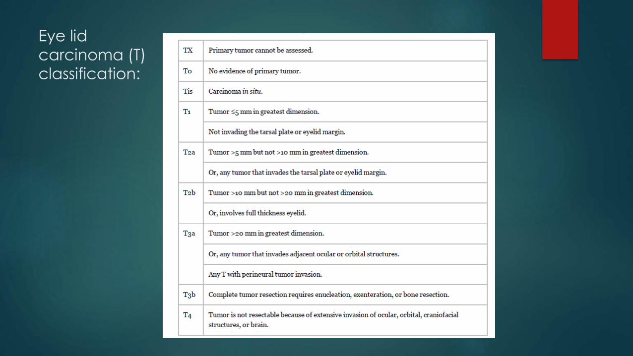
Eye lid
carcinoma (T) classification:

Patients with a primary cutaneous SCC or other cutaneous carcinoma with no evidence (i.e., clinical, radiologic, or pathologic) of regional or distant metastases are divided into the following two stages:
Stage I for tumors measuring 2 cm or less in size.
Stage II for tumors measuring more than 2 cm in size.
In instances where there is clinical concern about extension of the tumor into bone and radiologic evaluation has been performed (and is negative), these data may be included to support the stage I versus stage II designation.
Tumors that are 2 cm or less in size can be upstaged to stage II if they contain two or more high-risk features.
Stage III patients are those with either of the following:
Clinical, histologic, or radiologic evidence of one involved lymph node measuring 3 cm or less in size.
Tumor extension into bone; namely, the maxilla, mandible, orbit, or temporal bone.
Stage IV patients are those with any of the following:
Tumor with direct or peri-neural invasion of skull base or axial skeleton.
Two or more involved lymph nodes.
Single or multiple involved lymph nodes measuring more than 3 cm in size.
Distant metastases.
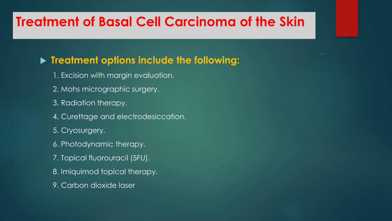
Treatment of Basal Cell Carcinoma of the Skin
Treatment options include the following:
1. Excision with margin evaluation.
2. Mohs micrographic surgery.
3. Radiation therapy.
4. Curettage and electrodesiccation.
5. Cryosurgery.
6. Photodynamic therapy.
7. Topical fluorouracil (5FU).
8. Imiquimod topical therapy.
9. Carbon dioxide laser

Excision with margin evaluation
Surgical margins ranging from 3 -10 mm, depending on the diameter of
the tumor.
Excision has been compared in randomized trials to radiation therapy, Mohs micrographic surgery, photodynamic therapy (PDT), and
cryosurgery Their overall assessments favored excision.
• In a single-center trial, 360 patients with facial BCCs <4 cm in diameter were randomly assigned to excision VS
radiation therapy.
• RTx was : 55% interstitial brachytherapy, 33% contact radiation therapy, and 12% conventional external beam
radiation therapy.
• Excisional margins, assessed during surgery by frozen section during the procedure in 91% of cases, had to be at
least 2 mm, with re-excision if necessary.
• At 4 years (mean follow-up of 41 months), the actuarial failure rates (confirmed persistent or recurrent tumor)
were 0.7% and 7.5% in the surgery and radiation therapy arms, respectively (P = .003).
• The cosmetic results were also rated as better after surgery by both patients and dermatologists, and also by
three independent judges. At 4 years, 87% of surgery patients rated cosmesis as good versus 69% of radiation
therapy patients.
Petit JY, Avril MF, Margulis A, et al.: Evaluation of cosmetic results of a randomized trial comparing surgery and radiotherapy in the treatment of basal cell carcinoma of the face. Plast Reconstr Surg 105 (7): 254451, 2000.

Mohs micrographic surgery
Principle:
specialized technique used with the intent to achieve the narrowest margins
necessary to avoid tumor recurrence, while maximally preserving cosmesis.
The tumor is microscopically delineated, with serial radial resection, until it is
completely removed as assessed with real-time frozen sections.
Indications:
1. tumors in cosmetically sensitive areas; (e.g., eyelid periorbital area,
nasolabial fold, nose-cheek angle, posterior cheek sulcus, pinna, ear
canal, forehead, scalp, fingers, and genitalia).
2. Tumors that have recurred after initial excision.
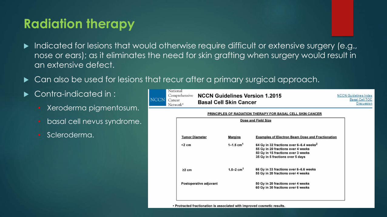
Radiation therapy
Indicated for lesions that would otherwise require difficult or extensive surgery (e.g.,
nose or ears); as it eliminates the need for skin grafting when surgery would result in
an extensive defect.
Can also be used for lesions that recur after a primary surgical approach.
Contra-indicated in :
• Xeroderma pigmentosum.
• basal cell nevus syndrome.
• Scleroderma.

Curettage & electrodesiccation (electro-surgery)
Principle: sharp curette is used to scrape away the tumor down to its base,
followed by electrodesiccation of the lesion base.
Indication: superficial lesions of the neck, trunk, and extremities that are
considered to be at low-risk for recurrence.
Evidence:
In a large, single-center case series of 2,314 previously untreated BCCs managed at a
major skin cancer unit.
The 5-year recurrence rate of BCCs of the neck, trunk, and extremities was 3.3%.
However, rates increased substantially for tumors larger than 6 mm in diameter at
other anatomic sites.
Silverman MK, Kopf AW, Grin CM, et al.: Recurrence rates of treated basal cell
carcinomas. Part 2: Curettage electrodesiccation. J Dermatol Surg Oncol 17 (9): 7206,
1991.

Topical fluorouracil (5FU)
Topical 5FU (5% cream) may be useful in specific limited circumstances. It is a FDA-approved treatment for superficial
BCCs in patients for whom conventional methods are
impractical, such as individuals with multiple lesions or difficult
treatment sites.
Safety and efficacy in other indications have not been
established.
Given the superficial nature of its effects, non-visible dermal
involvement may persist, giving a false impression of treatment
success. In addition, the brisk accompanying inflammatory
reaction may cause substantial skin toxicity and discomfort in a large proportion of patients.


Treatment for Recurrent BCC of the Skin
Most recurrences occur within 5 years, with about 18% of recurrences are diagnosed beyond that point.
Patients who develop a primary BCC are also at increased risk of
subsequent primary skin cancers because the susceptibility of their sun
damaged skin to additional cancers persists (field carcinogenesis).
Age at diagnosis of the first BCC (<65 years), red hair, and initial BCC
on the upper extremities appear to be associated with higher risk of
subsequent new BCCs.
Mohs micrographic surgery is commonly used for local recurrences of
BCC.
Treatment for Advanced & Metastatic BCC
Cisplatin, alone or in combination with other drugs, is the most commonly
reported systemic therapy and appears to be associated with the best tumor
response rates.
A variety of other agents have been reported but have low associated
response rates, including cyclophosphamide, vinblastine, 5FU, methotrexate,
and doxorubicin.
Since there is no standard therapy, clinical trials are appropriate if available.
Hedgehog/PTCH1signaling pathway inhibitor Vismodegib was approved by
FDA at 2012 foe advanced BCC.
Orally administered Hedgehog pathway inhibitor (GDC0449) has produced
objective responses in patients with advanced or metastatic sporadic BCC.

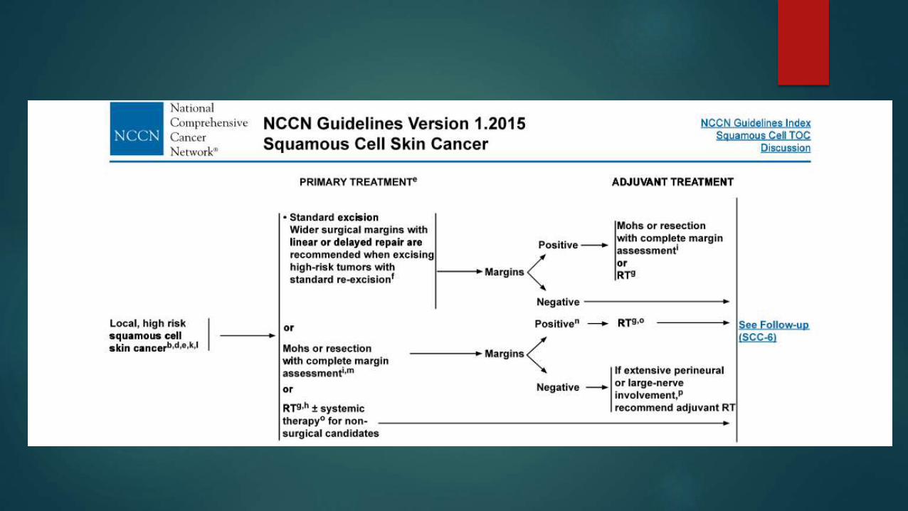
Treatment of Squamous Cell Carcinoma of the Skin
Localized squamous cell carcinoma (SCC) of the skin is a highly curable
disease.
Absent high-quality evidence from controlled clinical trials, the management
of clinically localized cutaneous SCC is based upon case series and consensus statements from experts.
Treatment options include the following:
1. Surgical excision with margin evaluation.
2. Mohs micrographic surgery.
3. Radiation therapy.
4. Curettage and electrodesiccation.
5. Cryosurgery.
Surgical excision with margin evaluation
Excision is probably the most common therapy for SCC.
This traditional surgical treatment usually relies on surgical margins ranging from
4 -10 mm, depending on the diameter of the tumor and degree of
differentiation.
In a prospective case series of 141 SCCs, a 4mm margin was adequate to
encompass all subclinical microscopic tumor extension in more than 95% of well-
differentiated tumors up to 19 mm in diameter.
Wider margins of 6 -10 mm were needed for larger or less-differentiated tumors
or tumors in high-risk locations (e.g., scalp, ears, eyelids, nose, and lips).
Re-excision may be required if the surgical margin is found to be inadequate on
permanent sectioning.
ـــــــــــــــــــــــــــــــــــــــــــــــــــــــــــــــــــــــــــــــــــــــــــــــــــــــــــــــــــــــــــــــــــــــــــــــــــــــــــــــــــــــــــــــــــــــــــــــــــBrodland DG, Zitelli JA: Surgical margins for excision of primary cutaneous squamous cellcarcinoma. J Am Acad Dermatol 27 (2 Pt 1): 2418, 1992. [PUBMED Abstract]

Radiation therapy
Radiation therapy is a logical treatment choice, particularly for patients with primary lesions requiring difficult or extensive surgery (e.g., nose, lip, or ears).
Radiation therapy eliminates the need for skin grafting when surgery would result in an extensive defect.
Cosmetic results are generally good, with a small amount of hypopigmentation or telangiectasia in the treatment port.
Radiation therapy can also be used for lesions that recur after a primary surgical approach.
Radiation therapy is avoided in patients with conditions that predispose them to radiation-induced cancers, such as xeroderma pigmentosum or basal cell nevus syndrome.
Although radiation therapy, with or without excision of the primary tumor, is used for histologically proven clinical lymph node metastases and has been associated with favorable disease-free survival rates, However it is difficult to know the impact of nodal radiation on survival.


Treatment for Recurrent SCC of the Skin
SCCs have definite metastatic potential, and patients should be followed regularly after initial treatment.
Overall, local recurrence rates after treatment of primary SCCs ranged from about 3% - 23%, depending upon anatomic site.
About 58% of local recurrences manifest within 1 year, 83% within 3 years, and 95% within 5 years.
The metastatic rate for primary tumors of sun-exposed skin is 5%; for tumors of the external ear, 9%; and for tumors of the lip, 14%. Metastases occur at an even higher rate for primary SCCs in scar carcinomas or in non-exposed areas of skin (about 38%).
About 69% of metastases are diagnosed within 1 year, 91% within 3 years, and 96% within 5 years.
Tumors that are 2 cm or larger in diameter, 4 mm or greater in depth, or poorly differentiated have a relatively bad prognosis and even higher local recurrence and metastasis rates than those listed.
Reported rates also vary by treatment modality, with the lowest rates associated with Mohs micrographic surgery, but at least some of the
variation may be the result of patient selection factors; no randomized
trials directly compare the various local treatment modalities.
Recurrent non-metastatic SCCs are considered high risk and are
generally treated with excision, often using Mohs micrographic surgery.
Radiation therapy is used for lesions that cannot be completely
resected.
As is the case with BCC, patients who develop a primary SCC are also
at increased risk of subsequent primary skin cancers because the susceptibility of their sun-damaged skin to additional cancers persists.
Treatment for Metastatic & Advanced SCC
As is the case with BCC, metastatic and far advanced SCC is
unusual, and reports of systemic therapy are limited to case reports
and very small case series with tumor response as the endpoint.
Cisplatin-based regimens appear to be associated with high initial
tumor response rates.
High response rates have also been reported with the use of 13-cis-
retinoic acid plus interferonalpha-2a.
Since there is no standard therapy, clinical trials are appropriate if available.

The main source of this presentation is:
National Cancer Institute: PDQ® Skin Cancer Treatment. Bethesda, MD: National
Cancer Institute.
Date last modified <4/28/2015>.
Available at: http://cancer.gov/cancertopics/pdq/treatment/skin/HealthProfessional