non-inflammatory laryngeal stridor...
TRANSCRIPT
NON-INFLAMMATORY LARYNGEAL STRIDOR ININFANTS
BY
JAMES CROOKSFrom The Hospitalfor Sick Children, Great Ormond Street, London
(RECEIVED FOR PUBLICATION OCTOBER 6, 1953)
'Congenital laryngeal stridor' is merely a clinicaldescription, and although perhaps 90 % of stridors inyoung babies are due to an exaggerated pattern ofinfantile larynx, which rectifies itself with increasingage, it is not safe to assume that this is the causeof the stridor. There are other conditions causinglaryngeal stridor in young babies, sometimes frombirth, which are graver than the exaggeratedinfantile larynx, and often prove fatal, and whichmay be amenable to surgical treatment.
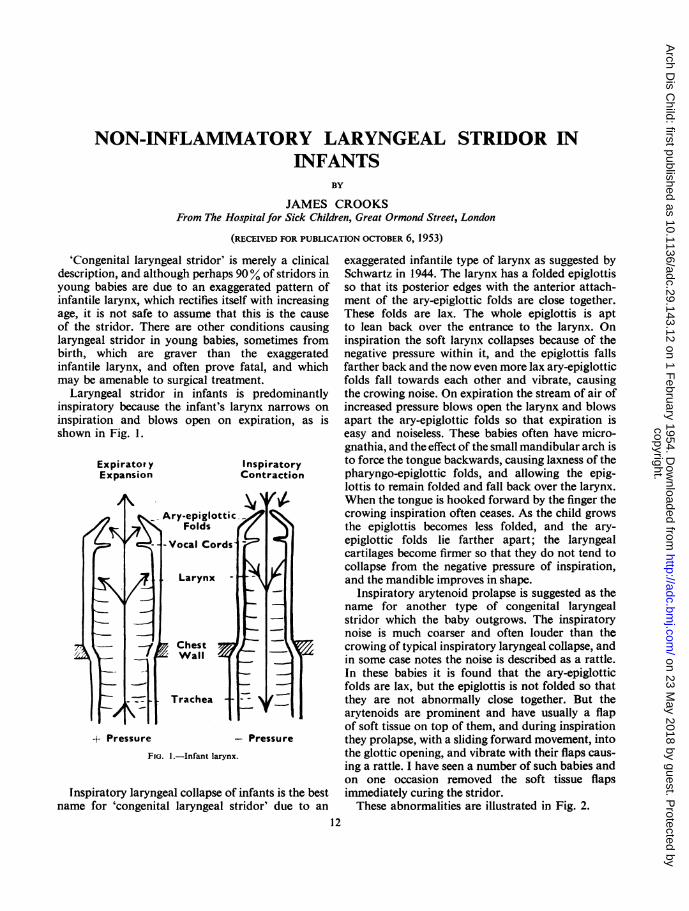
Laryngeal stridor in infants is predominantlyinspiratory because the infant's larynx narrows oninspiration and blows open on expiration, as isshown in Fig. 1.
Expirator y InspiratoryExpansion Contraction
t+ Pressure - Pressure
FIG. 1.-Infant larynx.
Inspiratory laryngeal collapse of infants is the bestname for 'congenital laryngeal stridor' due to an
exaggerated infantile type of larynx as suggested bySchwartz in 1944. The larynx has a folded epiglottisso that its posterior edges with the anterior attach-ment of the ary-epiglottic folds are close together.These folds are lax. The whole epiglottis is aptto lean back over the entrance to the larynx. Oninspiration the soft larynx collapses because of thenegative pressure within it, and the epiglottis fallsfarther back and the now even more lax ary-epiglotticfolds fall towards each other and vibrate, causingthe crowing noise. On expiration the stream of air ofincreased pressure blows open the larynx and blowsapart the ary-epiglottic folds so that expiration iseasy and noiseless. These babies often have micro-gnathia, and the effect of the small mandibular arch isto force the tongue backwards, causing laxness of thepharyngo-epiglottic folds, and allowing the epig-lottis to remain folded and fall back over the larynx.When the tongue is hooked forward by the finger thecrowing inspiration often ceases. As the child growsthe epiglottis becomes less folded, and the ary-epiglottic folds lie farther apart; the laryngealcartilages become firmer so that they do not tend tocollapse from the negative pressure of inspiration,and the mandible improves in shape.
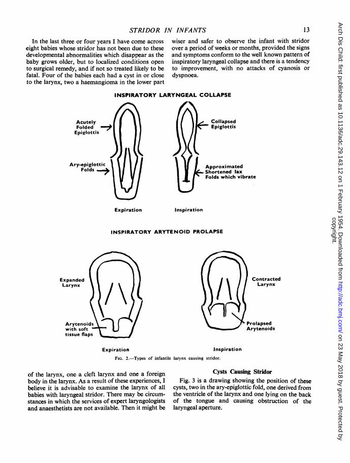
Inspiratory arytenoid prolapse is suggested as thename for another type of congenital laryngealstridor which the baby outgrows. The inspiratorynoise is much coarser and often louder than thecrowing of typical inspiratory laryngeal collapse, andin some case notes the noise is described as a rattle.In these babies it is found that the ary-epiglotticfolds are lax, but the epiglottis is not folded so thatthey are not abnormally close together. But thearytenoids are prominent and have usually a flapof soft tissue on top of them, and during inspirationthey prolapse, with a sliding forward movement, intothe glottic opening, and vibrate with their flaps caus-ing a rattle. I have seen a number of such babies andon one occasion removed the soft tissue flapsimmediately curing the stridor.These abnormalities are illustrated in Fig. 2.
12
P,
copyright. on 23 M
ay 2018 by guest. Protected by
http://adc.bmj.com
/A
rch Dis C
hild: first published as 10.1136/adc.29.143.12 on 1 February 1954. D
ownloaded from
STRIDOR IN INFANTSIn the last three or four years I have come across
eight babies whose stridor has not been due to thesedevelopmental abnormalities which disappear as thebaby grows older, but to localized conditions opento surgical remedy, and if not so treated likely to befatal. Four of the babies each had a cyst in or closeto the larynx, two a haemangioma in the lower part
wiser and safer to observe the infant with stridorover a period of weeks or months, provided the signsand symptoms conform to the well known pattern ofinspiratory laryngeal collapse and there is a tendencyto improvement, with no attacks of cyanosis ordyspnoea.
INSPIRATORY LARYNGEAL COLLAPSE
Collapsed
Epiglottis
i !I1 / 1 \ { | ApproximatedShortened laxFolds which vibrate
Expiration Inspiration
INSPIRATORY ARYTENOID PROLAPSE
piration inspFIG. 2.-Types of infantile larynx causing stridor.
oiration
of the larynx, one a cleft larynx and one a foreignbody in the larynx. As a result of these experiences, Ibelieve it is advisable to examine the larynx of allbabies with laryngeal stridor. There may be circum-stances in which the services of expert laryngologistsand anaesthetists are not available. Then it might be
Cysts Causing StridorFig. 3 is a drawing showing the position of these-
cysts, two in the ary-epiglottic fold, one derived fromthe ventricle of the larynx and one lying on the backof the tongue and causing obstruction of thelaryngeal aperture.
ContractedLarynx
ProlapsedArytenoids
Exi
13
copyright. on 23 M
ay 2018 by guest. Protected by
http://adc.bmj.com
/A
rch Dis C
hild: first published as 10.1136/adc.29.143.12 on 1 February 1954. D
ownloaded from

ARCHIVES OF DISEASE IN CHILDHOODAry-epiglottic Fold Cysts.-One was in a boy who had
difficulty in breathing from birth and was nursed in anoxygen tent for stridor and cyanosis. He had to havetracheotomy at 14 days old at his local hospital, and at3 weeks old the surgeon there could see that the right ary-epiglottic fold was thick. At 2 months old I could see thatthere was a cyst in the fold and aspirated clear fluid fromit. I removed a piece of the cyst then and upon two sub-
Cyst derived fromLaryngeal Ventricle
sequent occasions. It was lined with pseudo-stratifiedcolumnar ciliated epithelium. At 4 months old there was a
reasonably good aperture to the larynx, the tracheotomytube was removed and the baby went home. After twomonths at home he developed laryngo-tracheitis withspasm and a tracheotomy was made and he returned tomy ward. The trachea above the tracheostomy collapsed,and although he had a good laryngeal aperture after thelaryngitis settled, it took 18 months and the same numberof operations, including splinting the upper trachea openwith a wire bridge, before he could breathe without thetube. At 41 years he was strong and well with a goodvoice.The other ary-epiglottic cyst was also in a boy, aged 2
months, who was admitted to my ward at the request of
Dr. George Newns. He had had stridor since birth, caus-ing indrawing of the ribs, and increasing progressively.Feeding had become more and more difficult. Hedeveloped an upper respiratory infection five days beforeadmission (there were colds in the family), and his diffi-culty in breathing had become worse in these days. Thediagnosis on admission was inspiratory laryngeal collapse,with superadded laryngitis, but the physician suspected
Epiglottis
that there might be another abnormality of the larynx.The baby was given oxygen and aureomycin, andhis condition was maintained. Four days later laryngo-scopy was performed under general anaesthesia, and a
rounded cyst was seen occluding the anterior halfof the laryngeal opening, superficial to the cords. Itsorigin could not be determined, but as much as possibleof the cyst was seized in biting forceps and removed. Thebleeding was not great, and breathing was immediatelyeasier. The blood was sucked out repeatedly during thenext few hours while the baby lay in the theatre with allhands standing by. He settled into quiet respiration, andthereafter made uneventful progress, taking his feedswell. He left hospital in a week, and the larynx wasexamined a month later. It was normal but for a small
Ary-epiglottic Fold Cysts
)Cyst on back of Tonguepressing Epiglottis backover Entrance of Larynx
FIG. 3.-Cysts causing laryngeal stridor in infants.
14
copyright. on 23 M
ay 2018 by guest. Protected by
http://adc.bmj.com
/A
rch Dis C
hild: first published as 10.1136/adc.29.143.12 on 1 February 1954. D
ownloaded from

STRIDOR IN INFANTSraised pink scar at the junction of the right ary-epiglotticfold with the epiglottis. Section showed the cyst to belined with flattened respiratory columnar epithelium, andcovered with squamous epithelium, presumably derivedfrom that of the epiglottis. This ary-epiglottic fold cysthad been removed, fortunately and almost completely,at one operation without tracheotomy. The baby enjoyedgreat advantages over others who had to overcome thedangers of tracheotomy with collapse of the upper tracheaand many operations and a prolonged stay in hospital.
In a following paragraph the troubles of tracheos-tomy in very young babies are discussed.
CystDerived from the Laryngeal Ventricle.-This type ofcyst was in a baby girl I year old under the care of mycolleague Mr. Henry Sharp. She was in hospital withpylorospasm, and was found to have laryngeal stridorwhich rapidly became worse, so that after two weekstracheotomy was necessary. A large thin-walled cyst wasfound to rise up from the side of the larynx and overlap itsentrance. Thin grey fluid was aspirated and a large part ofthe cyst wall removed; it was lined with stratifiedsquamous epithelium. Thereafter the aperture of thelarynx looked adequate, but the baby could not bebrought to breathe through it because the tracheaabove the tracheotomy tube collapsed. She died a fewweeks later during an operative procedure.
These three cysts are representative of the usualtype of cyst found in the infant larynx. Ahlen andRanstrom (1944) report one and refer to 20 others,and Holinger and Steinmann (1947) report two. Withthe three described here there are 26 laryngeal cystsin the infant on record, and 17 died. Theoreticallythey were all open to surgical cure.
Thyro-glossal Duct Cyst.-This cyst was not in thelarynx itself, but on the back of the tongue and com-pressed the larynx causing stridor. It also caused difficultyin feeding and it was for this reason that the baby girl of6 weeks came into hospital. The physician looking afterher, Dr. Wilfrid Sheldon, put his finger in her mouthand remarked that there was not enough room low downin the pharynx for her to swallow easily, and he thoughtthat there must be a cyst there. I saw a large thin-walledcyst, and aspirated some fluid which digested starch. Alarge piece of cyst wall was removed and section showedit to be lined with stratified squamous epithelium with afew acini of salivary glands in the connective tissue. It wasthought to be the dilated upper end of the thyro-glossalduct. After the operation the baby made rapid progress,with easy feeding and no stridor.
Haemangiomata Causing StridorBoth of the patients with haemangiomata were boys
who developed inspiratory stridor at about 3 months ofage. The one who died had attended a hospital where adiagnosis of 'congenital laryngeal stridor' had been made,and the mother told that 'he had a small larynx and wouldgrow out of his trouble.' But he did not. He got worse, andwhen Dr. P. R. Evans saw the baby he noted that thestridor did not improve when he pulled the tongue for-
ward, and that there were several haemangiomata on theskin, and wrote in his out-patient notes: 'there might be alaryngeal angioma or something of that sort.' Onlaryngoscopy and bronchoscopy a rounded swelling wasseen protruding from a broad base on the posterior wallof the larynx about a quarter of an inch below the vocalcords. The airway was narrow but had proved to be
FIG. 4.-On the left normal baby's larynx, split from the front. Onthe right a haemangiomatous tumour protruding into the lumen from
the posterior wall of larynx and upper trachea.
adequate so far. Impressed with the difficulties I hadexperienced with the other similar baby, I decided totreat this haemangioma by x rays and the child wentback to the medical ward. He died 36 hours later. Figs. 4,5 and 6 show this tumour.The other baby boy came to us at 41 months from
another hospital for investigation, having had inspiratory
FIo. 5.-Haemangioma of posterior wall of larynx and upper tracheaon section.
15
copyright. on 23 M
ay 2018 by guest. Protected by
http://adc.bmj.com
/A
rch Dis C
hild: first published as 10.1136/adc.29.143.12 on 1 February 1954. D
ownloaded from

ARCHIVES OF DISEASE IN CHILDHOODstridor and attacks of pallor since he was 3 months old.The laryngoscope and bronchoscope showed a roundedswelling on the posterior wall of the larynx about half aninch below the vocal cords. A tracheotomy was made anda fortnight later the larynx was split by laryngo-fissureoperation and the rounded tumour, which was faintlyblue, was cut out. The larynx was sewn up and the babyrecovered from the operation well. Section of the tumourshowed it to be a capillary haemangioma with very fewformed vessels. It was not possible to get the baby tobreathe through the larynx, because yet again thetrachea above the tracheotomy collapsed. In addition,three months later, the angioma became evident again.He was given two treatments by deep x ray at fourmonths' interval, but it was not until he was 15 monthsold that we were able to get him to breathe through thelarynx. He has been very well for the last year, and arecent examination showed only a small pale raised scar,with a good airway.
Suehs and Herbut (1940) describe one and refer toseven other examples of haemangioma in the infantlarynx. and Ferguson (1944) records one. With thesetwo added there are 11, of whom five recovered.
FiG. 6.-Haemangioma of larynx x 75. Numerous endothelial cellsand few blood spaces.
Cleft Larynx Causing StridorThe cleft larynx baby was a member of a family of five
children, the first four of whom were described by Finlay(1949). Three of hersisters died in the samesort of way as she did,at about 3 or 4 monthsold, from ulcerative .
tracheitis or infectionof the lungs. I examinedthe larynx of this babygirl when she was 3months old and inhospital under Dr.George Newns for in-spiratory stridor andfeeding difficulty. I didnot notice the cleft be-tween the arytenoids.It seemed to me to bea larynx of the 'inspira-tory laryngeal collapse'type. But at necropsy FIG. 7--Cleft in posterior wall oftypeM. Butiat founeco larynx between arytenoids.Dr. M. Bodian found acleft between the ary-tenoids. Finlay had noticed a cleft in one of the sistersand it seems that the two others who died probably alsohad clefts.
I believe Dr. Bodian has found two other examplesof what he calls 'cleft larynx' apart from themembers of this family. Now that one is awarethat such a condition exists, a careful inspection ofthat part of the larynx on laryngoscopy should,sooner or later, enable a diagnosis to be made duringlife. Then perhaps oesophageal feeds will preventthe fatal consequences of food getting into the lungs,and a baby may reach a size when surgical repairof the cleft is possible.
Foreign Body causing StridorMy last example of non-inflammatory laryngeal stridor
is a very obvious one, after the event. But this baby boyof 11 months had a cold for a few days, developed suddenstridor and difficulty in breathing, and was taken to aLondon teaching hospital where a diagnosis of laryngo-tracheo-bronchitis was made. There was not a cot forhim so he was transferred to The Hospital for SickChildren with that diagnosis, which set our admissionofficer off on the wrong foot. An accurate history mighthave corrected the error, but the parents were Italian withvery little English, and our admission officer English withvery little Italian. Laryngoscopy of this baby with verysevere respiratory difficulty and laryngeal stridor, in theearly hours of the morning, revealed a large piece of bonejammed between the vocal cords, and there was a rapidrecovery after it was removed. The necropsy was notupon the child, but upon the history of his illness, when itwas elicited that the attack of respiratory difficulty had
16
copyright. on 23 M
ay 2018 by guest. Protected by
http://adc.bmj.com
/A
rch Dis C
hild: first published as 10.1136/adc.29.143.12 on 1 February 1954. D
ownloaded from

STRIDOR IN INFANTS 17
come on suddenly when the baby was drinking soup-Italian soup with bone in it.
These examples are from the writer's own experi-ence in the last few years, but they do not cover allthe causes of laryngeal stridor in infants. Forinstance, a considerable number of cases of webs inthe infant larynx have been described.
Tracheostomy in Young InfantsTracheostomy tubes had to be kept in position in
two of the babies here described for a year and a halfbefore they could be removed although the originalcause of the laryngeal obstruction, which hadnecessitated the tracheostomy, had been overcome inthe first few months. A third baby died during anoperative procedure to overcome tracheal obstruc-tion following tracheostomy.The trachea of an infant up to a few months old
collapses above a tracheostomy if the tube is kept infor more than a few days. This seems to be due toloss of strength of the trachea after one or two of itssoft rings have been divided, and to the negativepressure in the larynx and upper trachea which ispresent during inspiration (Fig. 1). This negativepressure is less above the tracheostomy than below,but the tracheotomy tube props open the lowertrachea. The collapse of the upper trachea preventsthe baby from resuming normal breathing evenwhen the larynx itself has a good lumen. In two of thebabies described here, numerous operations, andvarious modifications of tracheotomy tube, includ-ing varieties with a tube passing up as well as down,failed to hold the collapsed upper trachea open. Thepassage of time and growth of the child eventuallysucceeded where all our efforts had failed. Thetrachea enlarges and the rings become firmer. Thereis also a very important mental element. The babyfinds it easy to breathe through the tracheotomytube, and seems to lose the natural ability orinclination to breathe through the larynx. When he isabout a year and a half old it is possible to educatehim to breathe through the larynx again by takingout the tube for short periods, and blocking thetracheostomy with the finger. At this stage it may bepossible to dispense with the tube, with the tracheos-tomy closed, for hours or days at a time if the baby isput in an oxygen tent, whereas in ordinary air hebecomes cyanosed. The lesson that has been learntfrom all this is that a tracheotomy tube should onlyremain in a young baby for a very few days if at all
possible. For instance a tracheostomy should not bemade and at a subsequent date the obstruction in thelarynx removed. Both should be done at the sametime. It is better still to remove the laryngealabnormality without a tracheostomy. The last of theary-epiglottic cysts displays the enormous advan-tages to the baby if this can be done. He was only aweek in hospital, and had only one operation com-pared with 18 months in hospital and a similarnumber of operations suffered by the other ary-epiglottic cyst case which had a tracheostomy. Theone operation of the fortunate baby required greatskill and experience on the part of the anaesthetistat least. A general anaesthetic is necessary toexamine or operate on an infant's larynx, and thisbaby with laryngeal obstruction and cyanosis camestraight out of an oxygen tent to the theatre. Theoperation was followed by some anxious momentsduring the recovery period when the baby layin the theatre with everyone in attendance. He lefthospital in a week and when seen a month later wasin every respect a normal baby.
Summary'Congenital laryngeal stridor' is a clinical descrip-
tion and should not be used to denote the specificentity of inspiratory laryngeal collapse of infants.This condition is described, and also anotherdevelopmental abnormality the baby outgrows,which the writer calls inspiratory arytenoid prolapse.
Case records are given of eight babies with laryn-geal stridor due to localized conditions in thelarynx open to surgical treatment, and if not sotreated likely to prove fatal. Four of the babieshad a cyst, two a haemangioma, one a cleft larynxand one a foreign body.The literature reveals a high mortality in similar
cases.The larynx of a baby with stridor should be
inspected to establish the cause.The difficulties and dangers of tracheostomy in
young infants are discussed.
REFERENCES
Ahl6n, G. and Ranstrom, S. (1944). Acta Olo-laryng., Stockh., 32,483.
Finlay, H. V. L. (1949). Archives of Disease in Childhood, 24, 219.Ferguson, G. B. (1944). Arch. Otolaryng., Chicago, 40, 189.Holinger, P. H. and Steinmann, E. P. (1947). Pract. Oto-rhino-laryng.
Basel, 9. 129.Schwartz, L. (1944). Arch. Otolaryng., Chicago, 39, 403.Suehs, 0. W. and Herbut, P. A. (1940). Ibid., 32, 783.
2
copyright. on 23 M
ay 2018 by guest. Protected by
http://adc.bmj.com
/A
rch Dis C
hild: first published as 10.1136/adc.29.143.12 on 1 February 1954. D
ownloaded from