nlrp3-dependent il-1 inhibits cd103 dendritic · poliklinik iv, klinikum der universität münchen,...
TRANSCRIPT
Nlrp3-dependent IL-1bb inhibits CD103+ dendriticcell differentiation in the gut
Rachel Mak’Anyengo, … , Max Schnurr, Christian Bauer
JCI Insight. 2018;3(5):e96322. doi:10.1172/jci.insight.96322.
Inflammatory bowel disease (IBD) is associated with enhanced levels of the IL-1 familycytokines IL-1b and IL-18, which are activated by the Nlrp3 inflammasome. Here, weinvestigated the role of inflammasome-driven cytokine release on T cell polarization and DCdifferentiation in steady state and T cell transfer colitis. In vitro and in vivo data showed thatIL-1b induces Th17 polarization and increases GM�CSF production by T cells. Reduced IL-1b levels in Nlrp3–/– mice correlated with enhanced FLT3L levels and increased frequencyof tolerogenic CD103+ DC. In the T cell transfer colitis model, Nlrp3 deficiency resulted inlower IL�1b levels, reduced Th17 immunity, and less severe colitis. Unaltered IL-18 levels inboth mouse strains pointed toward Nlrp3-independent processing. Importantly, cohousingrevealed that the gut microbiome had no impact on the observed Nlrp3–/– phenotype. Thisstudy demonstrates that NLRP3 acts as a molecular switch of intestinal homeostasis byshifting local immune cells toward an inflammatory phenotype via IL-1b.
Research Article Gastroenterology Immunology
Find the latest version:
http://jci.me/96322
1insight.jci.org https://doi.org/10.1172/jci.insight.96322
R E S E A R C H A R T I C L E
Authorship note: RM and PD contributed equally to this work.
Conflict of interest: The authors have declared that no conflict of interest exists.
Submitted: July 17, 2017 Accepted: February 7, 2018 Published: March 8, 2018
Reference information: JCI Insight. 2018;3(5):e96322. https://doi.org/10.1172/jci.insight.96322.
Nlrp3-dependent IL-1β inhibits CD103+ dendritic cell differentiation in the gutRachel Mak’Anyengo,1 Peter Duewell,1 Cornelia Reichl,1 Christine Hörth,1 Hans-Anton Lehr,2 Sandra Fischer,3 Thomas Clavel,3,4 Gerald Denk,5 Simon Hohenester,5 Sebastian Kobold,1 Stefan Endres,1 Max Schnurr,1 and Christian Bauer6
1Center of Integrated Protein Science Munich (CIPS-M) and Division of Clinical Pharmacology, Medizinische Klinik und
Poliklinik IV, Klinikum der Universität München, Munich, Germany. 2Institute of Pathology, Medizin Campus Bodensee,
Friedrichshafen, Germany. 3Core Facility Microbiome, ZIEL-Institute for Food and Health, Technische Universität München,
Freising-Weihenstephan, Germany. 4Institute of Medical Microbiology, RWTH University Hospital, Aachen, Germany. 5Medizinische Klinik und Poliklinik II, Klinikum der Universität München, Munich, Germany. 6Division of Gastroenterology,
Endocrinology, Infectiology and Metabolism, University Hospital Giessen and Marburg, Campus Marburg, Philipps
University Marburg, Marburg, Germany.
IntroductionThe gastrointestinal tract comprises the body’s largest interface toward daily environmental factors that influ-ence inflammatory pathways, whereas genetic alterations provide a source of inherent mutations, associ-ated with intestinal inflammation and the predisposition to inflammatory bowel disease (IBD) (1, 2). The intracellular NOD-like receptor (NLR) NOD2/IBD1 was the first identified gene associated with a risk of developing Crohn’s disease (3, 4). NACHT, LRR, and PYD domains-containing protein 3 (NALP3) is one of the best-characterized NLRs, able to oligomerize with the adaptor apoptosis-associated speck-like pro-tein containing a CARD (ASC) and caspase-1 to form a multiprotein platform, termed the inflammasome (5). Unlike other inflammasomes, the ligand for NLRP3 remains elusive. Danger- and pathogen-associated molecular patterns, such as crystals or aggregated proteins, bacterial toxins, and ROS have been shown to activate inflammasome signaling (6). Upon assembly, caspase-1 is activated to process IL-1β and IL-18 into their active and secreted forms. Mutations of the NLRP3 gene have been linked to rare inherited autoinflam-matory diseases, summarized as cryopyrin-associated periodic syndrome (CAPS) (7). NLRP3 inflammasome activation is tightly regulated. It requires an initial priming step for the transcription of NLRP3, pro–IL-1β and pro–IL-18 and a second activation step leading to the secretion of the bioactive cytokines (6). Both IL-1β and IL-18 share common downstream signaling features, and the association with the pathogenesis of IBD leads back to the early 1990s (8–10).
In clinical studies, IL-1β levels have been reported to correlate with disease activity (11) and to act in concert with other proinflammatory cytokines to induce Th17 cells, which are key mediators of both Crohn’s disease and ulcerative colitis (12, 13). The role of IL-18 in IBD is a matter of ongoing debate (14–16). Although elevated IL-18 levels are observed in IBD patients and in animal models, both protective and deleterious effects of IL-18 signaling have been reported (17, 18). One explanation is that intestinal IL-1β is mostly produced by myeloid cells, whereas IL-18 is constitutively expressed in epithelial cells and seems to regulate mucosal homeostasis (18).
Inflammatory bowel disease (IBD) is associated with enhanced levels of the IL-1 family cytokines IL-1β and IL-18, which are activated by the Nlrp3 inflammasome. Here, we investigated the role of inflammasome-driven cytokine release on T cell polarization and DC differentiation in steady state and T cell transfer colitis. In vitro and in vivo data showed that IL-1β induces Th17 polarization and increases GM-CSF production by T cells. Reduced IL-1β levels in Nlrp3–/– mice correlated with enhanced FLT3L levels and increased frequency of tolerogenic CD103+ DC. In the T cell transfer colitis model, Nlrp3 deficiency resulted in lower IL-1β levels, reduced Th17 immunity, and less severe colitis. Unaltered IL-18 levels in both mouse strains pointed toward Nlrp3-independent processing. Importantly, cohousing revealed that the gut microbiome had no impact on the observed Nlrp3–/– phenotype. This study demonstrates that NLRP3 acts as a molecular switch of intestinal homeostasis by shifting local immune cells toward an inflammatory phenotype via IL-1β.
2insight.jci.org https://doi.org/10.1172/jci.insight.96322
R E S E A R C H A R T I C L E
The role of Nlrp3 in IBD has been mostly investigated using the model of dextran sulfate sodium–induced (DSS-induced) colitis. Our group has described a protective effect of NLRP3 deficiency in acute DSS colitis (19). The role of NLRP3- and IL-1β–mediated colonic inflammation has recently been con-firmed by a study on Nlrp3 regulation by miR-223 (20). We hypothesized that DSS compromises gut barrier integrity and allows priming of NLRP3 by bacterial components, with subsequent initiation of caspase-1–mediated IL-1β release by myeloid cells within the lamina propria (LP). Similar results have been observed in caspase-1–deficient mice or by administration of soluble IL-1Ra antibody, which both led to profound amelioration of DSS-induced colitis (21). In addition, our group has shown that pralnacasan, a small mol-ecule caspase-1 inhibitor, significantly reduced severity of DSS colitis (19, 22). Other groups have reported that Casp1–/– mice have aggravated colonic inflammation (23). In contradiction to our findings, others have reported an increased susceptibility to DSS-induced colitis in Nlrp3–/– mice, attributed to low IL-18 levels and defective epithelial repair (14, 24). In line with this, Il-18–/– and Il-18R–/– mice were shown to devel-op more severe DSS-induced colitis compared with WT mice (25). However, reduced disease severity of DSS-induced colitis was shown by other studies after treatment with anti–IL-18 antibody or IL-18–binding protein (26, 27). Interestingly, the study by Neudecker et al. found that IL-18 levels were unaltered by Nlrp3 expression (20). Recently, it was demonstrated that transfer of Il-18R–/– T cells did not suppress T cell trans-fer colitis, although IL-18R signaling was required for generation and maintenance of optimal Th1 T cell responses, indicating a possible dichotomal role of IL-18 in colitis (28).
Thus, a large body of evidence reveals a role for the NLRP3/caspase-1/IL-1β axis in IBD; however, experimental protocols and the genetic background of mice may affect results (14, 24, 29). Considering the peculiarities of biochemical inhibitor studies, including off-target effects versus genetic approaches with altered microbiome, most disease models currently available are susceptible to microbial diversity. In line with this, we have previously reported that cohousing led to comparable susceptibility to colitis-inducing agents in Nlrp3–/– and WT mice, and that treatment with antibiotics significantly reduced disease severity (30).
Intestinal DC comprise multifaceted cell populations with antigen presenting activity, effector T cell stimulation, and induction of Treg differentiation, depending on the local stimulus (31–34). Amongst these, local CX3CR1+CD11b+ monocyte–derived DC directly interact with the intestinal lumen while, migratory CD103+CD11c+MHC-II+ DC mediate T cell homing and differentiation (35, 36). Both DC subsets have dif-ferent progenitors, with CX3CR1+CD11b+ DC originating from macrophage precursors and CD103+ DC from DC progenitor cells. The balance of CD103+ and CD103– DC preserves gut homeostasis and is tightly regulated by cytokines such as FMS-like tyrosine kinase 3 ligand (FLT3L) and GM-CSF. FLT3L is required for CD103+ DC, whereas GM-CSF seems to influence CD11b+ expression (37, 38). Intestinal CD103+ DC are considered tolerogenic and promote Treg function (32, 38).
Recently, we have reported that the frequency of CD103+ DC in the LP was higher in Nlrp3–/– mice compared with WT mice at steady state (30). In the present study, we investigated the molecular basis for this observation and elucidated how Nlrp3 links innate to adaptive immune responses in intestinal DC using a T cell transfer colitis model.
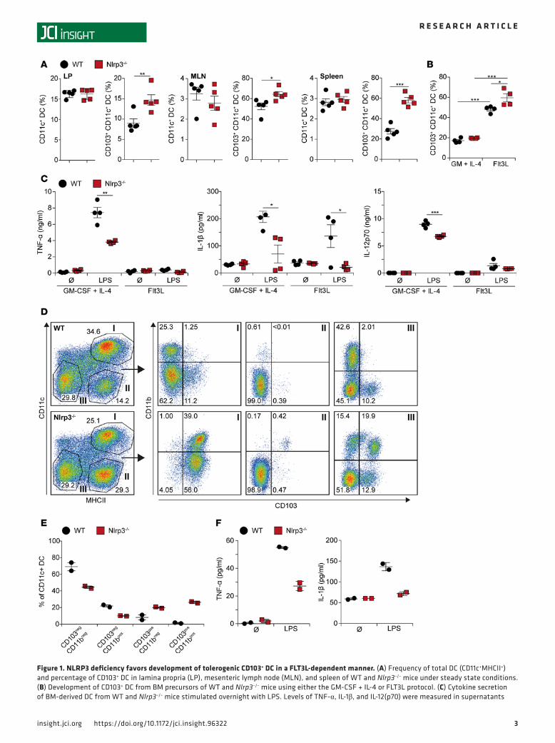
ResultsNLRP3 inflammasome limits FLT3L-mediated differentiation of tolerogenic CD103+ DC in vitro and in vivo. We recently reported an increased frequency of CD103+ DC in the LP of Nlrp3–/– mice. As CD103+ DC are considered to exert tolerogenic functions, we hypothesized that this finding might explain reduced colitis severity in Nlrp3–/– mice, as observed in DSS- and TNBS-induced colitis models (19, 30). To elucidate the relevance of our finding, we analyzed immune cells from LP, mesenteric lymph node, and spleen of WT and Nlrp3–/– mice in steady state. The number of total CD11c+ DC was similar in both genotypes, but the proportion of CD103+ DC in Nlrp3–/– mice was significantly increased (Figure 1A). Expression of CD103 by DC requires the growth factor FLT3L (37, 38). For further characterization of the observed DC phe-notype, we generated BM DC of WT and Nlrp3–/– mice using the GM-CSF + IL-4 or the FLT3L protocol. FACS analysis confirmed high frequency of CD103+ cells within the FLT3L-differentiated WT and Nlrp3–/– DC, whereas the GM-CSF + IL-4 protocol resulted in low numbers of CD103+ cells. Within FLT3L cul-tures, Nlrp3–/– DC expressed significantly higher CD103 levels when compared with DC from WT mice, pointing out a role for NLRP3 in controlling CD103+ DC differentiation (Figure 1B).
Flt3L DC cultures were shown to produce CD103+ DC with reduced cytokine production and tolero-genic features (39). This led us to investigate the inflammatory potential of DC from WT and Nlrp3–/– mice
3insight.jci.org https://doi.org/10.1172/jci.insight.96322
R E S E A R C H A R T I C L E
Figure 1. NLRP3 deficiency favors development of tolerogenic CD103+ DC in a FLT3L-dependent manner. (A) Frequency of total DC (CD11c+MHCII+) and percentage of CD103+ DC in lamina propria (LP), mesenteric lymph node (MLN), and spleen of WT and Nlrp3–/– mice under steady state conditions. (B) Development of CD103+ DC from BM precursors of WT and Nlrp3–/– mice using either the GM‑CSF + IL‑4 or FLT3L protocol. (C) Cytokine secretion of BM‑derived DC from WT and Nlrp3–/– mice stimulated overnight with LPS. Levels of TNF‑α, IL‑1β, and IL‑12(p70) were measured in supernatants

4insight.jci.org https://doi.org/10.1172/jci.insight.96322
R E S E A R C H A R T I C L E
after stimulation with the TLR4 ligand LPS. Compared with FLT3L, GM-CSF–differentiated DC pro-duced high levels of the proinflammatory cytokines TNF, IL-1β, and IL-12p70. Compared with WT DC, cytokine production was significantly lower in Nlrp3–/– DC. In FLT3-differentiated DC, IL-1β secretion was almost absent in Nlrp3–/– DC, in line with inflammasome-mediated IL-1β processing (Figure 1C). In con-clusion, Nlrp3 deficiency favors development of CD103+ DC with reduced inflammatory capacity.
We next investigated the role of FLT3L-dependent DC expansion in vivo. We injected WT or Nlrp3–/– mice with FLT3L-producing B16 melanoma cells and assessed the frequency of splenic CD103+ DC popu-lations (Figure 1D). As expected, high FLT3L levels led to strong expansion of DC populations. Compared with WT mice, a massive shift from CD103– toward CD103+ DC, in both CD11b+ and CD11b– DC pop-ulations, was observed in NLRP3-deficient mice (Figure 1, D and E). DC expanded in WT mice secreted higher levels of TNF-α and IL-1β than DC from Nlrp3–/– mice in response to LPS stimulation (Figure 1F). Together, these in vivo and in vitro findings indicate a decisive role for the NLRP3/IL-1β axis in controlling CD103 expression by DC.
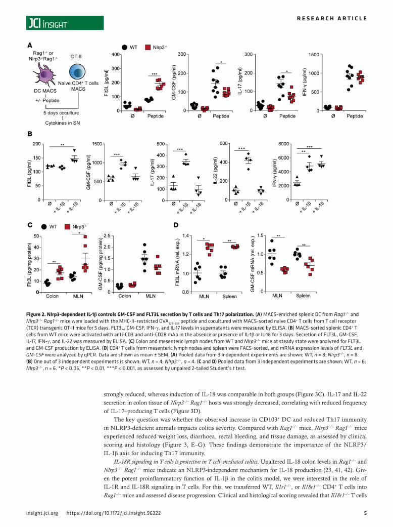
NLRP3-dependent IL-1β produced by DC limits FLT3L but enhances GM-CSF and IL-17 secretion by T cells. Upon DC and T cell interaction, T cells are polarized into an effector or regulatory phenotype with secre-tion of corresponding cytokines and growth factors. We hypothesized that NLRP3 in DC influences intes-tinal T cell phenotype and function. Hence, we analyzed cytokine secretion of naive CD4+ OT-II T cells cocultured with splenic, OVA peptide-pulsed DC. T cells cocultured with Nlrp3–/– DC produced significant-ly more FLT3L but lower levels of GM-CSF, as compared with WT DC. In addition, IL-17 levels were sig-nificantly reduced, whereas secretion of the Th1 cytokine IFN-γ was similar for both DC genotypes (Figure 2A). These findings argue for an NLRP3-dependent role in DC balancing GM-CSF and FLT3L secretion, as well as Th17 polarization of T cells.
The NLRP3 inflammasome controls the activation of IL-1β and IL-18, which are both involved in T helper cell development (40). To investigate the differential contribution of IL-1β and IL-18 on T cell polarization, we stimulated CD4+ T cells with IL-1β or IL-18 in the presence of antibody-mediated CD3/CD28 costimulation. The addition of IL-18 increased the production of FLT3L by activated T cells, where-as IL-1β enhanced GM-CSF production (Figure 2B). Moreover, only IL-1β induced the secretion of the Th17-associated cytokines IL-17 and IL-22, yet with similar levels of IFN-γ for both IL-1 family cytokines (Figure 2B). Thus, the observed phenotype of Nlrp3–/– DC may be explained by the lack of bioactive IL-1β.
As our in vitro data indicated a link between NLRP3 and FLT3L, we investigated the in vivo levels of FLT3L and GM-CSF in colonic tissue and mesenteric lymph nodes of WT and Nlrp3–/– mice under steady state conditions. Compared with WT mice, Nlrp3–/– mice exhibited significantly higher FLT3L tissue levels (Figure 2C). GM-CSF levels, however, were reduced but did not reach statistical significance at this point. As our results identified T cells as a source of both growth factors upon activation by DC and IL-1 family cytokines, we isolated CD4+ T cells from mesenteric lymph nodes and spleen and analyzed them for cytokine expression by quantitative PCR (qPCR). We observed significantly increased FLT3L mRNA expression in T cells of Nlrp3–/– mice and higher expression of GM-CSF mRNA in WT mice (Figure 2D). Together, these data show that NLRP3-mediated IL-1β tips the FLT3L/GM-CSF balance in T cells toward GM-CSF, which prevents the development of tolerogenic CD103+ DC favoring Th17-associated pathology.
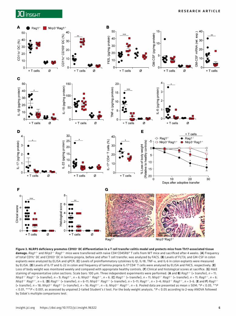
Enhanced frequency of CD103+ DC in Nlrp3–/– mice correlates with reduced Th17 immunity and protection from T cell transfer colitis. Rag1–/– mice lack functional B and T cells and are used to study the pathogenesis of colitis by adoptively transferred T cells. This model provides a platform for host-mediated education of naive T cells and their interaction with local DC. Naive CD4+ T cells were transferred into Rag1–/– or Nlrp3–/– Rag1–/– mice, and local DC as well as T cells were analyzed in different tissues after 4 weeks. Similar to our previous observations, the frequency of total CD11c+ DC in LP was comparable, but again, CD103+ DC were signifi-cantly increased in NLRP3-deficient Rag1–/– mice (Figure 3A). Increased numbers of CD103+ DC correlated with significantly enhanced FLT3L levels and reduced GM-CSF mRNA expression in colon tissue of Nlrp3–/– Rag1–/– hosts (Figure 3B). Colon tissue levels of the inflammatory cytokines IL-1β, TNF, and IL-6 were also
with ELISA. (D–F) WT and Nlrp3–/– mice were implanted with FLT3L‑producing B16 tumor cells. (D and E) Expression profile of whole splenic subpopu‑lations (D) and MACS‑sorted CD11c+ DC populations (E) were analyzed by FACS. (F) CD11c‑enriched DC were stimulated with LPS overnight. Secretion of TNF‑α and IL‑1β into supernatant was determined by ELISA. Data are shown as mean ± SEM; (A) WT n = 5, Nlrp3–/–, n = 5, 1 out of 3 experiments is shown; (B) WT, n = 4, Nlrp3–/–, n = 4, 1 out of 3 experiments shown; (C) WT (n = 4), Nlrp3–/– (n = 4), 1 out of 3 experiments shown; (D–F) WT (n = 2), Nlrp3–/– (n = 2), 1 out of 2 experiments is shown. *P < 0.05, **P < 0.01, ***P < 0.001, as assessed by unpaired 2‑tailed Student’s t test.
5insight.jci.org https://doi.org/10.1172/jci.insight.96322
R E S E A R C H A R T I C L E
strongly reduced, whereas induction of IL-18 was comparable in both groups (Figure 3C). IL-17 and IL-22 secretion in colon tissue of Nlrp3–/– Rag1–/– hosts was strongly decreased, correlating with reduced frequency of IL-17–producing T cells (Figure 3D).
The key question was whether the observed increase in CD103+ DC and reduced Th17 immunity in NLRP3-deficient animals impacts colitis severity. Compared with Rag1–/– mice, Nlrp3–/– Rag1–/– mice experienced reduced weight loss, diarrhoea, rectal bleeding, and tissue damage, as assessed by clinical scoring and histology (Figure 3, E–G). These findings demonstrate the importance of the NLRP3/IL-1β axis for inducing Th17 immunity.
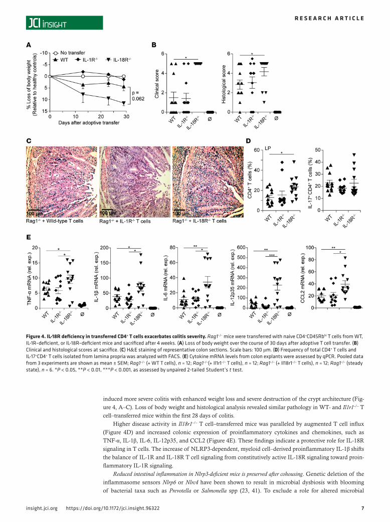
IL-18R signaling in T cells is protective in T cell–mediated colitis. Unaltered IL-18 colon levels in Rag1–/– and Nlrp3–/– Rag1–/– mice indicate an NLRP3-independent mechanism for IL-18 production (23, 41, 42). Giv-en the potent proinflammatory function of IL-1β in the colitis model, we were interested in the role of IL-1R and IL-18R signaling in T cells. For this, we transferred WT, Il1r1–/–, or Il18r1–/– CD4+ T cells into Rag1–/– mice and assessed disease progression. Clinical and histological scoring revealed that Il18r1–/– T cells
Figure 2. Nlrp3-dependent IL-1β controls GM-CSF and FLT3L secretion by T cells and Th17 polarization. (A) MACS‑enriched splenic DC from Rag1–/– and Nlrp3–/– Rag1–/– mice were loaded with the MHC‑II–restricted OVA323–339 peptide and cocultured with MACS‑sorted naive CD4+ T cells from T cell receptor (TCR) transgenic OT‑II mice for 5 days. FLT3L, GM‑CSF, IFN‑γ, and IL‑17 levels in supernatants were measured by ELISA. (B) MACS‑sorted splenic CD4+ T cells from WT mice were activated with anti‑CD3 and anti‑CD28 mAb in the absence or presence of IL‑1β or IL‑18 for 3 days. Secretion of FLT3L, GM‑CSF, IL‑17, IFN‑γ, and IL‑22 was measured by ELISA. (C) Colon and mesenteric lymph nodes from WT and Nlrp3–/– mice at steady state were analyzed for FLT3L and GM‑CSF production by ELISA. (D) CD4+ T cells from mesenteric lymph nodes and spleen were FACS‑sorted, and mRNA expression levels of FLT3L and GM-CSF were analyzed by qPCR. Data are shown as mean ± SEM. (A) Pooled data from 3 independent experiments are shown; WT, n = 8; Nlrp3–/–, n = 8. (B) One out of 3 independent experiments is shown; WT, n = 4; Nlrp3–/–, n = 4. (C and D) Pooled data from 3 independent experiments are shown; WT, n = 6; Nlrp3–/–, n = 6. *P < 0.05, **P < 0.01, ***P < 0.001, as assessed by unpaired 2‑tailed Student’s t test.
6insight.jci.org https://doi.org/10.1172/jci.insight.96322
R E S E A R C H A R T I C L E
Figure 3. NLRP3 deficiency promotes CD103+ DC differentiation in a T cell transfer colitis model and protects mice from Th17-associated tissue damage. Rag1–/– and Nlrp3–/– Rag1–/– mice were transferred with naive CD4+CD45Rbhi T cells from WT mice and sacrificed after 4 weeks. (A) Frequency of total CD11c+ DC and CD103+ DC in lamina propria, before and after T cell transfer, was analyzed by FACS. (B) Levels of FLT3L and GM‑CSF in colon explants were analyzed by ELISA and qPCR. (C) Levels of proinflammatory cytokines IL‑1β, IL‑18, TNF‑α, and IL‑6 in colon explants were measured by ELISA. (D) Levels of IL‑17 and IL‑22 in colon and frequency of lamina propria IL‑17+CD4+ T cells were analyzed by ELISA and FACS, respectively. (E) Loss of body weight was monitored weekly and compared with appropriate healthy controls. (F) Clinical and histological scores at sacrifice. (G) H&E staining of representative colon sections. Scale bars: 100 μm. Three independent experiments were performed. (A and B) Rag1–/– (+ transfer), n = 11; Nlrp3–/– Rag1–/– (+ transfer), n = 9; Rag1–/–, n = 6; Nlrp3–/– Rag1–/–, n = 6. (C) Rag1–/– (+ transfer), n = 11; Nlrp3–/– Rag1–/– (+ transfer), n = 11; Rag1–/–, n = 6; Nlrp3–/– Rag1–/–, n = 6. (D) Rag1–/– (+ transfer), n = 6–11; Nlrp3–/– Rag1–/– (+ transfer), n = 5–11; Rag1–/–, n = 3–4; Nlrp3–/– Rag1–/–, n = 3–6. (E and F) Rag1–/– (+ transfer), n = 18; Nlrp3–/– Rag1–/– (+ transfer), n = 16; Rag1–/–, n = 6; Nlrp3–/– Rag1–/–, n = 6. Pooled data are presented as mean ± SEM; *P < 0.05, **P < 0.01, ***P < 0.001, as assessed by unpaired 2‑tailed Student’s t test. For the body weight analysis, *P < 0.05 according to 2‑way ANOVA followed by Sidak’s multiple comparisons test.
7insight.jci.org https://doi.org/10.1172/jci.insight.96322
R E S E A R C H A R T I C L E
induced more severe colitis with enhanced weight loss and severe destruction of the crypt architecture (Fig-ure 4, A–C). Loss of body weight and histological analysis revealed similar pathology in WT- and Il1r1–/– T cell–transferred mice within the first 28 days of colitis.
Higher disease activity in Il18r1–/– T cell–transferred mice was paralleled by augmented T cell influx (Figure 4D) and increased colonic expression of proinflammatory cytokines and chemokines, such as TNF-α, IL-1β, IL-6, IL-12p35, and CCL2 (Figure 4E). These findings indicate a protective role for IL-18R signaling in T cells. The increase of NLRP3-dependent, myeloid cell–derived proinflammatory IL-1β shifts the balance of IL-1R and IL-18R T cell signaling from constitutively active IL-18R signaling toward proin-flammatory IL-1R signaling.
Reduced intestinal inflammation in Nlrp3-deficient mice is preserved after cohousing. Genetic deletion of the inflammasome sensors Nlrp6 or Nlrc4 have been shown to result in microbial dysbiosis with blooming of bacterial taxa such as Prevotella or Salmonella spp (23, 41). To exclude a role for altered microbial
Figure 4. IL-18R deficiency in transferred CD4+ T cells exacerbates colitis severity. Rag1–/– mice were transferred with naive CD4+CD45Rbhi T cells from WT, IL‑1R–deficient, or IL‑18R–deficient mice and sacrificed after 4 weeks. (A) Loss of body weight over the course of 30 days after adoptive T cell transfer. (B) Clinical and histological scores at sacrifice. (C) H&E staining of representative colon sections. Scale bars: 100 μm. (D) Frequency of total CD4+ T cells and IL‑17+CD4+ T cells isolated from lamina propria was analyzed with FACS. (E) Cytokine mRNA levels from colon explants were assessed by qPCR. Pooled data from 3 experiments are shown as mean ± SEM; Rag1–/– (+ WT T cells), n = 12; Rag1–/–(+ Il1r1–/– T cells), n = 12; Rag1–/– (+ Il18r1–/– T cells), n = 12; Rag1–/– (steady state), n = 6. *P < 0.05, **P < 0.01, ***P < 0.001, as assessed by unpaired 2‑tailed Student’s t test.
8insight.jci.org https://doi.org/10.1172/jci.insight.96322
R E S E A R C H A R T I C L E
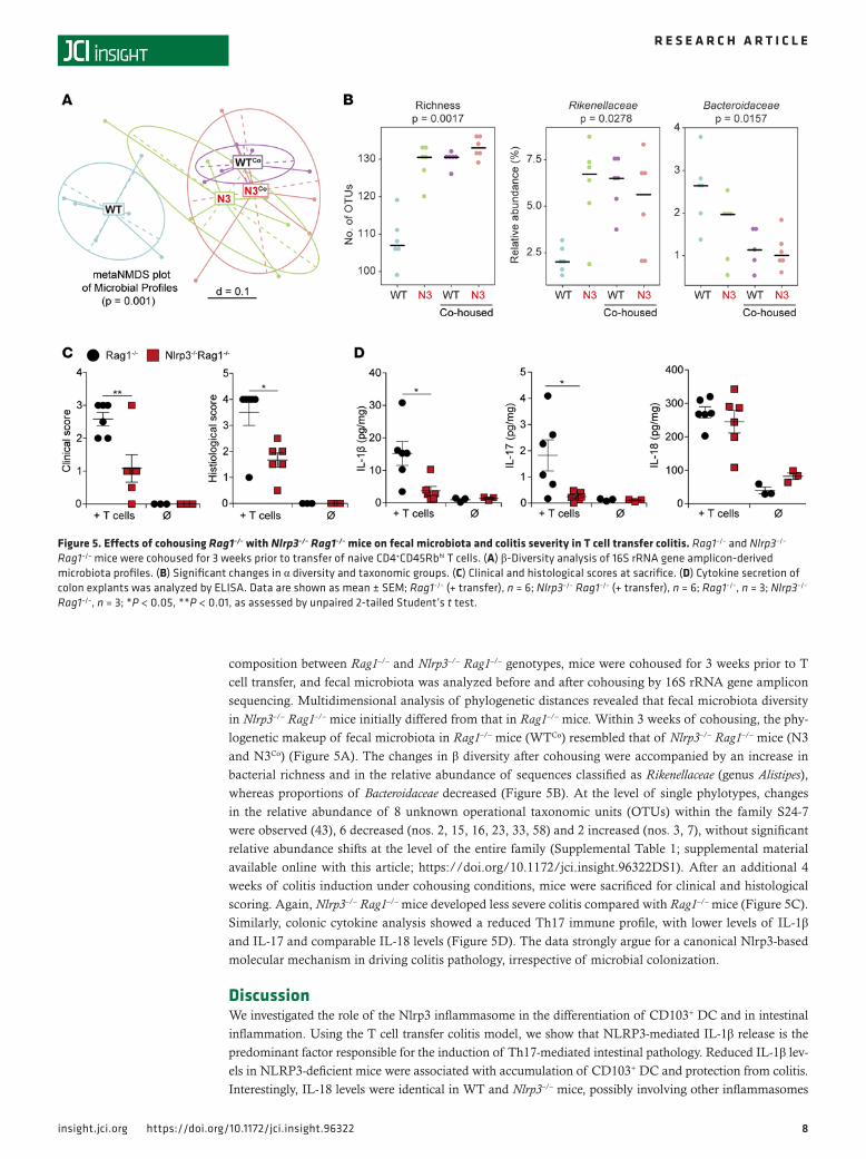
composition between Rag1–/– and Nlrp3–/– Rag1–/– genotypes, mice were cohoused for 3 weeks prior to T cell transfer, and fecal microbiota was analyzed before and after cohousing by 16S rRNA gene amplicon sequencing. Multidimensional analysis of phylogenetic distances revealed that fecal microbiota diversity in Nlrp3–/– Rag1–/– mice initially differed from that in Rag1–/– mice. Within 3 weeks of cohousing, the phy-logenetic makeup of fecal microbiota in Rag1–/– mice (WTCo) resembled that of Nlrp3–/– Rag1–/– mice (N3 and N3Co) (Figure 5A). The changes in β diversity after cohousing were accompanied by an increase in bacterial richness and in the relative abundance of sequences classified as Rikenellaceae (genus Alistipes), whereas proportions of Bacteroidaceae decreased (Figure 5B). At the level of single phylotypes, changes in the relative abundance of 8 unknown operational taxonomic units (OTUs) within the family S24-7 were observed (43), 6 decreased (nos. 2, 15, 16, 23, 33, 58) and 2 increased (nos. 3, 7), without significant relative abundance shifts at the level of the entire family (Supplemental Table 1; supplemental material available online with this article; https://doi.org/10.1172/jci.insight.96322DS1). After an additional 4 weeks of colitis induction under cohousing conditions, mice were sacrificed for clinical and histological scoring. Again, Nlrp3–/– Rag1–/– mice developed less severe colitis compared with Rag1–/– mice (Figure 5C). Similarly, colonic cytokine analysis showed a reduced Th17 immune profile, with lower levels of IL-1β and IL-17 and comparable IL-18 levels (Figure 5D). The data strongly argue for a canonical Nlrp3-based molecular mechanism in driving colitis pathology, irrespective of microbial colonization.
DiscussionWe investigated the role of the Nlrp3 inflammasome in the differentiation of CD103+ DC and in intestinal inflammation. Using the T cell transfer colitis model, we show that NLRP3-mediated IL-1β release is the predominant factor responsible for the induction of Th17-mediated intestinal pathology. Reduced IL-1β lev-els in NLRP3-deficient mice were associated with accumulation of CD103+ DC and protection from colitis. Interestingly, IL-18 levels were identical in WT and Nlrp3–/– mice, possibly involving other inflammasomes
Figure 5. Effects of cohousing Rag1–/– with Nlrp3–/– Rag1–/– mice on fecal microbiota and colitis severity in T cell transfer colitis. Rag1–/– and Nlrp3–/– Rag1–/– mice were cohoused for 3 weeks prior to transfer of naive CD4+CD45Rbhi T cells. (A) β‑Diversity analysis of 16S rRNA gene amplicon‑derived microbiota profiles. (B) Significant changes in α diversity and taxonomic groups. (C) Clinical and histological scores at sacrifice. (D) Cytokine secretion of colon explants was analyzed by ELISA. Data are shown as mean ± SEM; Rag1–/– (+ transfer), n = 6; Nlrp3–/– Rag1–/– (+ transfer), n = 6; Rag1–/–, n = 3; Nlrp3–/– Rag1–/–, n = 3; *P < 0.05, **P < 0.01, as assessed by unpaired 2‑tailed Student’s t test.
9insight.jci.org https://doi.org/10.1172/jci.insight.96322
R E S E A R C H A R T I C L E
(18, 23, 42). Our data are concordant with a recent report on miR-223–mediated regulation of Nlrp3 expres-sion and intestinal inflammation, describing Nlrp3-dependent elevation of IL-1, but not of IL-18, levels (20).
The activation of both IL-1β and IL-18 requires inflammasomes and caspase-1, with NLRP3 and NLRP6 as the best-characterized representatives. While NLRP3-dependent IL-1β is predominantly pro-duced in immune cells, NLRP6 is highly expressed in intestinal epithelial cells and controls gut barrier integrity in an IL-18–dependent manner (18). Colonic IL-1β levels correlate with disease activity and are critical for Th17 differentiation (44). We found reduced IL-1β levels in NLRP3-deficient mice correlating with reduced Th17 cell infiltrates and tissue preservation. Conversely, IL-18 has been shown to signal via the IL-18R on T cells to limit Th17 cell differentiation (17). Accordingly, we show that transfer of IL-18R–deficient T cells results in increased colitis pathology with vigorous T cell influx in the LP. The data argue that IL-18R signaling in T cells counteracts proinflammatory Nlrp3-dependent IL-1R signaling.
Contrary to published data on IL-1R deficiency of transferred T cells ameliorating colitis severity (45, 46), we did not observe significant differences between WT and Il1r1–/– T cells within the observation period of 4 weeks in our study, which focused on the early effects of inflammasome and cytokine signal-ing in colitis. This stands in contrast to the above-mentioned studies, which sacrificed the mice after 5–8 weeks, when mice were generally sicker. These findings hint toward a more prominent role of protective IL-18R signals in T cells in the early phase of colitis, whereas IL-1R–mediated effects occur later, when
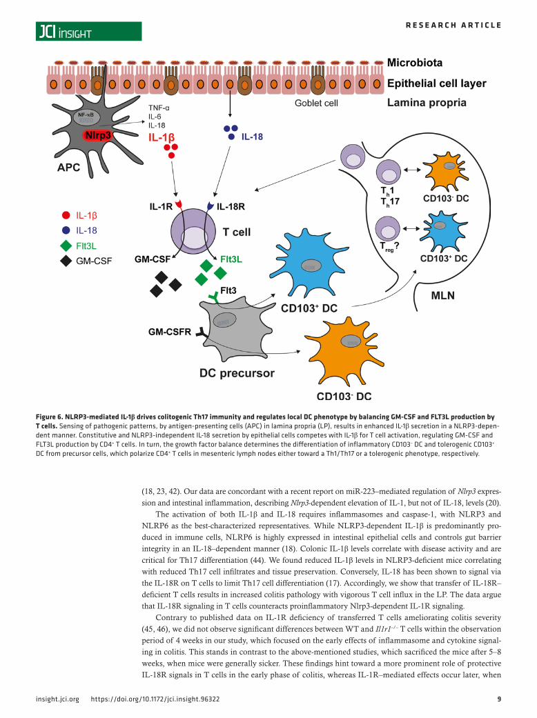
Figure 6. NLRP3-mediated IL-1β drives colitogenic Th17 immunity and regulates local DC phenotype by balancing GM-CSF and FLT3L production by T cells. Sensing of pathogenic patterns, by antigen‑presenting cells (APC) in lamina propria (LP), results in enhanced IL‑1β secretion in a NLRP3‑depen‑dent manner. Constitutive and NLRP3‑independent IL‑18 secretion by epithelial cells competes with IL‑1β for T cell activation, regulating GM‑CSF and FLT3L production by CD4+ T cells. In turn, the growth factor balance determines the differentiation of inflammatory CD103– DC and tolerogenic CD103+ DC from precursor cells, which polarize CD4+ T cells in mesenteric lymph nodes either toward a Th1/Th17 or a tolerogenic phenotype, respectively.
1 0insight.jci.org https://doi.org/10.1172/jci.insight.96322
R E S E A R C H A R T I C L E
the epithelial barrier function is worsening and the LP is infiltrated with innate immune cells secreting IL-1β. The precise kinetics of IL-18R and IL-1R signaling during T cell–mediated colitis remain to be elucidated in future studies.
Protection of NLRP3-deficient mice from colitis was associated with expansion of CD103+ DC. Their tolerogenic properties, potentially controlling disease severity, led us to investigate the DC/T cell axis. DC differentiation occurs via FLT3L and GM-CSF, which are produced by various immune and nonimmune cells upon inflammatory stimuli, including IL-1 family cytokines (47–50). We focused on IL-1β and IL-18, as inflammasome signaling pathways have already been shown to control cyto-kine release in other pathological conditions (48, 51). In Rag1–/– mice, as well as Nlrp3–/– Rag1–/– mice, absence of T cells resulted in reduced DC numbers in steady state. T cells are a source of DC-expand-ing cytokines, such as FLT3L and GM-CSF, and have the potential to influence DC homeostasis in the gut (51, 52). We identified both IL-1β and IL-18 to control FLT3L and GM-CSF secretion by activated T cells in vitro and in vivo. Within this paradigm, IL-1β favored the secretion of GM-CSF, whereas IL-18 induced FLT3L release by activated T cells.
Both IL-1R and IL-18R signal through the adapter molecule MyD88 and share common downstream elements. Interestingly, IL-18 plays a role in IL-18R–mediated inhibition of IL-1R downstream signaling (17). The absence of NLRP3-mediated IL-1β may, thus, enforce IL-18R signaling, with enhanced FLT3L secretion and differentiation of CD103+ DC. Conversely, IL-1R signaling enhances GM-CSF production by T cells, favoring inflammatory CD103– DC to promote Th17 immunity (Figure 6).
IL-1β has been shown to promote Th17 responses in the intestine via the IL-1R, and mice as well as humans with hyperactive Nlrp3 mutations exhibit strong Th17 immunity as a consequence of excess IL-1β (44, 45, 53). Accordingly, in our model, lack of NLRP3-dependent IL-1β was associated with decreased Th17 pathology with reduced levels of IL-1β, IL-6, and IL-17. Finally, we identified IL-1β as potent stimu-lus for IL-22 secretion by T cells, which acts in concert with IL-17 to mount a robust Th17-based immune response. Impaired IL-22 secretion in Nlrp3–/– mice further highlights the role for the Nlrp3 inflammasome in regulating Th17 immunity. However, IL-1 effects are not limited to Th17, as IL-1 signaling was shown to override Foxp3 expression, regulating plasticity of induced Tregs (54). The net effect of IL-1β is proin-flammatory; however, the role of IL-18 is complex. Depending on site of activation, the release of IL-18 can mediate protective or destructive effects. IL-18 activation within the intestinal barrier contributes to the preservation of the intestinal barrier, while — in the LP — it mainly synergizes with IL-12, resulting in T cell activation. Recent data indicate that steady state intestinal epithelial cells constitutively secrete IL-18, which acts directly on IL-18R–expressing T cells, resulting in 2 effects: limitation of colonic Th17 differentiation and promotion of suppressive function of FOXP3+CD4+ T cells, thereby preventing colitis (17). We suggest that IL-1β mediates increased susceptibility of NLRP3-sufficient mice toward T cell trans-fer colitis by overriding protective IL-18R signals, thereby shaping a highly plastic intestinal T cell pool. Dysbalance of IL-18R versus IL-1R signaling in T cells might explain beneficial versus detrimental effects of NLRP3-deficiency in the murine models of IBD, resulting in seemingly contradicting reports in the literature (14, 19).
The intestinal microbiota regulate gut homeostasis, and disrupted inflammasome signaling can result in dysbiosis with increased colonization of pathobionts (23, 29). Recently, Mamantopoulos et al. have questioned the role of inflammasomes in shaping intestinal ecology (55). In an attempt to control any potentially confounding microbiota effects, we cohoused mice and found that fecal microbiota profiles in WT mice shifted toward that in Nlrp3-deficient mice. Despite cohousing and acquisition of bacterial taxa in all cohoused mice, we observed the same protective phenotype in Nlrp3–/– mice, implying that the Nlrp3 inflammasome, not intestinal dysbiosis, modulates T cell responses and DC generation. This indicates that differences in intestinal ecology associated with NLRP3 deficiency are phenomena without causal influ-ence on the outcome of T cell responses in transfer colitis. Our data might help to reconcile contradicting reports in the literature.
In summary, our data identify the NLRP3 inflammasome as an important checkpoint regulating the IL-1β/IL-18 ratio in the intestine to control immune homeostasis and Th17 immunity. The balance of IL-1β and IL-18 controls the secretion of FLT3L and GM-CSF by T cells, which in turn influences the differentiation of CD103+ DC from their precursors. Pharmacological inhibition of the NNLRP3/IL-1β/GM-CSF axis might represent a promising approach for IBD treatment.
1 1insight.jci.org https://doi.org/10.1172/jci.insight.96322
R E S E A R C H A R T I C L E
MethodsMice. WT C57BL/6 mice were purchased from Janvier Labs. Nlrp3–/– mice were a gift of Jurg Tschopp (University of Lausanne, Lausanne, Switzerland) (56) and were crossed into Rag1–/– to obtain Nlrp3–/– Rag1–/– double-deficient mice. Rag1–/– mice, originally obtained from The Jackson Laboratory, were a gift of Norbert Gerdes (Institute for Cardiovascular Prevention [IPEK], University of Munich). Il18r1–/– mice (B6.129P2-Il18r1tm1Aki/J; 004131) and Il1r1–/– mice (B6.129S7-Il1r1tm1lmx/J; 003245) were purchased from The Jackson Laboratory. OT-II TCR transgenic mice (B6.Cg-Tg(TcraTcrb)425Cbn/J) were a gift from Thom-as Brocker (Institute of Immunology, University of Munich). Mice were kept on the C57BL/6 background for more than 15 generations and bred under SPF conditions with a 12-hour light/dark cycle, at the Uni-versity of Munich. Mice were fed a standard chow diet (ssniff R/M-H autoclavable chow diet; ssniff Spe-zialdiäten), received water ad libitum, and were used for experiments between 8 and 10 weeks of age.
T cell transfer colitis. Single cell suspensions from spleens of C57BL/6 mice were depleted of CD8+, MHC class II+, Mac-1+, and B220+ cells, leaving CD4+ T cells untouched (Dynabeads, Invitrogen). After staining with PE-conjugated CD4 and FITC-conjugated CD45Rb antibodies, naive CD4+CD45Rbhi T cells were isolated using a BD FACSAria III cell sort system (>98% purity). T cells (4 × 10⁵) were adoptively transferred into Rag1–/– recipient mice via i.p. injection.
Clinical and histological scoring. Scoring of disease activity was done as previously described (19, 30). Brief-ly, combined diarrhea and hematochezia (clinical) scoring was done in a blinded fashion. Stool consistency was classified as: 0, regular pellets; 1, soft pellets; 2, bulky pellets; 3, pasty and opaque pellets; and 4, liquid diarrhea. Hemoccult testing was staged as: 0, negative; 2, positive; 4, visual rectal bleeding. The overall clinical score was calculated as the sum of the diarrhea and hematochezia scores, reaching a maximum of 8 points. Combined histological scoring was done for distal colon sections by a trained pathologist in a blind-ed fashion. Formalin-fixed distal colon samples (1 cm each) were embedded in paraffin and cut into 4-μm sections. Subsequently, H&E staining was performed and analyzed. Focal inflammatory cell infiltration was graded as: 0, normal; 1, increased; 2, submucosal infiltration; and 3, transmural extension. Epithelial dam-age was scored as: 0, no damage; 1, scarce focal lesions; 2, focal erosions or ulcerations of the mucosal layer; and 3, severe damage extending to the intestinal wall. The overall histological score was calculated as the sum of the cell infiltration and epithelial damage scores, reaching a maximum of 6 points.
Ex vivo analysis of colonic cytokines. Colonic explants were weighed and cultured in complete RPMI media or shock frozen in liquid nitrogen, mechanically crushed, and incubated with cell lysis buffer (Bio-Rad) for 15 minutes on ice. The homogenate was centrifuged at 10,000 g at 4 °C for 15 minutes, and protein amounts were adjusted (Bradford analysis, Bio-Rad). Cytokines in supernatants or cell lysates were mea-sured by ELISA for IL-6 (BD Biosciences); IL-22 (eBioscience); and GM-CSF, FLT3L, IL-1β, TNF, IFN-γ, and IL-17 (all from R&D Systems). Concentrations were normalized to tissue weight or whole protein, as indicated.
Analysis of LP immune cells. LP immune cells were isolated using Mouse Lamina Propria Dissociation Kit (Miltenyi Biotec). Colon sections were cut into small pieces and incubated with 5 mM EDTA, 1 mM DTT (MilliporeSigma), and 5% FBS in HBSS buffer (Thermo Fisher Scientific) for 20 minutes and passed through a 70-μm cell strainer. Remaining tissue was collected into a gentleMACS C tube (Miltenyi Biotec) containing 5% FBS in HBSS (with Ca2+/Mg2+) and dissociated using gentleMACS Dissociator (Miltenyi Biotec). Cells were washed and stained for FACS analysis. For intracellular cytokine staining, cells were stimulated with 0.1 μg/ml PMA,1 μg/ml ionomycin, 10 μg/ml Brefeldin A (all from MilliporeSigma) for 4 hours; washed; and stained for surface markers. Intracellular staining was done using BD Cytofix/Cyto-perm solution (BD Biosciences).
Antibodies and flow cytometry. Rat anti–mouse CD4-PE (clone GK1.5), rat anti–mouse CD4-PerCP (clone RM4-5), rat anti–mouse CD45RB-FITC (clone 16A), rat anti–mouse IL-17 Alexa Fluor 488 (clone TC11-18H10), and rat anti–mouse CD11b-PerCP-Cy5.5 (clone M1/70), as well as hamster anti–mouse CD3-APC (clone 145-2C11), were purchased from BD Biosciences. Hamster anti–mouse CD11c-Pacific Blue (clone N418), hamster anti–mouse CD103 Alexa Flour 488 (clone 2E7), hamster anti–mouse CD3-Pacific Blue (clone 145-2C11), and rat anti–mouse IFN-γ–Pacific Blue (clone XMG1.2) were from BioLegend. Flow cytometry was performed using a FACSCanto II system (BD Biosciences).
qPCR analysis. Cells or colonic tissue were homogenized using an Ultra-turrax instrument (IKA). Total RNA was isolated with the peqGOLD total RNA kit (Peqlab) according to the manufacturers’ protocol. Purity and yield were determined using NanoDrop (Thermo Fisher Scientific). cDNA transcription was done with the

1 2insight.jci.org https://doi.org/10.1172/jci.insight.96322
R E S E A R C H A R T I C L E
RevertAID First stranded cDNA Synthesis kit (Invitrogen), and qPCR analysis was performed with the Kapa Probe Fast Kit (Peqlab) on a LightCycler 480 system (Roche Diagnostics). β-Actin served as housekeeping gene.
Generation of BM-derived and splenic DC, and T cell coculture assay. BM cells of femur and tibia were passed through a 40-μm cell strainer and centrifuged for 5 minutes at 400 g. After RBC lysis (BD Biosciences), cells were incubated with recombinant murine IL-4 and GM-CSF (20 ng/ml), or FLT3L (100 ng/ml), for 7–9 days, and DC were isolated by magnetic beads (Miltenyi Biotec). If indicated, cells were stimulated with 0.5 μg/ml LPS (InvivoGen), and cytokine levels in supernatants were assessed. For coculture experiments, splenic DC were pulsed with or without 25 μg/ml MHC-II–restricted OVA323-339 peptide for 1 hour at 37°C. Naive CD4+ T cells were isolated by negative depletion (Miltenyi Biotec) from OT-II mice. DC (2 × 10⁴) were cocultured with 1 × 10⁵ CD4+ OT-II T cells for 5 days, and cytokine levels in supernatants were determined by ELISA.
T cell stimulation assays. CD4+ T cells were isolated using CD4+ T Cell Isolation Kit mouse (Miltenyi Biotec) according to the manufacturer’s instruction and were labeled with PerCP rat anti–mouse CD4 (clone RM4-5) and APC hamster anti–mouse CD3 (clone 145-2C11) (BD Biosciences) for FACS-sort (FACSAria III, BD Biosciences). Cells were resuspended in RPMI medium and activated with plate-bound anti-CD3 (5 μg/ml) and soluble anti-CD28 (2 μg/ml) in 96-well U-bottom plates for 72 hours. Where indi-cated, 20 ng/ml recombinant murine (rm) IL-1β (Peprotech) or 10 ng/ml rmIL-18 (BioVision) was added to the cell cultures.
16S rRNA gene amplicon analysis. Fecal samples were collected before and 3 weeks after cohousing of animals, prior to T cell transfer. Samples were processed as described before (57). Briefly, genomic DNA was extracted using phenol/chloroform extraction after mechanical disruption. The variable region V3/V4 of 16S rRNA genes was amplified using the primers 341F and 785R following a 2-step procedure. Ampli-cons were purified using the AMPure XP system (Beckman Coulter) and sequenced in paired-end modus (PE275) using a MiSeq system (Illumina Inc.). Raw read files were processed using IMNGS (www.imngs.org) (43). OTUs were picked at a threshold of 97 % sequence similarity. Only those with a relative abun-dance above 0.5% total sequences per sample were kept. Readouts and statistics were generated in R using Rhea (https://lagkouvardos.github.io/Rhea/) (43).
Statistics. GraphPad Prism 5 was used for data analysis. Two-tailed Student’s t test or 2-way ANOVA were used for group comparisons. Data are expressed as means ± SEM, with at least 2 independent experi-ments performed. Differences were considered statistically significant at *P < 0.05, **P < 0.01, ***P < 0.001.
Study approval. Animal experiments were reviewed by an animal care authority and approved by the Regierung von Oberbayern.
Author contributionsPD, CB, and MS conceived the experimental strategy. PD, RM, CH, CR, and CB designed and performed experiments. HAL contributed histological analysis. GD and SH performed experiments on Il18r1–/– mice. TC and SF contributed the microbiota analysis. SK and SE gave conceptual advice. The paper was written by PD, CB, and MS.
AcknowledgmentsWe thank Michael Hristov (IPEK, Ludwig-Maximilians-Universität [LMU] Munich) for assistance with cell sorting; Nicole Lichter and Cäcillia Czogalla for technical assistance in animal work and with ex vivo assays; Caroline Ziegler and Angela Sachsenhauser for technical assistance with sample preparation for high-throughput 16S rRNA gene amplicon analysis; Irmina Karl for technical assistance; and Thomas Korn (Neuro-Kopf-Zentrum, Technical University of Munich, Munich, Germany) and Norbert Gerdes (IPEK, LMU Munich) for providing Rag1–/– mice. OT-II TCR transgenic mice were kindly provided by Thomas Brocker (Institute of Immunology, University of Munich). Flt3L-transduced B16 melanoma cells were pro-vided by Glenn Dranoff (Dana-Farber Cancer Institute, Boston, Massachusetts, USA). This work was fund-ed by Else Kröner-Fresenius-Stiftung 2012_A27 to CB and Deutsche Forschungs-gemeinschaft - SFB1123 to PD and MS.
Address correspondence to: Max Schnurr, Klinikum der Universität München, Lindwurmstrasse 2a, Munich 80337, Germany. Phone: 49.89.4400.57322; Email: [email protected].
1 3insight.jci.org https://doi.org/10.1172/jci.insight.96322
R E S E A R C H A R T I C L E
1. de Souza HS, Fiocchi C. Immunopathogenesis of IBD: current state of the art. Nat Rev Gastroenterol Hepatol. 2016;13(1):13–27. 2. Neurath MF. Cytokines in inflammatory bowel disease. Nat Rev Immunol. 2014;14(5):329–342. 3. Hugot JP, et al. Association of NOD2 leucine-rich repeat variants with susceptibility to Crohn’s disease. Nature.
2001;411(6837):599–603. 4. Ogura Y, et al. A frameshift mutation in NOD2 associated with susceptibility to Crohn’s disease. Nature. 2001;411(6837):603–
606. 5. Martinon F, Burns K, Tschopp J. The inflammasome: a molecular platform triggering activation of inflammatory caspases and
processing of proIL-beta. Mol Cell. 2002;10(2):417–426. 6. Latz E, Xiao TS, Stutz A. Activation and regulation of the inflammasomes. Nat Rev Immunol. 2013;13(6):397–411. 7. Paramel GV, Sirsjö A, Fransén K. Role of genetic alterations in the NLRP3 and CARD8 genes in health and disease. Mediators
Inflamm. 2015;2015:846782. 8. Casini-Raggi V, Kam L, Chong YJ, Fiocchi C, Pizarro TT, Cominelli F. Mucosal imbalance of IL-1 and IL-1 receptor antago-
nist in inflammatory bowel disease. A novel mechanism of chronic intestinal inflammation. J Immunol. 1995;154(5):2434–2440. 9. Pizarro TT, et al. IL-18, a novel immunoregulatory cytokine, is up-regulated in Crohn’s disease: expression and localization in
intestinal mucosal cells. J Immunol. 1999;162(11):6829–6835. 10. Cominelli F, et al. Interleukin 1 (IL-1) gene expression, synthesis, and effect of specific IL-1 receptor blockade in rabbit immune
complex colitis. J Clin Invest. 1990;86(3):972–980. 11. Reinecker HC, et al. Enhanced secretion of tumour necrosis factor-alpha, IL-6, and IL-1 beta by isolated lamina propria mono-
nuclear cells from patients with ulcerative colitis and Crohn’s disease. Clin Exp Immunol. 1993;94(1):174–181. 12. Ludwiczek O, et al. Imbalance between interleukin-1 agonists and antagonists: relationship to severity of inflammatory bowel
disease. Clin Exp Immunol. 2004;138(2):323–329. 13. Raza A, Yousaf W, Giannella R, Shata MT. Th17 cells: interactions with predisposing factors in the immunopathogenesis of
inflammatory bowel disease. Expert Rev Clin Immunol. 2012;8(2):161–168. 14. Zaki MH, Boyd KL, Vogel P, Kastan MB, Lamkanfi M, Kanneganti TD. The NLRP3 inflammasome protects against loss of
epithelial integrity and mortality during experimental colitis. Immunity. 2010;32(3):379–391. 15. Siegmund B. Interleukin-18 in intestinal inflammation: friend and foe? Immunity. 2010;32(3):300–302. 16. Reuter BK, Pizarro TT. Commentary: the role of the IL-18 system and other members of the IL-1R/TLR superfamily in innate
mucosal immunity and the pathogenesis of inflammatory bowel disease: friend or foe? Eur J Immunol. 2004;34(9):2347–2355. 17. Harrison OJ, et al. Epithelial-derived IL-18 regulates Th17 cell differentiation and Foxp3+ Treg cell function in the intestine.
Mucosal Immunol. 2015;8(6):1226–1236. 18. Nowarski R, et al. Epithelial IL-18 Equilibrium Controls Barrier Function in Colitis. Cell. 2015;163(6):1444–1456. 19. Bauer C, et al. Colitis induced in mice with dextran sulfate sodium (DSS) is mediated by the NLRP3 inflammasome. Gut.
2010;59(9):1192–1199. 20. Neudecker V, et al. Myeloid-derived miR-223 regulates intestinal inflammation via repression of the NLRP3 inflammasome.
J Exp Med. 2017;214(6):1737–1752. 21. Siegmund B, Lehr HA, Fantuzzi G, Dinarello CA. IL-1 beta -converting enzyme (caspase-1) in intestinal inflammation. Proc
Natl Acad Sci USA. 2001;98(23):13249–13254. 22. Bauer C, et al. The ICE inhibitor pralnacasan prevents DSS-induced colitis in C57BL/6 mice and suppresses IP-10 mRNA but
not TNF-alpha mRNA expression. Dig Dis Sci. 2007;52(7):1642–1652. 23. Elinav E, et al. NLRP6 inflammasome regulates colonic microbial ecology and risk for colitis. Cell. 2011;145(5):745–757. 24. Allen IC, et al. The NLRP3 inflammasome functions as a negative regulator of tumorigenesis during colitis-associated cancer.
J Exp Med. 2010;207(5):1045–1056. 25. Takagi H, et al. Contrasting action of IL-12 and IL-18 in the development of dextran sodium sulphate colitis in mice. Scand J
Gastroenterol. 2003;38(8):837–844. 26. Siegmund B, et al. Neutralization of interleukin-18 reduces severity in murine colitis and intestinal IFN-gamma and TNF-alpha
production. Am J Physiol Regul Integr Comp Physiol. 2001;281(4):R1264–R1273. 27. Sivakumar PV, et al. Interleukin 18 is a primary mediator of the inflammation associated with dextran sulphate sodium induced
colitis: blocking interleukin 18 attenuates intestinal damage. Gut. 2002;50(6):812–820. 28. Holmkvist P, Pool L, Hägerbrand K, Agace WW, Rivollier A. IL-18Rα-deficient CD4(+) T cells induce intestinal inflammation
in the CD45RB(hi) transfer model of colitis despite impaired innate responsiveness. Eur J Immunol. 2016;46(6):1371–1382. 29. Hirota SA, et al. NLRP3 inflammasome plays a key role in the regulation of intestinal homeostasis. Inflamm Bowel Dis.
2011;17(6):1359–1372. 30. Bauer C, Duewell P, Lehr HA, Endres S, Schnurr M. Protective and aggravating effects of Nlrp3 inflammasome activation in
IBD models: influence of genetic and environmental factors. Dig Dis. 2012;30 Suppl 1:82–90. 31. Cerovic V, et al. Intestinal CD103(-) dendritic cells migrate in lymph and prime effector T cells. Mucosal Immunol. 2013;6(1):104–113. 32. Coombes JL, et al. A functionally specialized population of mucosal CD103+ DCs induces Foxp3+ regulatory T cells via a
TGF-beta and retinoic acid-dependent mechanism. J Exp Med. 2007;204(8):1757–1764. 33. Laffont S, Siddiqui KR, Powrie F. Intestinal inflammation abrogates the tolerogenic properties of MLN CD103+ dendritic cells.
Eur J Immunol. 2010;40(7):1877–1883. 34. Uematsu S, et al. Regulation of humoral and cellular gut immunity by lamina propria dendritic cells expressing Toll-like recep-
tor 5. Nat Immunol. 2008;9(7):769–776. 35. Niess JH, et al. CX3CR1-mediated dendritic cell access to the intestinal lumen and bacterial clearance. Science.
2005;307(5707):254–258. 36. Pabst O, Bernhardt G. The puzzle of intestinal lamina propria dendritic cells and macrophages. Eur J Immunol.
2010;40(8):2107–2111. 37. Bogunovic M, et al. Origin of the lamina propria dendritic cell network. Immunity. 2009;31(3):513–525. 38. Varol C, et al. Intestinal lamina propria dendritic cell subsets have different origin and functions. Immunity. 2009;31(3):502–512. 39. Miller G, Pillarisetty VG, Shah AB, Lahrs S, DeMatteo RP. Murine Flt3 ligand expands distinct dendritic cells with both tolero-
1 4insight.jci.org https://doi.org/10.1172/jci.insight.96322
R E S E A R C H A R T I C L E
genic and immunogenic properties. J Immunol. 2003;170(7):3554–3564. 40. Sims JE, Smith DE. The IL-1 family: regulators of immunity. Nat Rev Immunol. 2010;10(2):89–102. 41. Franchi L, et al. NLRC4-driven production of IL-1β discriminates between pathogenic and commensal bacteria and promotes
host intestinal defense. Nat Immunol. 2012;13(5):449–456. 42. Wilson NS, et al. Inflammasome-dependent and -independent IL-18 production mediates immunity to the ISCOMATRIX adju-
vant. J Immunol. 2014;192(7):3259–3268. 43. Lagkouvardos I, et al. The Mouse Intestinal Bacterial Collection (miBC) provides host-specific insight into cultured diversity
and functional potential of the gut microbiota. Nat Microbiol. 2016;1(10):16131. 44. Lasigliè D, et al. Role of IL-1 beta in the development of human T(H)17 cells: lesson from NLPR3 mutated patients. PLoS One.
2011;6(5):e20014. 45. Coccia M, et al. IL-1β mediates chronic intestinal inflammation by promoting the accumulation of IL-17A secreting innate lym-
phoid cells and CD4(+) Th17 cells. J Exp Med. 2012;209(9):1595–1609. 46. Li B, Gurung P, Malireddi RK, Vogel P, Kanneganti TD, Geiger TL. IL-10 engages macrophages to shift Th17 cytokine depen-
dency and pathogenicity during T-cell-mediated colitis. Nat Commun. 2015;6:6131. 47. Brasel K, Escobar S, Anderberg R, de Vries P, Gruss HJ, Lyman SD. Expression of the flt3 receptor and its ligand on hemato-
poietic cells. Leukemia. 1995;9(7):1212–1218. 48. Guermonprez P, et al. Inflammatory Flt3l is essential to mobilize dendritic cells and for T cell responses during Plasmodium
infection. Nat Med. 2013;19(6):730–738. 49. Huleihel M, Douvdevani A, Segal S, Apte RN. Different regulatory levels are involved in the generation of hemopoietic cyto-
kines (CSFs and IL-6) in fibroblasts stimulated by inflammatory products. Cytokine. 1993;5(1):47–56. 50. Solanilla A, et al. Expression of Flt3-ligand by the endothelial cell. Leukemia. 2000;14(1):153–162. 51. Lukens JR, Barr MJ, Chaplin DD, Chi H, Kanneganti TD. Inflammasome-derived IL-1β regulates the production of GM-CSF
by CD4(+) T cells and γδ T cells. J Immunol. 2012;188(7):3107–3115. 52. Saito Y, Boddupalli CS, Borsotti C, Manz MG. Dendritic cell homeostasis is maintained by nonhematopoietic and T-cell-pro-
duced Flt3-ligand in steady state and during immune responses. Eur J Immunol. 2013;43(6):1651–1658. 53. Meng G, Zhang F, Fuss I, Kitani A, Strober W. A mutation in the Nlrp3 gene causing inflammasome hyperactivation potenti-
ates Th17 cell-dominant immune responses. Immunity. 2009;30(6):860–874. 54. Basu R, et al. IL-1 signaling modulates activation of STAT transcription factors to antagonize retinoic acid signaling and control
the TH17 cell-iTreg cell balance. Nat Immunol. 2015;16(3):286–295. 55. Mamantopoulos M, et al. Nlrp6- and ASC-Dependent Inflammasomes Do Not Shape the Commensal Gut Microbiota Compo-
sition. Immunity. 2017;47(2):339–348.e4. 56. Martinon F, Pétrilli V, Mayor A, Tardivel A, Tschopp J. Gout-associated uric acid crystals activate the NALP3 inflammasome.
Nature. 2006;440(7081):237–241. 57. Lagkouvardos I, et al. Gut metabolites and bacterial community networks during a pilot intervention study with flaxseeds in
healthy adult men. Mol Nutr Food Res. 2015;59(8):1614–1628.