nihms-6008
DESCRIPTION
jhgjTRANSCRIPT
Gonadal Hormone Modulation of Mu, Kappa, and Delta OpioidAntinociception in Male and Female Rats
Erin C. Stoffel*, Catherine M. Ulibarri†, John E. Folk‡, Kenner C. Rice‡, and Rebecca M.Craft**From the Department of Psychology, Washington State University, Pullman, Washington
†From the Departments of Veterinary and Comparative Anatomy, Pharmacology and Physiology,Washington State University, Pullman, Washington
‡From the Department of Laboratory of Medicinal Chemistry, NIDDK, NIH, Bethesda, Maryland.
AbstractPrevious studies suggest that sex differences in morphine antinociception in rodents might beattributed to the activational effects of gonadal hormones. The present study determined whetherhormonal modulation of opioid antinociception in adult rats extends to opioids other than theprototypic mu agonist morphine. Male and female rats were sham-gonadectomized (sham-GDX) orgonadectomized (GDX) and replaced with no hormone, estradiol (E2, females), progesterone (P4,females), E2+P4 (females), or testosterone (males). Approximately 28 days later, nociception wasevaluated on the 50°C hot plate and warm water tail withdrawal tests before and after subcutaneousadministration of hydromorphone, buprenorphine, U50,488, or SNC 80. In sham-GDX (gonadallyintact) rats, the mu agonists and U50,488 were less effective in females than in males in at least onenociceptive test, and the delta agonist SNC 80 was less effective in males than in females. In males,gonadectomy tended to decrease, and testosterone tended to increase antinociception produced by 3of the 4 agonists. In females, gonadectomy and hormone treatment had more variable effects,although E2 tended to decrease mu opioid antinociception. The present results suggest thatactivational effects of gonadal hormones are relatively modest and somewhat inconsistent onantinociception produced by various opioid agonists in the adult rat.
Perspective: This study demonstrates that reproductive hormones such as testosterone in males andestradiol in females do not consistently modulate sensitivity to the analgesic effects of opioids in theadult organism.
KeywordsTestosterone; estradiol; progesterone; pain; analgesia; sex differences
Accumulating evidence suggests that there are sex differences in the acute antinociceptiveeffects of opioids in rodents, with males generally displaying greater opioid sensitivity thanfemales.14 Several variables that might influence the observation of sex differences in opioidantinociception include the efficacy/selectivity of the opioid, the type and intensity of thenoxious stimulus, and the genotype of the subject.5,13,15,29,36,37,49 Sex differences in opioid
Correspondence to: Rebecca M. Craft.Address reprint requests to Rebecca M. Craft, PhD, Department of Psychology, Washington State University, Pullman, WA 99164-4820.E-mail: [email protected] by NIDA grant DA10284 (R.M.C.)
NIH Public AccessAuthor ManuscriptJ Pain. Author manuscript; available in PMC 2006 March 29.
Published in final edited form as:J Pain. 2005 April ; 6(4): 261–274.
NIH
-PA Author Manuscript
NIH
-PA Author Manuscript
NIH
-PA Author Manuscript
antinociception have been attributed to the differential organizational and/or activationaleffects of gonadal steroid hormones in males versus females.
Studies that have examined the organizational effects of gonadal hormones report that neonatalgonadal steroid manipulations (castration of males, androgen administration to females) caneliminate sex differences in morphine antinociception in adult rats.12,31 Results of studiesexamining activational effects of gonadal hormones on opioid antinociception are quitevariable. In male rats, gonadectomy increased,1,10 decreased,43,47,48 or failed tochange9,11,27,28,30 mu opioid antinociception. Similarly, in females, gonadectomyincreased,1,28,30,47,48 decreased,4 or did not change11,28,48 opioid antinociception.
The variability across studies that have manipulated gonadal hormones in adult rats might bedue to the wide array of methodologic differences across studies. Nearly every study has useddifferent gonadectomy test intervals, hormone replacement regimens, nociceptive testingprocedures, and opioid administration procedures. In a previous study from this laboratory, weexamined the ability of gonadal steroid hormones to modulate morphine antinociception byusing a gonadectomy test interval and hormone replacement regimen that have been usedextensively to examine the reproductive system in male and female rats.18-20,47,52 This protocolproduced well-documented changes in male and female reproductive behavior and physiologyand also altered morphine antinociception in male and female rats.47 In male rats, gonadectomydecreased morphine's potency, and chronic treatment with testosterone or the combination ofits 2 primary metabolites, estradiol (E2) and dihydrotestosterone, restored morphine's potencyto that seen in sham-gonadectomized males. In gonadally intact female rats, morphine wasmore potent during diestrus-1 and proestrus phases relative to the estrus phase. Gonadectomyincreased morphine's potency in females, and chronic treatment with E2 or E2 plusprogesterone (P4) decreased morphine's potency to that seen in gonadally intact estrus females.With a similar gonadectomy-nociceptive test interval, another laboratory also found thatmorphine's potency was increased in gonadectomized females relative to sham-gonadectomized females.48 In another study, chronic treatment with E2 also decreasedmorphine's potency relative to gonadectomized females.45 These studies suggest that bothtestosterone in adult male rats and E2 in adult female rats contribute to the sex difference inmorphine antinociception.
Previous studies examining gonadal hormone modulation of opioid antinociception haveprimarily tested only morphine,1,4,10-12,27,28,30,31,43,47 with the exception of one study thatincluded several other mu-selective and mixed action opioids.48 The latter study examined theeffect of gonadectomy on opioid antinociception, but it did not examine the effects ofreplacement hormones or estrous cycle phase in gonadally intact females, which are crucialfor confirming that effects of gonadectomy are due to loss of particular gonadal steroids. Thepurpose of the present study was to more fully characterize the modulatory role of gonadalsteroid hormones on antinociception produced by a variety of opioids. We examinedhydromorphone and buprenorphine, 2 opioids that produce antinociception in rodents via muopioid receptor activation,17,26,33 as well as the kappa receptor-selective opioid,U50,488,32,40 and the delta receptor-selective opioid, SNC 80.8 Buprenorphine was chosenbecause it is less efficacious than morphine, and previous studies have shown that sexdifferences tend to be larger with lower efficacy opioids.13 Previous studies suggest thatgonadal steroids might modulate morphine antinociception by modulating itsmetabolism.3,44,46 Thus, hydromorphone was chosen because it is structurally very similar tomorphine and metabolized similarly.2 Gonadal hormones were manipulated in male and femalerats via gonadectomy and hormone replacement by using dosing regimens that arereproductively relevant.47 In addition, gonadally intact female rats were tested during differentphases of the estrous cycle, and 2 nociceptive tests were used to determine the generalizabilityof gonadal hormone modulation of opioid antinociception.
Stoffel et al. Page 2
J Pain. Author manuscript; available in PMC 2006 March 29.
NIH
-PA Author Manuscript
NIH
-PA Author Manuscript
NIH
-PA Author Manuscript

Material and MethodsAll procedures were approved by the Washington State University Institutional Animal Careand Use Committee.
SubjectsMale and female Sprague-Dawley rats (bred in-house from Taconic stock, Germantown, NY)were used. Rats were housed in same-sex pairs, according to gonadal steroid status after surgery(eg, an orchidectomized rat with testosterone replacement was housed with another male inthe same treatment group). Male and female animals were housed in separate rooms maintainedat 21.5°C ± 2°C with a 12:12-hour light:dark cycle (lights on at 6:00 AM). Access to food andwater was ad libitum except during testing.
General ProcedureSurgery (gonadectomy or sham-gonadectomy) was performed on rats that were approximately3 months old, as described previously.47 In gonadectomized (GDX) rats, basal nociception andopioid antinociception were tested 28 days after surgery, when endogenous hormone levelsand associated reproductive behaviors have been shown to be minimal.18-20,52 In sham-gonadectomized (sham-GDX) female rats, antinociceptive testing occurred between 21 and 35days after surgery so that females in diestrus-1 (sham-D), proestrus (sham-P), and estrus (sham-E) could be identified and tested in approximately equal numbers.
Steroid Hormone ReplacementChronic steroid replacement was achieved via silicone rubber capsules implantedsubcutaneously immediately after gonadectomy. A small incision was made between theshoulder blades, capsules were inserted in the subcutaneous space, and the skin was closedwith wound clips. Capsules containing testosterone were prepared in 10-mm lengths; one 10-mm testosterone capsule/100 g of body weight was implanted in male animals. Capsulescontaining E2 were prepared in 1-mm lengths; one 1-mm capsule/rat was implanted in femaleanimals. We have previously shown that these hormone treatments are sufficient to reinstatenormal reproductive behavior and physiology in male and female rats, and that they alsosignificantly alter sensitivity to morphine antinociception.47 In terms of sexual receptivity anduterine weight, GDX females replaced with E2 as described in this study are similar to intactfemales in vaginal proestrus to estrus.47 To control for possible effects of capsule implantation,sham-GDX and GDX control rats were implanted with empty capsules (0); males received two10-mm 0 capsules/rat, and females received one 1-mm 0 capsule/rat.
Some female animals also received P4 treatment. Beginning 4 days after surgery, injections ofP4 (500 μg/rat in 0.1-mL volume, subcutaneous) were given every 4 days at approximately10:00 AM to mimic the preovulatory P4 surge observed in normally cycling females.22 All rats(females and males) not receiving P4 treatment received safflower oil vehicle injections (0.1mL/rat, subcutaneous) on the same schedule, so that all rats were handled/injected the samenumber of times. Table 1 summarizes the 10 different treatment groups in the study.
Nociceptive TestingNociceptive testing began at approximately 2:00 PM to occur within the period of P4's peakreproductive effect, 4 to 6 hours after injection.21 Rats were tested on both the hot plate andwarm water tail withdrawal assays (both 50°C) consecutively in that order (tests wereapproximately 10 seconds apart). To obtain non-drug response latencies (to lick a hind paw orjump off the hot plate, or to withdraw the tail from warm water), each rat was tested on eachassay 3 times: first at time 0 (baseline 1) and then again 10 to 15 minutes later (baseline 2);
Stoffel et al. Page 3
J Pain. Author manuscript; available in PMC 2006 March 29.
NIH
-PA Author Manuscript
NIH
-PA Author Manuscript
NIH
-PA Author Manuscript

immediately after this second baseline test, saline was administered (1 mL/kg, subcutaneous),and rats were tested a third time on each assay 10 to 15 minutes later (saline test). Immediatelyafter the saline test, increasing doses of one of the opioid agonists in ½-log (buprenorphine,SNC 80) or ¼-log (hydromorphone, U50,488) unit increments were administered every 10(SNC 80) or 15 (buprenorphine, hydromorphone, U50,488) minutes, with nociceptive latenciesobtained 10 or 15 minutes, respectively, after each injection. A 10-minute rather than 15-minuteinter-injection interval was used for SNC 80 because of its relatively short duration ofaction.8 For instance, 0.1 mg/kg hydromorphone was administered, and hot plate and tailwithdrawal latencies were obtained 15 minutes later; immediately after testing, an additional0.22 mg/kg (total 0.32 mg/kg) was administered, and hot plate and tail withdrawal latencieswere obtained 15 minutes later. Testing continued with increasing doses of agonist until therat reached the cutoff latency on both assays. Cutoff latencies of 80 and 25 seconds were usedon the hot plate and tail withdrawal assays, respectively. If a rat did not respond by the cutofflatency, the test was terminated, and the cutoff latency was recorded. Once the rat reachedcutoff on one assay, subsequent testing was conducted only on the other assay. Each rat wasonly tested with only one drug; in other words, separate rats were used to examine hormonemodulation of antinociception produced by each of the 4 agonists.
Vaginal CytologyTo further characterize gonadal steroid hormone state in female rats, daily vaginal smears wereobtained in sham-GDX female rats beginning on day 21 after surgery. Proestrus was identifiedby the prevalence (approximately 80% or more of epithelial cells in sample) of nucleatedepithelial cells; estrus was identified by the prevalence of dense sheets of cornified epithelialcells; diestrus-1 was identified by the presence of leukocytes and scattered nucleated and/orcornified epithelial cells; diestrus-2 was identified by the relative absence of any cells.24 Tokeep the stages highly distinct, sham-GDX female animals whose vaginal cytology indicatedthat they were in transition from one estrous stage to another were not tested. To represent a“normal” sample of females, testing in sham-GDX females was not limited to 4-day cyclers.Thus, the designations of diestrus-1 and diestrus-2 are based on vaginal cytology alone and donot necessarily indicate the first and second day after estrus.
Smears also were obtained in sham-GDX female rats immediately after nociceptive testing.Only data from females that remained in the same stage from before to after testing wereincluded in the analyses.
Data AnalysisLatency to respond in seconds was recorded for each hot plate and tail withdrawal test. Unlikethe other opioid agonists tested, U50,488 increased the number of rats that jumped off the hotplate rather than licked a hind paw; furthermore, rats that jumped off the hot plate after a lowdose of U50,488 were more likely to immediately jump off the plate on subsequent tests.Therefore data from rats that jumped off the plate more than 2 times during testing with U50,488were removed from further analyses. This resulted in the deletion of data from approximately2 rats per treatment group tested with U50,488. No data were deleted from any groups testedwith the other agonists.
Baseline response latency was calculated for each rat as the mean of the second baseline testand the saline test; latency from the first baseline test was not used because baseline hot platelatency shortens significantly from the first to second test in females.15 Baseline responselatencies were compared between sham-GDX males and females and within the male, sham-GDX, and GDX female groups by using analysis of variance (ANOVA). Because there weresignificant differences in baseline response latencies among treatment groups, individualresponse latencies obtained after each dose of drug were converted to % maximum possible
Stoffel et al. Page 4
J Pain. Author manuscript; available in PMC 2006 March 29.
NIH
-PA Author Manuscript
NIH
-PA Author Manuscript
NIH
-PA Author Manuscript
effect (%MPE): [(Drug latency – Baseline latency)/(Cutoff latency – Baseline latency)] × 100.Cutoff latency was adjusted so that the difference between the mean baseline and cutoff valuewas the same in all hormone treatment groups given the same drug (eg, for rats tested withhydromorphone, the cutoffs were 52 and 17.5 seconds over baseline on the hot plate and tailwithdrawal assays, respectively). These cutoff adjustments equalized the %MPE scale amongall hormone treatment groups within each drug, so that comparisons of drug potency amonghormone treatment groups were based on the same 0% to 100% scale. From these %MPE data,the estimated dose at which antinociception was 50% (ED50) was calculated for each rat bylinear interpolation, generally using 1 point above 50% and 1 point below 50% MPE. ED50values were not calculated when the %MPE value at the highest dose tested was less than 45%.
ANOVA of ED50 values was conducted to test for potency differences in opioid antinociceptionamong the various treatment groups. When ED50 values could not be calculated in more thanone third of the rats in a treatment group, treatment group ED50 comparisons were notconducted (Tables 2 and 3). Analyses of %MPE data also were conducted, because these allowfor efficacy comparisons among treatment groups based on all data, which is particularlycrucial for lower efficacy agonists (wherein ED50 values cannot always be calculated). Todetermine whether there were sex differences in opioid antinociception, %MPE and ED50values were compared between the sham-GDX male group and the 3 groups of sham-GDXfemales combined (data from sham-GDX females in proestrus, estrus, and diestrus werepooled). When significant, differences between particular sham-GDX female groups and thesham-GDX male group are also noted. To determine whether hormone treatments influencedopioid antinociception, %MPE and ED50 values were compared within the male groups, withinthe sham-GDX female groups, and within the GDX female groups by using ANOVA. Posthoc tests were conducted with Dunnett test, when comparing multiple treatment groups to acontrol group (eg, comparing GDX+E2, GDX+P4, GDX+E2/P4 groups to the GDX+0 group);with independent samples t tests with a Bonferroni correction, to determine at which dose therewere group differences in opioid antinociception; or with the Student-Newman-Keuls test,when there were no a priori comparisons (eg, comparing the different sham-GDX femalegroups). Significance level was set at P ≤.06, on the basis of Fisher's zone ofindeterminancy.50
DrugsHydromorphone (Sigma, St Louis, Mo) was dissolved in saline; saline served as the controlvehicle for hydromorphone. Buprenorphine (National Institute on Drug Abuse, Rockville, Md)and SNC 80 (synthesized by Drs J. Folk and K. Rice) were dissolved in lactic acid to whichdistilled water was added; pH was adjusted to approximately 5.5 with 1 N NaOH. A salinesolution of pH 5.5 was used as the control vehicle for buprenorphine and SNC 80. U50,488(National Institute on Drug Abuse) was dissolved in distilled water; distilled water served asthe control vehicle for U50,488. Vehicle and opioid injections were subcutaneous(hydromorphone, buprenorphine, U50,488) or intraperitoneal (SNC 808) in a volume of 1.0mL/kg. P4 (Steraloids, Wilton, NH) was dissolved in safflower oil; safflower oil served as thecontrol vehicle for this steroid. To administer steroid hormones by constantreleasesubcutaneous capsule, silicone rubber tubing (0.062 inches inner diameter/0.125 inches outerdiameter) was filled with crystallized hormone or nothing. Capsules were sealed with siliconerubber adhesive and allowed to dry for a minimum of 24 hours. They were then soaked in 70%ethanol for a minimum of 24 hours before use to test the integrity of the seal,18 and then theywere dried before implantation.
Stoffel et al. Page 5
J Pain. Author manuscript; available in PMC 2006 March 29.
NIH
-PA Author Manuscript
NIH
-PA Author Manuscript
NIH
-PA Author Manuscript

ResultsBasal Nociception
Baseline response latencies among rats tested with the different opioid agonists showed nosignificant interactions between agonist and hormonal status within male groups, sham-GDXfemale groups, or GDX female groups. Therefore, baseline response latency data from ratstested with the 4 different opioids were pooled for subsequent analysis of hormone effects onbasal nociception.
Among gonadally intact (sham-GDX) rats, there were sex differences in basal nociception inthe hot plate test. Fig 1 shows that sham-GDX females tested in diestrus-1, proestrus, or estrushad significantly longer hot plate latencies than sham-GDX males (F3,141 = 18.06, P < .001).In contrast to the hot plate, there were no significant sex differences in basal nociception in thetail withdrawal test (Fig 1).
Gonadal steroid manipulations in male rats did not significantly affect basal nociception in thehot plate test (Fig 1); however, in the tail withdrawal test, GDX+T males had significantlylonger latencies than GDX+0 males. In sham-GDX female rats, there were no significanteffects of estrous stage on basal nociception in either the hot plate or tail withdrawal assays(Fig 1). However, all sham-GDX female groups had significantly longer hot plate latenciesthan gonadectomized females with no hormone replacement (GDX+0 females) (F3,137 = 7.39,P < .001). In the tail withdrawal test, only sham-GDX females in diestrus had significantlylonger response latencies than GDX+0 females. Among the GDX females (Fig 1), GDX+E2and GDX+E2/P4 females had significantly longer response latencies than GDX+0 females inboth hot plate (F3,148 = 14.48, P < .001) and tail withdrawal tests (F3,147 = 7.20, P < .001).GDX+P4 females were not significantly different from GDX+0 females on either test.
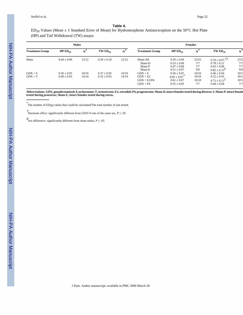
Hydromorphone AntinociceptionFig 2 shows the hydromorphone dose-effect curves in the hot plate and tail withdrawal testsin all treatment groups; Table 4 shows the corresponding ED50 values. Comparing gonadallyintact (sham-GDX) male versus female animals, analysis of %MPE values indicated thathydromorphone produced less hot plate antinociception (sex × dose: F4,132 = 2.31, P = .06)and less tail withdrawal antinociception in females than in males (sex × dose: F4,132 = 3.20,P = .02), particularly at the lower doses (0.32, 0.56 mg/kg). ED50 comparisons showed thathydromorphone was significantly more potent in gonadally intact males than in females in thetail withdrawal test (F1,34 = 4.84, P = .04), but not the hot plate test (Table 4). Gonadectomyeliminated the sex difference in hydromorphine antinociception (Table 4, compare GDX+0males vs females).
Among male treatment groups, there were no significant differences in ED50 values forhydromorphone in either the hot plate or tail withdrawal tests (Table 4), suggesting thathormonal manipulations in males did not significantly affect hydromorphone's antinociceptivepotency. However, analysis of the %MPE values revealed a significant interaction betweentreatment group and dose on the hot plate test (F8,132 = 2.37, P = .02); specifically, 0.56 mg/kg hydromorphone produced greater hot plate antinociception in GDX+T males than in GDX+0 males (Fig 2).
Among sham-GDX female animals tested in diestrus, proestrus, and estrus, hydromorphoneproduced essentially identical hot plate antinociception; furthermore, these gonadally intactfemales were not significantly different from gonadectomized females (GDX+0) in their hotplate response to hydromorphone (Fig 2, Table 4). However, in the tail withdrawal test, analysisof ED50 values revealed that hydromorphone was significantly less potent in estrus femalesthan in GDX+0 females (Table 4) (F3,29 = 3.39, P = .03).
Stoffel et al. Page 6
J Pain. Author manuscript; available in PMC 2006 March 29.
NIH
-PA Author Manuscript
NIH
-PA Author Manuscript
NIH
-PA Author Manuscript

Among GDX female animals, hydromorphone was significantly less potent in GDX+E2females compared to GDX+0 females in the hot plate test (Fig 2, Table 4)(F3,36 = 4.30, P = .01) and in GDX+E2/P4 females compared to GDX+0 females in the tail withdrawal test (Figure2, Table 4) (F3,36 = 3.45, P = .03). Analysis of %MPE values confirmed these ED50 analyses,showing that hydromorphone produced significantly less antinociception in GDX femalestreated with E2 in the hot plate test and in GDX females treated with E2+P4 in the tailwithdrawal test, compared to GDX females with no hormone replacement.
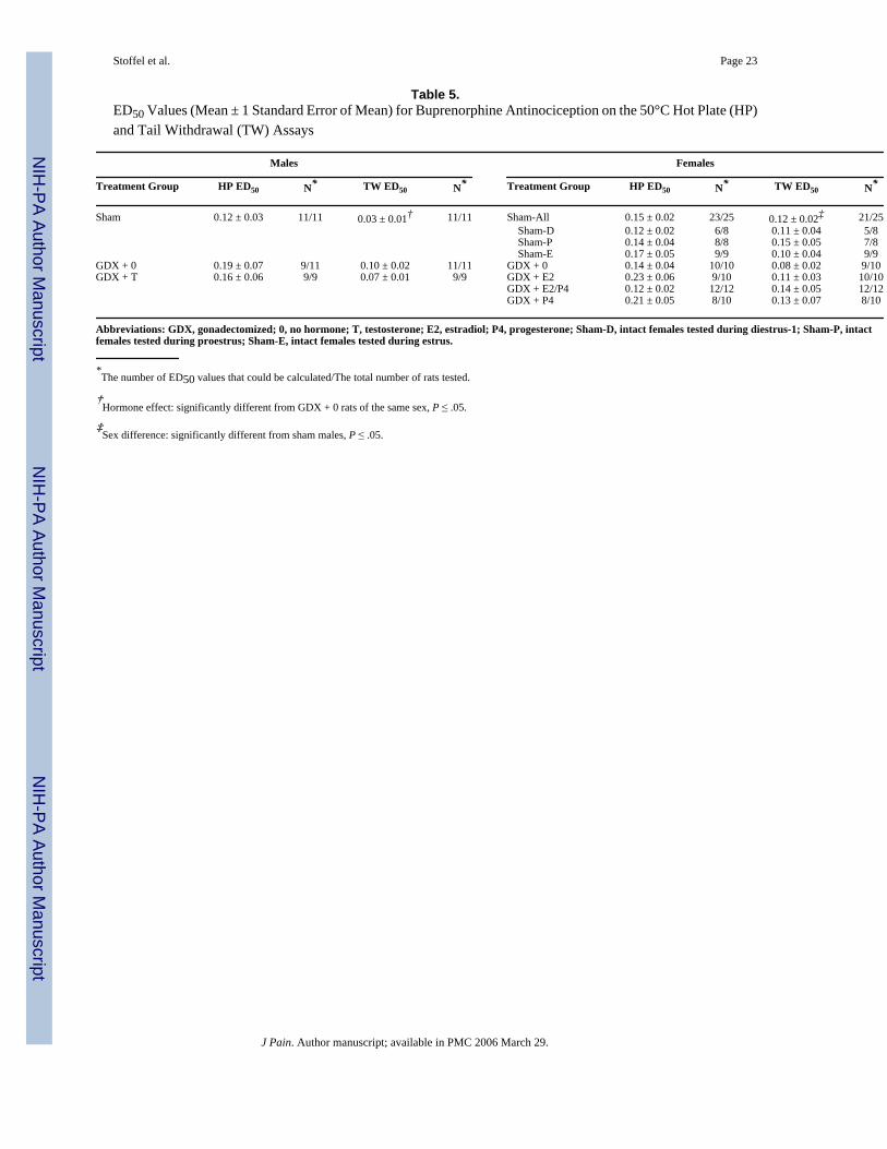
Buprenorphine AntinociceptionFig 3 shows buprenorphine dose-effect curves in the hot plate and tail withdrawal tests in alltreatment groups; Table 5 shows the corresponding ED50 values. Comparing gonadally intact(sham-GDX) males versus females, there were significant sex differences in buprenorphineantinociception in both hot plate and tail withdrawal tests (Fig 3). On the basis of analysis of%MPE values, buprenorphine produced less hot plate antinociception in females than in males(sex × dose: F4,136 = 2.91, P = .02); however, buprenorphine's potency did not differsignificantly on the basis of ED50 values that could be calculated (Table 5). In the tailwithdrawal test, buprenorphine produced less antinociception at the higher doses tested (%MPE analysis, sex × dose: F4,136 = 6.31, P < .001) and was less potent (ED50 analysis, Table5: F1,31 = 6.92, P = .01) in gonadally intact females than in males. Gonadectomy eliminatedthe sex difference in buprenorphine antinociception (Table 5, compare GDX+0 males vsfemales).
Among the male groups, in the hot plate test, buprenorphine was approximately equipotent,but it produced a slightly greater maximal effect in sham-GDX males than in GDX+0 males(Fig 3, Table 5); however, testosterone treatment in GDX males did not significantly increasebuprenorphine's maximal effect at the doses tested. In the tail withdrawal test, buprenorphineproduced a significantly greater maximal effect at the highest dose tested in sham-GDX relativeto GDX+0 males (Fig 3; F2,28 = 3.87, P = .03); buprenorphine's potency was also significantlygreater in sham-GDX relative to GDX+0 males (Table 5) (F2,28 = 4.32, P = .02). Testosteronetreatment increased buprenorphine's potency slightly but not significantly in the tail withdrawaltest.
Among female rats, neither %MPE nor ED50 analyses revealed any significant effects ofestrous stage, gonadectomy, or hormone replacement on buprenorphine antinociception ineither the hot plate or tail withdrawal tests (Fig 3).
U50,488 AntinociceptionFig 4 shows U50,488 dose-effect curves in the hot plate and tail withdrawal tests in all treatmentgroups; Table 2 shows the corresponding ED50 values. Comparing gonadally intact (sham-GDX) males versus females, there were no significant sex differences in U50,488antinociception in either the hot plate or tail withdrawal assays. Analyses of the %MPE valuesfor intact males and individual sham-GDX female groups revealed a significant interactionbetween treatment group and dose of U50,488 in the tail withdrawal test (F12,116 = 1.90, P = .04); specifically, 18 mg/kg U50,488 produced significantly less effect in sham-estrus femalesrelative to sham-GDX males (Fig 4).
Among the male groups, hormonal manipulations significantly influenced U50,488antinociception (Fig 4). Analysis of the hot plate %MPE values revealed a significantinteraction between treatment group and dose (F8,80 = 2.92, P = .007); specifically, 5.6 and 10mg/kg U50,488 produced greater antinociception in sham-GDX males than in GDX+0 males.Potency (ED50) comparisons among the 3 male groups yielded nonsignificant differences(Table 2)(F2,20 = 3.02, P = .07). Similar results were obtained in the tail withdrawal test;
Stoffel et al. Page 7
J Pain. Author manuscript; available in PMC 2006 March 29.
NIH
-PA Author Manuscript
NIH
-PA Author Manuscript
NIH
-PA Author Manuscript

although there were no significant differences in ED50 values among the male treatment groups(Table 2), analysis of %MPE values revealed a significant main effect of treatment group(F2,20 = 5.22, P = .02). Specifically, U50,488 produced greater tail withdrawal antinociceptionin GDX+T males than in GDX+0 males (Fig 4).
Among the sham-GDX females, U50,488 antinociception did not vary significantly across theestrous cycle in the hot plate test (Fig 4, Table 4). In the tail withdrawal test, analysis of %MPE values yielded a nonsignificant group by dose interaction (F8,96 = 1.90, P = .07).Gonadectomy of female rats decreased U50,488 antinociception on the hot plate (F12,124 =2.01, P = .03); specifically, 5.6 mg/kg U50,488 produced greater hot plate antinociception insham-GDX females in diestrus than in GDX+0 females (Fig 4). Analysis of ED50 valuesconfirmed that U50,488 tended to be more potent in gonadally intact females than in GDXfemales without hormone replacement on the hot plate test (F1,33 = 15.97, P = .02). In contrast,gonadectomy of female rats did not significantly alter U50,488 antinociception on the tailwithdrawal test.
Among the GDX females, there were no significant effects of hormone manipulation onU50,488 antinociception in the hot plate test (Fig 4). In the tail withdrawal test, however,gonadal hormone manipulations significantly affected U50,488 antinociception (% MPEanalysis: group × dose, F12,136 = 3.77, P < .001). Specifically, U50,488 produced significantlyless tail withdrawal antinociception in GDX+P4 females than in GDX+0 females (Fig 4).
SNC 80 AntinociceptionFig 5 shows SNC 80 dose-effect curves in the hot plate and tail withdrawal tests in all treatmentgroups; Table 3 shows the corresponding ED50 values. Comparing sham-GDX males versusfemales, analysis of ED50 values showed that SNC 80 was more potent in females than in malesin the hot plate (F1,34 = 4.84, P = .04) and tail withdrawal tests (F1,27 = 4.04, P = .06), on thebasis of the ED50 values that could be calculated (Table 3). Analysis of %MPE values, whichincluded data from all sham-GDX male and female rats, showed that SNC 80 produced greaterhot plate (F1,34 = 6.90, P = .01) but not tail withdrawal antinociception in females than in males.
Among the male groups, gonadal hormone manipulations did not significantly affect SNC 80antinociception in the hot plate test (Fig 5, Table 3). In the tail withdrawal test, gonadal hormonemanipulations in males did not affect SNC 80's potency on the basis of the ED50 values thatcould be calculated (Table 3). A comparison of %MPE data from all male rats showed,however, that SNC 80's maximal effect was significantly diminished in GDX+T males relativeto GDX+0 males (Fig 5; treatment group × dose: F6,78 = 4.59, P < .001).
Within the sham-GDX female groups, SNC 80 antinociception did not differ significantlyamong females tested in diestrus, proestrus, and estrus in either nociceptive test (Fig 5, Table3). However, in the tail withdrawal test, gonadectomy decreased SNC 80's potency in femalerats; ED50 values were significantly higher in GDX+0 females than in sham-GDX females(Table 3) (F1,27 = 8.75, P = .006).
Among GDX females tested in the hot plate test (Fig 5), gonadal hormone manipulationsmodulated SNC 80's potency (F3,29 = 3.10, P = .04) and maximal effects at the doses tested(treatment group × dose, F9,96 = 2.35, P = .02). However, none of the post hoc tests weresignificant, so it is difficult to draw conclusions about the specific hormone effects. Forexample, P4 appeared to increase the potency of SNC 80, but when combined with E2, maximaleffect was decreased. In the tail withdrawal test (Fig 5), analysis of %MPE values also showedthat there were significant differences in SNC 80 antinociception among GDX female groups(F3,32 = 4.64, P < .01), yet post hoc analyses yielded no differences between specific hormone-treated groups and the GDX+0 group.
Stoffel et al. Page 8
J Pain. Author manuscript; available in PMC 2006 March 29.
NIH
-PA Author Manuscript
NIH
-PA Author Manuscript
NIH
-PA Author Manuscript

DiscussionHormonal Modulation of Nociception
The present study demonstrates sex differences in latency to respond in the hot plate test butnot in the tail withdrawal test. As we reported previously,47 female animals had longer hotplate latencies than males, and this sex difference appeared to depend on the presence of E2.Whereas gonadectomy and testosterone replacement in males did not significantly affect hotplate latencies, gonadectomy in females shortened hot plate latency, and E2 replacement, withor without P4, reinstated the longer latencies seen in gonadally intact females. Hormonalmodulation of nociception in females was similar in the tail withdrawal test, with gonadectomyshortening and E2 lengthening tail withdrawal latencies. In contrast, in males, testosteronesignificantly lengthened tail withdrawal latencies despite the fact that gonadectomy had noeffect. Previous studies have reported similar gonadectomy and testosterone replacementeffects on nociceptive responses in male rats23,25,34,42 and gonadectomy/ovarian hormonereplacement effects in female rats,23,51 whereas other studies report no apparent hormonalmodulation of nociceptive thresholds.7,11,30,35,41 It should be noted that we also have observedno hormonal modulation of thermal nociception when using higher intensity nociceptivestimuli than those used in the present study (unpublished data).47
Sex Differences in Opioid AntinociceptionTable 6 summarizes the present results on sex differences in and hormonal modulation of opioidantinociception and compares these to results we previously obtained by using similarprocedures to evaluate hormonal modulation of morphine antinociception in male and femalerats.47 In the present study, agonists that are known to produce antinociception by actingpreferentially at mu opioid receptors (hydromorphone, buprenorphine) were more potent and/or efficacious in gonadally intact males than in females, similar to what we and others havereported previously.13,15,17,47,49 The opioid agonist that is known to act preferentially at kappaopioid receptors, U50,488, was also more effective in males than in females in the tailwithdrawal but not hot plate test; this result also agrees with previous studies from ourlaboratory15,17 as well as another.5 In contrast to the mu and kappa agonists, the delta receptor–preferring agonist SNC 80 produced greater antinociception in females than in males; this sexdifference was most reliable on the hot plate, because it occurred when all data were includedin the analysis. This result contrasts with our previous studies showing that delta agonists areeither equipotent in male and female rats or more effective in males.6,17 We can offer noplausible explanation for this discrepancy at present.
Hormonal modulation of antinociception was similar across opioid agonists in some ways butnot others. Table 6 shows that in male animals, removal of gonadal steroids via orchidectomysignificantly decreased opioid antinociception and/or testosterone replacement significantlyincreased opioid antinociception for all 3 mu agonists and U50,488 in at least one nociceptivetest. For these 4 opioids, the nonsignificant effects of gonadectomy and testosteronereplacement showed trends toward the same results. Only SNC 80 antinociception appearedto be modulated differently by testosterone in male rats; in the tail withdrawal test, testosteronedecreased rather than increased antinociception produced by SNC 80. Thus, testosteronemodulation of opioid antinociception in male rats appears to be similar for mu and kappaopioids, but not for delta opioids.
As described in a recent review article,16 gonadal hormone modulation of opioidantinociception in adult animals has been examined in approximately 20 previous studies. Allbut 2 of these studies were conducted in rodents, and nearly all examined only morphine (orstress-induced opioid antinociception). In adult males, gonadal hormones were found tomodulate opioid antinociception in 10 of 16 studies; of these 10, gonadal hormones most often
Stoffel et al. Page 9
J Pain. Author manuscript; available in PMC 2006 March 29.
NIH
-PA Author Manuscript
NIH
-PA Author Manuscript
NIH
-PA Author Manuscript

enhanced opioid antinociception.16 Thus, the present results generally agree with previousstudies in that gonadal hormones most often enhanced or did not significantly affect opioidantinociception in male rats. The single exception was for the delta agonist SNC 80; to ourknowledge no selective delta agonists have been examined previously in hormone-manipulatedmales of any species, so it will be important to determine whether antinociception producedby other delta agonists is similarly attenuated.
Table 6 shows that in gonadally intact female animals, antinociception varied significantlyacross the estrous cycle only in rats treated with morphine; furthermore, the effects ofovariectomy and hormone replacement were quite variable across the 5 opioid agonistsexamined. Whereas ovariectomy significantly increased antinociception produced bymorphine and hydromorphone, it significantly decreased antinociception produced by U50,488and SNC 80 on at least one nociceptive test and had no significant effect on buprenorphineantinociception. Treatment of ovariectomized females with E2 and/or P4 also producedinconsistent modulation of opioid antinociception, although the effects of morphine andhydromorphone—the most closely related agonists, chemically and pharmacologically—wereboth decreased by E2 treatment (with or without P4). Examination of the other (nonsignificant)cases showed no consistency in the pattern of hormonal modulation of antinociception infemale rats. Although the regimen of E2 replacement in this study was chronic and thereforedid not mimic the constantly fluctuating estrogen levels in gonadally intact females, the factthat there were no significant changes in opioid antinociception among sham-GDX femalestested in different stages of the estrous cycle further suggests that estrogen might not robustlyaffect antinociception produced by opioids other than morphine.
A recent review that included hormone modulation of opioid antinociception in femalesshowed that in cycling female rodents, opioid (mostly morphine) antinociception variedsignificantly across estrous stage in 5 of 7 studies, and comparisons of GDX females to intactor hormone-replaced adult females yielded significant gonadal hormone modulation of opioid(mostly morphine) antinociception in 14 of 19 studies.16 Although not all hormone effects werein the same direction, 10 of the 14 studies reported that gonadally intact or estradiol-replacedfemales were less sensitive to opioid antinociception than GDX females.16 Thus, the presentstudy agrees with previous studies showing that when gonadal hormones do significantly affectmu opioid antinociception in adult female rodents, they are more likely to attenuate rather thanenhance it.
There are very few previous studies examining gonadal hormone modulation of antinociceptionproduced by multiple types of opioid agonists. In rats, gonadectomy generally decreased opioidantinociception in males and increased it in females, but this effect depended on opioid efficacy(larger effects for lower efficacy opioids), rat genotype (larger effects in F344 than in Sprague-Dawley strain), and intensity of the nociceptive stimulus.48 In fact, the magnitude of thegonadectomy effect clearly paralleled the magnitude of the sex difference in this previousstudy, suggesting that the small, inconsistent gonadectomy and hormone effects in the presentstudy might simply reflect the relatively small sex differences observed in gonadally intact ratsunder the testing conditions we used. In the other previous studies conducted in ovariectomizedfemale rhesus monkeys,38,39 the effects of gonadal hormones were opposite to those reportedin many rodent studies; estradiol and/or progesterone enhanced antinociception produced bysome opioid agonists, including butorphanol, nalbuphine, and U50,488, but did not affectantinociception produced by morphine or fentanyl. Enhancement rather than attenuation ofopioid antinociception by gonadal hormones is rarely observed in female rodents, suggestingthat there might be species differences in gonadal hormone modulation of opioidantinociception.
Stoffel et al. Page 10
J Pain. Author manuscript; available in PMC 2006 March 29.
NIH
-PA Author Manuscript
NIH
-PA Author Manuscript
NIH
-PA Author Manuscript
Taken together, the results of the present and previous studies suggest that testosterone canincrease adult male rats' sensitivity to mu and kappa opioid antinociception, but the modulatoryeffects of E2 and P4 in adult female rats might depend on the specific opioid agonist. Thefindings further suggest that the existing literature demonstrating gonadal hormone modulationof morphine antinociception in both male and female rodents16 might not extend to all typesof opioid agonists. Of course, examining a greater variety of opioid agonists and nociceptiveend points will be necessary to fully test this hypothesis. In addition, factors such as rodentgenotype (strain), opioid efficacy, intensity of the noxious stimulus used in the nociceptivetest, and dose/duration of hormone treatment have all been identified as important variables inresearch on sex differences in opioid antinociception,14,16 and it will be important to considerthese variables when designing future studies on hormonal modulation of nociception. Overall,the results of the present and previous studies suggest that the gonadal steroid hormonestestosterone (in males) and estradiol (in females) have relatively modest effects on opioidanalgesia in the adult organism, and that sex differences in opioid analgesia might be morereadily explained by organizational effects of gonadal steroids.12,31
References1. Ali BH, Sharif SI, Elkadi A. Sex differences and the effect of gonadectomy on morphine-induced
antinociception and dependence in rats and mice. Clin Exp Pharmacol Physiol 1995;22:342–344.[PubMed: 7554425]
2. Armstrong SC, Cozza KL. Pharmacokinetic drug interactions of morphine, codeine, and theirderivatives: Theory and clinical reality, part II. Psychosomatics 2003;44:515–520. [PubMed:14597688]
3. Baker L, Ratka A. Sex-specific differences in levels of morphine, morphine-3-glucuronide, andmorphine antinociception in rats. Pain 2002;95:65–74. [PubMed: 11790468]
4. Banerjee P, Chatterjee TK, Ghosh JJ. Ovarian steroids and modulation of morphine-induced analgesiaand catalepsy in female rats. Eur J Pharmacol 1983;96:291–294. [PubMed: 6687119]
5. Barrett AC, Cook CD, Terner JM, Roach EL, Syvanthong C, Picker MJ. Sex and rat strain determinesensitivity to kappa opioid-induced antinociception. Psychopharmacology (Berl) 2002;160:170–181.[PubMed: 11875635]
6. Bartok RE, Craft RM. Sex differences in opioid antinociception. J Pharmacol Exp Ther 1997;282:769–778. [PubMed: 9262340]
7. Beatty WW, Beatty PA. Hormonal determinants of sex differences in avoidance behavior and reactivityto electric shock in the rat. J Comp Physiol Psychol 1970;73:446–455. [PubMed: 5514680]
8. Bilsky EJ, Calderon SN, Wang T, Bernstein RN, Davis P, Hruby VJ, McNutt RW, Rothman RB, RiceKC, Porreca F. SNC 80, a selective, nonpeptidic and systemically active opioid delta agonist. JPharmacol Exp Ther 1995;273:359–366. [PubMed: 7714789]
9. Candido J, Lufty K, Billings B, Sierra V, Duttaroy A, Intrurrisi CE, Yoburn BC. Effect of adrenal andsex hormones on opioid analgesia and opioid receptor regulation. Pharmacol Biochem Behav1992;42:685–692. [PubMed: 1325057]
10. Chatterjee TK, Das S, Banerjee P, Ghosh JJ. Possible physiological role of adrenal and gonadalsteroids in morphine analgesia. Eur J Pharmacol 1982;77:119–121. [PubMed: 7060631]
11. Cicero T, Nock B, Meyer E. Gender-related differences in the antinociceptive properties of morphine.J Pharmacol Exp Ther 1996;279:767–773. [PubMed: 8930182]
12. Cicero TJ, Nock B, O'Connor L, Meyer ER. Role of steroids in sex differences in morphine-inducedanalgesia: Activational and organizational effects. J Pharmacol Exp Ther 2002;300:695–701.[PubMed: 11805235]
13. Cook CD, Barrett AC, Roach EL, Bowman JR, Picker MJ. Sex-related differences in theantinociceptive effects of opioids: Importance of rat genotype, nociceptive stimulus intensity, andefficacy at the mu opioid receptor. Psychopharmacology 2000;150:430–442. [PubMed: 10958085]
14. Craft RM. Sex differences in opioid analgesia: “From mouse to man”. Clin J Pain 2003;19:175–186.[PubMed: 12792556]
Stoffel et al. Page 11
J Pain. Author manuscript; available in PMC 2006 March 29.
NIH
-PA Author Manuscript
NIH
-PA Author Manuscript
NIH
-PA Author Manuscript

15. Craft RM, Bernal SA. Sex differences in opioid antinociception: Kappa and ‘mixed action’ agonists.Drug Alcohol Depend 2001;63:215–228. [PubMed: 11418226]
16. Craft RM, Mogil JS, Aloisi AM. Sex differences in pain and analgesia: The role of gonadal hormones.Eur J Pain 2004;8:397–411. [PubMed: 15324772]
17. Craft RM, Tseng AH, McNiel DM, Furness MS, Rice KC. Receptor-selective antagonism of opioidantinociception in female versus male rats. Behav Pharmacol 2001;12:591–602. [PubMed:11856896]
18. Damassa DA, Smith ER, Tennent B, Davidson JM. The relationship between circulating testosteronelevels and male sexual behavior in rats. Horm Behav 1977;8:275–286. [PubMed: 881168]
19. Davidson JM. Characteristics of sex behavior in male rats following castration. Animal Behav1966;14:266–272.
20. Davidson JM, Rodgers CH, Smith ER, Bloch GJ. Stimulation of female sex behavior inadrenalectomized rats with estrogen alone. Endocrinology 1968;82:193–195. [PubMed: 5634993]
21. Fadem BH, Barfield RJ, Whalen RE. Dose-response and time-response relationships betweenprogesterone and the display of patterns of receptive and proceptive behavior in the female rat. HormBehav 1979;13:40–48. [PubMed: 521020]
22. Feder, HH. Estrous cyclicity in mammals. In: Adler, NT., editor. Neuroendocrinology ofReproduction. Plenum; New York, NY: 1981. p. 279-348.
23. Forman LJ, Tingle V, Estilow S, Cater J. The response to analgesia testing is affected by gonadalsteroids in the rat. Life Sci 1989;45:447–454. [PubMed: 2770410]
24. Freeman, ME. The neuroendocrine control of the ovarian cycle of the rat. In: Knobil, E.; Neill, J.,editors. The Physiology of Reproduction. Raven Press Ltd; New York, NY: 1988. p. 1893-1928.
25. Frye CA, Seliga AM. Testosterone increases analgesia, anxiolysis, and cognitive performance of malerats. Cogn Affect Behav Neurosci 2001;1:371–381. [PubMed: 12467088]
26. Hennies HH, Friderichs E, Schneider J. Receptor binding, analgesic and antitussive potency oftramadol and other selected opioids. Arzneimittelforschung 1988;38:877–880. [PubMed: 2849950]
27. Islam AK, Cooper ML, Bodnar RJ. Interactions among aging, gender, and gonadectomy effects uponmorphine antinociception in rats. Physiol Behav 1993;54:45–53. [PubMed: 8392209]
28. Kepler KL, Kest B, Kiefel JM, Cooper ML, Bodnar RJ. Roles of gender, gonadectomy and estrousphase in the analgesic effects of intracerebroventricular morphine in rats. Pharmacol Biochem Behav1989;34:119–127. [PubMed: 2626443]
29. Kest B, Wilson SG, Mogil JS. Sex differences in supraspinal morphine analgesia are dependent ongenotype. J Pharmacol Exp Ther 1999;289:1370–1375. [PubMed: 10336528]
30. Krzanowska EK, Bodnar RJ. Morphine antinociception elicited from the ventrolateral periaqueductalgray is sensitive to sex and gonadectomy differences in rats. Brain Res 1999;821:224–230. [PubMed:10064807]
31. Krzanowska EK, Ogawa S, Pfaff DW, Bodnar RJ. Reversal of sex differences in morphine analgesiaelicited from the ventrolateral periaqueductal gray in rats by neonatal hormone manipulations. BrainRes 2002;929:1–9. [PubMed: 11852025]
32. Lahti RA, VonVoigtlander PF, Barsuhn C. Properties of a selective kappa agonist, U-50,488H. LifeSci 1982;31:2257–2260. [PubMed: 6131357]
33. Lutfy K, Eitan S, Bryant CD, Yang YC, Saliminejad N, Walwyn W, Kieffer BL, Takeshima H, CarrollFI, Maidment NT, Evans CJ. Buprenorphine-induced antinociception is mediated by mu-opioidreceptors and compromised by concomitant activation of opioid receptor-like receptors. J Neurosci2003;23:10331–10337. [PubMed: 14614092]
34. Marks HE, Hobbs SH. Changes in stimulus reactivity following gonadectomy in male and femalerats of different ages. Physiol Behav 1972;8:1113–1119. [PubMed: 5074025]
35. Martinez-Gomez M, Cruz Y, Salas M, Hudson R, Pacheco P. Assessing pain threshold in the rat:Changes with estrus and time of day. Physiol Behav 1994;55:651–657. [PubMed: 8190790]
36. Mogil JS, Chesler EJ, Wilson SG, Juraska JM, Sternberg WF. Sex differences in thermal nociceptionand morphine antinociception in rodents depend on genotype. Neruosci Biobehav Rev 2000;24:375–389.
Stoffel et al. Page 12
J Pain. Author manuscript; available in PMC 2006 March 29.
NIH
-PA Author Manuscript
NIH
-PA Author Manuscript
NIH
-PA Author Manuscript
37. Mogil JS, Wilson SG, Chesler EJ, Rankin AL, Nemmani KV, Lariviere WR, Groce MK, WallaceMR, Kaplan L, Staud R, Ness TJ, Glover TL, Stankova M, Mayorov A, Hruby VJ, Grisel JE, FillingimRB. The melanocortin-1 receptor gene mediates female-specific mechanisms of analgesia in miceand humans. Proc Natl Acad Sci U S A 2003;100:4867–4872. [PubMed: 12663858]
38. Negus SS, Mello NK. Effects of gonadal steroid hormone treatments on opioid antinociception inovariectomized rhesus monkeys. Psychopharmacology (Berl) 2002;159:275–283. [PubMed:11862360]
39. Negus SS, Mello NK. Opioid antinociception in ovariectomized monkeys: Comparison withantinociception in males and effects of estradiol replacement. J Pharmacol Exp Ther 1999;290:1132–1140. [PubMed: 10454487]
40. Nock B, Giordano AL, Cicero TJ, O'Connor LH. Affinity of drugs and peptides for U-69,593-sensitiveand -insensitive kappa opiate binding sites: The U-69,593-insensitive site appears to be the betaendorphin-specific epsilon receptor. J Pharmacol Exp Ther 1990;254:412–419. [PubMed: 2166790]
41. Nomikos G, Spyraki C, Kazandijan A, Sfikakis A. Estrogen treatment to ovariectomized rats modifiesmorphine-induced behavior. Pharmacol Biochem Behav 1987;27:611–617. [PubMed: 3659086]
42. Pednekar JR, Mulgaonker VK. Role of testosterone on pain threshold in rats. Indian J PhysiolPharmacol 1995;39:423–424. [PubMed: 8582761]
43. Rao SS, Saifi AQ. Influence of testosterone on morphine analgesia in albino rats. Indian J PhysiolPharmacol 1985;29:103–106. [PubMed: 4093190]
44. Ratka A. Effects of estradiol and testosterone on pharmacodynamics and pharmacokinetics ofmorphine in male rats. J Idaho Acad Sci 1995;31:11–24.
45. Ratka A, Simpkins JW. Effects of Estradiol and progesterone on the sensitivity to pain and onmorphine-induced antinociception in female rats. Horm Behav 1991;25:217–228. [PubMed:2066081]
46. South SM, Wright AW, Lau M, Mather LE, Smith MT. Sex-related differences in antinociceptionand tolerance development following chronic intravenous infusion of morphine in the rat: Modulatoryrole of testosterone via morphine clearance. J Pharmacol Exp Ther 2001;297:446–457. [PubMed:11259573]
47. Stoffel EC, Ulibarri CM, Craft RM. Gonadal steroid hormone modulation of nociception, morphineantinociception and reproductive indices in male and female rats. Pain 2003;103:285–302. [PubMed:12791435]
48. Terner JM, Barrett AC, Grossell E, Picker MJ. Influence of gonadectomy on the antinociceptiveeffects of opioids in male and female rats. Psychopharmacology (Berl) 2002;163:183–193. [PubMed:12202965]
49. Terner JM, Lomas LM, Smith ES, Barrett AC, Picker MJ. Pharmacogenetic analysis of sex differencesin opioid antinociception in rats. Pain 2003;106:381–391. [PubMed: 14659521]
50. Tryon WW. Evaluating statistical difference, equivalence, and indeterminacy using inferentialconfidence intervals: An integrated alternative method of conducting null hypothesis statistical tests.Psychol Methods 2001;6:371–386. [PubMed: 11778678]
51. Walf AA, Frye CA. Anti-nociception following exposure to trimethylthiazoline, peripheral or intra-amygdala estrogen and/or progesterone. Behav Brain Res 2003;144:77–85. [PubMed: 12946597]
52. Young, WC. The hormones and mating behavior. In: Young, WC., 3rd, editor. Sex and InternalSecretions. Waverly; Baltimore, Md: 1961. p. 1173-1239.
Stoffel et al. Page 13
J Pain. Author manuscript; available in PMC 2006 March 29.
NIH
-PA Author Manuscript
NIH
-PA Author Manuscript
NIH
-PA Author Manuscript

Figure 1.Effects of estrous stage, gonadectomy, and hormone replacement on basal nociception in the50°C hot plate test (upper panels) and 50°C warm water tail withdrawal test (lower panels).Each bar is the mean ± 1 standard error of mean (SEM) of 21-28 rats. Treatment groupabbreviations: GDX, gonadectomized; 0, no hormone; T, testosterone; E2, estradiol; P4,progesterone; Sham-D, intact females tested during diestrus-1; Sham-P, intact females testedduring proestrus; Sham-E, intact females tested during estrus. *Significantly different fromGDX + 0 of same sex, P ≤ .05. #Significantly different from sham males, P ≤ .05.
Stoffel et al. Page 14
J Pain. Author manuscript; available in PMC 2006 March 29.
NIH
-PA Author Manuscript
NIH
-PA Author Manuscript
NIH
-PA Author Manuscript
Figure 2.Effects of estrous stage, gonadectomy, and hormone replacement on hydromorphoneantinociception in the 50°C hot plate test (upper panels) and 50°C warm water tail withdrawaltest (lower panels). Each point is the mean ± 1 SEM of 7-14 rats. Treatment groupabbreviations: GDX, gonadectomized; 0, no hormone; T, testosterone; E2, estradiol; P4,progesterone; Sham-D, intact females tested during diestrus-1; Sham-P, intact females testedduring proestrus; Sham-E, intact females tested during estrus. *Significantly different fromGDX + 0 of same sex, P ≤ .05.
Stoffel et al. Page 15
J Pain. Author manuscript; available in PMC 2006 March 29.
NIH
-PA Author Manuscript
NIH
-PA Author Manuscript
NIH
-PA Author Manuscript

Figure 3.Effects of estrous stage, gonadectomy, and hormone replacement on buprenorphineantinociception in the 50°C hot plate test (upper panels) and 50°C warm water tail withdrawaltest (lower panels). Each point is the mean ± 1 SEM of 8-12 rats. Treatment groupabbreviations: GDX, gonadectomized; 0, no hormone; T, testosterone; E2, estradiol; P4,progesterone; Sham-D, intact females tested during diestrus-1; Sham-P, intact females testedduring proestrus; Sham-E, intact females tested during estrus. *Significantly different fromGDX + 0 of same sex, P ≤ .05.
Stoffel et al. Page 16
J Pain. Author manuscript; available in PMC 2006 March 29.
NIH
-PA Author Manuscript
NIH
-PA Author Manuscript
NIH
-PA Author Manuscript

Figure 4.Effects of estrous stage, gonadectomy, and hormone replacement on U50,488 antinociceptionin the 50°C hot plate test (upper panels) and 50°C warm water tail withdrawal test (lowerpanels). Each point is the mean ± 1 SEM of 5-15 rats. Treatment group abbreviations: GDX,gonadectomized; 0, no hormone; T, testosterone; E2, estradiol; P4, progesterone; Sham-D,intact females tested during diestrus-1; Sham-P, intact females tested during proestrus; Sham-E, intact females tested during estrus. *Significantly different from GDX + 0 of same sex, P≤ .05.
Stoffel et al. Page 17
J Pain. Author manuscript; available in PMC 2006 March 29.
NIH
-PA Author Manuscript
NIH
-PA Author Manuscript
NIH
-PA Author Manuscript
Figure 5.Effects of estrous stage, gonadectomy, and hormone replacement on SNC 80 antinociceptionin the 50°C hot plate test (upper panels) and 50°C warm water tail withdrawal test (lowerpanels). Each point is the mean ± 1 SEM of 8-10 rats. Treatment group abbreviations: GDX,gonadectomized; 0, no hormone; T, testosterone; E2, estradiol; P4, progesterone; Sham-D,intact females tested during diestrus-1; Sham-P, intact females tested during proestrus; Sham-E, intact females tested during estrus. *Significantly different from GDX + 0 of same sex, P≤ .05. For visual clarity, some data points at 3.2 mg/kg (all at or near zero) are not shown.
Stoffel et al. Page 18
J Pain. Author manuscript; available in PMC 2006 March 29.
NIH
-PA Author Manuscript
NIH
-PA Author Manuscript
NIH
-PA Author Manuscript

NIH
-PA Author Manuscript
NIH
-PA Author Manuscript
NIH
-PA Author Manuscript
Stoffel et al. Page 19
Table 1.Treatment Groups
GROUP SURGERY STEROID HORMONECAPSULES
STEROID HORMONEINJECTION
TREATMENT ABBREVIATION
Males1 Sham-orchidectomy Blank Vehicle Sham2 Orchidectomy Blank Vehicle GDX + 03 Orchidectomy T Vehicle GDX + T
Intact females*4 Sham-ovariectomy Blank Vehicle Sham-D5 Sham-ovariectomy Blank Vehicle Sham-P6 Sham-ovariectomy Blank Vehicle Sham-E
GDX females7 Ovariectomy Blank Vehicle GDX + 08 Ovariectomy E2 Vehicle GDX + E29 Ovariectomy Blank P4 GDX + P410 Ovariectomy E2 P4 GDX + E2P4
Abbreviations: GDX, gonadectomized; 0, no hormone; T, testosterone; E2, estradiol; P4, progesterone; Sham-D, intact females tested duringdiestrus-1; Sham-P, intact females tested during proestrus; Sham-E, intact females tested during estrus.
*No sham females were tested during diestrus-2 because, in terms of gonadal steroid levels, reproductive behavior, and reproductive physiology, there
are few differences between diestrus-1 and diestrus-2.24
J Pain. Author manuscript; available in PMC 2006 March 29.

NIH
-PA Author Manuscript
NIH
-PA Author Manuscript
NIH
-PA Author Manuscript
Stoffel et al. Page 20
Table 2.ED50 Values (Mean ± 1 Standard Error of Mean) for U50,488 Antinociception on the 50°C Hot Plate (HP) andTail Withdrawal (TW) Assays
Males Females
Treatment Group HP ED50 N* TW ED50 N* Treatment Group HP ED50 N* TW ED50 N*
Sham 7.30 ± 2.72 5/5 10.61 ± 1.55 5/5 Sham-All 6.43 ± 0.81† 27/27 12.51 ± 1.51 24/27 Sham-D 5.27 ± 1.33 10/10 11.56 ± 3.06 10/10 Sham-P 7.28 ± 1.78 8/8 13.37 ± 1.33 6/8 Sham-E 6.96 ± 1.50 9/9 13.05 ± 3.00 8/9
GDX + 0 11.53 ± 0.62 9/9 15.85 ± 2.91 8/9 GDX + 0 10.45 ± 1.12 8/8 9.76 ± 2.68 8/8GDX + T 8.70 ± 1.58 9/9 9.88 ± 1.39 8/9 GDX + E2 7.88 ± 3.08 7/8 11.66 ± 2.08 7/8
GDX + E2/P4 6.40 ± 1.13 6/7 9.73 ± 1.93 6/7GDX + P4 8.31 ± 1.17 13/15 ‡ 7/15
Abbreviations: ED50, estimated dose at which nociception was 50%; GDX, gonadectomized; 0, no hormone; T, testosterone; E2, estradiol; P4, progesterone; Sham-D,intact females tested during diestrus-1; Sham-P, intact females tested during proestrus; Sham-E, intact females tested during estrus.
*The number of ED50 values that could be calculated/The total number of rats tested.
†Hormone effect: significantly different from GDX+0 rats of the same sex, P ≤ .05.
‡Mean ED50 value not calculated because of insufficient sample.
J Pain. Author manuscript; available in PMC 2006 March 29.
NIH
-PA Author Manuscript
NIH
-PA Author Manuscript
NIH
-PA Author Manuscript
Stoffel et al. Page 21Ta
ble
3.ED
50 V
alue
s (M
ean
± 1
Stan
dard
Err
or o
f Mea
n) fo
r SN
C 8
0 A
ntin
ocic
eptio
n on
the
50°C
Hot
Pla
te (H
P) a
nd T
ail W
ithdr
awal
(TW
) ass
ays
Mal
esFe
mal
es
Tre
atm
ent G
roup
HP
ED
50N
*T
W E
D50
N*
Tre
atm
ent G
roup
HP
ED
50N
*T
W E
D50
N*
Sham
42.5
5 ±
11.7
59/
973
.42
± 7.
007/
9Sh
am-A
ll22
.44
± 3.
82†
27/2
747
.40
± 6.
95,†‡
22/2
7
Sha
m-D
13.3
5 ±
2.79
8/8
52.3
8 ±
13.6
08/
8
Sha
m-P
25.9
5 ±
10.5
19/
951
.17
± 13
.35
8/9
S
ham
-E26
.70
± 4.
7710
/10
§6/
10G
DX
+ 0
50.4
7 ±
8.71
10/1
065
.86
± 13
.47
10/1
0G
DX
+ 0
33.1
3 ±
8.06
8/8
90.2
0 ±
14.6
67/
8G
DX
+ T
37.3
1 ±
9.65
8/10
62.6
7 ±
17.5
27/
10G
DX
+ E
228
.88
± 7.
1610
/10
54.6
1 ±
9.68
8/10
GD
X +
E2/
P413
.35
± 3.
687/
10§
5/10
GD
X +
P4
15.9
2 ±
2.53
9/9
87.6
6 ±
19.0
57/
9
Abb
revi
atio
ns: G
DX
, gon
adec
tom
ized
; 0, n
o ho
rmon
e; T
, tes
tost
eron
e; E
2, es
trad
iol;
P4, p
roge
ster
one;
Sha
m-D
, int
act f
emal
es te
sted
dur
ing
dies
trus
-1; S
ham
-P, i
ntac
t fem
ales
test
ed d
urin
gpr
oest
rus;
Sha
m-E
, int
act f
emal
es te
sted
dur
ing
estr
us.
* The
num
ber o
f ED
50 v
alue
s tha
t cou
ld b
e ca
lcul
ated
/The
tota
l num
ber o
f rat
s tes
ted.
† Hor
mon
e ef
fect
: sig
nific
antly
diff
eren
t fro
m G
DX
+ 0
rats
of t
he sa
me
sex,
P ≤
.05.
‡ sex
diff
eren
ce: s
igni
fican
tly d
iffer
ent f
rom
sham
mal
es, P
≤ .0
5.
§ Mea
n ED
50 v
alue
not
cal
cula
ted
beca
use
of in
suff
icie
nt sa
mpl
e.
J Pain. Author manuscript; available in PMC 2006 March 29.
NIH
-PA Author Manuscript
NIH
-PA Author Manuscript
NIH
-PA Author Manuscript
Stoffel et al. Page 22
Table 4.ED50 Values (Mean ± 1 Standard Error of Mean) for Hydromorphone Antinociception on the 50°C Hot Plate(HP) and Tail Withdrawal (TW) assays
Males Females
Treatment Group HP ED50 N* TW ED50 N* Treatment Group HP ED50 N* TW ED50 N*
Sham 0.44 ± 0.06 12/12 0.50 ± 0.10 12/12 Sham-All 0.50 ± 0.04 23/23 0.76 ± 0.07,†‡ 23/23 Sham-D 0.53 ± 0.06 7/7 0.78 ± 0.11 7/7 Sham-P 0.47 ± 0.06 7/7 0.63 ± 0.06 7/7 Sham-E 0.51 ± 0.07 9/9 0.85 ± 0.15‡ 9/9
GDX + 0 0.50 ± 0.05 10/10 0.37 ± 0.05 10/10 GDX + 0 0.56 ± 0.07 10/10 0.46 ± 0.04 10/10GDX + T 0.40 ± 0.03 14/14 0.32 ± 0.05 14/14 GDX + E2 0.85 ± 0.07* 10/10 0.52 ± 0.05 10/10
GDX + E2/P4 0.61 ± 0.07 10/10 0.73 ± 0.11‡ 10/10GDX + P4 0.55 ± 0.05 7/7 0.44 ± 0.04 7/7
Abbreviations: GDX, gonadectomized; 0, no hormone; T, testosterone; E2, estradiol; P4, progesterone; Sham-D, intact females tested during diestrus-1; Sham-P, intact femalestested during proestrus; Sham-E, intact females tested during estrus.
*The number of ED50 values that could be calculated/The total number of rats tested.
†Hormone effect: significantly different from GDX+0 rats of the same sex, P ≤ .05.
‡Sex difference: significantly different from sham males, P ≤ .05.
J Pain. Author manuscript; available in PMC 2006 March 29.
NIH
-PA Author Manuscript
NIH
-PA Author Manuscript
NIH
-PA Author Manuscript
Stoffel et al. Page 23
Table 5.ED50 Values (Mean ± 1 Standard Error of Mean) for Buprenorphine Antinociception on the 50°C Hot Plate (HP)and Tail Withdrawal (TW) Assays
Males Females
Treatment Group HP ED50 N* TW ED50 N* Treatment Group HP ED50 N* TW ED50 N*
Sham 0.12 ± 0.03 11/11 0.03 ± 0.01† 11/11 Sham-All 0.15 ± 0.02 23/25 0.12 ± 0.02‡ 21/25 Sham-D 0.12 ± 0.02 6/8 0.11 ± 0.04 5/8 Sham-P 0.14 ± 0.04 8/8 0.15 ± 0.05 7/8 Sham-E 0.17 ± 0.05 9/9 0.10 ± 0.04 9/9
GDX + 0 0.19 ± 0.07 9/11 0.10 ± 0.02 11/11 GDX + 0 0.14 ± 0.04 10/10 0.08 ± 0.02 9/10GDX + T 0.16 ± 0.06 9/9 0.07 ± 0.01 9/9 GDX + E2 0.23 ± 0.06 9/10 0.11 ± 0.03 10/10
GDX + E2/P4 0.12 ± 0.02 12/12 0.14 ± 0.05 12/12GDX + P4 0.21 ± 0.05 8/10 0.13 ± 0.07 8/10
Abbreviations: GDX, gonadectomized; 0, no hormone; T, testosterone; E2, estradiol; P4, progesterone; Sham-D, intact females tested during diestrus-1; Sham-P, intactfemales tested during proestrus; Sham-E, intact females tested during estrus.
*The number of ED50 values that could be calculated/The total number of rats tested.
†Hormone effect: significantly different from GDX + 0 rats of the same sex, P ≤ .05.
‡Sex difference: significantly different from sham males, P ≤ .05.
J Pain. Author manuscript; available in PMC 2006 March 29.

NIH
-PA Author Manuscript
NIH
-PA Author Manuscript
NIH
-PA Author Manuscript
Stoffel et al. Page 24
Table 6.Summary of Sex Differences in and Hormonal Modulation of Opioid Antinociception
Morphine* Hydromorphone Buprenorphine U50,488 SNC 80
Sex difference HP Male > female
†Male > female
‡Male > female
‡ - Female > male†,‡,§
TW Male > female†,‡ Male > female
†,‡ Male > female‡
Female > male‡,§
Estrous cycle Diestrus proestus> estrus
†- - - -
Ovariectomy HP ↑
Antinociception†
- - ↓Antinociception
‡-
TW ↑Antinociception
†,‡- - ↓
Antinociception†,‡
+ E2 HP ↓
Antinociception†
↓Antinociception
†,‡- - -
TW - - - -+ P4 HP - - - - - TW - - - ↓
Antinociception‡
-
+ E2/P4 HP ↓
Antinociception†
- - - -
TW - ↓ Antinociception† - - -
Orchidectomy HP ↓
Antinociception†
- - ↓Antinociception
‡-
TW - - ↓Antinociception
†,‡- -
+ T HP ↑
Antinociception† ↑ Antinociception
‡ - - -
TW - - - ↑Antinociception
‡ ↓ Antinociception‡
Abbreviations: HP, hot plate; TW, tail withdrawal; -, no significant group difference; E2, estradiol; P4, progesterone; T, testosterone; %MPE;% maximum possible effect; ED50, estimated dose at which nociception was 50%.
*From reference
47, rats were tested in HP only, but other methods were identical to those used in the present study.
†Based on ED50 analysis.
‡Based on %MPE analysis.
§ED50 comparison based on subset of data, because ED50 values could not be calculated in all cases.
J Pain. Author manuscript; available in PMC 2006 March 29.