nih public access horacio o. gonzalez mar environ res ...jianjunh/paper/tadpole.pdf · horacio o....
TRANSCRIPT

Dose-Responsive Gene Expression Changes in Juvenile andAdult Mummichogs (Fundulus heteroclitus) After ArsenicExposure
Horacio O. Gonzaleza, Jianjun Hub, Kristen M. Gaworeckic, Jonathan A. Rolinge, William S.Baldwinc, Jorge L. Gardea-Torresdeyd, and Lisa J. Bainc,*aDepartment of Biological Sciences, University of Texas at El Paso, El Paso, TX, USAbDepartment of Computer Science and Engineering, University of South Carolina, Columbia, SC,USAcDepartment of Biological Sciences, Clemson University, Clemson, SC, USAdDepartment of Chemistry, University of Texas at El Paso, El Paso, TX, USAeDepartment of Biological Sciences, Bridgewater State College, Bridgewater, MA, USA
AbstractThe present study investigated arsenic's effects on mummichogs (Fundulus heteroclitus), while alsoexamining what role that gender or exposure age might play. Adult male and female mummichogswere exposed to 172ppb, 575ppb, or 1,720ppb arsenic as sodium arsenite for 10 days immediatelyprior to spawning. No differences were noted in the number or viability of eggs between the groups,but there was a significant increase in deformities in 1,720ppb arsenic exposure group. Total RNAfrom adult livers or 6-week old juveniles was used to probe custom macroarrays for changes in geneexpression. In females, 3% of the genes were commonly differentially expressed in the 172 and575ppb exposure groups compared to controls. In the males, between 1.1-3% of the differentiallyexpressed genes were in common between the exposure groups. Several genes, includingapolipoprotein and serum amyloid precursor were commonly expressed in either a dose-responsivemanner or were dose-specific, but consistent across genders. These patterns of regulation wereconfirmed by QPCR. These findings will provide us with a better understanding of the effects ofdose, gender, and exposure age on the response to arsenic.
KeywordsArsenic; Fundulus heteroclitus; fish; apolipoprotein; serum amyloid precursor protein; arrays; geneexpression
© 2010 Elsevier Ltd. All rights reserved.*To whom correspondence should be addressed: Phone: +1 864 656 5050; FAX: +1 864 656 0435; [email protected]'s Disclaimer: This is a PDF file of an unedited manuscript that has been accepted for publication. As a service to our customerswe are providing this early version of the manuscript. The manuscript will undergo copyediting, typesetting, and review of the resultingproof before it is published in its final citable form. Please note that during the production process errors may be discovered which couldaffect the content, and all legal disclaimers that apply to the journal pertain.
NIH Public AccessAuthor ManuscriptMar Environ Res. Author manuscript; available in PMC 2011 August 1.
Published in final edited form as:Mar Environ Res. 2010 August ; 70(2): 133–141. doi:10.1016/j.marenvres.2010.04.003.
NIH
-PA Author Manuscript
NIH
-PA Author Manuscript
NIH
-PA Author Manuscript

1. IntroductionArsenic is an element present in water bodies throughout the world, due to both natural andanthropogenic processes (Mandal and Suzuki, 2002; Smedley and Kinniburgh, 2002). Becauseof its widespread presence, both the U.S. Agency for Toxic Substances and Disease Registryand the World Health Organization have classified arsenic as the number one substance ofhealth concern in the world (ATSDR, 2007; NRC, 1999). Long-term exposure to arsenic istoxic to organisms. For example, epidemiological studies have shown correlations betweendisease and human exposure to arsenic in drinking water in Taiwan, Chile, Mexico, Japan,Germany and Bangladesh (reviewed in Bates et al., 1992; Engel et al., 1994; Col et al., 1999;NRC, 1999; Tseng et al., 2000; Yoshida et al., 2004; Chen et al., 2007; Díaz-Villaseñor et al.,2007; Hays et al., 2008; NRC, 1999; Raqib et al., 2009). Disease outcomes reported includeincreases in hyperpigmentation, keratosis, cancer, diabetes, cardiovascular disease, respiratoryillnesses, and developmental and reproductive problems (Bates et al., 1992). In light of theepidemiological findings, the U.S. Environmental Protection Agency has placed arsenic as oneof the top pollutants of environmental concern, and implemented a reduction in the drinkingwater standard from 50ppb to 10ppb starting in 2006 (EPA, 2001). However, the effects ofarsenic at these low exposure levels are not yet clearly understood (NRC, 1999).
In aquatic organisms, sublethal exposure to arsenic results in several deleterious effects, suchas immune system suppression in zebrafish (Danio rerio) and in the Indian catfish (Clariasbatrachus) (Datta et al., 2009), hepatocyte proliferation in the Indian catfish (Datta et al.,2007), altered ability of mummichogs (Fundulus heteroclitus) to adapt to their environment(Bears et al., 2006; Shaw et al., 2007a; Shaw et al., 2007b), and changes in antioxidant enzymesin zebrafish (Ventura-Lima et al., 2009). Arsenic exposure has also caused mutations in thep53 gene in salamanders (Hynobius leechii) (Chang et al., 2009).
Additionally, recent studies in fish link changes in reproduction and development to arsenicexposure. For example, exposure to arsenic resulted in a reduction in 11-ketotestosterone-induced spermatogenesis in catfish (Pangasianodon hypophthalmus) (Yamaguchi et al.,2007), while zebrafish fed metal-contaminated oligochaetes for 42-68 days had a significantreduction in the cumulative number of eggs, number of spawns, and percent of hatching success(Boyle et al., 2008). Zebrafish exposed to high concentrations of arsenic during development(64,950-259,800ppb) showed dorsal curvature, cardiac edema along with cardiacmalformations, inappropriate apoptosis and methylation patterns, and altered development ofthe neuro-muscular system (Li et al., 2009). These findings corroborate studies in whichoffspring of mummichogs whose parents were exposed to 230ppb arsenic had a 2.8-foldincrease in trunk curvatures. These abnormalities were correlated with differential expressionof genes important in cellular and organismal structure, such as myosin light chain 2,tropomyosin, parvalbumin and type II keratin genes (Gonzalez et al., 2006). These effects aresupported by the fact that arsenic has been shown to be transferred from the mother to theoffspring in a variety of species (Concha et al., 1998; Kubota et al., 2005; Fängström et al.,2009). There also does appear to be gender-based differences in diseases caused by arsenic,although whether males or females are more sensitive seems to depend on the specific type ofeffect (reviewed in (Vahter et al., 2007).
Thus, there is a need to investigate arsenic's effects on aquatic organisms at low concentrations,while also examining what modifying role that gender or age of exposure may play. One wayto do this is through the use of microarrays. Arsenic exposure can alter the expression of genesincluding those involved in stress response, proto-oncogenes, signaling molecules,transcription factors, chemokine receptors, and DNA repair enzymes in a variety of cell linesat concentrations ranging from 0.125 μM to 25μM (Chen et al., 2001; Zheng et al., 2003; Snowet al., 2005; Posey et al., 2008; Yamamoto et al., 2008). A number of studies have demonstrated
Gonzalez et al. Page 2
Mar Environ Res. Author manuscript; available in PMC 2011 August 1.
NIH
-PA Author Manuscript
NIH
-PA Author Manuscript
NIH
-PA Author Manuscript
the utility of microarrays in environmental toxicogenomics. For example, a sheepsheadminnow estrogen responsive array was developed to monitor the action of xenoestrogens inaquatic environments (Larkin et al., 2002). Arrays have also been developed for Fundulus toexamine differential gene expression after chromium exposure to both adults and juveniles(Roling et al., 2006). Microarrays were used to identify biomarkers of cadmium exposure inthe European flounder (Platichthys flesus) (Sheader et al., 2006), and in rainbow trout(Koskinen et al., 2004). Clusters of rainbow trout genes involved in energy metabolism, proteinsynthesis, and metal ion transport were upregulated at high exposures (0.5mg/L cadmium),while downregulated at medium or low exposures (0.25mg/L, 0.05mg/L respectively). Incontrast, other genes involved in stress response, receptor signaling, G-protein coupledreceptor, lipid biosynthesis and sulfur metabolism were upregulated at low doses and repressedat high doses (Koskinen et al., 2004).
Only a few studies have used microarrays to examine arsenic's effects on aquatic organisms.Using arrays, zebrafish exposed to 15ppm arsenic had decreased hepatic glycogen, increasedincidence of cholestasis, and changes in overall hepatocyte morphology. Concurrent with thesefindings, the authors found genes involved in carbohydrate catabolism, DNA repair, andoxidant status-related proteins to be differentially expressed (Lam et al., 2006). Consequently,the present study used microarrays to investigate changes in the patterns of gene expressionafter adult and parentally-exposed juvenile mummichogs were exposed to three arsenicconcentrations. These findings will provide us with a better understanding of dose-responserelationships and the effects of gender and age on the response to arsenic.
2. Methods2.1. Fish exposures
Mummichogs (2 males and 5 females) were randomly assigned to one of 10 different aquaria,5 of which contained 20ppt saltwater (Coral Life, Burbank, CA) and 5 of which containedsodium arsenite in 20ppt saltwater. Three separate studies were conducted using the same set-up, but one exposure was with 172ppb (1.32μM), the second with 575ppb (4.43μM), and thethird with 1,720ppb (13.2μM) arsenic, provided as sodium arsenite. Fish were exposed for atotal of 10 days, maintained on a natural photoperiod, and fed daily with AquaTox flaked fishfood (Zeigler Brothers, Gardners, PA) supplemented with freeze-dried brine shrimp. Allstudies were conducted using a static-renewal exposure, with 80% of the water being replacedevery 48 hours. Although arsenic concentrations in the control aquaria water were below thedetection limits, arsenic concentrations in the food (0.03ng/g) are similar to what others havereported (Shaw et al., 2007b).
Spawning substrates were placed in each tank on the three days surrounding the full moon tocollect eggs. The total number of eggs produced was counted and their viability wasdetermined. Eggs from each of the aquaria on each day were placed into petri dishes containing20ppt saltwater to monitor their viability, development, and time to hatch. After completionof spawning, the parents were euthanized in 1g/L buffered MS-222 and two female fish fromeach group were stored at −80°C to determine arsenic body burdens. For the remaining adults,livers were removed, placed in TriReagent, and stored at −80°C. The hatchlings from eachexposure group were transferred to an individual 1 gallon aquarium containing 20ppt seawater,maintained on a 16-h light/8-h dark photoperiod at 25°C, and fed recently hatched brine shrimp.The juveniles were observed over the next six weeks to determine whether additionalmorphological changes, developmental delays, or death would occur, but no additionalphenotypes were noticed. Six weeks after hatching, the juveniles were euthanized with anoverdose of buffered MS-222 and were stored at −80°C.
Gonzalez et al. Page 3
Mar Environ Res. Author manuscript; available in PMC 2011 August 1.
NIH
-PA Author Manuscript
NIH
-PA Author Manuscript
NIH
-PA Author Manuscript

2.2. Chemical residue analysisOne intact adult female fish carcass from each aquaria in 1,720ppb experiment was freeze-dried (Labconco, Kansas City, MO), microwave digested (CEM, Mathews, NC) following theUSEPA 3051 method (Kingston and Jassie, 1988) and arsenic levels analyzed using anInductively Coupled Plasma Optical Emission Spectrometer (ICP/OES) Optima 4300 DV(Perkin-Elmer Instruments, Edgewood, NM). Arsenic was detected at 189nm, which has adetection limit of 3ppb. A standard curve for sodium arsenite was used to determine bodyburdens in the fish, and statistical differences between the groups were determined usingStudent's t-test.
2.3. Morphometric analyses of the vertebraeTen control and ten 575ppb parentally-exposed juveniles were examined for changes invertebral and fin bone size and number. Briefly, the fish were fixed in 10% formalin for 7 daysand placed in Alcian Blue dye in 80% ethanol/20% glacial acetic acid solution for 12 hours.After neutralization with potassium hydroxide, the specimens were bleached in 3% hydrogenperoxide. The fish were then digested in a sodium borate/trypsin solution until bones andcartilage were visible, and stored in glycerol (Taylor and Van Dyke, 1985). A confocalmicroscope (Carl Zeiss, Thornwood, NY) was used to quantify the area of each vertebra, whichwas normalized to the weight of each fish.
2.4. Gene Expression Changes using MacroarraysTargeted mummichog cDNA arrays were constructed using genes from subtractivehybridization or differential display experiments from fish exposed in the laboratory orcollected from field sites containing Cr(VI), Cr(III), anthracene, pyrene, and arsenic (Bain,2002; Maples and Bain, 2004; Peterson and Bain, 2004; Roling et al., 2004; Roling et al.,2006; Gonzalez et al., 2006). Each membrane contained 270 duplicate clones, including 13blanks or plasmid controls. Specific information on the array platform is available on the GeneExpression Omnibus website (GEO-GPL2535) (Roling et al., 2006).
For each experiment, five arrays were probed using RNA samples from control groups andfive were probed with samples from the arsenic-exposed groups. Array experiments wereconducted for the 172, 575, and 1,720ppb males and for the 172 and 575ppb females. RNAwas extracted from a pool of 7 to 10 juveniles per aquarium using TriReagent (Sigma, St. Louis,MO) and treated with DNase I. The pools of RNA were obtained from the 172, 575, and1,720ppb exposure groups, plus their respective controls. Probes were generated by reversetranscription using 2μg of RNA, 0.1μM oligo dT primer, 1.2mM dNTPs lacking dATP, [33P]dATP (3000Ci/mmol, Perkin Elmer, Boston, MA) and 200U MMLV-RT at 37°C for 2 hours.Meanwhile, the array membranes were prehybridized for 4-6 hour in Express hybridizationsolution (Clontech, Palo Alto, CA) containing 100mg/ml denatured salmon sperm. Each probewas purified using Amersham's Probe Quant-50 columns (Piscataway, NJ), denatured, andincubated overnight with the array at 68°C. Then, the arrays were washed four times with 2XSSC/0.5%SDS at 64°C and four times with 0.5X SSC/0.5%SDS at 64°C. The membranes werewrapped in plastic, placed on a phosphorimaging screen, and developed using a Bio-RadMolecular Imager FX (Hercules, CA). The intensity of each spot was quantified using theInvitrogen ResGen Pathways 4-Universal Microarray Analysis Software (Carlsbad, CA). Thedata for this series of experiments has been deposited in NCBI's Gene Expression Omnibus(Edgar et al., 2002) and can be accessed using GEO series accession number GSE16772(http://www.ncbi.nlm.nih.gov/geo/query/acc.cgi?acc=GSE16772).
Gonzalez et al. Page 4
Mar Environ Res. Author manuscript; available in PMC 2011 August 1.
NIH
-PA Author Manuscript
NIH
-PA Author Manuscript
NIH
-PA Author Manuscript

2.5. Microarray analysisThe CLEAR-test method was used to analyze the microarray data for identifying differentiallyexpressed genes between normal state and different degrees of arsenic exposure for each fishgroup (Valls et al., 2008). This method combines the z-test, which focuses on large changes,with a x2 test to evaluate variability, and has the advantage of not reporting genes with smallchanges and low variances as differentially expressed. For all the duplicate spots, the raw pixelintensity values were first averaged before running CLEAR-test. Average fold expressionchanges for each gene were obtained for each of the three arsenic concentrations in the male,female, and juvenile groups.
2.6. Changes in RNA abundance by real-time PCRTotal RNA from the liver of 5 adult fish of each gender per exposure group was isolated usingTRI-Reagent (Sigma) and then treated with RNase-free DNase. Total RNA from a pooledsample of 3 whole juveniles was also isolated after homogenization (n=5 pools per group). Toprepare cDNA, total RNA (2μg) was incubated with 50ng random heblxamers, RNasin, 10mMdNTP mix, and 200U Moloney murine leukemia virus (MMLV) reverse transcriptase at 37°Cfor 1 hour. Real-time PCR was performed in Bio-Rad's I-Cycler (Hercules, CA) using RT2
SYBR Green/Fluorescein qPCR master mix (SABiosciences, Frederick, MD), along individualsets of primers for the EST 9-7 (#CO897482; forward primer 5′-GCCAACACCATGTGTGCAAGTTCT -3′; reverse primer 5′-TCTGGACACAGTCACACACAGACT-3′), apolipoprotein (8-56) (#DN474949; forwardprimer 5′-ATCTTGCAGCTGCTGTTCTGTTGC-3′; reverse primer 5′-GTGTCAAAGTGGGTCTTGGCCTTT-3′), and serum amyloid precursor (1-31)(#AY735160, forward primer 5′-AGGTTATCAGCGATGCCAGAGAGT-3′; reverse primer5′- ATTTGCTTCCTGATCGGCCTCTGA-3′). 18S rRNA (#X00686; forward primer 5′-TTTCTCGATTCTGTGGGTGGTGGT-3′; reverse primer 5′-TAGTTAGCATGCCGGAGTCTCGTT-3′) was used as a housekeeping gene to normalize thetotal number of molecules in each sample. All PCR products had a denaturing step of 95 C for15 seconds, an annealing/extension step at 57°C for 1 minute for a total of 40 cycles. The cyclethreshold values obtained from the real-time PCR were converted into starting number ofmolecules per 100ng cDNA using known concentrations of the specific gene product, whichwas normalized to the number of 18S rRNA molecules (Gonzalez et al., 2006). The standardswere prepared by RT-PCR and sequenced at Clemson University to confirm their identity.
3. Results3.1. Arsenic body burdens
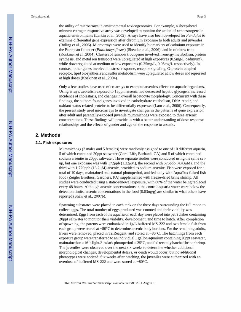
At the conclusion of spawning, female fish from each control and 1,720ppb arsenic aquariawere used to determine the body burdens of arsenic. In the adults, arsenic levels were 2.6-foldhigher in the exposed fish than in the controls after 10 days of exposure (Figure 1). This levelof uptake is consistent with other studies in Fundulus. For example, adult mummichogsexposed to 8,000ppb arsenic for 4 days had approximately 6 times more arsenic than controlswhile those fish exposed to 100ppb arsenic for 14 days had approximately 3 times more arsenicthan the controls (Miller et al., 2007). Similarly, mummichogs exposed to 787 ppb for 14 dayshad 2.8-fold higher hepatic arsenic levels than controls (Bears et al., 2006).
3.2. Hepatic gene expression changes in adult Fundulus exposed to arsenicMicroarrays were used to determine both the dose-dependent patterns of gene expressionchanges in livers following arsenic exposure and whether the response to arsenic differedbetween genders. Replicated test array experiments were conducted for 2 different exposureconcentrations in the females or 3 different exposure concentrations in the males. Of the 257
Gonzalez et al. Page 5
Mar Environ Res. Author manuscript; available in PMC 2011 August 1.
NIH
-PA Author Manuscript
NIH
-PA Author Manuscript
NIH
-PA Author Manuscript

genes on the array, between 4.7-8.2% were differentially expressed after arsenic exposureamongst the 5 groups of experiments using the CLEAR test method. Analyzing the data forsignificantly differential changes in gene expression in a dose-responsive manner indicatedthat in the two female exposure groups, 172ppb and 575ppb arsenic, 16 and 19 hepatic geneswere significantly changed compared to the controls, respectively. There were 7 commondifferentially expressed genes (Figure 2A). In comparing the livers of male fish exposed toarsenic with the controls, 21, 12, and 13 hepatic genes were differentially expressed in 172,575, and 1,720ppb exposures. There was only 1 gene shared in common between all three malegroups, which is an EST termed 10-15. Shared genes between two of the three male exposuresranged from 3 to 7 (Figure 2A). When examining the data from Table 1 for the number ofcommon genes changed after arsenic exposure based upon gender, a total of 9 differentiallyexpressed genes were common between males and females. The differential expression wasmore likely to occur in the 172ppb exposure group than the 575ppb group (Figure 2B).
Several genes were expressed in a dose-responsive manner. Only genes with statisticallydifferent expression are shown in Table 1. These included apolipoprotein, which wassignificantly downregulated in both female exposure groups and in two of the three maleexposure groups. Apolipoprotein is represented by two partially overlapping clones on thearray, 8-56 and Ab35, and both were changed in the same manner and the same magnitude(Table 1). Lysozyme precursor is also represented by two clones, 1-11 and 1-163, with bothconsistently demonstrating downregulation in the 172ppb males but upregulation in the1,720ppb males exposed to arsenic (Table 1). Other clones show differences based upongender, such as tributyltin (TBT)-binding protein, which is downregulated in females butupregulated in males (Table 1). A second overlapping clone for TBT-binding protein termedC3C61 (accession #BM084929) was also significantly downregulated (0.33-fold) in the575ppb arsenic-exposed females (data not shown). Several other genes were differentiallyexpressed in only one exposure group, but many of these genes are associated with cell stress,damage, and repair, such as heat shock proteins, glutathione S-transferases, transcriptionelongation factors, and ATP synthase (Table 2).
3.3. Reproductive effects, developmental effects, and gene expression changes in thejuveniles
In addition to changes in hepatic gene expression, we were interested in investigating the effectsof arsenic on embryonic development. After the exposure period, the fish were mated and eggscollected to determine changes in embryonic development and hatchling success. There wereno differences in the number of eggs laid per tank between the control and the arsenic-exposedgroups at any concentration, nor any differences in egg viability (Figure 3a). After hatching,any deformities in the embryos were recorded. In the offspring whose parents were exposedto 1,720ppb arsenic, there was a significant increase in deformities including embryos withabnormal trunk curvatures, and head and eye deformities (Figure 3b). Because of theseabnormalities, it was hypothesized that arsenic could be altering the structure of bone orcartilage. However, no differences were observed in the number, orientation, or position of thevertebrae in parentally-exposed offspring compared to control offspring (Figure 3c).
Unlike the adult hepatic gene expression, we used RNA extracted from whole 6-week oldjuveniles. There were very few genes in common between the three different parental exposuregroups, except for two ESTs, 2-80 and 9-7 (Table 1). There was only one additional gene,complement component C8, which was shared between the juveniles and the adults. However,this gene was only significantly differentially expressed in the highest juvenile exposure group.
Gonzalez et al. Page 6
Mar Environ Res. Author manuscript; available in PMC 2011 August 1.
NIH
-PA Author Manuscript
NIH
-PA Author Manuscript
NIH
-PA Author Manuscript

3.4. Confirmation of gene expression patternsTo confirm the expression patterns of several of the genes, we used quantitative PCR (QPCR)to examine expression levels of several selected genes in the livers of the adult males andfemales. Expression levels of the EST shared in common with the adults, 9-7, was alsoexamined in the juveniles (Figure 4). Apolipoprotein expression on the arrays wasdownregulated in all adult arsenic exposed groups, regardless of gender or concentration. Thiswas validated in four of the five QPCR reactions. The exception was the 172ppb female group,which based upon QPCR expression, showed no change in expression (fold change=1.03). TheEST termed 9-7 showed a bit more variability, but the arrays and QPCR validated one anotherin 5 of the 7 groups (Figure 4). The expression of 9-7 in the 172ppb female group wasdownregulated in the arrays but upregulated in the QPCR, while the 575ppb male group wasupregulated in the arrays but downregulated based upon QPCR. The levels of 9-7 in twojuvenile groups examined showed similar expression patterns, being upregulated in both thearrays and when using QPCR (Figure 4).
Serum amyloid precursor expression was downregulated in all of the male exposures and the172ppb female exposure, but upregulated in the 575ppb female exposure group in the arrays.Again, the direction of change using QPCR matched in four of the five groups, but this time,the 172ppb male group showed no changes in expression based upon the QPCR data (foldchange=1.07). For the higher exposure groups, both the directionality and magnitude of changewere similar. The expression of serum amyloid precursor in the 575ppb males wasdownregulated 0.83-fold in the arrays and 0.67-fold in the QPCR, while the expression in the1,720ppb males was downregulated 0.32-fold in the arrays and 0.28-fold in the QPCR.Interestingly, serum amyloid precursor expression was upregulated in the 575ppb femalegroup, and the magnitude of change between the arrays and QPCR was also similar (2.5-foldin arrays; 3.7-fold in the QPCR). Overall, the QPCR data showed similar fold-changes anddirectionality, validating the array data.
4. Discussion4.1. Arsenic-mediated changes in gene expression
The use of fish toxicogenomics is suited for studying contaminants such as arsenic, as fish canact as both models for human diseases as well as models for the health of the aquaticenvironment (Williams et al., 2003; Gorman and Breden, 2007). In the present study, adultmummichogs were exposed to three different arsenic concentrations, and the differentialexpression of genes was investigated by examining the effects of gender and maternal transferto offspring. Several genes were differentially expressed as a result of the arsenic exposure,including apolipoprotein, serum amyloid precursor, tributyltin-binding protein, andcytochrome c oxidase. Overall, arsenic appears to perturb genes involved in energy utilization.
Apolipoprotein was one gene that was downregulated in the adult livers at all exposureconcentrations tested. Using QPCR, the underexpression of apolipoprotein and the magnitudeof downregulation were in concordance with the array data. Apolipoproteins are lipid bindingproteins important in lipid secretion, lipolytic enzyme activation, and transport and binding oflipoproteins in cells (Atkinson, 1992). Several isoforms have been identified, which bind toand act as ligands for low density or very low density lipoprotein receptors (Atkinson, 1992).In our experiment, both clones of the apolipoprotein C-1 were down-regulated similarly infemales as well as males. The reduction in apolipoprotein expression is in concordance withother arsenic exposure studies. For example, arsenic downregulated apolipoprotein in the lungfluid of male mice exposed to 50ppb sodium arsenite for four weeks (Lantz et al., 2008), andwas differentially expressed in zebrafish embryos exposed to arsenic (Mattingly et al., 2009).
Gonzalez et al. Page 7
Mar Environ Res. Author manuscript; available in PMC 2011 August 1.
NIH
-PA Author Manuscript
NIH
-PA Author Manuscript
NIH
-PA Author Manuscript

Another protein, serum amyloid precursor, was also downregulated in the adult males and inthe 172ppb female exposure group. Its altered expression was examined by QPCR, whichconfirmed a dose-responsive reduction in the male 172, 575, and 1720ppb groups. Serumamyloid precursor proteins (APPs) are also a family of apolipoproteins (Zheng and Koo,2006). Although most often associated with the development of Alzheimer's disease (reviewedin: (Esler and Wolfe, 2001; Jaeger and Pietrzik, 2008), APPs are thought to function to controlcholesterol transport, ApoE metabolism, and help maintain cholesterol homeostasis (Yao andPapadopoulos, 2002; Liu et al., 2007). Additionally, cellular cholesterol can modify the boththe processing and transcription of APP (Bodovitz and Klein, 1996; Fassbender et al., 2001;Kiyosawa et al., 2004). In general, the downregulation of APP in the adult mummichogsmimics the reduction in apolipoprotein expression.
Both apolipoprotein and APPs are involved in lipid homeostasis, and in other studies, exposureto arsenic does appear to alter lipid levels. Mice exposed to arsenic via drinking water fromweaning to 1 year of age, or from 8 weeks to 1 year of age had significant reductions in serumtriglycerides, total cholesterol, and HDL cholesterol compared to controls (Ahlborn et al.,2009). The authors hypothesized that the reduction in lipids could be due to alterations in fattyacid production, as there was a reduction in the mRNA expression of stearoylCoA desaturase1, the rate limiting enzyme in monounsaturated fatty acid synthesis (Ahlborn et al., 2009).Interestingly, the mice in their study exposed to arsenic only in utero did not have alterationsin serum lipids (Ahlborn et al., 2009). These findings are in concordance with our studiesshowing a marked reduction in both apolipoprotein and APP proteins in the adults, but not inthe offspring. Thus, arsenic exposure may impair cholesterol homeostasis, which could haveadverse physiological consequences for fish (Heath, 1995). Examining lipid levels in the liversand serum of arsenic-exposed fish, and how that may impact adult health and egg quality wouldbe worthy of future investigation.
Indeed the present study, along with many others, shows that arsenic often exhibits a complexgene expression profile. For example, arsenic exposure in zebrafish embryos resulted in thedifferential expression of several immune-responsive genes following a 10μg/L exposure, butno change in expression at 100μg/L (Mattingly et al., 2009). In additional microarray studiesusing cell lines or rodent models, arsenic exposure results in very few commonly differentiallyexpressed genes between the exposure groups (Ahlborn et al., 2008), or can alter the expressionof genes in opposite directions at low versus high concentrations (Andrew et al., 2003; Lau etal., 2004; Snow et al., 2005; Andrew et al., 2007). This may indicate that differingtranscriptional networks are activated at low arsenic levels versus high arsenic levels, resultingin differing biological processes being altered (Bodwell et al., 2004; Bodwell et al., 2006).
4.2. Gender differences in gene expressionWe also found differing responses depending on the gender of the fish. For example, serumamyloid precursor was upregulated in the livers of the highest female exposure group, butdownregulated in the livers of the males. In contrast, several ESTs were upregulated in themales by downregulated in the females. Gender-based differences in disease outcomes afterarsenic exposure have been reported in the literature. For example, men have a higher incidenceof pathological liver changes after arsenic exposure, while women are at greater risk of arsenic-related kidney, lung, and bladder cancers than men (Vahter et al., 2007; Lindberg et al.,2008b). The same increase in bladder lesions in females has also been reported in studies withrodents (Shen et al., 2006; Waalkes et al., 2006), while increases in liver tumors after arsenicexposure are seen in male rodents (Waalkes et al., 2003; Waalkes et al., 2004a; Waalkes et al.,2004b; Ahlborn et al., 2009). It is known that hepatic metabolism of contaminants in someorganisms can vary by gender (reviewed in Burger et al., 2007) and it has been speculated thatsex hormones affect in arsenic metabolism, altering its methylation, excretion, and thus its
Gonzalez et al. Page 8
Mar Environ Res. Author manuscript; available in PMC 2011 August 1.
NIH
-PA Author Manuscript
NIH
-PA Author Manuscript
NIH
-PA Author Manuscript

pathology (Shen et al., 2006; Lindberg et al., 2008a; Lindberg et al., 2008b). As the fish in ourstudy were reproductively active, hormonal changes may have influenced the effects of arsenicon gene expression.
4.3. Effect of arsenic on developmentThe second part of this study was to examine changes in development and gene expression inthe offspring whose parents were exposed to arsenic. Viability was not different between thecontrol and arsenic-exposed groups at any exposure concentration. However, parental exposureto arsenic increased the incidence of developmental deformities, such as spinal curvatures andsmaller heads, by 17.4-fold at the highest concentration as compared to controls. In a previousexperiment using a concentration of 230ppb arsenic, there was a 2.8-fold increase in theincidence of spinal deformities in the hatchlings of exposed parents (Gonzalez et al., 2006). Astudy with zebrafish revealed various similar morphological abnormalities including dorsalcurvature, flattened head, and pericardial edema (Li et al., 2009), and mice exposed in uterovia maternal oral treatment also exhibited skeletal abnormalities (Hill et al., 2008). In humans,a decreased head and chest size was noted in children exposed to arsenic in utero (Rahman etal., 2009).
It has been well documented that arsenic can cross the maternal blood supply and be detectedin the fetus (Concha et al., 1998; Kubota et al., 2005; Fängström et al., 2009). A study lookingat the maternal transfer of arsenic from organisms such as adult black-tailed gulls noted that11.3% of the arsenic burden was transferred to eggs (Kubota et al., 2002), fetal porpoise arsenicburden was about 36% of that measured in the mothers (Kubota et al., 2005), and newbornmouse livers had 10% of the average arsenic concentration in maternal livers (Xie et al.,2007). Thus, even the low amounts of arsenic transferred to the offspring may impact its geneexpression and subsequent development.
It is interesting to note that offspring exposed to arsenic in utero have very different pathologiesbased upon their gender. In mice exposed to arsenic in utero, males have increase in liver andadrenal tumors, while females have increases in urogenital tumors, but no changes in theincidence of liver or adrenal tumors (Waalkes et al., 2003; Waalkes et al., 2004a; Waalkes etal., 2004b; Ahlborn et al., 2009). We did not determine the sex of the juveniles in our studybefore using them in the microarray study. Given that in rodents, very different arsenicmetabolite profiles and pathologies are seen, not sexing our juvenile fish might havecontributed to the lack of gene expression changes and the high amount of variability associatedwith the offspring.
In summary, we observed changes in lipid responsive genes at environmentally-relevantconcentrations, indicating that arsenic exposure may impair cholesterol homeostasis, whichcould have adverse effects on organismal health. These findings will provide us with a betterunderstanding of the effects of dose, gender, and exposure age on the response to arsenic.
AcknowledgmentsThis work was supported by NCRR #2G12 RR008124, NSF/EPA #EF 0830117, and CSREES/USDA, under projectnumber #SC-1700380. It is technical contribution No. 5647 of the Clemson University Experiment Station. The authorswould also like to thank Marian Viveros and Judi Ellzey (UTEP) for technical assistance.
ReferencesAhlborn GJ, Nelson GM, Grindstaff RD, Waalkes MP, Diwan BA, Allen JW, Kitchin KT, Preston RJ,
Hernandez-Zavala A, Adair B, Thomas DJ, Delker DA. Impact of life stage and duration of exposureon arsenic-induced proliferative lesions and neoplasia in C3H mice. Toxicology 2009;262:106–113.[PubMed: 19450653]
Gonzalez et al. Page 9
Mar Environ Res. Author manuscript; available in PMC 2011 August 1.
NIH
-PA Author Manuscript
NIH
-PA Author Manuscript
NIH
-PA Author Manuscript

Ahlborn GJ, Nelson GM, Ward WO, Knapp G, Allen JW, Ouyang M, Roop BC, Chen Y, O'Brien T,Kitchin KT, Delker DA. Dose response evaluation of gene expression profiles in the skin of K6/ODCmice exposed to sodium arsenite. Toxicol. Appl. Pharmacol 2008;227:400–416. [PubMed: 18191166]
Andrew AS, Bernardo V, Warnke LA, Davey JC, Hampton T, Mason RA, Thorpe JE, Ihnat MA, HamiltonJW. Exposure to arsenic at levels found in U.S. drinking water modifies expression in the mouse lung.Toxicol. Sci 2007;100:75–87. [PubMed: 17682005]
Andrew AS, Warren AJ, Barchowsky A, Temple KA, Klei L, Soucy NV, O'Hara KA, Hamilton JW.Genomic and proteomic profiling of responses to toxic metals in human lung cells. Environ.HealthPerspect 2003;111:825–835. [PubMed: 12760830]
ATSDR. Agency for Toxic Substances and Disease Registry. SUDHHS; PHS, City: 2007. CERCLApriority list of hazardous substances that will be the subject of toxicological profiles and supportdocument. 2007
Bain LJ. cDNA cloning, sequencing, and differential expression of a heart-type fatty acid-binding proteinin the mummichog (Fundulus heteroclitus). Mar. Environ. Res 2002;54:379–383. [PubMed:12420700]
Bears H, Richards JG, Schulte PM. Arsenic exposure alters hepatic arsenic species composition andstress-mediated gene expression in the common killifish (Fundulus heteroclitus). Aquat. Toxicol2006;77:257–266. [PubMed: 16445994]
Bodovitz S, Klein WL. Cholesterol modulates alphasecretase cleavage of amyloid precursor protein. J.Biol. Chem 1996;271:4436–4440. [PubMed: 8626795]
Bodwell JE, Gosse JA, Nomikos AP, Hamilton JW. Arsenic disruption of steroid receptor gene activation:Complex dose-response effects are shared by several steroid receptors. Chem. Res. Toxicol2006;19:1619–1629. [PubMed: 17173375]
Bodwell JE, Kingsley LA, Hamilton JW. Arsenic at very low concentrations alters glucocorticoid receptor(GR)-mediated gene activation but not GR-mediated gene repression: complex dose-response effectsare closely correlated with levels of activated GR and require a functional GR DNA binding domain.Chem. Res. Toxicol 2004;17:1064–1076. [PubMed: 15310238]
Boyle D, Brix KV, Amlund H, Lundebye AK, Hogstrand C, Bury NR. Natural arsenic contaminated dietsperturb reproduction in fish. Environ. Sci. Technol 2008;42:5354–5360. [PubMed: 18754393]
Burger J, Fossi C, McClellan-Green P, Orlando EF. Methodologies, bioindicators, and biomarkers forassenting gender-related differences in wildlife exposed to environmental chemicals. Environ. Res2007;104:135–152. [PubMed: 17207477]
Chang JS, Gu MB, Kim KW. Effect of arsenic on p53 mutation and occurrence of teratogenicsalamanders: their potential as ecological indicators for arsenic contamination. Chemosphere2009;75:948–954. [PubMed: 19203779]
Chen CJ, Wang SL, Chiou JM, Tseng CH, Chiou HY, Hsueh YM, Chen SY, Wu MM, Lai MS. Arsenicand diabetes and hypertension in human populations: A review. Toxicol. Appl. Pharmacol2007;222:298–304. [PubMed: 17307211]
Chen H, Liu J, Merrick BA, Waalkes MP. Genetic events associated with arsenic-induced malignanttransformation: applications of cDNA microarray technology. Mol. Carcinogen 2001;30:79–87.
Col M, Col C, Soran A, Sayli BS, Ozturk S. Arsenic-related Bowen's disease, palmar keratosis, and skincancer. Environ. Health Perspect 1999;196:687–689. [PubMed: 10417369]
Concha G, Vogler G, Lezcano D, Nermell B, Vahter M. Exposure to inorganic arsenic metabolites duringearly human development. Toxicol. Sci 1998;44:185–190. [PubMed: 9742656]
Datta S, Ghosh D, Saha DR, Bhattacharaya S, Mazumder S. Chronic exposure to low concentration ofarsenic is immunotoxic to fish: role of head kidney macrophages as biomarkers of arsenic toxicityto Clarias batrachus. Aquat. Toxicol 2009;92:86–94. [PubMed: 19237206]
Datta S, Saha DR, Ghosh D, Majumdar T, Bhattacharya S, Mazumder S. Sub-lethal concentration ofarsenic interferes with the proliferation of hepatocytes and induces in vivo apoptosis in Clariasbatrachus L. Comp. Biochem. Physiol. C Toxicol. Pharmacol 2007;145:339–349. [PubMed:17336163]
Díaz-Villaseñor A, Burns AL, Hiriart M, Cebrián ME, Ostrosky-Wegman P. Arsenic-induced alterationin the expression of genes related to type 2 diabetes mellitus. Toxicol. Appl. Pharmacol2007;225:123–133. [PubMed: 17936320]
Gonzalez et al. Page 10
Mar Environ Res. Author manuscript; available in PMC 2011 August 1.
NIH
-PA Author Manuscript
NIH
-PA Author Manuscript
NIH
-PA Author Manuscript

Engel RR, Hopenhayn-Rich C, Receveur O, Smith AH. Vascular effects of chronic arsenic exposure: areview. Epidemiol. Rev 1994;16:184–209. [PubMed: 7713176]
EPA. National primary drinking water regulations; arsenic and clarifications to compliance and newsource contaminants monitoring from the environmental protection agency; Final Rule. Fed Reg2001;66:6976–7066.
Esler WP, Wolfe MS. A portrait of Alzheimer secretases—new features and familiar faces. Science2001;293:1449–1454. [PubMed: 11520976]
Fängström B, Hamadani J, Nermell B, Grandér M, Palm B, Vahter M. Impaired arsenic metabolism inchildren during weaning. Toxicol. Appl. Pharmacol 2009;239:208–214. [PubMed: 19167415]
Fassbender K, Simons M, Bergmann C, Stroick M, Lutjohann D, Keller P, Runz H, Kuhl S, Bertsch T,von Bergmann K, Hennerici M, Beyreuther K, Hartmann T. Simvastatin strongly reduces levels ofAlzheimer's disease beta-amyloid peptides Abeta 42 and Abeta 40 in vitro and in vivo. Proc. Natl.Acad. Sci. USA 2001;98:5856–5861. [PubMed: 11296263]
Gonzalez HO, Roling JA, Baldwin WS, Bain LJ. Physiological changes and differential gene expressionin mummichogs (Fundulus heteroclitus) exposed to arsenic. Aquat. Toxicol 2006:43–52. [PubMed:16356559]
Gorman KF, Breden F. Teleosts as models for human vertebral stability and deformity. Comp. Biochem.Physiol. C Toxicol. Pharmacol 2007;145:28–38. [PubMed: 17240199]
Hays AM, Lantz RC, Rodgers LS, Sollome JJ, Vaillancourt RR, Andrew AS, Hamilton JW, CamenischTD. Arsenic-induced decreases in the vascular matrix. Toxicol. Pathol 2008;36:805–817. [PubMed:18812580]
Heath, AG. Water pollution and fish physiology. CRC Press; Boca Raton, Fl.: 1995.Hill DS, Wlodarczyk BJ, Finnell RH. Reproductive consequences of oral arsenate exposure during
pregnancy in a mouse model. Birth Defects Res 2008;88(Part B):40–47.Jaeger S, Pietrzik CU. Functional role of lipoprotein receptors in Alzheimer's disease. Curr Alzheimer
Res 2008;5:15–25. [PubMed: 18288927]Kingston, HM.; Jassie, LB., editors. Am. Chem. Soc. Washington, DC: 1988. ACS Professional
Reference Book Series.Kiyosawa N, Ito K, Niino N, Sakuma K, Kanbori M, Yamoto T, Manabe S, Matsunuma N. Effect of
serum cholesterol on the mRNA content of amyloid precursor protein in rat livers. Toxicol. Lett2004;150:157–166. [PubMed: 15093671]
Koskinen H, Pehkonen P, Vehniäinen E, Krasnov A, Rexroad C, Afanasyev S, Mölsa H, Oikari A.Response of rainbow trout transcriptome to model chemical contaminants. Biochem. Biophys. Res.Comm 2004;320:745–753. [PubMed: 15240111]
Kubota R, Kunito T, Fujihara J, Tanabe S, Yang J, Miyazaki N. Placental transfer of arsenic to fetus ofDall's porpoises (Phocoenoides dalli). Mar. Pollut. Bull 2005;51:845–849. [PubMed: 16291195]
Kubota R, Kunito T, Tanabe S, Ogi H, Shibata Y. Maternal transfer of arsenic to eggs of blacktailed gull(Larus crassirostris) from Rishiri Island, Japan. Appl. Organomet. Chem 2002;16:463–468.
Lam SH, Winata CL, Tong Y, Korzh S, Lim WS, Korzh V, Spitsbergen J, Mathavan S, Miller LD, LiuET, Gong Z. Transcriptome kinetics of arsenic-induced adaptive response in zebrafish liver. Physiol.Genomics 2006;27:351–361. [PubMed: 16882884]
Lantz RC, Chau B, Sarihan P, Witten ML, Pivniouk VI, Chen GJ. In utero and postnatal exposure toarsenic alters pulmonary structure and function. Toxicol. Appl. Pharmacol 2008;235:105–113.[PubMed: 19095001]
Larkin P, Sabo-Attwood T, Kelso J, Denslow ND. Gene expression analysis of largemouth bass exposedto estradiol, nonylphenol, and p,p′-DDE. Comp. Biochem. Physiol. B Biochem. Mol. Biol2002;133:543–557. [PubMed: 12470818]
Lau AT, Li M, Xie R, He QY, Chiu JF. Opposed arsenite-induced signaling pathways promote cellproliferation or apoptosis in cultured lung cells. Carcinogenesis 2004;25:21–28. [PubMed:14514659]
Li D, Lu C, Wang J, Hu W, Cao Z, Sun D, Xia H, Ma X. Developmental mechanisms of arsenite toxicityin zebrafish (Danio rerio) embryos. Aquat. Toxicol 2009;91:229–237. [PubMed: 19110324]
Gonzalez et al. Page 11
Mar Environ Res. Author manuscript; available in PMC 2011 August 1.
NIH
-PA Author Manuscript
NIH
-PA Author Manuscript
NIH
-PA Author Manuscript

Lindberg AL, Ekström EC, Nermell B, Rahman M, Lönnerdal B, Persson LA, Vahter M. Gender andage differences in the metabolism of inorganic arsenic in a highly exposed population in Bangladesh.Environ. Res 2008a;106:110–120. [PubMed: 17900557]
Lindberg AL, Rahman M, Persson LA, Vahter M. The risk of arsenic induced skin lesions in Bangladeshimen and women is affected by arsenic metabolism and the age at first exposure. Toxicol. Appl.Pharmacol 2008b;230:9–16. [PubMed: 18336856]
Liu Q, Zerbinatti CV, Zhang J, Hoe HS, Wang B, Cole SL, Herz J, Muglia L, Bu G. Amyloid precursorprotein regulates brain apolipoprotein E and cholesterol metabolism through lipoprotein receptorLRP1. Neuron 2007;56:66–78. [PubMed: 17920016]
Mandal B, Suzuki T. Arsenic around the world: a review. Talanta 2002;58:201–235. [PubMed: 18968746]Maples NL, Bain LJ. Trivalent chromium alters gene expression in the mummichog (Fundulus
heteroclitus). Environ. Tox. Chem 2004;23:626–631.Mattingly CJ, Hampton TH, Brothers KM, Griffin NE, Planchart A. Perturbation of defense pathways
by low-dose arsenic exposure in zebrafish embryos. Environ. Health Perspect 2009;117:981–987.[PubMed: 19590694]
Miller DS, Shaw JR, Stanton CR, Barnaby R, Karlson KH, Hamilton JW, Stanton BA. MRP2 and acquiredtolerance to inorganic arsenic in the kidney of killifish (Fundulus heteroclitus). Toxicol. Sci2007;97:103–110. [PubMed: 17324950]
NRC. Arsenic in drinking water. National Research Council; Washington, D.C.: 1999.Peterson JSK, Bain LJ. Differential gene expression in anthracene-exposed mummichogs (Fundulus
heteroclitus). Aquat. Toxicol 2004;66:345–355. [PubMed: 15168943]Posey T, Weng T, Chen Z, Chintagari NR, Wang P, Jin N, Stricker H, Liu L. Arsenic-induced changes
in the gene expression of lung epithelial L2 cells: implications in carcinogenesis. BMC Genomics2008;9:115. [PubMed: 18315880]
Rahman A, Vahter M, Smith AH, Nermell B, Yunus M, El Arifeen S, Persson LA, Ekström EC. Arsenicexposure during pregnancy and size at birth: A prospective cohort study in Bangladesh. Am. J.Epidemiol 2009;169:304–312. [PubMed: 19037006]
Raqib R, Ahmed S, Sultana R, Wagatsuma Y, Mondal D, Hoque AM, Nermell B, Yunus M, Roy S,Persson LA, Arifeen SE, Moore S, Vahter M. Effects of in utero arsenic exposure on child immunityand morbidity in rural Bangladesh. Toxicol. Lett 2009;185:197–202. [PubMed: 19167470]
Roling JA, Bain LJ, Baldwin WS. Differential gene expression in mummichog (Fundulus heteroclitus)following treatment with pyrene and fluoranthene: comparison to a creosote contaminated site. Mar.Environ. Res 2004;57:377–395. [PubMed: 14967520]
Roling JA, Bain LJ, Gardea-Torresdey J, Bader J, Baldwin WS. Hexavalent chromium reduces larvalgrowth and alters gene expression in mummichogs (Fundulus heteroclitus). Environ. Tox. Chem2006;25:2725–2733.
Shaw JR, Gabor K, Hand E, Lankowski A, Durant L, Thibodeau R, Stanton CR, Barnaby R, CoutermarshB, Karlson KH, Sato JD, Hamilton JW, Stanton BA. Role of glucocorticoid receptor in acclimationof killifish (Fundulus heteroclitus) to seawater and effects of arsenic. Am. J. Physiol. Regul. Integr.Comp. Physiol 2007a;292:R1052–R1060. [PubMed: 17038445]
Shaw JR, Jackson B, Gabor K, Stanton S, Hamilton JW, Stanton BA. The influence of exposure historyon arsenic accumulation and toxicity in the killifish, Fundulus heteroclitus. Environ. Toxicol. Chem2007b;26:2704–2709. [PubMed: 18020683]
Sheader DL, Williams TD, Lyons BP, Chipman JK. Oxidative stress response of European flounder(Platichthys flesus) to cadmium determined by a custom cDNA microarray. Mar. Environ. Res2006;62:33–44. [PubMed: 16624402]
Shen J, Wanibuchi H, Waalkes MP, Salim EI, Kinoshita A, Yoshida K, Endo G, Fukushima S. Acomparative study of the sub-chronic toxic effects of three organic arsenical compounds on theurothelium in F344 rats; gender-based differences in response. Toxicol. Appl. Pharmacol2006;210:171–180. [PubMed: 15950995]
Smedley P, Kinniburgh D. A review of the source, behaviour and distribution of arsenic in natural waters.Appl, Geochem 2002;17:517–568.
Gonzalez et al. Page 12
Mar Environ Res. Author manuscript; available in PMC 2011 August 1.
NIH
-PA Author Manuscript
NIH
-PA Author Manuscript
NIH
-PA Author Manuscript

Snow ET, Sykora P, Durham TR, Klein CB. Arsenic, mode of action at biologically plausible low doses:what are the implications for low dose cancer risk? Toxicol. Appl. Pharmacol 2005;207:557–564.[PubMed: 15996700]
Taylor WR, Van Dyke GC. Revised procedures for staining and clearing small fishes and other vertebratesfor bone and cartilage study. Cybium 1985;9:107–119.
Tseng CH, Tai TY, Chong CK, Tseng CP, Lai MS, Lin BJ, Chiou HY, Hsueh YM, Hsu KH, Chen CJ.Long-term arsenic exposure and incidence of non-insulin-dependent diabetes mellitus: a cohort studyin arseniasis-hyperendemic villages in Taiwan. Environ. Health Perspect 2000;108:847–851.[PubMed: 11017889]
Vahter M, Akesson A, Lidén C, Ceccatelli S, Berglund M. Gender differences in the disposition andtoxicity of metals. Environ. Res 2007;104:85–95. [PubMed: 16996054]
Ventura-Lima J, de Castro MR, Acosta D, Fattorini D, Regoli F, de Carvalho LM, Bohrer D, GeracitanoLA, Barros DM, Marins LF, da Silva RS, Bonan CD, Bogo MR, Monserrat JM. Effects of arsenic(As) exposure on the antioxidant status of gills of the zebrafish Danio rerio (Cyprinidae). Comp.Biochem. Physiol. C Toxicol. Pharmacol 2009;149:538–543. [PubMed: 19138757]
Waalkes MP, Liu J, Ward J, Diwan BA. Animal models for arsenic carcinogenesis: inorganic arsenic isa transplacental carcinogen in mice. Toxicol. Appl. Pharmacol 2004a;198:377–384. [PubMed:15276417]
Waalkes MP, Liu J, Ward JM, Powell DA, Diwan BA. Urogenital carcinogenesis in female CD1 miceinduced by in utero arsenic exposure is exacerbated by postnatal diethylstilbestrol treatment. CancerRes 2006;66:1337–1345. [PubMed: 16452187]
Waalkes MP, Ward JM, Diwan BA. Induction of tumors of the liver, lung, ovary and adrenal in adultmice after brief maternal gestational exposure to inorganic arsenic: promotional effects of postnatalphorbol ester exposure on hepatic and pulmonary, but not dermal cancers. Carcinogenesis 2004b;25:133–141. [PubMed: 14514661]
Waalkes MP, Ward JM, Liu J, Diwan BA. Transplacental carcinogenicity of inorganic arsenic in thedrinking water: induction of hepatic, ovarian, pulmonary, and adrenal tumors in mice. Toxicol. Appl.Pharm 2003;186:7–17.
Williams TD, Gensberg K, Minchin SD, Chipman JK. A DNA expression array to detect toxic stressresponse in European flounder (Platichthys flesus). Aquat. Toxicol 2003;65:141–157. [PubMed:12946615]
Xie Y, Liu J, Benbrahim-Tallaa L, Ward JM, Logsdon D, Diwan BA, Waalkes MP. Aberrant DNAmethylation and gene expression in livers of newborn mice transplacentally exposed to ahepatocarcinogenesis dose of inorganic arsenic. Toxicology 2007;236:7–15. [PubMed: 17451858]
Yamaguchi S, Miura C, Ito A, Agusa T, Iwata H, Tanabe S, Tuyen BC, Miura T. Effects of lead,molybdenum, rubidium, arsenic and organochlorines on spermatogenesis in fish: monitoring atMekong Delta area and in vitro experiment. Aquat. Toxicol 2007;83:43–51. [PubMed: 17448548]
Yamamoto M, Hirano S, Vogel CF, Cui X, Matsumura F. Selective activation of NF-kappaB and E2Fby low concentration of arsenite in U937 human monocytic leukemia cells. J. Biochem. Mol. Toxicol2008:136–146. [PubMed: 18418899]
Yao Z, Papadopoulos V. Function of beta-amyloid in cholesterol transport: a lead to neurotoxicity.FASEB J 2002;16:1677–1679. [PubMed: 12206998]
Yoshida T, Yamauchi H, Fan Sun G. Chronic health effects in people exposed to arsenic via the drinkingwater: dose-response relationships in review. Toxicol. Appl. Pharmacol 2004;198:243–252.[PubMed: 15276403]
Zheng H, Koo E. The amyloid precursor protein: beyond amyloid. Mol. Neurodegener 2006;1:5.[PubMed: 16930452]
Zheng XH, Watts GS, Vaught S, Gandolfi AJ. Low-level arsenite induced gene expression in HEK293cells. Toxicology 2003;187:39–48. [PubMed: 12679051]
Gonzalez et al. Page 13
Mar Environ Res. Author manuscript; available in PMC 2011 August 1.
NIH
-PA Author Manuscript
NIH
-PA Author Manuscript
NIH
-PA Author Manuscript
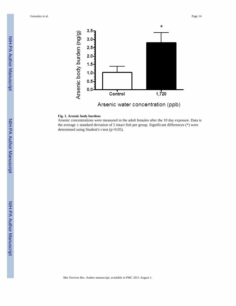
Fig. 1. Arsenic body burdensArsenic concentrations were measured in the adult females after the 10 day exposure. Data isthe average ± standard deviation of 5 intact fish per group. Significant differences (*) weredetermined using Student's t-test (p<0.05).
Gonzalez et al. Page 14
Mar Environ Res. Author manuscript; available in PMC 2011 August 1.
NIH
-PA Author Manuscript
NIH
-PA Author Manuscript
NIH
-PA Author Manuscript

Fig. 2. Gene expression profiles for males and female Fundulus exposed to different concentrationsof arsenicHepatic gene expression was examined using the Fundulus cDNA array (GEO-GPL2535) andanalyzed for differential expression using the CLEAR Test Method. The numbers of geneswith significantly different expression (p≤0.05) are indicated, and those changed by more thanone exposure are found in the overlapping areas. A. Dose-responsive changes in hepatic geneexpression in male and female fish B. Gender-responsive changes in hepatic gene expressionin male and female fish.
Gonzalez et al. Page 15
Mar Environ Res. Author manuscript; available in PMC 2011 August 1.
NIH
-PA Author Manuscript
NIH
-PA Author Manuscript
NIH
-PA Author Manuscript

Fig. 3. Percentage viability and morphological abnormalities following arsenic exposureAdults were exposed to 172, 575 or 1,720ppb arsenic for 10 days prior to spawning. Eggs werecollected for three days following arsenic exposure, and their viability, along with anydevelopmental abnormalities observed after 7 days, was recorded. A. Viability and deformitypercentages are expressed as the average + standard deviation for offspring from 4 –5 tanks,with statistical differences (*)determined by Mann-Whitney (p<0.05). B. Representativephotographs from normal and deformed offspring. C. Morphological analysis of the vertebrae.The total size (area) for individual vertebrae were quantified using the LSM5 Pascal software.Values are the average area in square micrometers divided by the weight of the 6-week oldfish. Significant differences (*) were determined using Student's t-test (p<0.05).
Gonzalez et al. Page 16
Mar Environ Res. Author manuscript; available in PMC 2011 August 1.
NIH
-PA Author Manuscript
NIH
-PA Author Manuscript
NIH
-PA Author Manuscript

Fig. 4. Comparisons of gene expression changes between the arrays and quantitative real-time PCR(QPCR)Expression of three genes from the arrays that were differentially expressed in almost allgenders, ages, and exposure groups were verified by QPCR using specific primers (n=5 foreach group). Array fold changes were plotted against QPCR fold changes to determine theirdirectionality and magnitude. Genes examined were apolipoprotein, serum amyloid precursor,and an EST termed 9-7. The letter indicates females (F), males (M), or juveniles (J), while thenumber (172, 575, or 1,720) indicates the exposure group.
Gonzalez et al. Page 17
Mar Environ Res. Author manuscript; available in PMC 2011 August 1.
NIH
-PA Author Manuscript
NIH
-PA Author Manuscript
NIH
-PA Author Manuscript

NIH
-PA Author Manuscript
NIH
-PA Author Manuscript
NIH
-PA Author Manuscript
Gonzalez et al. Page 18
Tabl
e 1
Gen
e Ex
pres
sion
Cha
nges
in F
undu
lus E
xpos
ed to
Ars
enic
. Fol
d-ch
ange
s in
gene
exp
ress
ion
wer
e ca
lcul
ated
and
sign
ifica
nt d
iffer
ence
s wer
e de
term
ined
usin
g th
e C
LEA
R T
est m
etho
d (p
<0.0
5). O
nly
gene
s tha
t are
diff
eren
tially
exp
ress
ed a
nd sh
ared
bet
wee
n m
ore
than
1 e
xpos
ure
grou
p ar
e lis
ted.
Fold
cha
nge
(Ars
enic
/con
trol
)
Clo
ne ID
Gen
e N
ame
Gen
Ban
k #
Fem
ales
Mal
esJu
veni
les
172p
pb57
5ppb
172p
pb57
5ppb
1,72
0ppb
172p
pb57
5ppb
1,72
0ppb
8--5
6ap
olip
opro
tein
DN
4749
490.
590.
750.
850.
85
Ab3
5ap
olip
opro
tein
CN
9926
960.
590.
790.
850.
84
9--7
EST
CO
8974
820.
611.
221.
141.
714.
11.
45
9--1
2TB
T-bi
ndin
g pr
otei
nA
Y72
5225
0.56
0.6
1.95
8-6-
T7ES
TD
N59
6277
5.71
0.45
Bb5
6ES
TD
N59
6273
0.67
1.79
1--3
1se
rum
am
yloi
d pr
ecur
sor
AY
7351
600.
632.
520.
770.
32
2--8
0ES
TD
N47
4945
0.54
0.81
2.13
1.54
1.29
9--1
6ES
TC
O89
7478
0.7
0.74
1.39
pa 1
--15
com
plem
ent c
ompo
nent
C8
AY
5216
640.
70.
821.
931.
58
Bb5
3cy
toch
rom
e c
oxid
ase
subu
nit I
IC
V82
1001
1.57
0.7
8--4
0cy
toch
rom
e c
oxid
ase
subu
nit I
IC
N97
6180
0.8
0.6
10--
15ES
TC
O89
7479
1.14
1.8
0.87
10--
57ES
TC
O89
7481
1.41
0.55
1--1
1ly
sozy
me
prec
urso
rA
Y73
5156
0.3
2.65
1--1
63ly
sozy
me
prec
urso
rA
Y73
5156
0.29
2.85
8--4
6ES
TD
N47
4948
1.37
0.82
8--8
EST
CO
8974
781.
680.
82
Mar Environ Res. Author manuscript; available in PMC 2011 August 1.

NIH
-PA Author Manuscript
NIH
-PA Author Manuscript
NIH
-PA Author Manuscript
Gonzalez et al. Page 19
Tabl
e 2
Gen
es sh
owin
g un
ique
diff
eren
tial e
xpre
ssio
n. F
old-
chan
ges i
n he
patic
gen
e exp
ress
ion
in th
e mal
es an
d fe
mal
es w
ere c
alcu
late
d an
d si
gnifi
cant
diff
eren
ces
wer
e de
term
ined
usi
ng th
e C
LEA
R T
est m
etho
d (p≤0
.05)
. Onl
y ge
nes t
hat w
ere
diff
eren
tially
exp
ress
ed in
onl
y 1
expo
sure
gro
up a
re li
sted
. Gen
Ban
kac
cess
ion
num
bers
are
indi
cate
d in
par
enth
eses
.
Clo
ne Id
entit
y an
d G
enB
ank
Acc
essi
on N
umbe
r
172p
pb fe
mal
es57
5ppb
fem
ales
172p
pb m
ales
575p
pb m
ales
1,72
0ppb
mal
es
Smal
l ind
ucib
le c
ytok
ine
A4
(AY
7351
53)
EST
(CX
7003
81)
GLU
T2 (A
Y52
1663
)El
onga
tion
fact
or1α
(AY
7351
80)
EST
(CO
8974
78)
Myo
sin
light
cha
in 2
(CX
7003
95)
Coa
gula
tion
fact
or X
IIIB
prec
urso
r (C
N98
8088
)H
emop
exin
pre
curs
or(A
Y73
5182
)70
kDa
HSP
(AY
7351
59)
War
p65
(AY
7351
66)
EST
(CX
7003
86)
14-3
-3 (A
Y72
5221
)ES
T (C
O89
7478
)G
STμ
(AY
7252
20)
EST
(DN
5962
72)
Map
kin
ase
kina
se(B
I993
599)
Dei
odin
ase
(U70
869)
trans
ferr
in (A
Y73
5165
)ES
T (C
O89
7478
)ES
T (C
X70
0393
)
EST
(DN
4749
42)
Vite
lloge
nin
(U07
055)
Cyt
ochr
ome
b (A
Y72
5226
)
EST
(CO
8974
90)
ATP
synt
hase
Fo c
ompl
ex(A
Y73
5178
)fu
cole
ctin
(AY
7351
52)
EST
(CO
8974
85)
EST
(CX
7003
90)
Mar Environ Res. Author manuscript; available in PMC 2011 August 1.