nih public access aileen y. alontaga lawrence pautsch juan
TRANSCRIPT

Kinetic and spectroscopic studies of hemin acquisition in thehemophore HasAp from Pseudomonas aeruginosa†
Erik T. Yukl1,§, Grace Jepkorir2, Aileen Y. Alontaga2, Lawrence Pautsch1, Juan C.Rodriguez2, Mario Rivera2, and Pierre Moënne-Loccoz*,1
1Department of Science and Engineering, School of Medicine, Oregon Health & ScienceUniversity, 20,000 NW Walker Road, Beaverton, Oregon 97006-8921, USA.2Department of Chemistry, University of Kansas, Lawrence, Kansas 66047, USA
AbstractThe extreme limitation of free iron has driven various pathogens to acquire iron from the host inthe form of heme. Specifically, several Gram negative pathogens secrete a heme binding proteinknown as HasA to scavenge heme from the extracellular environment and to transfer it to thereceptor protein HasR for import into the bacterial cell. Structures of heme-bound and apo-HasAhomologues show that the heme iron(III) ligands, His32 and Tyr75, reside on loops extendingfrom the core of the protein and that a significant conformational change must occur at the His32loop upon heme binding. Here, we investigate the kinetics of heme acquisition by HasA fromPseudomonas aeruginosa (HasAp). The rate of heme acquisition from human met-hemoglobin(met-Hb) closely matched that of heme dissociation which suggests a passive mode of hemeuptake from this source. The binding of free hemin is characterized by an initial rapid phaseforming an intermediate before further conversion to the final complex. Analysis of this samereaction using an H32A variant lacking the His heme ligand shows only the rapid phase to form aheme-protein complex spectroscopically equivalent to that of the wild type intermediate. Furthercharacterization of these reactions using EPR and resonance Raman spectroscopy of rapid freezequench samples provided support for a model where heme is initially bound by the Tyr75 to forma high-spin heme-protein complex before slower coordination of the His32 ligand upon closing ofthe His loop over the heme. The slow rate of this loop closure implies that the induced-fitmechanism of heme uptake in HasAp is not based on a rapid sampling of the H32 loop betweenopen and closed configurations, but rather, that the H32 loop motions are triggered by theformation of the high-spin heme-HasAp intermediate complex.
Certain opportunistic pathogens, such as Pseudomonas aeruginosa, are able to overcome theextremely low levels of available free iron within their mammalian host by deployingseveral iron acquisition systems. One of these, encoded by the has (heme acquisitionsystem) operon found in a number of gram-negative bacteria, involves the secretion of aheme-binding protein HasA1 (1). HasA binds extracellular heme with high affinity (5.3 ×10¹⁰ M-1) (2) and delivers it to a specific outer membrane receptor, HasR (1,3). Once
†This work was supported in part by the National Science Foundation (P.M.-L., MCB-0811888; M.R., MCB-0818488) and theNational Institutes of Health (P.M.-L., GM0 74785; M.R., GM 50503).*Corresponding author: Pierre Moënne-Loccoz, Oregon Health & Science University, 20,000 NW Walker Road, Beaverton, Oregon97006. Tel: 503-748-1673; Fax: 503-748-1464; [email protected].§Current address: Department of Biochemistry, Molecular Biology and Biophysics, University of Minnesota, Minneapolis, Minnesota55455SUPPORTING INFORMATION PARAGRAPHEPR spectrum of control-RFQ sample of hemin + buffer, and RR spectra of wt and H32A HasAp at room temperature and 105 K.This material is available free of charge via Internet at http://pubs.acs.org.
NIH Public AccessAuthor ManuscriptBiochemistry. Author manuscript; available in PMC 2011 August 10.
Published in final edited form as:Biochemistry. 2010 August 10; 49(31): 6646–6654. doi:10.1021/bi100692f.
NIH
-PA Author Manuscript
NIH
-PA Author Manuscript
NIH
-PA Author Manuscript
imported into the cell, heme oxygenases degrade the heme, providing the organism with asource of iron (4). This is an effective strategy given that heme, as opposed to “free” iron, isabundant within the mammalian host with hemoglobin (Hb) representing a particularly largereservoir. P. aeruginosa maximizes the utilization of this reservoir by secreting hemolysins(5,6) and cytotoxins (7) to lyse the host's erythrocytes, liberating Hb.
HasA from P. aeruginosa (HasAp) has been shown to be essential for the growth of thisorganism using Hb as the sole iron source (8), thus supporting its vital role in the acquisitionof heme from Hb. However, the mechanism by which this process occurs is still underdebate. The inability of ultracentrifugation experiments to detect a stable complex betweenhuman met-Hb and apo-HasA from S. marcescens (HasAs) suggests a passive mode ofacquisition, with HasAs binding heme only subsequent to its dissociation from met-Hb(9,10). Such a mechanism may be sufficient given that rapid dilution of oxy-Hb uponerythrocyte lysis promotes dissociation of Hb into αβ dimers (11,12), acceleration ofautoxidation (13), and hemin dissociation (14). Conversely, NMR analyses of mixtures ofholo-HasAp and human met-Hb show perturbations consistent with the formation of atransient complex between these two proteins (15). Knowing that the rates of hemedissociation from met-Hb, at various concentrations, have been carefully studied (14), we setout to measure the rate of heme acquisition from met-Hb by apo-HasAp to determinewhether the formation of a transient complex facilitates the loss of heme from met-Hb. Toour knowledge, such experiments have not been reported for HasAp and homologues.
Also of interest is the possibility of observing intermediates in the heme-binding process toapo-HasAp. Structural characterizations of HasAp (15,16) and HasAs (17-19) show thatthese ~19 kDa proteins adopt a monomeric structure composed of an extended antiparallelβ-sheet and a four α-helix wall from which extrudes two loops that sandwich the hememolecule. The Fe(III) heme is axially coordinated by His32 and Tyr75 which also engages ahydrogen bond interaction with His83 (15,17). In the apo form of the hemophore, the loopbearing Tyr75 is largely unaffected by the absence of the heme molecule and the interactionbetween Tyr75 and His83 is maintained, but the His32 loop adopts a very differentconformation (16,19). Specifically, the His32 loop moves 30 Å away from the heme bindingsite to interact with the outer surface of the protein core. This open configuration of the apoform led Wolff and coworkers to propose an “induced fit” model of the heme uptake processin HasA, with a two-step mechanism where the heme interacts first with the Tyr75 loopbefore the heme pocket closes. Closure of the His32 loop over the heme is presumablymediated by the binding of His32 to the heme iron and by the formation of hydrogen bondsbetween heme propionate groups and backbone nitrogen atoms of the His32 loop (18,19).Several other indirect lines of evidence support the idea that Tyr75 is the predominant of thetwo axial ligands: 1) Among HasA homologues from four Gram negative bacteria, His32 isconserved among only 3 while the Tyr75/His83 pair is fully conserved (20). 2) Whilemutation of either Tyr75 or His83 to Ala results in a loss of binding affinity of two orders ofmagnitude in HasAs, the affinity of H32A HasAs is decreased by only 5-fold relative to wildtype (wt) (2). 3) Formation of a Fe(II)-CO complex in both wt and H83A HasAs proceedsby displacement of His32 rather than Tyr75 (21). 4) Crystal structures of holo-HasAscomplexed with wt and variant receptor proteins HasR show the heme iron(III)-Tyr75coordination retained while the H32 loop is disordered (22). The fact remains that directevidence for a two-step mechanism for the heme uptake process in HasA is lacking.
1Abbreviations: HasA, heme binding protein acquisition; HasR, HasA-specific outer membrane receptor; met-Hb, met-hemoglobin;RFQ, rapid freeze quench; RR, resonance Raman; EPR, electron paramagnetic resonance; HEPES, N-(2-hydroxyethyl)piperazine-N'-(2-ethanesulfonic acid); HS/LS, high-spin/low-spin.
Yukl et al. Page 2
Biochemistry. Author manuscript; available in PMC 2011 August 10.
NIH
-PA Author Manuscript
NIH
-PA Author Manuscript
NIH
-PA Author Manuscript

In this work, we strive to answer two fundamental questions about heme binding by HasAp:1) Does the transient complex that has been proposed to form between HasAp and humanHb (15) enhance the rate of heme loss from Hb? 2) What is the mechanism by which apo-HasAp binds hemin? Specifically, can we identify coordinate intermediate(s) in the heminbinding process and determine which ligand initially binds the iron or if both ligands bindsimultaneously? Intermediates are not observed in the hemin-binding reactions of the 6-coordinate hemophores HtsA (likely His/Met ligation (23,24)) or Shp (bis-Met ligation(25)), but the asymmetric His/Tyr ligand and the structural differences in the ligand-carryingloops in HasAp could lead to distinct hemin binding kinetics. To answer the first question,we followed the transfer of heme from human met-Hb to apo-HasAp using UV-visspectroscopy and compared the rate of transfer to literature values for the rate of hemedissociation from met-Hb. To address the hemin-binding mechanism, we analyzed thekinetics of hemin binding to wt and H32A apo-HasAp using stopped-flow spectroscopy. Wealso characterized rapid freeze quench (RFQ) samples of these reactions using resonanceRaman (RR) and EPR spectroscopy. The work presented herein demonstrates that wt apo-HasAp acquires free hemin in two steps via the formation of a coordination intermediatewhich is spectroscopically equivalent to the resting state of H32A holo-HasAp beforecompletion of the heme binding pocket with a t in the hundred millisecond range (k ~ 5 s-1 at4 °C).
MATERIALS AND METHODSExpression and Purification of apo-HasAp
The truncated form of HasAp, omitting the last 21 C'-terminal amino acids was constructedas described previously (15). The H32A mutation was introduced by site directedmutagenesis using the QuickChange mutagenesis kit (Stratagene) and the mutation wasconfirmed by DNA sequencing (SeqWright). The plasmid was transformed into E. coliBL21 GOLD (DE3) competent cells for subsequent protein expression. A 50 mL starterculture in Luria-Bertani (LB) medium (100 μg/mL ampicillin) was grown overnight in a 125mL flask from a single colony of transformed cells from a freshly streaked LB agarampicillin plate. The growth was used to inoculate 30 mL of M9 minimal mediumcontaining 200 μg/mL ampicillin and grown to an OD600 of 0.8-0.9. The cells werecentrifuged at 4800 rpm for 10 min at 4 °C, resuspended in 1.0 L of fresh minimal M9medium (200 μg/mL ampicillin), and cultured to an OD600 of ~ 0.9 at 37 °C. Thetemperature was lowered to 30 °C with continuous incubation to an OD600 of ~ 1.0 andprotein expression was induced with the addition of isopropyl β-D-thiogalactopyranoside(IPTG) to a final concentration of 1 mM. After 5 h of culture, the cells were harvested andstored at -20 °C. The pellet was resuspended in 20 mM Tris-HCl (pH 7.6) (3 mL/gram ofcell paste), DNase (Sigma-Aldrich, St. Louis, MO) was added, the cells were disrupted bysonication and the cell debris were separated by centrifugation at 19500 rpm at 4 °C for 40min. Purification of apo wild type HasAp or its H32A mutant was carried out by ionexchange chromatography at 4 °C and by hydrophobic interaction chromatography atambient temperature. The clarified supernatant obtained after removal of cell debris wasloaded onto a Q-Sepharose Fast flow column (2.6 cm i.d. × 15 cm; GE Healthcare) preequilibrated with 20 mM Tris-HCl (pH 7.6). The column was washed with at least 3 bedvolumes of the same buffer before protein elution with a linear NaCl concentration gradient(0-600 mM). Fractions containing HasAp were pooled and exchanged into 50 mM sodiumphosphate buffer containing 0.7 M ammonium sulfate (pH 7.0) prior to loading onto a ButylSepharose Fast Flow (GE Healthcare) column (2.6 cm i.d. x 12 cm) equilibrated with thesame buffer. Elution of weakly bound contaminating proteins and holo-HasAp was achievedwith 50 mM sodium phosphate/0.5 M ammonium sulfate, before eluting apo-HasAp with alinear gradient of sodium phosphate buffer (50 mM–20 mM)/ammonium sulfate (0.50 M – 0
Yukl et al. Page 3
Biochemistry. Author manuscript; available in PMC 2011 August 10.
NIH
-PA Author Manuscript
NIH
-PA Author Manuscript
NIH
-PA Author Manuscript

M). Fractions containing the apo protein were loaded onto a Phenyl Sepharose 6 Fast Flowhigh substitution (GE Healthcare) column (2.6 cm i.d. × 12 cm) and eluted using the twosteps described above. The purity of the protein in the resultant fractions was assessed bySDS-PAGE and if necessary, fractions containing apo-HasAp were purified again on asecond Butyl Sepharose column. The MW of apo- wild type and H32A HasAp wasdetermined by electrospray mass spectrometry.
Transfer of Heme from Hemoproteins to wt apo-HasApElectronic absorption spectra were obtained using a Cary 50 Varian spectrophotometer usinga resolution of 1 nm and a scanning rate of 1200 nm/min. The concentration of wt apo-HasAp was determined by titration with a solution of bovine hemin (Sigma). Lyophilizedhuman Hb (Sigma) was dissolved in phosphate buffer (μ = 0.1, pH 7.0) and purified in aSephacryl S-200 size exclusion column preequilibrated and eluted with phosphate buffer (μ= 0.1, pH 7.0). Fractions with purity ratio (A280/A406) < 0.25 were pooled and concentratedusing Amicon ultracentrifuge filters to a final concentration of 3.5 mM. This purificationprocess was necessary to remove adventitiously bound hemin which would confound theresults of the experiment. Purified Hb was then incubated with a 10-fold excess of potassiumferricyanide (Mallinkrodt) for 10 min followed by removal of excess ferricyanide usingdesalting spin columns (ZebaTM 0.5 mL; Pierce). Reactions were initiated by mixing Hb andwt apo-HasAp solutions to final concentrations of 50 μM (in heme) and 200 μM,respectively, in 50 mM HEPES pH 7.0 containing 150 mM NaCl. Solutions were loadedinto 2 mm pathlength cuvettes, and scans were taken every 5 min for 120 min. For transferreactions from reduced Hb, concentrated solutions of met-Hb and wt apo-HasAp werepurged of oxygen with Ar and brought into a glove box with a controlled atmosphere of lessthan 1 ppm O2 (Omni-Lab System; Vacuum Atmospheres Co.). Met-Hb was diluted in theabove buffer and sodium dithionite was added to a final concentration of 500 μM. ReducedHb was loaded into a 2 mm pathlength cuvette and sealed. Reactions were initiated by theinjection of anaerobic wt apo-HasAp to a final concentration of 200 μM and 50 μM in Hbheme. Horse heart Mb (Sigma) was oxidized with ferricyanide as described above anddesalted twice to remove excess ferricyanide and adventitiously- bound heme. Hemetransfer reactions were conducted identically to those for met-Hb. Single wavelengthabsorbance data were fit to a double exponential expression with identical amplitudes usingOrigin 7.0 (Origin Lab Corporation).
Stopped-flow UV-vis SpectroscopyThe concentrations of stock wt and H32A apo-HasAp solutions were determined as above toyield extinction coefficients at 280 nm of 28.6 and 29.8 mM-1cm-1, respectively. Thesevalues were subsequently used to determine apo-HasAp concentrations in reaction mixtures.Hemin was dissolved in 10 mM NaOH, and its concentration was determined using ε385 =58.4 mM-1cm-1 (26); this was diluted to a final concentration of 13 μM in 10 mM NaOH andused immediately. Apoproteins were diluted in 200 mM HEPES pH 7.5. Solutions wereloaded into an SX20 stopped-flow UV-vis spectrometer (Applied Photophysics) equilibratedto 4.2 °C. Excess solution was recovered from the stopped-flow apparatus after eachexperiment in order to re-measure the concentrations of hemin and apo-HasAp after loading;this was necessary due to the design of the instrument, which leads to some sample dilutionupon loading. Controls using 1:1 mixtures of hemin solution and buffer were run to ensurethat the UV-vis spectrum of hemin alone did not change in the course of a stopped-flowexperiment. The fitting of kinetic data at 408 nm was performed using Origin 7.0. The first100 ms of the H32A HasAp data was fitted to a single exponential while the wt data wassplit into 0 - 40 ms and .04 - 4s segments, each fitted to a single exponential to determinek1obs and k2obs, respectively. Amplitudes for the initial phase were fixed based on theabsorbance contribution of hemin before binding.
Yukl et al. Page 4
Biochemistry. Author manuscript; available in PMC 2011 August 10.
NIH
-PA Author Manuscript
NIH
-PA Author Manuscript
NIH
-PA Author Manuscript

Rapid Freeze Quench (RFQ)Concentrations of apo-HasAp and hemin were determined as above and were then diluted to~ 1 mM in 200 mM HEPES pH 7.5 and 10 mM NaOH, respectively. Solutions were thenloaded into the System 1000 Chemical/Freeze Quench Apparatus (Update Instruments)equipped with a water bath maintained at 3-5 °C. Reaction times were controlled by varyingthe syringe displacement rate from 2-8 cm/s or by varying the length of reactor hose afterthe mixer. 5 ms were added to the calculated reaction times to account for time of flight andfreezing in liquid ethane. Samples of 250 μL were ejected into a glass funnel attached toEPR or NMR tubes filled with liquid ethane at ≤ -120° C. The frozen sample was packedinto the tube as the assembly sat within a Teflon block cooled with liquid nitrogen to ≤-100° C. Liquid ethane was subsequently removed by immersing tubes in ethanol cooled to~ -100° C and applying vacuum for 5-10 min. RR analysis before and after cryosolventremoval showed no perturbation of spectra due to this treatment apart from the loss of bandsoriginating from ethane. End point samples were generated by collecting reaction mixturesinto a microcentrifuge tube, and then transferring them to EPR tubes. 1:1 mixtures of bufferand 10 mM NaOH were checked to ensure that the final pH of RFQ samples remained near7.5.
RR SpectroscopyRR spectra were obtained using a custom McPherson 2061/207 spectrograph (0.67 m withvariable gratings) equipped with a Princeton Instruments liquid N2-cooled CCD detector(LN-1100PB). Excitation at 413 nm was provided by a krypton laser (Innova 302,Coherent), and a Kaiser Optical supernotch filter was used to attenuate Rayleigh scattering.Off-Soret excitations were provided by an argon laser (Innova 90, Coherent). Spectra atroom temperature were collected in a 90° scattering geometry on samples mounted on areciprocating translation stage. Frequencies were calibrated relative to indene and CCl4 andare accurate to ±1 cm-1. CCl4 was also used to check the polarization conditions. Theintegrity of the RR samples, before and after laser illumination, was confirmed by directmonitoring of their UV-vis spectra in the Raman capillaries. Low temperature spectra wereobtained in a backscattering geometry on samples maintained at ~ 105 K in a liquid nitrogencold finger. Frequencies were calibrated relative to aspirin and are accurate to ±1 cm-1.Sodium selenate (Sigma) was chosen as an internal standard (27) and added at 100 mM(final concentration) to 500 μM samples of wt holo-HasAp, H32A holo-HasAp, and freehemin to determine their relative resonance enhancement based on the ν4 intensity at 413 nmexcitation and 105 K.
EPR SpectroscopyEPR spectra were obtained on a Bruker E500 X-band EPR spectrometer equipped with asuperX microwave bridge and a dual mode cavity with a helium-flow cryostat (ESR900,Oxford Instruments, Inc.). The experimental conditions, i.e., temperature, microwave power,and modulation amplitude, were varied to optimize the detection of all potential EPR activespecies and to ensure non-saturating conditions. Known concentrations of wt and H32Aholo-HasAp were used as standards to quantify low-spin (LS) (2300 - 4300 G) and high-spin(HS) (900 -1900 G) signals, respectively, in RFQ samples by double integration. The signalintensity of a packed RFQ sample was typically ~ 75% that of a hand-frozen sample of thesame concentration, reflecting the density to which the frozen powders were packed.
Yukl et al. Page 5
Biochemistry. Author manuscript; available in PMC 2011 August 10.
NIH
-PA Author Manuscript
NIH
-PA Author Manuscript
NIH
-PA Author Manuscript

RESULTSPurification of apo HasAp proteins and Heme Acquisition from Hb
Expression of wild type and H32A HasAp in E. coli produces a 1:4 mixture of holo and apo-protein. While preparations of apo-HasAp by heme extraction in cold acid-acetone retain 1-5% holo proteins, hydrophobic affinity interaction columns allow complete separation of apoand holo-HasAp. The absorption spectra of wt holo-HasAp and H32A holo-HasAp producedafter reconstitution with hemin are shown in Figure 1. These UV-vis spectra can bedistinguished from that of human met-Hb at equivalent heme concentration (Figure 1). Uponmixing met-Hb with wt apo-HasAp at a ratio of 4:1 apo-HasAp:heme, we observed, over thecourse of 120 min, changes in the spectra consistent with unidirectional transport of ferricheme from Hb to HasAp (Figure 2). The change of absorbance at 618 nm was fitted to adouble exponential expression forced to have equal amplitudes, representing hemedissociation from α and β subunits (Figure 2). Observed rates of 4.0 h-1 and 0.6 h-1 wereobtained from this analysis. These slow rates do not significantly deviate from literaturevalues for the dissociation of ferric heme from the β and α subunits of human met-Hb(14,28).
These data support a mechanism of heme acquisition from met-Hb which relies on thedissociation of hemin from Hb and suggest that formation of a transient complex between wtapo-HasA and met-Hb may not influence the heme dissociation rate. As expected, wt apo-HasAp is unable to acquire heme over the course of 2 h from either reduced Hb, where hemedissociation is precluded, or from met-myoglobin, where the dissociation rate is extremelyslow (0.01 h-1) (14) (data not shown).
HasAp Hemin BindingThe much more rapid binding of free hemin to apo-HasAp required monitoring by stopped-flow spectroscopy at 4 °C. The concentration ratio of hemin to apo-HasAp was kept low (<0.25) to maintain pseudo-first order conditions. Mixing hemin with excess wt apo-HasApresults in a rapid increase in absorbance at 403 nm followed by a more gradual increase inabsorbance and red shift to 408 nm (ε408 = 116 mM-1 cm-1) that corresponds to the Soretabsorption maximum of wt holo-HasAp (Figure 3A). This second phase of the reaction ischaracterized in the 450 – 700 nm range of the spectra by a diminution in absorbance at 622nm and a growth at 570 nm, consistent with the partial conversion of heme iron(III) from HSto LS (29). When the same experiment is performed with H32A apo-HasAp, only the initialrapid phase is observed, with a growth in absorbance at 403 nm (ε403 = 106 mM-1 cm-1)(Figure 3B); in the 450 – 700 nm range, where absorption maxima from hemin and H32Aholo-HasAp are similar, an overall increase in absorbance is observed. Control experimentswith hemin in the absence of HasAp confirm that no spectral changes occur on this timescale after mixing with the buffer blank (blue traces, Figure 3A&B). Of particular interest isthat the absorption changes observed upon mixing hemin with the wt and variant proteinsare essentially the same in the first 20 ms (red traces, Figure 3A&B), after which only the wtprotein exhibits the onset of a second phase (black traces, Figure 3A&B). Indeed, the 20-msabsorption spectrum of the heme-binding reaction with wt HasAp is nearly identical to thatof H32A HasAp after 20 ms and beyond (Figure 3C), which suggests similar hemecoordination environments in the transient complex of wt HasAp and the resting holo-H32Avariant protein.
The kinetics measured at 408 nm clearly illustrate that hemin binding by wt apo-HasAp isbiphasic with a fast rate (k1obs) and a slow rate (k2obs), while the H32A mutant binds heminin a single rapid step (k1obs) (Figure 4A). Measurements at different apo-HasApconcentrations yield similar k1obs values for wt and H32A HasAp, with a hyperbolic
Yukl et al. Page 6
Biochemistry. Author manuscript; available in PMC 2011 August 10.
NIH
-PA Author Manuscript
NIH
-PA Author Manuscript
NIH
-PA Author Manuscript

dependence of k1obs on apo-HasAp concentration (Figure 4B). The slower phase k2obs,observed only with wt apo-HasAp, exhibits no significant dependence on [apo-HasAp], andhas an average value of 5.3 ± 0.6 s-1 at 4 °C.
The hyperbolic dependence of hemin-binding rates on apoprotein concentration has beensuccessfully modeled by a two-step process for a number of hemoproteins (23,30). Itincludes the formation of a hemin–apoprotein reversible complex, within the mixing time ofthe experiment and prior to iron coordination (Scheme 1) (23,30). Relying on theassumption that k1[Apoprotein] and k2 >> kcoord >> k-hemin, the dependence of k1obs on theconcentration of apo-HasAp can be fitted using equation 1.
Equation 1
This model can be used to analyze the rapid first phase of hemin binding to wt apo-HasAp(k1obs) prior to the onset of k2obs. This analysis yields kcoord and Kd values of 552 s-1 and 35μM for wt apo-HasAp and 830 s-1 and 61 μM for H32A apo-HasAp, respectively. In turn,these values lead to apparent bimolecular rate constants k'hemin ≈ kcoord / Kd of 16 μM-1 s-1
and 14 μM-1 s-1, for wt and H32A HasAp, respectively. Since these initial binding steps arevery rapid, even at 4 °C, and only a few points are available to determine k1obs values(Figure 4A), these kinetic parameters should be viewed as evidence of conservedcharacteristics of hemin binding in wt and H32A HasAp proteins, as well as in other heme-acquisition proteins (Table 1) (23, 30, 31). Thus, the data suggest a model for hemin bindingto wt apo-HasAp where the first observable step is the rapid coordination of the heme iron toTyr75 with accumulation of an intermediate species closely resembling H32A HasAp. Thisfirst coordination step is followed by a second step, only observed with wt HasAp andindependent of the protein concentration, which corresponds to the binding of the His32ligand to the heme iron(III) and the closing of the His32 loop to complete the heme-bindingpocket.
In order to better characterize the heme-binding intermediate in wt HasAp, the reactions ofhemin with both wt and H32A apo-HasAp were rapid freeze-quenched (RFQ) at 7, 21, 100and 500 ms for EPR and RR analyses. EPR spectra of RFQ samples of wt HasAp reveal aconversion from HS to LS ferric heme which is essentially complete by 500 ms (Figure 5A).2 The HS g = 6 signal is composed of multiple resonances with major signals at g = 6.09 and5.62, and smaller features at g = 6.69 and 5.13. The minor signals cannot be assigned to freehemin, which shows very weak EPR signals in control experiments, presumably because ofsignificant antiferromagnetic coupling between iron(III) centers in hemin-stacked dimers(Figure S1). EPR spectra of RFQ samples of H32A HasAp exhibit the same major andminor resonances around g = 6, but the conversion to LS is not observed (Figure 5B). Inboth proteins, the minor resonances at g = 6.69 and 5.13 disappear after 100 ms (Figure5A&B). Double integrations of the g ~ 2 and g ~ 6 regions of the EPR spectra ofreconstituted wt and H32A holo-HasAp samples allow for quantification of the relativeabundance of LS and HS species in the RFQ samples of wt HasAp (see Materials andMethods). This analysis produces an apparent rate of conversion for the HS intermediate tothe LS resting state of 9.0 s-1 (Figure 5C), in good agreement with the k2obs = 5.3 s-1 valuededuced from the UV-vis stopped-flow data.
The same RFQ samples were also characterized by RR spectroscopy. High-frequency RRspectra of heme proteins obtained with Soret excitation are diagnostic of the heme-iron
2Our earlier EPR analyses of wt HasAp (15) revealed approximately 15% high-spin ferric heme contributions, but the improvedpurification procedure used here to isolate the apo form leads to high-spin contributions no more than 5% after hemin reconstitution.
Yukl et al. Page 7
Biochemistry. Author manuscript; available in PMC 2011 August 10.
NIH
-PA Author Manuscript
NIH
-PA Author Manuscript
NIH
-PA Author Manuscript

status, with the porphyrin skeletal mode ν4 correlating with the oxidation state of the iron,and the ν3, ν2, and ν10 modes reporting on the iron coordination and spin states (32). The RRspectra of the RFQ samples trapped within 7 ms are mixtures of 6-coordinate high-spin(6cHS) and 5-coordinate high-spin (5cHS) species as indicated by the presence of ν modesat 1479 and 1489 cm-1, respectively (Figure 6A&B). Over time, this species converts to apredominantly 6-coordinate low-spin (6cLS) product in wt HasAp as shown by the growthof ν3 at 1504 cm-1 concurrent with the disappearance of the other ν3 modes (Figure 6A). Itshould be noted that the spin state of wt holo-HasAp is temperature dependent, with 6cHS to6cLS conversion occurring with decreasing temperature (Figure S2). Thus, although theEPR spectrum at 7.4 K suggests an essentially pure 6cLS heme in wt holo-HasAp, the RRspectrum of the same species at 105 K indicates the presence of some 6cHS. In comparison,the RR spectra of H32A HasAp RFQ samples show relatively little change over time (Figure6B). The mixture of 6cHS and 5cHS species observed in the 7-ms RFQ sample with H32Aevolves only slightly with time to favor the 6cHS coordination and this transformationappears to be largely completed within 100 ms (Figure 6B). This decay in 5cHS signals inthe RR spectra seems to correlate with that of the minor EPR resonances at g = 6.69 and5.13 (Figure 5). The RR spectra of RFQ samples with long reaction times and those ofreconstituted H32A HasAp are undistinguishable, but it is important to make the comparisonat the same temperature since the RR spectra of H32A holo-HasAp are temperaturesensitive, exhibiting an increase of 6cHS components with decreasing temperature (FigureS2).
The low-frequency region of the RR spectra of RFQ samples from H32A show nosignificant changes for different reaction times (Figure 7B). A RR band at 489 cm-1 is aclose match for the 491-cm-1 HS ν(Fe-OH) of alkaline met-Mb (33); however, this feature isnot sensitive to solvent exchange with D2O and H2
18O (data not shown), leading us tospeculate that the sixth ligand in these H32A samples is an aqua ligand. The low-frequencyRR spectrum of the 7-ms RFQ sample of the wt protein is nearly identical to that of H32AHasAp, while that of the 500-ms RFQ sample is nearly indistinguishable from that of theresting wt protein (Figure 7A). The sharper RR bands at 544, 562, and 634 cm-1 are likely tocorrespond to out-of-plane pyrrole vibrations, while bands at ~380 and 426 cm-1 includesignificant contributions from out-of-plane modes of peripheral substituents (34-37).
RR spectra were collected also with a 514-nm excitation as Q-band excitations have beenshown to enhance phenolate vibrations in several tyrosinate ligated ferric heme proteins(38-43). However, these experiments did not reveal RR bands assignable to phenolatevibrations in the HS intermediate trapped in wt HasAp nor in the H32A variant (data notshown). Earlier efforts by us and others (44) to identify tyrosinate RR vibrations in restingferric HasA wt and variant proteins were equally unsuccessful.
DISCUSSIONThe mechanisms by which hemophores are used by pathogenic bacteria to acquire iron fromthe host are of considerable interest given their apparent role in virulence. The observationof transient complex formation between met-Hb and holo-HasAp (15) suggested thatspecific interactions between these proteins may predispose apo-HasAp to this largereservoir of heme and facilitate its extraction. However, our kinetic study shows no evidencefor an increase in hemin dissociation rate from Hb upon the addition of apo-HasAp, and thussuggests that, although apo-HasAp is capable of acquiring heme from met-Hb, it may do soby simply scavenging dissociated hemin. This mechanism could be sufficient given therelatively rapid dissociation of hemin from met-Hb at low concentrations (14), a conditionthat is likely to be relevant in vivo upon erythrocyte lysis. Moreover, apo-HasAp may
Yukl et al. Page 8
Biochemistry. Author manuscript; available in PMC 2011 August 10.
NIH
-PA Author Manuscript
NIH
-PA Author Manuscript
NIH
-PA Author Manuscript

actively target other host hemoproteins such as albumin or hemopexin; experiments toexplore these possibilities are currently underway in our laboratories.
The second part of our investigation sought to characterize the mechanism by which apo-HasAp acquires free hemin. The mechanism of hemin binding to apo-HasAp is proposed toinvolve its initial coordination to Tyr75 followed by closing of the His32 loop and His32coordination (19), but, until now, kinetic parameters and spectroscopic evidence for thistwo-step binding mechanism were lacking. Alternative mechanisms can be envisioned,including initial coordination by His32 or simultaneous coordination by both axial ligands.We used rapid spectroscopic techniques on both wt and H32A apo-HasAp in an attempt todescribe the binding of hemin in solution.
Our stopped-flow experiments showed that hemin binding to wt apo-HasAp proceeds in twodistinct phases via the formation of a HS intermediate whereas H32A apo-HasAp bindshemin in a single rapid step. Completion of the first phase of hemin binding to wt apo-HasAp within a few ms results in the buildup of an intermediate whose UV-vis spectrumclosely matches that of H32A holo-HasAp. Kinetic parameters for this initial binding phasecorrespond to a combination of two components, a bimolecular association and a first hemecoordination event, and are essentially the same in the wt and variant proteins (Table 1). Thesecond phase observed only with wt HasAp is relatively slow with a k2obs of ~5 s-1 at 4 °Cand suggests a conversion of the HS intermediate species to a LS species, as observed in wtholo-HasAp (Figure S2). Since this second phase is not observed with H32A-HasAp, it isreasonable to propose that this step represents the closing of the His32 loop and coordinationof the heme iron by His32.
Kinetic analyses of hemin binding to proteins are potentially complicated by the presence ofhemin dimers in aqueous solution. Dimers are stabilized by non-covalent π – π stackinginteractions and exist even at very low hemin concentrations with a dimerization constantKdimer of ~ 6 μM-1 at pH 7.5 (45). An aqueous hemin monomer-dimer equilibrium has beenproposed to be the source of biphasic kinetic binding rates of hemin to the iron-regulatedsurface determinant protein IsdA of Staphylococcus aureus (30); the fast phase was assignedto hemin monomer binding, and the slow phase, which was independent of the apo-IsdAconcentration, was to represent hemin-dimer dissociation. However, stopped-flowexperiments with H32A apo-HasAp do not reveal any evidence of biphasic binding kineticssuggesting that the hemin monomer-dimer conversion rates are extremely rapid and do notaffect the observed rates of hemin binding to wt- and H32A-HasAp. Stopped-flowexperiments performed at different hemin concentrations in an effort to vary the heminmonomer-dimer distribution did not affect the kinetic data.3
RR and EPR spectroscopy of our RFQ samples support the interpretation of the stopped-flow UV-vis data and also provide interesting additional details. The RFQ samples obtainedwith H32A HasAp show minor changes in the high-frequency RR spectra over the first 100ms, with a loss of 5cHS contributions relative to 6cHS signals (Figure 6B); these changesseem to correlate with the loss of g = 6.69 and 5.13 resonances in the EPR spectra (Figure
3Stopped-flow analyses of heme uptake in wt and H32A apoproteins with starting hemin concentrations between 30 to 40 μM showabsorption traces and kinetic behaviors that are qualitatively equivalent to measurements carried out at 1/10 these heme concentration.Since the vast majority of hemin is expected to be dimerized, a slow heme monomer-dimer equilibrium could lead to the formation ofheme-containing stack HasAp dimers. However, the absorption spectra observed 20 ms after mixing for the wt and H32A HasAp areidentical to those of monomeric H32A holo-HasAp with no evidence of broadening or weakening of the Soret absorption. This pointis noteworthy because dimerization of HasA systems is known to occur. Specifically, wt holo-HasAs dimer with domain swappingbetween subunits has been characterized by X-ray crystallography (Czjzek, M.; Letoffe, S.; Wandersman, C.; Delepierre, M.;Lecroisey, A.; Izadi-Pruneyre, N., J. Mol. Biol., 2007, 365, 1176-1186). Also, H32A-HasAp can form holodimers with each subunitbinding one heme to Tyr75 (Ref. 16). In this structure, the two hemes are stacked, and in solution, this interaction results in a distinctweakening and broadened of the heme Soret absorptions (data not shown).
Yukl et al. Page 9
Biochemistry. Author manuscript; available in PMC 2011 August 10.
NIH
-PA Author Manuscript
NIH
-PA Author Manuscript
NIH
-PA Author Manuscript

5B). Similar early changes in the RR and EPR spectra are observed in the RFQ samples ofwt-HasAp. These early RR and EPR features do not match those of free hemin controls(Figure S1), and they are not replicated by reconstituting HasAp with excess hemin nor byaltering the pH of H32A holo-HasAp between 6.0 and 9.2 (data not shown). Thus, thesespectral features are unlikely to originate from a second adventitious hemin binding site, andthey may instead identify a precursor to resting H32A holo-HasAp. Hemin may bind toHasAp initially as a 5cHS species before coordination of a solvent molecule to form a 6cHSspecies, but other interpretations exist. RR experiments in D2O or H2
18O solvent did notidentify iron(III)-Osolvent stretching vibrations, but such modes are rarely observed except inthe case of hydroxide ligands. Although direct identification of iron(III)-axial ligandvibrations could not be obtained from the RR analysis, the close similarity of the UV-vis,EPR and RR spectra of the wt HS intermediate and H32A holo-HasAp provide strongsupport for a conserved heme iron(III) coordination by Tyr75 in these two heme-proteincomplexes. Our recent structural investigation of H32A holo-HasAp supports thisconclusion (16).
The relatively slow rate associated with the closing of the H32 loop on the heme-binding site(t½ ~ 140 ms at 4 °C) is consistent with structures of the apoprotein where the H32 loop isflipped away from the heme-binding site (16,19). The low-frequency RR spectra of wt andH32A holo-proteins exhibit marked differences in pyrrole and peripheral group out-of-planedeformation modes and may reflect increased out-of-plane porphyrin deformation in the wtprotein upon formation of hydrogen bonds between heme propionates and residues locatedon the His32 loop as observed in the crystal structures of both HasAs and HasAp (15,17,19).Important to the present study, the RR spectra of the RFQ samples of wt HasAp do notreveal any conformational states other than the “H32A-like” intermediate and the wt holo-HasAp complex. Indeed, the RFQ experiments do not kinetically resolve the formation ofthe iron(III)-N(His32) bond which results in the HS to LS transition from the changes inlow-frequency modes which primarily reflect interactions between the His32 loop and hemeperipheral groups.
In conclusion, our kinetic studies of heme acquisition by apo-HasAp from met-Hb show thatapo-HasAp scavenges dissociated hemin from met-Hb via a passive mechanism. Time-resolved analysis of free hemin acquisition by apo-HasAp reveals a near stoichiometricbuildup of a millisecond intermediate which can be trapped using RFQ techniques. Theintermediate shows striking spectral similarities with the variant H32A holo-HasAp,supporting the idea of iron(III) ligation by Tyr75 as the initial step in the binding of hemin toHasAp. Conversion of this HS intermediate to the LS state seen in wt holo-HasA occurs,without the appearance of other intermediate species, with a first order rate of 5 s-1 at 4 °C.This second phase in the formation of wt holo-HasAp is assigned to the closing of the H32loop leading to further stabilization of the heme-protein complex. This loop motion isexpected to be facilitated by two hinge regions consisting of Phe27-Gly28 and Gly43-Gly44-Phe45 (16,19), but while similar protein dynamics have been shown to occur in thenanoseconds to milliseconds (46-48), the rate of closure of the H32 loop in HasAp is slow.This slow rate is consistent with the large number of contacts the H32 loop engages with theapoprotein core (16,19), and it does not support an induced fit process based on a rapiddynamic equilibrium between open and closed configurations of the H32 loop prior to hemecapture. Instead, the data indicate that binding of hemin to Tyr75 must be established beforethe His32 loop can sample its closed configuration. Time-resolved spectroscopic studies, asreported here, will be useful to test proposals from molecular-dynamics based models of thesequence of events leading to the closure of the H32 loop on the bound high-spin ferricheme ligand (16).
Yukl et al. Page 10
Biochemistry. Author manuscript; available in PMC 2011 August 10.
NIH
-PA Author Manuscript
NIH
-PA Author Manuscript
NIH
-PA Author Manuscript
Supplementary MaterialRefer to Web version on PubMed Central for supplementary material.
REFERENCES1. Letoffe S, Ghigo JM, Wandersman C. Iron acquisition from heme and hemoglobin by a Serratia
marcescens extracellular protein. Proc. Natl. Acad. Sci. U. S. A. 1994; 91:9876–9880. [PubMed:7937909]
2. Deniau C, Gilli R, Izadi-Pruneyre N, Letoffe S, Delepierre M, Wandersman C, Briand C, LecroiseyA. Thermodynamics of heme binding to the HasA(SM) hemophore: effect of mutations at three keyresidues for heme uptake. Biochemistry. 2003; 42:10627–10633. [PubMed: 12962486]
3. Ghigo JM, Letoffe S, Wandersman C. A new type of hemophore-dependent heme acquisitionsystem of Serratia marcescens reconstituted in Escherichia coli. J. Bacteriol. 1997; 179:3572–3579.[PubMed: 9171402]
4. Ratliff M, Zhu W, Deshmukh R, Wilks A, Stojiljkovic I. Homologues of neisserial heme oxygenasein gram-negative bacteria: degradation of heme by the product of the pigA gene of Pseudomonasaeruginosa. J. Bacteriol. 2001; 183:6394–6403. [PubMed: 11591684]
5. Berka RM, Vasil ML. Phospholipase C (heat-labile hemolysin) of Pseudomonas aeruginosa:purification and preliminary characterization. J. Bacteriol. 1982; 152:239–245. [PubMed: 6811552]
6. Pritchard AE, Vasil ML. Nucleotide sequence and expression of a phosphate-regulated geneencoding a secreted hemolysin of Pseudomonas aeruginosa. J. Bacteriol. 1986; 167:291–298.[PubMed: 3087958]
7. Vasil ML, Kabat D, Iglewski BH. Structure-activity relationships of an exotoxin of Pseudomonasaeruginosa. Infect. Immun. 1977; 16:353–361. [PubMed: 406204]
8. Letoffe S, Redeker V, Wandersman C. Isolation and characterization of an extracellular haem-binding protein from Pseudomonas aeruginosa that shares function and sequence similarities withthe Serratia marcescens HasA haemophore. Mol. Microbiol. 1998; 28:1223–1234. [PubMed:9680211]
9. Letoffe S, Nato F, Goldberg ME, Wandersman C. Interactions of HasA, a bacterial haemophore,with haemoglobin and with its outer membrane receptor HasR. Mol. Microbiol. 1999; 33:546–555.[PubMed: 10417645]
10. Cescau S, Cwerman H, Letoffe S, Delepelaire P, Wandersman C, Biville F. Heme acquisition byhemophores. Biometals. 2007; 20:603–613. [PubMed: 17268821]
11. Turner GJ, Galacteros F, Doyle ML, Hedlund B, Pettigrew DW, Turner BW, Smith FR, Moo-PennW, Rucknagel DL, Ackers GK. Mutagenic dissection of hemoglobin cooperativity: effects ofamino acid alteration on subunit assembly of oxy and deoxy tetramers. Proteins. 1992; 14:333–350. [PubMed: 1438173]
12. Benesch RE, Kwong S. Coupled reactions in hemoglobin. Heme-globin and dimer-dimerassociation. J. Biol. Chem. 1995; 270:13785–13786. [PubMed: 7775434]
13. Zhang L, Levy A, Rifkind JM. Autoxidation of hemoglobin enhanced by dissociation into dimers.J. Biol. Chem. 1991; 266:24698–24701. [PubMed: 1761565]
14. Hargrove MS, Whitaker T, Olson JS, Vali RJ, Mathews AJ. Quaternary structure regulates hemindissociation from human hemoglobin. J. Biol. Chem. 1997; 272:17385–17389. [PubMed:9211878]
15. Alontaga AY, Rodriguez JC, Schonbrunn E, Becker A, Funke T, Yukl ET, Hayashi T, Stobaugh J,Moënne-Loccoz P, Rivera M. Structural characterization of the hemophore HasAp fromPseudomonas aeruginosa: NMR spectroscopy reveals protein-protein interactions between Holo-HasAp and hemoglobin. Biochemistry. 2009; 48:96–109. [PubMed: 19072037]
16. Jepkorir G, Rodriguez JC, Rui H, Im W, Lovell S, Battaile KP, Alontaga AY, Yukl ET, Moënne-Loccoz P, Rivera M. Structural, NMR spectroscopy and computational investigation of heminloading in the hemophore HasAp from Pseudomonas aeruginosa. J . Am. Chem. Soc. 2010; 132 inpress.
Yukl et al. Page 11
Biochemistry. Author manuscript; available in PMC 2011 August 10.
NIH
-PA Author Manuscript
NIH
-PA Author Manuscript
NIH
-PA Author Manuscript

17. Arnoux P, Haser R, Izadi N, Lecroisey A, Delepierre M, Wandersman C, Czjzek M. The crystalstructure of HasA, a hemophore secreted by Serratia marcescens. Nat. Struct. Biol. 1999; 6:516–520. [PubMed: 10360351]
18. Arnoux P, Haser R, Izadi-Pruneyre N, Lecroisey A, Czjzek M. Functional aspects of the hemebound hemophore HasA by structural analysis of various crystal forms. Proteins. 2000; 41:202–210. [PubMed: 10966573]
19. Wolff N, Izadi-Pruneyre N, Couprie J, Habeck M, Linge J, Rieping W, Wandersman C, Nilges M,Delepierre M, Lecroisey A. Comparative analysis of structural and dynamic properties of theloaded and unloaded hemophore HasA: functional implications. J. Mol. Biol. 2008; 376:517–525.[PubMed: 18164722]
20. Rossi MS, Fetherston JD, Letoffe S, Carniel E, Perry RD, Ghigo JM. Identification andcharacterization of the hemophore-dependent heme acquisition system of Yersinia pestis. Infect.Immun. 2001; 69:6707–6717. [PubMed: 11598042]
21. Lukat-Rodgers GS, Rodgers KR, Caillet-Saguy C, Izadi-Pruneyre N, Lecroisey A. Novel hemeligand displacement by CO in the soluble hemophore HasA and its proximal ligand mutants:implications for heme uptake and release. Biochemistry. 2008; 47:2087–2098. [PubMed:18205408]
22. Krieg S, Huche F, Diederichs K, Izadi-Pruneyre N, Lecroisey A, Wandersman C, Delepelaire P,Welte W. Heme uptake across the outer membrane as revealed by crystal structures of thereceptor-hemophore complex. Proc. Natl. Acad. Sci. U. S. A. 2009; 106:1045–1050. [PubMed:19144921]
23. Nygaard TK, Blouin GC, Liu M, Fukumura M, Olson JS, Fabian M, Dooley DM, Lei B. Themechanism of direct heme transfer from the streptococcal cell surface protein Shp to HtsA of theHtsABC transporter. J. Biol. Chem. 2006; 281:20761–20771. [PubMed: 16717094]
24. Ran Y, Liu M, Zhu H, Nygaard TK, Brown DE, Fabian M, Dooley DM, Lei B. Spectroscopicidentification of heme axial ligands in HtsA that are involved in heme acquisition byStreptococcus pyogenes. Biochemistry. 49:2834–2842. [PubMed: 20180543]
25. Aranda, R. t.; Worley, CE.; Liu, M.; Bitto, E.; Cates, MS.; Olson, JS.; Lei, B.; Phillips, GN, Jr..Bis-methionyl coordination in the crystal structure of the heme-binding domain of thestreptococcal cell surface protein Shp. J. Mol. Biol. 2007; 374:374–383. [PubMed: 17920629]
26. Dawson, RMC.; Elliott, DC.; Elliott, WH.; Jones, KM. Data for Biochemical Research. OxfordUniversity Press; Oxford, England: 1986. p. 230-231.
27. Song SH, Asher SA. Internal intensity standards for heme protein UV resonance Raman studies:excitation profiles of cacodylic acid and sodium selenate. Biochemistry. 1991; 30:1199–1205.[PubMed: 1846749]
28. Zhu H, Xie G, Liu M, Olson JS, Fabian M, Dooley DM, Lei B. Pathway for heme uptake fromhuman methemoglobin by the iron-regulated surface determinants system of Staphylococcusaureus. J. Biol. Chem. 2008; 283:18450–18460. [PubMed: 18467329]
29. Makinen, MW.; Churg, AK. Iron Porphyrins. Lever, ABP.; Gray, HB., editors. Addison-WesleyPublishing Company; Reading, MA: 1983. p. 141-236.
30. Liu M, Tanaka WN, Zhu H, Xie G, Dooley DM, Lei B. Direct hemin transfer from IsdA to IsdC inthe iron-regulated surface determinant (Isd) heme acquisition system of Staphylococcus aureus. J.Biol. Chem. 2008; 283:6668–6676. [PubMed: 18184657]
31. Ran Y, Zhu H, Liu M, Fabian M, Olson JS, Aranda R. t. Phillips GN Jr. Dooley DM, Lei B. Bis-methionine ligation to heme iron in the streptococcal cell surface protein Shp facilitates rapidhemin transfer to HtsA of the HtsABC transporter. J. Biol. Chem. 2007; 282:31380–31388.[PubMed: 17699155]
32. Spiro, TG.; Li, XY. Biological Applications of Raman Spectroscopy. Vol. 3. Resonance Ramanspectra of hemes and metalloproteins. Spiro, TG., editor. John Wiley & Sons; New York: 1988. p.1-37.
33. Feis A, Marzocchi MP, Paoli M, Smulevich G. Spin state and axial ligand bonding in thehydroxide complexes of metmyoglobin, methemoglobin, and horseradish peroxidase at room andlow temperatures. Biochemistry. 1994; 33:4577–4583. [PubMed: 8161513]
Yukl et al. Page 12
Biochemistry. Author manuscript; available in PMC 2011 August 10.
NIH
-PA Author Manuscript
NIH
-PA Author Manuscript
NIH
-PA Author Manuscript

34. Hu S, Smith KM, Spiro TG. Assignment of protoheme resonance Raman spectrum by hemelabeling in myoglobin. J. Am. Chem. Soc. 1996; 118:12638–12646.
35. Peterson ES, Friedman JM, Chien EY, Sligar SG. Functional implications of the proximalhydrogen-bonding network in myoglobin: a resonance Raman and kinetic study of Leu89, Ser92,His97, and F-helix swap mutants. Biochemistry. 1998; 37:12301–12319. [PubMed: 9724545]
36. Mak PJ, Podstawka E, Kincaid JR, Proniewicz LM. Effects of systematic peripheral groupdeuteration on the low-frequency resonance Raman spectra of myoglobin derivatives.Biopolymers. 2004; 75:217–228. [PubMed: 15378481]
37. Mak PJ, Kaluka D, Manyumwa ME, Zhang H, Deng T, Kincaid JR. Defining resonance Ramanspectral responses to substrate binding by cytochrome P450 from Pseudomonas putida.Biopolymers. 2008; 89:1045–1053. [PubMed: 18655143]
38. Nagai M, Yoneyama Y, Kitagawa T. Characteristics in tyrosine coordinations of four hemoglobinsM probed by resonance Raman spectroscopy. Biochemistry. 1989; 28:2418–2422. [PubMed:2730874]
39. Egeberg KD, Springer BA, Martinis SA, Sligar SG, Morikis D, Champion PM. Alteration of spermwhale myoglobin heme axial ligation by site-directed mutagenesis. Biochemistry. 1990; 29:9783–9791. [PubMed: 2176857]
40. Adachi S, Nagano S, Ishimori K, Watanabe Y, Morishima I, Egawa T, Kitagawa T, Makino R.Roles of proximal ligand in heme proteins: replacement of proximal histidine of human myoglobinwith cysteine and tyrosine by site-directed mutagenesis as models for P-450, chloroperoxidase,and catalase. Biochemistry. 1993; 32:241–252. [PubMed: 8380334]
41. Liu Y, Moënne-Loccoz P, Hildebrand DP, Wilks A, Loehr TM, Mauk AG, Ortiz de MontellanoPR. Replacement of the proximal histidine iron ligand by a cysteine or tyrosine converts hemeoxygenase to an oxidase. Biochemistry. 1999; 38:3733–3743. [PubMed: 10090762]
42. Jin Y, Nagai M, Nagai Y, Nagatomo S, Kitagawa T. Heme structures of five variants ofhemoglobin M probed by resonance Raman spectroscopy. Biochemistry. 2004; 43:8517–8527.[PubMed: 15222763]
43. Eakanunkul S, Lukat-Rodgers GS, Sumithran S, Ghosh A, Rodgers KR, Dawson JH, Wilks A.Characterization of the periplasmic heme-binding protein shut from the heme uptake system ofShigella dysenteriae. Biochemistry. 2005; 44:13179–13191. [PubMed: 16185086]
44. Caillet-Saguy C, Turano P, Piccioli M, Lukat-Rodgers GS, Czjzek M, Guigliarelli B, Izadi-Pruneyre N, Rodgers KR, Delepierre M, Lecroisey A. Deciphering the structural role of histidine83 for heme binding in hemophore HasA. J. Biol. Chem. 2008; 283:5960–5970. [PubMed:18162469]
45. de Villiers KA, Kaschula CH, Egan TJ, Marques HM. Speciation and structure offerriprotoporphyrin IX in aqueous solution: spectroscopic and diffusion measurementsdemonstrate dimerization, but not mu-oxo dimer formation. J. Biol. Inorg. Chem. 2007; 12:101–117. [PubMed: 16972088]
46. Waldman AD, Hart KW, Clarke AR, Wigley DB, Barstow DA, Atkinson T, Chia WN, HolbrookJJ. The use of genetically engineered tryptophan to identify the movement of a domain of B.stearothermophilus lactate dehydrogenase with the process which limits the steady-state turnoverof the enzyme. Biochem. Biophys. Res. Commun. 1988; 150:752–759. [PubMed: 3422557]
47. Nicholson LK, Yamazaki T, Torchia DA, Grzesiek S, Bax A, Stahl SJ, Kaufman JD, WingfieldPT, Lam PY, Jadhav PK, Hodge CN, Domaille PJ, Chang C-H. Flexibility and function in HIV-1protease. Nat. Struct. Biol. 1995; 2:274–280. [PubMed: 7796263]
48. Gulotta M, Deng H, Dyer RB, Callender RH. Toward an understanding of the role of dynamics onenzymatic catalysis in lactate dehydrogenase. Biochemistry. 2002; 41:3353–3363. [PubMed:11876643]
49. Hargrove MS, Singleton EW, Quillin ML, Ortiz LA, Phillips GN, Olson JS, Mathews AJ.His64(E7)-->Tyr apomyoglobin as a reagent for measuring rates of hemin dissociation. J. Biol.Chem. 1994; 269:4207–4214. [PubMed: 8307983]
Yukl et al. Page 13
Biochemistry. Author manuscript; available in PMC 2011 August 10.
NIH
-PA Author Manuscript
NIH
-PA Author Manuscript
NIH
-PA Author Manuscript

Scheme 1.
Yukl et al. Page 14
Biochemistry. Author manuscript; available in PMC 2011 August 10.
NIH
-PA Author Manuscript
NIH
-PA Author Manuscript
NIH
-PA Author Manuscript

Figure 1.Absorption spectra of human met-Hb (blue), wt holo-HasAp (black), and H32A holo-HasAp(red) normalized to 5.0 μM heme.
Yukl et al. Page 15
Biochemistry. Author manuscript; available in PMC 2011 August 10.
NIH
-PA Author Manuscript
NIH
-PA Author Manuscript
NIH
-PA Author Manuscript

Figure 2.Absorption spectra of human met-Hb (50 μM heme) combined with 200 μM wt apo-HasApat room temperature and pH 7.0. Solutions were loaded in a 2 mm cuvette and scans weretaken every 5 min. Arrows show direction of change over time. The inset shows theAbsorbance change at 618 nm vs. time (scattered points) and double exponential fit (redline).
Yukl et al. Page 16
Biochemistry. Author manuscript; available in PMC 2011 August 10.
NIH
-PA Author Manuscript
NIH
-PA Author Manuscript
NIH
-PA Author Manuscript
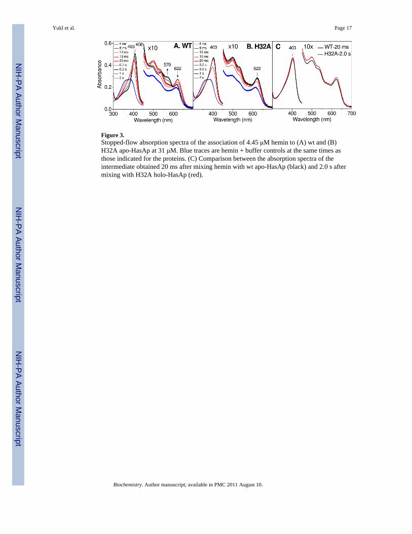
Figure 3.Stopped-flow absorption spectra of the association of 4.45 μM hemin to (A) wt and (B)H32A apo-HasAp at 31 μM. Blue traces are hemin + buffer controls at the same times asthose indicated for the proteins. (C) Comparison between the absorption spectra of theintermediate obtained 20 ms after mixing hemin with wt apo-HasAp (black) and 2.0 s aftermixing with H32A holo-HasAp (red).
Yukl et al. Page 17
Biochemistry. Author manuscript; available in PMC 2011 August 10.
NIH
-PA Author Manuscript
NIH
-PA Author Manuscript
NIH
-PA Author Manuscript

Figure 4.(A) Kinetics of hemin association measured at 408 nm for wt (black) and H32A (red)HasAp. (B) Observed rate constants plotted as a function of apo-HasAp concentration for wt(black) and H32A (red) HasAp as determined by single exponential fitting of the 408-nmabsorbance.
Yukl et al. Page 18
Biochemistry. Author manuscript; available in PMC 2011 August 10.
NIH
-PA Author Manuscript
NIH
-PA Author Manuscript
NIH
-PA Author Manuscript

Figure 5.X-band EPR spectra of RFQ samples of the reaction of one equivalent hemin with (A) wtand (B) H32A apo-HasAp at 760 and 950 μM, respectively. Spectra are normalizedaccording to double integration of EPR signals from known concentrations of holo-HasAp(see Materials and Methods) (0.0317 mW microwave power, 10 G modulation amplitude, T= 7.4 K). (C) Quantification of relative concentration of LS heme in hemin + wt apo-HasApRFQ samples and fit to a single exponential.
Yukl et al. Page 19
Biochemistry. Author manuscript; available in PMC 2011 August 10.
NIH
-PA Author Manuscript
NIH
-PA Author Manuscript
NIH
-PA Author Manuscript

Figure 6.High-frequency RR spectra of RFQ samples of the reaction of one equivalent hemin with(A) wt and (B) H32A apo-HasAp at 760 and 950 μM, respectively (λexc = 413 nm, 20 mW;all spectra are normalized on the intensity of the ν4 observed between 1370 and 1375 cm-1).
Yukl et al. Page 20
Biochemistry. Author manuscript; available in PMC 2011 August 10.
NIH
-PA Author Manuscript
NIH
-PA Author Manuscript
NIH
-PA Author Manuscript

Figure 7.Low-frequency RR spectra of RFQ samples of the reaction of one equivalent hemin with(A) wt and (B) H32A apo-HasAp at 760 and 950 μM, respectively (λexc = 413 nm, 20 mW;all spectra are normalized on the intensity of the ν7 observed between 676 and 677 cm-1).
Yukl et al. Page 21
Biochemistry. Author manuscript; available in PMC 2011 August 10.
NIH
-PA Author Manuscript
NIH
-PA Author Manuscript
NIH
-PA Author Manuscript

NIH
-PA Author Manuscript
NIH
-PA Author Manuscript
NIH
-PA Author Manuscript
Yukl et al. Page 22
Tabl
e 1
Kin
etic
par
amet
ers f
or h
emin
bin
ding
to w
t and
mut
ant h
eme-
acqu
isiti
on p
rote
ins.
Kin
etic
Par
amet
erH
emop
hore
(roo
m te
mpe
ratu
re c
oord
inat
ion
of th
e re
stin
g ho
lo fo
rm)
Wt H
asA
pa,b
(6cH
S/L
S)H
32A
Has
Apa
,b (5
cHS/
6cH
S)W
t Hts
Ac
(6cL
S)W
t Shp
c (6
cLS)
M15
3A S
hpd
(5cH
S)W
t Isd
Ae
(5cH
S)
Kd
(μM
)35
618
± 0.
722
± 2
62 ±
73.
3 ±
0.7
k coo
rd (s
-1)
552
830
655
± 47
35 ±
449
0 ±
2433
0 ±
27
k'he
min
(μM
-1 s-1
)16
1480
1.6
7.9
100
k 2ob
s (s-1
)5.
3 ±
0.6f
n.o.
n.o.
n.o.
n.o.
n.o.
a This
wor
k fr
om m
easu
rem
ents
at 4
°C
b Aff
inity
con
stan
ts fo
r the
bin
ding
of h
emin
to w
t and
H32
A H
asA
m h
ave
been
mea
sure
d by
dis
plac
emen
t iso
ther
mal
titra
tion
calo
rimet
ry a
t 25
°C in
ref.(
2)
c ref.(
23) f
rom
mea
sure
men
ts a
t 25
°C
d ref.(
31) f
rom
mea
sure
men
ts a
t 25
°C
e ref.(
30) f
rom
mea
sure
men
ts a
t 25
°C
f Stan
dard
dev
iatio
n fr
om 5
mea
sure
men
ts a
t diff
eren
t Has
Ap
conc
entra
tions
n.o
.(not
obs
erve
d)
Biochemistry. Author manuscript; available in PMC 2011 August 10.