nice guidance on cvc ultrasound
TRANSCRIPT

8/8/2019 NICE Guidance on CVC Ultrasound
http://slidepdf.com/reader/full/nice-guidance-on-cvc-ultrasound 1/24
Guidance on
the use ofultrasound
locating devices
for placing central
venous catheters
T e c h n o l o g y A p
p r a i s a l G u
i d a n c e - N
o .
4 9
September 2002

8/8/2019 NICE Guidance on CVC Ultrasound
http://slidepdf.com/reader/full/nice-guidance-on-cvc-ultrasound 2/24
Technology Appraisal No. 49
Guidance on the use of ultrasound locating devices for placing central venous catheters
Issue date: September 2002
Review date: August 2005
Ordering Information:
Copies of this guidance can be obtained from the NHS Response Line by telephoning 0870 1555 455 and
quoting ref: N0146. A patient version of this document can be obtained by quoting ref: N0148. A bi-lingual
patient leaflet is also available, ref: N0149.
National Institute for Clinical Excellence
11 Strand
London
WC2N 5HR
Web: www.nice.org.uk
ISBN: 1-84257-213-X
Published by the National Institute for Clinical ExcellenceSeptember 2002
© National Institute for Clinical Excellence September 2002. All rights reserved. This material may be freely reproduced for
educational and not for profit purposes within the NHS. No reproduction by or for commercial organisations is permitted
without the express written permission of the Institute.
Distribution of guidance
This document has been circulated to the following:
• PCT Chief Executives• NHS Trust Chief Executives in England and Wales• Local Health Group General Managers• Medical and Nursing Directors in England and Wales• Consultant anaesthetists in England and Wales• Consultant cardiologists in England and Wales• Consultant cardiothoracic surgeons in England and Wales• Consultant radiologists in England and Wales• ICU/ITU critical care specialists in England and Wales• Strategic Health Authority Chief Executives in England and Wales• NHS Director Wales• Chief Executive of the NHS in England• NHS Executive Regional Directors• Special Health Authority Chief Executives• Community Health Councils in England and Wales
• Patient advocacy groups• Commission for Health Improvement• NHS Clinical Governance Support Team• Chief Medical, Nursing Officers and Pharmaceutical Officers in England and Wales• Medical Director & Head of NHS Quality – Welsh Assembly Government• Representative bodies for health services, professional organisations and statutory bodies, Royal Colleges
This guidance is written in the following context:
This guidance represents the view of the Institute which was arrived at after careful consideration of the
available evidence. Health professionals are expected to take it fully into account when exercising their
clinical judgement. This guidance does not, however, override the individual responsibility of health
professionals to make appropriate decisions in the circumstances of the individual patient, in consultation
with the patient and/or guardian or carer.

8/8/2019 NICE Guidance on CVC Ultrasound
http://slidepdf.com/reader/full/nice-guidance-on-cvc-ultrasound 3/24
1. Guidance
1.1 Two-dimensional (2-D) imaging ultrasound guidance is recommendedas the preferred method for insertion of central venous catheters (CVCs)into the internal jugular vein (IJV) in adults and children in electivesituations.
1.2 The use of two-dimensional (2-D) imaging ultrasound guidance shouldbe considered in most clinical circumstances where CVC insertion isnecessary either electively or in an emergency situation.
1.3 It is recommended that all those involved in placing CVCs using two-dimensional (2-D) imaging ultrasound guidance should undertakeappropriate training to achieve competence.
1.4 Audio-guided Doppler ultrasound guidance is not recommended forCVC insertion.
Guidance on
the use of
ultrasound
locating devices
for placing
central venous
catheters
This section (Section 1) constitutes the Institute's guidance on the use ofultrasound locating devices for placing central venous catheters The remainderof the document is structured in the following way:
2 Clinical need and practice3 The technology4 Evidence and interpretation5 Recommendations for further
research6 Resource impact for the NHS7 Implementation and audit
A bi-lingual summary is available from our website at www.nice.org.uk or by telephoning0870 1555 455 and quoting the reference number N0147.
Mae crynodeb ar gael yn Gymraeg ac yn Saesneg ar ein gwefan yn www.nice.org.uk neu
drwy ffonio 0870 1555 455 gan ddyfynnu cyfeirnod N0147 .
8 Related guidance9 Review of guidanceAppendix A: Appraisal CommitteeAppendix B: Sources of evidenceAppendix C: Information for the publicAppendix D: Technical detail on criteria
for auditTechnology Appraisal
Guidance No. 49ssue date September 2002Review date August 2005

8/8/2019 NICE Guidance on CVC Ultrasound
http://slidepdf.com/reader/full/nice-guidance-on-cvc-ultrasound 4/24
2.1 Central venous catheters (CVCs) are inserted for a number of reasons including haemodynamic monitoring, intravenousdelivery of blood products and drugs (for example,chemotherapy and antibiotics), haemodialysis, total parenteralnutrition, cardiac pacemaker placement and management of
perioperative fluids. Central venous catheterisation may berequired for patients undergoing cancer treatment, dialysis, orcoronary or other major surgery, and for those admitted tointensive therapy units (ITUs), high dependency units (HDUs)or accident and emergency departments. It has been estimatedthat about 200,000 CVCs are inserted annually in the NHS.
2.2 Central venous access has traditionally been achieved by puncturing a central vein (venepuncture) and passing theneedle along the anticipated line of the relevant vein by usingsurface anatomical landmarks and by knowing the expected
anatomical relationship of the vein to its palpable companionartery. This is known as the ‘landmark method’. Direct surgicalaccess to a peripheral vein (‘cut-down’) is a less frequently usedmethod for central venous access catheter insertion.
2.3 CVCs are inserted in a wide range of clinical settings by adiverse group of clinicians including radiologists, anaesthetists,nephrologists, oncologists, surgeons, general physicians andpaediatricians. In the USA and increasingly in the UK, nursespecialists are also undertaking CVC procedures. The range of settings in which CVCs are inserted includes operatingtheatres, emergency rooms, nephrology, oncology and other
wards, radiology departments, ITUs and HDUs.
2.4 Central venous access can be achieved using various puncturesites but the most common are the internal jugular vein (IJV),the subclavian vein (SV), the femoral vein (FV), and the upperlimb veins (using peripherally inserted central catheters –PICCs). The choice of access route depends on multiple factorsincluding the reason for CVC insertion, the anticipatedduration of access, the intact venous sites available and theskills of the operator.
2.5 Whilst experienced operators using the landmark method canachieve relatively high success rates with few complications, inthe literature failure rates for initial CVC insertion have beenreported to be as high as 35%.
2.6 The most common complications associated with CVCplacement are arterial puncture, arteriovenous fistula,pneumothorax, nerve injury and multiple unsuccessfulattempts at catheterisation, which delay treatment. The risksand the consequences of complications vary substantially acrossdifferent patient groups depending on the patient’s anatomy (for example, morbid obesity, cachexia, short neck, or localscarring from surgery or radiation treatment), thecircumstances in which CVC insertion is carried out (for
Clinical needand practice
2
2 NICE Technology Appraisal Guidance – No. 49
8/8/2019 NICE Guidance on CVC Ultrasound
http://slidepdf.com/reader/full/nice-guidance-on-cvc-ultrasound 5/24
NICE Technology Appraisal Guidance – No. 49 3
example, for a patient receiving mechanical ventilation orduring emergencies such as cardiac arrest) and co-morbidities(for example, bullous emphysema or coagulopathy). TheNational Confidential Enquiry into Perioperative Deathsrecently reported that in a survey of over 3000 CVC procedures
undertaken in the NHS, one fatality occurred as a result of aprocedure-induced pneumothorax.
3.1 Ultrasound technology has long been used in interventionalradiology to guide percutaneous procedures at sites such as thekidneys, liver, arterial and venous circulation, pleural cavity,gallbladder and joints. Real-time ultrasound guidance of CVCinsertion provides the operator with visualisation of the desiredvein and the surrounding anatomical structures before andduring the insertion. The advantages of ultrasound-guidedcentral venous catheterisation include the identification of the
precise position of the target vein and the detection of anatomical variants and of thrombosis within the vessel,together with the avoidance of inadvertent arterial puncture.Ultrasound guidance therefore has the potential to reduce theincidence of complications related to initial venous puncture,
which is the first stage of CVC insertion.
3.2 Two types of real-time ultrasound guidance are described: two-dimensional (2-D) imaging ultrasound guidance and audio-guided Doppler ultrasound guidance. Two-dimensionalimaging ultrasound, which is the more commonly usedmethod, provides a real-time grey-scale imaging of the
anatomy. Audio-guided Doppler ultrasound generates anaudible sound from flowing venous blood, which helps theoperator localise the vein and differentiate it from itscompanion artery. The portable ultrasound machines can beused in operating theatres, accident and emergency departments, ITUs, HDUs and radiology suites, as well as atthe bedside on the hospital ward.
3.3 Operators need to be trained to use ultrasound-guidedtechniques. Training involves not only acquiring the necessary manual skills, but also having a basic understanding of
ultrasound principles and being able to interpret ultrasoundimages.
The Appraisal Committee considered evidence from a numberof sources (see Appendix B).
4.1 Clinical effectiveness
4.1.1 Twenty randomised clinical trials (RCTs) wereidentified. Of these, six evaluated audio-guidedDoppler ultrasound against the landmark method,thirteen evaluated 2-D ultrasound guidance against thelandmark method and one evaluated both audio-guided Doppler ultrasound and 2-D ultrasoundguidance against the landmark method. There were no
The technology
3
Evidence andinterpretation
4

8/8/2019 NICE Guidance on CVC Ultrasound
http://slidepdf.com/reader/full/nice-guidance-on-cvc-ultrasound 6/24
4 NICE Technology Appraisal Guidance – No. 49
trials that compared the use of ultrasound locatingdevices (ULDs) against the surgical cut-down method.
4.1.2 Insertion sites were the IJV (fifteen trials), SV (fourtrials) or FV (one trial). One trial did not specify the
insertion point, and one investigated both the IJV andthe SV as insertion sites. None addressed the placementof PICCs or ports, both of which can be consideredtypes of CVCs.
4.1.3 For most of the trials, the setting within the hospital where the cannulation took place was not reportedclearly. In six of the trials the central venouscatheterisation took place in an ITU or trauma unit,and in two trials catheterisations took place inemergency rooms. In the seven studies involving
patients scheduled for cardiac surgery, the cannulationis most likely to have taken place on the way intotheatre. In only three of the trials does it seem likely that CVCs were inserted on wards or in clinics.
4.1.4 The CVC procedure was carried out by anaesthetists inseven studies and by other medical staff in four studies.One study involved 2-D ultrasound-guidedcatheterisation by junior radiologists. None of thestudies involved nurses. The remaining nine studies didnot make the specialty or profession of the operatorclear. The range of experience of the operator, both with
respect to medical career and use of the intervention,differed greatly from study to study. Six studiesdescribed the operators as having up to 5 years’postgraduate experience, eight described them ashaving more than 5 years’ experience, and twodescribed them as varying in experience. Four trials didnot record the career experience of the operator.
2-D ultrasound imaging
Internal jugular vein
4.1.5 Pooled results from seven RCTs suggested that real-time2-D ultrasound guidance was significantly better thanthe landmark method for all five outcome variablesmeasured for insertions into the IJV in adults.Compared with the landmark method, 2-D ultrasoundguidance was associated with reduced risks of failedcatheter placements (86% reduction in relative risk,95% confidence interval [CI] 67% to 94%, p < 0.001),catheter placement complications (57% reduction inrelative risk, 95% CI 13% to 78%, p = 0.02), and failureon the first catheter placement attempt (41% reduction in
relative risk, 95% CI 12% to 61%, p = 0.009), and fewerattempts to achieve successful catheterisation (on average,1.5 fewer attempts, 95% CI 0.47 to 2.53, p = 0.004).

8/8/2019 NICE Guidance on CVC Ultrasound
http://slidepdf.com/reader/full/nice-guidance-on-cvc-ultrasound 7/24
NICE Technology Appraisal Guidance – No. 49 5
4.1.6 The difference between the 2-D ultrasound method andthe landmark method in the time taken to insert acatheter successfully was small and not statistically significant (2-D ultrasound-guided catheterisation was20 seconds faster, 95% CI –83 to 124 seconds).
However, there was significant heterogeneity for thisendpoint (p < 0.01), which indicated that it might notbe appropriate to pool these results. In the study whichreported the longest time to achieve a successfulcatheterisation, the time taken to set up the ULD wasalso included in the outcome measurement. When theanalysis was repeated, excluding this study,heterogeneity was no longer significant and the pooledresult from the included trials showed thatcatheterisation was, on average, 69 seconds faster (95%CI 46 to 92 seconds) with the ULD than with the
landmark method, which was a highly statistically significant difference (p < 0.001). It is acknowledgedthat the importance of this endpoint will vary betweenclinical situations.
4.1.7 Three trials evaluated the effect of 2-D ultrasoundguidance on the cannulation of the IJV in infants. Inthese trials, 2-D ultrasound guidance was significantly better than the landmark method in terms of reductionsin the risk of failed catheter placements (85% reductionin relative risk, 95% CI 36% to 97%, p = 0.01), the risk of catheter placement complications (73% reduction in
relative risk, 95% CI 8% to 92%, p = 0.03), and thenumber of attempts required before catheterisation wassuccessful (reduced by an average of 2, 95% CI 1.2 to2.8, p = 0.001). Using 2-D ultrasound guidance,successful cannulation was achieved, on average, 349seconds (95% CI –103 to 802 seconds) more quickly than with the landmark method, although this result
was not statistically significant.
Subclavian vein
4.1.8 Only one RCT was identified that analysed the effect of 2-D ultrasound guidance on SV catheterisation inadults. In the trial, in comparison with the landmark method, 2-D ultrasound guidance was associated withreduced risks of catheter placement failure (86%reduction in relative risk, 95% CI 43% to 96%,p = 0.006) and catheter placement complications (90%reduction in relative risk, 95% CI 29% to 99%,p = 0.02). However, in this trial, the operators wererelatively inexperienced in both the landmark methodand 2-D ultrasound guidance. The failure rate with thelandmark method was 55%, which is higher than that
reported in trials that involved more experiencedoperators (around 9–19%).

8/8/2019 NICE Guidance on CVC Ultrasound
http://slidepdf.com/reader/full/nice-guidance-on-cvc-ultrasound 8/24
4.1.9 No studies were found that investigated the effect of 2-D ultrasound guidance on SV catheterisation ininfants.
Femoral vein
4.1.10 One study was identified that evaluated the effect of 2-D ultrasound guidance on femoral catheterisation inadults. In this trial, the operators took, on average, 2.7(95% CI 0.1 to 5.3) fewer attempts to insert a catheterusing 2-D ultrasound guidance than using thelandmark method (p = 0.04). Compared with thelandmark method, 2-D ultrasound guidance reducedthe risk of failed catheter placement and the time tosuccessful catheterisation, but the differences were notstatistically significant. No studies in infants were
found.
4.1.11 No studies were found that investigated the effect of 2-D ultrasound guidance on FV catheterisation ininfants.
Audio-guided Doppler ultrasound
Internal jugular vein
4.1.12 Four RCTs were found that compared audio-guidedDoppler ultrasound guidance with the landmark
method for IJV catheterisation in adults. Pooled resultsfrom these RCTs suggest that audio-guided Dopplerultrasound guidance was significantly better than thelandmark method in terms of risk of failed catheterplacement (61% reduction in relative risk, 95% CI 8%to 83%, p = 0.03) and the risk of failure on the firstcatheter placement attempt (43% reduction in relativerisk, 95% CI 12% to 63%, p = 0.01). With the audio-guided Doppler ultrasound method, the risk of catheterplacement complications was reduced (57% reductionin relative risk, 95% CI –5% to 83%) and there were
fewer attempts to achieve successful catheterisation(0.6 fewer attempts, 95% CI –0.6 to 1.8); however, thedifferences did not reach statistical significance(p = 0.06 and p = 0.40, respectively) so they could havearisen by chance. It took, on average, 35 seconds longer(95% CI –54 to 124 seconds) to successfully insert acatheter using Doppler ultrasound guidance than it did
with the landmark method, although this difference was also not statistically significant.
4.1.13 Only one trial was identified that studied the effect of audio-guided Doppler ultrasound in infants. The
sample size of this study was small (n = 29) and so itlacked statistical power. It failed to show any differences
with the landmark method.
6 NICE Technology Appraisal Guidance – No. 49

8/8/2019 NICE Guidance on CVC Ultrasound
http://slidepdf.com/reader/full/nice-guidance-on-cvc-ultrasound 9/24
Subclavian vein
4.1.14 The pooled results from three RCTs (all involvingadults) suggest that for SV catheterisation there was asignificantly increased risk of failed catheter placement
when the audio-guided Doppler ultrasound method was used compared with the landmark method (48%increased in relative risk, 95% CI 3% to 114%,p = 0.03) – in other words the landmark method waspreferable to the audio-guided Doppler ultrasoundguidance technique. In contrast, the pooled resultsfrom two of the trials, which reported the risk of catheter placement, showed a 43% fall (95% CI 89%to 188%) in relative risk in the audio-guided Dopplerultrasound group, although this result was notstatistically significant.
4.1.15 Only one study reported the effect of audio-guidedDoppler ultrasound guidance on the risk of failure of the first catheter placement in adults. There was a slightincrease (4%, 95% CI –24% to 43%) in the risk of catheter placement complications associated with theuse of audio-guided Doppler ultrasound guidancecompared with the landmark method, although thisresult was not statistically significant. Only one study recorded the effect of audio-guided Doppler ultrasoundguidance on the number of attempts required toachieve successful catheterisation. This study found
that an average of 0.4 (95% CI 0.2 to 0.6) fewerattempts were needed to achieve successfulcatheterisation with the audio-guided Dopplerultrasound guidance method compared with thelandmark method, a highly statistically significantdifference (p < 0.001). The same study was the only one to record the effect of Doppler ultrasoundguidance on the time to achieve successfulcatheterisation. Catheterisation using the Dopplerultrasound guidance method was significantly (onaverage, 209 seconds, 95% CI 175 to 242) slower than
catheterisation using the landmark method (p < 0.001).
4.2 Cost effectiveness
4.2.1 No relevant economic evaluations were identified inthe literature. Furthermore, none of the submissionsmade to the Institute included economic evaluations.
4.2.2 The Assessment Group developed an economicanalysis, based on the evidence from the systematicreview of RCTs, to evaluate the cost effectiveness of 2-D ultrasound guidance compared with the landmark method. This model is a simple decision analyticmodel, and is based on a theoretical cohort of 1000
NICE Technology Appraisal Guidance – No. 49 7

8/8/2019 NICE Guidance on CVC Ultrasound
http://slidepdf.com/reader/full/nice-guidance-on-cvc-ultrasound 10/24
adult patients who required IJV cannulation beforesurgery and who had a low to moderate risk of complications.
4.2.3 This model adopted a set of conservative assumptions.
It was assumed that: the operators were experienced inusing the landmark method; the time to achievesuccessful puncture was the same for both methods;complications were limited to arterial puncture; there
was a 10-minute delay between the prior failure andthe new attempt at another insertion site; there was a100% success rate at the second insertion site; andeach machine was used for 15 procedures per week.
4.2.4 The results of the Assessment Group’s model suggestedthat the ultrasound guidance not only avoided 90
arterial punctures for every 1000 patients treated, butalso reduced costs by an average of almost £2 perpatient. In other words the 2-D ultrasound guidancemethod was found to be both more effective and lesscostly than the landmark method.
4.2.5 A threshold sensitivity analysis was undertaken toexamine by how much key variables in the modelneeded to change to make the ultrasound guidancemethod cost-neutral instead of cost-saving. Themodelled result was most sensitive to the utilisation of the ultrasound equipment. The cost-saving result was
eradicated if the number of ultrasound proceduresassumed per machine per week was less than around11, or if the number of ultrasound procedures carriedout by an individual trained practitioner was less thanaround 3 per month on average.
4.2.6 Given that the model used relatively conservativeestimates, the Assessment Group concluded that theresults were probably generalisable to all anatomicalcatheter insertion sites, to infants, and to other sites
within the hospital including the clinical wards.
4.3 Consideration of the evidence
4.3.1 The Committee reviewed the evidence on both theclinical effectiveness and the cost effectiveness of ULDsfor placing CVCs, having also considered the evidencefrom clinical experts. Furthermore, the Committee
was mindful of the need to ensure that its advice took account of the efficient use of NHS resources.
4.3.2 The Committee took note of the fact that the evidenceon the effectiveness of CVC placement into IJVs in
adult patients was more robust than that available forother insertion sites. For infants, evidence was available
8 NICE Technology Appraisal Guidance – No. 49

8/8/2019 NICE Guidance on CVC Ultrasound
http://slidepdf.com/reader/full/nice-guidance-on-cvc-ultrasound 11/24
only from trials that evaluated central venouscatheterisation of the IJV, and there was very limitedevidence on the use of this technology in very smallinfants (i.e. those weighing less than 3 kg). In addition,the economic analysis presented to the Committee was
based on an evaluation of the cost effectiveness of 2-Dultrasound-guided elective CVC placement into theIJV in the operating theatre prior to surgery. The
Assessment Report provided justifications forextrapolating this analysis to other settings including
ward-based management, other sites of CVC insertionand also to CVC placement in infants.
4.3.3 Given the constraints outlined in 4.2.2, theCommittee concluded that there was evidence of boththe clinical and cost effectiveness of 2-D imaging
ultrasound guidance as an adjunct for placing CVCs inthe majority of clinical scenarios, but that the degree to which this technology would be most suitably applied would vary according to the clinical situation and thecompetence/previous experience of the operator. Inaddition, there could be potential benefits for patientsarising from reduced discomfort from the procedureand reduced risk of complications compared with thelandmark method, particularly for IJV insertions.
4.3.4 The Committee found the evidence for the use of audio-guided Doppler ultrasound guidance less
satisfactory, and therefore concluded that the 2-Dimaging ultrasound guidance should be used inpreference to audio-guided Doppler ultrasoundguidance.
4.3.5 While accepting that, from a patient’s perspective, 2-Dultrasound imaging guidance in CVC insertion mightbe more appropriate and probably superior to thetraditionally used landmark method in many circumstances, the Committee also considered thefinancial and service implications of purchasing the
required equipment and of training sufficient numbersof competent practitioners.
4.3.6 The Committee also considered that although 2-Dultrasound imaging guidance in CVC placement may eventually become the routine method for placingCVCs, the landmark method would remain importantin some circumstances, such as emergency situations,
when ultrasound equipment and/or expertise mightnot be immediately available. Consequently, theCommittee thought it important that operatorsmaintain their ability to use the landmark method and
that the method continues to be taught alongside the2-D-ultrasound-guided technique.
NICE Technology Appraisal Guidance – No. 49 9

8/8/2019 NICE Guidance on CVC Ultrasound
http://slidepdf.com/reader/full/nice-guidance-on-cvc-ultrasound 12/24
5.1 Good quality studies are needed:
• to investigate the possible economic and clinicalimplications to the NHS of nurse specialists or otherhealthcare practitioners carrying out routine insertion of
CVCs
• to evaluate the use of ultrasound-guided central venouscatheterisation in small infants (i.e. those weighing lessthan 3 kg).
6.1 The purchase cost of a portable 2-D ultrasound machinecurrently lies between £7000 and £15,000. The additionaldisposables necessary for the ultrasound-guided procedure costless than £1 per procedure. Estimates made by the AssessmentGroup analysis indicate that the additional cost of using
ultrasound equipment for the CVC placement procedure islikely to be less than £10 per procedure.
6.2 It is likely that the NHS will need to invest in a significantnumber of additional 2-D ultrasound machines, although it isimpossible to predict how many will be required, as localcircumstances will vary considerably. Implementing theguidance will require local decisions regarding optimal numberof machines, staff training and device service contracts.
6.3 The Assessment Group analysis suggests that in the long termthe implementation of ultrasonic locating devices will be cost-
saving. The majority of these savings are likely to be due toreleasing resources such as staff, and operating theatre andITU/HDU time and beds.
6.4 A constraint upon the implementation of this technology willbe the need to ensure that there are adequately trainedcompetent operators to support the services. Many CVCplacement procedures are performed on an emergency basis atthe bedside in a diverse number of locations and therefore thenecessary skills need to be spread across several relateddisciplines.
7.1 NHS Trusts in which CVCs are used, all those who routinely insert CVCs and those responsible for clinical trainingprogrammes should review policies and practices regarding theinsertion of CVCs to take account of the guidance set out inSection 1. The recommendations in this guidance willrepresent a significant service development for most NHSorganisations. The Appraisal Committee has advised theInstitute that the nature of the resource consequences of theguidance and the time it will take to put them in place shouldbe brought to the attention of the Department of Health andthe Welsh Assembly Government.
10 NICE Technology Appraisal Guidance – No. 49
Implementationand audit
7
Resource impactfor the NHS
6
Recommendationsfor furtherresearch
5

8/8/2019 NICE Guidance on CVC Ultrasound
http://slidepdf.com/reader/full/nice-guidance-on-cvc-ultrasound 13/24
7.2 Local guidelines or care pathways which relate to the use of CVCs should incorporate the guidance set out in Section 1.
7.3 To enable healthcare practitioners to audit their owncompliance with this guidance, it is recommended that a system
is available to identify patients who have a CVC inserted ineither an elective or an emergency situation.
7.4 To measure compliance locally with the guidance in Section 1,the following criteria should be used. Further details onsuggestions for audit are presented in Appendix D.
• When a CVC is being inserted into the IJV of an adult ora child in an elective situation, 2-dimensional (2-D)imaging ultrasound guidance is used.
• All healthcare practitioners involved in the placement of CVCs using 2-D imaging ultrasound guidance undertakeappropriate training to achieve competence in thistechnique.
• Audio-guided Doppler ultrasound guidance is not used forCVC insertion.
7.5 All NHS Trusts in which CVCs are used should identify thenumber of 2-D imaging ultrasound units required and theappropriate location for each unit, should plan to train asufficient number of healthcare practitioners from a range of
disciplines in the proper use of the units and should identify other financial and service implications of implementing theguidance in Section 1.
7.6 Healthcare practitioners should consider the most appropriatemethod of CVC insertion that is in the best interest of thepatient in his or her specific clinical situation, particularly interms of minimising the risk of adverse events such as failedcatheter placements or catheter placement complications. Trustsshould recognise that the decision to use 2-D imagingultrasound guidance or the landmark method will be informed
by:
• the competence and previous experience of the operator(s)
• the anatomical site of CVC insertion and other anticipatedtechnical difficulties
• the urgency of clinical need.
8.1 There is no related NICE guidance for this technology.
NICE Technology Appraisal Guidance – No. 49 11
Relatedguidance
8

8/8/2019 NICE Guidance on CVC Ultrasound
http://slidepdf.com/reader/full/nice-guidance-on-cvc-ultrasound 14/24
9.1 The review date for a technology appraisal refers to the monthand year in which the Guidance Executive will consider any new evidence on the technology, in the form of an updated
Assessment Report, and decide whether the technology shouldbe referred to the Appraisal Committee for review.
9.2 The guidance on this technology is reviewed in August 2005
Andrew DillonChief Executive
September 2002
12 NICE Technology Appraisal Guidance – No. 49
Reviewof guidance
9

8/8/2019 NICE Guidance on CVC Ultrasound
http://slidepdf.com/reader/full/nice-guidance-on-cvc-ultrasound 15/24
NICE Technology Appraisal Guidance – No. 49 13
Appendix A
Appraisal Committee members
NOTE The Appraisal Committee is a standing advisory committee of the
Institute. Its members are appointed for a 3-year term. A list of theCommittee members appears below. The Appraisal Committee meets
twice a month other than in December, when there are no meetings. The
Committee membership is split into two branches, with the Chairman,
Vice-chairman and a number of other members attending meetings of
both branches. Each branch considers its own list of technologies and
topics are not moved between the branches.
Committee members are asked to declare any interests in the technology
to be appraised. If there is a conflict of interest, the member is excluded
from participating further in that appraisal.
The minutes of each Appraisal Committee meeting, which include thenames of the members who attended and their declaration of interests,
are posted on the NICE website.
Dr Jane Adam
Radiologist, St George’s Hospital,
London
Professor R L Akehurst
Dean, School of Health Related
Research, Sheffield University
Dr Sunil Angris
General Practitioner,Waterhouses
Medical Practice
Professor David Barnett
(Chairman)
Professor of Clinical Pharmacology,
University of Leicester
Dr Sheila Bird
MRC Biostatistics Unit, Cambridge
Professor Carol Black
Consultant Physician, Royal Free
Hospital & UCL, London
Professor John Brazier
Health Economist, University of
Sheffield
Professor Martin Buxton
Director of Health Economics Research
Group, Brunel University
Professor Mike Campbell
Statistician, Institute of General
Practice & Primary Care, Sheffield
Dr Karl Claxton
Health Economist, University of York
Professor Sarah CowleyProfessor of Community Practice
Development, Kings College, London
Professor Jack Dowie
Health Economist, London School of
Hygiene & Tropical Medicine, London
Mr Chris Evennett
Chief Executive, Mid-Hampshire
Primary Care Trust
Dr Paul EwingsStatistician, Taunton & Somerset NHS
Trust
Professor Terry Feest
Clinical Director and Consultant
Nephrologist, Richard Bright Renal
Unit, and Chairman of the UK Renal
Registry
Professor Gary A Ford
Professor of Pharmacology of Old Age/
Consultant Physician, Wolfson Unit of Clinical Pharmacology, University of
Newcastle

8/8/2019 NICE Guidance on CVC Ultrasound
http://slidepdf.com/reader/full/nice-guidance-on-cvc-ultrasound 16/24
14 NICE Technology Appraisal Guidance – No. 49
Mrs Sue Gallagher
Chief Executive, Merton, Sutton and
Wandsworth Health Authority
Dr Trevor Gibbs
Head, Global Clinical Safety &Pharmacovigilance, GlaxoSmithKline
Sally Gooch
Director of Nursing, Mid-Essex
Hospital Services Trust
Mr John Goulston
Director of Finance,The Royal Free
Hampstead NHS Trust
Professor Trisha Greenhalgh
Professor of Primary Health Care,University College London
Miss Linda Hands
Consultant Vascular Surgeon, John
Radcliffe Hospital, Oxford
Professor Philip Home
Professor of Diabetes Medicine,
University of Newcastle
Dr Terry John
General Practitioner, The Firs, London
Dr Diane Ketley
Research into Practice Programme
Leader, NHS Modernisation Agency
Dr Mayur Lakhani
General Practitioner, Highgate Surgery,
Leicester, and Lecturer, University of
Leicester
Ruth LesirgeLay Representative; Director, Mental
Health Foundation
Dr George Levvy
Lay Representative; Chief Executive,
Motor Neurone Disease Association
Dr Gill Morgan
CEO, North & East Devon Health
Authority
Professor Miranda Mugford
Health Economist, University of East
Anglia
Mr M Mughal
Consultant Surgeon, LancashireTeaching Hospitals NHS Trust
Mr James Partridge
Lay Representative; Chief Executive,
Changing Faces
Siân Richards
General Manager, Cardiff Local Health
Group
Professor Philip Routledge
Professor of Clinical Pharmacology,University of Wales
Dr Rhiannon Rowsell
Pharmaceutical Physician, AstraZeneca
UK Ltd
Dr Stephen Saltissi
Consultant Cardiologist, Royal
Liverpool University Hospital
Professor Andrew Stevens
(Vice-Chairman)
Professor of Public Health, University
of Birmingham
Professor Ray Tallis
Consultant Physician, Hope Hospital,
Salford
Dr Cathryn Thomas
General Practitioner, and Senior
Lecturer, Department of Primary Care
and General Practice, University of Birmingham
Professor Mary Watkins
Head of Institute of Health Studies,
University of Plymouth
Dr Norman Waugh
Public Health Consultant, University of
Southampton

8/8/2019 NICE Guidance on CVC Ultrasound
http://slidepdf.com/reader/full/nice-guidance-on-cvc-ultrasound 17/24
NICE Technology Appraisal Guidance – No. 49 15
A. Assessment report prepared bythe School of Health Related
Research (ScHARR), University of Sheffield: The effectiveness and cost effectiveness of ultrasound locating devices for central
venous access, 24 January 2002.
B. Manufacturer/sponsorsubmissions:
• KeyMed (Medical & Industrial
Equipment) Ltd
• Jade Medical UK and Dymax
Corporation
• SonoSite Inc
• Siemens
• Dynamic Imaging Limited
C. Professional/specialist groupsubmissions:
• British Association of Critical
Care Nurses
• Royal College of Physicians
• Renal Association
• Intensive Care Society
• Royal College of Anaesthetists
• Lincolnshire Health
Authority/West Lincolnshire
PCT
• Royal College of Nursing
• Royal College of Radiologists
• Department of Health andWelsh Assembly Government
• Health Technology Board for
Scotland
Appendix B
Sources of evidence considered by theCommittee
D. Patient/carer groupsubmissions:
• No submissions received
E. Expert perspective:
• Dr A R Bodenham, Consultant
in Anaesthesia and Intensive
Care, Leeds General Infirmary
The following documentation and opinion were made available to the
Committee:

8/8/2019 NICE Guidance on CVC Ultrasound
http://slidepdf.com/reader/full/nice-guidance-on-cvc-ultrasound 18/24
16 NICE Technology Appraisal Guidance – No. 49
The National Institute for Clinical Excellence (NICE) is part of theNHS. It produces guidance for both the NHS and patients on theuse of medicines, medical equipment, diagnostic tests and clinicaland surgical procedures and under what circumstances they shouldbe used.
To produce this guidance, NICE looks at how well the medicine,equipment or procedure works and also how well it works inrelation to how much it costs. This process is called an appraisal.The appraisal process involves the manufacturer of the medicine or
equipment for which guidance is being produced and theorganisations that represent the healthcare professionals, patientsand carers who will be affected by the guidance. Each appraisal takesabout 12 months to complete.
NICE was asked to look at the available evidence on ultrasoundlocating devices for placing central venous catheters and provideguidance that would help the NHS in England and Wales decide
when they should be used.
A central venous catheter is a tube that is inserted into a vein (a
blood vessel that carries blood to the heart). There are many reasons why a patient might need a central venous catheter – for example, itmight be necessary to slowly deliver blood products, certain liquiddrugs or other fluids into the body (this is called an intravenousinfusion or a ‘drip’) or to carry out certain surgical procedures suchas fitting a heart pacemaker. And there lots of hospital situations in
which central venous catheters are used – for example, for patientsundergoing cancer treatment, dialysis, or major surgery, and forthose admitted to accident and emergency departments or intensivetherapy units.
The most common sites for insertion of a central venous catheter arethe internal jugular vein (in the neck, carrying blood from the headto the heart), the subclavian vein (under the collar bone, carryingblood from the arm to the heart), the femoral vein (the main vein inthe leg), and veins in the arm.
Appendix C
Patient information
Guidance on the use of ultrasound locatingdevices for placing central venous catheters
What is NICEguidance?
What are centralvenous cathetersand when arethey used?
The patient information in this appendix has been designed tosupport the production of your own information leaflets. Youcan download it from our website at www.nice.org.uk where itis available in English and Welsh. If you would like printedcopies of the leaflets please ring the NHS Response Line on 08701555 455 and quote reference number N0148 for the Englishpatient leaflet and N0149 for the bi-lingual patient leaflet.

8/8/2019 NICE Guidance on CVC Ultrasound
http://slidepdf.com/reader/full/nice-guidance-on-cvc-ultrasound 19/24
NICE Technology Appraisal Guidance – No. 49 17
Central venous catheters are usually inserted by doctors, butsometimes a specialist nurse carries out the procedure. It has beenestimated that about 200,000 CVCs are inserted annually in theNHS.
A vein has to be punctured to insert a central venous catheter. Thisis done by inserting a needle into the body and along the vein.Traditionally, doctors have found the right place to insert the needleby using their knowledge of body structure to look for certainfeatures and to feel for the pulse in the artery that lies close to thevein. (An artery is a blood vessel that carries blood away from theheart. The catheter must be inserted into a vein, not into an artery.)This is known as the ‘landmark method’.
Ultrasound devices are now available to help the doctor to guide theneedle into the vein. There are two types of ultrasound device
available to do this: two-dimensional (2-D) imaging ultrasounddevices and audio-guided Doppler ultrasound devices. Allultrasound devices send very high frequency sound waves (whichcan’t be heard by the human ear) into the body and detect theechoes that are reflected back. In 2-D imaging ultrasound, theseechoes are analysed by the machine and translated into an image of the vein and the tissues surrounding it, which is displayed on ascreen. Audio-guided Doppler ultrasound devices don’t show apicture of the vein; instead they emit a sound when they detectblood flowing in a vein.
There are a number of complications that can be associated with
inserting a central venous catheter. These include puncturing anartery instead of a vein, puncturing the wall of the pleural cavity that surrounds the lungs, causing injury to a nerve or having tomake several attempts at inserting the catheter, which can delay treatment. The risks and the consequences of complications differsubstantially across different patient groups depending on thepatient’s body structure, the circumstances in which the procedureis carried out and what illnesses or injuries the patient has.
Studies have investigated whether ultrasound locating devices haveadvantages over the landmark method for placing central venous in
terms of factors such as failure to place a catheter, the number of attempts made before a catheter is placed successfully and theoccurrence of complications. NICE has looked at the evidenceavailable and has made recommendations to the NHS in Englandand Wales about when ultrasound locating devices should be used.
NICE has made the following recommendations.
• 2-D imaging ultrasound guidance should be the preferredmethod when inserting of central venous catheter into theinternal jugular vein in adults and children in ‘electivesituations’. (‘Elective situation’ means that the operation, orother treatment, has been planned – that is, it is not anemergency.)
Why mightultrasoundlocating devicesbe used?
How areultrasoundlocating devicesused?
What has NICErecommended?

8/8/2019 NICE Guidance on CVC Ultrasound
http://slidepdf.com/reader/full/nice-guidance-on-cvc-ultrasound 20/24
18 NICE Technology Appraisal Guidance – No. 49
• 2-D imaging ultrasound guidance should be considered inmost clinical situations where CVC insertion is necessary,
whether the situation is elective or an emergency.
• Everyone who uses 2-D imaging ultrasound guidance to insert
central venous catheters should have appropriate training toensure they are competent to use the technique.
• Audio-guided Doppler ultrasound guidance is notrecommended for use when inserting central venous catheters.
If you or someone you care for is going to have a clinical procedure which might involve inserting a central venous catheter (forexample, major surgery), you should discuss this guidance withyour doctor or nurse.
Yes. The guidance will be reviewed in August 2005.
The NICE website (www.nice.org.uk) has further information onNICE and the full guidance on the use of ultrasound locatingdevices for placing intravenous catheters that has been issued to theNHS. The guidance can also be requested from the NHS ResponseLine by phoning 0870 1555 455 and quoting reference N0146.
Will NICE reviewits guidance?
What shouldI do?
Furtherinformation

8/8/2019 NICE Guidance on CVC Ultrasound
http://slidepdf.com/reader/full/nice-guidance-on-cvc-ultrasound 21/24
NICE Technology Appraisal Guidance – No. 49 19
Appendix D
Detail on criteria for audit of the use ofultrasound locating devices for placing central
venous catheters
Possibleobjectives foran audit
Possible patientsto be included inthe audit andtime period forselection
An audit on the appropriate use of ultrasound locating devicescould be carried out to ensure that:
• when a central venous catheter (CVC) is being inserted intothe internal jugular vein (IJV) of an adult or a child in anelective situation, 2-dimensional (2-D) imaging ultrasoundguidance is used
• healthcare practitioners involved in the placement of CVCs
using 2-D imaging ultrasound guidance have appropriatetraining
• audio-guided Doppler ultrasound guidance is not used forCVC insertion.
If healthcare practitioners have agreed locally on the clinicalcircumstances where 2-D imaging ultrasound guidance is to beused when a CVC insertion is necessary, the audit also could becarried out to ensure that the technique is used as agreed locally.
All patients who have a CVC inserted either in the IJV in anelective situation (or for any purpose on either an elective oremergency basis, if 2-D imaging ultrasound is more widely used)over a reasonable period of time for audit data collection, forexample, for 1 to 3 months. A sample of patients stratified by clinical areas most likely to be involved, for example, critical careareas, theatres, and accident and emergency, could be used for theaudit or the audit could be staged to include one clinical area at atime, working through all clinical areas.
8/8/2019 NICE Guidance on CVC Ultrasound
http://slidepdf.com/reader/full/nice-guidance-on-cvc-ultrasound 22/24
20 NICE Technology Appraisal Guidance – No. 49
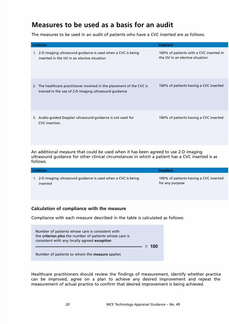
Measures to be used as a basis for an audit
The measures to be used in an audit of patients who have a CVC inserted are as follows.
An additional measure that could be used when it has been agreed to use 2-D imagingultrasound guidance for other clinical circumstances in which a patient has a CVC inserted is asfollows.
Number of patients whose care is consistent with
the criterion plus the number of patients whose care is
consistent with any locally agreed exception
Number of patients to whom the measure applies
100
Criterion Standard
100% of patients with a CVC inserted in
the IJV in an elective situation
Calculation of compliance with the measure
Compliance with each measure described in the table is calculated as follows:
Healthcare practitioners should review the findings of measurement, identify whether practicecan be improved, agree on a plan to achieve any desired improvement and repeat the
measurement of actual practice to confirm that desired improvement is being achieved.
2. The healthcare practitioner involved in the placement of the CVC is
trained in the use of 2-D imaging ultrasound guidance
3. Audio-guided Doppler ultrasound guidance is not used for
CVC insertion
100% of patients having a CVC inserted
100% of patients having a CVC inserted
1. 2-D imaging ultrasound guidance is used when a CVC is being
inserted in the IJV in an elective situation
Criterion Standard
100% of patients having a CVC inserted
for any purpose
1. 2-D imaging ultrasound guidance is used when a CVC is being
inserted

8/8/2019 NICE Guidance on CVC Ultrasound
http://slidepdf.com/reader/full/nice-guidance-on-cvc-ultrasound 23/24
NICE Technology Appraisal Guidance – No. 49 21
Exception Definition of Terms
Local clinical teams should agree on the types of elective situations
to be included in the audit and should agree to any exceptions forthe use of the technique such as an infant weighing less than 3 kg
None
For audit purposes, it should be agreed at NHS Trust level how
training to achieve competence in the technique is documented
None
None
Exception Definition of Terms
Local healthcare practitioners may specify circumstances in which 2-
D ultrasound guidance is to be used when a CVC is being inserted or
may specify exceptions, for audit purposes
None

8/8/2019 NICE Guidance on CVC Ultrasound
http://slidepdf.com/reader/full/nice-guidance-on-cvc-ultrasound 24/24
National Institute for
Clinical Excellence
11 Strand
London
WC2N 5HR
Web: www.nice.org.uk
N0146 1P 15k Sep 02 (ABA)