next tuesday
DESCRIPTION
Next Tuesday. Bring Blue Scan sheet Bring #2 pencils Bring erasers if you want to change your mind. Plant Development and Anatomy. Fig. 40.2. Plant Development. Seed: baby plant (embryo) in box (seed coat) with its lunch (endosperm) Some babies finish lunch early - PowerPoint PPT PresentationTRANSCRIPT

Next Tuesday• Bring Blue Scan sheet
• Bring #2 pencils
• Bring erasers if you want to change your mind

Plant Development and Anatomy

Plant Development• Seed: baby plant
(embryo) in box (seed coat) with its lunch (endosperm)
• Some babies finish lunch early
• All that’s left in box (seed coat) is baby (embryo)!
• Example, bean seed.
Fig. 40.2

Plant Development
• Embryo parts:– Cotyledons: seed leaves. Bean is dicot (2
cotyledons). These contain stored food.– Epicotyl: Located above cotyledons. Will
develop into shoot system (stem and leaves) of plant
– Radicle: Will develop into root system of plant– Hypocotyl: Below cotyledons. Stem that
connects cotyledons to radicle.
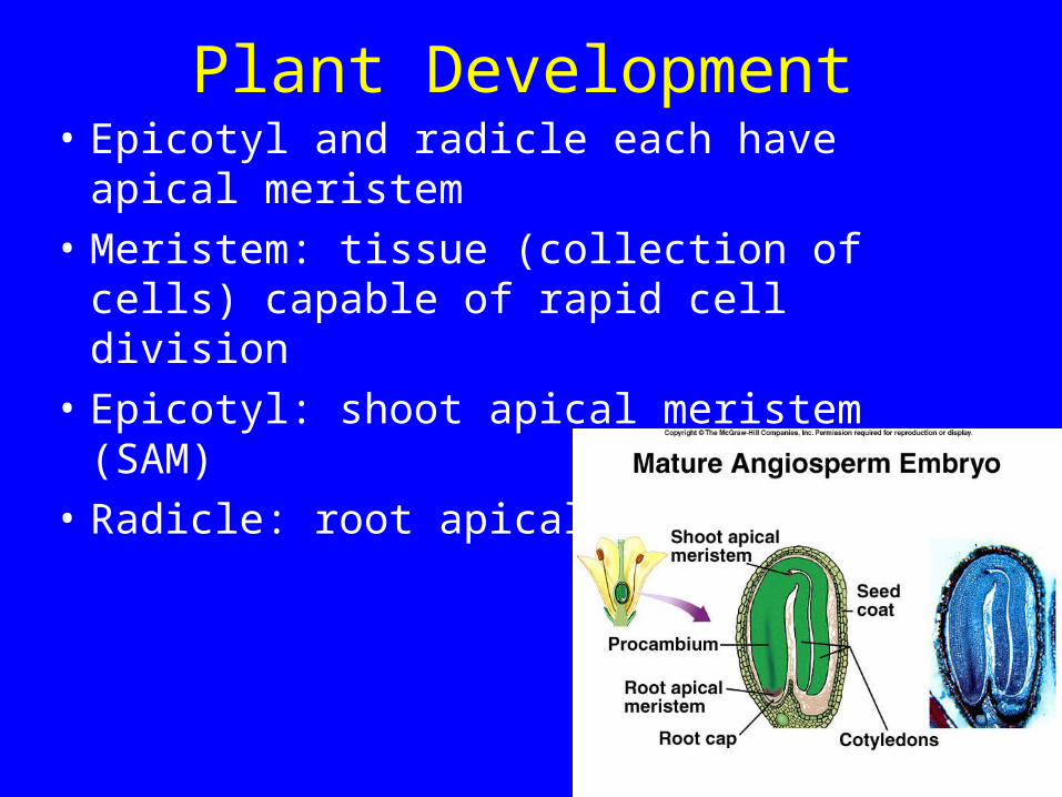
Plant Development• Epicotyl and radicle each have apical meristem
• Meristem: tissue (collection of cells) capable of rapid cell division
• Epicotyl: shoot apical meristem (SAM)
• Radicle: root apical meristem (RAM).

Plant Development• Bean seed germination
• Hypocotyl pulls cotyledons/epicotyl from soil
• Root and shoot systems start to grow.
Fig. 40-15a

Plant Development• Primary growth occurs due to activity of
meristems (SAM and RAM)
• These produce specialized dividing cells called primary meristems
• Primary meristems develop into primary tissues.

Plant Cells• Focus on cell walls:
form plant skeleton (supports body)
• Primary wall: cellulose, penetrated by plasmodesmata
• Middle lamella: “cement” between adjacent walls.

Plant Cells• Secondary cell wall: laid
down inside primary wall
• Thick, contains lignin (strong, rot resistant)
• Note: In our lab slides, secondary wall will stain __RED_____ whereas primary wall will stain __GREEN______.
primary
secondary

Plant Cells• Secondary cell wall: laid
down inside primary wall• Thick, contains lignin
(strong, rot resistant)• Opening in secondary
wall called pit• Note pits still have
primary wall present (pit membrane: pit not a completely open space between cells).

Tissues and cell types
• Tissue: Group of cells organized as a functional unit
• Tissues made of cell types
• Simple tissues contain 1 cell type
• Complex tissues contain >1 cell type.

Tissues and cell types• 3 main types of tissues:
– 1) ground (yellow)– 2) dermal (white)– 3) vascular (purple).

Ground Tissues• Usually simple tissues;
– Parenchyma– Collenchyma– Sclerenchyma.
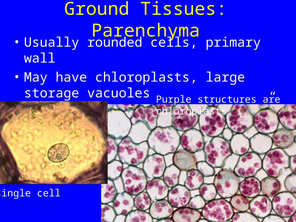
Ground Tissues: Parenchyma• Usually rounded cells, primary wall
• May have chloroplasts, large storage vacuoles
• May be used as “packing tissue” to fill space.Purple structures are chloroplasts
single cell

Ground Tissues: Collenchyma• Contains collenchyma
cells• Elongate cell, thick
primary walls, especially thick at corners, alive at maturity
• Used as strengthening tissue in young plant parts.

Ground Tissues: Sclerenchyma• Simple tissue: contains 1
cell type• But two types of cells
possible:• 1) fiber: elongate, thick
secondary wall, often dead at maturity
• Strengthening cell/tissue.

Ground Tissues: Sclerenchyma• Simple tissue: contains 1
cell type• But two types of cells
possible:• 2) sclereid: Not elongated
(mostly branched or rounded), thick secondary wall, often dead at maturity
• Used to strengthen tissues, protect areas (hard endocarp of drupe is sclereids).
1 cell

Ground Tissues: Sclerenchyma• An astrosclereid alone and
others in leaf section. 1 cell

Dermal Tissues• Epidermis: Surface covering of all plant body
• Typical epidermal cells: flattened, no chloroplasts, primary wall, thick outer walls
• In shoot/stem, outer walls have cuticle (waxy layer) on outside.
Thick cuticle (orange layer)

Epidermis• Special cell types: guard cell
• In pairs on stems/leaves
• Have chloroplasts, primary wall, open and close pore of stoma (plural: stomata)
• Control water loss, CO2 uptake.

Epidermis• Special cell types: guard cell
• In pairs on stems/leaves
• Have chloroplasts, primary wall, open and close pore of stoma (plural: stomata)
• Control water loss, CO2 uptake.

Epidermis• Special cell types: trichome cells
• On stems/leaves
• Have primary wall, elongate.

Epidermis• Special cell types: trichome cells
• Function to slow water loss
• Function to reflect light to keep leaf cool

Epidermis• Special cell types: trichome
cells
• Function: defense against herbivores.
sticky trichomes
stinging nettle trichome

Epidermis• Special cell types: root hairs• On roots• No chloroplasts, little/no cuticle,
hair is extension of epidermal cell wall
• Increases root surface to take up water/minerals
• Ephemeral, hair withers after few days.

Vascular Tissues• Complex tissues (<1 cell type)
• Phloem (conducts sugars)
• Xylem (conducts water and minerals).

Vascular Tissues• Phloem (conducts sugars)• 4 cell types possible:• 1) Sieve tube elements: conducting
cells• Elongate, primary wall, no nucleus• Ends connect by sieve plate (large
plasmodesmata).
sieve platesface view sieve plates
side view

Vascular Tissues• 2) Companion cell:
smaller cells with nuclei, direct action of sieve tube element.

Vascular Tissues• Phloem (conducts sugars)
• 3) parenchyma cells
• 4) fibers.
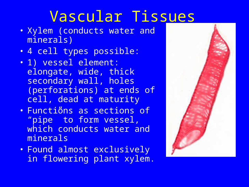
Vascular Tissues• Xylem (conducts water and
minerals)• 4 cell types possible:• 1) vessel element: elongate,
wide, thick secondary wall, holes (perforations) at ends of cell, dead at maturity
• Functions as sections of “pipe” to form vessel, which conducts water and minerals
• Found almost exclusively in flowering plant xylem.

Vascular Tissues• Xylem (conducts water and
minerals)• 4 cell types possible:• 2) tracheid: elongate, wide, thick
secondary wall, many pits in wall• Functions to conduct water and
minerals also. These must pass through pits to reach next cell (pits have primary wall in center and are not open holes)
• Found in xylem of both flowering plants and gymnosperms.
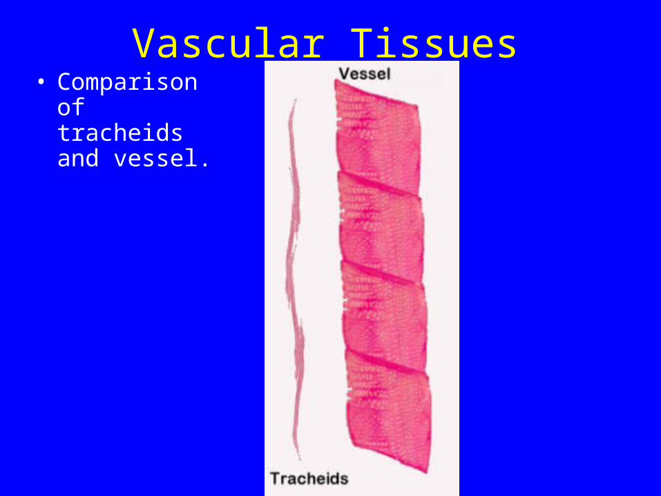
Vascular Tissues• Comparison of
tracheids and vessel.

Vascular Tissues• Comparison of
tracheids and vessel.
Vascular Tissues• Xylem (conducts water and
minerals)• 4 cell types possible:• 3) fibers: covered earlier.
Note they have secondary wall and are usually dead at maturity
• 4) parenchyma: covered earlier. Note they have primary walls and are alive.

Plant Organs• 1) Flower (covered
previously)• 2) Stem• 3) Leaf• 4) Root.

Plant Organs• 2) Stem:
aboveground portion of shoot, bearing leaves
• Divided into nodes (where leaves attach) and internodes (where no leaves attached)
• Note also: bundle scar, leaf scar, terminal bud scale scar.
Fig. 38.23

Plant Organs• 2) Special stem
modifications– Photosynthesis (cactus)
– Climbing (some tendrils).

Plant Organs• 2) Special stem
modifications– Protection (thorn).
Honey locust (Gleditsia)

Plant Organs• 2) Special stem
modifications– Storage (tuber). Also
asexual reproduction
– Explore world (runner). Also asexual reproduction.

Plant Organs• 2) Special stem
modifications– Underground stems
(bulb, rhizome).
Bulb (A) and corm (B)

Plant Organs• 3) leaf
– major site of photosynthesis
– stalk is petiole, flattened part is blade
– axillary bud: small meristem (contains a SAM) at base of leaf: can grow to make branch.

Plant Organs• 3) leaf
– note stipules: pairs of appendages at base of petiole of some leaves.

Plant Organs• 3) leaf
– Venation: netted for dicots, parallel for monocots.

Plant Organs• 3) leaf
– Simple– Compound (pinnate
and palmate, doubly compound).

Plant Organs• 3) leaf
– Arrangement.

Plant Organs• 3) Special leaf
modifications– Carnivory! (obtain nutrients)– Venus flytrap: leaf like steel
trap (leg-hold trap).
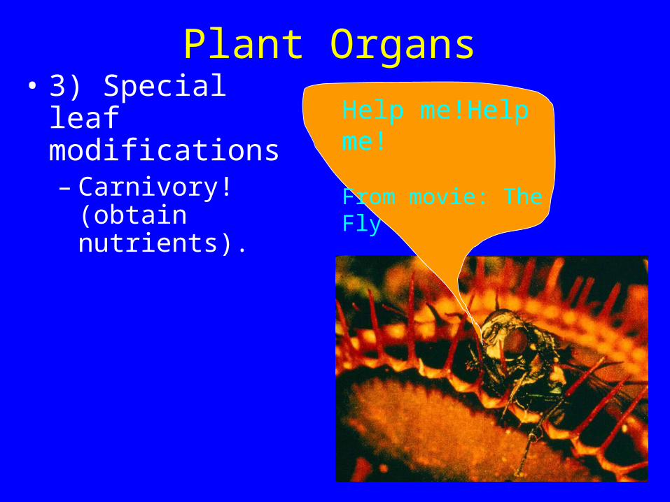
Plant Organs• 3) Special leaf
modifications– Carnivory! (obtain
nutrients).
Help me!Help me!
From movie: The Fly

Plant Organs• 3) Special leaf
modifications– Carnivory! (obtain
nutrients)– Pitcher plants (leaf
acts as pitfall trap).
white-top pitcher plant

Plant Organs• 3) Special leaf
modifications– Carnivory!
(obtain nutrients)– Sundew: uses
sticky hairs to catch prey on leaf.

Plant Organs• 3) Special leaf
modifications– Asexual
reproduction (ex, plantlets).
Plantlets on Kalanchoe leaves

Plant Organs• 3) Special leaf
modifications– Pollinator
attraction (bracts).

Plant Organs• 3) Special leaf
modifications– water storage:
succulent leaf.
Lithops: stone plant

Plant Organs• 4) Root
– anchors plant– absorbs
water/nutrients.
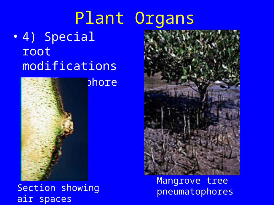
Plant Organs• 4) Special root
modifications– Pneumatophores.
Mangrove treepneumatophoresSection showing
air spaces
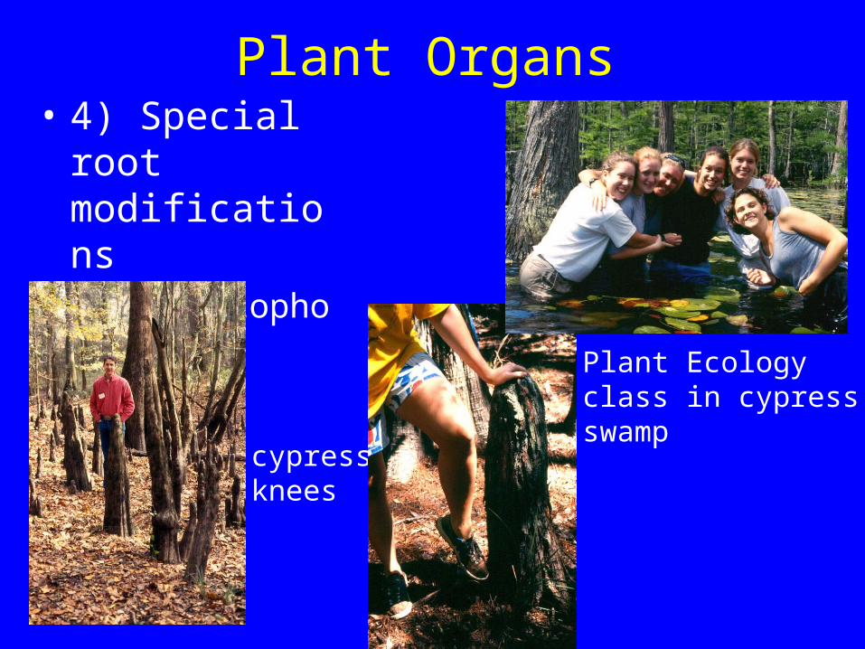
Plant Organs• 4) Special root
modifications– Pneumatophores– Cypress knees.
cypressknees
Plant Ecologyclass in cypress swamp

Plant Organs• 4) Special root
modifications– Contractile roots

Plant Organs• 4) Special root
modifications– Parasitic roots
(haustoria)
Leafy mistletoe (parasite on trees)
tree branch

Plant Organs• 4) Special root
modifications– Storage roots
– Ex, carrot

Plant Organs• 4) Special root
modifications– Climbing (aerial
roots)

Root anatomy

Root anatomy• At tip: apical
meristem covered by root cap
• Protects RAM from abrasion by soil particles.

Root anatomy• Next: primary
meristems– protoderm:
becomes epidermis– procambium:
becomes vascular tissue
– ground meristem: becomes rest of tissue (region called cortex).

Root anatomy• Mature root tissues
– Epidermis: root hairs
– Cortex (region): parenchyma tissue (storage)
• endodermis

Root anatomy• Mature root tissues
– Cortex (region): parenchyma tissue (storage)
• endodermis: cells with Casparian strips (waxy material called suberin: blocks water from going thru walls).

Root anatomy• Mature root tissues
– Vascular cylinder• pericycle• phloem and xylem

Root anatomy• Lateral roots
– develop from pericycle.

Stem anatomy

Stem anatomy• SAM at tip
• Protected by developing leaves called primordia (-ium).
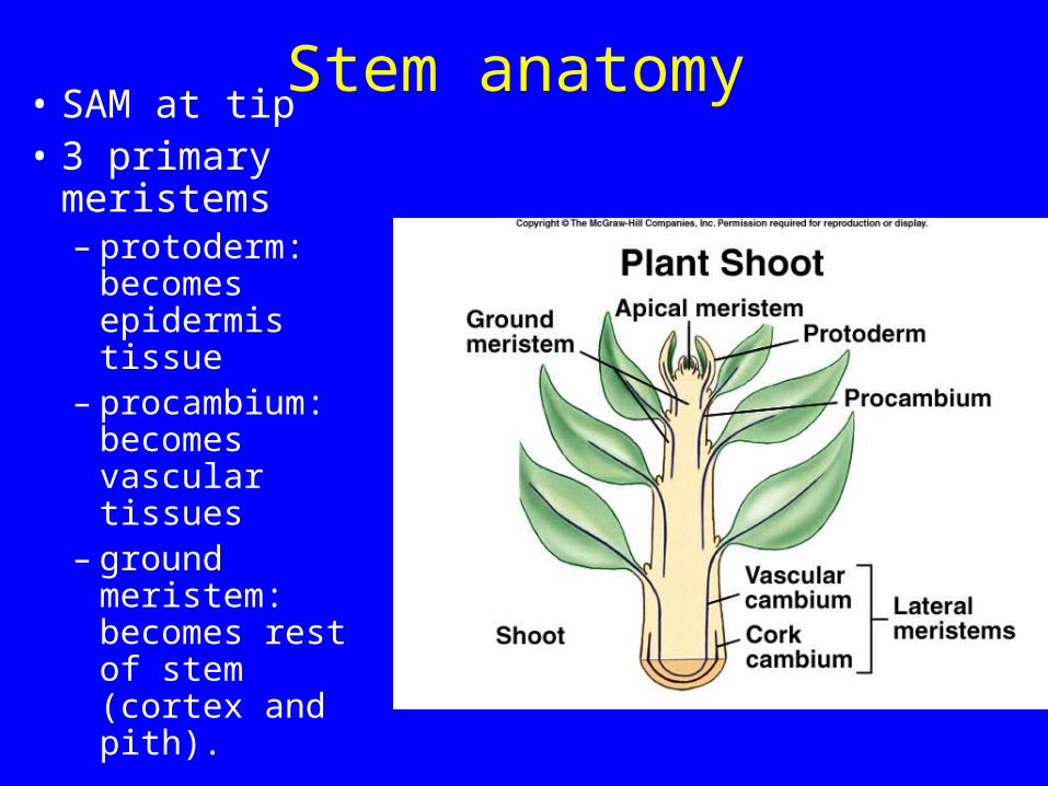
Stem anatomy• SAM at tip• 3 primary
meristems– protoderm:
becomes epidermis tissue
– procambium: becomes vascular tissues
– ground meristem: becomes rest of stem (cortex and pith).

Stem anatomy• Epidermis on
outside• Vascular tissue
in bundles • Note regions:
– cortex– pith
(parenchyma).

Stem anatomy• Epidermis• Cortex• Vascular bundle.
Helianthus stem

Stem anatomy• Cortex closeup: note
collenchyma here. Also parenchyma deeper.
Helianthus stem

Stem anatomy• Vascular bundle
close
• Note phloem on outside, xylem on inside.

Leaf anatomy• Leaves start as
outgrowths from apical meristem: leaf primordia.

Leaf anatomy• 3 primary
meristems– protoderm:
becomes epidermis tissue
– procambium: becomes vascular tissues
– ground meristem: becomes rest of leaf.

Leaf anatomy• Epidermis:
note cuticle, stomata
• Veins with vascular tissues (xylem on top, phloem on bottom)
• Supply water & nutrients, remove sugars for transport elsewhere.

Leaf anatomy• Mesophyll
– Parenchyma tissue layers (palisade and spongy: do photosynthesis.

Plant Growth Phenomena• Hormones: molecules produced in small
amounts that change plant physiology or growth
• Can inhibit or stimulate processes to occur
• 5 major types: auxins, cytokinins, gibberellins, ethylene, abscisic acid

Auxins• Promote stem
elongation and growth
• Example, phototropism. Bending of stem toward light

Auxins• Also involved in apical dominance: suppression
of lateral meristems by apical meristem

Cytokinins• Stimulate cell division where auxin is
also present
• Acts as anti-aging hormone (keeps detached leaves green).

Gibberellins• Promote stem elongation
• Mutant plants with low amounts are dwarfs (internode lengths short)

Ethylene• Promotes fruit ripening
• Stimulates abscission (dropping) of leaves, flowers

Abscisic acid• Induces formation of
winter buds (bud scales, dormant meristem)
• Involved in opening and closing of stomata
• Can cause seed dormancy

Plant transport• Phloem: sugars and water (often from leaf to root)
• Xylem: water and minerals from root to shoot
• Movement driven by water potential: measure of tendency of water to move from one place to another
• Affected by solutes (high solutes low tendency to move), pressure (high pressure high tendency to move), tension (pull: high tension high tendency to move).

Water transport• Xylem: water and minerals from root to shoot
• How much of water remains in plant?

Water transport• Transpiration: evaporation of water from leaves
• Driven by pull from leaves. Water under tension. Water potential high in soil and low in air.

Water transport• Transpiration greatly controlled by stomata
• Stomata open in light but can close if plant lacks sufficient water.
Stomata!

Sugar transport• Phloem: sugars and water
• Flow from source to sink
• Pressure flow mechanism

Sugar transport• Source: lots of sugar
dissolved in water. Generates pressure as water flows in to dilute sugar
• Sink: little sugar dissolved in water. Low pressure as water flows out
• Creates pressure gradient that moves fluid thru sieve tubes.

Sugar transport• Result: sugar flows to
wherever demand is high