newsletter of the friends - harvard university
TRANSCRIPT
Newsletter of the FRIENDS
OF THE
FARLOW
Number 54 Fall 2009 K. Griffith, Editor
FoF Annual Meeting
Saturday, November 7. See page 7.
Cemetery Lichens: Study Collections for Beginners
Elizabeth J. Kneiper, FoF President
Specimens, both historic and current, are the foundation of the study of cryptogams. Herbaria, such as the Farlow Herbarium of Cryptogamic Botany, exchange specimens around the globe with specialists doing research. These collections generate data that increase our understanding of the biology and ecology of cryptogams, support taxonomic revisions, and document current and historic species ranges. The significance of the specimens held by the Farlow Herbarium can be seen by the frequency of their citation in the literature.
Because of the importance of the
Farlow’s collections, the Friends of the Farlow offers both a Graduate Student Fellowship and the Harvey Pofcher Visiting Scholars Fellowship for specialists to work at the Farlow. Researchers who work on specimens learned to do so by exposure to herbaria. Collections are invaluable in research and as teaching tools for students with research careers in mind, and for naturalists who want to learn more about cryptogams.
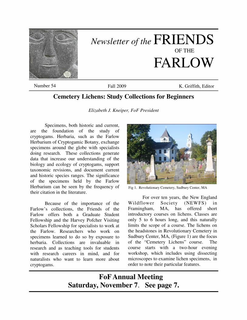
For over ten years, the New England Wildflower Society (NEWFS) in Framingham, MA, has offered short introductory courses on lichens. Classes are only 5 to 6 hours long, and this naturally limits the scope of a course. The lichens on the headstones in Revolutionary Cemetery in Sudbury Center, MA, (Figure 1) are the focus of the “Cemetery Lichens” course. The course starts with a two-hour evening workshop, which includes using dissecting microscopes to examine lichen specimens, in order to note their particular features.
Fig 1. Revolutionary Cemetery, Sudbury Center, MA

2
Chemical spot tests are used to confirm the presence of particular compounds, and keys are used to identify species. The subsequent three-hour site visit reinforces the class work and expands on the discussion of the natural history of lichens with special focus on issues that pertain to the lichens in the field site. What is done in the introductory session is tailored to the field trip experience; what happens in the field trip reinforces what was covered in class. Course participants have noticed lichens in the field and are generally aware of forms and colors of lichens. They know that lichens occur in different quantities in different habitats. Some know the common names of a handful of macrolichens. But only a few are familiar with the life cycles of fungi, much less lichens. Hardly any of the participants have examined lichens up close, using a hand lens or a dissecting microscope, and therefore they do not yet know how to go about studying them.
As with other specialized fields, lichenology has its own descriptive language without which the understanding of the field is superficial at best. A short introductory course has to be a rapid-paced “lichen language” course that teaches simultaneously the basic descriptive language and the biology of lichens. Teaching from collections makes lichen language acquisition less difficult. The students work on collections of the same species at the same time so that discussions around the samples reinforce the use of lichen terminology in class.
The need for the descriptive
terminology used in field guides becomes obvious once different features of a lichen are studied, but a list of 20-25 unfamiliar terms can overwhelm beginners. It is important to remind students that field guides, such as Lichens of North America (Brodo, Sharnoff and Sharnoff 2001) and The Macrolichens of
New England (Hinds and Hinds 2007), have glossaries that define the terms used in keys and to point out that glossaries are posted on the web (Selva). Students are encouraged to use the Internet as an invaluable resource for follow-up work on lichens.
Most lichen-forming fungi are
Ascomycetes that form a nutritive association with a photosynthetic alga or a cyanobacterium or both. The name of a lichen however, is the name of the fungus in the association. Species lists with both Latin names and vernacular names are good references for class work. Latin names are less user-friendly than vernacular names. The common names for lichens are included in Lichens of North America (Brodo, Scharnoff and Scharnoff 2001) and Simplified Field Key
to Maine Macrolichens (Hinds and Hinds 2007). Students are made aware of the online checklist of North American lichen-forming fungi (Esslinger 2009).
Lichen colors are best understood by
comparing fresh collections of species of different colors. The varying morphologies of a thallus (lichen body) are appreciated more readily when actually seen on a thallus. When viewed this way, the functions of structures are easier to relate to lichen biology and ecology. Structures are obviously easier to see when they are magnified under the dissecting scope than with a hand lens in the field.
Recognition of features in the field is
improved if specimens are studied under magnification before the site visit. By working on collections students learn that examining lichens is a form of multitasking that requires close attention to details, making sections, and doing spot tests. Phil May’s (another FoF member) web site “How to Identify a Macrolichen” is an excellent reference that outlines the process of studying a macrolichen collection (May).
3
Morphology is central to the
classification of lichens. Examining specimens helps beginners recognize that a lichen thallus changes as it grows and matures, and that a thallus consists of a mix of dissimilar older and younger regions. The form of a young lichen thallus can differ from that of an older thallus.
Terms used in keys such as “becoming” or “developing” refer to changes in thallus growth form and to the development of new features on the thallus. In Sudbury’s Revolutionary Cemetery, encrusting species on the headstones exist as isolated thalli; others grow intermixed with other species; and some form large irregular colonies. Appreciating how growth patterns account for visible forms on a thallus connects morphology to growth and solidifies the concept that lichens undergo change.
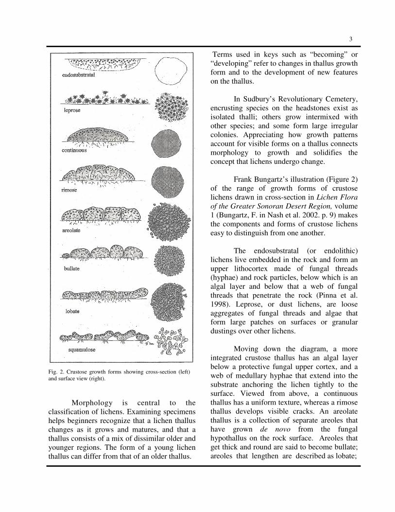
Frank Bungartz’s illustration (Figure 2) of the range of growth forms of crustose lichens drawn in cross-section in Lichen Flora
of the Greater Sonoran Desert Region, volume 1 (Bungartz, F. in Nash et al. 2002. p. 9) makes the components and forms of crustose lichens easy to distinguish from one another.
The endosubstratal (or endolithic) lichens live embedded in the rock and form an upper lithocortex made of fungal threads (hyphae) and rock particles, below which is an algal layer and below that a web of fungal threads that penetrate the rock (Pinna et al. 1998). Leprose, or dust lichens, are loose aggregates of fungal threads and algae that form large patches on surfaces or granular dustings over other lichens. Moving down the diagram, a more integrated crustose thallus has an algal layer below a protective fungal upper cortex, and a web of medullary hyphae that extend into the substrate anchoring the lichen tightly to the surface. Viewed from above, a continuous thallus has a uniform texture, whereas a rimose thallus develops visible cracks. An areolate thallus is a collection of separate areoles that have grown de novo from the fungal hypothallus on the rock surface. Areoles that get thick and round are said to become bullate; areoles that lengthen are described as lobate;
Fig. 2. Crustose growth forms showing cross-section (left) and surface view (right).
4
and those that pull off the surface but remain attached to the surface by fungal threads (rhizoidhyphen) are referred to as squamulose.
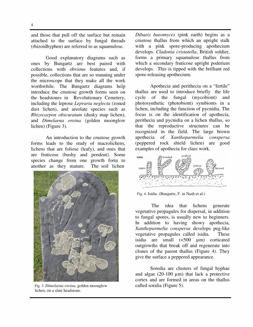
Good explanatory diagrams such as ones by Bungartz are best paired with collections with obvious features and, if possible, collections that are so stunning under the microscope that they make all the work worthwhile. The Bungartz diagrams help introduce the crustose growth forms seen on the headstones in Revolutionary Cemetery, including the leprose Lepraria neglecta (zoned dust lichen), and areolate species such as Rhizocarpon obscuratum (dusky map lichen), and Dimelaena oreina (golden moonglow lichen) (Figure 3).
An introduction to the crustose growth
forms leads to the study of macrolichens, lichens that are foliose (leafy), and ones that are fruticose (bushy and pendent). Some species change from one growth form to another as they mature. The soil lichen
Dibaeis baeomyces (pink earth) begins as a crustose thallus from which an upright stalk with a pink spore-producing apothecium develops. Cladonia cristatella, British soldier, forms a primary squamulose thallus from which a secondary fruticose upright podetium develops. This is tipped with the brilliant red spore-releasing apothecium.
Apothecia and perithecia on a “fertile”
thallus are used to introduce briefly the life cycle of the fungal (mycobiont) and photosynthetic (photobiont) symbionts in a lichen, including the function of pycnidia. The focus is on the identification of apothecia, perithecia and pycnidia on a lichen thallus, so that the reproductive structures can be recognized in the field. The large brown apothecia of Xanthoparmelia conspersa (peppered rock shield lichen) are good examples of apothecia for class work.
The idea that lichens generate vegetative propagules for dispersal, in addition to fungal spores, is usually new to beginners. In addition to having showy apothecia, Xanthoparmelia conspersa develops peg-like vegetative propagules called isidia. These isidia are small (<500 µm) corticated outgrowths that break off and regenerate into clones of the parent thallus (Figure 4). They give the surface a peppered appearance.
Soredia are clusters of fungal hyphae and algae (20-100 µm) that lack a protective cortex and are formed in areas on the thallus called soralia (Figure 5).
Fig. 4. Isidia. (Bungartz, F. in Nash et al.)
Fig. 3. Dimelaena oreina, golden moonglow lichen, on a slate headstone.

5
Learning to differentiate the vegetative propagules from one another on a thallus takes practice. Keying out collections of assorted common species using the Simplified Field
Key to Maine Macrolichens (Hinds and Hinds 1998) provides the needed practice for distinguishing isidiate from sorediate lichens and the practice for using lichen colors, growth forms, thallus sizes, reproductive strategies, and substrate preferences to write descriptions of the lichens that are identified.
Revolutionary Cemetery in Sudbury Center, MA, is an historic New England cemetery with slate and marble headstones and monuments. Granite is restricted to the bases of marble headstones and monuments, partially buried boulders, and the rock walls that surround the cemetery. The headstones and monuments are arranged in rows that run north to south. The front of the tombstones face westward, toward Presbyterian Church, which the cemetery abuts. The southwestern corner of the cemetery is partially shaded by trees.
Living lichens on headstones are
specimens that allow the class to see the importance of microhabitat conditions and rock composition to the establishment and growth of lichens. By comparing the lichen assemblages on slate and marble headstones, the students get to practice distinguishing different lichen species and different stages of growth of individual species and to note patterns of dispersal of lichens on the headstones. The fieldwork builds on what was covered in class.
A field trip to a cemetery introduces the r e a l - l i f e c o n t r o v e r s y between the conservation of lichens and the conservation of h i s t o r i c a l monuments. All the monuments and headstones in Revolutionary Cemetery are
weathered. None have been protected as the Girl Under Glass in the Forest Hills Cemetery in Boston has been (Figure 6). Rain, airborne pollutants, and particulates stain and weather rocks. Colonizing bacteria, cyanobacteria, non-lichenized fungi, and lichens form biofilms thought to weather rocks. Lichens can be pioneering colonizers on rock and are known to play a role in the biogeochemical breakdown of rock by fragmentation, chemical alteration, and dissolution of minerals in the rock. How, at what rate, under what conditions, and by what agent each rock type is weathered is an interdisciplinary field of
Fig. 6. The Girl Under Glass. Forest Hills Cemetery, Boston, MA.
Fig. 7. Polsporina simplex, common coal-dust lichen. Black apothecia are lifting rock flakes.
Fig. 5. Soredia. (Bungartz, F. in Nash et al. 2002. )

6
research with an enormous body of literature (Piervittori, et al. 1994, 1996, 1998, 2004). A close examination of Acarospora
fuscata (brown cobblestone lichen) colonizing the roughly cut backs of the older slate headstones in Revolutionary Cemetery shows flaking of the slate caused by the growth and swelling of wet areoles. Apothecia of the endolithic Polsporina simplex (common coal-dust lichen) flakes off rock as they break through the surface of the rock (Figure 7). On the other hand, the foliose lichen Physciella
melanchra (cryptic rosette lichen) forms a protective blanket over marble. Where a thallus separates from the surface, a window of clean marble is left surrounded by gray weathered marble. In Revolutionary Cemetery the widespread colonization of slate by Dimelaena
oreina forms chromatic changes on the slate headstones, altering their original appearance. Inscriptions on headstones seem to disappear under lichens and the information inscribed on the stone is viewed by historians and genealogists as having been lost. Conservators refer to the biofilms that include lichens as “tough biologicals” that need to be removed from monuments. Dimelaena oreina, growing on the slate face in the upper left corner of the first page as well as in Figure 3, is an example of a “tough biological”.
T h e S u d b u r y C o n s e r v a t i o n
Commission has had 100 of the headstones in Revolutionary Cemetery cleaned. This cleaning involves wetting the headstone, rubbing the surface with a soft brush and applying Simple Green, a biocide, that kills the remaining lichens (Kai Nalenz, owner of Gravestone Services of New England, personal communication). The eroded and dulled patina of the cleaned headstones is attributed in part to lichen erosion of the rock surfaces, which is evident on the slate face shown on page one.
The Living Churchyard Project of the British Lichen Society is committed to lichen conservation. As far as they are concerned, churchyards should be viewed as undisturbed refugia for lichens. After all, 300 of the 1700 lichen species in the flora of Great Britain are cemetery lichens records (Churchyard Lichens). The British Lichen Society suggests this compromise between monument conservationists and lichen conservationists: whenever tombstones and monuments are scheduled to be cleaned, enlist a lichenologist to determine whether a rare species is at risk. If rare species are found, the lichens should be spared.
In New England, poor air quality and
habitat destruction have had a negative impact on lichen flora diversity. Could a rare lichen on a cemetery headstone in our area receive the same protection and have the same public draw that headstones of famous authors or colonists do? As more field guides and internet resources popularize lichenology and as more naturalists are committed to learning about the lichens they see in the field, by taking courses such as “Cemetery Lichens,” we hope conservation of living lichen collections in cemeteries and in other habitats will become a reality.
Acknowledgments
Cecily Miller, Forest Hills Educational
Trust, Forest Hills Cemetery, Boston, provided the image of the Girl Under Glass.
Tom Nash III and Frank Bungartz
generously allowed the FoF to use illustrations from the Lichen Flora of the Greater Sonoran
Desert Region, volume 1. Kai Nalenz, of Gravestone Service of New
England, kindly provided information on gravestone cleaning.
Thank you all.

7
References Brodo, I. M., S. D. Sharnoff, & S. Sharnoff. 2001. Lichens of North America. Yale University Press, New Haven, CT, 795 pp. Bungartz, F. 2002. Introduction, pp 1–43. In T. H. Nash, III, B. D. Ryan, C. Gries, & F. Bungartz (eds.). Lichen Flora of the Greater Sonoran Desert Region, volume 1. Lichens Unlimited, Arizona State University, Tempe. Dobson, F. S. 1992. Lichens. An Illustrated Guide to the British and Irish Species. Richmond Publishing. 3rd edition. Esslinger, T. L. 2009. A cumulative checklist for the lichen-forming, lichenicolous and allied fungi of the continental United States and Canada. North Dakota State University: http://www.ndsu.nodak.edu/instruct/esslinge/chcklst/ chcklst7.htm (First Posted 1 December 1997, Most Recent Version (#15) 27 August 2009), Fargo, North Dakota. Churchyard Lichens On British Lichen Society website: http://www.thebls.org.uk/contents/chlich.html Hinds, J. W. and P. L. Hinds. 2007. The Macrolichens of New England. The New York Botanical Garden Press. 584 pp. Hinds, P. L. & J.W. Hinds. 1998. Simplified Field Key to Maine Macrolichens, published and distributed by the authors. 48 pp. May, P. F. How to Identify a Macrolichen http://www.huh.harvard.edu/collections/lichens/howto.html New England Wildflower Society. http://www.newfs.org/ Piervittori, R., O. Salvadori & A. Laccisaglia. 1994. Literature on Lichens and Biodeterioration of Stonework I. Lichenologist 26(2):171-192. Piervittori, R., O. Salvadori & A. Laccisaglia. 1996. Literature on Lichens and Biodeterioration of Stonework II. Lichenologist 28: 471-483. Piervittori, R., O. Salvadori & A. D. Isocrono. 1998. Literature on Lichens and Biodeterioration of Stonework III. Lichenologist 30(3):263-277. Piervittori, R., O. Salvadori & A. D. Isocrono. 2004. Literature on Lichens and Biodeterioration of Stonework IV. Lichenologist 36(2):145-157. Pinna, D., O Salvadori and M. Tretiach. 1998. An anatomical investigation of calcicolous endolithic lichens from the Trieste karst (NE Italy). Plant Biosystems,132 (3) 183-195. Selva, S. B. Lichen Terminology http://csdept.umfk.maine.edu/lichensWEbsite/glossary.asp
FoF Annual Meeting The FoF Annual Meeting will be held on Saturday, November 7, with the business meeting beginning at 3:30 p.m.
At 4:00 p.m. our speaker, Dr.
Michaela Schmull, Farlow Research and Curatorial Associate, will talk about “Lichens
- what are they, what are their relatives, and
what are they good for.” Her lecture will give an overview of the partners that are involved in forming lichens, how lichens appear when they are being examined closely, and how they are related to other organisms. Dr. Schmull will also talk about the important (but often unknown) role lichens play in our lives, in human culture and economics. All are welcome to attend all or part of the meeting. Members are encouraged to bring their friends. A reception will take place in the Farlow Library Reading Room following the meeting. There are three new exhibits in the Farlow and Botany Libraries curated by Botany Libriaries’ archivist Lisa DeCesare: The Young Botanist: Juvenile Literature in
the Collections of the Botany Libraries; Witches or Wise Women: Plant Lore of
Early Female Herbalists; and Dying with
Cryptogams. Those attending the Annual Meeting will have an opportunity to enjoy some of these exhibits during the reception.

8
Fragile Science
Text and photos by Tessa Updike
Botanical specimens are inherently
fragile; they are composed of once-living matter doomed to wilt, dry, and crumble to dust. The specimens deposited in herbaria are housed there to be preserved, their characters suspended indefinitely in time. Many precautions are taken to insure the longest preservation with the best conditions possible for each incoming specimen. But despite the care and devotion from the curators of herbaria, there are destructive elements waiting around each bend.
Specimens are sent to herbaria from all over the world. They arrive in boxes well worn from travel, the specimens within wrapped in newspaper or stored temporarily within commercial containers and bags. The packaging from the field is sometimes nearly as interesting as the specimens themselves. Some come in exotic bags and wrappings, with
moth balls sometimes s c a t t e r e d i n s i d e among the specimens to deter insects from eating them in transit.
Time is an element we cannot control. As the years pass, the acids in the paper and glue of older labels become brittle and dry. They will discolor so that labels cannot be read, they will crack and fall to the floor or to the bottom of a drawer, or will be lost in the crevice of a shelf. With the loss of these labels, often the identity and provenance of a specimen is gone. Without these identifying signs, they are simply objects.
Temperature and humidity control in herbaria are vital to the survival of the specimens. High temperature and high humidity will invite mold to grow, insects to swarm and thrive, and specimens to deteriorate more quickly. Low humidity will dry paper and specimens, making them brittle. Fluctuations in temperature and humidity cause further damage to the specimens. The comfort level for humans differs greatly from that of botanical specimens.
This label has become discolored, dry and broken with age.
The label on this piece of fossilized wood has become unreadable.
A newly-arrived specimen, still in its packaging.

9
The cabinets in which the specimens are housed have changed over the years. Older cabinets, made of wood or metal, were not airtight and allowed insects to enter, as well as allowing light, temperature, and humidity to damage the specimens. More modern cabinets are airtight and better protect specimens from harmful particles in the air, as well as from fluctuating temperature and humidity. Throughout the years there have been changes in technologies used to control insects and pests in herbaria. Fumigants were used in the past to protect specimens from insects. Many of these fumigants are now banned from use because they are known to be carcinogenic or otherwise harmful to people and the environment.
One such fumigant formerly used by herbaria was methyl bromide, which is now known to cause ozone depletion and is banned by the Montreal Protocol. Not only are many of the formerly used fumigants harmful to the people who used them, but over time the chemicals have burned through both paper and specimens. Another poison that was regularly used was arsenic, which was applied to help kill insects and pests. While arsenic is no longer actively used, specimens that were treated with it remain in herbaria today. Currently one of the best practices for killing insects and their eggs is to freeze specimens. All specimens arriving at the herbaria are frozen before coming into contact with other specimens. Cabinets need to be periodically checked, and, as appropriate, specimens refrozen. The old fumigation room at the Harvard University Herbaria, which still bears a Danger sign, will eventually become the new freezing room … replacing an outdated practice with a newer method.
Despite the best efforts and care of collections staff to prevent insect infestation and damage, most herbaria will have examples of past damage. The most common and potentially dangerous beetles have been Lasioderma serricorne (Cigarette Beetle), Anthrenus verbasci (Varied Carpet Beetle), Stegobium paniceum (Drugstore Beetle) and Trogoderma spp. (Warehouse Beetle).
Damage from silverfish, cockroaches and book lice often comes in the form of damaged paper, book spines and labels. The glue used on the backs of labels, in addition to the paper itself, is the desired meal and little may be left behind besides scraps and frass. The larvae of these beetles, however, do not go after labels and paper, but the actual dried plant material. Vigilance is needed in the fight against insects. Freezing must be calibrated to kill both adult insects and their eggs.
Modern cabinets are now used to protect specimens.
Something has been eating this specimen.
10
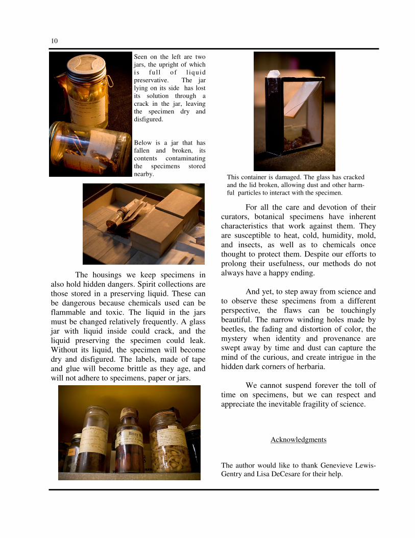
Seen on the left are two jars, the upright of which i s ful l o f l iquid preservative. The jar lying on its side has lost its solution through a crack in the jar, leaving the specimen dry and disfigured. Below is a jar that has fallen and broken, its contents contaminating the specimens stored nearby.
The housings we keep specimens in also hold hidden dangers. Spirit collections are those stored in a preserving liquid. These can be dangerous because chemicals used can be flammable and toxic. The liquid in the jars must be changed relatively frequently. A glass jar with liquid inside could crack, and the liquid preserving the specimen could leak. Without its liquid, the specimen will become dry and disfigured. The labels, made of tape and glue will become brittle as they age, and will not adhere to specimens, paper or jars.
For all the care and devotion of their curators, botanical specimens have inherent characteristics that work against them. They are susceptible to heat, cold, humidity, mold, and insects, as well as to chemicals once thought to protect them. Despite our efforts to prolong their usefulness, our methods do not always have a happy ending. And yet, to step away from science and to observe these specimens from a different perspective, the flaws can be touchingly beautiful. The narrow winding holes made by beetles, the fading and distortion of color, the mystery when identity and provenance are swept away by time and dust can capture the mind of the curious, and create intrigue in the hidden dark corners of herbaria. We cannot suspend forever the toll of time on specimens, but we can respect and appreciate the inevitable fragility of science.
Acknowledgments
The author would like to thank Genevieve Lewis-Gentry and Lisa DeCesare for their help.
This container is damaged. The glass has cracked and the lid broken, allowing dust and other harm-ful particles to interact with the specimen.

11
News from the Farlow
Matthew E. Smith, a post-doctoral fellow in the Farlow for two years, has moved on to Duke University where he has begun a National Science Foundation sponsored research project on mycorrhizal fungi from areas of Guiana. His paper, with D. H. Pfister, on tuberculate ectomycorrhizae on oaks in the Boston area and Mexico was published in American Journal of Botany (96: 1665-1675. 2009) earlier this year. Also leaving us at the end of September to return to the Kunming Institute of Botany in Kunming, China was Zai-wei Ge. Zai-wei was here for a year and worked on lepiotoid fungi and also on Cudonia and Spathularia (Rhytismataceae) from China. Michaela Schmull, Farlow Research and Curatorial Associate, attended a workshop on Molecular Evolution in Woods Hole, MA, from July 26th to August 7th which was partially funded by the Friends of the Farlow. She increased her knowledge of data analysis and interpretation through lectures, demonstrations, and computer laboratories which will assist her in understanding species concepts and the clarification of uncertain species.
Rosanne Healy, a graduate student at the University of Minnesota, was here as a Farlow Graduate Student Fellow for two weeks in May. She worked with Matt Smith and Don
Pfister on Pachyphoeus and related hypogeous ascomycetes. Jesus Hernandez of the Fundación Instituto Botánico de Venezuela spent a week looking at specimens of the lichen family Graphidaceae from Venezuela, part of his Ph. D. thesis work. Young-Joon Choi from Seoul, South Korea, recently arrived for a year’s stay. He will be working on Pezizales with Don Pfister. He is supported by a fellowship from the Science Foundation of Korea. Professor Gier Hestmark from the University of Oslo, Norway, visited the Farlow from October 5th to October 7th to work on Umbilicaria lichens in the general, Tuckerman and Dodge herbaria. Friends of the Farlow brought Frank
Bungartz and Franke Ziemmeck to the Farlow under the Harvey Pofcher Fellowship fund. Bungartz, who is stationed at the Charles Darwin Research Station in the Galapagos Islands, is compiling a checklist of the lichens that occur in the islands. The Farlow Herbarium contains a number of early and more recent collections from the Galapagos. During his stay Bungartz was able to locate and confirm or re-identify over 350 specimens from the general collection as well as from the herbarium of Edward Tuckerman. Thanks to the work of Michaela Schmull
these have now been entered in the Harvard University Herbaria specimen database. Other visitors to the Herbarium during the past 6 months have included: Terry Henkel, Humboldt State University, Susan Brawley, University of Maine, and Lee Crane, Illinois Natural History Survey.
Zai-wei Ge and his wife Qingying Zhang

Join us ! Receive the FOF Newsletter, notification of the annual book sale, discount on Farlow publications and services, invitations to the annual meeting and other events, and a special welcome when visiting the Farlow. Name _____________________________________________ Address ___________________________________________ City, State, Zip/Postal Code ___________________________ Country ___________________________________________ Telephone / Fax _____________________________________ E-mail address ______________________________________
Visit the Friends of the Farlow Website today!
www.huh.harvard.edu/collections/fof/fof.html
Membership Categories Associate Member ……..($10-15) Full Member ………….. ($25) Sponsor …………..…….($50-100) Benefactor …………..….($1000) Amount enclosed $____________
Please make checks payable to: Friends of the Farlow Applications should be sent to: Friends of the Farlow, Harvard University Herbaria 22 Divinity Avenue, Cambridge, MA 02138 USA
FRIENDS of the FARLOW
FA
RL
OW
R
EF
ER
EN
CE
L
IBR
AR
Y
OF CRYPTOGAMIC
BO
TA
NY
* HA
RV
AR
D U
NIV
ER
SIT
Y
22 Divinity Avenue, Cambridge, MA 02138 USA www.huh.harvard.edu/collections/fof/fof.html
F I R S T C
L A S S