news & features clinical center 25th anniversary - office of nih
TRANSCRIPT
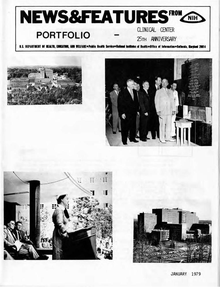
NEWS&FEATURESFR0~ CLINICAL CENTER
PORTFOLIO 25TI-i ANNIVERSARY . U. HPHTIHT If IOI.Tl, a.arm. m IUHll•hMlc 1111111 Slnlce•llllllll llllllllts 11 lell••lfflu If l1ttrutill•h1Mda, ..,._. 21114 ·
JANUARY 1979

2
Marc Stern, Editor Thomas H. Flavin , Associate Editor NIH News Branch Building 31, Room 2B-10 Bethesda, MD 20014 Phone: (301) 496-1206 Designed by Joyae F. MaCarthy
PORTFOLIO: 25th ANNIVERSARY OF THE CLINICAL CENTER
1
3
2
4
COVER PHOTOS
(1) The Cliniaal Center in the final stages of ·aonstruation , (CP- 1)
(2) President Harry S. Truman, trowel in hand, prepares to lay t he aornerstone for t he Cliniaal Center on June 22, 1951 . The three men in the first row behind the President are (l . to r.) Dr . William H. SebreU, Jr., then NIH Direator; Contraator John MaShain; and Dr. Leonard A. Saheele, USPHS Surgeon General . <CP- 2)
(3) Mrs . OVeta Culp Hobby, Sear etary of Health, Eduaation, and Welfare, dedi aated the Cliniaal Center on July 2, 1953. Seated behind her are the Rev. Dr. Frederiak Brown Harris , ahaplain of the U.S. Senate; and Nelson A. Roakefeller, then Under Searetary of DREW. <CP- 3)
(4) The Cliniaal Center as it is today. <CP- 4)

3 Clinical Center
The Clinical Center, in the right upper quadrant of this aerial view, dominates the 306-n~~g National InstituteA of Health " · 1. rrus". <NIH Photo #CC- 1)
PORTFOI rn · ?5th ANNIVERSARY OF THE CLINICAL CE.NT ER
INTRODUCTION
A visiting foreign scientist working at the Clinical Center in this, its 25th year, was overheard to say, "What is hard to comprehend is that the amount of research done in this building in one week is equal to all the medical research done in my country in one year."
With over 2,300 employees devoted to biomedical exploration and clinical excellence, the volume of work done at the Clinical Center indeed makes it unique. But what makes it most special, perhaps is the setting in which that work is done. There are 541 hospital beds, with twice as much space devoted to laboratories as to patient care areas. This means that biochemists peering down microscopes at proteins isolated from diseased lungs can hear patients with emphysema coughing across the hallway. And neurophysiologists studying nerve axons in squids glance occasionally at television monitors taping epileptic seizures in infants. Pediatricians sit down to coffee in the cafeteria with molecular biologists and biostatisticians as children skim by in wheelchairs.
Ther e have been many research achievements made at the Clinical Center in the past twenty-five years . Its greatest achievement may have been simply to be here, to be the setting where the patients' presence, amid -so much scientific brilliance, prevented its becoming an ivory tower and made the Clinical Center instead a place of care and creativity.

4 Clinical Center
THE CLINICAL CENTER
Physically the Clinical Center is a 14-story red brick building, with each of twelve stories accommodating approximately twenty-five patients and one hundred laboratories. The Center was specially designed to bring patient care facilities close to research laboratories so that the new findings of basic and clinical scientists can be moved quickly from the labs to the treatment of patients.
Nearly one thousand physicians from the eleven NIH institutes conducting research at the Center provide the medical care. Nursing care and other support services are provided by Clinical Center staff.
Patients, upon referral by their physicians, are admitted to clinical studies conducted by the Institutes. Over six thousand patients are treated as fulltime admissions annually, and with the completion of the new addition in the early 1980's, the Clinical Center will accommodate over 300,000 outpatients visits.
The high quality of medical and support care required for clinical research at the Center have led to innovations that have influenced patient care throughout the nation. This portfolio will touch on but a few of them.

5 Clinical Center
CONQUERING HEPATITIS
The Dane pa:rticles and fi lamentous form of hepatitis B antigen a:re shOl.Vn in this photomicrograph magnified 150, 000 times . (NIH Photo #CC- 2)
Significant advances have been made in the control of post-transfusion hepatitis since the Clinical Center opened its doors in 1953. A large number of these advances are due to the Center's Blood Bank.
As early as 1957 the Blood Bank had published its first research paper delineating the post-transfusion hepatitis problem, firing the first salvo in what was to be a long but largely successful campaign.
In 1964 an antigen - the Australia antigen - was discovered, first in a blood sample collected from an aborigine and then in the blood of leukemia patients by Blood Bank Imm~nology Chief Dr. Harvey Alter and National Cancer Institute investigator Dr. Barruch Blumberg. Later Dr. Blumberg, who was then working in Philadelphia, linked the antigen with hepatitis. For this discovery he was awarded the Nobel Prize.
Paralleling this laboratory work was a clinical study conducted by the Blood Bank that was published in 1970. Patients undergoing open-heart surgery at the Clinical Center were divided into two groups, one that received commercial blood, the other group that received blood from volunteers who had donated at the Clinical Center or the Washington Red Cross Center. Within six months after the operation, over 50% of those receiving commercial blood developed hepatitis. No hepatitis developed in those receiving voluntary blood. With this concrete evidence in hand, the Clinical Center implemented a supply program that relied exclusively on voluntary blood provided by NIH employee donors and the Red Cross.

6Cl i nical Center
Subsequently Blood Bank Chief Dr. Paul Holland, working with other reseArchers in the Blood Bank, the National Institute of Allergy and Infectious Diseases, and the National Heart, Lung, and Blood Institute, demonstrated in another clinical study that transfused blood containing the antigen of ten results in patients getting hepatitis. These studies laid the foundation for the current FDA ruling that every blood unit be tested for the antigen - now called hepatitis B surface antigen (HBsAg) - before transfusion.
With the sole use of volunteer donors and hepatitis screening with a radioactive test for the hepatitis B antigen, it was hoped that the blood from the Blood Bank would soon be hepatitis free. This was not the case. Instead, 8% of the transfused patients continued to develop hepatitis after transfusions. This frustrating experience prompted the most recent and perhaps most important discovery made at the Blood Bank the identification of a new form of viral hepatitis, called non-A, non-B hepatitis. Dr. Alter and his colleagues have developed an animal model for this newly recognized disease. The Blood Bank researchers are currently attempting to develop a test to identify carriers of non-A, non-B hepatitis who may inadvertently transmit this disease to patients through their blood donations.
A Blood Bank teahniaian prepares blood samples for analysis . c
l
At a press aonferenae on Marah 27, 1978, Dr . Tabor of the FDA Bureau of Biologias and Dr. Alter of the CC Blood Bank announae that their independent s tudies have demonstrated for the f i r st time that a t ransmissible agent i s responsible f or non-A, non- B hepatitis.
7 Clinical Center
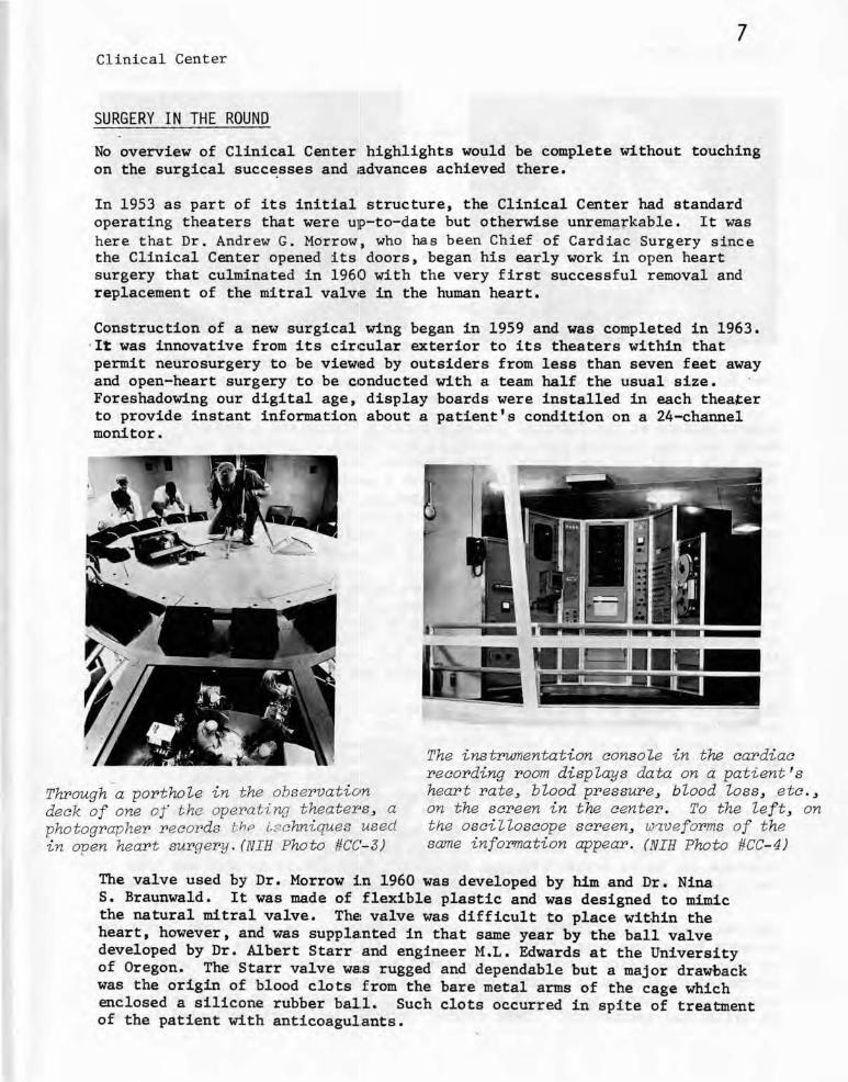
SURGERY IN THE ROUND
No overview of Clinical Center highlights would be complete without touching on the surgical successes and 1advances achieved there.
In 1953 as part of its initial structure, the Clinical Center had standard operating theaters that were u1p-to-date but otherwise unremarkable. It was here that Dr. Andrew G. Morrow, who has been Chief of Cardiac Surgery since the Clinical Center opened its doors, began his early work in open heart surgery that culminated in 196!0 with the very first successful removal and replacement of the mitral valv1e in the human heart.
Construction of a new surgical wing began in 1959 and was completed in 1963 . ·I't was innovative from its cir1cular exterior to its theaters within that permit neurosurgery to be vieW1ed by outsiders from less than seven feet away and open-heart surgery to be c•onducted with a team half the usual size. Foreshadowing our digital age, display boards were installed in each theater to provide instant information about a patient's condition on a 24-channel monitor.
··- l -~ I 11 _J
The instrumentation console in the cardiac recording room displays data on a patient ' s
Through a porthoie in the observation heart rate, blood pressure , biood ioss, eta. , deck of one of t he operating theater•s , a on the screen in the center. To the ieft, on photographer records t.hP l~chniques used t he osaiiloscope screen, w~veforms of t he in open heart surgery . (NIH Photo #CC- 3) same information appear. (NIH Photo #CC- 4)
The valve used by Dr. Morrow :f.n 1960 was developed by him and Dr. Nina S. Braunwald. It was made of flexible plastic and was designed to mimic the natural mitral valve. ThE! valve was difficult to place wit.bin the heart, however, and was supplalnted in that same year by the ball valve developed by Dr. Albert Starr and engineer M.L. Edwards at the University of Oregon. The Starr valve WBlS rugged and dependable but a major drawback was the origin of blood clots f rom the bare metal arms of the cage which enclosed a silicone rubber ball. Such clots occurred in spite of treatment of the patient with anticoagulants.

8Clinical Cent er
Hancock porcine xenograft . (NIB Photo #CC- 5)
Fabric- covered Starr- Eib.uards vaive . (NIH Photo #CC- 6)
Investigations at the Clinical Center's Surgery Branch and elsewhere found that the clotting problem could be alleviated by covering the cage arms with a teflon-type fabric. This improved design, the cloth-covered Starr-Edwards valve, was first implanted in a patient by Dr. Morrow, in the new Surgical Wing of the Clinical Center in February 1967. By December of 1968, more than one hundred patients had received the improved valve at the Clinical Center. Follow-up studies have since confirmed that the fabric covering indeed markedly reduces the clotting problems.
During the same period further research was being done on other suitable materials and sources for artificial valves. A promising development was a pig heart valve preserved and sterilized by glutaraldehyde and mounted on a flexible polypropylene frame or "stent." This design is known as the Hancock porcine xenograft, named after W.D. Hancock who perfected this valve configuration with Dr. Robert Reis and Dr. Morrow at the NIH.
The Hancock xenograft was first used to replace a mitral valve in a patient at the Clinical Center in July of 1970. The porcine valve has proven itself to cause far fewer clotting problems, so much so that anticoagulant drugs are not usually prescribed for its recipients as they usually are for other valve replacement patients. The Hancock valve also seems not to cause hemolysis, the destruction of red blood cells by the rougher surfaces of other artificial valves which necessitates iron supplementation. Its size makes it especially suitable for mitral valve replacement where space limitations are an important factor. Its only drawback may be in durability, but this seems less likel y as more and more early recipients cross the critical five-year period without valve failure.
Another area of contribution by the National Heart, Lung, and Blood Institute's Surgery Branch has been in the design of artificial pacemakers. When one or more of the major elements of the heart's own conduction system are disrupted by disease or injury, heart block of varying degrees of sever~ty may result. Normal heart rate and pacing can be res tored in these patients by artificial pacemakers. These compact devices, which are implanted in the

9 Clinical Cen ter
chest cavity, are usually powered by mercury batteries which must be replaced every two or three years. Though a simple operation,. this replacement is not without risk of infection or other complication and it adds to a patient's expenses.
It was Dr. Andrew Morrow who first conceived of using a nuclear power source, plutonium 238, to extend a pacemaker's useful life from 11 to 20 years. Such a pacemaker power cell was subsequently developed under contract by the Atomic Energy Commission. The capsule, which contained the radioactive plutonium, withstood a whole battery of tests such as point blank impact from a .44 magnum handgun and a simulated cremation at 1,000' C.
After extensive testing in dogs, the first of these pacemakers was implanted by Dr. Charles Mcintosh, in a patient in the Clinical Center in April of 1973. The patient had received her first conventional pacemaker in 1962 and had undergone 10 pacemaker-related operations in the following decade. Since that time, the nuclear-powered pacemakers have shown themselves to be particularly suitable for younger patients who have a longer life expectancy and thus can derive full benefit from the lengthy service· and reduced operating costs.
Apart from these remarkable contributions in devising artificial parts for the heart, the cardiac surgery unit has also made important advances in corrective surgery for various congenital malformations. A good example of these advances are the strategies and techniques used to treat idiopathic hypertrophic subaortic stenosis (IHSS), a genetically transmitted disease of the heart muscle. In all cases of IHSS, the ventricular septum (the wall between the ventricles) is thickened, especially in the vicinity of the aortic valve, thro·ugh which all blood destined for the systemic circulation must pass.
RADIOISOTOPE POWERED CARDIAC PACEMAKER NU-5
10 Cl inical Center
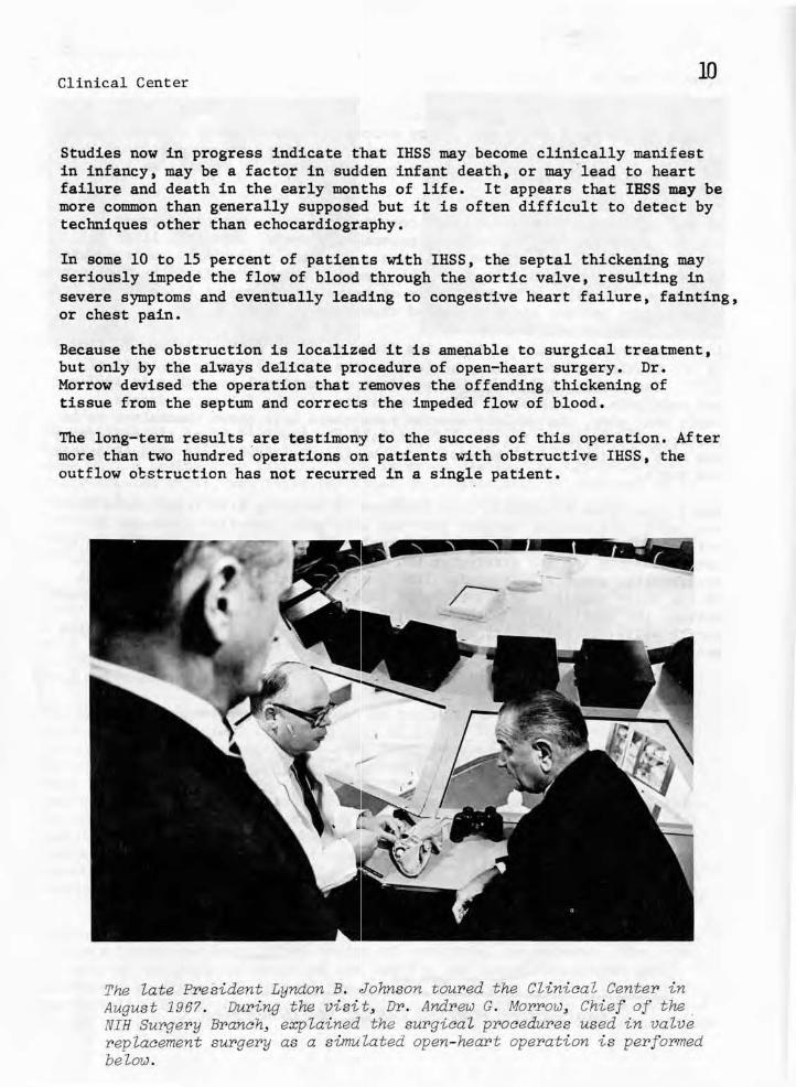
Studies now in progress indicate that IHSS may become clinically manifest in infancy, may be a factor in sud·den infant death, or may ' lead to heart failure and death in the early months of life. It appears that IHSS may be more common than generally supposed but it is often difficult to detect by techniques other than echocardiogr.aphy.
In some 10 to 15 percent of patients with IHSS, the septa! thickening may seriously impede the flow of blood through the aortic valve, resulting in severe symptoms and eventually leading to congestive heart failure, fainting, or chest pain.
Because the obstruction is localiz1ed it is amenable to surgical treatment, but only by the always delicate prc~cedure of open-heart surgery . Dr. Morrow devised the operation that :removes the offending thickening of tissue from the septum and correct is the impeded flow of blood.
The long-term results .are testimony to the success of this operation . After more than two hundred operations 0 1n patients with obstructive IHSS, the outflow obstruction has not recurr1ed in a single patient.
a L41i1 ·
The Late President Lyndon B. ,Johnson toured the Clinical Center in August 1967 . During the visi t , Dr. Andrew G. Morrow, Chief of the . NIH Surgery Branch, explained the surgical procedures used in valve replacement surgery as a simulated open- heart operation is performed below.
ll
Clin ical Center
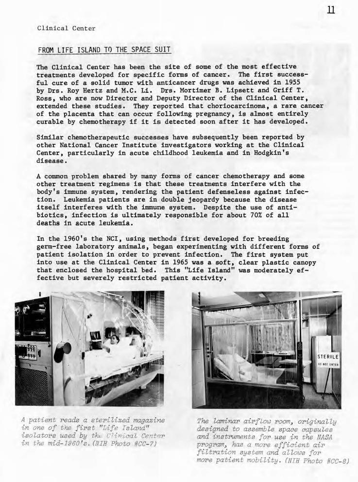
FROM LIFE ISLAND TO THE SPACE SUIT
The Clinical Center has been thE~ site of some of the most effective treatments developed for specifjlc forms of cancer. The first successful cure of a -solid tumor with anticancer drugs was achieved in 1955 by Drs. Roy Hertz and M.C. Li. Drs. Mortimer B. Lipsett and Griff T. Ross, who are now Director and Deputy Director of the Clinical Center, extended these studies. They rE~ported that choriocarcinoma, a rare cancer of the placenta that can occur following pregnancy, is almost entirely curable by chemotherapy if it in detected soon after it has developed.
Similar chemotherapeutic succesues have subsequently been reported by other National Cancer Institute investigators working at the Clinical Center, particularly in acute childhood leukemia and in Hodgkin's disease.
A common problem shared by many forms of cancer chemotherapy and some other treatment regimens is that: these treatments interfere with the body's immune system, rendering the patient defenseless against infection. Leukemia patients are in double jeopardy because the disease itself interferes with the immune system. Despite the use of antibiotics, infection is ultimately responsible for about 70% of all deaths in acute leukemia.
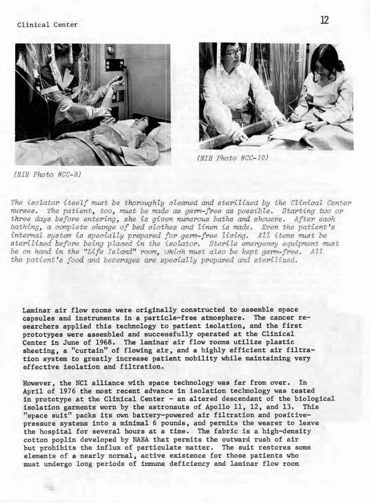
In the 1960's the NCI, using met:hods first developed for breeding germ-free laboratory animals, bE!gan experimenting with different forms of patient isolation in order to p1~event infection. The first system put into use at the Clinical Center in 1965 was a soft, clear plastic canopy that enclosed the hospital bed. This "Life Island" was moqerately effective but severely restricted p~tient activity.
A patient reads a steri 'lized magazine in one of the first "Life Island" isolators used by the r'lin ·/,,cal Centr?r in t he mid- 1960 's . (NIH Photo #CC- ?)
The laminar airflow room, originally designed to assemble space capsules and instruments for use in the NASA program, ha.s a more efficient air filtration system and allows for more patient mobility . (NIH Photo #CC- 8)
12Clinical Center
(NIH Photo #CC- 10)
(NIH Photo #CC- 9)
The isolator itself must be t horoughly cleaned and sterilized by the Clinical Center nurses . 'l'he patient, too, must be made as germ- free as possible . Starting two OY"
t hY'ee days befoY'e entering, she i s given numerous baths and showers . After each bathing, a complete change of bed clothes and Unen is made . Even the patient 's internal system is specially prepared .for germ- free living. All items must be sterilized before being placed in the ·isolator . Sterile emergency equipment must be on hand in the "Life Island" room, <iJhich must also be kept germ- fY'ee . AU the patient 's f'ood and beverages are specially prepared and ste1'ilized.
Laminar air flow rooms were originally constructed to assemble space capsules and instruments in a partic:.le- free atmosphere. The cancer researchers applied this technology tc:> patient isolation, and the first prototypes were assembled and succe1;sfully operated at the Clinical Center in June of 1968. The lamina1r air flow rooms utilize plastic sheeting, a "curtain" of flowing ai1r, and a highly efficient air filtration system to greatly increase pat:l.ent mobility while maintaining very effective isolation and filtration.
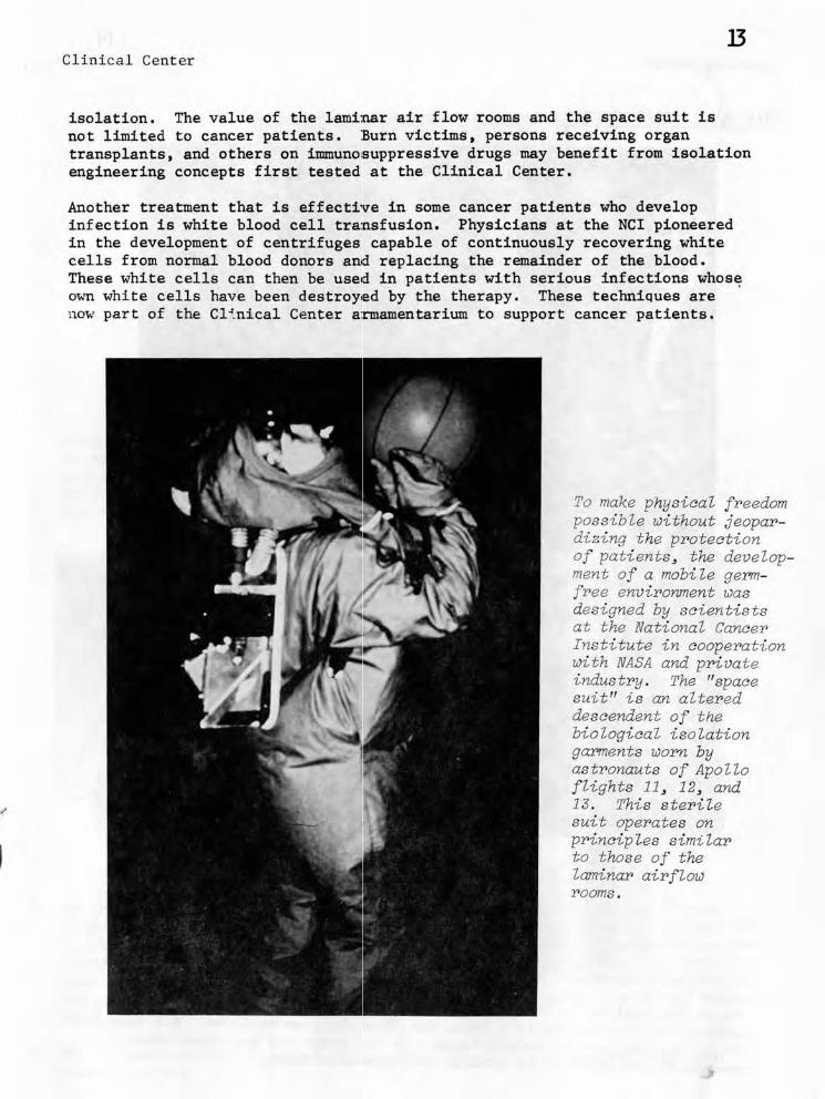
However, the NCI alliance with spac1e technology was far from over. In April of 1976 the most recent advan•ce in isolation technology was tested in prototype at the Clinical Center - an altered descendant of the biological isolation garments worn by the astr1onauts of Apollo 11, 12 , and 13. This "space suit" packs its own battery-:powered air filtration and positivepressure systems into a minimal 6 p1ounds, and permits the wearer to leave the hospital for several hours at a time. The fabric is a high-density cotton poplin developed by NASA that permits the outward rush of air but prohibits the influx of particulate matter. The suit restores some elements of a nearly normal, active existence for those patients who must undergo long periods of immune deficiency and laminar flow room
13 Cl inical Center
isolation. The value of the lami:nar air flow rooms and the space suit is not limited to cancer patients. :Burn victims . persons receiving organ transplants. and others on immuno:suppressive drugs may benefit from isolation engineering concepts first tested at the Clinical Center.
Another treatment that is effective in some cancer patients who develop infection is white blood cell tra:nsfusion. Physicians at the NCI pioneered in the development of centrifuges capable of continuously recovering white cells from normal blood donors antd replacing the remainder of the blood. These white cells can then be usetd in patients with serious infections whos~ o·wn whi te cells have been destroy1ed by the therapy. These techniques are now part of the Cl~.nical Center a:rmamentarium to support cancer patients.
To make physiaal freedom possible without jeopardizing t he proteation of patients , the development of a mobile germfree environment was designed by saientists at the National Caneer Institute in aooperation with NASA and private industry . The "spaae suit" is an altered desaendent of the biologiaal isolation garments worn by astronauts of Apollo flights 11 , 12, and 13. This s ter i l e suit operates on prinaiples similar to those of the laminar airflow rooms .

14 Clinical Center
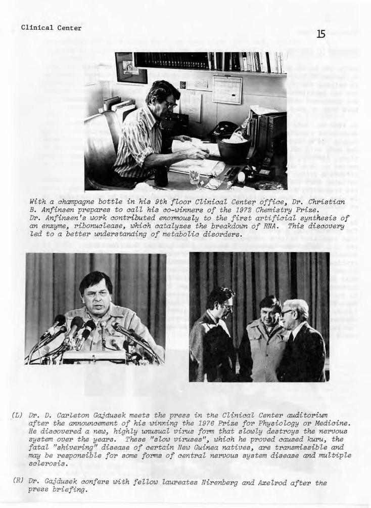
FOUR NOBEL PRIZE WINNERS
Dr. Mars'hall W. Nirenberg (center) at the 1968 award ceremonies in Stockholm. He received his prize for discovering the key to deciphering the genetic code, lea~ing the way to recombinant DNA research. ·
Dr. Julius AxeZ~od in the 'hallway of his Clinical Center lab after hearing word of his winning the 1970 Prize for Physiology or Medicine. His research traced the function of catechoZcunines in the brain and how these chemical messengers are affected by various antidepressant drugs.
Clinical Center 15
With a champagne bottle in his 9th floor Clinical Center office, Dr . Christian B. Anfinsen prepares to call his ao-winner s of the 19?2 Chemistry Prize . Dr. Anfinsen 's work contributed enormousl y to the first artificial synthesis of an enzyme, ribonuclease, which catalyzes the ~reakdown of RNA . This discovery ied to a better understanding of metabolic disorders .
(L) Dr. D. Carieton Gajdusek meets the press in the Clinical Center auditorium after the announcement of his winning the 19?6 Prize for Physiology or Medicine . He discovered a new~ highly unusual virus form that slowly destroys the nervous system over the years . The se "slow viruses", which he proved caused kuru, the fatal "shivering" disease of certain New Guinea natives, CU'e transmissible and may be responsible for some forms of central nervous system disease and mult~ple salerosis .
(RJ Dr . Gajdusek confers with fellow laureates Nirenberg and Axelrod after the press briefing.

16 Cl i nical Center
DIAGNOSTIC IMAGING
Early and detailed diagnosis is of.ten the key to effective treatment. A picture or image can truly be wor th the proverbial thousand words in helping the physician make such a diagnosis. The Clinical Center has two departments , Diagnostic Radiology and Nuclear Medicine, that are concerned with the production of diagnostic images. Their successes have been so many and varied that it is possible to touch on only a few.
In 1964 Dr. John L. Doppman, now Chief of Diagnostic Radiology, and his associates reported the first successful imaging of the arteries that supply the spinal cord. The technique, knoWQ. as spinal angiography, has made surgical intervention possible where spinal arterial malformations , lesions, or tumors cause paralysis. The researchers also experimented at this time with embolization (deliberate blockage) of the offending spinal arteries by injecting a clotting agent through the catheter that carries the radiologic contrast medium. Embolization can frequently eliminate the need for subsequent surgery. The technique is also used in cancer patients to decrease the pain caused by untreatable ot unresponsive tumors .
Dr . Doppman and his associates in 1968 developed a method for locating the parathyroid, a group of glands, each about the size of a BB pellet , that regulates calcium metabolism. These glands are usually found in the neck behind the thyroid. Determining the precise location of a diseased parathyroid is a difficult and frus~rating exercise but a critical one for successful surgery. The method developed at the Clinical Center is called "selective venous catheterization". The veins that drain the thyroid gland are sampled by a fluoroscopically directed catheter . The blood samples drawn are tagged by location and subjected to the sensitive radioimmunoassay for the presence of parathyroid hormone . When the levels of the hormones are higher than those in the peripheral blood, this pinpoints the location of the gland or tumor and suggest whether multiple glands are involved in the overproduction of hormones . The technique has been proven nearly 100% accurate in patients with newly diagnosed parathyroid dis~ase .
Radionuclide cinearzgiography aLZows the heart to be viewed during and just after intense exercise. The patient is peda.Zing an exercise machine whiZe a scintiiiation camera coZZects data for the computerbased movie of his heart. (NIH Photo #CC- 11)
17 Clinical Center
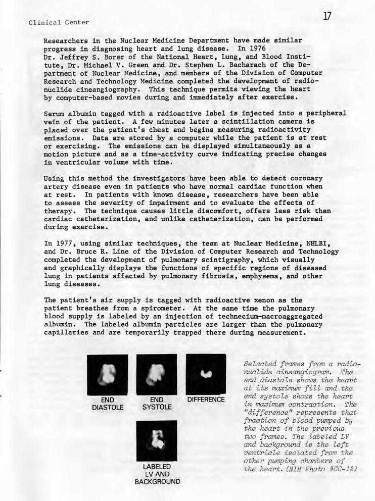
Researchers in the Nuclear Medicine Department have made similar progress in diagnosing heart and lung disease. In 1976 Dr. Jeffrey s. Borer of the National Heart, Lung, and Blood Insti tute, Dr. Michael V. Green and Dr. Stephen L. Bacharach of the Department of Nuclear Medicine, and members of the Division of Computer Research and Technology Medicine completed the development of radionuclide cineangiography. This technique permits viewing the heart by computer-based movies during and immediately after exercise.
Serum albumin tagged with a radioactive label is injected into a peripheral vein of the patient. A few minutes later a scintillation camera is placed over the patient's chest and begins measuring radioactivity emissions. Data are stored by a computer while the patient is at rest or exercising. The emissions can be displayed simultaneously as a motion picture and as a time-activity curve indicating precise changes in ventricular volume with time.
Using this method the investigators have been able to detect coronary artery disease even in patients who have normal cardiac function when at rest. In patients with known disease, researchers have been able to assess the severity of impairment and to evaluate the effects of therapy. The technique causes little discomfort, offers less risk than cardiac catheterization, and unlike catheterization, can be performed during exercise.
In 1977, using similar techniques, the team at Nuclear Medicine, Nlil.BI, and Dr. Bruce R. Line of the Division of Computer Research and Technology completed the development of pulmonary scintigraphy, which visually and graphically displays the functions of specific regions of diseased lung in patients affected by pulmonary fibrosis, emphysema, and other lung diseases.
The patient's air supply is tagged with radioactive xenon as the patient breathes from a spirometer. At the same time the pulmonary blood supply is labeled by an injection of technecium-macroaggregated albumin. The labeled albumin particles are larger than the pulmonary capillaries and are temporarily trapped there during measurement.
END END DIFFERENCE DIASTOLE SYSTOLE
LABELED LVAND
BACKGROUND
Selected frames from a radionuclide cineangiogram. The end diastole shows the heart at its maximwn fill and the end systole shows t he hea:r>t in maximwn contraction. The "difference" rep:resents that fraction of bZood pumped by the heart i~ the previous two frames . The labeled LV and background is the left ventricle isolated from the other pwnping chambers of the hea:r>t . (NIH Photo #CC- 12)

18Clinical Center
NORMAL ABNORMAL NORMAL ABNORMAL
A.B: A,B:
LUNG LUNG PERFUSION VENTILATION
(BLOOD FLOW) (AIR FLOW)
c,o: C,O: AIR SPACE VENTILATION
VOLUME PERFUSION RATIO
E,F: E,F: E CURVE OF GALLIUM-67
TRACER ACTIVITY LUNG SCAN •IN LUNG AIR
(INFLAMMATION) ...;a.(ARRIVAL & ~,
DISAPPEARANCE)
Gamma rays emitted by the tracers are then measured during respiration by a gamma camera that is placed behind the patient's chair . The tracer activity is visualized and measured by imaging equipment linked to a computer . The computer then constructs pictures that visualize from 1,200 to 1,500 regions in the lung and identifies any diseased area. The information can also be used to determine whether the problem is linked to air flow or pulmonary blood flow . (See photos above. )
Ultrasound , a different physical technique, utilizes high-f requency sound waves that are reflected by varying tissue densities and shapes revealing the body's internal structure and form.
Ultrasound has two major advantages over X-rays in noninvasive visualization. First , it is capable of visualizing soft tis sues without the physician having to resort to injecting "tagged" contrast mediums, which can be hazardous. Secondly, while X-rays are knol!m to cause cellular and tissue damage, ultrasound appears to be safe at the levels of intensity used in the existing equipment. The Diagnostic Radiology Department has made some very real contributions to the technology of u~trasound , many of them as recently as this year.
The first generation of ultrasound scanners were somewhat awkward mechanically. The images they produced were of limited value because vibration and lack of control destroyed much of the image quality.
The second generation devices, called "phased array", used sophisticated electronics to compensate for these earlier drawbacks. While they gave a very good image, their price was quite high, approaching $100,000 per unit.
The mechanically controlled ultrasound scanner developed by Dr . Thomas H. Shawker of Diagnostic Radiology and Mr . William Schuette of the Division of Research Services produces images comparable to the phased-array systems at approximately one- tenth the expense. This i s a real-time scanner, which gives a continuous "flouroscopic" image.

19 Clinical Center
The mechanicaUy- contr>oUed hand- held ultr>asound scanner> pr>oduces i mages corrrpa:r>able to the elect~onically -con
tr>olled phased- aITay system at approximately one- tenth of the expense. (NIH Photo #CC- 13)
Vibration from the hand-held motor that drives the scanning head was nullified by giving the same motor an equally demanding and necessary task to perform - in the opposite direction - namely to power a position feedback control system. The two torques cancel each other out thus producing zero vibration.
The investigators have been able to f.urther enhance image quality by integrating their scanner to operate "in synch" with television hardware. For many reasons television is the ideal means to both display and store ultrasound-generated images. The Clinical Center researchers use a TV camera to record the position of the scanner on the pa tient's body on the same videotape that records the ultrasound image, thereby creating a third dimension for diagnostic purposes.
Beyond improving image quality. Dr. Shawker is collaborating with researchers at the National Bureau of Standards to link ultrasound scanners directly with a computer to generate numerical pictures similar to those being used in the CT scanner. The inunediate goal is to gain computer-generated quantified measurements. This system could measure the size and volume of a tumor, for instance, before and after chemotherapy. The long-range goal is to perform, via computer-li.nked ultrasound, an "acoustic biopsy" to find through t he proper interpretation on the numerical data not only the volume and location of a tumor , but its particula.r type, such as melanoma or sarcoma. Establishing such . "tissue signatures" will take much additional investigation. The tremendous value of such a system would be the margin of safety that ultrasound has over X-ray - scans could be done repeatedly with no hazard to the patient .
Whether in ultrasound , scintigraphy, angiography, gallium scan, or the r efining of new techniques for the CT scanners, the investigators at the Clinical Center are constantly developing new approaches to improve t he utility of the diagnostic image.
Clinic~l Cen ter
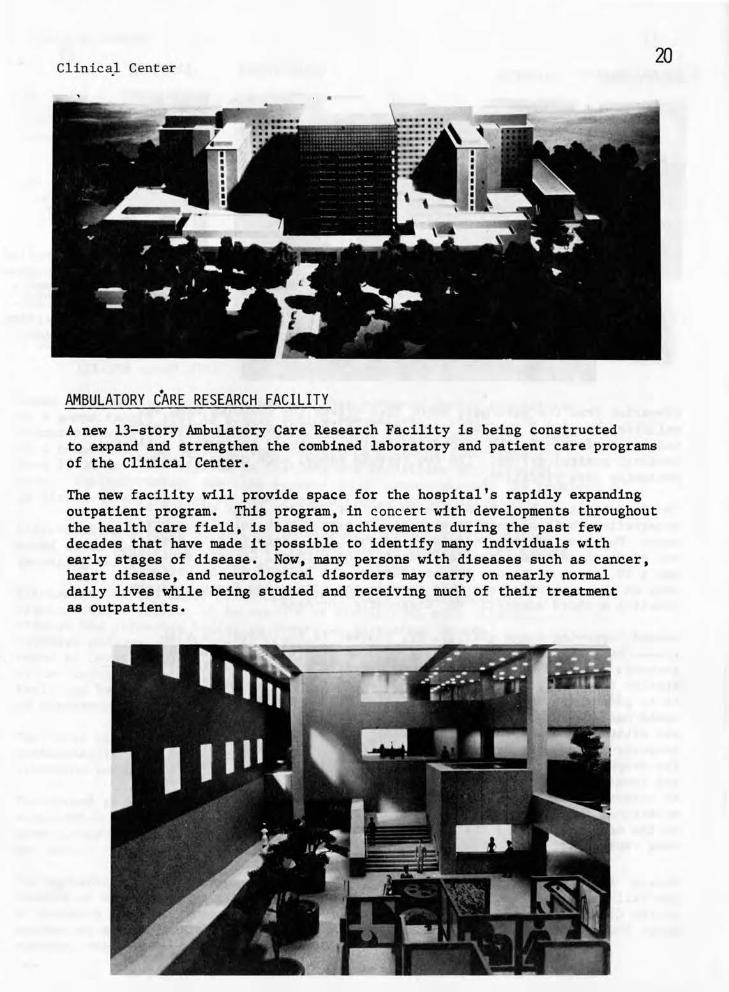
•AMBULATORY CARE RESEARCH FACILITY
A new 13-story Ambulatory Care Research Facility is being constructed to expand and strengthen the combined laboratory and patient care programs of the Clinical Center.
The new facility will provide space for the hospital's rapidly expanding outpatient program. This program, in conc ert with developments throughout the health care field, is based on achievements during the past few decades that have made it possible to identify many individuals with early stages of disease . Now, many persons with diseases such as cancer, heart disease, and neurological disorders may carry on nearly normal daily lives while being studied and receiving much of their treatment as outpatients.
21 Clinical Center
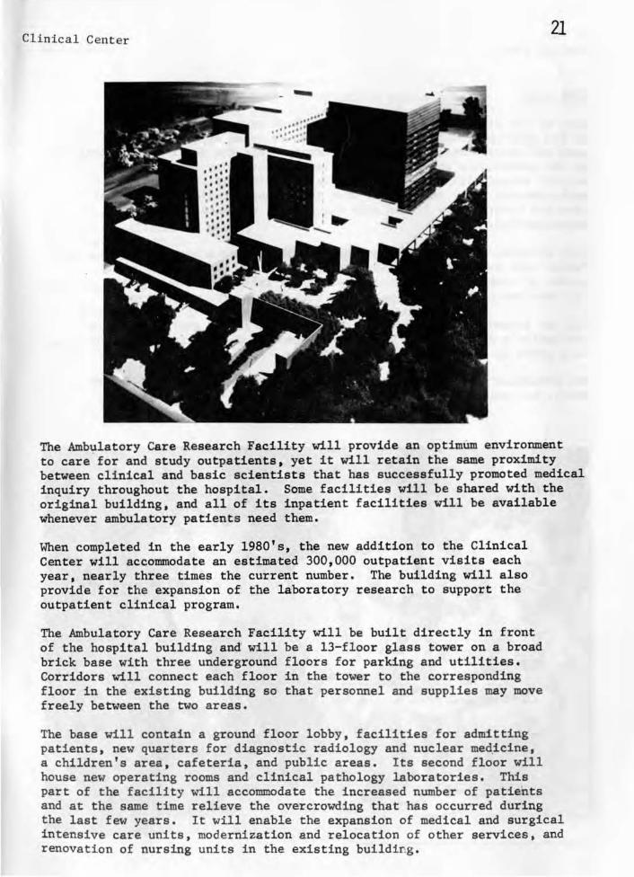
The Ambulatory Care Research Facility will provide an optimum environment to care for and study outpatients. yet it will retain the same proximity between clinical and basic scientists that has successfully promoted medical inquiry throughout the hospital. Some facilities will be shared with the original building, and all of its inpatient facili ties will be available whenever ambulatory patients need them.
When completed in the early 1980's , the new addition to the Clinical Center will accommodate an estimated 300,000 outpatient visits each year. nearly three times the current number. The building will also provide for the expansion of the laboratory research to support the outpatient clinical program.
The Ambulatory Care Research Facility will be built directly in front of the hospital building and will be a 13-floor glass tower on a broad brick base with three under ground floors for parking and utilities. Corridors will connect each floor in the tower to the corr esponding floor in the existing building so that personnel and supplies may move freely between the two areas .
The base will contain a ground floor lobby, facilities for admitting patients , new quarters for diagnostic radiology and nuclear med.icine, a children's area , cafeteria , and public areas . Its second f loor will house new operating rooms and clinical pathology laboratories. This part of the facility will accommodate the increased number of patients and at the same time relieve the overcrowding that has occurred during the l ast few years . It will enable the expansion of medical and surgical intensive care units, modernization and relocation of other services, and renovation of nursing units in the existing building .

Clinical Center
CONCLUSION
None of the discoveries reported here, which represent only a tip of the iceberg, would be possible without an ocean of support from each and every Clinical Center department. From the nursing department to the maintenance staff, from the clinical pathology section to the medical records office, from social services to spiritual ministry, each operates under the premise that the highest standards in patient care are inextricably linked with the highest standards in clinical investigation.
Most Clinical Center departments have a research component, even the Fabr'ic Care department is doing investigations in the most efficient manner of cleaning and storing hospital and laboratory linens. All the departments are participating in this most important ~xperiment.
Can two thousand people working together for a single goal - improving the nation's health - make a real difference in the quality of medical care given to the American people?
The preliminary results of the experiment, after the first twenty-five years, are overwhelmingly affirmative.

24