new york college of podiatric medicine podiatric medical ... · review (pmr). collectively, we all...
TRANSCRIPT

Podiatric Medical Review
New York College of Podiatric Medicine
Volume 27 2018-2019
Dynamic Gait & Static Changes to the Lower Extremity During Pregnancy: A Literature Review Sanjna Sanghvi, BS, Karla De La Mata, BS
A Systematic Review of Surgical Management for Neurogenic Neuroarthropathy Mohammed Gheith, BA, Thomas Milisits, BS, Sapan Patel, BS
Imaging Modalities for Diagnosis of Supination External Rotation Ankle Injuries: A Literature Review Ashima Choudhary, BA, BS,Tinisha Ricks, BA, Yumna Siddiqui, BS, Treatment Modalities for Idiopathic Toe-Walking in Neurotypical and Autistic Individuals Jenna Friedman, BA, Joann Li, BA, Farah Naz, MS, BS, Nadia Hussain, BA, Manali
Naik, BSIntamedullary Fixation Technique for Correction Distal Tibia Fractures in Pediatric Patients: A Systematic Review Joe Dobbs, BA, Sruti Karwa, BS, Rahim Lakhani, BS
The Efficacy of Opioids in the Treatment of Diabetic Peripheral Neuropathy Sohaib Asalam, BA, Michael Chung, BS, Vikal Singh, MPH, BS, Zain Ul-Suhleri, BS,
Complications of Overcorrected Clubfoot Leading to Dorsal Bunion: A Literature Review Farah Naz, MS, BS, Jenna Friedman, BA, Joann Li, BA.
4
20
28
42
53
62
71
STUDENT JOURNAL OF THE NEW YORK COLLEGE OF PODIATRIC MEDICINE
Editor-in-Chief Sanjna Sanghvi, BS
Senior EditorsKarla De La Mata, BS
Mohammed Gheith, BA
Faculty AdvisorAnthony Iorio, DPM
PMRStudent Peer Reviewers Karla De La Mata, BSMohammad Gheith, BAThomas Milisits, BA Farah Naz, MS,BSSanjna Sanghvi, BSVikal Singh, MPH, BSTinisha Ricks, BSZain Ul-Suleri, BS
Faculty Reviewers Marie Christine-Bergeron, DPMJeffrey J. Cusack, DPMJaveria Hussani, DPMBarbara Resseque, DPMThomas Vitale, DPM

Evidence-based medicine the cornerstone of podiatric practice. The continued enthusiasm of rigorous research within our field has shaped us to become more knowledgeable and capable clinicians. The dedicated initiatives undertaken by our colleagues have truly advanced the field of podiatry with creative surgical techniques, new medical applications, and innovate products to provide superior patient care.
It is with great pleasure, I present volume twenty seven of the Podiatric Medical Review (PMR). Collectively, we all have contributed a great deal of hard work and dedication to publish quality manuscripts. We hope you enjoy the novelty and range of the topics showcased this year.
I would like to express great gratitude to my senior editors, Karla De La Mata and Mohammad Gheith for their efforts to see this volume from start to finish. A sincere thanks to our meticulous student peer reviewers and dedicated student collaborators whose submissions made this year’s publication possible. Thank you to our advisor and professors for providing constructive edits and supporting us.
A Note from the Editor
Sanjna Sanghvi, BS Editor-in-Chief
3

Dynamic Gait & Static changes to the lower extremity during pregnancy: A Literature Review
By: Karla De La Mata, BS, Sanjna Sanghvi, BS ABSTRACT Introduction The robust nature of the lower extremity, especially the foot, is best exemplified in a pregnant patient where this temporary condition manifests as dynamic gait changes. There is much curiosity regarding the pregnant population, as they present with a multitude of quantitative and qualitative changes. Minimal research has been conducted with regard to its podiatric relevance. Our objective is to assemble the most current research regarding gait kinematics and temporospatial parameters observed in pregnant women. Thereby, showcasing the need for a podiatric evaluation long-term musculoskeletal injury, reduction of pain and improved quality of life for this subset of patients can be achieved. Proper counsel and foot care should be an essential part of a woman’s prenatal workup as this type of preventative care measure improves the health of the expectant mother, subsequently the health of the fetus, and ultimately lowers healthcare costs. Study design: Systematic Review of the Literature Methods A search on PubMed was performed with the following phrases: “pregnancy AND gait” yielded 324 articles and then sorted by “Best Match” results from which we narrowed it down to 36 by modifying to “pregnancy AND gait parameters.” We then applied our inclusion and exclusion criteria. The inclusion criteria encompassed studies on women during all trimesters of pregnancy with varying lifestyles. Exclusion criteria included non-English articles written prior to the year 2000 (up till October 2018), studies of pregnant animals, cadavers/skeletal remains, and the obese population. In accordance with these criteria, 11 articles were selected for review. Results After appraising several parameters it can be deduced that there is a direct impact of added weight on gait and in turn a woman’s overall stability. In our evaluation, statistical significance was defined as a p-value less than 0.05 for the following parameters: velocity, double and single limb support time, stride length, and pelvic, thoracic and trunk range of motion. The aforementioned parameters culminate in an altered gait pattern. In addition to the dynamic gait changes, some women, when evaluated postpartum, present with a persistent reduction in arch height and arch rigidity with a corresponding increase in foot length. Conclusion The dynamic and static changes that take place during pregnancy increases the susceptibility of musculoskeletal injuries that can lead to permanent foot and/or joint damage. It has been shown across many trials that the effects of weight gain in combination with hormonal modulation (relaxin) are significant. All women, especially those with an increased risk of residual deficits due to genetics or acquired deformity such as tendonitis, should seek preventative care prior to, during, and after pregnancy. A podiatric consult and proper footwear can alleviate physical strain and prevent long term anatomical abnormalities. Key Words: Pregnancy, Pregnant, Gait, Weight Gain, Relaxin, Joints, Kinematics, Lower Extremity Level of evidence: 4
4

INTRODUCTION The experience of being pregnant is a unique bond shared by many women, but each woman faces individual health challenges; including - genetic predispositions, health care access and affordability, and overall medical literacy. The dynamic set of variables amongst pregnant women makes it difficult to study and understand the changes necessary to improve their condition during gestation. Signs and symptoms of gestation, particularly those involving the lower extremity, are often dismissed as secondary issues on the assumption that they will spontaneously revert to their “normal” pre-gestational state. However, the dismissal of what may be minor foot pain, backache or joint pain can lead to long term complications. In the United States, an estimated 4% of the female population is pregnant per year; that is approximately 6 million women.1 Each woman adjusts to pregnancy differently, making it difficult to identify overarching trends. Despite the given limitations, several research teams have assessed the changes in kinetic, temporal, and spatial parameters. Temporospatial parameters are those that survey the position and time of contact of a body part. Kinematic parameters examine joint motion. Regarding physical changes, anterior weight gain is easily observed in comparison to the less obvious increase in joint laxity, which reflects hormonal fluctuations. Pregnancy is divided into three trimesters, each of which is characterized by different
physical changes. Many gait studies were executed during the second and third trimester, as those trimesters corresponded to the most noticeable changes in weight gain - the predominating and most impactful parameter2. Studies that followed a longitudinal approach and assessed the women before pregnancy until a year postpartum are held in higher regard. A gait cycle is comprised of a predominating stance phase and swing phase, taking up 60% and 40% of the cycle, respectively. The stance phase corresponds to the time from heel strike to toe-off the same foot. The swing phase is when the opposite foot swings through and in front of the foot that was in contact with the ground to propel the body forward.3 Pregnant women spend an increased percentage of their gait cycle in the stance phase.4 Changes in the gait cycle are accompanied by an increase in step width and an altered tilt with external rotation of the pelvis; together these factors produce what is known as the pregnant “waddle”.4
The purpose of this paper is to review how physical stresses of pregnancy affect the lower extremity and its direct impact on gait. The lack of unanimous conclusions regarding gait in the pregnant female led us to review, summarize, and spotlight the efforts of research teams for their significance in the podiatric field.5 Based on our interpretation of the literature, pregnant women should, at a minimum, receive a podiatric consult during their second or third trimester. A consult would include a gait
5
evaluation, education about proper lower limb care and appropriate exercises, and footwear advice to reduce the potential of long-term musculoskeletal impairment, and thus injury.
METHODS A general search was performed on PubMed with keywords (Pregnancy, Pregnant, Gait, Weight Gain, Relaxin, Joints, Kinematics, Lower Extremity). We used the Boolean operators “AND” and “OR” to ensure that all related articles were identified. Searching “pregnancy AND gait” yielded 324 using sort by “Best Match” results from which we narrowed it down to 36 by modifying the search to “pregnancy AND gait parameters.”
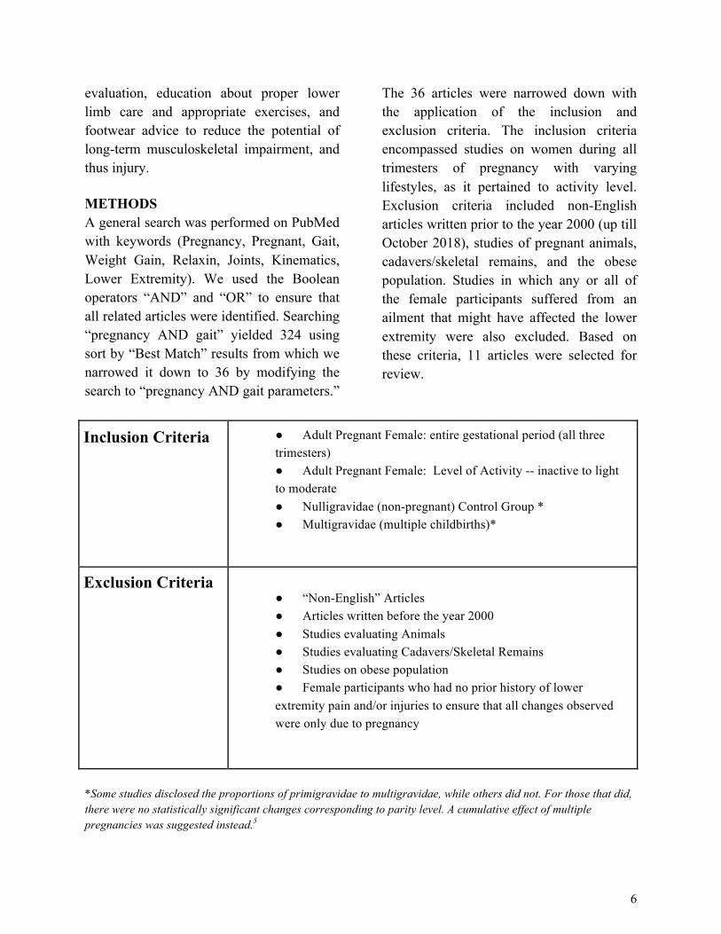
The 36 articles were narrowed down with the application of the inclusion and exclusion criteria. The inclusion criteria encompassed studies on women during all trimesters of pregnancy with varying lifestyles, as it pertained to activity level. Exclusion criteria included non-English articles written prior to the year 2000 (up till October 2018), studies of pregnant animals, cadavers/skeletal remains, and the obese population. Studies in which any or all of the female participants suffered from an ailment that might have affected the lower extremity were also excluded. Based on these criteria, 11 articles were selected for review.
Inclusion Criteria ● Adult Pregnant Female: entire gestational period (all three trimesters) ● Adult Pregnant Female: Level of Activity -- inactive to light to moderate ● Nulligravidae (non-pregnant) Control Group * ● Multigravidae (multiple childbirths)*
Exclusion Criteria ● “Non-English” Articles ● Articles written before the year 2000 ● Studies evaluating Animals ● Studies evaluating Cadavers/Skeletal Remains ● Studies on obese population ● Female participants who had no prior history of lower extremity pain and/or injuries to ensure that all changes observed were only due to pregnancy
*Some studies disclosed the proportions of primigravidae to multigravidae, while others did not. For those that did, there were no statistically significant changes corresponding to parity level. A cumulative effect of multiple pregnancies was suggested instead.5
6

RESULTS The goal was to examine a causal link between gait alterations, the likelihood of residual lower limb changes and potential sequella following pregnancy. Studies that addressed the qualitative and quantitative temporal, spatial, and kinematic adjustments made during pregnancy were selected. All articles selected for review agree that obtaining a comprehensive understanding of pregnancy's effect on gait requires an evaluation of body kinematics, as there is a direct impact on temporospatial parameters. Evaluation of stationary posture would not suffice. The literature has acknowledged the multifactorial impacts on gait during pregnancy, but not all have evaluated the same variables. Refer to Table 1 for a listing of the analyzed studies and their respective measured variables.
There was potential for elucidating significant changes when comparing pregnant versus non-pregnant, and when comparing the pregnant woman at each trimester. Statistical significance was defined as a p-value less than 0.05. Studies that evaluated all three trimesters and a postpartum period greater than six weeks are held in higher regard because no extrapolation or data-based inferences were necessary. Unfortunately, only a few studies were able to execute a longitudinal design. Changes in the musculoskeletal system following pregnancy have not been reported to persist past six weeks and hormone levels stabilize after 48 hours.6 There was no significant difference in the values obtained when using the right or left leg as the reference, so the values were averaged.4
Due to the interplay of many factors creating
the cyclic nature of gait, even if one variable was not directly measured there was a high likelihood that it was factored into the results of the measured variable of interest. For instance, to measure ground reactive forces (GRF), velocity had to be considered as a covariant.
Velocity All studies had the participants walk at their natural pace while acclimating to the platform. Walking at one’s optimal pace, that which maintains stability, consumes as much as ⅓ of the net metabolic energy.4,7 A healthy asymptomatic individual typically has a walking speed of 1-1.4 m/s.3A reduction in walking speed, an average pace of 1 ±0.2 m/s, was most noticeable during the third trimester of 28 pregnant women; normal pace of nulliparous being approx 1.4 m/s.7 A pregnant female walking at a velocity lower than optimum will expend more energy, greater than ⅓ of the net metabolic energy, to accommodate the change. A greater quantity of energy is expended as a trade-off for increased reaction time to gait disturbances. Following pregnancy, once the anterior weight gain is lost the gait cycle becomes more efficient. 8
Following a study conducted by Branco et al. examining the gait of 22 pregnant women during the second and third trimesters, walking speed remained unchanged.9
Participants were asked to walk barefoot across a 10-meter platform for three minutes with a one-minute break between trials. In contrast, evaluation of 58 women during their last four months of pregnancy by Bertuit et al. found that there was a reduced
7

speed at slow, preferred, and fast pace.4 For each of the nine trials, 5-10 steps were sampled and the order at which the test subjects walked at one out of the three speeds was randomized by dice throwing. As a consequence of reduced speed, gait cycle time increased. Decreased step length, a measurement of the distance from contact of the ipsilateral limb to contact of the contralateral limb, also contributed to an increase in gait cycle time. After childbirth, speed and gait cycle time stabilized to values similar to those of the 23 nulliparous women of the control group.4
Overground walking is not the same as walking on a treadmill; the body’s adaptation to each surface is very different. Albeit very different surfaces, the same reduction in speed of pregnant women was noted.5 On a treadmill programmed with 15 different speeds, each 0.11 m/s apart, there was a significant reduction in the walking speed of 12 women out of 25 participants between 20 to 34 weeks pregnant. Their comfortable walking velocity fell between 0.72 m/s to 1.28 m/s. At each level, the participant was asked if the pace was too fast. If she responded yes, the level prior was designated as her maximum velocity at which she would continue walking for three minutes. The nulliparous control group (n=13) had a higher comfortable walking speed of 0.83 m/s to 1.50 m/s. The average difference was significant, p<0.05, an average of 1.03 m/s versus 1.19 m/s (13% reduction in speed).5 Gait Cycle In some cases, reduced speed accompanied an increase in double support time. Double
support time, when both feet are in contact with the ground, was evaluated as a percentage of the gait cycle and as a parameter that parallels step width. During the third trimester, an increase in step width of up to approximately 2 cm may be observed; Blaszczyk et al. recorded a change from 27.7 cm to 29.7 cm.7 Postpartum, the stance width spontaneously reduced. Gilleard et al. cites the reduction in stance width at eight weeks postpartum, but were unable to conclude whether an increase in step width was a consequence of increased pelvic width or implemented to increase stability.10
Foti et al. found that pelvic width averaged a 4.3 cm increase with a p<0.001, and a mean ankle separation width, averaging 2.4 cm with a p<0.001- both values corroborate an increased step width.11 McCrory et al. also confirmed an increase in step width as pregnancy progressed and did detect a “waddle.”12
Stride Length A decrease in stride length, measured as the distance between two consecutive heel strikes of the same limb, was only observed between the second and third trimester.9,10,13
During pregnancy, women walked at least 10 cm/s slower and had a decreased stride length of at least 10 cm per step compared to initial non-pregnant baseline. Postpartum, Hagan et al. and Foti et al. both reported a 10 cm increase in stride length and 10 cm/s (0.1 m/s) increase in gait speed, compared to values of the third trimester.11,14 In the case study published by Hagan et al. both women underwent similar changes in stride length and walking speed but Subject 1 gained 14.5
8

kg (~32 lbs) and Subject 2 gained 4.8 kg (~11 lbs).14 Differences in weight gain suggests that there are other factors accounting for the gait variability accompanying pregnancy.7,14
Range of Motion - Pelvis, Thorax, and Trunk Traditional theory of motion divides the body into a “passenger” (head, neck, trunk, and arms) and “locomotor” (lower limbs and pelvis).15 One’s trunk works to minimize extreme changes in the body’s oscillations to maintain equilibrium. In a pregnant female with a correspondent decrease in truncal range of motion, due to increased anterior abdominal mass, the body’s oscillations are not as firmly controlled resulting in gait imbalance.10 Gait modifications are made to counteract the imbalance. Trunk tilt is measured as the angle of forward or backward inclination relative to one’s vertical axis (standing in the upright position). On average, there was a four-degree increase of anterior pelvic tilt, p-value of 0.018. Although this finding was significant, it was noted that not all women undergo the same quantitative postural adjustments even if qualitatively an anterior pelvic tilt and lumbar lordosis are apparent.11 Branco et al. study substantiates an observed increase in anterior pelvic tilt that occurs between the first and second trimester.16 Between four and six months postpartum there was a reduction in pelvic tilt, an indication of recovery.16
Pelvic measurements were the most variable parameter. Even while walking on a treadmill, the pregnant subjects had pelvic
and thoracic rotations that were approximately one degree smaller in amplitude than the controls. Keeping in mind the increased inertia of the pelvis and thorax, greatly propelled forward by the additional anterior mass, it becomes more critical for the pregnant female to remain cautious and not exceed the relative timing of pelvis and thoracic rotations. Gilleard et al. also recorded decreased rotational moments, potentially exacerbated by a decrease in stride length and changes in muscle activity.10 However, the findings of McCrory et al. and Foti et. al did not attenuate the same conclusion.11,12 This difference is not too surprising considering genetic variability among different races and fitness levels. Any residual changes in the range of motion may be related to the functionality of anterior and posterior trunk muscles that experience greater fatigability up to eight weeks postpartum. It is those same trunk muscles that are engaged at a greater rate to help maintain stability.10
Joints Kinematic analysis is particularly useful and better appreciated when prescribing rehabilitative and treatment modalities. After evaluating the sagittal, frontal and transverse planes of motion, it was concluded that the hip and pelvis withstand the largest impact. Nevertheless, all other lower limb joints undergo angular changes - primarily in the sagittal plane. Angular data was strongly influenced by pregnancy.16 The hip joints, being the closest in proximity to the pelvis, are subject to greater angular adjustments primarily during the stance phase.9 Eccentric contraction of hip flexors,
9

contraction that occurs as the muscle lengthens, becomes reduced following an increase in thigh fat.8 In contrast, larger areas of fat surrounding the calf and thigh cause greater engagement of the abductors and extensor muscles surrounding each hip joint.8,18 A progressive increase in hip flexion paired with an anterior pelvic tilt was emphasized; both parameters underwent a reduction postpartum.13,14,16
Internal rotation of the hip in the transverse plane causes an in-toe position. Inward toe rotation might temporarily increase knee stability but also prevents forward movement; over time there is undue pressure on the knee joint. Increased stability of the knee was most pronounced during the third trimester.13At the end of the swing phase, there was an observed increase in knee flexion coincident with a decrease in knee extension.9,16 Hagan et al. did not note any significant change in knee mobility, but discerned a decrease in ankle dorsiflexion during the third trimester in all phases of gait by at least 5° that did persist postpartum.14 The decrease in ankle plantarflexion minimizes the propulsive force available following heel strike.9 A 3° ankle inversion was present up until the mid-swing phase of the gait cycle, between the first and third trimester.16 Ankle inversion does not recover, if at all, until six months after childbirth; although subtle, it may contribute to fall risks. Studies that lack a longitudinal approach may fail to observe this change.16,17
Foot Structure Biomechanical measures of foot structure and function are related and are particularly relevant to clinicians because many pathologies affecting the foot and ankle have a biomechanical origin.3 A change in foot structure during pregnancy is anticipated due to increased downward forces that impact bones, tendons, and ligaments of the foot. There remains speculation whether the changes persist.6 Segal et al. hypothesized that a significant reduction in arch height persists during static and dynamic conditions in 49 women who were evaluated during their first trimester and later at 19 weeks postpartum. There was a reduction in arch height, analogously cited as an increase in arch drop, and a reduced arch rigidity index (ARI) leading to flatter and more flexible arches following pregnancy.6
Women generally have a reduced ARI compared to men even before gestation.3
Arch drop as an indicator of foot flexibility was calculated by subtracting the arch height index (AHI) at standing from the AHI while seated. AHI was the dorsum height at 50% of total foot length divided by the ipsilateral truncated foot length.6 Arch Rigidity Index, defined as the ability of the foot to maintain the structural arch in a weight-bearing position, was calculated by dividing standing AHI by the AHI while seated.6 A pregnant female may not fully understand the consequences from a clinical viewpoint, but they do notice a change in shoe size and possibly pain. Pain symptomatology is often linked to over-pronation. As the foot pronates, the presence of rotational torques causing shear stress can ascend to the knee and eventually the hip.6
10

Table 1. Variables Measured by the Eleven Publications Analyzed
Author Pregnant (primigravidae &
multigravidae) Test Individuals
Time Period of Observation
Nulligravidae (non-pregnant) Control Group
Method Parameters Evaluated
Bertuit4 58 6mos, 7mos, 8mos, and 9mos
23 9 randomized gait trials, 3 at each speed (slow, preferred, and fast). Rest period between trials.
-Velocity -Cadence -Step Length -Step Width -Stride Length -Single & Double Limb Support Times -Cycle Time: Stance & Swing
Blaszcyk7 28 Early & Late Pregnancy, 2mos & 6mos postpartum
N/A Walking back and forth across 10m platform ten times
-Velocity -Cadence -Stride Length -Single & Double Limb Support Times -Cycle Time: Stance & Swing
Branco9 22 2nd (25-29wks) and 3rd (35-38wks) trimesters
12 Walking across 10m platform for 3mins. 1min break between trials
-Velocity -Stride Width -Single & Double Limb Support Times -Cycle Time: Stance & Swing -Joint Kinematics**
Branco16 11 End of all three trimesters, 4mos and 6mos postpartum
N/A Walking barefoot straight across a 10m platform for 3mins.
-Velocity -Step Length -Stride Length -Stride Width -Single & Double Limb Support Times -Cycle Time: Stance & Swing -Joint Kinematics**
11

Author Pregnant (primigravidae &
multigravidae) Test Individuals
Time Period of Observation
Nulligravidae (non-pregnant) Control Group
Method Parameters Evaluated
Foti11 15 End of 3rd trimester (35-40wks) and 1 yr postpartum
N/A Walking across 20m platform
-Joint Kinematics**
Gilleard10 9 18wks or less, 24wks, 32wks, 38wks, and 8wks postpartum *first test session designated for acclimation
12 tested initially and at 16wks
3 trials walking at self-determined pace across 20m platform
-Velocity -Stride Length -Step Width -Pelvic, Thoracic, and Trunk ROM
Hagan14 2 Before pregnancy, end of each trimester, and 12-16 wks postpartum
N/A 3 walking trials across 30ft walkway at each of the 5 testing sessions with a 20 min duration.
-Velocity -Cadence -Stride Length -Stride Time -Ankle, Knee, Hip Joint Angles -Cervical & Thoracic Spine Angles in the sagittal plane
McCrory12 29 Mid-second (20.9±1.2wks ) and Mid-third (35.8±1.5wks ) trimesters
40 for a single data collection session in the week following menses [33 no previous pregnancies, 6 had a single previous pregnancy, 1 with two previous pregnancies]
5 trials per subject -ROM of thorax & pelvis -mediolateral translation of C7 & L4 vertebrae -thoracic & pelvic angles at Right Heel Strike -Step Width - (Covariate) Velocity
12

Author Pregnant (primigravidae &
multigravidae) Test Individuals
Time Period of Observation
Nulligravidae (non-pregnant) Control Group
Method Parameters Evaluated
McCrory18 29 Mid-second (20.9±1.2wks) and Mid-third (35.8±1.5wks) trimesters
40 5 trials of walking across 8m platform.
-Ground Reactive Forces - (Covariate) Velocity
Segal6 49 1st trimester (~10-12wks) and 19wks postpartum
N/A Static and dynamic arch measurements obtained
-Arch Height
Wu5 12 20-34wks 13 Walking on treadmill at preset velocities ranging from 0.17-1.72m/s
-Velocity
**Joint Kinematics include: pelvic tilt + rotation, hip flexion + extension, hip adduction + abduction, ankle dorsiflexion + plantarflexion. Knee flexion + extension DISCUSSION Pregnancy certainly takes a physical toll on a woman’s body, the most pronounced being weight gain during the second and third trimester.4 The increase in weight localized to the trunk, gluteal region and lower extremity is a major predictor of joint kinematic efficiency throughout pregnancy and postpartum.8 However, fundamentally the root of the issue is purely physics based; as the abdominal, gluteal, thigh, and calf weight increases, a woman’s center of gravity (COG) is altered, thus impacting gait.8,9,19
Weight gain is one cause of decreased stride length, decreased step length and increased step width - gait variables that impact
locomotive stability by modifying one’s COG.2,14 Changes in stride length comparing the second trimester to four months postpartum, and between the third trimester and postpartum were not significant.13 It was theorized that a change in stride length might be due to altered eye contact with the floor due to increased abdominal volume.9
From the eighth month of pregnancy onward, stride length gradually decreased.4
An increase in step width results in an increase in the lateral positioning of the feet and when taken into consideration with an increased pelvic tilt the pregnant female is said to walk with a “waddle.” 4,11,12,15 As the woman leans from side-to-side her COG is displaced. As the fetus grows, the woman’s vertebral column attempts to withstand the
13

shifting COG by taking on a more pronounced lordotic posture, requiring greater energy expenditure.20 Postural changes come with biomechanical costs, visible as an increase in postural sway and pelvic anteversion.9,13,17 Two-thirds of recent longitudinal studies corroborate postural sway (the “waddle”) as it relates to instability-induced injuries.21
The pregnant female reduces time spent in the single limb support phase and takes smaller steps in an attempt to reduce energy expenditure. Shortened steps paired with a greater double support time allows for a reduction of the forces generated during the gait cycle.7 As double support time came to occupy 5-7% more of the gait cycle, single limb support time occupied 2% less. This inverse relationship remained postpartum.4 It is important to recognize that a woman’s pre-existing body mass index (BMI) and prior medical conditions will impact the course and nature of the pregnancy.8 As part of our review we limited the study population to those women that did not already have lower extremity pain and/or injuries to ensure that any observations noted were a sequela of pregnancy. Women on average experience a 14% (10.3 kg) weight gain during gestation.4 Overweight or obese women have a larger quantity of skinfold thickness and body fat, placing them at a higher risk for gestational hypertension, gestational diabetes, a stressful childbirth and subsequent maternal obesity.17 Moreover, pre-existing lower back pain and joint pain adds to the strain of pregnancy.20
Expectant mothers are generally anxious in their efforts to prepare themselves for the arrival of their baby. This added anxiety can often impair their perception of movement.2
In turn pregnant women walk at a significantly slower and cautious pace in order to contest their compromised sense of balance and perception.22 On average the girth of the abdomen increases by 16.5 cm (6.5 in) which can significantly hinder a person’s field of vision.8 Anatomical changes that increase the potential for falls account for nearly 27% of pregnancy emergency room traumas.21 Of those visits, two-thirds of the women are in their second or third trimester; this statistic parallels the observation that women typically gain the most weight during the second and third trimester.21 Our intent, nor the intent of the authors of the studies reviewed, is not to stigmatize weight gain during gestation as it is entirely expected and even recommended. The American College of Obstetricians and Gynecologists recommends that women with a healthy pre-pregnancy weight, a BMI between 19.8 kg/m2and 26.0 kg/m2, gain approximately 25% of their body weight (~11 to 16 kg).8,14 Overweight BMI ranges from 26 kg/m2 and 29 kg/m2 and obese is greater than 29 kg/m2.8 Excessive weight gain combined with ligamentous laxity seriously changes the landscape of this discussion. Postural adjustments, persisting up to two months postpartum, cause an overuse of certain muscle groups and joints leading to lower back and hip pain, as well muscle
14

cramps in the calf and other regions of the lower extremity.11,13 Pregnancy-related hip pain is one of the most common conditions (64%) causing women to seek medical attention due to the pain and difficulty experienced while walking quickly and/or over long distances.5, 23 The hip joints play a dominating role in trying to control the angular momentum of the trunk.10,15 Pain in the pelvic muscles, lower back and sacroiliac joint are accompanied by decreased participation of the knee extensors and ankle plantarflexors.16,17 As pelvic width increased there was an increase in external hip rotation causing an increased lateralization of each foot.4 On the contrary, at later stages of pregnancy as weight gain is maximized there was a reduction in arch height with an accompanying increase in pronation stemming from an internal rotation of the hip.6
Second to the hip, the ankle is very important in executing proper gait and balance. Musculoskeletal movements encircling the ankle joint perform the mechanical work needed for walking. The ankle plantarflexors control the rotation of the tibia over the foot as the ankle dorsiflexes during stance phase.11
Discomfort and pain around the ankle can lead to a fall and/or permanent damage. Lingering post-partum conditions such as flat-footedness (due to a reduction in arch height), slower gait (weight gain), and longer double support time were also observed postpartum.6 Persistent changes could lead to permanent pain and discomfort until and unless proper care is taken. Analysis of the results from the selected
studies demonstrate the tangible impact on the foot and ankle as a result of global weight gain, that further displaces the COG superiorly and anteriorly until the fetus reaches 40% of expected final weight.4,17 This interconnectedness between one’s hips and feet sheds light on how minor secondary pregnancy symptoms could become more severe problems. The proper alignment of the spine, hips and feet is a critical aspect of normal gait. As resilient as a woman’s body may be in enduring the changes of pregnancy, there are limitations and cautionary signs. The impact of added weight alone is remarkable, however hormonal changes during pregnancy also perpetuate gait defects. Relaxin, a peptide hormone produced by the placenta, is most influential during tissue remodeling and tendon metabolism.6,24 This hormone has been found to peak during the twelfth week - a tenfold increase in relaxin has been measured.6 Relaxin stimulates the release of matrix metalloproteinases (MMPs), collagenase and plasminogen activators. These enzymes enhance the degradation of various connective tissue components.24 The complete effects of relaxin are not entirely understood but it is agreed upon that it has a systemic effect of increasing joint laxity and peripheral joint limitation.6,11,14,19
In pregnant women, weakening of normally very stiff ligaments facilitates labor but will negatively impact the bones and muscles supporting the weight of the growing fetus. The decreased strength and increased laxity hinders the ability to maintain proper
15

posture while bearing additional weight effectively.14 When surveyed, 45% of women reported sacroiliac joint pain, stemming from hyperlaxity of each hip joint and pelvic girdle, that often times radiates to the buttocks and posterior aspect of thigh.14,25 In an attempt to ease the pain, women initiate compensatory gait mechanisms. Although sufficient during gestation, compensatory gait mechanisms pose immediate and residual risks. Ligament laxity in the foot and ankle, in conjunction with changes in foot structure, have been reported as a possible permanent loss that can contribute to increased risk of injury.6,16 Research teams have observed laxity of the spring ligament (plantar calcaneonavicular ligament) alongside a weakened tibialis posterior resulting in lowering of the talar head up to 1 cm resulting in midfoot pronation along with a collapsed longitudinal arch.23 Although falls related to flat feet are not necessarily common, it is important to highlight the preventative aspect of stabilizing the foot with proper grip and form. This approach could not only help the patient but also reduce healthcare costs over time.21
Modification of the pregnant female’s gait pattern accompanied by a change in foot structure includes an increase in foot length, width, and volume during pregnancy.6
Changes in foot structure were often attributed to edema as opposed to bony changes.20,23 The increase in anterior mass and increased inertia of the pelvis lead to a concurrent increase in the pressure and time spent on the forefoot. Patents have been issued for insoles designed specifically to
counterbalance such changes. A postpartum survey revealed that women often require an increased shoe size during their last trimester.23 In 2001 a patent was issued for a full-length insole with a sloped heel, raised medial arch, and cupped heel to prevent over-pronation and to shift the COG. The inventors of US patent 6286232B1 found that the pregnant woman spends more time on the rear of her foot as a way to counterbalance the added weight gain that is propelling her forward.26
Although further research and insole design improvement are necessary, the use of inexpensive and widely available insoles and orthotics are available to protect and potentially prevent musculoskeletal injury.6
However, the practice of commonly prescribing and encouraging them has not been successful. Out of 49 women that completed the study by Segal et. al., only two reported their foot concerns to a physician. In terms of specific podiatric care, we were unable to find clear recommendations, defined protocols or guidelines solely designed with the pregnant population in mind. There is the potential for greater attention to foot care at both the patient and physician level. Clinicians are not entirely comfortable in providing recommendations meant for non-pregnant individuals to parous women.11
There is uncertainty among primary care physicians if a particular exercise or traditional treatment plan is appropriate.11
Physicians often hope for symptom resolution postpartum so some issues might be glazed over during pregnancy.23 This practice adds to the high incidence of
16

musculoskeletal issues among women. This is a reason why a collaborative approach to patient care could improve patient health and resolve the gaps in healthcare management. Under the care of a podiatrist, pregnant women can become more knowledgeable in learning how to best accommodate the inevitable anatomical changes that lead to gait alterations. Pregnancy produces considerable modifications in the structure and function of the human body to allow for the development of the fetus and parturition. Many of these changes, including weight gain, ligamentous laxity, and alterations in spinal alignment contribute to the characteristic posture and gait associated with the pregnant woman.16 These alterations, although transient, are believed to lead to postural complaints such as back, pelvis, knee, foot and ankle pain.16 Any means of lowering the risk of injury, permanent deformity and/or pain is an important tenet of prenatal healthcare worth investigating and improving. The information synthesized in this review will be useful in informing medical professionals about the necessity of a podiatric consult for pregnant woman. For our podiatric community, this information will be useful in understanding how to approach and help pregnant women.
CONCLUSION A deviation from the woman’s pre-gestational center of gravity requires that she maintain balance by increasing her base of gait, stride length, double-support time, and decreasing her step length and speed; in addition to alterations in pelvic and thoracic range of motions. Analysis of these parameters elucidates the importance of having a podiatric consult and assessment as a preventative measure for pregnant women. Unfortunately, a practice of benign neglect is common when evaluating secondary pregnancy symptoms due to the fear that certain treatment modalities would impose an increased risk of the pregnant condition. The medical expertise of the podiatric community can improve a woman’s prenatal health by performing a simple visual gait analysis, identifying an individual’s susceptible joints and prescribing the appropriate care. Authors’ Contribution Two authors contributed equally to the production of this article. All conceived the topic, performed initial literature reviews, and authored the introduction, results, discussion, and conclusion. All authors drafted, read, reviewed, and agreed upon the final manuscript. Statements of Competing Interests The authors declare that they have no competing interests.
17

REFERENCES 1. Statistic brain. Pregnancy Statistics.
http://www.statisticbrain.com/pregnancy-statistics/. Updated 2015.
2. Gill SV, Ogamba M, Lewis CL. Effects of additional anterior body mass on gait. BMC Pregnancy and Childbirth. 2016;16(109). doi:10.1186/s12884-016-0893-0.
3. Altchek DW, DiGiovanni CW, Dines JS, Positano RG. Foot and ankle sports medicine. 1st ed. Philadelphia,PA: LWW; 2012. http://replace-me/ebraryid=10825379.
4. Bertuit J, Feipel V, RoozeM. Temporal and spatial parameters of gait during pregnancy. Acta of Bioengineering and Biomechanics. 2015;17(2):93. doi:10.5277/ABB-00092-2014-03.
5. Wu W, Meijer OG, Lamoth CJC, et al. Gait coordination in pregnancy: Transverse pelvic and thoracic rotations and their relative phase. Clinical Biomechanics. 2004;19(5): 480-488. doi:10.1016/j.clinbiomech.2004.02.003.
6. Segal NA, Boyer ER, Teran-Yengle P, Glass NA, Hillstrom HJ, Yack HJ. Pregnancy leads to lasting changes in foot structure. American Journal of Physical Medicine & Rehabilitation. 2013;92(3):232-240. doi:10.1097/PHM.0b013e31827443a9.
7. Błaszczyk JW, Opala-Berdzik A, Plewa M. Adaptive changes in spatiotemporal gait characteristics in women during pregnancy.Gait & Posture. 2015;43(2016):160-164. doi:10.1016/j.gaitpost.2015.09.016.
8. Branco M, Santos-Rocha R, Vieira F, Silva M, Aguiar L, Veloso AP. Influence of body composition on gait kinetics throughout pregnancy and postpartum period. Scientifica.
2016;2016:1-12. doi:10.1155/2016/3921536.
9. Branco M, Santos-Rocha R, Aguiar L, Vieira F, Veloso A. Kinematic analysis of gait in the second and third trimesters of pregnancy. Journal of Pregnancy. 2013;2013:1-9. doi:10.1155/2013/718095.
10. Gilleard WL. Trunk motion and gait characteristics of pregnant women when walking: report of a longitudinal study with a control group. BMC Pregnancy and Childbirth. 2013;13:71-78. doi:10.1186/1471-2393-13-71.
11. Foti T, Davids JR, Bagley A. A biomechanical analysis of gait during pregnancy. The Journal of Bone & Joint Surgery. 2000;82(5):625-632. doi:10.2106/00004623-200005000-00003.
12. McCrory JL, Chambers AJ, Daftary A, Redfern MS. The pregnant "waddle": An evaluation of torso kinematics in pregnancy. Journal of Biomechanics. 2014;47(12):2964-2968. doi:10.1016/j.jbiomech.2014.07.009.
13. Carpes FP, Griebeler D, Kleinpaul JF, Mann L, Mota CB. Women able-bodied gait kinematics during and post pregnancy period.Brazilian Journal of Biomechanics. 2008:33-40.
14. Hagan L, Wong CK. Gait in pregnant women: spinal and lower extremity changes from pre- to postpartum. Journal of Women’s Health Physical Therapy. 2010;34(2):46-56. doi:10.1097/JWH.0b013e3181e8fd4d.
15. Sawa R, Doi T, Asai T, Watanabe K, Taniguchi T, OnoR. Differences in trunk control between early and late pregnancy during gait. Gait & Posture. 2015;42(4):455-459. doi:10.1016/j.gaitpost.2015.07.058.
18

16. Branco MAC, Santos-Rocha R, Vieira F, Aguiar L, Veloso AP. Three-dimensional kinematic adaptations of gait throughout pregnancy and post-partum. Acta of Bioengineering and Biomechanics. 2016;18(2):153. doi:10.5277/ABB-00418-2015-05.
17. Branco M, Santos-Rocha R, Vieira F. Biomechanics of gait during pregnancy. The Scientific World Journal. 2014;2014:1-5. doi:10.1155/2014/527940.
18. McCrory J, Chamber A, Daftary A, Redfern M. Ground reaction forces during gait in pregnant fallers and non-fallers. Gait & Posture. 2011; 34:524-528. doi:10.1016/j.gaitpost.2011.07.007.
19. Jang J, Hsiao KT, Hsiao-Wecksler ET. Balance (perceived and actual) and preferred stance width during pregnancy.Clinical Biomechanics. 2008;23(4):468-476. doi:10.1016/j.clinbiomech.2007.11.011.
20. Borg-Stein J, Dugan SA. Musculoskeletal disorders of pregnancy, delivery and postpartum.Physical Medicine and Rehabilitation Clinics of North America.2007;18:459-476. doi:10.1016/j.pmr.2007.05.005.
21. Dunning K, LeMasters G, Bhattacharya A. A major public health issue: The high
incidence of falls during pregnancy. Maternal Child Health Journal.2010;14(5):720-725. doi:10.1007/s10995-009-0511-0.
22. Aguiar L, Santos-Rocha R, Vieira F, Branco M, Andrade C,Veloso A. Comparison between overweight due to pregnancy and due to added weight to simulate body mass distribution in pregnancy. Gait & Posture. 2014:42;511-517. doi:10.1016/j.gaitpost.2015.07.065.
23. Ponnapula P, Boberg JS. Lower extremity changes experienced during pregnancy. The Journal of Foot and Ankle Surgery. 2010;48: 452-458. doi:10.1053/j.jfas.2010.06.018.
24. DehghanF, Haerian BS, Muniandy S, Yusof A, Dragoo JL, Salleh N. The effect of relaxin on the musculoskeletal system. Scandinavian Journal of Medicine & Science in Sports.
25. Proisy M, RouilA, Raoult H, et al. Imaging of musculoskeletal disorders related to pregnancy. 2014:2020(4);828-838. doi:10.2214/AJR.13.10988.
26. Crane,L, Snyder DB, inventors; Schering-Plough Healthcare, Inc., assignee. Pregnancy/maternity insoles. US patent 6286232B1. 2001 Sep 11.
19

A Systematic Review of Surgical Management for Neurogenic Neuroarthropathy
By: Mohammed Gheith, BA; Thomas Milisits, BS; Sapan Patel, BS ABSTRACT INTRODUCTION Neuropathic Arthropathy (NA) (or Neurogenic Neuroarthropathy or Charcot arthropathy) is a progressive syndrome that involves the destruction of joints in the lower extremity leading to the loss of mechanical movement and sensation in the foot. Because the pathogenesis for NA is multifactorial, metabolic abnormalities such as Diabetes Mellitus often lead to the progression of neuropathic arthropathy. The treatment for NA is dependent on the stage of progression of the pathology. For less severe NA, conservative, non-surgical treatment such as applying a cast to offload the affected lower extremity is sufficient in allowing existing wounds to heal and to inhibit further deformities. For more severe NA, surgical intervention is used to reconstruct damaged joints in the foot and ankle. The objective of this paper is to evaluate the effectiveness and limitations of different surgical interventions for NA through a detailed qualitative systematic review. Study Design: Qualitative systematic review of literature. METHODS A literature search was conducted on PubMed. The search used the term ("Arthropathy, Neurogenic/surgery"[Mesh]) AND "Foot Diseases"[Mesh] and yielded 42 articles. The inclusion criteria included articles that assessed surgical treatments for patients diagnosed with neurogenic arthropathy, articles that were written as a systematic review, and articles published between 01/01/2010 and 10/01/2018. Exclusion criteria included articles that were written in a language other than English, articles that focused on non-surgical interventions, and studies that focused on adolescents. After applying the aforementioned inclusion and exclusion criteria, 2 papers were included in this literature review. Another literature search was conducted on PubMed. The search used the term ("Arthropathy, Neurogenic/surgery"[Mesh]) AND "Diabetes Mellitus"[Mesh] and yielded 197 articles. The inclusion criteria included articles that were systematic reviews and published between 01/01/2010 and 11/01/2018. Exclusion criteria included articles that were written in a language other than English, and articles that focused on non-surgical interventions. After applying the aforementioned inclusion and exclusion criteria, 1 paper was included in this literature review. A total of 3 papers from both search terms were included in this literature review. RESULTS: There are multiple effective options available for surgically treating foot pathology caused by Neurogenic Neuroarthropathy. Surgeries success profiles are based on the Eichenholtz Classification system that allows a standardized approach to treating this patient base. In addition, since this pathology is under ongoing research, there have been newer approaches developed and employed over the last couple of years that show efficacy for treating the Charcot Foot. CONCLUSION: Although the first line of treatment for Neuropathic Arthropathy is the conservative approach, use of surgical intervention is often required to avoid amputation. The rate of amputation post-surgical intervention of NA in several studies was approximately 6%. The procedures shown in this review have shown to be the most effective to date. Medullary medial column bolt fusion and multilevel external fixation were the main procedures, each of which addresses osseous instability and fusion of the midfoot, as well as the promotion of ulcer healing. Timely surgical management can provide more positive outcomes for these patients. Keywords: Neurogenic Neuroarthropathy, Charcot Foot, Diabetes, Eichenholtz Classification, Surgery Level of Evidence: 4
20

INTRODUCTION General Background of Charcot Foot Diabetes Mellitus affects just over 30 million people throughout the United States, with another 85 million being prediabetic, and another 7.2 million people currently undiagnosed.1 There are a wide range of serious and potentially life-threatening consequences of this condition. One of the more debilitating is Charcot Neuropathic Arthropathy (CN) and potential for lower extremity amputation.
Charcot Neuropathic Arthropathy is a destructive, chronic condition typically manifesting in the bones and soft tissue of the foot and ankle. This condition is most often seen as a result of peripheral neuropathy from Diabetes Mellitus. In its early stages, there is a local inflammatory process that progresses to deformation of the joints and bones in the area that can further result in ulceration from faulty biomechanics and ultimately osteomyelitis.1 Pathogenesis The Neurovascular (French) Theory and Neurotraumatic (German) Theory are widely known for describing the pathogenesis of CN. The most recent evidence shows that it is actually a combination of these two theories that gives the most accurate pathophysiology of CN. The Neurovascular Theory (French Theory) was proposed by Mitchell and Charcot.2 They suggested that neuropathy can lead to diminished vascular reflexes with arteriovenous shunting and greater arterial perfusion (believed by them to be the principal etiological factor).3 As a result, this could exacerbate bone resorption and biomechanical weakness, leading to morbidities such as fractures or collapse.3 Clinically, this presents with rubor and calor with venous dilatation of the foot and ankle. The Neurotraumatic Theory (German Theory) was proposed by Volkman and Virchow.2 This
theory proposes that the loss of sensation from peripheral neuropathy increases the likelihood of an inflammatory response and injury after trauma. This is particularly evident in patients that are more active and spend much of their time in a weight-bearing position.4 This can lead to soft tissue damage, fracture or breaks in the bones, and a wide range of mechanical deformities. Epidemiology While the etiology of CN suggests uncontrolled diabetes to be the main causative factor, CN cannot be purely attributed to diabetes due to variance in diagnostic criteria in case series about CN. In a study involving 85 patients with CN, patients with diabetes developed CN in their fifth and sixth decade of life.5 Another published study involving upwards of 500,000 patients with diabetes reported the annual frequency of CN in 2003 to be 0.12%.1 When moving from the general patient population to the incidence of CN in patients at specialist diabetes centers, the incidence grew to 0.3%.3 The highest reported incidence of CN was in patients with severe, long standing diabetes that had previously undergone pancreatic and kidney transplantations.4 While the incidence of CN may not categorize the pathology as “common”, the morbidity and prognosis of CN remain to be very debilitating on patient lives. Clinical Features/Diagnosis In an effort to determine appropriate treatments for patients with CN, the severity of CN was classified into stages using the Eichenholtz system.6,7 While earlier stages of CN, Stage 0 and Stage 1, involve inflammatory processes and mild bony changes, later stages of CN might require surgical intervention due to their increased complexity and destructive progression. Stage 2 CN, or “Coalescence”, can be identified by the presence of small bone
21

fragments, while Stage 3 CN, or “Remodeling” is unique with decreased sclerosis and permanent deformities with bone remodeling.1
Treatment Acute CN can be addressed best by offloading the affected foot for an average of 11 months.1 In addition to using medications to mediate the inflammatory changes of CN, antiresorptive therapy can be used with bisphosphates and calcitonin.3 Chronic CN, on the other hand, can be addressed with surgical interventions.8 Orthopedic and Podiatric surgeons are often consulted to evaluate the risks and benefits of surgery.4 In an effort to avoid amputation, surgeons are tasked to fix the alignment of deformed bones while preserving the integrity and viability of soft tissues. Surgical intervention includes, but is not limited to, the removal of exostoses to relieve bony pressure, Achilles tendon lengthening to reduce forefoot pressure, and arthrodesis to improve stability of the foot structure.5 The objective of this paper is to evaluate the effectiveness and limitations of different surgical interventions for CN through a detailed qualitative systematic review.
METHODS A literature search was conducted on PubMed. The search used the term ("Arthropathy, Neurogenic/surgery"[Mesh]) AND "Foot Diseases"[Mesh] and yielded 42 articles. The inclusion criteria included articles that assessed surgical treatments for patients diagnosed with neurogenic arthropathy, articles that were written as a systematic review, and articles published between 01/01/2010 and 11/01/2018. Exclusion criteria included articles that were written in a language other than English, articles that focused on non-surgical interventions, and studies that focused on adolescents. After applying the aforementioned inclusion and exclusion criteria, 2 papers were included in this literature review. Another literature search was conducted on PubMed. The search used the term ("Arthropathy, Neurogenic/surgery"[Mesh]) AND "Diabetes Mellitus"[Mesh] and yielded 197 articles. The inclusion criteria were articles that were systematic reviews and published between 01/01/2010 and 11/01/2018. Exclusion criteria were articles that were written in a language other than English, and articles that focused on non-surgical interventions. After applying the aforementioned inclusion and exclusion criteria, 1 paper was included in this literature review. A total of 3 papers were included in this literature review.
22

RESULTS Lowery et al. did a systematic review with the purpose of evaluating the literature regarding surgical decision making in the treatment of patients with CN of the foot and ankle. In their search for literature, they found 499 articles that were cited from 1963-2009 for CN surgical management, of these, 96 articles met their inclusion criteria. Of the 96 articles, there were 42 articles (43.8%) that were Level V evidence (expert opinion or case reports) and 54 articles (56.3%) that were Level IV evidence (retrospective case series without control group). Of these 96 articles, there were 1143 patients who underwent a surgical procedure for CN. 18 patients were treated in the acute phase (Eichenholtz stage I) and the remainder 1125 patients were treated in later stages of CN (Eichenholtz stage II and III). The anatomic location of the surgical procedure could be determined in 897 patients, of which 534 were at the midfoot, 263 at the ankle, and 100 at the hindfoot. In Lowery et al. the surgical treatment of CN patients included exostectomy, ulcer debridement, realignment arthrodesis, and amputation, with the addition of ancillary procedures like Tendo-Achilles lengthening for
equinus. Exostectomy is used to relieve plantar prominences that are associated with ulceration of the plantar foot, with one study cited showing a 74% healing rate for patients undergoing the procedure to treat a plantar ulcer. Arthrodesis is employed after failure of conservative non-operative treatments in patients with instability, pain, and recurrent ulcerations. There were 246 patients reported to undergo arthrodesis, with the results showing 76.4% complete fusion and 22.4% incomplete fusion; even the patients with an incomplete fusion presented with an ulcer-free plantigrade foot in those patients. Lowery et al. found no direct comparison studies for external and internal fixation treatments. They did find studies that held some support for the use of external fixation over internal fixation because of its ability to provide stability with access to open wounds, achieve distraction or compression, and allow patients to mobilize earlier. The downfall with external fixation devices were the minor complications reported, with one study showing 80-100% of patients incurred them. Achilles tendon lengthening was employed by some surgeons to decrease equinus contracture that increased forefoot plantar pressure, leading to ulcerations. The authors
23

found that patients undergoing Tendo-Achilles lengthening with application of a total contact cast afterwards reported lower SF-36 health survey scores compared to patients who used a total contact cast alone. Shazadeh Safavi et al. did a systematic review for surgical interventions used to treat midfoot CN. In their literature search they found 136 articles from 2006-2016, of which 9 were included in their paper with an average follow-up time of 12-63 months. All the studies were case series level of evidence 4. The procedures described were medial column fusion and multilevel external fixation because the literature search identified them as the most effective and commonly used procedures for midfoot CN. The rate of amputation following these procedures was 6%, and the rate of bony fusion was 91%. The clinical indications for use of these aforementioned procedures could not be determined by the present study. Shazadeh Safavi et al. was able to find 5 reports in their literature search that described the surgical treatment of midfoot CN in Eichenholtz stage I. All the reports were published after 2010, suggesting surgeons are progressively more willing to do a surgical intervention in early stage CN. The study found that comparison of patient groups receiving midfoot CN surgical treatment was difficult because of patient diversity, which highlighted the importance of a surgeon’s discretion in choosing a surgical procedure. The overall trend of choosing midfoot CN procedures was for use of limb salvage procedures instead of amputation, mostly to satisfy patient preference. Schneekloth et al. did an updated systematic review for surgical management of CN in patients with diabetes. Their search found a total of 209 reports for the period of 2009-2014, and
of these reports 30 met their criteria to be included in their study. A total of 860 patients were reported to have surgical treatment for CN, with the breakdown of the procedures being the following: 330 of the ankle, 358 of the hindfoot, 231 of the midfoot, and 2 of the forefoot. Of the 860 procedures, 196 of them included a tendo-Achilles lengthening. For the 860 cases, the surgical procedures that were done were 170 tibiotalocalcaneal arthrodesis (TTC), 195 exostectomy with incision and drainage, and 116 midfoot arthrodesis. The authors found no direct comparisons for surgical outcomes in acute phase of CN versus chronic phase of CN, so an ideal timing for surgery remains uncertain. Over a 54-year review from 1960-2014, surgical intervention for patients with CN is most commonly done in the midfoot (43.5%), followed by the ankle (33.8%). The authors found no significant data favoring either internal or external fixation for CN treatment.
24

Study Procedure Outcome Lowery et al. Exostectomy/Arthrodesis
External Fixation
Exostectomy: 74% healing rate Arthrodesis: 76.4% complete fusion & 22.4% incomplete fusion External fixation: 80-100% reoccurrence
Shazadeh Safavi et al. Medial Column Fusion & Multilevel External Fixation
91% complete fusion rate & 6& amputation rate
Schneekloth et al. Internal & External Fixation No significant results
DISCUSSION
Although the first line of treatment for Neuropathic Arthropathy is the conservative approach of offloading the area, employment of surgical intervention is often required to avoid amputation as was seen in the review of the literature. Early surgical intervention leads to a much more desirable outcome for patients in terms of limb salvage and minimal post-surgical
complications, as was reported by Shazadeh Safavi et al.
The vast majority of the patients from 1960-2014 that underwent surgery for Neuropathic Arthropathy presented with symptoms in the midfoot (43.5%), followed by the ankle.9 However, in a study by Schneekloth et al from 2009-2014, 38% of the CN cases were in the ankle, 41% were in the hindfoot, and only 26% were in the midfoot. This is suggestive of certain
25
systematic reviews not representing the entire population of patients with CN. The hallmark symptom of this disease is a collapse of the midfoot, making ground reactive forces much more significant in the midfoot joints as the disease progresses.9 Additionally, the patients that opt for surgical repair are typically in the later stages of diseases, either “Coalescence” Stage 2 or “Remodeling” Stage 3.9 It is thought that the later progression of CN to the chronic phase can lead to a poorer outcome, however the research is still not certain if this is the case. Despite the varying level of significance in terms of early treatment, it is still advisable to surgically treat in the acute phase for optimal outcome.
Of the invasive procedures that were performed, the literature most strongly supports the performance of medial column fusion and multilevel external fixation due to post-procedural statistics of 6% amputation and 91% bony fusion with increased stability and promotion of ulcer healing rates as reported by Shazadeh Safavi et al. Not all of the papers analyzed supported this conclusion. Schneekloth et al. found no significant data favoring either internal or external fixation as an invasive treatment option for CN. The arthrodesis procedure also showed positive results, with nearly 80% of the patients experiencing complete fusion, while the remaining 20% had an incomplete fusion. However, the patients with incomplete fusions showed no more signs of ulceration plantarly than did with patients with a complete fusion.
Tendo-Achilles lengthening was another procedure that was discussed in the papers that we reviewed.9-11 This intervention is thought to decrease the forces and morbidity associated with midfoot collapse of CN.9-11 However, the papers do not have significant evidence that this procedure leads to a full recovery. Similarly,
exostectomy was examined as a way to decrease pressure in the midfoot by addressing bony prominences that form as the disease becomes chronic. The literature did not show much support for this procedure, however Lowery et al reported a study showing approximately 75% healing rate from ulcerations post-exostectomy. CONCLUSION
The prognosis and treatment for CN is variable depending on the stage of the pathology and its location in the foot and ankle. While conservative treatment is appropriate for less severe stages, surgical treatments for more severe cases of CN include, but are not limited to, exostectomy, ulcer debridement, realignment arthrodesis, and amputation, with the addition of ancillary procedures like Tendo-Achilles lengthening for equinus. For patients that underwent and failed conservative treatment, arthrodesis can be employed with complete fusion being preferable to an incomplete fusion. For patients with plantar prominences associated with ulceration of the plantar foot, exostectomy can be used. For other patients with ulcerations attributed to increased equinus contracture, Tendo-Achilles lengthening can be employed followed by the utilization of a total contact cast. Most surgical interventions for CN are on the midfoot, followed next by the ankle. For midfoot CN, medial column fusion and multilevel external fixation were used with no significant data showing preference for one over the other. AUTHORS’ CONTRIBUTIONS: All authors’ contributed equally to this literature review. All authors’ agreed upon the final submission of this draft. Statement of Competing Interests: All author’s state that they have no competing interests
26

REFERENCES
1. Charcot arthropathy. (n.d.). SpringerReference. doi:10.1007/springerreference_37922
2. Zhao HM, Diao JY, Liang XJ, Zhang F, Hao DJ. Pathogenesis and potential relative risk factors of diabetic neuropathic osteoarthropathy. Journal of Orthopaedic Surgery and Research. 2017. 12(1):142. doi: 10.1186/s13018-017-0634-8.
3. Selby, P., Young, M., & Boulton, A. (1994). Bisphosphonates: A New Treatment for Diabetic Charcot Neuroarthropathy? Diabetic Medicine,11(1), 28-31. doi:10.1111/j.1464-5491.1994.tb00225.x
4. Bono, J. V., Roger, D. J., & Jacobs, R. L. (1993). Surgical Arthrodesis of the Neuropathic Foot. Clinical Orthopaedics and Related Research,&NA;(296). doi:10.1097/00003086-199311000-00004
5. Jude, E. B. (2006). Charcot Foot: Whats New in Pathogenesis and Medical Management? The Foot in Diabetes,265-273. doi:10.1002/0470029374.ch22
6. Rosenbaum AJ, DiPreta JA. Classifications in brief: Eichenholtz classification of charcot arthropathy. Clinical Orthopaedics and Related Research. 2015. 473(3):1168-71. doi: 10.1007/s11999-014-4059-y.
7. Kucera T, Shaikh HH, Sponer P. Charcot neuropathic arthropathy of the foot: a literature review and single-center experience. Journal of Diabetes Research. 2016;2016:3207043
8. Rettedal D, Parker A, Popchak A, Burns PR. Prognostic Scoring System for Patients Undergoing Reconstructive Foot and Ankle Surgery for Charcot Neuroarthropathy: The Charcot Reconstruction Preoperative Prognostic Score. The Journal of Foot and Ankle Surgery. 2018 May - Jun;57(3):451-455. doi: 10.1053/j.jfas.2017.10.021.
9. Schneekloth BJ, Lowery NJ, Wukich DK. Charcot neuroarthropathy in patients with diabetes: an updated systematic review of surgical management. The Journal of Foot and Ankle Surgery. 2016. 55(3): 586-590. doi: 10.1053/j.jfas.2015.12.001
10. Shazadeh Safavi P, Jupiter DC, Panchbhavi V. A systematic review of current surgical interventions for charcot neuroarthropathy of the midfoot. The Journal of Foot and Ankle Surgery. 2017. 56(6): 1249-1252. doi: 10.1053/j.jfas.2017.06.011.
11. Lowery NJ, Woods JB, Armstrong DG, Wukich DK. Surgical management of charcot neuroarthropathy of the foot and ankle: a systematic review. Foot & Ankle International. 2012. 33(2): 113-121. doi: 10.3113/FAI.2012.0113.
27

Imaging Modalities for Diagnosis of Deltoid Ligament Disruption in Ankle Injuries: A Literature Review
Ashima Choudhary, BA, BS, Tinisha Ricks, BA, Yumna Siddiqui, BS, ABSTRACT Introduction The purpose of this study is to evaluate and compare the current imaging modalities used to diagnose a deltoid ligament ankle injury. Study Design Qualitative Systematic Review of Literature Methods: A Pubmed search was conducted: ((deltoid ligament) AND ankle injury[MeSH Terms]) AND diagnostic imaging[MeSH Terms]. Inclusion criteria required articles to be published in the English language, published within the last ten years, and conducted in human models. Exclusion criteria was applied, and 18 articles were rejected due to lack of sufficient radiographic analysis on the deltoid ligament during or following the time of injury. At the end, 21 articles were reviewed. Results: 10 articles were selected for final review. Three articles focused on the use of weightbearing plain radiography. One article detailed the use of external rotation vs lateral stress radiography. Two articles appraised the use of Magnetic Resonance Imaging (MRI). Four articles compared the diagnostic value of plain radiographs when compared to arthroscopy, MRI, and Ultrasound in comparison to plain radiographs Conclusion: Choosing an effective imaging study is critical for the identification of an ankle injury and deltoid ligament pathology. While there is not a consensus on a sole imaging modality being the gold standard for diagnosis, arthroscopy was found to be statistically superior but the most invasive. All studies conclude that assessment of ankle joint congruity is essential in the categorization and treatment of an acute ankle injury with deltoid ligament compromise. Key Words: Deltoid ligament, ankle injury, diagnostic imaging, radiology Level of Evidence: 4
28

INTRODUCTION Ankle sprains and fractures are among the most common foot and ankle injuries documented in the ambulatory population.1 Studies have shown that damage to the ankle joint and its surrounding ligaments cause approximately 23,000 injuries per day in the United States.2 Although ankle injuries are highly prevalent in both athletes and lay people, the severity of an ankle sprain is often overlooked as benign which leads to mismanaged treatment and chronic ankle instability (CAI).1,2 Soft tissue anatomy of the ankle: The ankle joint, being a crucial synovial and hinge-type joint in bipedal gait, is surrounded by many ligamentous structures that allow for its fluid movement. Its motions are further enhanced with the activity of the subtalar joint inferior to it and the distal tibiofibular syndesmosis superior to it. All three joints together comprise the rearfoot complex. The ankle joint, together with the subtalar joint, allow for ankle motion in all three planes. Excessive motions in one or more planes can cause the ligamentous structures as well as the distal tibiofibular syndesmosis to tear or rupture. The ankle itself is stabilized and surrounded by three sets of ligament complexes. These are the syndesmotic ligament complex, the lateral collateral ligaments complex, and the medial collateral (deltoid) ligament complex.3 The distal tibiofibular syndesmosis is a strong fibrous joint that connects the tibia and fibula by a strong ligamentous membrane. The ligaments comprising this membrane are the distal anterior tibiofibular
ligament, the distal posterior tibiofibular ligament, the transverse ligament and the interosseous ligament. The syndesmosis is a unique joint because of its ligamentous feature, and any injury occurring to its ligaments is referred to in literature as a syndesmotic injury. In an estimated 1–11% of all ankle sprains, injury of the distal tibiofibular syndesmosis occurs.4 The lateral ligament complex is the most easily injured in a supination and external rotation (SER) injury. It is composed of the anterior talofibular (ATFL), posterior talofibular (PTFL), and calcaneofibular ligaments. Additionally, the anterior-inferior tibiofibular ligament of the syndesmotic ligament complex is commonly injured in the ankle.5 The medial collateral ligament (MCL) complex of the ankle, also known as the deltoid ligament, is the strongest ligament complex of the ankle because it consists of a superficial and a deep layer and comprises of six ligaments in total. The superficial layer, which spans from the medial malleolus to the navicular, includes the tibionavicular, posterior superficial tibiotalar, tibiospring, and tibiocalcaneal ligaments. The deep layer consists of the anterior tibiotalar as well as the posterior deep tibiotalar ligaments. The MCL complex ligaments are difficult to rupture but may be injured in rotational ankle injuries.3 Presentation of an Acute Ankle Injury: Evaluation of an acute ankle injury starts with a detailed patient history and physical exam that would delineate the mechanism of
29

injury and the amount of force sustained. The initial presenting symptom will be pain and discomfort. Patients with ligament sprains or tears also describe a delayed swelling and bruising but still possess the ability to bear weight on the affected limb. Complete ligament ruptures and fractures of bones are described as having immediate swelling, ecchymosis, and the inability to bear weight on the affected limb or continue activity.6 Patient demographics can provide information to define the extent of injury and determine who is most likely to sustain the greatest damage.7 Although a meta-analysis of epidemiological studies investigating ankle injuries has not been demonstrated in the literature, demographical analysis depicted by Waterman et al, concluded that gender is not a risk factor for ankle sprains between the age range of 10-19 years. The study also showed males had higher rates of ankle
injury between 15-24 and after 30 years old females are at higher risks of ankle injury than their male counterparts. As previously reported more than half of assessed ankle sprains occur during a period of athletic activity.8
Danis-Weber Classification of an Ankle Injury: One method of describing ankle fractures is through the Danis-Weber classification system. Danis-Weber uses plain radiographs to classify ankle fractures based on the level of fibular fracture in relation to the ankle joint. Type A consists of a transverse avulsion fibular fracture below the level of the ankle joint. Type B is a spiral or oblique fibular fracture at the level of the ankle joint. And Type C is a fibular fracture above the level of the ankle joint, also known as a Maisonneuve fracture. Each of the Danis-Weber classification types correspond to a specific mechanism of injury.9
Table 1: Danis-Weber Classification System9
CATEGORY Fracture Level and Mechanism of Injury Type A Below the level of syndesmosis
MOI: Supination Adduction (SAD) Type B At the level of syndesmosis
MOI: Pronation Abduction (PAB) or Supination External Rotation (SER)
Type C
Above the level of syndesmosis MOI: Pronation External Rotation (PER)
30

Lauge-Hansen Classification of an Ankle Injury: Most ankle sprain injuries are due to inversion forces about the foot.6 The most popular system used to diagnose ankle sprains and fractures is the Lauge-Hansen (LH) Classification (Table 1). The LH classification categorizes ankle fractures based on the position of the foot at the time of the injury as well as the direction of the applied force or stress to the foot. The different types of injuries include Supination-Adduction (SAD), Supination-External Rotation (SER), Pronation-Abduction (PAB), and Pronation-External Rotation (PER). Of these four mechanisms, the SER injury is the most common mechanism of fracture as it accounts for 40-70% of all ankle fractures. 9 Table 2: Lauge-Hansen Classification System 10,11,12 CATEGORY STAGE Supination-External Rotation (SER) I – Injury of the anterior inferior tibiofibular ligament
II – Oblique/spiral fracture of the distal fibula III – Injury of the posterior inferior tibiofibular ligament or avulsion of the posterior malleolus IV – Medial malleolus fracture or injury to the deltoid ligament
Supination Adduction (SAD) I – Transverse fracture of the distal fibula II – Vertical fracture of the medial malleolus
Pronation-External Rotation (PER) I – Medial malleolus fracture or injury to the deltoid ligament II – Injury of the anterior inferior tibiofibular ligament III - Oblique/spiral fracture of the fibula proximal to the tibial plafond IV - Injury of the posterior inferior tibiofibular ligament or avulsion of the posterior malleolus
Pronation-Abduction (PAB) I – Medial malleolus fracture or injury to the deltoid ligament II – Injury of the anterior inferior tibiofibular ligament III – Transverse or comminuted fracture of the fibula proximal to the tibial plafond
Purpose and Objective: The purpose of this systematic review is to analyze the different imaging modalities used to diagnose an ankle injury with deltoid ligament pathology. We will consider the strengths and weaknesses of various imaging modalities, in the hopes of delineating which modality or imaging technique is more effective in identifying
injury to the deltoid ligament. We also seek to determine if said modality can detect injury in any detailed aspect of the deltoid ligament complex.
31

METHODS For this qualitative systematic review of the literature, a Pubmed search was conducted. The search utilized MeSH terms, the Boolean operators “AND,” and one free text entry. The search was: ((deltoid ligament) AND ankle injury[MeSH Terms]) AND diagnostic imaging[MeSH Terms]. This search yielded 113 results which were further narrowed down to include only articles published within 10 years,
conducted on human models, and published in the English language. These filters yielded 39 articles. Of the 39 articles included, 18 articles were excluded because there was no mention of a deltoid ligament injury, and 6 articles were excluded because the deltoid ligament was neither the primary nor secondary topic discussed in the results, and 3 articles had no discussion of imaging modalities for diagnosis. This yielded a total of 10 articles selected for final review.
32

RESULTS A prospective cross sectional study performed by Lee et al. observed ultrasound averaged 100% sensitivity, 96.6% specificity, 82.85% positive predictive value, 100% negative predictive value, and 97.05% accuracy in diagnosis of a partial deltoid ligament tear. The team stated that while plain radiographs may be the standard for ankle instability or recurrent ankle sprains, radiographs are heavy influenced by the technique and reliability of the radiographer. They also acknowledged that MRI identified pathology and the extent of the ligament injury, but stated MRI is not a dynamic study. Nevertheless, leaving physicians unable to determine the stability of the ankle joint. Lee et al concluded ultrasound was just as efficient and precise as MRI in detection of deltoid ligament injury.13 A cadaveric study completed by Jiang et al showed stress radiography was sufficient in detecting isolated deltoid ligament injuries versus syndesmotic injuries. ER stress radiograph showed a significant increase in MCS of 2.5mm. However, upon lateral stress radiography significant widening of the MCS was seen only when the interosseous membrane was also transected. It was concluded that in a SER deltoid variant an ER stress radiograph could best determine a deltoid ligament injury and a lateral stress radiograph would be best for diagnosis of a syndesmotic injury.14 In a prospective study Mengiardi et al showed that in 20 patients with medial ankle instability or stage adult acquired flatfoot, MRI was more successful in detecting
injuries in both the superficial and deep layer of the deltoid ligament than the gold standard plain radiograph. MRI detected abnormalities in the superficial layer with 78% sensitivity and 80% specificity, and in the deep layer with 89% sensitivity and 83% specificity. MRI diagnosed partial or complete tears of the superficial layer with 68% specificity and 89% specificity, and in the deep layer with 78% sensitivity and 96% specificity. Mengiardi et al concluded that both plain and stress radiographs had diagnostic limitations and that MRI is required for accurate diagnosis of a deltoid ligament injury.15 The prospective study done by Jeong et al used MRI findings to grade acute injury to the deltoid ligament. A Grade 0 indicated an intact ligament, Grade I: edema surrounding the ligament, Grade II: partial tear with hyperintensity of the ligament, and Grade III: complete ligament tear. In the 36 patients MRI was used to find and grade the superficial and deep deltoid ligament represented by the tibionavicular (TNL), tibiospring (TSL), tibiocalcaneal (TCL), and the anterior and posterior tibiotalar ligaments (aTTL and pTTL respectively). 16 of the 36 patients underwent primary DLR and surgical findings correlated with MRI findings in 100% of cases. When MRI findings were compared to initial ankle mortise radiographs taken upon intake 17/36 showed > 4mm increase in MCS that coincided with tears in the superficial and deep deltoid ligaments.16 Crim et al. performed a prospective MRI study on 87 cases in which the results were
33

later used to supplement a retrospective case review. MRI findings from intake were compared to later surgical findings. Of the 87 cases MRI prospectively identified 45 ankles as having superficial deltoid ligament tears. However, upon retrospective comparison surgically evaluation it showed 54 ankles had tears to the superficial deltoid ligament. This led to the to the conclusion that MRI is 83.3% sensitive and 93.9% specific. When it came to the deep deltoid ligament, 26 ankles were prospectively diagnosed as injured on MRI. Retrospectively 27 ankles were surgically found to have deep deltoid ligament injury. In this comparison to MRI versus surgical findings, MRI was 96.3% sensitive and 97.9% specific.17 Hastie et al. also conducted a prospective study to delineate whether weightbearing radiographs could reliably measure ankle instability and whether they could guide the treatment of undisplaced ankle fractures (i.e. those fractures with an intact deep deltoid ligament which would prevent displacement the talus). Their team measured MCS on weightbearing radiographs at both pre-bracing and post-brace treatment intervals for 211 patients with undisplaced malleolar fractures. All patients had radiographic evidence of trabeculae crossing the fracture site on the final weightbearing radiograph without the brace, implying that treatment outcomes are favorable and often can follow a conservative route when patients are treated based on weightbearing radiographs as they do not overestimate the number of unstable injuries as stress radiographs often do.18
Feller et al. performed a cadaveric study to quantify syndesmotic instability on stress radiography versus arthroscopy after sectioning the anteroinferior tibiofibular ligament, the interosseous membrane, the posteroinferior tibiofibular ligament, and the deltoid ligament. Disruption of the deltoid ligament was quantified using medial clear space. They found that MCS measurement on stress radiography did not reliably distinguish between intact and single-ligament disruption, but it was able to distinguish statistically significant differences between intact and 3- and 4-ligament transections. Also, they state that arthroscopy significantly predicted isolated disruption of the deltoid ligaments and could differentiate between different patterns of ligament injury.19 Henari et al. performed a prospective study and compared the results of an arthrogram, ultrasonography, and radiography to detect deltoid ligament integrity in 12 patients who sustained a SER ankle injury. They found that arthrography was the gold standard when it came to assessing the deltoid ligament, but ultrasound offered 100% sensitivity and specificity of ligament injury compared with arthograms. Radiography was the least valuable modality and MCS was an unreliable measure. If one does use radiography though, they found that stress views along with clinical findings are more useful than plain radiographs alone in diagnosis of deltoid ligament injury.20 Another prospective case series by Saldua et al. revealed that as ankle plantarflexion increased incrementally from 15 to 30 to 45
34

degrees on ankle mortise radiographs, the also positive rate of deltoid ligament injury also increased. This was because ankle plantarflexion deceptively increased MCS width by about 0.15 mm per 15 degrees increase. The authors noted that small increases in MCS were not statistically significant in their study but could have larger effects when the ratio of MCS to the superior clear space is used to establish the presence of deltoid ligament injury.21 Lastly, Weber et al. performed a retrospective study to identify the usefulness of weightbeari ng radiographs with respect
to nonoperative treatment outcomes. They studied 78 patients with closed isolated lateral malleolar SER-type fractures and assessed their weightbearing radiographs with measures including MCS and lateral talar subluxation. They found that recognizing deltoid ligament injury was imperative to decide operative vs. non-operative treatment. However, radiographs were not always reliable modalities. In situations where the instability was not clearly visible on the initial radiographs, they noted that stress radiographs were indicated and more revealing.22
Table 3: Summary of Articles Reviewed
Study Authors
Study Design &
Sample Size
Imaging Modality
Results Conclusion
Crim & Longenecker17
Prospective & Retrospective chart review N = 87 cases
ProspectiveMRI results confirmed via surgical findings
MRI detected 45 out of 54 later surgically diagnosed superficial deltoid ligament tears. This deems MRI as 83.3% sensitive and 93.9% specific to tears on the superficial layer of the deltoid ligament. MRI detected 26 out of the confirmed 27 surgical tears of the deep deltoid ligament, making MRI 96.3% sensitive and 97.9% specific.
MRI is a good tool to use in the detection of both superficial and deep deltoid ligament tears. Injuries were best detected primarily on the axial plane.
Feller et al.19 Cadaveric Study N = 10 cadaveric models
Arthroscopy vs. Plain film radiography
Stress radiography using MCS measurement did not distinguish between intact and single-ligament disruption, and was unreliable in distinguishing between sequential transection models. MCS was only able to distinguish statistically significant differences between intact and 3- and 4-ligament transections.
Ankle arthroscopy is a more useful diagnostic modality than stress radiography in predicting isolated deltoid ligament rupture and syndesmostic diastasis.
35

Study Authors
Study Design &
Sample Size
Imaging Modality
Results Conclusion
Arthroscopy significantly predicted isolated disruption of the AITFL or deltoid ligaments.
Hastie et al.18 Prospective Case Series N = 211 patients
X-ray Studies report variations in the reliability of medial ankle symptoms (tenderness, ecchymosis) as indicators of deltoid insufficiency. A positive manual stress radiograph is an indicator of deltoid insufficiency. Weightbearing radiographs are currently the most useful guide for determining ankle fracture stability.
Weightbearing radiographs can be used to reduce the number of unnecessary surgeries.
Henari et al.20 Prospective Study N = 12 patients with SER injuries
Ultrasound vs Radiography vs Arthroscopy
Medial clear space is not a reliable predictor of deltoid ligament disruption in displaced lateral malleolar fractures. Ultrasonography accurately diagnosed medial deltoid rupture with a sensitivity of 100% and specificity of 100%. Plain film radiographs of the ankle had a sensitivity of 57.1% and a specificity of 60%. The difference between these was significant (χ(2)=.0091).
Ultrasonography is an accurate diagnostic modality in assessing medial deltoid ligament integrity in patients with supination external rotation fractures. It offers the same sensitivity and specificity as arthrography without the need for additional invasive procedures or ionizing radiation.
Jeong et al.16 Prospective study N = 36 with acute deltoid ligament injury
MRI vs. Plain film radiography
21/36 injuries classified as grade II and III with tears in both the superficial and deep deltoid ligament. 4/36 tears were localized to the deep deltoid only. 6/36 tears in superficial deltoid only 15/36 had complete (grade III) tears in all 3 bands of the superficial layer. 14/36 complete tears of the pTTL (deep layer) 78.3% of those with pTTL tears has concomitant tearing of the whole superficial layer
MRI is highly reliable in evaluating the deltoid ligament integrity of the ankle. The MRI based study showed higher rates of injury to the superficial layer of the deltoid ligament. MRI is the best modality to depict injury to the deep deltoid, more specifically the aTTL. When compared to widening MCS on radiographs MRI was in moderate agreement. Deltoid ligament injury was most frequently observed in SER & PER
36

Study Authors
Study Design &
Sample Size
Imaging Modality
Results Conclusion
Intraoperative integrity of deltoid ligament correlated with MRI findings in 15/16 of patient who underwent surgical treatment
mechanisms and are often associated with concomitant ankle pathology. MRI allows evaluation of a complex injury, has high correlation when compared to surgical and arthroscopic findings, and can be helpful in diagnosis and treatment decisions.
Jiang et al.14 Cadaveric study N = 11 fresh frozen human lower limbs
External rotation stress vs lateral stress radiographs
Upon isolated sectioning of the deltoid ligament as would be in a SER deltoid injury, the external rotation stress radiographs were able to detect a significant increase in MCS of 2.5mm from baseline while lateral stress radiographs detected a significant increase in MCS of 0.5mm only when the interosseous membrane was also transected.
In an SER deltoid variant, deltoid ligament disruption is depicted by an increase in MCS upon stress radiography even with an intact syndesmotic complex. Therefore, a significant widening of the MCS on an ER or lateral stress radiograph would best reflect a deltoid ligament injury over a syndesmotic injury.
Lee et al.13 Prospective cross sectional study N = 85 patients
Ultrasound vs MRI
5/85 ankle had a partial tear of deltoid ligament. Ultrasound had 100% sensitivity, 96.% specificity, 82.85% positive predictive value, 100% negative predictive value, 97.05% accuracy in determining a deltoid ligament injury.
MRI can identify the pathology and extent of injury but it is expensive and a static study that does not determine ankle stability. Ultrasound is just as efficient and precise as MRI in diagnosing deltoid ligament ankle injury. It is dynamic and can determine ankle instability of both the affected and contralateral limb faster and at a more cost-effective price point.
Mengiardi et al.15
Prospective study N = 20 patients with medial instability or adult acquired flatfoot stage II
MRI MRI detection was 78% sensitive and 80% specific when identifying an abnormality in the superficial layer of the deltoid ligament. In detecting an abnormality in the deep deltoid MRI was 89% sensitive and 83% specific. With a partial or
Widening of MCS on plain radiograph is gold standard of deltoid insufficiency but lacks sensitivity in diagnosis of a deep deltoid ligament injury. Stress radiographs are unsuccessful in identifying deltoid ligament sufficiency. MRI is required for accurate
37

Study Authors
Study Design &
Sample Size
Imaging Modality
Results Conclusion
complete tear of the superficial deltoid MRI was 68% sensitive and 89% specific, and 78% sensitive and 96% specific for the deep deltoid.
deltoid ligament diagnosis based on the limitations of plain radiographs.
Saldua et al.21 Case Series N = 25 volunteers
Mortise radiographs
Mortise radiographs were made for twenty-five healthy volunteers, with the ankle in four positions of plantarflexion (0, 15, 30, and 45). Found an average increase of 0.38 mm in MCS with ankle plantar flexion from 0° to 45°, using mortise radiographs.
Plantar flexion of the ankle produces changes in radiographic measurements of the medial clear space. The potential for false-positive findings of deltoid disruption increases with increasing ankle plantar flexion. Study highlights the importance of the ankle being in neutral position when radiographs are taken to measure the MCS.
Weber et al.22 Retrospective Study N = 78 patients
Plain film radiography
Manual and gravity stress radiographs are supposed to be performed with the ankle in a neutral dorsiflexion-plantarflexion position to be most predictive of deep deltoid ligament disruption. However, many patients have their ankles in slight plantarflexion during examination which leads to overestimation of ankle instability.
The use of weight bearing radiographs is a reliable method to exclude need for operative treatment. Manual or gravity stress radiographs seem to overestimate the need for operation.
DISCUSSION Out of the 10 articles that were reviewed, radiography was deemed to be the most rudimentary and least useful imaging modality out of all of them to assess a deltoid ligament injury. For best radiographic evaluation, the consensus seemed to be that gravity stress and weightbearing radiographs be used over plain film radiography. Weber et al. and
Saldua et al. also recognized the importance of keeping the ankle in neutral instead of plantarflexed in order to minimize false positive on weightbearing radiographs. Furthermore, if it comes to identifying deltoid ligament injury versus syndesmotic injury with only radiographs available, Jiang et al. recommended measuring and comparing MCS on an ER and lateral stress radiograph.22,21,14
38

In comparing radiographs to MRI, Jeong et al. identified the superiority of MRI due to its ability to demonstrate detailed anatomy of other concomitant structures in relation to the deltoid ligament. The researchers also noted the ability of MRI to differentiate between the layers of the deltoid ligament complex to provide a more definitive diagnosis between pathology, tear, and complete rupture. The relative inaccuracy of radiographs due to the low correlation between increased MCS (>4 mm) and deltoid ligament injury was also discussed. 16 After reviewing the literature we found, statistically arthroscopy is the most reliable diagnostic modality. However, it is invasive and should be used secondarily as a less intrusive surgical investigative tool. According to Henari et al., diagnosis via ultrasound came very close with 100% reliability and specificity. In comparison to ultrasonography, MRI’s seem to be just as efficient and precise in diagnosing injury but are much more expensive and do not determine ankle stability. Jeong et al. also found high correlation between MRI and arthroscopy findings. Finally, MRI is especially useful to visualize deep deltoid injury according to Mengiardi et al.20,16,15 CONCLUSION The objective of this systematic literature review was to evaluate diagnostic modalities used to diagnose an ankle injury with deltoid ligament pathology. Most authors agreed plain radiographs are the initial step in diagnosing an ankle injury. However, six authors agree x-ray is not enough to definitively diagnose either a superficial or deep deltoid ligament injury, and other
modalities must follow for an accurate and complete diagnosis. In the case of syndesmotic or interosseous membrane involvement Jiang et al. suggests the use of gravity stress radiographs as a primary diagnostic tool.13,15,16,17,19,20
While literature does seem to support arthroscopy and ultrasound as superior imaging modalities to diagnose an ankle fracture with deltoid ligament injury, all authors agreed it does not replace the need for a detailed patient history depicting the pattern of injury. The LH classification suggests plain radiographs are all that is needed for identification of deltoid ligament injuries. Nevertheless, the analyzed studies advise the further use of imaging studies after initial plain radiographs to identify any soft tissue or ligament damage that might accompany the ankle injury. More advanced studies such as arthroscopy, MRI, and ultrasound aid in surgical planning as ways to identify a deltoid ligament injury and best anatomically reduce the ankle fracture. Which if left untreated could cause medial or chronic ankle instability.10,13,15,16,17,19,20 AUTHORS’ CONTRIBUTIONS All authors participated equally in the conception of the research topic, literature review, and extraction of data. All authors agreed upon the final submission. STATEMENT OF COMPETING INTERESTS All authors declare they have no competing interests.
39

REFERENCES 1. Doherty C, Delahunt E, Caulfield B,
Hertel J, Ryan J, Bleakley C. The incidence and prevalence of ankle sprain injury: a systematic review and meta-analysis of prospective epidemiological studies. Sports Med. 2014;44(1):123-40.
2. Rosen AB, Than NT, Smith WZ, et al. Attention is associated with postural control in those with chronic ankle instability. Gait Posture. 2017;54:34-38.
3. Okanobo H, Khurana B, Sheehan S, Duran-mendicuti A, Arianjam A, Ledbetter S. Simplified diagnostic algorithm for Lauge-Hansen classification of ankle injuries. Radiographics. 2012;32(2):E71-84.
4. Hermans JJ, Beumer A, De jong TA, Kleinrensink GJ. Anatomy of the distal tibiofibular syndesmosis in adults: a pictorial essay with a multimodality approach. J Anat. 2010;217(6):633-45.
5. Golanó P, Vega J, De leeuw PA, et al. Anatomy of the ankle ligaments: a pictorial essay. Knee Surg Sports Traumatol Arthrosc. 2010;18(5):557-69.
6. Czajka CM, Tran E, Cai AN, Dipreta JA. Ankle sprains and instability. Med Clin North Am. 2014;98(2):313-29.
7. Choi Y, Kwon SS, Chung CY, Park MS, Lee SY, Lee KM. Preoperative Radiographic and CT Findings Predicting Syndesmotic Injuries in Supination-External Rotation-Type Ankle Fractures. J Bone Joint Surg Am. 2014;96(14):1161-1167.
8. Waterman BR, Owens BD, Davey S, Zacchilli MA, Belmont PJ. The epidemiology of ankle sprains in the United States. J Bone Joint Surg Am. 2010;92(13):2279-84.
9. Singh, Rohit, et al. “Ankle Fractures: A Literature Review of Current Treatment Methods.” Open Journal of Orthopedics, vol. 04, no. 11, Nov. 2014, pp. 292–303., doi:10.4236/ojo.2014.411046.
10. Tartaglione JP, Rosenbaum AJ, Abousayed M, Dipreta JA. Classifications in Brief: Lauge-Hansen Classification of Ankle Fractures. Clin Orthop Relat Res. 2015;473(10):3323-8.
11. Tejwani NC, Park JH, Egol KA. Supination external rotation ankle fractures: A simpler pattern with better outcomes. Indian J Orthop. 2015;49(2):219-22.
12. Russo A, Reginelli A, Zappia M, et al. Ankle fracture: radiographic approach according to the Lauge-Hansen classification. Musculoskelet Surg. 2013;97 Suppl 2:S155-60.
13. Lee, Sun Hwa, and Seong Jong Yun. “The Feasibility of Point-of-Care Ankle Ultrasound Examination in Patients with Recurrent Ankle Sprain and Chronic Ankle Instability: Comparison with Magnetic Resonance Imaging.” Injury, vol. 48, no. 10, 2017, pp. 2323–2328., doi:10.1016/j.injury.2017.07.015.
14. Jiang, Kevin N., et al. “Comparison of Radiographic Stress Tests for Syndesmotic Instability of Supination–External Rotation Ankle Fractures.” Journal of Orthopaedic Trauma, vol. 28, no. 6, 2014, doi:10.1097/bot.0000000000000010.
15. Mengiardi, Pinto, and Marco Zanetti. “Medial Collateral Ligament Complex of the Ankle: MR Imaging Anatomy and Findings in Medial Instability.” Seminars in Musculoskeletal Radiology, Edited by Bernard Mengiardi, vol. 20, no. 01, 2016, pp. 091–103., doi:10.1055/s-0036-1580617.
16. Jeong, Min Sun, et al. “Deltoid Ligament in Acute Ankle Injury: MR Imaging Analysis.” Skeletal Radiology, vol. 43, no. 5, 2014, pp. 655–663., doi:10.1007/s00256-014-1842-5.
17. Crim, Julia, and Loren G. Longenecker. “MRI and Surgical Findings in Deltoid Ligament Tears.” American Journal of
40

Roentgenology, vol. 204, no. 1, 2015, doi:10.2214/ajr.13.11702.
18. Hastie, Graham Ross, et al. “Weightbearing Radiographs Facilitate Functional Treatment of Ankle Fractures of Uncertain Stability.” The Journal of Foot and Ankle Surgery, vol. 54, no. 6, 2015, pp. 1042–1046., doi:10.1053/j.jfas.2015.04.025.
19. Feller, Ross, et al. “Arthroscopic Quantification of Syndesmotic Instability in a Cadaveric Model.” Arthroscopy: The Journal of Arthroscopic & Related Surgery, vol. 33, no. 2, 2017, pp. 436–444., doi:10.1016/j.arthro.2016.11.008.
20. Henari, Shwan, et al. “Ultrasonography as a Diagnostic Tool in Assessing Deltoid Ligament Injury in Supination External Rotation Fractures of the
Ankle.” Orthopedics, 2011, doi:10.3928/01477447-20110826-11.
21. Saldua, Nelson S, et al. “Plantar Flexion Influences Radiographic Measurements of the Ankle Mortise.” The Journal of Bone and Joint Surgery-American Volume, vol. 92, no. 4, 2010, pp. 911–915., doi:10.2106/jbjs.i.00084.
22. Weber, Martin, et al. “The Use of Weightbearing Radiographs to Assess the Stability of Supination-External Rotation Fractures of the Ankle.” Archives of Orthopaedic and Trauma Surgery, vol. 130, no. 5, 2010, pp. 693–698., doi:10.1007/s00402-010-1051-1.
41

Treatment Modalities for Idiopathic Toe-Walking in Neurotypical and Autistic Individuals: A Literature Review
By: Jenna Friedman, BA, Joann Li, BA, Farah Naz, MS, BS, Nadia Hussain, BA, Manali Naik, BS ABSTRACT Introduction: The prevalence of idiopathic toe-walking (ITW) in children diagnosed with Autism spectrum disorder (ASD) is 20.1%. Untreated toe-walking can adversely impact a child’s motor development and increases the risk of ankle sprains, falls, lower extremity pain, and equinus deformities. Treatment modalities for ITW include observation, simplified habit reversal, serial casting, orthoses, physical therapy, Botulinum Toxin type A injection, and surgical interventions. Our investigation summarizes the body of research exploring ITW treatment options and seeks to provide comprehensive insight regarding the treatment of ITW in neurotypical individuals as well as in those with ASD. Study Design: Systematic Review of Literature Methods: A systematic review of PubMed and Google Scholar for literature elucidating the treatment options for neurotypical and autistic individuals that present as toe-walkers. Results: Application of inclusion and exclusion criteria and elimination of redundant entries resulted in a total of 16 articles: 3 from the PubMed database and 13 from the Google Scholar database. The authors carefully reviewed the remaining 16 articles and found that they pertained to the treatment of idiopathic toe-walking and were thus included in this review. Discussion and Conclusion: Based upon the reported literature, the most promising treatments are both the tendo-Achilles lengthening and Vulpius gastrocnemius recession surgeries. Unfortunately, more conservative treatment modalities for ITW fail to demonstrate long-term success; therefore, further prospective investigations are required both to evaluate the aforementioned treatments and to explore alternative methods to address ITW. Key Words: Toe-walking, equinus gait, autism, neurotypical, treatment, child, gait Level of Evidence: 4
42

INTRODUCTION Idiopathic toe-walking (ITW) is defined as persistent bilateral forefoot gait with diminished heel strike during the gait cycle. Toe-walking, also referred to as toe-to-toe gait, is a typical characteristic of ambulation in children up until two years of age.1 If toe-walking persists beyond two years of age, and defined known causes of the observed forefoot gait have been excluded, an individual can be diagnosed with ITW.2 Known causes of toe-walking include, but are not limited to cerebral palsy, muscular dystrophy, spinal amyotrophy, ankylosing spondylitis, and Charcot-Marie-Tooth disease.3, 4, 5 Research to date varies as whether to exclude ITW as a diagnosis for individuals with neuropsychiatric disorders, such as autism spectrum disorder (ASD) and attention deficit hyperactivity disorder (ADHD).2 As there has not yet been a commonly accepted, proven causal link established between ASD and toe-walking, we consider ASD and ITW two distinct and possibly coexisting entities. The incidence of ITW is approximated to be 7% in the pediatric population, and can be observed in up to 20% of children diagnosed with ASD.6, 7 ITW is more frequently observed in males than females, and research suggests that some individuals are more genetically predisposed to ITW than others.8 Children diagnosed with ITW ambulate with absent heel strike and a limitation of dorsiflexory motion at the ankle joint. This leads to diminished control of closed chain plantarflexion of the foot in the stance phase of the gait cycle. Individuals diagnosed with ITW often enter the stance phase of gait with a flat foot and then proceed to have a premature heel rise.3 When instructed on proper walking mechanics, individuals with ITW could walk
with a normal heel-to-toe gait, as well as stand plantigrade for a short period of time.9
Limited research exists, but defined causes of ITW range from biomechanical abnormalities, musculoskeletal restrictions, neuropsychiatric disorders, and sensory processing dysfunction.3 Some researchers hypothesize that congenital contracture of the Achilles tendon or the gastrocnemius-soleus muscle complex could lead to digitigrade gait. One study by Davies suggested a positive association between delayed sensory and motor processing and toe-walking, noting that children with toe-walking had decreased vibration and perception compared with typically developing children of the same age.3 Persistent ITW has negative short- and long-term skeletal impacts, which include increased risk of falling, ankle sprains, leg pain, and fatigue.10 Abnormal gait deviations may result, such as increased anterior pelvic tilt, genu recurvatum, out-toeing, and equinus contracture of the foot and ankle.3, 11, 12 Various treatment modalities have been proposed, however no single treatment has been beneficial for all individuals with ITW.13 The most commonly utilized modalities include physical therapy, serial casting, orthoses, simplified habit reversal, botulinum toxin type A, and surgical interventions.10, 14 Our literature review identifies and evaluates the short- and long-term efficacy of currently available treatment modalities for ITW. METHODS Literature searches were performed using both PubMed and Google Scholar databases. The PubMed search was conducted using the query “autism spectrum disorder", "toe-walking", and "treatment," using the
43

"and"/"or" operators (i.e. ((autism spectrum disorder[MeSH Terms]) AND toe-walking AND treatment)), yielding 5 results. The Google Scholar search was conducted using the advanced search function on the database. Following the prompt with all of the words, authors input "autism AND idiopathic toe-walking AND treatment," and following the prompt with the exact phrase, authors input "treatment of ITW" (i.e. (autism AND toe-walking AND treatment "treatment of ITW")). This Google Scholar search yielded 38 articles. Authors used the inclusion criteria of articles printed within the last ten years between 2008 and 2018, articles printed in English, articles with abstract availability, and articles with full text availability through the New York College of Podiatric Medicine library or
interlibrary loan. Exclusion criteria included articles printed before 2008, printed in non-English languages, non-human subjects, and irrelevance to treatment of idiopathic toe-walking. Additionally, resulting articles were divided amongst the five authors, and were manually selected if the key term "treatment" was in the article title and/or abstract. Application of inclusion and exclusion criteria, and elimination of redundant entries resulted in a total of 16 articles; 3 from the PubMed database, and 13 from the Google Scholar database. The authors carefully reviewed the remaining 16 articles, and found they pertained to the treatment of idiopathic toe-walking and were thus included in this review.
44

Figure 1. Summary of article acquisition with inclusion and exclusion criteria
45

RESULTS Article Treatment Modality Study Aim
Davies 2016 - No treatment (n=20) - Casting +/- Botulinum toxin type A
injections (n=23) - Tendo-Achilles lengthening (n=1)
- Compare the natural history of individuals with toe-walking gait to individuals receiving various treatments
Engstrom 2012 - No treatment including both neurotypical children and children with comorbid neuropsychiatric conditions (n=1487)
- Botulinum toxin type A injections (n=15)
- Botulinum toxin A injections +/- casting (n=47)
- Determine the prevalence of ITW in young children (age 5.5 years)
- Note the occurrence of neuropsychiatric conditions in children with ITW
- Determine the benefits of Botulinum toxin A injections
- Observe results of Botulinum toxin A injections +/- casting
- Determine success of treatments for ITW in children diagnosed with neuropsychiatric problems
Marcus et al. 2010 - GaitSpot auditory squeakers with positive reinforcement (n=3)
- Measure the effectiveness of simplified habit reversal (using GaitSpot Auditory squeakers) and positive reinforcement on reduction of ITW gait in autistic children
Mulpuri et al. 2018 - Botulinum toxin type A injections +/- casting (n=52)
- Analyze the findings of a clinical study conducted on children receiving Botulinum toxin A injections with or without casting and use these results to influence their clinical practice
Ruzbarky et al. 2016 - Physical therapy - Foot orthoses - Casting - Gastric-soleus-Achilles lengthening - Surgery + casting
- Report on the causes, epidemiology, assessment, and treatment of ITW
Williams et al. 2013 - Foot orthoses, custom footwear, whole-body vibrations (n=15)
- Determine whether footwear, foot orthoses, or whole-body vibrations improve ITW gait
Collins 2011 - Observation - Stretching - Serial casting + AFO - Tendo-Achilles lengthening
- Observe the natural progression of normal heel to toe gait in comparison with idiopathic toe walking and explore effectiveness of various treatment options
Williams et al. 2014 - Observation - Stretching - Footwear change - Augmented verbal feedback - Orthoses/AFO - Serial casting - Botulinum toxin type A injection
- Determine the presentation of idiopathic toe walking while ruling out any neurological conditions in children who present with ITW
- Observe the best long-term
46

- Tendo-Achilles lengthening treatment options and how they affect gait
Morozova et al. 2018 - Stretching - Bracing - Tendo-Achilles lengthening
- Determine when to diagnose abnormal persistent toe walking with no underlying medical condition as ITW
Barkocy et al. 2017 - Serial casting and bracing (n=1) - Evaluate the benefits of serial casting in reduction of toe-walking gait in a child with Autism
Herrin and Geil 2016 - AFO (n=10) - FO (n=9)
- Compare orthoses in treating ITW
Pomarino et al. 2016 - 3-step Pyramid Insole - Physical therapy - Night splints - Botulinum toxin type A injections
- Provide information on the treatment options for ITW
Radtke et al. 2018 - Pyramid insoles - Orthoses
- Evaluate the effectiveness of conservative pyramid insole treatment for reduction of ITW
Persicke et al. 2013 - Auditory reinforcement using TAGteach TM clicker (n=1)
- Evaluate the effectiveness of an acoustical conditioned reinforcer (TAG) in decreasing toe-walking in autistic children
Thompson 2015 - Physical therapy - Passive exercise and stretching - Below-knee serial casting
- Determine the effectiveness of short-term treatments (i.e. serial casting) on decreasing toe-walking gait and increasing ankle passive range of motion
Van Kujik 2014 - Observation - Physical therapy - Serial casting - Botulinum toxin type A injection - Gastric-soleus-Achilles lengthening
- Assess the effects of the currently available treatment options for patients with ITW on three main levels of the WHO Interactional Classification of Functioning, Disability, and Health for Children and Youth: body structure and function; activities; participation
Table 1. Summary of results from literature review. Treatment options for ITW range from conservative measures to surgical intervention. Prior to treatment, a baseline of the patient’s gait is obtained either by video observation or three-dimensional gait analysis.1, 12 A tread mat can be used during three-dimensional gait analysis to measure pressure, force, shock, and weight transference patterns. Patients are sometimes
instructed to step in powder and walk along dark paper to analyze the extent of heel-to-ground contact.9 Parents may also be asked to complete a satisfaction survey, which records their perception of their child’s toe-to-toe gait.10 Having knowledge of the patient’s baseline ambulation pattern can help to determine the severity of the ITW and serves as a comparison from pre- to
47
post-intervention. The goal of these treatments is to reduce visible tiptoe gait, increase ankle dorsiflexion, induce heel strike, and lengthen the Achilles tendon. 9 Physical therapy A typical physical therapy session targeting ITW includes stretching, strengthening, and balance exercises. One such exercise involves the patient standing on the balls of their feet and repetitively dropping their heels onto the ground, aiming to strengthen the plantar flexors and posterior calf musculature.5, 9 Parents are instructed to continue these exercises with their child at home on a regular basis.3 Physical therapy is typically prescribed alongside casting, bracing, or following surgical intervention, thus limiting the statistical data available to determine if physical therapy is successful alone. Components of some studies involve comparison of treating ITW solely with physical therapy to other treatment modalities; however, outcomes show minimal effectiveness to correct the patient’s pathological gait pattern.2
Serial casting Serial casting entails placing the leg and foot in sequential semi-rigid fiberglass flex casts in positions of increased dorsiflexion. This ideally forces the patient to achieve a proper heel strike during ambulation.11, 14 Casting an individual with their leg in an elongated position adds sarcomeres to the gastrocnemius-soleus muscle complex fibers, promoting muscle growth.3 The patient is instructed to wear the cast for 5-6 consecutive days, and then must remove the cast for 1-2 days to prevent muscle weakness from forced immobilization.11 Casting cycles are reported to last anywhere from 3-16 weeks.5, 11 Determination of whether casts should be bilateral or
unilateral is dependent upon patient compliance, evaluation of their ability to maintain balance, and their expression of pain and/or discomfort.3 A study by Barkocy et. al. evaluated the effectiveness of serial casting on a seven-year old autistic male diagnosed with ITW. After a two-year follow-up, the patient showed improvements in maintaining heel-to-toe gait, as well as increased passive dorsiflexion range of motion, suggesting positive short-term effects of serial casting.11 However, other data suggests minimal long-term effectiveness. Thompson referenced five studies which examined children diagnosed with ITW after periods of serial casting. Two of the studies, which were retrospective in nature, showed no long-term improvements in ankle dorsiflexion or heel-to-toe gait, which may have contributed to the overall statistical insignificance because of their prolonged follow-up time (i.e. 10.9-14.5 years) after the serial casting intervention. This suggests little to no long-term effectiveness of serial casting in reduction of digitigrade gait for individuals diagnosed with ITW.5 Orthoses Standard ankle-foot orthoses (AFO) made from rigid ⅛” polypropylene plastic with Tamarack joints allow for free dorsiflexion mobility. Because AFO’s extend from the foot to just distal to the fibular head, they biomechanically block tiptoe gait by limiting ankle plantarflexion and toe flexion. A foot orthotic (FO) can also be prescribed, which is designed with a cork base, layered with ⅛” poron and ⅛” puff, as well as a carbon fiber footplate. These materials do not extend beyond the plantar aspect of the foot and are used to treat sensory-perceptual equinus. Herrin and Geil conducted a study on 19 individuals diagnosed with ITW, 10 of whom were prescribed an AFO, and 9 of
48

whom were prescribed a FO. While wearing the orthoses, the AFO group showed greater improvement reducing digitigrade gait, but these positive results subsided immediately after removal of the orthoses. The FO group showed improvement in the gait analysis laboratory with regards to early heel rise both while wearing and not wearing the orthoses. Parental satisfaction surveys were not as promising, indicating that their children continued their toe-to-toe gait at home. Researchers stress the importance of taking into consideration the parents’ perceptions of their child’s toe-walking gait, as the participants may have walked differently in the laboratory because they were being examined, as opposed to their comfortable home environment.12 Cessation of ITW and improvement of functional mobility were found to be statistically insignificant in both the AFO and FO group; long-term effects were not studied.3, 12 Simplified habit reversal Walking is an automatic behavior, which after the initial learning period requires little conscious attention or awareness. Marcus et. al. claims that by the time individuals diagnosed with ASD begin to persistently toe-walk, it can be considered a habituated or repetitive behavior. A process known as simplified habit reversal can be used to reverse ITW in neurotypical and autistic individuals. Simplified habit reversal helps the individual become aware of the habituated behavior, trains them to complete a competing action to the one that should be eliminated and rewards the person with positive reinforcement. To reinforce the desired heel-to-toe gait, Marcus et. al. fitted three autistic boys ages 8-9 years old with GaitSpot Auditory Shoe Squeakers are devices that make noise when compressed and are attached to the shoe. Each time a noise was heard, the participant received a
reward (i.e. edible treats, tokens, social praise). While wearing the GaitSpot Auditory Shoe Squeakers, each of the children showed decreased ITW; however when removed, there was some regression to tiptoe gait, and in one case an increase in toe-walking. Researchers are unsure of why ITW recurred or increased in the participants but hypothesized that it may be due to decreased satisfaction with the positive reinforcement.1 Another method involves TAGteachTM, paired with a positive reinforcement token (i.e. potato chip). TAGteachTM is an auditory clicker stimulus used by an observer each time an individual has a flat-footed step. Results of the case study utilizing TAGteachTM showed decreased ITW and increased plantigrade gait.7 Botulinum toxin type A It has frequently been hypothesized that one of the mechanical reasons for ITW is out-of-phase functioning of the gastrocnemius muscle, resulting in a toe strike. Injection of botulinum toxin type A (BTX-A) into the gastrocnemius-soleus muscle complex weakens these muscles, allowing for extended function of the tibialis anterior in terminal swing phase and loading response, thus inducing heel strike. Engstrom evaluated the effect of BTX-A on walking patterns of children diagnosed with ITW and later followed up with the children 3 months and 12 months post-injection.15 Complete cessation of ITW was not seen, however improvements were made with regards to increased dorsiflexion angle and decreased plantarflexion during the gait cycle.2 Surgical intervention Individuals with severe equinus contracture may lack the ability to stand plantigrade and may require surgery to anatomically correct
49

their feet to achieve a normal gait. Two of the most common surgical interventions are known to treat ITW are tendo-Achilles lengthening (TAL) and Vulipus gastrocnemius recession. In a retrospective study comparing six individuals undergoing TAL and seven individuals undergoing Vulpius gastrocnemius recession, it was found that both surgical procedures resulted in improvements among many gait parameters, including increased ankle dorsiflexion. McMulkin et. al. reported that TAL is more beneficial to those with more severe equinus contractures, while the Vulpius gastrocnemius recession should be used for those with more severely contracted gastrocnemius muscles. Hemo et. al. conducted a retrospective study and reported encouraging outcomes after TAL, with 12 out of 15 children walking with heel-to-toe gait all of the time, and the remaining three children walking with heel-to-toe gait most of the time at a three-year post-surgical follow-up.2
DISCUSSION Physical therapy A sparse amount of literature exists on the effects of physical therapy on the reduction of ITW. Researchers often define the practices used during ITW-related physical therapy in varying ways, including heel-cord stretching, passive stretching, strengthening, balance-and-coordination exercises, gait training, etc..3, 9, 14 Despite the various methods of how to conduct physical therapy to reduce digitigrade gait, the dearth of evidence does not support physical therapy as a role treatment option to reduce ITW.14 Serial casting Serial casting has been reported as being a successful short-term treatment for ITW. Two years after undergoing a 16 week long treatment period of below-knee casts,
Barkocy et sl. reported marked improvements in passive dorsiflexion range of motion, balance, cadence, and stair walking.11 Despite this positive evidence of short-term effectiveness, other researchers have monitored individuals’ gait more long-term (i.e. 10.9-14.5 years after intervention) and have reported that a majority of individuals reverted to toe-to-toe gait patterns. Thompson reported that while serial-casting may have long-term effects in increased muscle length, this intervention does not help with regards to strengthening plantar flexors (i.e. gastrocnemius and soleus) which could be the reason for the lack of long-term successful elimination of ITW following serial casting.5
Orthoses The use of orthoses (AFO and FO) is intended to be short-term, which could be a possible explanation for the significant lack of evidence with regards to successful reduction of ITW. Herrin and Geil reported that AFO’s fully prevented children from initially contacting the ground with their forefoot, while FOs only prevented 87% percent of children from initial forefoot contact. Despite these high numbers, researchers reported early heel rise in the majority of observed individuals. Based upon parental satisfaction surveys and gait analysis, toe-walking gait did not cease after treatment using orthoses.12
Simplified habit reversal The use of simplified habit reversal to reduce ITW had varying results in both short- and long-term observation. Marcus et al. fitted two children with GaitSpot Auditory speakers and used verbal and tangible positive reinforcement in order to promote initiation of ground contact with the heel. One child successfully and consistently used heel-to-toe gait following intervention, while the other regressed to digitigrade gait
50

and was once again fitted with the GaitSpot Auditory squeakers even after the phases of treatment had ceased.1 Persicke et al. conducted a study using TAGteachTM, an auditory clicker stimulus, in order to reduce tiptoe gait in a child with autism. Each time heel contact occurred, the child received positive reinforcements. An overall increase in the number of flat-footed steps was seen post-intervention.7 Very little research exists on the use of simplified habit reversal for reduction of ITW, and that which does exist is primarily limited to case reports and case series. Additionally, the varying results in the success of this intervention across all reports prevents simplified habit reversal from being a recommended sole treatment option for reduction of ITW. Botulinum toxin type A The use of BTX-A injections as a sole treatment for ITW resulted in some improvements of various components of the gait cycle (i.e. decreased plantarflexion and increased dorsiflexion angle). However, there were no reported cases of complete cessation of forefoot gait.2 No significant differences were seen when comparing the use of BTX-A alongside a casting intervention with casting alone.14 Because the goal of stopping ITW was not achieved, Engstrom reports that BTX-A injections cannot be recommended to clinicians as a either a sole treatment, or a complement treatment for cessation of ITW.2
Surgical intervention Arguably the most successful treatment option in reducing ITW, according to both observed gait analysis and parental satisfaction, is surgical intervention.2, 10 Both TAL and Vulpius gastrocnemius recession surgeries resulted in an improvement in multiple gait parameters, including increased dorsiflexion, improved ankle “push-off”, and an overall increase in heel-to-toe gait. Both short- and long-term reports of marked reduction of digitigrade
gait following surgery exist, and thus can be recommended as a successful treatment of ITW.2
CONCLUSION ITW is a pathological gait abnormality that is widely under-researched. Regardless of whether the individual is diagnosed as neurotypical or diagnosed with ASD, individuals experiences pain in their calf musculature, are prone to falling, and often develop other detrimental lower-limb physical changes as a result of their toe-to-toe gait.13 Despite many available treatment options, most have been deemed unsuccessful in completely eliminating ITW thus far. The most promising treatment is surgery; however, as with any surgical procedure there can be complications. For a child, this could be traumatic and thus surgery is only recommended for cases which may lead to severe functional deficits.16 Future research must be conducted to delve deeper into the causes of ITW, which could lead to better conservative and surgical treatment options and outcomes.
REFERENCES
1. Marcus A, Sinnott B, Bradley S, Grey I. Treatment of Idiopathic Toe-Walking in Children With Autism Using GaitSpot Auditory Speakers and Simplified Habit Reversal. Research in Autism Spectrum Disorders. 2010;4(2),260-267.
2. Engstrom P. Idiopathic Toe-Walking in Children; Prevalence, Neuropsychiatric symptoms, and the Effect of Botulinum Toxin A Treatment. [doctoral dissertation]. Stockholm, Sweden: Karolinska Institute; 2012.
51

3. Davies KR. Long-Term Follow-Up of Children With Idiopathic Toe Walking. [master’s thesis]. Vancouver, Canada: The University of British Columbia; 2016.
4. Radtke K, Karch N, Goede F, et al. Outcomes of Noninvasively Treated Idiopathic Toe Walkers. Foot & Ankle Specialist. 2018:193864001876660.
5. Thompson JL. Examining Effective Short-Term Interventions for Children with Idiopathic Toe-Walking in Ankle Range of Motion and Normalization of Gait: A Meta-Analysis. [doctoral dissertation]. Fresno, CA: California State University, Fresno; 2015.
6. Williams CM, Michalitsis J, Murphy A, Rawicki B, Haines TP. Do External Stimuli Impact the Gait of Children With Idiopathic Toe Walking? A Study Protocol For a Within-Subject Randomised Control Trial. BMJ Open. 2013;3(3).
7. Persicke A, Jackson M, Adams A. Brief Report: An Evaluation of TAGteach Components to Decrease Toe-Walking in a 4-Year-Old Child with Autism. Journal of Autism and Developmental Disorders. 2013;44(4),965-968.
8. Pomarino D, Ramírez-Llamas J, Martin S, Pomarino A. The 3-Step Pyramid Insole Treatment Concept for Idiopathic Toe Walking. Foot & Ankle Specialist. 2016;9(6):543-549.
9. Collins P. An Introduction to the Role of Physical Therapy in Idiopathic Toe Walking. [honor’s thesis]. DeKalb, Illinois: Northern Illinois University; 2011.
10. Kuijk A, Kosters R, Vugts M, Geurts A. Treatment for Idiopathic Toe Walking: A Systematic Review of the Literature. Journal of Rehabilitation Medicine. 2014;46(10),945-957.
11. Barkocy M, Dexter J, Petranovich C. Kinematic Gait Changes Following Serial Casting and Bracing to Treat Toe Walking in a Child With Autism. Pediatric Physical Therapy. 2017;29(3):270-274.
12. Herrin K, Geil M. A Comparison of Orthoses in the Treatment of Idiopathic Toe Walking: A Randomized Controlled Trial. Prosthetics and Orthotics International. 2015;40(2):262-269.
13. Williams CM, Tinley P, Rawicki B. Idiopathic Toe Walking: Have We Progressed in our Knowledge of the Causality and Treatment of this Gait Type? Journal of the American Podiatric Medical Association. 2014;104(3);253-262.
14. Ruzbarky JJ, Scher D, Dodwell E. Toe walking: Causes, Epidemiology, Assessment, and Treatment. Current Opinion in Pediatrics. 2016;28(1):40-46.
15. Mulpuri K, Schaeffer EK, Graham HK, Kocher MS, Sanders J, Zaltz I. Evidence-based Recommendations for Pediatric Orthopaedic Practice. Journal of Pediatric Orthopaedics. 2018;38(4).
16. Morozova OM, Chang TF, Brown ME. Toe Walking: When Do We Need to Worry? Current Problems in Pediatric and Adolescent Health Care. 2017;47(7):156-160.
52

Intramedullary Fixation Technique for Correcting Distal Tibia Fractures in Pediatric Patients: A Systematic Review
By: Joe Dobbs, BA, Sruti Karwa, BS, Rahim Lakhani, BS ABSTRACT Introduction: Tibia fractures are one of the most common long bone fractures in the pediatric population. The mechanism of injury varies in this population. Low energy forces or twisting injuries are two common causes of distal tibia fractures. These fractures can be managed by a variety of techniques. Our aim is to portray the importance of using intramedullary fixation for treatment of displaced distal tibia fractures in pediatric patients requiring surgical intervention as well as surgical technique. Study Design: Systematic Review of Literature Methods: A literature search using the Pubmed database was performed using boolean operators (AND, OR and NOT). The search included the terms “Distal tibial fracture” AND “Intramedullary” AND “fixation” AND “Pediatric”. This search yielded fifteen articles. Applying the exclusion criteria of English language and human subjects produced nine articles and additional screening resulted in a total of six articles included in this systematic review. Results: Multiple cases amongst 6 articles were analyzed in this systematic review. Our study demonstrates a significant number of positive outcomes for pediatric patients that underwent intramedullary fixation of distal tibia fractures. Discussion: Our study demonstrates that intramedullary fixation of distal tibia fractures is a favorable procedure in pediatric patients. Such surgical intervention provides high rates of union, periosteal preservation, and relatively low post-surgical complications, including recurrence of the fracture. Conclusion: Intramedullary fixation of distal tibia fractures is a favorable surgical intervention in pediatric patients. Key Words: Intramedullary fixation, Distal Tibia Fracture, Pediatrics, Metaizeau, Elastic Intramedullary Nail
53

INTRODUCTION Distal tibia fractures are commonly seen amongst the pediatric population. Distal tibial fractures are the third most common long bone fracture in pediatric patients and account for approximately 12% of trauma induced procedures in an average orthopedic practice 1, as well as 7.8% of all leg fractures 2. Conservative treatment for tibial fractures, particularly non-displaced fractures, typically includes closed reduction and immobilization due to the significant remodeling capacity seen in children 1,2 ; however, a definitive surgical treatment plan for distal tibial fractures around the metaphyseal and diaphyseal area is debatable and has not been extensively researched in the pediatric field of podiatry, especially those that are displaced. A recent publication studied the various ages in which epiphyseal growth plates of the distal tibia and fibula completely fuse 3. It was concluded that complete fusion can occur as early as 12 years of age and as late as 16 years of age in females. Additionally, in males, complete fusion was shown to be as early as 14 years old and as late as 19 years old 3. The unique situation of trauma induced injury causing a fracture with an unfused growth plate brings upon several open-ended approaches in terms of treating such a patient. For the purposes of this literature review, “pediatric” patients are considered those under the age of 19 years old, as this is the latest that the distal tibia epiphyseal growth plate has been shown to fuse.
Several treatment options have been discussed to achieve a properly aligned and fixated bone segment in these types of fractures: external fixation, open reduction internal fixation (ORIF) involving screws, and intramedullary pins are a few to name. However, there is a lack of specific guidelines in the literature for the treatment of distal tibia fractures in skeletally immature patients 2. External fixation devices and compression plates with ORIF are common surgical interventions, but they require extensive surgical dissection and longer periods of recovery and non-weight bearing time. Intramedullary pins have shown to be effective in managing and treating immature skeletal long bone fractures, particularly, unstable distal tibia fractures. Intramedullary pins, also known as elastic intramedullary nails (EIN) or intramedullary rods, are metal devices inserted into the medullary canal of bones and have historically been used to treat long bone fracture. It is a suitable treatment for unstable simple extra articular fractures of adequate length to allow for distal locking 5. Such treatment has demonstrated predictable bone realignment, bone regeneration, and early return of function of the affected limb when used to treat long bone fractures 4. As most pediatric distal tibia fractures are Salter Harris Type II, intramedullary pinning is an efficient and logical choice for treatment of such fractures. A review of articles concerning the surgical treatment and technique of distal tibia fractures with intramedullary pins in pediatric patients currently does not exist in
54

the literature. In this literature review, we analyzed 6 articles to assess the technique and importance of such surgical intervention. The efficacy of intramedullary pin technique was based on rates of union, fracture alignment, ease of application, range of motion and weight-bearing capabilities of the patient following treatment, and post-op complications. In this literature review, we evaluate the use and technique of intramedullary pins based on the aforementioned criteria for surgically treating pediatric patients with distal tibia fractures. METHODS The authors performed a literature search using the online database, PubMed. A search of the terms “intramedullary,” “distal tibia fracture,” “fixation,” and “pediatric” was performed (i.e. distal[All Fields] AND ("tibial fractures"[MeSH Terms] OR ("tibial"[All Fields] AND "fractures"[All Fields]) OR "tibial fractures"[All Fields] OR ("tibia"[All Fields] AND "fracture"[All Fields]) OR "tibia fracture"[All Fields]) AND intramedullary[All Fields] AND fixation[All Fields] AND ("pediatrics"[MeSH Terms] OR "pediatrics"[All Fields] OR "pediatric"[All Fields]). The inclusion criteria included studies published in English and those involving human subjects. The primary search yielded fifteen articles from PubMed. After applying the inclusion criteria and screening, a total of six articles were carefully analyzed and included in this systematic review (Figure 1).
Figure 1. Literature Search Using PubMed Database RESULTS A literature search using PubMed yielded a total of nine articles for screening. The outcomes are six articles are illustrated below. Three articles were excluded from the review (Korsh et al., Petje et al., and Brantley et al.) as they were irrelevant to the study topic of this systematic review. Pennock et al. 1 conducted a retrospective review of 70 patients who suffered a diaphyseal pediatric Salter-Harris Type I fracture. According to this study there were
55

44 patients treated using EIN. The average follow-up for the procedures was 1.4 years and it was concluded that 97% of the fractures healed by this time. EIN patients had an average time of 10.5 weeks in the cast and an average of 8.5 weeks of weight-bearing restrictions. Reviewing the procedure, Pennock et al. discussed how intramedullary nailing was meant to be used for long bones without open physes that had significant growth remaining. Over time, flexible intramedullary nails were developed in order to spare the physis. The nail would then be able to be inserted distal from the fracture site and allowed for a method that avoided opening the fracture site. Other advantages of EIN include a shorter surgical/operating room time as compared to ORIF in this study. Additionally, EIN procedures were less likely to lead to wound dehiscence complications. Pennock et al. assessed limitations of flexible intramedullary nails compared to rigid locked intramedullary nails as well. With the use of flexible intramedullary nails, there is a potential for reduction loss due to the lack of rigidity. However, overall, Pennock et al. demonstrated that although EIN procedures achieved a slower healing rate and allowed its patients to resume daily activities later, as compared to ORIF procedures, the EIN procedure proves to have its benefits that include fewer wound complications and faster surgical time without the need to open up the fracture site. Cravino et al. 2 examined the benefit of using EIN. The report studied the outcome of 18 children (5 females and 13 males) with
a mean age of 11-years-old, who were treated for closed fractures of distal metaphysis of tibia using elastic stable intramedullary nails (ESIN) after a fall, car accident or sports related injury. The goal of Cravino et al. was to obtain fixation and proper alignment in both the coronal and sagittal plane using Metaizeau’s surgical technique rather than a conservative treatment plan, which often lacks specific parameters. Metaizeu’s surgical technique is a well known approach when using ESIN. It uses retrograde insertion of a flexible nail such as ESIN through the medullary canal, for treatment and fixation of displaced diaphyseal or metaphyseal fractures in children using ESIN 9. Today, the technique is used as an approach to minimize invasiveness and post-operative complications, as well as to avoid damage to the open physis 10. All patients from the Cravino et. al. 2 study underwent closed reduction and stabilization of the fracture using two ESIN. The results of this study showed that all fractures, except for one, consolidated without evidence of delayed union, re-fracture, or hardware migration. The one exception, experienced a fifteen degree limitation in eversion and inversion. Additionally, all patients included in the study returned to daily activities including sports with no limitation, with the exception of one, in their range of movement. The patients did not experience any residual pain from the surgery. In addition, the patients did not suffer from cutaneous-like pin tract infection or hardware irrigation. Range of motion (ROM) was preserved in all patients but one,
56

who underwent a surgery for secondary displacement fracture. Bauer et al. 11 executed a retrospective review on the use of modified ESIN or quadruple ESIN to stabilize distal tibial fractures in the pediatric population. The gold standard treatment method for tibia shaft fractures, declared by Bauer et al., is to properly align the fracture site using 2 Prevot nails, a type of intramedullary nail, with the use of Metaizeau’s retrograde insertion technique. The use of Metaizeau’s retrograde insertion technique helped avoid any injury to the open physis in the pediatric population. Bauer et al. indicates ESIN use for unstable fractures of the distal tibia diaphysis or metaphysis, irreducible fractures, unstable fractures in which alignment cannot be achieved by external immobilization alone, open fractures classified as Gustilo 2 or 3, or fractures that involve vessel or nerve damage. This study demonstrated that the quadruple ESIN method used in 8 children from the ages of 8-13 years-old proved to result in greater stability, with full weight-bearing activity an average of 14.5 days, full range of motion and without the need for post-casting care. Additionally, the procedure led to no major complications such as post-op wound, compartment syndrome, malunion, limb length discrepancy, or malposition. The modified ESIN technique was described to be beneficial because the nails were spread latero-medially and antero-posteriorly to the distal tibial metaphysis, which led to more stability and prevented secondary dislocations. Bauer et al. concluded that modified ESIN is indicated in distal tibial
shaft and metaphyseal fractures, multi-fragment fractures, and secondary dislocations. Lascombes et al. 12 further supported the use of ESIN as an ideal treatment of osteosynthesis of diaphyseal fractures particularly in unstable and polytraumatic fractures involving the tibia. Lascombes et al. discusses the operative technique of ESIN: introducing 2 elastic nails into the medullary canal through the metaphysis. The publication discusses three important steps to ESIN technique. First is the introduction of the nail into the long bone, second is the progression of the nail across the fracture site and third is the final orientation of the nail to achieve reduction and stabilization of the long bone fracture fragments. As shown in the publication, intramedullary nails must have their maximum bend or curve at the level of the fracture. The pre-bend orientation of 30-40 degrees allows for the nail to spread into the medullary cavity and across the fracture site. This is advantageous because it allows for primary bone union without damaging the growth plate and minimal hardware subsidence. Overall, Lascombes et al. demonstrates that ESIN is a favored treatment for diaphyseal fractures because it avoids the need for post-op casting and it is a minimally invasive surgery that leads to rapid recovery, low infection rate, and quicker return to normal range of motion. Shen et al. 13 reinforced the idea that ESIN developed into efficacious results when treating distal tibial diaphyseal-metaphyseal junction fractures. In a 6-year-study, Shen et
57

al. studied 21 children (11 males and 10 females) with a mean age of surgery of 7.8 years-old. These children suffered from either transverse or mildly oblique fractures, multi-fragmented fractures, or polytrauma involving the fibula. Shen et al. concluded that ESIN should be the treatment of choice for pediatric fractures that are severely displaced at the diaphyseal-metaphyseal junction and cannot be treated conservatively. Shen et al. states that the use of ESIN is significant due to the rapid fracture healing and early weight bearing, ease and reliability of technique, and few postoperative complications. DISCUSSION Fracture Healing and Average Time to Union Complete union of a tibial fracture can be expected within 6 months of treatment. Fracture realignment is generally considered a delayed union if it occurs 7-12 months following treatment and a non-union if it is still not healed after 12 months 6. In all cases throughout the studies, the distal tibia fractures treated with EIN achieved complete union within the course of treatment. There was only one isolated case of delayed union, as documented by Shen et al. 12 There were no reported cases of non-union. These combined results indicate EIN can be used to effectively treat distal tibia fractures in pediatric patients, achieving complete union without delay in majority of the patients. In regards to time to union following treatment, Pencock et al. 1 found that
fractures treated with EIN resolved healed around 13.2 weeks, on average.. Shen et al. 12, however, documented an average time of union of 9.6 weeks using the same surgical technique. The remaining articles did not document average time to union values; only that complete union was achieved. Based on these findings, it can be concluded that EIN provides adequate union in pediatric distal tibia fractures with low risk for delayed union and non-union. However, more clinical trials should be completed on the subject, as time-to-union seems to vary between studies. Post-Operative Course: Casting Requirements and Time to Weightbearing An important consideration in treating distal tibia fractures is the post-operative course of treatment, including casting and how long the patient must remain non-weightbearing. In comparing EIN to ORIF, Pencock et al. 1 demonstrated that patients treated with EIN required casting for 10.5 weeks and a non-weightbearing period of 8.5 weeks following surgery, on average. The remaining articles, however, did not indicate the need for casting following surgical treatment 2, 10, 11, 12 and short periods of non-weightbearing, ranging from 3 to 6 weeks 12 while utilizing the typical EIN technique. Additionally, Bauer et al. described a modified EIN surgical technique that incorporates the use of 4 Prevot nails. While the authors noted that this hardly differs from the classic method, they reported the shortest needed non-
58

weightbearing period following surgery: an average of 14.5 days. The majority of the literature indicates that casting is not required after post-surgical treatment of pediatric distal tibia fractures using EIN. In addition, EIN may offer reduced periods of non-weightbearing for these patients, allowing them to return to daily activities more rapidly. More research should be performed on the modified EIN technique described by Bauer et al. 10, as this method offered markedly reduced periods of required non-weightbearing. Post-Surgical Complications In general, very few post-surgical complications were documented when treating pediatric distal tibia fractures with EIN. None of the authors reported any wound complications or cutaneous pin tract infections following surgery, indicating that this treatment is effective in minimizing the risk of post-operative infections. In one study, only minor post-operative surgical infection occurred in 9% of subjects treated with EIN 1. Isolated, more serious complications following EIN placement were reported, however, and included limb length discrepancy, delayed healing, and reduced first metatarsophalangeal joint range of motion 12. Surgical Times The ability to complete a surgical operation quickly and efficiently is important when treating tibial fractures, especially in pediatric patients. Pencock et al. 1 showed that the time it takes to complete an EIN procedure for treatment of a pediatric distal
tibia fracture averaged 59 minutes. Other articles cite similar results, with the EIN procedure being consistently shorter 7, 12. The literature suggests that EIN can reduce surgical times in the treatment of pediatric distal tibia fractures; however, more clinical studies should be performed to confirm. CONCLUSION The 6 articles retrieved in this systematic review of literature search demonstrated that intramedullary fixation of distal tibial fractures is a favorable procedure in pediatric patients. Pennock et al. 1 performed a retrospective review comparing elastic intramedullary nails (EIN) and ORIF in treating diaphyseal fractures classified as a single pediatric level I trauma. Pennock et al. concluded that EIN had short operating room times and few rates of wound complications. Cravino et al. 2 is another publication that illustrated the benefit of using elastic stable intramedullary nails. The study observed 18 children, in which the mean age of trauma was 11 years old. Cravino et al. concluded that all the fracture sites healed with proper alignment and no hardware movement. Additionally, patients were pain free and able to perform daily activities to their full-strength potential. Bauer et al. further supported the use of elastic stable intramedullary nailing (EISN) by observing the advantage of being ability to achieve early full range of motion and weight-bearing capabilities at an average of 14.5 days. Furthermore, none of Bauer et al. 10 patients required a post-operative cast. After performing a systematic review of literature using the PubMed database, the 6 articles retrieved from the search lead us to
59

believe and conclude that intramedullary fixation of distal tibial fractures is a superior surgical intervention in the pediatric population due to the ease of the technique, the short surgical time required for the surgery, the minimal invasiveness involved in the surgical process, as well as the minimal post-operative complications that can result with the surgery.
AUTHOR’S CONTRIBUTION Three authors contributed equally to the construction of this paper. All authors performed the initial literature reviews, evaluated abstracts, and collaborated on each section of this paper. All authors drafted, read, reviewed, and agreed upon the final manuscript before publication. Statement of Competing Interests The authors declare that they have no competing interests associated with this manuscript REFERENCES 1. Pennock AT, Bastrom TP, Upasani VV. Elastic Intramedullary Nailing Versus Open Reduction Internal Fixation of Pediatric Tibial Shaft Fractures. J Pediatr Orthop. 2017;37(7):e403-e408. 2. Cravino M, Canavese F, De rosa V, et al. Outcome of displaced distal tibial metaphyseal fractures in children between 6 and 15 years of age treated by elastic stable intramedullary nails. Eur J Orthop Surg Traumatol. 2014;24(8):1603-8. 3. Crowder C, Austin D. Age ranges of epiphyseal fusion in the distal tibia and fibula of contemporary males and females. J Forensic Sci. 2005;50(5):1001-7.
4. Bong MR, Kummer FJ, Koval KJ, Egol KA. Intramedullary nailing of the lower extremity: biomechanics and biology. J Am Acad Orthop Surg. 2007;15(2):97-106. 5. AO Foundation. Intramedullary pin fixation with cerclage wires. https://www2.aofoundation.org/wps/portal/surgery?showPage=redfix&bone=CatFemur&segment=Shaft&classification=c32-A2&treatment=&method=Intramedullary%20pin%20fixation%20with%20cerclage%20wires&implantstype=&approach=&redfix_url=1415871619943&Language=en. Accessed December 23, 2018 6. Coley BD. Caffey's Pediatric Diagnostic Imaging. 2019. 7. Nawaf CB, Kelly DM, Warner WC, Beaty JH, Rhodes L, Sawyer JR. Fat embolism syndrome in an adolescent before surgical treatment of an isolated closed tibial shaft fracture. Am J Orthop. 2012;41(12):565-8. 8. Aoki S, Yokoyama K, Itoman M. Effects of reamed or unreamed intramedullary nailing under non-damaged conditions on pulmonary function in sheep. J Trauma. 2005;59(3):647-58. 9. Ligier JN, Metaizeau JP, Prévot J, Lascombes P. Elastic stable intramedullary pinning of long bone shaft fractures in children. Z Kinderchir. 1985;40(4):209-12. 10. Luo J, Halanski M, Noonan K. The Métaizeau Technique for Pediatric Radial Neck Fracture With Elbow Dislocation: Intraoperative Pitfalls and Associated Forearm Compartment Syndrome. American Journal of Orthopedics. 2018;27(3):411-417. 11. Bauer J, Hirzinger C, Metzger R. Quadruple ESIN (Elastic Stable
60

Intramedullary Nailing): Modified Treatment in Pediatric Distal Tibial Fractures. J Pediatr Orthop. 2017;37(2):e100-e103. 12. Lascombes P, Haumont T, Journeau P. Use and abuse of flexible intramedullary
nailing in children and adolescents. J Pediatr Orthop. 2006;26(6):827-34. 13. Shen K, Cai H, Wang Z, Xu Y. Elastic stable intramedullary nailing for severely displaced distal tibial fractures in children. Medicine (Baltimore). 2016;95(39):e4980.
61

The Efficacy of Opioids in the Treatment of Diabetic Peripheral Neuropathy
By: Sohaib Asalam, BA, Michael Chung, BS, Vikal Singh, MPH, BS, Zain Ul-Suhleri, BS
Abstract Introduction According to the American Diabetes Association (ADA), most diabetic patients have some form of nerve damage known as neuropathy. Diabetic neuropathy can be further classified as either peripheral or autonomic. There are many classes of drugs that treat diabetic peripheral neuropathy (DPN), one class being opioids. The goal of this paper is to conduct a systematic review of literature regarding the efficacy of opioids in the treatment of DPN. We hypothesized that the application of opioids is a practical approach and should be considered for use in combination therapy with other classes of drugs for a therapeutically viable outcome. Study Design: Qualitative Systematic Review of Literature Methods Using PubMed MeSH database, a search entry using the MeSH key term “diabetic neuropathy” was used. This search entry yielded a total of 167 articles. The inclusion criteria included human clinical trials, papers written in English and papers published after 2016. The exclusion criteria consisted of papers that did not contain the words “diabetic peripheral” or “diabetic neuropathy” in the article title. Using both the inclusion and exclusion criteria, a total of three articles were found and used for this paper. Results The studies selected for this paper showed mixed opinions in the efficacy of opioids and opioid-like drugs towards treatment of neuropathic pain caused by DPN. Snyder et. al found that an opioid-like drug (tapentadol) had a significant impact in reducing the neuropathic pain, whereas Simpson et al. found opioids (tramadol) to do due to the same.10,11 Mai et. al found that compared to placebos and other medications, opioids (buprenorphine) had no major difference in reducing the neuropathic pain when compared to other groups of medications and placebos.12 Discussion & Conclusion DPN is one of many complications associated with both type 1 and type 2 diabetes. Many treatments options are currently available in treating the symptoms of DPN. This paper examines the efficacy of opioids compared to other drugs available on the market. Despite aforementioned articles finding opioids to be of clinical therapeutic use, all three of the studies used for this literature review found mixed results. It is likely due to the conflicting evidence of their usage and results as well as a political climate that has increasingly shed light on opioid abuse. Key Words Diabetic Neuropathy (DPN); Diabetic Peripheral Neuropathic Pain (DPNP); opioid-like; Tramadol; Polyneuropathy; Pain; Neuropathic Pain Level of Evidence: 4
62

INTRODUCTION
According to the American Diabetes Association (ADA), about half of all people with diabetes have some form of nerve damage known as diabetic neuropathy.1 In both type 1 and type 2 diabetes, the prevalence of diabetic neuropathy varies with severity and duration of hyperglycemia. Diabetic neuropathy can be further classified as either peripheral (affecting the extremities) or autonomic (affecting a wide spectrum of organ systems such the cardiovascular or the gastrointestinal).1 Some of the symptoms of DPN are the feelings of numbness in feet, tingling, stabbing and burning sensation that worsens at night.2 A complete past medical history along with the history of recent illnesses, other systemic symptoms, new medications, alcohol use, and a family history of neurological disease, aids physicians in diagnosing DPN.3
Clinicians can also diagnose DPN with electrodiagnostic testing through electromyography (EMG). An EMG study of a patient with DPN would indicate a slow and small amplitude of waves due to the disease affecting nerve conduction.3
Clinically, DPN is primarily a symmetrical sensory polyneuropathy that initially affects the distal lower extremities. As the disease progresses, signs of sensory loss ascend and when reaching mid-calf, it appears in the hands. Without treatment, DPN can lead to the formation of foot ulcers and muscle and joint disease.4
The treatment of DPN involves three components: glycemic control, proper diabetic foot care, and treatment of pain.5 It is essential for patients to control their glucose to help prevent DPN. Regular foot screening help detect early neuropathy and are also a crucial component of the treatment of diabetic patients. Symptoms such as, treatment induced neuropathy in the setting of rapid glycemic control and DPN that occurs in the context of severe weight loss, should both be confirmed in order to start drug therapy. Drug therapy should begin if there are no signs of spontaneous resolution of DPN. Treating a painful DPN includes various class of drugs such as antidepressants, anticonvulsants, anesthetics, opioids, and others.6
Although they are not the preferred first choice, opioids have shown efficacy in the treatment of pain in the presence of DPN. Opioids can be administered through various routes, but the most common ways are oral and transdermal. These drugs produce analgesia by acting on the central and peripheral mu-, kappa-, and delta receptors.7
Specifically, opioids inhibit the transmission of nociceptive input and the perception of pain through these receptors.8
Use of opioids in the management of chronic pain is common, and their per capita prescription has increased from 2007 to 2012.9 However, opioids should only be used when other alternative therapies have not provided pain relief. Other indications include when pain is adversely affecting a patient’s function and or quality of life and
63

when the potential benefits of opioids treatments outweigh potential harms. Besides DPN, opioids can be used to manage lower back pain, chronic pancreatitis, fibromyalgia and other types of chronic pain.11 The objective of this paper is to conduct a systematic review of the literature regarding the efficacy of opioids in the treatment of DPN. METHODS
PubMed MeSH database was used for this systematic literature review. The MeSH key term “diabetic neuropathy” was used in the search entry. Then into the search builder box, the following entry was copied and pasted, ("Diabetic Neuropathies/drug therapy"[Mesh] OR "Diabetic Neuropathies/prevention and control"[Mesh] OR "Diabetic Neuropathies/therapy"[Mesh]) AND ("analgesics, opioid"[Pharmacological
Action] OR "analgesics, opioid"[MeSH Terms] OR ("analgesics"[All Fields] AND "opioid"[All Fields]) OR "opioid analgesics"[All Fields] OR "opioid"[All Fields]). This search entry yielded a total of 167 articles. The article search was further filtered using inclusion and exclusion criteria. The inclusion criteria included human clinical trials, papers written in English and papers published after 2016. From the inclusion criteria a total of seven articles were found. From the seven articles, only articles that contained with the words “diabetic peripheral” or “diabetic neuropathy” in the title were included for the final review of this paper and the rest were excluded. From this exclusion criteria the search yielded three articles which were summarized for this paper. Figure 1 depicts a flow chart which summarizes the methods.
64

Figure 1: Flow chart used for initial search of literatures.
RESULTS
In all three of the articles used for this review, none had opioids as the first line of treatment in treating pain caused by DPN.10-
12 Pregabalin, an anticonvulsant, and
duloxetine, a serotonin-norepinephrine reuptake inhibitor (SNRI), were the two most common medications used as first line of treatment for DPN.10-12 Pregabalin and
65

duloxetine are also the only two medications FDA approved in treating DPN.12 Figure 2 depicts the different line of therapies used in
the management of painful DPN (PDPN), to further understand the types of therapies available to treat DPN.
Figure 2: Flow chart showing management of PDPN. Table reproduced from reference 11.
The following studies demonstrate the use of opioid drugs in reducing pain. In a randomized, double-blind, placebo-controlled study of 186 patients taking the opioid drug versus the placebo, Simpson et al. found a 30% reduction in DPN pain among the patients taking buprenorphine
compared to those taking placebo at week 12. Of the 186 patients, 2 groups of 93 patients, were randomly assigned buprenorphine or placebo.
Patients that were ultimately enrolled in the study had been diagnosed with either type 1
66

or type 2 diabetes within the past 6 months and had stable glycemic control for the past 3 months, along with experiencing PDPN for a minimum of 6 months. In addition to taking buprenorphine, the patients could continue taking any other medications meeting the guidelines of the study, which included antidepressants, antiepileptics, or other medications used for treating neuropathic pain. The patients received 5 µg/h of buprenorphine patches or the placebo weekly for the first 6 weeks, and biweekly from weeks 7 to 12. Patients were allowed a maximum dosage 40 µg/h of buprenorphine dosage if neuropathic pain increased throughout the study. This group did not see any improvement in physical function, mood, or any health-related quality of life. Side effects of buprenorphine included nausea, vomiting, constipation.10
Snyder et al. found in a 2006 Cochrane review of 4 trials comparing tramadol, an opioid drug, compared to placebo showed daily dosage of 100 to 400 mg resulted in significant reduction in pain caused by PDPN.11 Snyder et al. found taking tapentadol showed a significant reduction in neuropathic pain. Of the 395 patients, those taking tapentadol, 30% experienced pain reduction by 65%, and 50% experienced pain reduction by 34.9% after having taken tapentadol for 12 weeks of 100 to 250 mg twice daily compared to the placebo.11
In another study, Mai et al. conducted a cohort study of patients with type 1 and type 2 diabetes experiencing chronic neuropathic
pain using anticonvulsants and opioids. 60 patients were selected from 7 Canadian tertiary pain centers, of which only 47 completed the study. Of these, 37.2% saw pain reduction greater than 30%, 51.2% saw improvement with a reduction of greater than or equal to 1 using the Pain Interference Scale reduction, and 30.2% had achieved both these results. Of the 47 patients, 16 patients were treated with tramadol with an initial dosage of 90 mg/day to increasing dosage of 173.5 mg/day. It was seen that patients that did not receive opioid drugs had similar outcomes to those patients that were given opioids.12
DISCUSSION
Managing and treating DPN is challenging, given the multi-factorial nature of the pathology. When it comes to first line therapy for treating neuropathic pain, opioids are often not used as a first line of treatment.13-15 One of the main reasons of not using opioids as the first-line of treatment for neuropathic pain is due to the issue of abuse/dependency and the withdrawal symptoms associated with them.16 Another major issue using opioids long-term are the associated negative side effects such as respiratory depression, hyperalgesia, constipation, diarrhea, vomiting and neuronal symptoms like seizures.16
The following studies demonstrate the mixed opinions towards the effectiveness of opioids towards treating neuropathic pain. According to Simpson et al., although patients saw a 30% reduction in pain taking
67

buprenorphine, there was also a large withdrawal rate in this group, another negative effect of using an opioids. Withdrawal from buprenorphine occurred due to untreated nausea and vomiting. Higher withdrawal rate was observed more in the buprenorphine group compared to the placebo group, resulting in more treatment failures being recorded. Side effects of opioid analgesics have been reported in other studies as well, where 80% of the patients experienced constipation and nausea. Patients in the placebo group saw an average reduction in pain by 1.2 using the 11-point numeric rating scale (NRS). Simpson et al. concluded that although buprenorphine was seen to reduce the pain in the patients taking it, other medications such as anti-emetics and/or aperients should be taken alongside opioids to aid with the side effects.10
In a systematic review, Snyder et al. found that there are 6 other classes of drug used in first line treatment before the consideration of tramadol and tapentadol as shown in Figure 2. Snyder et al. pointed out the drug interactions that occur when taking opioids along with other medications. These negative effects increased the toxic effects of central nervous system depressants, monoamine oxidase inhibitors (MAOIs) and the selective serotonin reuptake inhibitors (SSRIs). The effectiveness of opioid analgesics may be increased by CYP3A4 inducers like carbamazepine and phenytoin. Snyder et al. also state that monotherapy with opioids treatments should only be considered for treating patients with PDPN that have not received significant improvement with other medications first.11
Neuropathic pain caused by DPN responding to opioids therapy shows conflicting evidence of effectiveness. Of the 16 patients in the opioid group, there was no significant improvement seen towards pain reduction, even despite the increased dosage of opioids. It was seen that patients in the other medication groups saw similar outcomes just as frequently as those in the opioid therapies. This shows that opioids alone do not have much significant properties in reducing pain as compared to the other medications used in the study. Mai et al. also found that at the end of the 12 months, the patients in the opioid groups experienced increase in pain as a result of paradoxical hyperplasia.12
LIMITATIONS
Although no major limitations were mentioned, Simpson et al. state that no one therapy is effective in treating PDPN. Also, there is no way to predict which patient will respond to which therapy, thus additional analgesics with robust evidence of clinical effectiveness are needed. Mai et al. stated two principal limitations of their observational study: the small and the 22% dropout rate at 12-month follow-up. Nevertheless, Mai et al. study helps to clarify the long-term outcomes that are specific to DPN. No limitations were reported by Snyder et al.
CONCLUSION
Opioids continued to be used to treat diabetic patients experiencing neuropathic pain. Whether they are the best line of treatment for treating neuropathic pain remains to be a matter of controversy. Some
68

studies have found a significant improvement in reducing pain by over 50% compared with placebos while others have found that opioids compared with placebos and other medications show similar and/or no major difference in reducing pain. In addition to taking opioids, patients may experience other harmful symptoms alongside their neuropathic pain. Nausea, vomiting, and gastrointestinal related problems like constipation may arise from prolonged use of these opioid treatments. Furthermore, long term opioids use may lead to paradoxical hyperplasia and an increase in pain.
Although they are not considered as the best nor the first line of treatment in treating neuropathic pain, opioids are still a viable option. Studies have suggested taking other medications alongside opioid treatments that minimize the drug-drug interactions and may decrease the other symptoms resulting from taking opioid treatments.
However, given the results of the literature review, opioids seem to inconsistently address the patient's pain with the additional burden of the significant side effects mentioned above. At this time, it is probably not convenient for the patient to elevate opioids to a first line modality, since it is best served as an adjunct option, adapted to the patient's pain profile and at the discretion of the provider, until more robust research proves otherwise.
AUTHORS’ CONTRIBUTIONS
All authors contributed equally to the production of this manuscript. All authors conceived on the topic and preformed
literature searches. All authors drafted, read, reviewed and agreed upon the final submission of this manuscript.
Statement of Competing Interests
The authors declare that they have no competing interests associated with this manuscript.
REFERENCES
1. Peripheral Neuropathy. (2018). American Diabetes Association (ADA), retrieved on December 25, 2018, from http://www.diabetes.org/living-with-diabetes/complications/neuropathy/peripheral-neuropathy.html
2. Pop-Busui R., Boulton, A.J., Feldman, E.L. Diabetic Neuropathy: A Position Statement by the American Diabetes Association. Diabetes Care. 2017; 40:136.
3. Dyck, P.J., Kratz, K.M., Karnes, J.L. The prevalence by staged severity of various types of diabetic neuropathy, retinopathy, and nephropathy in a population-based cohort: the Rochester Diabetic Neuropathy Study. Neurology. 1993; 43:817.
4. Partanen, J., Niskanen, L., Lehtinen, J. Natural history of peripheral neuropathy in patients with non-insulin-dependent diabetes
mellitus. N Engl J Med. 1995; 333:89. 5. Edwards, J.L., Vincent, A.M., Cheng,
H.T., Feldman, E.L. Diabetic neuropathy: mechanisms to management. Pharmacol Ther. 2008; 120:1.
6. Pappagallo, M., Sokolowska, M. The implications of tamper-resistant formulations for opioid-like rotation. Postgrad Med. 2012; 124:101.
7. Severtson, S.G., Bartelson, B.B., Davis, J.M. Reduced abuse, therapeutic errors, and diversion following reformulation
69

of extended-release oxycodone in 2010. J Pain. 2013; 14:1122.
8. Watkins, E.A., Wollan, P.C., Melton, L.J. 3rd, Yawn, B.P. A population in pain: report from the Olmsted County health study. Pain Med. 2008; 9:166.
9. Nelson, K.A., Park, K.M., Robinovitz, E. High-dose oral dextromethorphan versus placebo in painful diabetic neuropathy and postherpetic neuralgia. Neurology. 1997; 48:1212.
10. Simpson, R.W., Wlodarczyk, J.H. Transdermal Buprenorphine Relieves Neuropathic Pain: A Randomized, Double-Blind, Parallel-Group, Placebo-Controlled Trial in Diabetic Peripheral Neuropathic Pain. Diabetes Care. 2016;39(9):1493-1500. doi:10.2337/dc16-0123.
11. Snyder, M.J., Gibbs, L.M., Lindsay, T.J. Treating Painful Diabetic Peripheral Neuropathy: An Update. American Family Physician. 2016;94(3):227-234. http://search.ebscohost.com/login.aspx?direct=true&AuthType=sso&db=cmedm&AN=27479625&site=eds-live&custid=s3453015. Accessed December 18, 2018.
12. Mai, L.M., Clark, A.J., Gordon, A.S. Long-Term Outcomes in the Management of Painful Diabetic Neuropathy. The Canadian Journal of Neurological Sciences Le Journal Canadien Des Sciences Neurologiques. 2017;44(4):337-342. doi:10.1017/cjn.2016.429.
13. Ziegler D, Fonseca V. From guideline to patient: a review of recent recommendations for pharmacotherapy of painful diabetic neuropathy. Journal Of Diabetes And Its Complications. 2015;29(1):146-156. doi:10.1016/j.jdiacomp.2014.08.008.
14. Spallone, V., Lacerenza, M., Rossi, A., Sicuteri, R., Marchettini, P. Painful diabetic polyneuropathy: approach to diagnosis and management. Clin J Pain. 2012;28(8):726–743.
15. Bril, V., England, J., Franklin, G.M. American Academy of Neurology; American Association of
Neuromuscular and Electrodiagnostic Medicine; American Academy of Physical Medicine and Rehabilitation. Evidence-based guideline: Treatment of painful diabetic neuropathy [published correction appears in Neurology. 2011;77(6):603]. Neurology. 2011;76(20):1758–1765.
16. Ziegler, D., Fonseca, V. From guideline to patient: a review of recent recommendations for pharmacotherapy of painful diabetic neuropathy. J Diabetes Complications. 2015;29(1):146–156.
70

Complications of Overcorrected Clubfoot Leading to Dorsal Bunion: A Literature Review
By: Farah Naz, MS, BS, Jenna Friedman, BA, Joann Li, BA. ABSTRACT Introduction Clubfoot, also known as congenital talipes equinovarus (CTEV), is one of the most common pediatric orthopedic surgeries performed on children around two years of age.1 Surgical overcorrection of clubfoot can result in bony deformities, such as a dorsal bunion, which can be painful in adult life and may require future conservative treatment and/or surgical correction. The purpose of this literature review is to summarize the causes, conservative and surgical treatment of dorsal bunions, which have developed post sequelae clubfoot. Study Design: Qualitative Systematic Review of Literature Methods A PubMed database search was conducted to include “clubfoot surgery and bunion.” The search was further restricted to include papers that used human subjects, consisted of “clubfoot surgery and bunion” keywords, printed in English, and articles with full text availability. Results The PubMed database search resulted in 10 articles. After applying inclusion and exclusion criteria, 6 articles were ultimately used in the literature review to discuss the cause of dorsal bunions post clubfoot surgery, along with conservative and surgical treatments for dorsal bunion. Procedures including muscular tendon release, metatarsocuneiform/tarsometatarsal joint fusion, osteotomy of the first metatarsal head, and Reverse Jones procedure were evaluated to assess the best treatment possible for an individual with bunion deformity. Conclusion Bunion deformities are a common occurrence after an overcorrected clubfoot surgery in both males and females. Conservative or non-surgical intervention is not needed until the patient becomes symptomatic and pain/discomfort affects the activities of daily living. There hasn’t been a procedure that has been deemed superior to the other thus a goal oriented treatment is necessary to provide the patient with the greatest comfort and range of motion. Keywords Clubfoot surgery, bunion deformity, dorsal bunion
71
INTRODUCTION Talipes Equinovarus, more commonly known as clubfoot, is a common structural deformity seen in infants around two years of age.1 Patients with this pathologic condition present with ankle equinus and varus foot as a result of the osseous, soft tissue, and Achilles tendon contracture present at birth.1,2 The clinical presentation of clubfoot is characterized by forefoot supination, metatarsus adductus, and a cavus component which makes independent ambulation difficult.2 Early intervention is crucial to achieving a brace-able and shoe-able foot.3 Clubfoot deformity could be treated both conservatively and surgically; however, in recent years, nonsurgical treatments have shown promising long-term outcomes.3 For patients with more severe clubfoot, surgeons encourage comprehensive surgical soft tissue release as an effective treatment.2 The anatomic positioning of the bones of the foot that can be transfixed depends on the surgical release upon the severity of contractures; requiring lengthening of musculotendinous and ligamentous structures.4 Despite a high success rate of the posteromedial surgical approach, 16% of cases require additional surgery due to residual deformities, with 70% of them being overcorrected clubfoot.3,4 Overcorrection of clubfoot is one of the hardest conditions to salvage postoperatively, often resulting in pathological conditions such as pronated foot, lateral translation of the calcaneus under the talus, and bony deformities -
including arthritis, dorsal subluxation of the navicular, flat top talus, and dorsal bunion.1,4 According to Docquier et al. and Kuo et al., dorsal bunion accounts for 4.4% to 16% of overcorrected clubfoot.2,4 A dorsal bunion is an elevation of the first ray which occurs as a result of the anterior tibial tendon overpowering the peroneus longus, strong flexor hallucis brevis, and a weak Achilles tendon.1,4,5 This presents clinically with metatarsal head elevation, plantar-flexion contracture at the metatarsophalangeal joint, and dorsiflexion contracture at the tarsometatarsal joint.5 Many individuals can tolerate the plantigrade position but seek intervention when shoewear becomes uncomfortable. Development of calluses, the arthritic progression of joints, and rigidity of forefoot are all additional reasons which demand intervention.3,5 Patients can opt for conservative or surgical treatment to correct dorsal bunion deformity. Conservative measures help to alleviate painful symptoms with the use of custom semi-rigid orthoses to support the elevation of the first metatarsal.1 Patients with exacerbated dorsal bunion require surgical treatment, including muscular tendon release, metatarsocuneiform /tarsometatarsal arthrodesis, osteotomy of the first metatarsal head, or Reverse Jones procedure.1,3,6 This literature review will summarize the causes and treatment modalities used to correct dorsal bunions, which develop post sequelae clubfoot.
72

METHODS All authors performed a thorough and detailed keyword literature search on PubMed database. Many key phrases were used throughout this process however, we were able to narrow our search to include the keyword search of “clubfoot surgery and bunion,” which yielded 10 articles. Inclusion criteria comprised articles which included both children and adults, consisted of “clubfoot surgery and bunion” keywords,
printed in English, and articles with full text availability. Exclusion criteria consisted of non-English articles, non-human subjects, articles which included veteran subgroup as subjects, and occurrence of bunion secondary to cerebral palsy. After reading, evaluating and assessing the abstracts for the relevance to the topic, 6 out of 10 articles were included. A summary of the methods employed in this paper is depicted in figure 1.
Figure 1: Acquisition of studies from the PubMed Database RESULTS Upon analyzing our sources, it was found that there are multiple treatment modalities that can be used to treat dorsal bunion deformity as a complication of an overcorrected clubfoot. Muscular imbalance
that occurs during clubfoot surgeries have been suggested as a factor that leads to eventual dorsal bunion deformity but there is a lack of statistical support to establish a definitive cause. Evaluation post surgical intervention indicated decreased elevation of
73

the first metatarsal head when measuring the metatarsal-horizontal angle; where the lateral angle improved from 23 degrees plantar flexion to 1 degree in dorsiflexion.5 The choice of treatment for dorsal bunion deformity is selected based upon what
would be most effective for the patient and the severity of the deformity as indicated by the articles included in this review. The results are portrayed in Table 1.
Study Treatment Approach
Burger et al. Muscular tendon release Metatarsocuneiform (MTC) joint fusion
- Transfer of the anterior tibial tendon - Fusion of the MTC joint in plantar flexion by removing a plantar-based wedge at the MTC joint.
Johnston and Roach
Osteotomy of the 1st metatarsal - Release of abductor and adductor hallucis, flexor hallucis brevis (FHB), and plantar capsulotomy of the 1st metatarsal head - Corrective 1st metatarsal osteotomy
Yong et al. Reverse Jones procedure - Flexor hallucis longus tendon transfer to the first metatarsal head
Kuo et al. Reverse Jones Procedure - Transfer of the distal flexor hallucis longus to the neck of the first metatarsal head through a drilled hole
Zide et al. Muscular imbalance correction with arthrodesis of the first tarsometatarsal (TMT) joint Anterior tibial tendon transfer
- Transfer of the anterior tibial tendon at the beginning to release the deforming force on the medial cuneiform - Removed the plantar based wedge at the first TMT joint - Hallux is forcibly dorsiflexed at the MTP joint
Table 1: Article Summaries
74

DISCUSSION Dorsal bunion is a common bone deformity that occurs following overcorrected clubfoot surgical tendon release and is commonly seen in older children and adults.4 There is weakness noted in the triceps surae, where the bunion develops as the patient attempts to push off during the gait cycle with the toe flexors (i.e., flexor hallucis longus or flexor hallucis brevis) to compensate for the weakness of the triceps.1,5,6 The plantarflexed hallux and proximal phalanx employs a powerful force at the MP joint each time the toe strikes the ground, causing the first ray to be even more elevated during ambulation, which repetitively leads to a worsening deformity.1 Patients seek medical advice when they become uncomfortable with their bunion or, it proves to be problematic in their daily routine. Non-surgical Many patients who have developed dorsal bunions have managed the problem for many years without healthcare intervention; however, due to difficulties with shoe-wear, callosity, and pain they may opt for medical assistance.5 Many physicians attempt first to treat the condition non-surgically. The primary choice of conservative treatment for dorsal bunions is the use of custom orthotics which are semi-rigid with accommodating arch support to accentuate forefoot varus.3 Another treatment option is casting, where the goal is to supinate the forefoot because increased pronation will worsen the cavus deformity. However, some patients that have
been treated with a cast can develop a dorsal bunion despite sustained supination.1 Surgical Muscular tendon release and metatarsocuneiform/tarsometatarsal joint arthrodesis. Anterior tibial tendon inserts on the medial cuneiform and the base of the first metatarsal. Burger et al. described the development of muscular imbalance in dorsal bunion deformity which required anterior tibial tendon transfer laterally to the middle or lateral cuneiform to balance the supination force and lessen the force on the middle column.1,3 In the study by Johnston et al., short flexors including flexor hallucis brevis, abductor and adductor hallucis, are considered the deforming force since the deformity is located at the metatarsophalangeal (MTP) joint instead of the interphalangeal (IP) joint.6 It is essential to correct the muscular imbalance; otherwise, it can lead to persistent dorsal bunion.1 Metatarsocuneiform fusion is performed in plantarflexion by removing plantar-based wedge at the metatarsocuneiform joint. This technique allows for relaxation of the flexor hallucis brevis (FHB) with shortening of the metatarsal, anterior tibial tendon transfer to improve muscular imbalance, and improvement in dorsiflexion at the first MP joint.1,3 The purpose of transferring the anterior tibial tendon, as stated by Zide et al., was to improve foot balance and alleviate the force on the medial first metatarsal.3 The plantar-wedge must be
75

closed with forceful dorsiflexion of the hallux MP joint, and the blade is used multiple times at the metatarsocuneiform joint along the plantar surface to decompress and plantar flex the arthrodesis.1 After evaluating the elevated first ray, it is crucial to address the range of motion and posture at the first MP joint with the ankle in a plantigrade position. The arthrodesis of the first MTP joint is preferred over osteotomy because it doesn’t provide substantial power to correct the deformity of the first ray.3 Other procedures that can be conducted to correct tension in the FHB with limited dorsiflexion, include shortening oblique osteotomy of the first metatarsal head, Moberg osteotomy of the proximal phalanx of the hallux, or fusion of the hallux MP joint.1 Osteotomy of the 1st metatarsal. Johnston and Roach treated a patient by performing a corrective first metatarsal osteotomy, including the release of short flexors and plantar capsulotomy of the first MTP joint, allowing for a reduced elevation of the metatarsal head. In addition, flexor hallucis longus (FHL) was transferred to the first metatarsal head on the dorsal surface so it could act as a first metatarsal depressor and tibialis anterior was eliminated as the first metatarsal elevator by transferring it the tendon midline of the foot.6 The procedure allowed for maintenance of motion in the midfoot and maintained the correction of the dorsal bunion deformity. Other authors argue that bunions are a rigid deformity for which full correction is not possible with osteotomy since further lengthening of the metatarsal can lead to increased stiffness of
the MP joint and causing more tension on FHB, thus other means of treatment should be sought.1 Reverse Jones Procedure. In a study conducted by Yong et al. 27 people were evaluated who had undergone clubfoot surgery, of whom 13 individuals developed dorsal bunions at an average age of 13.7 years. The choice of treatment was the reverse Jones procedure in which a longitudinal incision is made from the medial cuneiform bone to the distal first metatarsal head. FHL is identified and divided distally to the MTP joint and into the proximal part of the wound.5 A drilled hole is made at the junction of the head and neck of the first metatarsal through the bone, at which the distal end of FHL tendon is brought in from the plantar aspect to dorsal aspect of the drilled hole.4 This causes the FHL to function as a plantar flexor of the first metatarsal so it can pull the toe downwards. CONCLUSION This literature review collectively contains retrospective studies, topical review, and case report which show various treatment modalities for dorsal bunion deformity following overcorrected clubfoot surgery. Due to two of the studies being retrospective, a limitation we found in the review was undocumented severity of the deformity before correction, thus a comparison between pre-op and post-op was not possible. It is important to create a goal-oriented treatment plan to obtain painless, functional plantigrade foot with muscular balance, and maximal range of motion.
76

Authors’ Contributions All three authors equally contributed to the design of the study and evaluated the available articles. All authors contributed equally in the writing of this literature review and reviewed the final version for submission. Statement of Competing Interests The authors declare that they have no competing interest associated to this manuscript. REFERENCES
1. Burger D, Aiyer A, Myerson MS. Evaluation and Surgical Management of the Overcorrected Clubfoot Deformity in the Adult Patient. Foot and Ankle Clinics. 2015;20(4):587-599.
2. Docquier P-L, Leemrijse T, Rombouts J-J. Clinical and Radiographic Features of
Operatively Treated Stiff Clubfeet after Skeletal Maturity: Etiology of the Deformities and How to Prevent Them. Foot & Ankle International. 2006;27(1):29-37.
3. Zide JR, Myerson M. The Overcorrected Clubfoot in the Adult. Foot & Ankle International. 2013;34(9):1312-1318.
4. Kuo KN, Smith PA. Correcting Residual Deformity Following Clubfoot Releases. Clinical Orthopaedics and Related Research. 2008;467(5),1326-1333.
5. Yong SM, Smith PA, Kuo KN. Dorsal Bunion After Clubfoot Surgery. Journal of Pediatric Orthopaedics. 2007;27(7),814-820.
6. Johnston CE, Roach JW. Dorsal Bunion Following Clubfoot Surgery. Journal of Pediatric Orthopaedics. 1986;6(1):117.
77