new therapeutic options for the treatment of lung cancer ......new therapeutic options for the...
TRANSCRIPT

New Therapeutic Options
for the
Treatment of Lung Cancer by Telomerase Inhibition
Dissertation
zur Erlangung des Grades
des Doktors der Naturwissenschaften
der Naturwissenschaftlich-Technischen Fakultät III
Chemie, Pharmazie, Bio- und Werkstoffwissenschaften
der Universität des Saarlandes
von
Sebastian Tätz
Saarbrücken
2008

II

III
Tag des Kolloquiums: 20.02.2009
Dekan: Prof. Dr. Uli Müller
Mitglieder des Prüfungsausschusses:
Vorsitzender: Prof. Dr. Rolf Müller
1. Gutachter: Prof. Dr. Claus-Michael Lehr
2. Gutachter: Prof. Dr. Elias Fattal
Akademische Mitarbeiterin: Dr. Christiane Baldes

IV

V
Meinen Eltern

VI

Table of Contents
I
Table of Contents
TABLE OF CONTENTS................................................................................... I
ABSTRACT ...................................................................................................VI
KURZZUSAMMENFASSUNG ......................................................................VII
CHAPTER 1: GENERAL INTRODUCTION.................................................... 1
1.1 Lung Cancer .....................................................................................................1
1.2 Telomeres, Telomerase and their Implication in Cancer Development........7
1.3 Objectives of the studies ...............................................................................14
CHAPTER 2: BIOPHARMACEUTICAL CHARACTERIZATION OF THE TELOMERASE INHIBITOR BRACO19........................................................ 19
2.1 Introduction ....................................................................................................21
2.2 Materials and Methods...................................................................................23 2.2.1 Substances and buffers ................................................................................................23 2.2.2 Cells and cell culture conditions...................................................................................23 2.2.3 Transport studies ..........................................................................................................25 2.2.4 Transport experiments with different inhibitors ...........................................................25 2.2.5 Calculation of apparent permeability coefficient Papp .................................................26 2.2.6 Determination of uptake into cells and adsorption to filter material...........................26 2.2.7 Sample analysis ............................................................................................................26 2.2.8 IAM chromatography measurements ..........................................................................27 2.2.9 HSA binding...................................................................................................................28 2.2.10 MTT cytotoxicity assay .................................................................................................28
2.3 Results............................................................................................................29 2.3.1 Solubility, cytotoxicity, protein binding and IAM chromatography
measurements...............................................................................................................29 2.3.2 Transport experiments..................................................................................................29 2.3.3 Experiments with transport inhibitors and uptake/ adsorption studies ......................30
2.4 Discussion......................................................................................................32
2.5 Conclusion .....................................................................................................35

Table of Contents
II
CHAPTER 3: DECOMPOSITION OF THE TELOMERE TARGETING AGENT BRACO19 IN PHYSIOLOGICAL MEDIA RESULTS IN PRODUCTS WITH DECREASED INHIBITORY POTENTIAL...................... 37
3.1 Introduction ....................................................................................................39
3.2 Materials and Methods...................................................................................41 3.2.1 BRACO19 ......................................................................................................................41 3.2.2 Buffers and cell culture medium for stability studies ..................................................41 3.2.3 HPLC- DAD analysis of BRACO19 and decomposition products .............................41 3.2.4 Decomposition experiments .........................................................................................42 3.2.5 Decomposition of BRACO19 for structural analysis of decomposition
products by LC/MS and NMR ......................................................................................43 3.2.6 LC/MS analysis of decomposition products ................................................................43 3.2.7 NMR analysis of BRACO 19 and decomposition products ........................................44 3.2.8 TRAP Assay ..................................................................................................................44
3.3 Results............................................................................................................45 3.3.1 Stability experiments.....................................................................................................45 3.3.2 LC/MS and NMR analysis ............................................................................................49 3.3.3 TRAP Assay ..................................................................................................................51
3.4 Discussion......................................................................................................52
CHAPTER 4: THE INFLUENCE OF CHITOSAN CONTENT IN
CATIONIC CHITOSAN/PLGA NANOPARTICLES ON THE DELIVERY EFFICIENCY OF ANTISENSE 2’-O-METHYL-RNA DIRECTED AGAINST TELOMERASE IN LUNG CANCER CELLS................................ 55
4.1 Introduction ....................................................................................................57
4.2 Materials and Methods...................................................................................59 4.2.1 Materials ........................................................................................................................59 4.2.2 Nanoparticle preparation ..............................................................................................59 4.2.3 Purification of nanoparticles .........................................................................................60 4.2.4 Characterization of nanoparticle properties ................................................................61 4.2.5 Formation of 2OMR-Chitosan/PLGA nanoplexes.......................................................61 4.2.6 HPLC analysis of 2’-O-Methyl RNA.............................................................................62 4.2.7 Quantification of 5’-FAM-2’-O-Methyl RNA .................................................................62 4.2.8 Cell cultures...................................................................................................................63 4.2.9 Assessment of 2OMR association with cells by flow cytometry ................................63 4.2.10 Visualization of cellular uptake by confocal laser scanning microscopy...................64

Table of Contents
III
4.2.11 Cytotoxicity experiments and monolayer integrity ......................................................66 4.2.12Telomerase activity measurement (TRAP).......................................................67
4.2.13Terminal restriction fragment length determination (TRF) ...........................68 4.2.14 Statistical analysis.........................................................................................................68
4.3 Results............................................................................................................69 4.3.1 Nanoparticle properties ................................................................................................69 4.3.2 Complexation of 2OMR ................................................................................................71 4.3.3 Uptake of nanoplexes ...................................................................................................73 4.3.4 Cytotoxicity and monolayer integrity ............................................................................79 4.3.5 Inhibition of telomerase activity and telomere shortening ..........................................80
4.4 Discussion......................................................................................................82
4.5 Conclusion .....................................................................................................85
CHAPTER 5: PURIFICATION OF CHITOSAN/PLGA NANOPARTICELS BY SIZE EXCLUSION CHROMATOGRAPHY ............. 87
5.1 Introduction ....................................................................................................89
5.2 Materials and Methods...................................................................................90 5.2.1 The size exclusion chromatography system ...............................................................90 5.2.2 Selection of mobile phase ............................................................................................90 5.2.3 Particle suspensions .....................................................................................................90 5.2.4 Evaluation of the separation of particles from polymers ............................................90 5.2.5 Quantification of chitosan and PVA in fractions after purification..............................91
5.3 Results and Discussion.................................................................................92 5.3.1 Selection of the mobile phase ......................................................................................92 5.3.2 Quantification of chitosan and PVA in different fractions ...........................................95 5.3.3 Purification of different nanoparticle preparations ......................................................98 5.3.4 Repeated injection of particle suspensions.................................................................99
5.4 Conclusion ...................................................................................................100
CHAPTER 6: HYALURONIC ACID- MODIFIED LIPOSOMES FOR THE TARGETED DELIVERY OF SIRNA TO CD44 EXPRESSING LUNG
CANCER CELLS ........................................................................................ 101
6.1 Introduction ..................................................................................................103
6.2 Materials and Methods.................................................................................105 6.2.1 Materials ......................................................................................................................105

Table of Contents
IV
6.2.2 siRNAs .........................................................................................................................105 6.2.3 Radiolabeling of siRNA...............................................................................................105 6.2.4 Conjugation of DOPE to hyaluronic acid ...................................................................106 6.2.5 Preparation of liposomes............................................................................................106 6.2.6 Preparation of lipoplexes ............................................................................................107 6.2.7 Characterization of liposomes and lipoplexes ..........................................................107 6.2.8 Binding efficiencies of lipoplexes ...............................................................................107 6.2.9 Colloidal stability of liposomes and lipoplexes in serum-free cell culture
medium ........................................................................................................................107 6.2.10 Protection of siRNA in lipoplexes in the presence of RNase V1 .............................108 6.2.11 Stability of siRNA and lipoplexes in the presence of human serum........................108 6.2.12 Cell cultures and cell culture conditions ....................................................................109 6.2.13 Western blot analysis for the CD44 receptor ............................................................109 6.2.14 Cytotoxicity tests .........................................................................................................110 6.2.15 Flow cytometry ............................................................................................................111 6.2.16 Determination of telomerase activity by the TRAP-qPCR assay.............................112
6.3 Results..........................................................................................................114 6.3.1 Properties of liposomes and lipoplexes.....................................................................114 6.3.2 Binding of siRNA.........................................................................................................116 6.3.3 Influence of cell culture medium as dispersion medium ..........................................117 6.3.4 Protection of siRNA in lipoplexes...............................................................................118 6.3.5 Uptake of lipoplexes....................................................................................................119 6.3.6 Cytotoxicity of liposomes and lipoplexes...................................................................122 6.3.7 Inhibition of telomerase activity ..................................................................................123
6.4 Discussion....................................................................................................125
6.5 Conclusion ...................................................................................................128
CHAPTER 7: SUMMARY AND OUTLOOK................................................ 129
CHAPTER 8: ZUSAMMENFASSUNG UND AUSBLICK ........................... 133
ABBREVIATIONS....................................................................................... 137
REFERENCES............................................................................................ 139
DANKSAGUNGEN ACKNOWLEDGEMENTS........................................... 157

Table of Contents
V
PUBLICATION LIST................................................................................... 159 Publications .............................................................................................................................159 Poster Presentations ...........................................................................................................160 Oral Presentations ...............................................................................................................161
CURRICULUM VITAE ................................................................................ 163
Abstract
VI
Abstract The enzyme telomerase plays an important role in cell immortalization and
hence cancer development. It can be detected in most kinds of tumors but is
not expressed in the majority of healthy cells. Therefore, telomerase inhibition
appears to be a promising approach for a specific cancer therapy with
reduced side effects.
The work presented here concentrated on the treatment of non-small cell lung
cancer by telomerase inhibition and can be divided into three parts:
1. Characterization of the G-quadruplex stabilizing substance BRACO19
under biopharmaceutical and stability aspects. BRACO19 showed a poor
permeability across biological barriers and was instable under
physiological conditions.
2. Evaluation of cationic chitosan/PLGA nanoparticles as a carrier system for
2’-O-Methyl-RNA antisense oligonucleotides (2OMR), which was directed
against the template region of the telomerase specific RNA hTR. The
chitosan content in the particles considerably influenced the uptake
improvement of 2OMR into cells. Despite a poor complex stability in
physiological media a successful inhibition of telomerase activity in lung
cancer cells could be achieved.
3. Targeted delivery of anti-telomerase siRNA to CD44-overexpressing lung
cancer cells using hyaluronic acid modified DOTAP/DOPE liposomes.
These liposomes efficiently bound siRNA, protected it from degradation by
nucleases and increased its uptake into CD44-overexpressing lung cancer
cells. The modification improved the colloidal stability of lipoplexes in cell
culture medium and their cytotoxicity.

Kurzzusammenfassung
VII
Kurzzusammenfassung Das Enzym Telomerase spielt eine wichtige Rolle bei der Immortalisierung
von Zellen und damit auch bei der Entwicklung von Krebserkrankungen. Es
wird in einer Vielzahl von Tumoren exprimiert, jedoch nicht in den meisten
gesunden Zelltypen. Durch seine Hemmung erhofft man sich eine sehr
spezifische und nebenwirkungsarme Krebstherapie.
Die durchgeführten Arbeiten konzentrierten sich auf die Behandlung von
nicht-kleinzelligen Lungenkrebs mittels Telomeraseinhibitoren und unterteilen
sich in drei Teile:
1. Charakterisierung des G-Quadruplex stabilisierenden Wirkstoffs
BRACO19 unter biopharmazeutischen und stabilitätsrelevanten Aspekten.
BRACO19 permeierte schlecht über biologische Barrieren und war unter
physiologischen Bedingungen instabil.
2. Evaluierung von kationischen Chitosan/PLGA-Nanopartikeln als
Trägersystem für ein gegen die Template Region der Telomerase-
spezifischen RNA hTR gerichtetes 2’-O-Methyl-RNA Antisense
Oligonukelotid. Der Chitosangehalt der Partikel beeinflusste die
Aufahmeverbesserung von 2OMR in Zellen deutlich. Trotz geringer
Komplexstabilität in physiologischen Medien konnte das Enzym
Telomerase in Lungenkrebszellen effizient gehemmt werden.
3. Targeted Delivery von Anti-Telomerase siRNA mit Hilfe von
Hyaluronsäure-modifizierten kationischen DOTAP/DOPE Liposomen in
CD44-überexprimierende Lungenkrebszellen. Diese Liposomen konnten
effizient die siRNA binden, gegen Abbau durch Nucleasen schützen und
steigerten ihre Aufnahme in CD44-überexprimierende Lungenkrebszellen.
Weiterhin verbesserte die Modifizierung die kolloidale Stabilität der
Lipoplexe in Zellkulturmedium sowie deren Zytotoxizität.

Chapter 1 – General Introduction
1
Chapter 1
General Introduction
1.1 Lung Cancer
Together with cancers of the genital and digestive systems, lung cancer is the
most prominent type of malignancies in Germany and the United States [1, 2].
In contrast to these other kinds of cancer, it still has a very poor prognosis. It
is the leading cause for cancer related deaths and the overall five-year
survival rate is only 15 – 20%. Therefore it is considered as one of the most
malignant kind of cancers. The reason for this poor prognosis is mostly due to
the advanced stage of tumors at the time of discovery. Smoking, both active
or passive, is the major cause for the development of lung cancer while other
factors like exposure to the radioactive gas radon, carcinogenic substances or
fibers like asbestos play a minor role.
Histologically lung cancers are divided into several subtypes. For treatment
purposes, the majority of malignancies are roughly classified as small cell
lung cancer (SCLC) and non-small cell lung cancer (NSCLC). SCLC accounts
for approximately 20% of lung cancers and can be considered a class of its
own. The term NSCLC comprises most other types of lung cancer, which are
according to the World Health Organization (WHO) [3]:
• squamous cell carcinoma
• adenocarcinoma
• large cell carcinoma
• adenosquamous carcinoma
• sarcomatoid carcinoma
• carcinoid tumors
• salivary gland tumors.
These classes are further subdivided for more specific categorization.
For treatment, an assessment of the progress of the disease is important for
the choice of a suitable therapy. This procedure is called “staging”.

Chapter 1 – General Introduction
2
Diagnostic methods are x-ray radiography, which has a very low sensitivity,
computed tomography (CT), positron emission tomography (PET) or magnetic
resonance imaging (MRI). These are noninvasive methods for the detection
and localization of malignant lesions. For the correct classification biopsies
from the tumors are required. They are obtained by endoscopic methods such
as brochoscopy, mediastinoscopy or thoracoscopy or after surgical
intervention.
Patient with SCLC are categorized as having either limited disease (LD) or
extensive disease (ED), which depends on the spread of the tumor to distant
sites [4]. LD tumors are confined to the ipsilateral hemitorax while ED tumors
include malignant pleural or pericardial effusions or hematogenous
metastases. Since most SCLC tumors are not resectable and already formed
metastases at the time of discovery, chemotherapy and radiotherapy are the
treatments of choice for SCLC.
Staging of NSCLC is more complex. For this large group the so-called TNM
staging system, which has been developed by the Union International Contre
le Cancer (UICC; International Union Against Cancer; http://www.uicc.org/)
and American Joint Committee on Cancer (AJCC;
http://www.cancerstaging.org/), is applied to provide a description for the
status of the disease. In this system T describes the extent of the primary
tumor (T0 – T4), N the involvement of regional lymph nodes (N0 – N3) and M
the absence (M0) or presence (M1) of distant metastasis. Using these
descriptors, NSCLC is then classified into four stages I – IV, where stages I to
III are further subdivided in A and B [5, 6]. Figure 1-1 gives an overview of the
staging system and the relevant criteria (adopted from Lababede et al. [7]).

Chapter 1 – General Introduction
3
Figure 1-1: TNM staging system of non-small cell lung cancer (NSCLC) according to Union International Contre le Cancer and American Joint Committee on Cancer. The figure was adopted from Lababede et al. [7]. T = extent of the primary tumor (T0 – T4), N = involvement of regional lymph nodes (N0 - N3) and M = absence (M0) or presence (M1) of distant metastasis
Unfortunately, most patients are diagnosed at the late stages III and IV where
the 5-year survival rates are only minimal. The low probability of early
detection is due to the fact that symptoms for lung cancer like cough,
dyspnea, weight loss or chest pain are not very specific and therefore might
be attributed to other diseases.
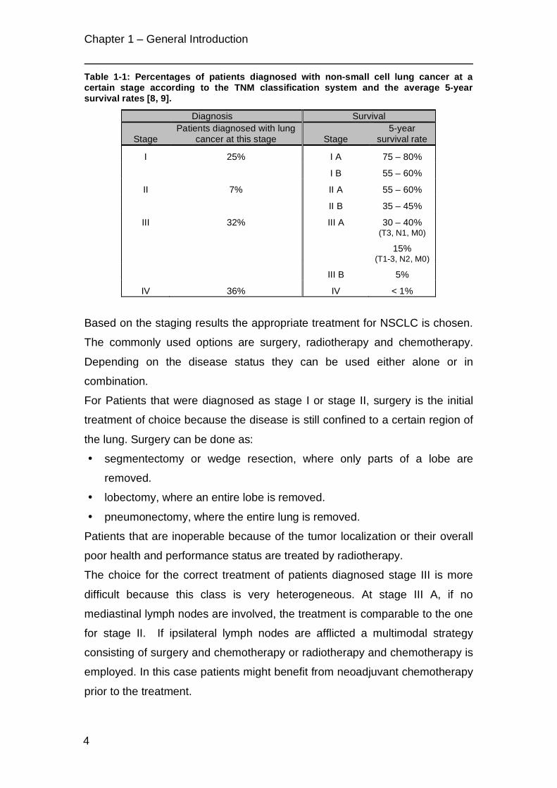
Table 1-1 gives an overview on the chance of discovery at a certain stage and
the 5-year survival rates [8, 9].
Chapter 1 – General Introduction
4
Table 1-1: Percentages of patients diagnosed with non-small cell lung cancer at a certain stage according to the TNM classification system and the average 5-year survival rates [8, 9].
Diagnosis Survival
Stage Patients diagnosed with lung
cancer at this stage Stage 5-year
survival rate
I 25% I A 75 – 80%
I B 55 – 60%
II 7% II A 55 – 60%
II B 35 – 45%
III 32% III A 30 – 40% (T3, N1, M0)
15% (T1-3, N2, M0)
III B 5%
IV 36% IV < 1%
Based on the staging results the appropriate treatment for NSCLC is chosen.
The commonly used options are surgery, radiotherapy and chemotherapy.
Depending on the disease status they can be used either alone or in
combination.
For Patients that were diagnosed as stage I or stage II, surgery is the initial
treatment of choice because the disease is still confined to a certain region of
the lung. Surgery can be done as:
• segmentectomy or wedge resection, where only parts of a lobe are
removed.
• lobectomy, where an entire lobe is removed.
• pneumonectomy, where the entire lung is removed.
Patients that are inoperable because of the tumor localization or their overall
poor health and performance status are treated by radiotherapy.
The choice for the correct treatment of patients diagnosed stage III is more
difficult because this class is very heterogeneous. At stage III A, if no
mediastinal lymph nodes are involved, the treatment is comparable to the one
for stage II. If ipsilateral lymph nodes are afflicted a multimodal strategy
consisting of surgery and chemotherapy or radiotherapy and chemotherapy is
employed. In this case patients might benefit from neoadjuvant chemotherapy
prior to the treatment.
Chapter 1 – General Introduction
5
Lung cancers at stage III B with contralateral involvement of lymph nodes are
considered as being unresectable. Therefore, chemoradiation is the
treatment of choice. If the tumor spread can be reduced by this therapy,
surgery might follow the initial treatment. Stage III B tumors with malignant
pleural effusions are treated as being M1 and hence the same therapeutic
conditions as for stage IV tumors apply.
As can be seen from Table 1-1 the prognosis for patients with metastatic
tumors (stage IV) is extremely poor. Since patients are considered as being
incurable a palliative chemotherapy and/or radiotherapy are applied to relieve
pain and other distressing symptoms and improve the patient’s quality of life.
After initial therapy an adjuvant chemotherapy or radiotherapy or
chemoradiation often continues the treatment to eradicate remaining
cancerous cells.
Table 1-2 gives an overview on the drugs that are currently used for
chemotherapy. Usually, a platin-based drug is given in combination with
another cytostatic agent. New and promising substances are epidermal
growth factor receptor tyrosine kinase inhibitors and the angiogenesis inhibitor
Bevacizumab, a monoclonal antibody that is directed against the vascular
endothelial growth factor.
Table 1-2: Chemotherapeutic agents currently used for the treatment of NSCLC and their mode of action.
Mechanism Drugs
Platin-based DNA-crosslinker Cisplatin Carboplatin
Mitotic inhibitors Paclitaxel Docetaxel Vinorelbine Vinblastine
Antimetabolites Gemcitabine Pemetrexed
Topoisomerase inhibitors Etoposide Irinotecan
Antitumor antibiotics Mitomycin C
Alkylating agents Ifosfamide
Epidermal growth factor receptor (EGFR) tyrosine kinase inhibitors
Gefitinib Erlotinib
Vascular endothelial growth factor (VEGF) inhibitors Bevacizumab

Chapter 1 – General Introduction
6
All these therapeutic agents are administered systemically resulting in more or
less severe unwanted side effects. The most prominent are nausea and
vomiting, diarrhea or obstipation, pain, fatigue, reduction of red and white
blood cells due to bone marrow depression or hair loss.
To circumvent such problems, regional drug delivery to the lungs via
aerosolized chemotherapy has been suggested. This route of administration
not only reduces the systemic burden but also avoids hepatic first-pass
metabolism and allows the deposition of higher drug levels at the site of
interest. Tatsumura et al. reported a high local deposition of 5-Fluorouracil
after inhalation and very low systemic drug concentrations while obtaining a
good anti-tumor response [10, 11]. Similar results were obtained in other
studies with e.g. aerosolized doxorubicine [12], cisplatin [13] or 9-nitro-20(s)-
camptothecin [14]. However, inhalative therapy is not yet established as a
standard treatment because the effect of high doses of chemotherapeutics
administered locally to the lung tissue still has to be evaluated to avoid
pulmonary toxicity and drug-induced lung diseases. Therefore, further studies
on new anti-cancer agents and drug formulations are needed to help this
promising therapeutic approach reach the clinics.
An interesting target for new anti-cancer agents is the enzyme telomerase,
which plays an important role in cell immortalization and hence cancer
development (see section 1.2). Telomerase has been found to be upregulated
in about 85% of lung cancers and its expression correlated with a poor
prognosis for NSCLC patients [15-18].

Chapter 1 – General Introduction
7
1.2 Telomeres, Telomerase and their Implication in Cancer
Development
In 1961 Hayflick and Moorehead demonstrated that human skin fibroblast in
culture could only undergo a limited number of about 40 to 50 cell divisions
before they entered the state of cellular senescence [19]. Furthermore, in
1965 Hayflick also reported a lower number of divisions for cells in culture
derived from older people than for cells from younger people [20]. He
suggested that this “countdown for senescence” is initiated at birth. Harley et
al. demonstrated in 1990 that the length of chromosomal ends, the telomeres,
shortens during aging of human fibroblasts [21]. The connection between
telomere shortening and cellular senescence and/or apoptosis was shown
after the discovery of the enzyme telomerase [22], a ribonucleoprotein that
maintains the chromosomal ends. Ectopic expression of the catalytic subunit
of this enzyme resulted in the elongation of telomeres and significantly
extended the life span of human primary cells [23, 24].
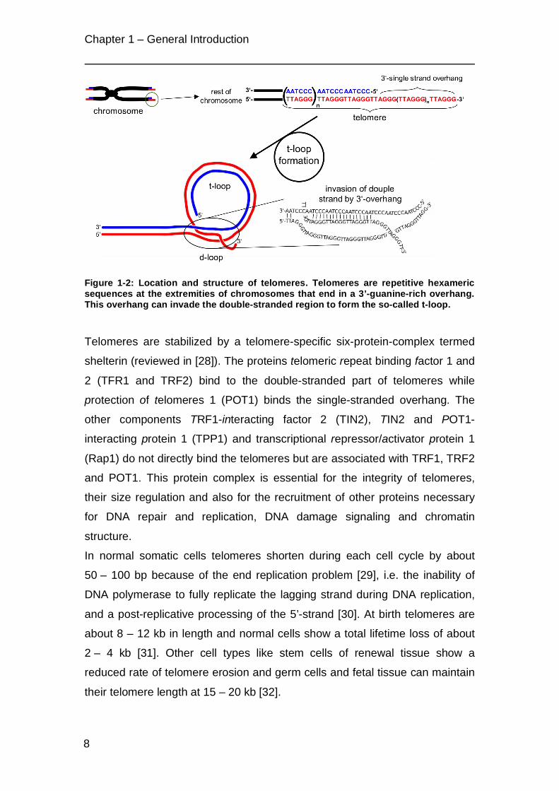
Telomeres are non-coding guanine-rich repetitive sequences that end in a
single-stranded 3’-overhang [25]. Humans telomeres are tandem repeats of
the hexameric sequence 5’-(TTAGGG)-3’, which is highly conserved in higher
eukaryotes [26] (Figure 1-2). Due to their special structure, telomeres protect
chromosomal ends from being recognized as damaged DNA. This prevents
the initiation of such events like non-homologous end joining or homology
directed repair, two mechanisms that strongly compromise the integrity of
chromosomal ends. To fulfill their protective function, telomeres form a lasso-
like structure, the so-called t-loop [27]. For the t-loop formation the G-rich
single strand 3’-overhang invades the double stranded region of telomeres to
form base pairs with the C-rich strand, thereby displacing the G-strand
(d-loop; Figure 1-2).
Chapter 1 – General Introduction
8
Figure 1-2: Location and structure of telomeres. Telomeres are repetitive hexameric sequences at the extremities of chromosomes that end in a 3’-guanine-rich overhang. This overhang can invade the double-stranded region to form the so-called t-loop.
Telomeres are stabilized by a telomere-specific six-protein-complex termed
shelterin (reviewed in [28]). The proteins telomeric repeat binding factor 1 and
2 (TFR1 and TRF2) bind to the double-stranded part of telomeres while
protection of telomeres 1 (POT1) binds the single-stranded overhang. The
other components TRF1-interacting factor 2 (TIN2), TIN2 and POT1-
interacting protein 1 (TPP1) and transcriptional repressor/activator protein 1
(Rap1) do not directly bind the telomeres but are associated with TRF1, TRF2
and POT1. This protein complex is essential for the integrity of telomeres,
their size regulation and also for the recruitment of other proteins necessary
for DNA repair and replication, DNA damage signaling and chromatin
structure.
In normal somatic cells telomeres shorten during each cell cycle by about
50 – 100 bp because of the end replication problem [29], i.e. the inability of
DNA polymerase to fully replicate the lagging strand during DNA replication,
and a post-replicative processing of the 5’-strand [30]. At birth telomeres are
about 8 – 12 kb in length and normal cells show a total lifetime loss of about
2 – 4 kb [31]. Other cell types like stem cells of renewal tissue show a
reduced rate of telomere erosion and germ cells and fetal tissue can maintain
their telomere length at 15 – 20 kb [32].

Chapter 1 – General Introduction
9
Critically short telomeres are identified as damaged DNA most probably
because of insufficient protection by shelterin proteins. They are recognized
by the two phosphatidyinositol-3-kinase related protein kinases, ATM and
ATR. This recognition triggers a molecular mechanism that results in the
activation of p53, RB and p21. As a consequence cell cycle progression is
inhibited and cells enter the state of senescence or even undergo apoptosis
(reviewed by [28, 31]). Telomere erosion alone can also serve as a
p53-independent checkpoint for cell proliferation. Cells that continue dividing
will face serious chromosomal damages that finally result in cell death. This
second checkpoint is termed “crisis” (Figure 1-3).
However, in the absence of functional tumor suppressors and after activation
of oncogenes like c-Myc because of the genomic instabilities and
rearrangements, cancer cells can escape these cell cycle checkpoints by
expressing the enzyme telomerase [31, 33, 34]. Telomerase is a
ribonucleoprotein complex that synthesizes new TTAGGG-sequences at the
3’-ends of chromosomes to maintain telomeres at a stable length. It is
composed of human telomerase RNA (hTR) [35], which contains the template
region for the synthesis of new telomeric repeats, and human telomerase
reverse transcriptase (hTERT), the catalytic subunit of the enzyme (Figure
1-4) [36]. In the active complex two hTR/hTERT units are associated with one
dyskerin molecule [37].
Telomerase is normally expressed at high levels during embryonic
development but is downregulated after birth in the larger parts of tissues. In
the healthy organism telomerase activity is limited to germ lines and certain
stem cell compartments, i.e. specialized cells that need to retain their
unlimited proliferative capacity [38, 39].

Chapter 1 – G
eneral Introduction
10
Figure 1-3: Mechanism
of cellular senescence and apoptosis and the influence of telomerase expression on cell survival. In norm
al cells telomeres
shorten until they reach a critical length and become dysfunctional. This triggers a m
echanism that results in cell cycle arrest, senescence and
apoptosis. Cells that do not stop dividing w
ill face serious chromosom
al damages due to the loss of telom
eres. Few cells like stem
cells, germ line
cells or fetal cells express the enzyme telom
erase and can therefore divide indefinitely. For cancer development the (re)activation of telom
erase is a crucial step tow
ard imm
ortality. They normally have very short telom
eres that constantly require telomere m
aintenance by telomerase.

Chapter 1 – General Introduction
11
Telomerase has been reported to be active in about 80-90% of cancer cells
[40, 41]. Its expression is a crucial step for their limitless replicative potential
and evasion from apoptosis, which are two of the six hallmarks of cancer [42].
Those cancer cells that do not express telomerase maintain their telomeres
by a homologous recombination-mediated process termed alternative
lengthening of telomeres (ALT; reviewed in [43, 44]).
Figure 1-4: Elongation of the 3’-strand overhang by telomerase. hTERT = human telomerase reverse transcriptase (the catalytic subunit); hTR = human telomerase RNA (contains the template region for the synthesis of new TTAGGG-repeats).
Due to its crucial role in carcinogenesis the inhibition of telomerase became
quickly an interesting target for the development of new anti-cancer
strategies. It could be shown that telomerase inhibition results in telomere
shortening and suppression of tumor growth both in vitro and in vivo.
To date numerous strategies for telomerase inhibition, either directly or
indirectly, have been investigated. The direct approaches targeted both the
catalytic subunit hTERT and the RNA component hTR. Potent inhibitors of
hTERT are the reverse transcriptase inhibitors BIBR 1532 [45] or AZT [46].
hTR could be successfully targeted with chemically modified oligonucleotides
that block the template region for the synthesis of new telomeric repeats [47,
48]. The lipid-linked oligonucleotide and telomerase template antagonist
GRN163L [49-51] has entered several clinical trials to prove its efficacy either
administered alone or in combination with established anti-cancer drugs
(www.clinicaltrias.gov).

Chapter 1 – General Introduction
12
Another strategy does not focus on telomerase but on its substrates, the
telomeres. Here the class of so-called G-quadruplex stabilizing substances
has been widely investigated. G-quadruplexes are special structures that can
form in guanine-rich DNA sequences like the telomeres. Their stabilization by
specially designed molecules prevent telomerase from recognizing the single-
strand overhangs. Furthermore, G-quadruplex stabilizers were reported to
induce telomere dysfunction due to the displacement of telomere-associated
proteins such as the shelterin complex. Therefore, a treatment with this kind
of inhibitors results in a faster response as could be expected from telomere
erosion alone [52-54].
Vaccination against hTERT as a tumor antigen is another approach currently
under investigation in clinical trials. Cancer patients are immunized against
short hTERT peptide sequences that can be recognized by cytotoxic
T-lymphocytes [55-57].
Other strategies employ adenoviral-mediated gene therapy. The gene
constructs contain promoter regions for hTR or hTERT so that transcription of
the actual gene sequence will only occur in telomerase expressing cells. This
mechanism is currently exploited for suicide gene therapy [58, 59] and
oncolytic viral therapy [60].
Concerns were raised that telomere inhibition might lead to severe side
effects because other telomerase expressing cells, i.e. germ cells and stem
cells, will also be affected by the treatment. However, telomeres from cancer
cells are normally considerably shorter than those from other telomerase
expressing cells [32]. Since cancer cells divide more rapidly, telomere erosion
in tumors should be much faster. Therefore the effect of telomerase inhibition
is expected to be much more pronounced in cancer cells than in other cell
types.
Another point of criticism in anti-telomerase therapy might be the lag time
between the start of the treatment and first effects on cell proliferation. This
period strongly depends on the initial telomere length of tumor cells. However,
it has been demonstrated that telomerase inhibition enhances the efficacy of
other anti-cancer therapies [61-66]. So even if telomerase inhibition alone

Chapter 1 – General Introduction
13
cannot be used because of a retarded effect this synergism might be
exploited in future therapies.

Chapter 1 – General Introduction
14
1.3 Objectives of the studies
This project was done in cooperation with the group of Professor Dr. Ulrich
Klotz from the Dr. Margarete Fischer-Bosch Institute for Clinical
Pharmacology, Stuttgart, and University of Tübingen and financially supported
by the Deutsche Krebshilfe e.V. (Project no.: 10-2035-Kl I) and the Robert
Bosch Foundation (Stuttgart, Germany).
The work described in Chapter 6 was done in the group of Professor Dr. Elias
Fattal, UMR CNRS 8612, Pharmaceutical Faculty of the University Paris Sud
11, Châtenay-Malabry, France, and financially supported by the GALENOS
Fellowship in the Framework of the EU Project "Towards a European PhD in
Advanced Drug Delivery, Marie Curie Contract MEST-CT-2004-404992.
The aim of the project was the development of new strategies for the
treatment of non-small lung cancer by telomerase inhibition with formulations
that are suitable for a local application via the inhalative route.
For this purpose different telomerase inhibitors that have been described in
the literature were tested. From these studies three substances were selected
as potential drug candidates:
• the acridine derivative BRACO19, which belongs to the telomere-targetig
G-quadruplex stabilizing substances [67].
• an antisense 2’-O-methyl-RNA (2OMR) that is complementary to the
template region of hTR [48].
• a small interfering RNA (siRNA) that is directed against the mRNA of the
catalytic subunit hTERT.
BRACO19 (Figure 1-5) could be regarded as the prototype of a new class of
drugs based on 3,6,9-substituted acridine derivatives. Although it has been
reported to be a very potent telomerase inhibitor and effective in targeting the
telomeres, nothing was known about its biopharmaceutical and physico-
chemical properties. Since these information are essential for the
development of delivery strategies, the studies with BRACO19 concentrated
on a thorough characterization regarding its transport across biological
barriers and stability under physiological conditions and a categorization

Chapter 1 – General Introduction
15
according to the biopharmaceutical classification system (BCS) as introduced
by Amidon et al. [68] (Figure 1-6; Chapter 2 and Chapter 3).
Figure 1-5: Structure of the 3,6,9-substituted acridine derivative BRACO19
Figure 1-6: The categorization of drug compounds according to the biopharmaceutical classification system. The key factors are the dissolution of the drug in biological fluids and its permeation accros biological barriers.
The oligonucleotide-based drugs 2OMR and siRNA are known to be very
effective and specific. However, due to their size and negative charge the
main obstacle for these molecules is their poor uptake into cells. Furthermore,
they are rapidly degraded in the presence of nucleases. Therefore, this kind of
drugs requires a suitable carrier system to ensure an efficient and safe
uptake.

Chapter 1 – General Introduction
16
For 2OMR a delivery system based on cationic chitosan/PLGA nanoparticles
was chosen. Chitosan and PLGA (Figure 1-7) are both biocompatible and
biodegradable.
Figure 1-7: Structures of the polyester poly(lactic-co-glycolic acid) (PLGA) and the polysaccharide chitosan which is composed of randomly distributed β-(1-4)-linked D-glucoseamine and N-acetyl-D-glucoseamine.
Nanoparticles composed of these polymers were previously developed for the
delivery of plasmid DNA. In our studies they were evaluated as carriers for the
short oligonucleotides 2OMR. The work focused on the influence of chitosan
content in the particle formulation on binding and delivery efficiency and the
fate of nanoplexes in different cell types as well as the efficacy regarding the
inhibition of telomerase activity (Chapter 4 and Chapter 5).
RNA interference (RNAi) is the latest mechanism described for gene
silencing. RNAi is mediated via short double stranded RNA sequences with a
length of normally 21 base pairs and overhanging 3’-ends [69, 70] termed
short inhibitory RNA (siRNA). After its discovery, it became rapidly a powerful
tool in molecular biology for studying the downregulation of gene expression.
These properties made it also interesting for medical applications.
For the delivery of siRNA a more refined system based on cationic
DOTAP/DOPE liposomes was investigated. These liposomes were modified
with the endogenous glycosaminoglycan hyaluronic acid (Figure 1-8) for the
targeting of cancer cells that overexpress the CD44-receptor.

Chapter 1 – General Introduction
17
Figure 1-8: Structures of the lipids DOTAP (1,2-dioleoyl-3-trimethylammonium-propane) and DOPE (1,2-Dioleoyl-sn-glycero-3-phosphoethanolamine) and the endogenous polymer hyaluronic acid.
In these studies the influence of the modification on liposome properties was
examined in comparison with non-modified liposomes. Special attention was
given to the liposome properties, binding and protection of siRNA in the
complex, the cytotoxicity of the new system and its efficiency for targeting
CD44-expressing lung cancer cells (Chapter 6).

18

Chapter 2 – Biopharmaceutical Characterization of BRACO19
19
Chapter 2
Biopharmaceutical Characterization of the
Telomerase Inhibitor BRACO19
The data presented in this chapter has been published as a short
communication in the journal Pharmaceutical Research:
Taetz, S., Baldes, C., Mürdter, T. E., Kleideiter, E., Piotrowska, K., Bock, U.,
Haltner-Ukomadu, E., Mueller, J., Huwer, H., Schaefer, U. F., Klotz, U.,
Lehr, C.-M.
Biopharmaceutical characterization of the telomerase inhibitor BRACO19.
(2006) Pharm. Res., 23(5), 1031-1037.
DOI:
10.1007/s11095-006-0026-y
Weblink:
http://www.springerlink.com/content/w06750366765q138/

20

Chapter 2 – Biopharmaceutical Characterization of BRACO19
21
2.1 Introduction
Telomerase, a ribonucleoprotein enzyme that belongs to the class of reverse
transcriptases, plays an important role in the control of proliferation and
carcinogenesis by maintaining the length of telomeres. Telomeres are lasso-
like structures at the end of chromosomes. They protect them from
recombination, nuclease degradation, DNA repair mechanisms and end-to-
end fusions. They also act as a kind of “mitotic clock” by constant erosion
after each cell cycle. Reaching a critical length is a signal for the cell to stop
dividing and enter the state of replicative senescence (M1 stage). Cells that
bypass replicative senescence and continue dividing will face serious DNA
damages and subsequently cell death due to further telomere shortening
(M2 stage or crisis). However, cells expressing high levels of telomerase, like
cancer cells, can escape this mechanism by keeping the telomere length
above this limit and therefore divide indefinitely [71, 72].
The approach of telomerase inhibition for the treatment of cancer attracted
increasing interest in the last years because telomerase is expressed in most
types of cancer cells but not in normal cells with the exception of
hematopoetic stem cells, germ cells, stem cells of the intestine and the skin.
Non- small cell lung cancer (NSCLC) is the most common form of lung cancer
and has a poor prognosis. Misawa et al. showed that telomerase inhibition in
the NSCLC cell line A549 led to an increase in apoptosis and higher
sensitivity to chemotherapeutic agents [64]. Inhibiting telomerase with suitable
drugs would ideally affect cancer cells only and spare healthy tissue. Due to
such inherent selectivity an inhalative application of telomerase inhibitors
might be possible. The drug could be delivered directly to the lung, thereby
further reducing unwanted systemic drug effects.
A screening for potential telomerase inhibitors led us to the acridine derivative
BRACO19 (9- [4- (N,N- dimethylamino)phenylamino]- 3,6- bis(3- pyrrolodino-
propionamido) acridine x 3HCl; Figure 2-1) [73]. This substance inhibits
telomerase activity and can lead to telomere dysfunction by G- quadruplex
stabilization in telomeres [67, 74].

Chapter 2 – Biopharmaceutical Characterization of BRACO19
22
Figure 2-1: The 3,6,9- substituted acridine derivative BRACO19 (9-[4-(N,N-dimethylamino)phenylamino]-3,6-bis(3-pyrrolodinopropionamido) acridine x 3HCl) according to [67].
For the development of a new drug formulation knowledge about
biopharmaceutical properties like solubility, cytotoxicity, permeation of the
drug across biological barriers (like the lung epithelia) or protein binding are
as important as the cytotoxic or pharmacological properties of the drug.
Protein binding and interaction with membrane lipids were tested by two
HPLC methods using immobilized human serum albumin (HSA) and
immobilized artificial membrane (IAM) chromatography, respectively.
To investigate the permeability of BRACO19 across relevant biological
barriers we used the SV40 virus immortalized cell line 16HBE14o- [75, 76]
and the adenocarcinoma cell line Calu-3 [77, 78] as models of the bronchial
epithelium. The alveolar epithelium was represented by primary human
alveolar type II cells which develop type I characteristics when cultivated
under appropriate conditions [79, 80]. For comparison we also included the
intestinal adenocarcinoma cell line Caco-2, an established model for intestinal
drug absorption [81].

Chapter 2 – Biopharmaceutical Characterization of BRACO19
23
2.2 Materials and Methods
2.2.1 Substances and buffers
BRACO19 was synthesized by ENDOTHERM GmbH (Saarbruecken,
Germany) according to Harrison et al. [82]. Identity was proven by NMR,
purity by HPLC. Propranolol- HCl was purchased from Synopharm GmbH &
Co. KG (Barsbuettel, Germany), cyclosporin A from Calbiochem® (Merck
Bioscience GmbH, Bad Soden, Germany), fluorescein- Na and
tetraethylammonium chloride (TEAC) were obtained from Fluka Chemie
GmbH (Buchs, Switzerland).
HBSS (Hank’s balanced salt solution) buffer was composed of 137.0 mM
NaCl, 5.36 mM KCl, 4.26 mM NaHCO3, 0.18 mM Na2HPO4 x 7 H2O, 0.44 mM
KH2PO4, 5.55 mM Glucose, 10.0 mM HEPES (N-[2-hydroxyethyl]piperazine-
N’-[2-ethanesulfonic acid]), 0.13 mM CaCl2 x 2 H2O, 0.05 mM MgCl2 x 6 H2O,
0.04 mM MgSO4 x 7 H2O. For transport experiments 0.25% BSA (Sigma-
Aldrich Chemie GmbH, Taufkirchen, Germany) was added to the HBSS buffer
(HBSS/BSA buffer).
Sterile BSS (balanced salt solution) contained 137.0 mM NaCl, 5.0 mM KCl,
0.7 mM Na2HPO4 x 7 H2O, 1.2 mM MgSO4 x 7 H2O, 5.5 mM Glucose,
10.0 mM HEPES, 0.18 mM CaCl2, 100 units/ml penicillin and 100 µg/ml
streptomycin. All reagents were of cell culture grade. The pH was adjusted to
7.4 with 1 M NaOH.
2.2.2 Cells and cell culture conditions
Calu-3 cells (HTB- 55, ATCC, Manassas, VA, USA) were cultivated in
Minimum Essential Medium (MEM) with Earl’s Salts and L- glutamine (PAA
Laboratories GmbH, Pasching, Austria) supplemented with 10% FCS, 1%
MEM non- essential amino acid (NEAA) solution and 1 mM sodium pyruvate
(all from Sigma- Aldrich).
16HBE14o- cells were a gift from Dr. Dieter Gruenert (Department of
Medicine, University of Vermont, Burlington, VT, USA). They were grown in
MEM with Earl’s Salts and L- glutamine (PAA Laboratories GmbH)

Chapter 2 – Biopharmaceutical Characterization of BRACO19
24
supplemented with 10% FCS, 1% NEAA solution and 3 mM glucose (Sigma-
Aldrich).
Caco-2 cells (C2BBe1, ATCC) were cultivated in Dulbecco’s Modified Eagle
Medium (DMEM) with high glucose (4.5 g/ml) and L- glutamine (PAA
Laboratories GmbH) supplemented with 10% FCS and 1% NEAA solution.
All cell lines were kept in 5% CO2 and 90- 98% humidity at 37°C.
For transport studies Calu-3 (passage 27 and 32), 16HBE14o- (passage 2.68)
and Caco-2 (passage 83) cells were seeded at a density of 60,000 cells/cm2
on Transwell® polyester (PET) filter inserts with a growth area of 1.13 cm2 and
a pore size of 0.4 µm (Transwell® Permeable Supports, Corning Inc., NY,
USA).
Primary human alveolar epithelial cells (hAEpC) were isolated from non-
tumor lung tissue of patients undergoing partial lung resection according to
Elbert et al. [79] with slight modifications of the enzymatic digestion and cell
purification [83]. In brief, the chopped tissue was digested using a
combination of 150 mg trypsin type I (Sigma) and 3 mg elastase (Worthington
Biochemical Corp., Lakewood, NJ, USA) in 30 ml BSS for 40 min. at 37°C.
The AT II cell population was purified by a combination of differential cell
attachment, percoll density gradient centrifugation and positive selection of
epithelial cells with magnetic beads (human Anti- HEA (Ep-CAM) MicroBeads,
Miltenyi Biotec, Bergisch Gladbach, Germany). Cell viability was assessed by
trypan blue staining.
The isolated hAEpC were seeded at a density of 600,000 cells/cm2 on
collagen/ fibronectin coated Transwell® polyester filter inserts (Corning Inc.)
with a growth area of 0.33 cm2. They were grown in SAGM medium (Cambrex
BioScience Walkersville Inc., Walkersville, MD, USA) supplemented with 1%
FCS, 100 units/ml penicillin and 100 µg/ml streptomycin (Sigma- Aldrich).
The formation of confluent monolayers and tight junctions was verified by
measuring the transepithelial electrical resistance (TEER) using an epithelial
voltohmmeter (EVOM, World Precision Instruments, Berlin, Germany) with a
STX-2 electrode.
For transport experiments, Calu-3 cells were used 14 days post seeding,
16HBE14o- cells and Caco-2 cells were used after 7 and 21 days,

Chapter 2 – Biopharmaceutical Characterization of BRACO19
25
respectively. hAEpC were cultivated for 8 days. TEER values were as
reported before or higher for transport experiments [79, 84]: about
1000 Ω x cm2 for Calu-3, 800 Ω x cm2 for 16HBE14o-, 750 Ω x cm2 for Caco-2
and 1400 Ω x cm2 for hAEpC.
2.2.3 Transport studies
Prior to transport experiments cell culture media were removed and the apical
and basolateral compartments were washed twice with HBSS/BSA buffer.
Cells were equilibrated for 2- 2.5 hours with this buffer.
After the equilibration period buffer was exchanged with drug solutions of the
same buffer in either the apical or basolateral compartment. The volumes for
the apical and basolateral side for 16HBE14o-, Calu-3 and Caco-2 were
500 µl and 1500 µl, respectively. The volumes for hAEpC were 200 µl for the
apical side and 800 µl for the basolateral side.
50 µl samples from the donor compartments were drawn immediately at the
beginning and at the end of the transport experiments. 50 µl samples from the
acceptor compartments were drawn after 30, 60, 120, 180, 240 and
300 minutes. An equal volume of fresh buffer was returned to the acceptor
compartment after each sampling. The filter plates were kept under cell
culture conditions and were slightly shaken with an orbital shaker. TEER
values were measured after the equilibration period and at the end of
transport experiments to verify that the barrier function of the monolayers was
not compromised. The drug concentrations in the donor compartments were
as follows: 16HBE14o-: 20 µg/ml of propranolol, fluorescein and BRACO19,
respectively; Calu-3 and Caco-2: 20 µg/ml fluorescein, 100 and 200 µg/ml
BRACO19; hAEpC: 100 and 200 µg/ml BRACO19.
Each experiment was performed fivefold in either direction.
2.2.4 Transport experiments with different inhibitors
Calu-3 cells are known to express the P- glycoprotein (P-gp) efflux system
[85] and organic cation transporter proteins (influx/ efflux of organic cations)
[84]. Inhibition of BRACO19 transport was studied at a donor concentration of
200 µg/ml as described in Transport studies with the following modifications.
For the inhibition of P-gp and other multi drug resistance efflux systems we

Chapter 2 – Biopharmaceutical Characterization of BRACO19
26
added 10 µM cyclosporin A [86] to the HBSS/BSA buffer. For the inhibition of
organic cation transporter proteins the buffer was supplemented with 5 mM
TEAC [87]. The inhibitors were present in both compartments. Transport
experiments were also performed at 4°C to check for other active transport
mechanisms as well as at 37°C without inhibitors for comparison. The results
were compared to those of the previous experiments with Calu-3 cells.
2.2.5 Calculation of apparent permeability coefficient Papp
The apparent permeability coefficient Papp was calculated according to :
!
Papp (cm / sec) =J
A "Co
Equation 2-1
where J is the linear section of the flux (µg/sec), A the filter area (cm2) and C0
the initial donor concentration (µg/cm3).
2.2.6 Determination of uptake into cells and adsorption to filter material
For adsorption/ uptake studies filters were washed twice with HBSS buffer
after the transport experiments and the filter membranes with cells were cut
out. Cells were lysed in 1 ml of a 80:20 mixture of methanol:HBSS (v/v) by
means of ultrasonication for 30 minutes. For removal of cell fragments the
suspensions were first kept at -80°C for 45 minutes and were then centrifuged
for 20 minutes at 14,000 rpm and 0°C. An aliquot of the supernatant was used
for analysis.
2.2.7 Sample analysis
BRACO19 and propranolol samples were analyzed by reversed phase HPLC
using an isocratic Dionex HPLC system consisting of an ASI 100 automated
sample injector, UVD 340U diode array detector and P680 pump with
Chromeleon® software (version 6.60 SP1 build 1449) (Dionex, Idstein,
Germany).
For BRACO19 analysis a Gemini® RP- 18 column (150 x 4.6 mm/ 5 µm/
110 Å) (Phenomenex, Aschaffenburg, Germany) was used. The mobile phase
was composed of 80:20 (v/v) methanol:borate buffer pH 10.0 (100 mM). At a
Chapter 2 – Biopharmaceutical Characterization of BRACO19
27
flow rate of 0.6 ml/min the retention time of BRACO19 was 8.9 ± 0.2 minutes.
The UV- detector was set at 268 nm.
Propranolol was analyzed with a Lichrospher® RP- 18 column (125 x 4 mm/
5 µm) (Merck, Darmstadt, Germany). The mobile phase was composed of
45:33:22 (v/v/v) water:methanol:acetonitrile supplemented with 0.033% (v/v)
triethylamine and 0.044% (v/v) phosphoric acid. The flow rate was set to
1.2 ml/min and the retention time was 2.9 ± 0.25 minutes. Propranolol was
detected at 215 nm.
Injection volumes for both substances were 20 µl per sample.
Fluorescein was analysed by fluorimetry using a Cytofluor II fluorescence
reader with Cytofluor software version 4.2 (PerSeptive Biosystems,
Wiesbaden- Norderstedt, Germany). 50 µl samples were diluted to 200 µl with
1 mM NaOH in a 96- well plate. The excitation wavelength was set to 485 nm
and the emission wavelength to 530 nm.
All unknown samples were calculated against known standards. Standards
were in the range of 0.02- 20 µg/ml. If necessary, samples were diluted 1:10.
2.2.8 IAM chromatography measurements
Immobilized artificial membrane (IAM) chromatography of BRACO19 was
performed with a phosphatidylcholine-functionalized column (Regis
Technologies). The mobile phase was composed of potassium buffer pH 6.8
and acetonitrile. A Waters HPLC system W2790 and PDA detector W2996
with Millenium32 software (Milford, Massachusetts, USA) was used for
analysis. The classification of the drug cellular membrane interaction in terms
of permeability was performed by the marker molecules uracil (eluting with the
injection peak), atenolol (3.07, 50 %), ketoprofen (5.55, 92 %),
carbamazepine (7.64, 70 %) and propranolol (11.55, 90 %). The values in
brackets indicate the KIAM values for the compounds and the corresponding
fraction absorbed according to the Biopharmaceutical Classification
System [88].

Chapter 2 – Biopharmaceutical Characterization of BRACO19
28
2.2.9 HSA binding
HSA binding of BRACO 19 was investigated by using a human serum albumin
functionalized column (Thermo Hypersil) according to a method developed by
Across Barriers GmbH. The mobile phase was composed of ammonium
acetate buffer pH 7.4 and propanol. An isocratic Waters HPLC system W2690
and dual wavelength detector W2487 with Millenium32 software (Milford,
Massachusetts, USA) was used for analysis. For the classification of drug
binding the following system was established from different literature values:
0- 40% for weak, 40- 50% for weak/ medium, 50- 85% for medium, 85- 95%
for medium/ strong and > 95% for strong drug binding to HSA. Substances for
calibration were acetaminophen (24% protein binding), carbamazepine
(76 %), propranolol (90 %) and naproxen (99 %).
2.2.10 MTT cytotoxicity assay
Cytotoxicity of BRACO19 was tested using the methyl-thiazolyl-tetrazolium
(MTT) assay. 16HBE14o-, Calu-3 and Caco-2 cells were grown at a density of
60,000 cells/ cm2 on 96 well Cellstar® tissue culture plates (Greiner bio-one,
Frickenhausen, Germany) for 6 days under cell culture conditions (see above)
in 200 µl medium with different concentrations of BRACO19. hAEpC were
grown under the same conditions but at a density of 30,000 cells/cm2.
BRACO19 concentrations were in the range of 0- 50 µM. Each concentration
was tested in quadruplicate. Cell culture medium with or without BRACO19
was changed every other day. After 6 days the medium was exchanged for
200 µl BRACO19- free medium and 10 µl of 5 mg/ml MTT solution in
phosphate buffered saline (PBS) were added to each well. After 3.5 hours
incubation the medium was removed and the cells were washed with PBS.
200 µl of 100% isopropanol were added to each well and the plate was left for
crystal dissolution on an orbital shaker over night. Absorption was measured
at a wavelength of 550 nm with an UV/Vis reader (SLT Spectra, Tecan
Deutschland GmbH, Crailsheim, Germany). Viability of cells treated with
BRACO19 was related to cells grown in BRACO19- free medium. The IC50
value was determined by non- linear regression using SigmaPlot® Version 9
(SPSS Inc., Chicago, Illinois).
Chapter 2 – Biopharmaceutical Characterization of BRACO19
29
2.3 Results
2.3.1 Solubility, cytotoxicity, protein binding and IAM chromatography measurements
BRACO19 (Figure 2-1) showed good and rapid solubility of at least 2 mg/ml in
distilled water and physiological buffer solutions in a pH range of 2.8- 7.4. For
transport experiments we used a 2 mg/ml stock solution in HBSS/BSA buffer
(pH 7.4) which was diluted to the desired concentration before each
experiment in the same buffer system.
The IC50 values found in the MTT cytotoxicity test were as follows: Calu-3
13.6 ± 3.8 µM, 16HBE14o- 3.6 ± 1.2 µM and hAEpC 6.0 ± 0.4 µM.
Interestingly, Caco-2 cells were not affected by BRACO19 concentrations up
to 50 µM.
BRACO19’s binding to human serum albumin was found to be 38% indicating
a weak to medium protein binding referring to the reference compounds. By
the IAM chromatography measurements BRACO19 was eluted very early
from the column together with the injection peak. This points out a low
potential for BRACO19 to overcome barriers like phospholipid membranes.
2.3.2 Transport experiments
In the first transport experiment the permeability of BRACO19 was compared
with the high permeability marker propranolol and the low permeability marker
fluorescein in 16HBE14o-- cells. BRACO19 transport was found to be
asymmetrical and very low. For the apical to basolateral (AB) direction no
transport could be detected at all. In basolateral to apical (BA) direction
transport was about tenfold lower than for fluorescein.
The results for Calu-3, Caco-2 and hAEpC were comparable to those found
with 16HBE14o-- cells. Even after increasing the donor concentrations of
BRACO19 to 100 and 200 µg/ml to facilitate detection in the receiver
compartment no transport could be detected in AB direction for neither cell
line. Also, in BA direction the apparent permeability coefficients were still
lower than the permeability of fluorescein. The results of all transport
experiments are summarized in Table 2-1.

Chapter 2 – Biopharmaceutical Characterization of BRACO19
30
Table 2-1: Apparent permeability coefficients (Papp; in cm/sec x 107) of BRACO19 in comparison with propranolol and fluorescein in different cell lines (16HBE14o-, Calu-3, Caco-2), primary human alveolar epithelial cells (hAEpC) and cell free filters. n.d. = no substance detectable in acceptor compartment; X = high BRACO19 concentrations led to a collapse of monolayer integrity.
16HBE14o- Calu-3 Caco-2 hAEpC filters without cells
Substance initial donor concentration A B B A A B B A A B B A A B B A A B B A
BRACO19 20 µg/ml n.d. 0.98 ± 0.23 - - - - - - 70.08
± 10.47 94.50 ± 1.85
BRACO19 100 µg/ml - - n.d. 0.25 ± 0.05 n.d. 0.31
± 0.03 n.d. 0.66 ± 0.07 - -
BRACO19 200 µg/ml - - n.d. 0.32 ± 0.05 n.d. 0.57
± 0.03 X X - -
Fluorescein 20 µg/ml 4.59 ± 0.66
9.59 ± 0.67
1.15 ± 0.18
1.26 ± 0.14
9.46 ± 1.16
8.95 ± 1.35 - - - -
Propranolol 20 µg/ml 131.59 ± 7.37
106.51 ± 5.97 - - - - - - - -
Transport experiments with cell free filters and BRACO19 solution were
performed under analogous conditions to look for the influence of the
Transwell® system. The Papp values were about 100- fold higher than for filters
with cells (data not shown), indicating that the permeation through cell free
filters is not rate limiting.
TEER values of all cell monolayers remained stable or even increased slightly
during transport studies. Only in the experiment with hAEpC and 200 µg/ml
BRACO19 donor concentrations a strong decrease in TEER to about
200 Ω x cm2 could be found. This was accompanied by a comparably strong
non- linear increase in BRACO19 concentrations in the respective acceptor
compartments.
2.3.3 Experiments with transport inhibitors and uptake/ adsorption studies
The results for transport experiments with Calu-3 cells in the presence of
different inhibitors are summarized in Table 2-2. They were comparable to
those under normal conditions: no transport in AB direction was detected and
the Papp values for BA transport were within the range of the former
experiments. This indicates that efflux/ influx systems or active transport are
not involved in the transport of BRACO19.

Chapter 2 – Biopharmaceutical Characterization of BRACO19
31
Table 2-2: Papp values (in cm/sec x 107) of BRACO19 (200 µg/ml donor concentration) in Calu-3 cells under normal conditions, in the presence of the P-gp inhibitor cyclosporin A (10 µM), the organic cation transporter protein inhibitor TEAC (5 mM) and at 4°C. n.d. = no substance detectable in acceptor compartment.
control Cyclosporin A TEAC 4°C
Substance initial donor concentration A B B A A B B A A B B A A B B A
BRACO19 200 µg/ml n.d. 0.27 ± 0.03 n.d. 0.92
± 0.10 n.d. 0.31 ± 0.03 n.d. 0.18
± 0.04
The asymmetry found in the transport experiments could also be observed in
the uptake/ adsorption studies (Figure 2-2). Filters that were in direct contact
with BRACO19 solution from the basolateral side (BA transport) contained
more BRACO19 than those that were in contact with the solution from the
apical side (AB transport). An exception was the transport experiment with
hAEpC at 200 µg/ml BRACO19 donor concentration where the integrity of the
monolayers collapsed. Here the amounts of BRACO19 in filters found for AB
transport were similar to those of BA transport. Amounts of BRACO19 found
in cell free filters submitted to the same conditions were comparable to those
of BA transport.
Figure 2-2: Amounts of BRACO19 (µg/cm2) recovered from excised filters. Concentrations refer to initial donor concentration of BRACO19. * = integrity of monolayer compromised by high BRACO19 concentration.

Chapter 2 – Biopharmaceutical Characterization of BRACO19
32
2.4 Discussion
Our studies showed that BRACO19 has a good solubility in aqueous media in
the concentrations that were used. The in vitro cytotoxicity of BRACO19 for
our pulmonary epithelial cells was comparable to values found in literature for
other cancerous cell lines derived from other organs (e.g. vulva carcinoma
cells, breast cancer cells or uterus carcinoma cells) [67, 74, 89]. In
comparison to “classic” anti- cancer drugs, the cytotoxicity of BRACO19 was
within the range of cisplatin (10- 25 µM) [90, 91] but lower than for doxorubicin
(0.4 - 2.4 µM) [92], paclitaxel (0.7- 1.8 nM) [91] or vincristine (1.9- 3.5 nM)
[93]. However, it has to be kept in mind that the strategy of this therapeutic
approach is the (re)induction of senescence and apoptosis by telomerase
inhibition (i.e. a controlled cell death) and not the simple poisoning of
malignant cells. Therefore, a high IC50 value is desired.
Protein binding of BRACO19 to HSA was rather weak compared to reference
drugs. The results from IAM chromatography measurements suggest that
interaction of BRACO19 with membrane lipids is negligible because there is a
proportional relationship between the capacity factor k’IAM and the membrane
partitioning coefficient Km [94].
Our transport studies showed that BRACO19 has great problems to overcome
biological barriers. The transport experiments with Caco-2 cells, a standard
model for intestinal drug absorption, indicate that BRACO19 might not be
suitable for oral administration. Burger et al. already demonstrated that tumor
xenografts in mice did not respond to an oral treatment with BRACO19. Even
when BRACO19 was administered intraperitoneally, only early stage tumors
were susceptible to the medication but not late stage tumors [74].
Since the results for the intestinal cell line Caco-2 are comparable to those of
our bronchial cell lines 16HBE14o- and Calu-3 as well as to the primary
alveolar cells hAEpC, we can assume that a topical application, like an
inhalative treatment of lung cancer, will face similar problems as the oral
route. Although a systemic availability is not needed, BRACO19 has at least
to be able to overcome the epithelial (air/ blood) barrier to reach deeper
regions of a lung tumor. The transport results are confirmed by

Chapter 2 – Biopharmaceutical Characterization of BRACO19
33
IAM chromatography measurements because the capacity factor k’IAM can be
correlated with the apparent permeability coefficient Papp [95].
Also, the concentrations used for the transport experiments are not
representative for possible therapeutic applications because they were far
above the IC50 values of our cells (10 µM ≈ 7 µg/ml). hAEpC were most
sensitive to high BRACO19 concentrations in the transport experiments as
could be seen in the strong decrease in TEER at 200 µg/ml donor
concentration. Since the major part of the pulmonary surface belongs to the
alveolar region, high drug concentrations would result in unwanted side
effects. Lower concentrations, however, would result in even less drug
absorption and inefficacy.
The apparent asymmetry in transport can be explained with a relatively strong
adsorption to the filter material as has been demonstrated in the experiments
with cell free filters (Fig. 2). For transport experiments in BA direction the
filters were rapidly saturated with BRACO19 because they were in direct
contact with the solution. Hence, transport was not hindered. However, when
BRACO19 was applied from the apical side, the small amounts that
permeated across the monolayer were completely adsorbed to the filter
material. Since the filter was not saturated at the end of the transport
experiment no substance could be detected in the basolateral compartment.
Differences in adsorptions (Fig. 2) were most likely due to varying amounts of
BRACO19 adsorbed to surface mucus and proteins (for AB direction) or
entrapped in the intercellular space.
Our observations can be explained by BRACO19’s chemical properties. It
contains two basic pyrrolidine rings (Fig. 1), which are very likely to be
protonated under physiological conditions, i.e. the molecule is positively
charged. This results in good water solubility but strongly decreases the
interaction with hydrophobic structures like cell membranes. The small
amounts that were transported in BA direction most probably took the
paracellular route by passive diffusion because no active transport
mechanisms or efflux/ influx systems like P-gp or organic cation transporter
proteins could be identified. Also the fact that BRACO19 does not interact with
membrane phospholipids and that higher amounts where only detectable in

Chapter 2 – Biopharmaceutical Characterization of BRACO19
34
the acceptor compartments and filters after the monolayer integrity collapsed
argue for the paracellular way
Chapter 2 – Biopharmaceutical Characterization of BRACO19
35
2.5 Conclusion
From our results we would suggest that BRACO19 has the typical properties
of a class III drug substance according to the Biopharmaceutical Classification
System (BCS): a good aqueous solubility and a very poor permeability across
biological barriers.
Obviously, BRACO19 is a potent substance with an interesting new mode of
action, i.e. telomerase inhibition by G- quadruplex stabilization, but with
challenging biopharmaceutical properties. Hence, suitable formulations for the
efficient delivery of this compound must be developed first in order to further
evaluate this new therapeutic concept.

36

Chapter 3 - Decomposition of BRACO19 in Physiological Media
37
Chapter 3
Decomposition of the Telomere Targeting Agent
BRACO19 in Physiological Media Results in
Products with Decreased Inhibitory Potential
The data presented in this chapter has been published as a research article in
the journal International Journal of Pharmaceutics:
Taetz, S., Murdter, T. E., Zapp, J., Boettcher, S., Baldes, C., Kleideiter, E.,
Piotrowska, K., Schaefer, U. F., Klotz, U., Lehr, C.-M.
Decomposition of the Telomere-Targeting agent BRACO19 in physiological
media results in products with decreased inhibitory potential. (2008) Int. J.
Pharm., 357(1-2), 6-14
DOI:
10.1016/j.ijpharm.2008.01.026
Weblink:
http://www.sciencedirect.com/science?_ob=ArticleURL&_udi=B6T7W-
4RMNYGY-
5&_user=10&_rdoc=1&_fmt=&_orig=search&_sort=d&view=c&_acct=C00005
0221&_version=1&_urlVersion=0&_userid=10&md5=1bccab3d5f2ceb071d53
ea25b525033f

38

Chapter 3 - Decomposition of BRACO19 in Physiological Media
39
3.1 Introduction
During the last years the concept of telomerase inhibition for the treatment of
cancer became an area of intensive research. Telomerase is a reverse
transcriptase composed of the catalytic subunit human telomerase reverse
transcriptase (hTERT) and the template RNA human telomerase RNA (hTR).
Its substrates, the telomeres, are located at the ends of each chromosome.
Telomeres are repetitive TTAGGG/AATCCC sequences that end in a
3’-(TTAGGG)n single strand overhang. They form a so-called t- loop, a
structure stabilized by associated proteins, where the single strand invades
the double stranded region. The t- loops prevent the chromosome ends from
end-to-end fusions and being recognized as damaged DNA. Due to the end
replication problem during DNA duplication telomeres erode at each cell cycle
until they approach a certain limit. Reaching this limit is an important signal for
a cell to enter the state of cellular senescence. Cells that do not stop dividing
will experience severe chromosomal damages leading to cell death
(apoptosis). Since most malignant cells express telomerase they are able to
maintain their telomeres above this limit and therefore escape these pathways
[71, 72, 96]. Various telomerase inhibitors have been developed and
successfully tested. They either target hTERT, hTR or the telomeres [97].
Our studies concentrate on the topical treatment of non-small cell lung cancer
(NSCLC) via the inhalative route. A search for suitable drug candidates led to
the telomere targeting 3,6,9- aminoacridine derivative BRACO19 ((9- [4- (N,N-
dimethylamino)phenylamino]- 3,6- bis (3- pyrrolodino-propionamido) acridine;
figure 1) which acts by G-quadruplex stabilization [67, 73]. G-quadruplexes
are planar G-quartet motifs that can form in guanine-rich DNA sequence like
the telomeres. These structures interact with the inhibitor via π-π stacking.
Positively charged side chains of the inhibitor interact with the negatively
charged phosphate DNA backbone and thereby stabilize the G-quadruplex –
inhibitor complex. Due to this mechanism telomeres are not able to form their
native structure and hence fail to protect the chromosomes [53, 54].
BRACO19 has shown some promising results in studies in tumor cell cultures
and mouse xenografts. It was reported that a treatment with BRACO19 not

Chapter 3 - Decomposition of BRACO19 in Physiological Media
40
only resulted in telomerase inhibition but also in general telomere dysfunction
that led to atypical mitosis and consequently to apoptosis [74].
In a previous study we characterized BRACO19 with respect to its
biopharmaceutical properties and found it to be a typical class III drug
substance with a good aqueous solubility but a very poor permeability across
epithelial cell monolayers [98]. During these studies we also found that
BRACO19 has stability problems when dissolved in aqueous media at
physiological pH.
In the present study we examined the influence of pH, temperature and
dissolution media on the stability of BRACO19. We have identified potential
decomposition products and evaluated whether they can contribute to the
inhibitory action of this drug candidate.

Chapter 3 - Decomposition of BRACO19 in Physiological Media
41
3.2 Materials and Methods
3.2.1 BRACO19
BRACO19 (9- [4- (N,N- dimethylamino)phenylamino]- 3,6- bis (3- pyrrolidino-
propionamido) acridine x 3HCl; figure 1) was synthesized by ENDOTHERM
GmbH (Saarbruecken, Germany) according to Harrison et al. [82]. Identity
and puritiy were proven by NMR (Table 3-3) and HPLC, respectively, in
comparison with the original compound.
3.2.2 Buffers and cell culture medium for stability studies
For stability studies different HBSS (Hank’s balanced salt solution) buffers
and diluted McIlvain buffer were used.
The basic compositions of HBSS buffers were: 137.0 mM NaCl, 5.36 mM KCl,
4.26 mM NaHCO3, 0.18 mM Na2HPO4 x 7 H2O, 0.44 mM KH2PO4, 5.55 mM
Glucose, 0.13 mM CaCl2 x 2 H2O, 0.05 mM MgCl2 x 6 H2O, 0.04 mM
MgSO4 x 7 H2O. For the adjustment of the appropriate pH values in HBSS
buffers the buffering substances were exchanged as follows: pH 7.4 and 7.0:
10.0 mM HEPES (N-[2-hydroxyethyl]piperazine-N’-[2-ethanesulfonic acid]),
pH 6.0: 10.0 mM MES (2-morpholinoethanesulfonic acid), pH 4.0 and 5.0:
10.0 mM sodium acetate. pH values were adjusted with 1 M NaOH or 1 M
HCl, respectively.
Diluted McIlvain buffer contained 50 mM citric acid and 1.4 mM Na2HPO4.
The pH was 2.8. Phosphate buffer was composed of 5 mM KH2PO4 and the
pH was adjusted with NaOH to pH 7.4.
RPMI cell culture medium was purchased from PAA Laboratories GmbH
(Pasching, Austria) and supplemented with 10% FCS (Sigma- Aldrich Chemie
GmbH, Taufkirchen, Germany). The pH of the cell culture medium was 7.5.
3.2.3 HPLC- DAD analysis of BRACO19 and decomposition products
BRACO19 was analyzed by reversed phase HPLC using an isocratic Dionex
HPLC system consisting of an ASI 100 automated sample injector with
adjustable temperature, UVD 340U diode array detector (DAD) and

Chapter 3 - Decomposition of BRACO19 in Physiological Media
42
P680 pump with Chromeleon® software (version 6.60 SP1 build 1449)
(Dionex, Idstein, Germany).
We used a Gemini® RP- 18 column/ 150 x 4.6 mm/ 5 µm/ 110 Å
(Phenomenex, Aschaffenburg, Germany). The mobile phase was composed
of 80:20 (v/v) methanol:borate buffer pH 10.0 (100 mM). At a flow rate of
0.6 ml/min the retention time of BRACO19 was 8.9 ± 0.2 minutes. The
wavelength of the detector was set at 268 nm. The detection and
quantification limit of BRACO19 were 0.010 µg/ml and 0.025 µg/ml,
respectively. Quantification was linear in the range from 0.025 to 12 µg/ml.
3.2.4 Decomposition experiments
All decomposition experiments were performed with the HPLC-DAD system
described above in HPLC brown glass vials (hydrolysis grade 1;
CS-Chromatography, Langerwehe, Germany). Decomposition experiments
over 7 hours were performed at 37° with HBSS buffer of different pH values
and diluted McIlvain buffer of pH 2.8. The influence of temperature on
BRACO19 decomposition was assessed at 4°C in comparison to the
decomposition at 37°C in HBSS buffer pH 7.4. BRACO19 was dissolved at a
concentration of 12 µg/ml in each respective buffer. 20 µl samples were
drawn every 45 minutes for 7 hours and analyzed by HPLC as described
above. Each experiment was done in triplicate.
Long-term decomposition experiments were performed for four days in HBSS
buffer pH 7.4, phosphate buffer pH 7.4 and RPMI cell culture medium
containing 10% FCS (pH 7.5), respectively. The concentration of BRACO19
was 12 µg/ml and the temperature was set to 37°C. Samples were drawn
every 2.54 hours. All experiments were performed in triplicate.
UV spectra of degradation products were recorded with the DAD detector
during these long-term experiments.
The decomposition of BRACO19 in different media was described
mathematically by assuming first order kinetics. A non-weighted curve fitting
was performed using the software Origin (Version 7.5, OriginLab Corp.,
Northampton, MA, USA) according to the equation:

Chapter 3 - Decomposition of BRACO19 in Physiological Media
43
!
A = A0" e
(#k" t ) Equation 3-1
where A is the amount of BRACO19 (in %) at time point ti, A0 the amount at
time point t = 0, k the rate constant (in h-1) and t the time (in hours).
3.2.5 Decomposition of BRACO19 for structural analysis of decomposition products by LC/MS and NMR
For the analysis of the decomposition products by LC/MS and NMR
BRACO19 was dissolved in phosphate buffer pH 7.4 at a concentration of
55 µg/ml. The solution was kept at room temperature for 14 days (~ 20°C).
The longer decomposition time was necessary to obtain larger amounts of
secondary decomposition products. Decomposition was confirmed by HPLC.
LC/MS and NMR analyses were performed with the mixture of decomposition
products without isolation and purification of degradation products.
3.2.6 LC/MS analysis of decomposition products
The Surveyor®-LC-system consisted of a pump, an autosampler, and a PDA
detector. Mass spectrometry was performed on a TSQ® Quantum (Thermo
Electron Corporation, Dreieich, Germany). The triple quadrupole mass
spectrometer was equipped with an electrospray interface (ESI). The system
was operated by the standard software FinniganTM Xcalibur® (Thermo
Electron Corporation).
A RP C18 NUCLEODUR® 100-5 (125 × 3 mm) column (Macherey-Nagel
GmbH, Duehren, Germany) was used as stationary phase. The solvent
system consisted of 0.1% formic acid (A) and 0.1% formic acid in methanol
(B). Injection volume was 20 µl and flow rate was set to 350 µl/min. From 0 to
10 min the percentage of B in the mixture was increased from 20% to 100%
and kept at 100% for 3 min. From 13 to 15 min the percentage of B was
decreased to the initial 20 %. MS analysis was carried out at a spray voltage
of 4200 V, a capillary temperature of 350 °C and a source CID of 10 V. The
polarity of the mass spectrometer was positive and as scan mode a full scan
from 100 to 800 m/z was chosen as first scan event.
Chapter 3 - Decomposition of BRACO19 in Physiological Media
44
In a second scan event, the most intense ion determined in scan event one
was collided with argon, at a collision gas pressure of 0.9 Pa and a collision
energy of 35 V. The resulting fragments were recorded in MS/MS mode.
3.2.7 NMR analysis of BRACO 19 and decomposition products
In NMR analysis the 1H NMR spectra of (a) the free base of BRACO19 and
(b) the decomposition products were compared. For this purpose aqueous
solutions of both were lyophilized and redissolved in CD3OD. The 1H NMR
spectra (500 MHz) of (a) and (b) were recorded at 298K on a Bruker DRX500
spectrometer using the standard pulse program zg30. Chemical shifts are
given in parts per million (ppm) on the δ scale referenced to the solvent peak
at 3.30 ppm.
3.2.8 TRAP Assay
The TRAP (Telomeric Repeat Amplification Protocol) assay was performed
according to the instructions of the TRAPEZE® Telomerase Detection Kit
(Chemicon International, Temecula, CA, USA), a modified version of the
TRAP assay originally developed by Kim et al. [40, 99]. 50 ng of a protein
extract from lysed A549 lung cancer cells was used per reaction.
Decomposition products were obtained from a sample of the solution used for
LC/MS and NMR analysis. The absence of intact BRACO19 in the mixture of
decomposition products was verified by the HPLC-DAD method described
above. The concentrations of BRACO19 and of decomposition products were
2.0, 1.0, 0.5, 0.25, and 0.1 µM. The concentrations refer to the initial
BRACO19 concentration. 1 µM BRACO19 and decomposition products,
respectively, were added to the control template TSR8. Cell lysis buffer was
included as negative control. TRAP products were separated on a 12.5 %
non- denaturating polyacrylamide gel. DNA fragments were visualized after
SYBR® Green I staining (Molecular Probes, Eugene, OR, USA) with the Gel
DocTM 2000 Gel Documentation System from BioRad (BioRad Laboratories
GmbH, Munich, Germany).

Chapter 3 - Decomposition of BRACO19 in Physiological Media
45
3.3 Results
3.3.1 Stability experiments
The stability of BRACO19 in solution was found to be dependent on pH and
temperature. As can be seen from Figure 3-1 A at 37°C the stability of
BRACO19 increased gradually with decreasing pH of the HBSS buffer. It was
least stable at pH 7.4 while at pH 2.8 (McIlvain buffer) no decomposition
occurred. Also, when BRACO19 was dissolved in HBSS buffer pH 7.4 and
analyzed at 4°C and 37°C the decomposition at 4°C was considerably slower
than at 37°C (Figure 3-1 B). An exchange of the brown glass HPLC vials with
clear glass vials or polypropylene vials did not affect the results of the stability
assays (data not shown).
Figure 3-1: (A) Stability of BRACO19 at 37°C in HBSS buffers of different pH values (pH 7.4 – pH 4) and McIlvain buffer (pH 2.8); (B) Stability of BRACO19 in HBSS buffer pH 7.4 at 4°C and 37°C.

Chapter 3 - Decomposition of BRACO19 in Physiological Media
46
In long-term experiments the type of dissolution medium exerted a less
important influence on the stability of BRACO19 than the variation of pH or
temperature. As can be seen from Figure 3-2 and Table 3-1 the
decomposition was fastest in HBSS buffer pH 7.4. In standard phosphate
buffer and RPMI cell culture medium the stability was only slightly better. The
half-life in HBSS buffer was about 2.9 hours. In phosphate buffer and RPMI
cell culture medium half-lives were about 4 and 4.4 hours, respectively.
Table 3-1: Rate constants, half-lifes (t1/2) (± standard error) and R2s of fits for the decomposition of BRACO19 in HBSS buffer, phosphate buffer and RPMI cell culture medium at 37°C.
Medium Rate constant (h-1) t1/2 (hours) R2 of fit
HBSS buffer pH 7.4 0.303 ± 0.026 2.9 ± 0.17 0.986 Phosphate buffer pH 7.4 0.174 ± 0.012 4.0 ± 0.27 0.985
RPMI cell culture medium pH 7.5 0.157 ± 0.016 4.4 ± 0.45 0.967
Total decomposition occurred within one day in all solutions at 37°C. Four
decomposition products (termed Decomp1 – 4) were identified during these
long-term experiments in HBSS buffer and phosphate buffer. Their retention
times in the HPLC-DAD system are given in Table 3-2.
Table 3-2: Retention times of BRACO19’s decomposition products after decomposition in HBSS buffer pH 7.4 (average ± standard deviation). For HPLC-DAD parameters see Material and Methods.
BRACO19 Decomp1 Decomp2 Decomp3 Decomp4
Retention time (min) 8.20 ± 0.20 6.71 ± 0.02 5.16 ± 0.01 5.38 ± 0.02 4.44 ± 0.02
The decomposition products could easily be distinguished by their UV spectra
and were the same in HBSS buffer and phosphate buffer (Figure 3-3). In cell
culture medium only Decomp2 could be detected under the given conditions
(data not shown). As can be seen from Figure 3-4 Decomp1 and Decomp2
appeared first but were also instable. Their decay curves resemble a
Bateman-function as seen in pharmacokinetics after oral administration of a
drug. Decomp3 and Decomp4 appeared much slower and showed a steady
increase over time and might be the decomposition products of Decomp1 and

Chapter 3 - Decomposition of BRACO19 in Physiological Media
47
Decomp2. However, since the exact structures and concentrations of the
decomposition products were unknown a calculation of kinetic parameters
was not possible.
Figure 3-2: Decomposition of BRACO19 in HBSS buffer pH 7.4 (A), phosphate buffer pH 7.4 (B) and RPMI cell culture medium pH 7.5 (C). Curves were fitted according to Equation 3-1 assuming first order kinetics. Fit results are given in Table 3-1.

Chapter 3 - Decomposition of BRACO19 in Physiological Media
48
Figure 3-3: UV/Vis spectra of BRACO19 and its decomposition products in the range from 200 to 600 nm. The spectra were obtained during long-term HPLC-DAD analysis with methanol : borate buffer pH 10 (80:20) as mobile phase. A: BRACO19, B: Decomp1, C: Decomp2, D: Decomp3, E: Decomp4. Spectra are corrected for spectra of mobile phase.
Figure 3-4: Decomposition of BRACO19 at 37°C in HBSS buffer pH 7.4 over 4 days as determined by HPLC-DAD analysis. Decomp1 and Decomp2 are the first decomposition products of BRACO19 while Decomp3 and Decomp4 appear to be the decomposition products of Decomp1 and Decomp2. Each data point represents mean ± standard deviation of 3 independent measurements.

Chapter 3 - Decomposition of BRACO19 in Physiological Media
49
3.3.2 LC/MS and NMR analysis
The analysis of the decomposition mixture by LC/MS resulted in the discovery
of a molecule with a mass (m/z) of 144.2, which corresponds to the mass of
the [M+H]+ of 3-pyrrolidino propionic acid (structure 2 in Figure 3-5), i.e. the
residue of the amide groups in position 3 and 6 of BRACO19. Thus,
hydrolysis of these amides seems to be likely. However, no molecule ions
were found that would match the corresponding reaction products (structures
3 and 4 in Figure 3-5). Instead, molecule ions with masses of 567.6, 442.5
and 317.3 were detected. These masses correspond to the [M+H]+ of
structures 5, 6, and 7, respectively, indicating a deamination in 4’ position, i.e.
an exchange of the dimethyl amino group for a hydroxyl group. In MS/MS
scans a fragment with the mass of 84.2 was found for structures 2, 5, and 6
but not for structure 7. This fragment matches the structure of N-methyl
pyrrolidine and confirms the hypothesis of a hydrolysis of the amide groups.
Figure 3-5: The suggested reaction scheme for the decomposition of BRACO19 (1 = BRACO19). The most important reactions for the decomposition seem to be the hydrolysis of the amide groups in position 3 and 9 and the deamination of the dimethyl amine group in position 4’ of the phenyl ring.

Chapter 3 - Decomposition of BRACO19 in Physiological Media
50
The NMR analyses of the decomposition products confirm the findings of
LC/MS analysis. The results of the measurements are given in Table 3-3. In
the aromatic part of the 1H NMR spectrum signals of two compounds (i) and
(ii) can be found in a ratio of 5 to 1. The data of the major compound (i) are
close to those of BRACO19 but lack signals for a dimethyl amino group in the
aliphatic part of the spectrum, indicating the presence of a hydroxyl instead of
the dimethyl amino group. The data of (i) are in good accordance with
structure 5. Investigation of the NMR spectral data of the minor compound (ii)
showed that in contrast to BRACO19 the signals of the outer rings of the
acridine system are not identical anymore. The difference of the chemical
shifts of both benzene subunits (δ 8.33, 7.99, 7,21 vs. 7.71, 6.69, 6.65)
suggests that the molecule has only one 3-pyrrolidino propionic acid group.
Correspondingly, the aliphatic part of the 1H NMR spectrum reveals only
signals for one 3-pyrrolidino propionic acid moiety. This together with the fact
that the dimethyl amino group is absent leads to structure 6 for compound (ii).
The solution must also contain free 3-pyrrolidino propionic acid (structure 2),
since the values for the integrals of the 3-pyrrolidino propionic acid groups are
bigger than required for pure compound i and ii.
Table 3-3: 1H NMR spectral data of compounds BRACO 19, (i) and (ii). Coupling constants are given in parentheses. s = singlet, d = douplet, brs = broad singlet, brd = broad douplet. Signals indicated as m were unresolved or overlapped multiplets.
BRACO 19 (i) (ii) δH (ppm) δH (ppm) δH (ppm) 1 7.91 brd (J = 9) 7.99 brd (J = 9) 7.71 brd (J = 9) 2 7.20 brd (J = 9) 7.25 brd 6.69 brd (J = 9) 4 8.09 brs 8.39 brs 6.65 brs 5 8.09 brs 8.39 brs 8.33 brs 7 7.20 brd (J = 9) 7.25 brd (J = 9) 7.21 brd (J = 9) 8 7.91 brd (J = 9) 7.99 brd (J = 9) 7.99 brd (J = 9) 2´, 6´ 6.80 d (J = 8.5) 7.13 d (J = 8.5) 7.13 d (J = 8.5) 3´, 5´ 6.90 d (J = 8.5) 6.89 d (J = 8.5) 6.89 d (J = 8.5) 1´´ 2.88 m (2H) 3.07 m 3.07 m 2´´ 2.64 m (2H) 2.79 m 2.79 m 3´´, 6´´ 2.66 m (4H) 2.84 m 2.84 m 4´´, 5´´ 1.84 m (4H) 1.91 m 1.91 m NCH3 2.88 s (6H) ------------- -------------

Chapter 3 - Decomposition of BRACO19 in Physiological Media
51
3.3.3 TRAP Assay
Decomposed BRACO19 (1 and 2 µM) was still able to reduce the number and
intensities of the bands representing the “telomere ladder” (Figure 3-6,
D1 - D5). When compared to intact BRACO19 (Figure 3-6, B1 - B5) the
decomposition products showed a reduced inhibitory effect. Inhibition of
telomerase with BRACO19 at a concentration of 0.25 µM clearly reduced the
band intensities of the telomerase products. At 0.5 µM the length of the
telomere ladder was considerably shorter. For the decomposition products the
same effects were observed at 1 and 2 µM, respectively, demonstrating their
reduced inhibitory potential. Both, BRACO19 and the mixture of
decomposition products, influenced the amplification of the TSR8 internal
standard. For BRACO19 no bands could be found. The decomposition
products reduced the band intensities slightly (Figure 3-6 T, TB and TD). The
bands in the control Lane (Figure 3-6 C) are most likely due to primer-dimer
artifacts. Figure 3-6 is representative for two TRAP assays that were
performed with BRACO19 and its decomposition products.
Figure 3-6: The TRAP assay shows the effects of BRACO19 and its decomposition products on telomerase activity. The assay was performed with the same material that has been used for LC/MS and NMR analysis. 0 = positive control; B1 – B5 = BRACO19 2, 1, 0.5, 0.25 and 0.1 µM; D1 – D5 = mixture of decomposition products 2, 1, 0.5, 0.25 and 0.1 µM (referred to initial BRACO19 concentrations); T = TSR8 internal standard, TB = TSR8 internal standard with 1 µM BRACO19; TD = TSR8 internal standard with 1 µM decomposition products; C = negative control (lysis buffer). This figure is representative for two TRAP assays that were performed with BRACO19 and its decomposition products.
Chapter 3 - Decomposition of BRACO19 in Physiological Media
52
3.4 Discussion
Our experiments demonstrate that the stability of BRACO19 considerably
depends on pH and temperature of standard buffers used in biological test
systems. At physiological pH BRACO19 decomposed in simple phosphate
buffer as well as in more complex cell culture medium. One important
mechanism of this decomposition seems to be a hydrolysis of the amide
bonds in position 3 and 6 of the acridine part of the molecule as has been
shown by LC/MS and NMR analysis. The decomposition is pH dependent and
can be approximated by first-order kinetics. The primary decomposition
products were also unstable and decomposed further into other products.
However, since the exact structures and concentrations of the decomposition
products are unknown a determination of kinetic parameters was not feasible.
Derivatives of 9-aminoacridine are described to be prone to hydrolysis in
position 9 resulting in acridones [100]. In our studies such products could not
be found. Instead, a deamination in 4’- position of the phenyl residue seems
to be more likely.
BRACO19 has been described as the prototype of a new generation of
aminoacridine based anti-cancer drugs that act as telomerase inhibitors by
G-quadruplex stabilization in telomeres. A modification of the acridine ring in
position 3 and 6 with an aminoalkylamide side chain containing a basic
heterocycle (i.e. the pyrrolidine ring in BRACO19) is essential to obtain stable
complexes between the quadruplex structures and the drug molecule. The
heterocycle is protonated under physiological conditions and thus positively
charged. The side chains are directed towards the G-quadruplex grooves
where they can interact with the negatively charged phosphate backbones of
the DNA [82, 101, 102]. Moore et al. have shown that 9-aminoacridine
derivatives without these side chains in position 3 and 6 form only very weak
complexes and are not able to inhibit telomerase activity [103]. Our results
show that the decomposition, i.e. the hydrolysis of the side chains and the
deamination in 4’- position strongly influences the inhibitory effect of
BRACO19. In the TRAP assay a significant reduction of telomerase activity
only occurs at the highest concentration of decomposition products derived
from 2 µM BRACO19 (Figure 3-6 D1). The interference with the amplification

Chapter 3 - Decomposition of BRACO19 in Physiological Media
53
of the TSR8 standard is not as pronounced as with BRACO19 as can be seen
in figure 7 when lines T, TB and TD are compared. TSR8 is an oligonucleotide
composed of the TS-primer + 8 telomeric repeats. In the TRAP assay it is
normally used as a quantitation control in the absence of telomerase. In a
previous study we were able to show that BRACO19 strongly interferes with
the amplification of the TSR8 oligonucleotide during PCR [73]. The
mechanism for this interference is the formation and stabilization of
G-quadruplexes within TSR8, which prevents a proper PCR reaction [104].
The reduced inhibition of the amplification of TSR8 by the decomposition
products indicates that the affinity of the decomposition products to
G-quadruplex structures is strongly reduced. Whether the remaining inhibitory
effect can be attributed to one of the decomposition products alone or to all of
them will be the matter of further studies. A reasonable order for inhibitory
potential can be established by following the degree of hydrolysis of the
pyrrolidino-propionamido groups: structure 5 > structure 6 > structure 7
(Figure 3-5).
The instability of BRACO19 under physiological conditions will cause some
problems when the agent is used in biological test systems. Concerning the
animal studies that were performed so far [74] it would be interesting to gain
more knowledge about the metabolism of BRACO19, i.e. if the effects that
have been described are really due to BRACO19 or one of its decomposition
products.
In conclusion, G-quadruplex stabilizing substances from the class of
alkylamidoacridines like BRACO19 appear to be promising new drug
molecules for the treatment of cancer by telomerase inhibition and telomere
dysregulation.
However, the results presented in this study show that this class of
compounds may face stability problems during the preparation of dosage
forms, their storage and after application. This might have serious
consequences for the therapeutic success. Any new drug candidate should be
submitted to thorough stability investigations prior to in vitro and in vivo tests.
We have shown previously that BRACO19 is also a problematic substance
from the biopharmaceutical point of view [98]. Since research in this field is

Chapter 3 - Decomposition of BRACO19 in Physiological Media
54
continued and new substances based on the alkylamidoacridine structure are
being developed [105] it can be concluded that compounds like BRACO19 will
require either further chemical modifications or a suitable formulation and
delivery strategy to i) improve their biopharmaceutical properties and
ii) reduce stability problems.

Chapter 4 - Chitosan/PLGA Nanoparticles for the Delivery of Antisense RNA
55
Chapter 4
The Influence of Chitosan Content in Cationic
Chitosan/PLGA Nanoparticles on the Delivery
Efficiency of Antisense 2’-O-Methyl-RNA Directed
Against Telomerase in Lung Cancer Cells
The data presented in this chapter has been published as a research article in
the journal European Journal of Pharmaceutics and Biopharmaceutics:
Taetz, S., Nafee, N., Beisner, J., Piotrowska, K., Baldes, C., Murdter, T. E.,
Huwer, H., Schneider, M., Schaefer, U. F., Klotz, U., Lehr, C.-M.
The influence of chitosan content in cationic chitosan/PLGA nanoparticles on
the delivery efficiency of antisense 2'-O-methyl-RNA directed against
telomerase in lung cancer cells. (2008) Eur. J. Pharm. Biopharm., in press
DOI:
10.1016/j.ejpb.2008.07.011
Weblink:
http://www.sciencedirect.com/science?_ob=ArticleURL&_udi=B6T6C-
4T2YMWM-
1&_user=10&_rdoc=1&_fmt=&_orig=search&_sort=d&view=c&_version=1&_u
rlVersion=0&_userid=10&md5=4748b484e95616d7feb5af7d56b6f479

56

Chapter 4 - Chitosan/PLGA Nanoparticles for the Delivery of Antisense RNA
57
4.1 Introduction
The treatment of genetic disorders by the administration of exogenous
plasmid DNA or the inhibition of gene expression by antisense
oligonucleotides or siRNA are promising approaches for the treatment of
severe diseases like cancer in the future. However, it is a well-recognized fact
that the main obstacle for gene therapy or the therapeutic application of
antisense oligonucleotides and siRNA is the lack of a safe and efficient
delivery strategy for nucleotide-based drugs in vivo.
Our studies focus on the treatment of non-small cell lung cancer by
telomerase inhibition. Telomerase is a ribonucleoprotein with reverse
transcriptase activity that plays an important role in cell immortalization and
cancer development by maintaining chromosomal ends, the telomeres.
Telomeres are non-coding, repetitive hexameric (TTAGGG)n sequences that
form lasso-like structures. They are important for chromosomal protection and
the control of cell proliferation. In normal cells telomeres shorten during each
cell cycle due to the end-replication problem. When they reach a certain limit it
is a signal for the cell to stop dividing and enter the state of cellular
senescence. Further cell divisions would result in non-functional telomeres
and severe chromosomal damages. Cells that express the enzyme
telomerase, like most cancer cells, escape this mechanism and can divide
indefinitely. Therefore, the reintroduction of the normal cell cycle by
telomerase inhibition is an interesting strategy for cancer treatment [71, 72].
A search for suitable telomerase inhibitors resulted in an antisense
2’-O-methyl-RNA (2OMR) [73] directed against the template region of human
telomerase RNA (hTR) that has been reported to be a highly potent and
sequence-selective inhibitor of human telomerase [48, 106]. Our group
previously developed chitosan-coated poly(lactic-co-glycolic acid) (PLGA)
nanoparticles based on an emulsion – diffusion – solvent-evaporation method.
These nanoparticles proved to be efficient carriers for the delivery of plasmid
DNA into A549 lung cancer cells in vitro and lungs of mice after intranasal
administration in vivo [107, 108]. The advantage of these nanoparticles is that
their properties with respect to size and surface charge can easily be modified

Chapter 4 - Chitosan/PLGA Nanoparticles for the Delivery of Antisense RNA
58
in a controlled manner by varying the amounts of PLGA, chitosan and PVA
during particle preparation [109]. However, although plasmid DNA and
antisense oligonucleotides might be regarded to be similar from the chemical
point of view their differences in size are likely to influence the properties of
complexes formed with nanoparticles and hence their biological efficacy.
Therefore, a re-evaluation of these chitosan/PLGA nanoparticles as a carrier
system for antisense 2OMR was necessary. The aim of the present study was
to evaluate the nanoparticles with regard to binding efficiency, complex
stability of nanoplexes in biologically relevant media, their delivery efficacy of
the telomerase inhibitor to lung cancer cells and the inhibition of telomerase
activity.

Chapter 4 - Chitosan/PLGA Nanoparticles for the Delivery of Antisense RNA
59
4.2 Materials and Methods
4.2.1 Materials
Poly(lactic-co-glycolic acid) (PLGA) 70:30 was purchased from Polysciences
Europe GmbH (Eppelheim, Germany), polyvinylalcohol (PVA) Mowiol® 4-88
from Kuraray Specialities Europe GmbH (Frankfurt, Germany), ultrapure
chitosan chloride ProtasanTM UP CL113 with a molecular weight of
50 - 150 kDa and a degree of deacetylation between 75 and 90% from
NovaMatrix (FMC BioPolymer, Drammen, Norway) and ethyl acetate from
Fluka Chemie GmbH (Buchs, Switzerland).
Antisense oligonucleotide 2’-O-Methyl-RNA (2OMR) with a phosphorothioate
(ps) backbone (5' -2' -O- methyl [C(ps)A(ps)GUUAGGGUU(ps)A(ps)G]-3')
directed against human telomerase RNA (hTR), its carboxyfluoresceinamine
labeled derivative 5'-FAM-2OMR and a mismatch sequence (5' -2' -O- methyl
[C(ps)A(ps)GUUAGAAUU(ps)A(ps)G]-3') were obtained from Biomers.net
GmbH (Ulm, Germany).
4.2.2 Nanoparticle preparation
Chitosan-coated PLGA nanoparticles were prepared by an emulsion-diffusion-
evaporation technique as described by Kumar et al. [107] and Nafee et al.
[109]. In brief, 5 ml of PLGA dissolved in ethyl acetate (20 mg/ml) were added
dropwise to 5 ml of an aqueous chitosan solution containing 2.5% w/v PVA
under magnetic stirring. The emulsion was stirred at 1000 rpm for 1 hour.
Afterwards, it was homogenized using an UltraTurrax T25 (Janke & Kunkel
GmbH & Co-KG, Germany) at 13,500 rpm for 15 minutes. The homogenized
emulsion was diluted to a volume of 50 ml under constant stirring with
MilliQ-water. Remaining ethyl acetate was evaporated by continuous stirring
over night at room temperature. The concentrations of chitosan in the
aqueous phase were varied to obtain nanoparticles with different surface
charges as has been shown by Nafee et al. [109]: 0 mg/ml (pure PLGA
nanoparticles; PLGA-NP), 0.5 mg/ml (05CPNP), 1 mg/ml (1CPNP), 3 mg/ml
(3CPNP) and 6 mg/ml (6CPNP). The abbreviations in parentheses will be

Chapter 4 - Chitosan/PLGA Nanoparticles for the Delivery of Antisense RNA
60
used throughout the document as reference to the different particle
preparations. CPNP stands for chitosan-coated PLGA nanoparticles.
Red fluorescently labeled chitosan/PLGA nanoparticles were prepared as
described above but with 2 µM of the lipophilic dye DiI (1,1′-Dioctadecyl-
3,3,3′,3′-tetramethylindocarbocyanine perchlorate; from Sigma-Aldrich
Chemie GmbH, Taufkirchen, Germany) in the organic phase. The chitosan
concentration for this preparation in the aqueous phase was 3 mg/ml
(DiICPNP).
4.2.3 Purification of nanoparticles
Chitosan/PLGA nanoparticles were purified from excess PVA by preparative
size exclusion chromatography (SEC) using an FPLC® system from
Pharmacia Biotech (now Amersham Biosciences; Uppsala, Sweden)
equipped with two P-500 pumps, a LCC-501 Plus controller, a MV-7 injection
valve, a 50 ml Superloop, a C 16/70 column with one AC 16 adaptor, an
Uvicord SII detector with an interference filter of 206 nm and a FRAC-100
fraction collector. Stationary phase was Sephacryl 1000-SF® from
GE Healthcare (Munich, Germany) with a dimension of 65 cm in height and
1.6 cm in diameter. Mobile phase was degassed MilliQ-water containing
0.1 mM HCl. The system was operated by the FPLCdirectorTM version 1.3
(Pharmacia Biotech).
Nanoparticles were filtered through non-sterile a 0.2 µm cellulose acetate filter
(Chromafil® GF/PET-20/25, Macherey-Nagel, Düren, Germany) prior to
purification to prevent clogging of the stationary phase. Injection volumes
were 5 – 7 ml per run. Flow rate of mobile phase was 1 ml/min under isocratic
conditions. Purified particles were collected and concentrated by ultrafiltration
using Vivaspin 20® (Sartorius, Göttingen, Germany) with a molecular weight
cut-off of 300,000 Da at 1000 x g. Afterwards they were sterile filtered
(0.2 µm, Minisart® sterile cellulose acetate filter, Sartorius). Concentrations of
nanoparticles were determined gravimetrically after lyophilization. PVA from
pure PLGA nanoparticle suspension was removed by five centrifugation/
resuspension (with MilliQ- water) cycles in Vivaspin 20® because it was not
possible to purify the pure PLGA nanoparticles by the size exclusion method
described above. Purified and sterile filtered particle suspensions were stored

Chapter 4 - Chitosan/PLGA Nanoparticles for the Delivery of Antisense RNA
61
at 4°C. For a more detailed description of the purification method see Chapter
5.
4.2.4 Characterization of nanoparticle properties
Particle characteristics were determined directly after preparation, after
purification, concentration and filtration and after a storage period of about two
months at 4°C. They were characterized with respect to size, polydispersity
index (PdI) and surface charge. Size and PdI were determined by photon
correlation spectroscopy and surface charge by measuring the zeta potential
in a ZetaSizer Nano ZS from Malvern Instruments Ltd (Worcestershire, UK).
The ZetaSizer was equipped with a 4 mW He-Ne laser. Measurements were
performed at 633 nm and a backscattering angle of 173°. Samples were
diluted in MilliQ water. All measurements were performed in triplicate at
25°C using the standard settings for water as dispersion medium.
4.2.5 Formation of 2OMR-Chitosan/PLGA nanoplexes
Complexation experiments were performed in MilliQ water, phosphate
buffered saline (PBS) pH 7.4 and RPMI cell culture medium (without phenol
red; PAA Laboratories GmbH, Pasching, Austria) containig 10% fetal calf
serum (FCS; Sigma- Aldrich). For this purpose a fixed amount of
2’-O-Methyl-RNA (2OMR) was mixed and incubated with different amounts of
purified chitosan/PLGA nanoparticles. The concentration of oligonucleotides
was 4 µM which corresponds to 17.72 µg/ml for non-labeled 2OMR and
19.84 µg/ml for 5’-FAM-2OMR, respectively.
For complex formation 100 µl of a 5-fold concentrated oligonucleotide solution
in MilliQ water was mixed with particles at the following weight:weight ratios
(concentrations in parentheses refer to the final concentrations of particles in
the suspensions): 1:0 (= 100% unbound oligonucleotides), 1:1 (17.72 µg/ml
particles), 1:5 (88.6 µg/ml particles), 1:10 (177.2 µg/ml particles), 1:25
(443 µg/ml particles), 1:50 (886 µg/ml particles) and 1:100 (1772 µg/ml
particles). The mixtures were vortexed thoroughly and incubated at room
temperature for about 10 minutes. Afterwards the volume was made up to
500 µl with MilliQ water, PBS buffer or RPMI cell culture medium (without

Chapter 4 - Chitosan/PLGA Nanoparticles for the Delivery of Antisense RNA
62
phenol red) supplemented with 10% FCS, respectively, vortexed again and
incubated at room temperature for another 15 minutes.
After incubation the nanoplex suspensions were centrifuged at about
23,000 x g and 25°C for one hour. Samples from the supernatant were
analyzed for unbound oligonucleotide. 2OMR was determined by HPLC
analysis. 5’-FAM-2OMR in cell culture medium was quantified by fluorimetry.
All experiments were performed in triplicate.
4.2.6 HPLC analysis of 2’-O-Methyl RNA
2’-O-Methyl RNA (2OMR) from complexation experiments was quantified by
reversed phase HPLC using an gradient Dionex HPLC system consisting of
an ASI 100 automated sample injector, UVD 170S detector and P580 gradient
pump with Chromeleon® software (version 6.50 SP5 build 1023) (Dionex,
Idstein, Germany).
The column was a Gemini® RP- 18 column (150 x 4.6 mm/ 5 µm/ 110 Å) from
Phenomenex (Aschaffenburg, Germany). Mobile phase A was composed of
10 mM triethylammonium acetate buffer pH 7.0, mobile phase B was pure
acetonitrile. The gradient was as follows: 0 – 0.3 min: 100% A; 0.3 – 3.3 min:
100% A – 80% A, 20% B; 3.3 – 6.0 min: 80% A, 20% B; 6.0 – 9.0 min: 80% A,
20% B – 100% A; 9.0 – 17.0 min: 100% A. Column temperature was 40°C
and the UV detector was set to 260 nm. Injection volume was 50 µl per
sample. At a flow rate of 1 ml/min the retention time of 2OMR was
6.47 ± 0.04 minutes. Detection and quantification limits were 0.025 and
0.050 µg/ml, respectively. Standards were in the linear range of
0.05 - 20 µg/ml.
4.2.7 Quantification of 5’-FAM-2’-O-Methyl RNA
5’-FAM labeled 2OMR was quantified by fluorimetry using a Cytofluor II
fluorescence reader with Cytofluor software version 4.2 (PerSeptive
Biosystems, Wiesbaden- Norderstedt, Germany). 150 µl samples from the
supernatant were transferred into a 96- well plate and directly measured. The
excitation wavelength was set to 485/20 nm and the emission wavelength to
530/30 nm. Standards were in the linear range of 0.5- 20 µg/ml.

Chapter 4 - Chitosan/PLGA Nanoparticles for the Delivery of Antisense RNA
63
4.2.8 Cell cultures
A549 (CCL-185; ATCC, Manassas, VA, USA) were cultivated in RPMI with
L-glutamine (PAA Laboratories GmbH, Pasching, Austria) supplemented with
10% fetal calf serum.
Calu-3 cells (HTB- 55; ATCC) were cultivated in Minimum Essential Medium
(MEM) with Earl’s Salts and L- glutamine (PAA Laboratories GmbH)
supplemented with 10% FCS, 1% MEM non- essential amino acid (NEAA)
solution and 1 mM sodium pyruvate (all from Sigma- Aldrich).
Primary human alveolar epithelial cells (hAEpC) were isolated from non-tumor
lung tissue of patients undergoing partial lung resection according to Elbert et
al. [79] with slight modifications of the enzymatic digestion and cell purification
[83]. In brief, the chopped tissue was digested using a combination of 150 mg
trypsin type I (Sigma) and 3 mg elastase (Worthington Biochemical Corp.,
Lakewood, NJ, USA) in 30 ml BSS (137.0 mM NaCl, 5.0 mM KCl, 0.7 mM
Na2HPO4 x 7 H2O, 1.2 mM MgSO4 x 7 H2O, 5.5 mM Glucose, 10.0 mM
HEPES, 0.18 mM CaCl2, 100 units/ml penicillin and 100 µg/ml streptomycin)
for 40 minutes at 37°C. The alveolar type II (AT II) cell population was purified
by a combination of differential cell attachment, percoll density gradient
centrifugation and positive selection of epithelial cells with magnetic beads
(human Anti- human epithelial antigen (epithelial cells adhesion molecule;
Ep-CAM) MicroBeads, Miltenyi Biotec, Bergisch Gladbach, Germany). Cell
viability was assessed by trypan blue staining. They were grown in small
airway growth medium (SAGM) (Cambrex BioScience Walkersville Inc.,
Walkersville, MD, USA) supplemented with 1% FCS, 100 units/ml penicillin
and 100 µg/ml streptomycin (Sigma- Aldrich). The use of the human material
for isolation of primary cells was reviewed and approved by the Ethics
Committee of the State Medical Board of Registration of the Saarland.
All cells were kept in an incubator set to 37°C, 5% CO2 and 95% humidity.
4.2.9 Assessment of 2OMR association with cells by flow cytometry
The association of 5’-FAM-labeled 2OMR with A549 cells was studied by flow
cytometry by measuring the green fluorescence of the oligonucleotides after

Chapter 4 - Chitosan/PLGA Nanoparticles for the Delivery of Antisense RNA
64
incubation with nanoplexes. A549 cells were seeded in 6-well plates at a
density of 25,000 cells per well and grown for 3 days. Nanoplexes of
5’-FAM-2OMR and different chitosan/PLGA nanoparticles were prepared at
ratios of 1:50 (weight2OMR/weightparticles) by incubating the different particle
suspensions with oligonucleotides at room temperature for 15 minutes.
Afterwards the nanoplex suspensions were diluted with RPMI cell culture
medium containing 10% FCS to the desired final concentration of 4 µM
5’-FAM-2OMR. The cells were incubated with these suspensions for 6 hours
in the incubator. Subsequently, the incubation medium was exchanged for
normal cell culture medium without nanoplexes. Controls were non-treated
cells, cells treated with 4 µM 5’-FAM-2OMR alone and cells treated with
particle suspensions without 5’-FAM-2OMR. Analysis by flow cytometry was
performed the next day with a FACSCalibur flow cytometer from Becton
Dickinson (BD) Biosciences (Heidelberg, Germany) using the software
CellQuestTM Pro Version 4.02 (BD Biosciences). For this purpose cells were
detached by incubation with trypsin/EDTA, washed with PBS and
resuspended in sheath fluid (BD Biosciences). Green fluorescence was
excited at 488 nm and measured after passing a 530/30 nm band pass filter.
The instrument was adjusted with non-treated cells. During each run 20,000
cells were counted. The extent of cell associated 5’-FAM-2OMR was
evaluated after gating and selection of a fluorescence threshold referred to
non-treated cells. The experiment was performed in triplicate.
4.2.10 Visualization of cellular uptake by confocal laser scanning microscopy
Preparation of cells
Cells were grown in 16 well LabTek chamber slides (Nunc GmbH,
Wiesbaden, Germany; growth area: 0.4 cm2). A549 cells were seeded at a
density of 2500 cells per well 3 days prior to the experiment. Calu-3 cells were
seeded at a density of 20,000 cells per well 6 days prior to the experiment.
hAEpC were seeded in collagen/ fibronectin coated wells at a density of
10,000 cells per well 7 days prior to the experiment. All cells were kept under
the conditions described above.

Chapter 4 - Chitosan/PLGA Nanoparticles for the Delivery of Antisense RNA
65
For uptake experiments cells were treated with nanoplexes prepared of
5’-FAM-2OMR and 3CPNP at a w/w ratio of 1:50. Nanoplexes were prepared
by mixing and incubating 5’-FAM-2OMR solution and 3CPNP suspension for
15 minutes at room temperature. The nanoplex suspension was diluted to
4 µM 5’-FAM-2OMR with RPMI cell culture medium containing 10% FCS.
Cells were incubated with this medium for 6 hours in the incubator. Afterwards
the incubation medium was replaced by normal cell culture medium without
nanoplexes. If necessary, cell culture medium was exchanged every other
day.
Staining of cell membranes and nuclei
Cell membranes were stained with the red fluorescent rhodamine-labeled
ricinus communis agglutinin I (Rho-RRCA; excitation maximum 552 nm,
emission maximum 577 nm; Vector Laboratories Peterborough, UK). Cells
were washed once with PBS and afterwards incubated with 25 µg/ml of
Rho-RRCA in PBS for 15 minutes in the incubator. After two washing steps
with PBS the cells were fixed with 4% paraformaldehyde in PBS for
10 minutes at room temperature.
Cell nuclei were stained with TOPRO3 (excitation maximum 642 nm, emission
maximum 661 nm; Molecular Probes, Invitrogen GmbH, Karlsruhe, Germany).
For this purpose the cells were washed once with PBS. Afterwards they were
fixed and permeabilized with ice-cold (-20°C) pure ethanol for 10 minutes at
4°C. After two washing steps with PBS the cells were incubated with 1 µM
TOPRO3 in PBS for 15 minutes at room temperature.
After fixation and staining the cells were washed again twice with PBS and
were subsequently mounted in the fluorophor protector FluorSafe® reagent
(Calbiochem, San Diego, CA, USA) and studied by confocal laser scanning
microscopy.
Image acquisition and processing
Confocal laser scanning microscopy was performed with a Bio-Rad
1024 MRC system (BioRad, München, Germany) in combination with an
inverted Zeiss Axiovert 100 microscope (Carl Zeiss MicroImaging GmbH,
Göttingen, Germany) equipped with a krypton/ argon laser and a 40x oil

Chapter 4 - Chitosan/PLGA Nanoparticles for the Delivery of Antisense RNA
66
immersion objective (NA 1.3). Excitation wavelengths and emission filter sets
were dependent on the staining method and are summarized in Table 4-1.
Table 4-1: Excitation wavelengths and filters for confocal microscopy depending on the staining method. (BP = band pass filter; LP = long pass filter)
staining method excitation wavelength emission filter
two colors (dyes)
green (5’-FAM-2OMR) 488 nm 522/35 nm BP
red (Rho-RRCA) 568 nm 585 nm LP
three colors
green (5’-FAM-2OMR) 488 nm 522/35 nm BP
red (DiI) 568 nm 605/32 nm BP
far red (TOPRO3) 647 nm 680/32 nm BP
A step motor was used for the acquisition of 3D images. Images were
processed using the software Volocity® (Improvision, Tübingen, Germany).
XY-images in Figure 4-5 and Figure 4-6 are presented in “extended focus”
mode, i.e. an overlay of all images in an image stack merged into one image
for better presentation. XZ- and YZ cross sections from the image stacks were
used to verify the localization of the green fluorescence of 5’-FAM-2OMR with
respect to the stained membrane.
4.2.11 Cytotoxicity experiments and monolayer integrity
Cytotoxicity of 3CPNP was determined by the MTT ((3-(4,5-dimethylthiazol-2-
yl)-2,5-diphenyltetrazolium bromide) assay. A549 cells were seeded on
96-well plates at a density of 10,000 cells per well and were allowed to adhere
overnight. The next day cells were incubated for 6 hours with 3CPNP and
nanoplexes of 2OMR and 3CPNP in cell culture medium at particle
concentrations of 443, 886 and 1772 µg/ml. The corresponding nanoplex
ratios were 1:25, 1:50 and 1:100 (weight2OMR /weight3CPNP ratios; where
1 = 17.72 µg/ml = 4 µM 2OMR final concentration). Cells treated with cell
culture medium and cell culture medium diluted with water (at a volume
corresponding to the highest CPNP concentration) were included for
comparison. After incubation the medium was exchanged for normal cell
culture medium and the cells were grown for three more days. On the third
day 10 µl of 10 mg/ml MTT solution in PBS pH 7.4 were added to each well.

Chapter 4 - Chitosan/PLGA Nanoparticles for the Delivery of Antisense RNA
67
After an incubation time of 2 hours under cell culture conditions 90 µl lysis
buffer (15% SDS in a 1:1 mixture of dimethylformamide and water, pH
adjusted to 4.5 with 80% acetic acid) were added to each well. Cell lysis was
performed at room temperature overnight on an orbital shaker. Absorbance
was measured the next day at 560 nm. Each concentration was measured in
quadruplicate.
The influence of chitosan/PLGA nanoparticles on the integrity of cell
monolayers was assessed by measuring the transepithelial electrical
resistance (TEER) using an epithelial voltohmmeter (EVOM, World Precision
Instruments, Berlin, Germany) with an STX-2 electrode. Calu-3 cells were
grown on 12 mm polyester (PET) Transwell® filters (Corning Inc., NY, USA) at
a density of 100,000 cells per filter for 3 weeks. Cells were treated with 0.5,
1 and 2 mg/ml of purified 3CPNP in cell culture medium containing 10% FCS.
Control cells were treated with cell culture medium only and a 1:1 mixture of
cell culture medium with sterile MilliQ water, respectively. The mixture with
water was used to check for the influence in changes of osmotic pressure that
might occur upon dilution of cell culture medium. Cells were incubated for
6 hours. Afterwards the incubation medium was replaced by normal cell
culture medium. TEER values were measured before incubation, after 3 and
6 hours and after 3 days of incubation. Measurements were performed in
hexaplicate.
4.2.12 Telomerase activity measurement (TRAP)
Cells (1 x 10⁶) were collected and lysed in 200 µL ice-cold CHAPS lysis buffer
(10 mM Tris-HCl (pH 7.5), 1 mM MgCl2, 1 mM EGTA, 0.1 mM Benzamidine,
5 mM β-mercaptoethanol, 0.5 % CHAPS (3-[(3-cholamidopropyl)dimethyl-
ammonio]-1-propanesulfonate), 10 % Glycerol) for 30 min on ice. Lysates were
centrifuged at 12,000 g for 20 min at 4°C and the supernatants were snap-frozen
and stored at -80°C. Telomerase activity was measured in lysates containing
0.05µg protein using a modified protocol of the TRAPeze Telomerase Detection
Kit. The manufacturer’s protocol was modified as follows: A 6-carboxy-
fluorescein (6-FAM) labeled TS primer 5´-AATCCGTCGAGCAGAGTT-3´ and
CX primer 5´-CCCTTACCCTTACCCTTACCCTAA-3´ were used. An internal
telomerase assay standard (ITAS) which represents a 150 bp fragment of the

Chapter 4 - Chitosan/PLGA Nanoparticles for the Delivery of Antisense RNA
68
rat myogenin cDNA which is amplified by TS and CX primer was added in the
reaction mix. PCR products were separated by fluorescence capillary
electrophoresis (ABI PRISM 310 Genetic Analyzer). Fragment sizes were
determined using the internal size standard GeneScan-500 ROX and
collected data were analyzed with GeneScan Analysis software.
4.2.13 Terminal restriction fragment length determination (TRF)
DNA was isolated from cells using the phenol/chloroform extraction method,
digested with 10 U of Hinf I, electrophoresed and transferred to a nylon
membrane. The nylon membrane was hybridized with a digoxigenin labeled
5′-(TTAGGG)7 telomere-specific probe and incubated with anti-DIG-alkaline
phosphatase. The immobilized telomere probe was visualized by a
chemiluminescent substrate for alkaline phosphatase (CDP-star, Roche). The
membrane was exposed to X-ray film (Hyperfilm; Amersham Biosciences)
and analyzed using AIDA software (Raytest, Straubenhardt, Germany).
4.2.14 Statistical analysis
One-Way-ANOVA was used for statistical analysis using the software
SigmaStat® version 3.01 from SPSS Inc. (Chicago, Illinois, USA). Results
were regarded to be statistically different when P < 0.05.

Chapter 4 - Chitosan/PLGA Nanoparticles for the Delivery of Antisense RNA
69
4.3 Results
4.3.1 Nanoparticle properties
The physico-chemical characteristics of the different nanoparticles in terms of
size, polydispersity and zeta potential were determined directly after the
preparation, after purification by size exclusion chromatography and after a
storage period of about two months at 4°C.
As can be seen from Figure 4-1 A particle sizes were in the range of 135 nm
(PLGA-NP) to 175 nm (6CPNP). The increase in size correlated with the
amount of chitosan. The purification procedure and storage did not affect the
mean particle size for all preparations. In all cases the nanoparticles showed
monomodal distribution. However, there was an increase in polydispersity
indices (PDIs; Figure 4-1 B) after purification and storage except for 05CPNP.
After preparation PDIs were in the range of 0.060 (PLGA-NP) to 0.160
(6CPNP). After purification they were in the range of 0.070 (05CPNP) to 0.240
(6CPNP) indicating a change in the size distribution because of the
purification procedure. This observation is most probably due to a
deglomeration of small particle agglomerates. A comparison of the size
distribution curves before and after purification revealed that the fraction of
smaller particles increased slightly after purification, giving rise to a
broadening of the size distribution curves and hence an increase in PDI (data
not shown). PDIs decreased again during storage to values below 0.190
(6CPNP). All zeta potentials (ZPs) were positive for preparations containing
chitosan while those for PLGA-NP were negative (Figure 4-1 C). There was a
good correlation between chitosan content and increase in zeta potentials
after purification. For 05CPNP, 1CPNP and 3CPNP the ZPs remained stable
during storage. For 6CPNP there was a drop in ZP from about 42 mV to
30 mV and PLGA-NP showed an increase from about -19 mV to -8 mV.

Chapter 4 - Chitosan/PLGA Nanoparticles for the Delivery of Antisense RNA
70
Figure 4-1: Properties of different chitosan/PLGA nanoparticles: after preparation (striped bars), after purification (bars with grey circles) and after storage for two months at 4°C (grey bars); A = size (nm), B = polydispersity index, C = zeta potential (mV). Measurements were performed in triplicate. Data represents mean values ± SD.
Chapter 4 - Chitosan/PLGA Nanoparticles for the Delivery of Antisense RNA
71
4.3.2 Complexation of 2OMR
To examine complexation efficiencies and nanoplex stabilities nanoplexes at
different 2OMR : nanoparticle w/w ratios were preformed in water and
subsequently diluted with either water, PBS or RPMI cell culture medium +
10% FCS. The amounts of unbound 2OMR were determined from the
supernatant after removal of nanoplexes by centrifugation.
In MilliQ water the amount of unbound 2OMR decreased with increasing
amounts of nanoparticles (Figure 4-2 A). Up to a weight2OMR/weightparticles ratio
of 1:25 there was a good correlation between the chitosan content and the
binding: particles prepared with higher amount of chitosan bound 2OMR
better than particles with lower amounts of chitosan. At higher concentrations
of nanoparticles the binding efficiency was between 80 – 100% for all
preparations. As expected, there was no binding of oligonucleotides to
negatively charged PLGA nanoparticles. For all preparations the pH values
decrease gradually with increasing particle concentrations from about pH 5.3
to pH 3.2 (see insert Figure 4-2 A).
In PBS the stabilities of preformed nanoplexes proved to be very weak. As
can be seen from Figure 4-2 B there was practically no binding of 2OMR after
dilution and incubation of the nanoplexes with PBS for all preparations. pH
values remained in the neutral range (pH 7.0 – 7.4) with increasing amounts
of chitosan-coated particles.
A similar result was found in RPMI cell culture medium containing 10% FCS
(Figure 4-2 C). Only at the highest particle concentrations about 10 to 20% of
2OMR were still bound to the particles. For all preparations pH values
remained in the neutral range of 7.4 to 7.7.

Chapter 4 - Chitosan/PLGA Nanoparticles for the Delivery of Antisense RNA
72
Figure 4-2: Binding efficiencies and complex stabilities of nanoplexes between 2OMR and chitosan/PLGA nanoparticles. Nanoplexes were preformed in MilliQ water and then diluted with: MilliQ water (A), PBS pH 7.4 (B) and RPMI cell culture medium + 10% FCS (C). The figure shows the percentages of unbound 2OMR recovered from the supernatant after centrifugation referred to the initial amounts. w2OMR/wparticles ratios are given on top of the figure and indicated by vertical dashed lines. Inserts: pH profiles for different particle concentrations in respective suspension medium. All experiments were performed in triplicate. Data represents mean values ± SD.

Chapter 4 - Chitosan/PLGA Nanoparticles for the Delivery of Antisense RNA
73
4.3.3 Uptake of nanoplexes
Cell-associated levels of FAM-labeled 2OMR after incubation with different
nanoplex preparations were studied by flow cytometry. Confocal laser
scanning microscopy (CLSM) was used to visualize the uptake of nanoplexes
(w2OMR/wNP ratio 1:50) and nanoparticles into A549, Calu-3 and hAEpC.
Flow cytometry (Figure 4-3) showed an increase in green fluorescence, i.e.
cell-associated 5’-FAM-2OMR levels, which was dependent on the
formulation. While the incubation of A549 cells with nanoplexes of PLGA-NP
did not increase the green fluorescence intensities, there was a shift to higher
values with the chitosan-modified PLGA nanoparticles. Best results were
obtained for 3CPNP and 6CPNP with about 88% and 83%, respectively.
(Figure 4-3). An incubation of cells with plain nanoparticles (i.e. not
complexed with 2OMR) showed no interference with this experiment (data not
shown).
Figure 4-3: Histogram from flow cytometry with A549 cells treated with nanoplexes of FAM-labeld 2OMR and different particle preparations at a w2OMR/wparticles ratio of 1:50. The green fluorescence of cells was measured 24 hours after incubation with nanoplexes. The results in the table were obtained after gating and selection of a fluorescence threshold. 20000 cells were counted per sample. Values are presented as mean values ± SD (n = 3).

Chapter 4 - Chitosan/PLGA Nanoparticles for the Delivery of Antisense RNA
74
The characterization of the nanoplexes with respect to size and surface
charge at the 1:50 ratio (Figure 4-4) demonstrated that nanoplexes prepared
with PLGA-NP and 05CPNP were negatively charged. Their sizes were
comparable to nanoparticles without 2OMR. 1CPNP exhibited a surface
charge around zero mV and the increase in size indicates an agglomeration of
the nanoplexes due to the loss of electrostatic repulsion. The sizes of
nanoplexes with 3CPNP and 6CPNP were again comparable to the
nanoparticles without 2OMR. Both formulations possessed positive surface
charges > 10 mV. Since the formulation with 6CPNP did not further increase
the cell-associated fluorescence, further experiments were performed with
3CPNP only.
Figure 4-4: Properties of nanoplexes at a ratio of 1:50 that were used for flow cytometry experiments measured in MilliQ water. Bars: size (z-average; nm); dots: zeta potential (mV). All measurements were performed in triplicate. Data represents mean values ± SD.

Chapter 4 - Chitosan/PLGA Nanoparticles for the Delivery of Antisense RNA
75
CLSM demonstrated that the increase of fluorescence intensities 24 hours
after incubation as observed in the flow cytometry experiments is due to an
uptake and not only an adsorption of nanoplexes to the cell surface (Figure
4-5). As can be seen in Figure 4-5 A2 – A4 the treatment of A549 cells with
nanoplexes of 5’-FAM-2OMR and 3CPNP resulted in a strong green
fluorescence compared to cells incubated with 5’-FAM-2OMR alone (Figure
4-5 A1). Interestingly, the uptake of nanoplexes into A549 cells appeared to be
a very slow process. After 6 hours of incubation the green fluorescence was
still colocalized with the red fluorescence of the cell membranes (Figure 4-5
A2). An uptake into the cells was observed 24 hours post incubation as can
be seen in Figure 4-5 A3. The green fluorescence appeared mostly
point-shaped indicating an entrapment of the nanoplexes in intracellular
vesicles. A comparable result was obtained 48 hours after incubation (Figure
4-5 A4).
The uptake of nanoplexes into A549 cells was compared to the uptake into
Calu-3 cells and non-cancerous hAEpC. In contrast to A549 cells Calu-3 cells
also showed an uptake of 5’-FAM-2OMR alone. The green fluorescence
intensity and distribution was comparable to cells treated with nanoplexes.
Also, there was no adsorption to the cell membrane. The green fluorescence
could be observed inside the cells directly after 6 hours of incubation (Figure
4-5 B1 and B2). Comparable to A549 cells the green fluorescence could be
observed for at least 48 hours (Figure 4-5 B3 and B4). In case of
non-cancerous hAEpC a colocalization of nanoplexes with the cell membrane
was observed but optical sections of these rather thin cells (< 1 µm) at least
suggested that the nanoplexes were not internalized. The nanoplexes showed
a patch-like pattern and where permanently colocalized with the cell
membranes for 48 hours (Figure 4-5 C1-C4).
Nanoplexes prepared of DiI-stained 3CPNP and 5’-FAM-2OMR (Figure 4-6)
allowed to demonstrate that 2OMR uptake in A549 cells occurred as a
complex with the nanoparticles although the complex stability in cell culture
medium was very weak (compare Figure 4-2 C). As can be seen in Figure 4-6
the green fluorescence of the oligonucleotides and the red fluorescence of the
nanoparticles are colocalized after incubation resulting in a yellowish/orange

Chapter 4 - Chitosan/PLGA Nanoparticles for the Delivery of Antisense RNA
76
color. A similar result was obtained 1 and 2 days after incubation indicating a
slow release of FAM-2OMR from complexes. However, in comparison to the
red fluorescence the green fluorescence seemed to decrease over time. After
5 days red cloudy structures could be found around the cell nuclei (white
arrows in Figure 4-6). This observation is most probably due to the
degradation of the nanoparticles and the release of DiI into the cytoplasm.
Next page: Figure 4-5: Confocal images of A549 (A1-A4), Calu-3 (B1-B4) and hAEpC (C1-C4) presented in extended focus mode. Yellow lines indicate the positions of XZ- and YZ-cross sections through image stacks. The location of apical and basolateral sides are given in A1. Bars = 50 µm. A1, B1, C1: cells after 6 hours of incubation with 4 µM of FAM-2OMR only; A2, B2, C2: cells after 6 hours of incubation with nanoplexes of FAM-2OMR and 3CPNP (w2OMR/wparticles ratio: 1:50); A3, B3, C3: cells 24 hours after incubation; A4, B4, C4: cells 48 hours after incubation. Green fluorescence = FAM-labeled 2OMR; red fluorescence: cell membranes counterstained with Rho-RRCA. FAM-2OMR alone is not taken up into A549 cells and hAEpC (A1 and C1) but into Calu-3 cells (B1). A549: after 6 hours of incubation nanoplexes of FAM-2OMR and 3CPNP are colocalized with the cell membrane (A2) but can be found inside the cells after 24 and 48 hours (A3 and A4). Calu-3: nanoplexes are directly taken up after 6 hours of incubation (B2). hAEpC: The green fluorescence is colocalized with the cell membrane for 48 hours (C2-C4).
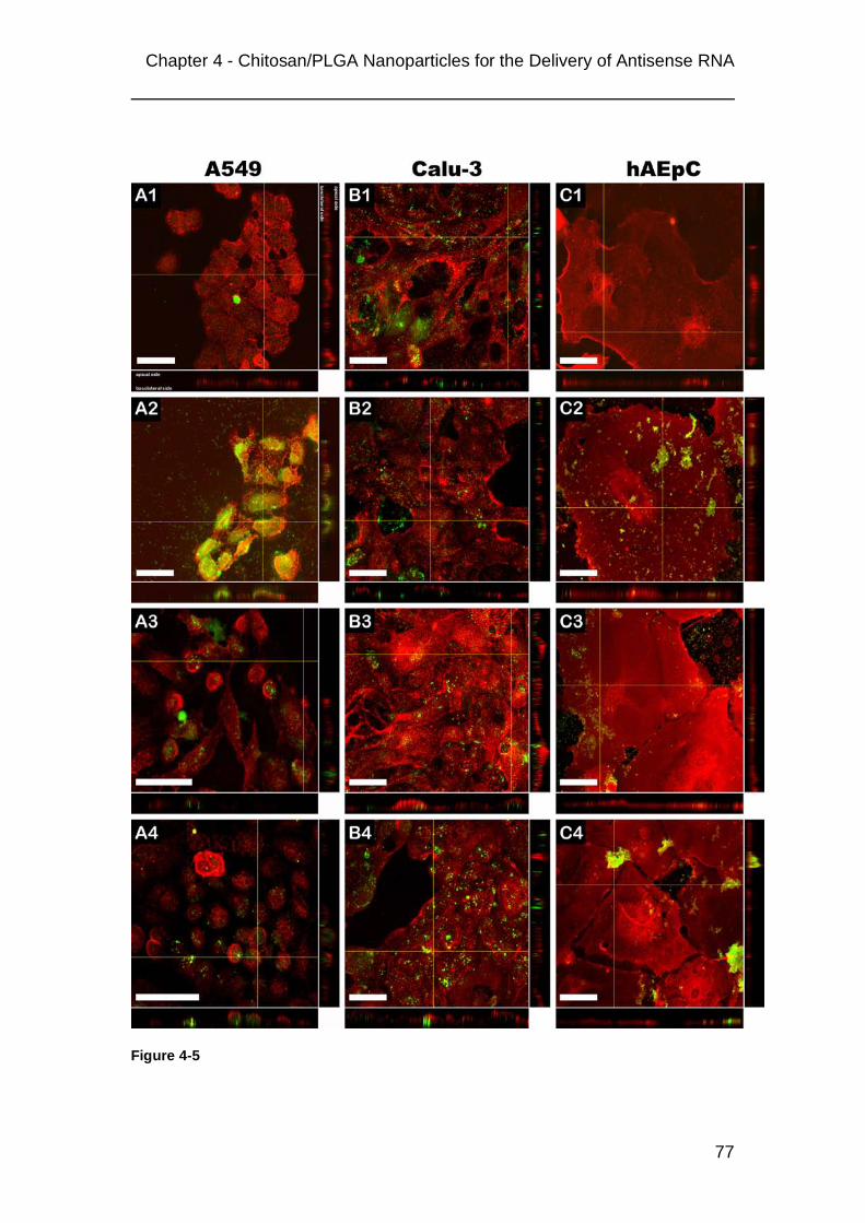
Chapter 4 - Chitosan/PLGA Nanoparticles for the Delivery of Antisense RNA
77
Figure 4-5

Chapter 4 - Chitosan/PLGA Nanoparticles for the Delivery of Antisense RNA
78
Figure 4-6: The uptake of 2OMR in A549 cells is mediated via nanoplexes. The green fluorescence of FAM-2OMR is colocalized with the red fluorescence from DiI-stained 3CPNP. Colocalization can be observed for at least two days by the yellowish/orange color in the left column (“merge”). Middle column and right column: fluorescence of FAM-2OMR (green) and DiI-3CPNP (red), respectively. After five days red cloudy structures (white arrows) indicate the degradation of nanoparticles and release of DiI into the cytoplasm. Purple = cell nuclei counterstained with TOPRO-3. Bars = 25 µm

Chapter 4 - Chitosan/PLGA Nanoparticles for the Delivery of Antisense RNA
79
4.3.4 Cytotoxicity and monolayer integrity
The MTT cytotoxicity assay showed that cell survival after a treatment with
different concentrations of 3CPNP and nanoplexes of 3CPNP and 2OMR,
respectively, is reduced to 70%- 80% of control independent of nanoparticle
concentration (Figure 4-7). However, a treatment of cells with a mixture of cell
culture medium and water also led to a reduced cell survival (about 90% of
control).
Furthermore, the treatment with nanoparticles did not show an effect on
monolayer integrity of Calu-3 cells. When grown on Transwell® filters Calu-3
cells typically form tight monolayers with transepithelial electrical resistance
(TEER) values between 1300 and 1500 Ω x cm2 [110]. TEER for cells
incubated with nanoparticles was comparable to controls during the
incubation period and 72 hours after treatment (data not shown).
Figure 4-7: MTT assay with purified 3CPNP: A549 cells show a reduced survival in the presence of nanoparticles and nanoplexes. Cells were treated with nanoplexes of the w2OMR/wparticles ratios 1:25 (= 443 µg/ml nanoparticles), 1:50 (= 886 µg/ml nanoparticles) and 1:100 (=1772 µg/ml nanoparticles). Cells treated only with plain nanoparticles in the corresponding concentrations were used as controls. Each concentration was measured in quadruplicate. Data represents mean values ± SD.

Chapter 4 - Chitosan/PLGA Nanoparticles for the Delivery of Antisense RNA
80
4.3.5 Inhibition of telomerase activity and telomere shortening
The treatment of A549 cells with nanoplexes of either purified (Figure 4-8 A)
or non-purified (Figure 4-8 B) 3CPNP and 2OMR resulted in comparable
reductions of telomerase activity for both batches.
The strongest inhibitory effect was observed after treatment with nanoplexes
at a w2OMR/wparticles ratio of 1:100 resulting in about 60% inhibition of
telomerase activity. However, at this ratio the mismatch control, which
contains two mismatches relative to the template sequence [48], also showed
a considerable telomerase inhibition. This observation is most probably due to
non-specific effects. Best results were obtained with non-purified particles at
the 1:50 ratio. Here a statistically significant difference between normal 2OMR
and the mismatch sequence has been found (P < 0.05). Telomerase activity
was decreased by about 50% with normal 2OMR while mismatch 2OMR
showed only a slight effect. Such a statistically significant difference between
normal and mismatch 2OMR could not found for purified particles. The
telomerase inhibition by nanoplexes prepared with non-purified nanoparticels
was as efficient as using the lipid-based transfection reagents DOTAP and
MegaFectinTM, which was described recently by our group [111].
Figure 4-8: Reduction of telomerase activities in A549 cells 72 hours after incubation with 2OMR:3CPNP nanoplexes at different w/w ratios in comparison to a 2OMR mismatch control. Telomerase activities were normalized to non-treated cells. A: nanoplexes were formed with purified 3CPNP nanoparticles; B: nanoplexes were formed with non-purified 3CPNP nanoparticels. Data represent mean values ± SD of at least 3 independent experiments. * = statistically significant difference between normal 2OMR and mismatch control (P < 0.05, One-Way ANOVA).

Chapter 4 - Chitosan/PLGA Nanoparticles for the Delivery of Antisense RNA
81
Reduction of telomere length was examined after repetitive treatments (twice
weekly) of A549 cells over 35 days in comparison with the lipid-based
transfection reagents DOTAP and MegaFectinTM. As can be seen in Figure
4-9, 3CPNP were as effective as the two commercial transfection reagents.
Reduction in telomere length was significant for all three preparations
compared to control (P < 0.05; One-Way-ANOVA) but there was no significant
difference between the transfection reagents.
Figure 4-9: Telomere length in A549 cells after repeated treatment with 2OMR:3CPNP nanoplexes over 35 days in comparison to cells treated with the commercially available transfection reagents DOTAP and MegaFectinTM. 3CPNP are as efficient as DOTAP and MegaFectinTM. * = all treated cells were statistically significant different from control (One-way-ANOVA; P < 0.05). There was no statistical significant difference between the lipid-based transfection reagents and 3CPNP.

Chapter 4 - Chitosan/PLGA Nanoparticles for the Delivery of Antisense RNA
82
4.4 Discussion
Our results show that changing the amounts of chitosan in the preparation
procedure can easily vary particle properties. These variations significantly
influence their efficiency for the delivery of antisense 2OMR to lung cancer
cells.
The particles proved to be stable after purification and storage over two
months and they showed a high binding of 2OMR and complex stability in
water. However, dilution in buffered media strongly reduced complex
formation. In our previous study, suspensions of CPNP either as particles
alone or in complex with 2OMR proved to be stable in NaCl solution, PBS
pH 7.4 and HBSS buffer pH 7.4 with regard to their colloidal properties.
However, they showed a strong reduction in their surface charge when diluted
in buffered media, which is due to the neutralization of the protonated primary
amine groups of chitosan [109]. This charge neutralization results in a
reduced ability to bind the negatively charged 2OMR as could be
demonstrated by our binding/complexation experiments. Chitosan is a weak
base. The type that was used for these experiments has a pKa value of about
6.5 [112]. Complexes diluted in pure MilliQ water were stable because pH
values were below this pKa resulting in protonation of most of the amino
groups. In contrast, due to the neutralization of the positive charges, complex
stability was strongly reduced and dissociation occurred when the preformed
complexes were diluted in buffered media like PBS and cell culture medium
with pH values ≥ 7.
From these results one could expect that nanoplexes in cell culture medium
are unstable and do not improve the uptake of 2OMR into cells. But this was
not the case. As we could demonstrate in the following experiments with A549
cells, uptake was significantly increased for nanoplexes prepared with 3CPNP
and 6CPNP and telomerase was activity successfully inhibited (Figures 4-3,
4-5, 4-6, 4-8 and 4-9).
From the nanoplex characterizations it could be argued that the uptake
correlated with the particle surface charge (Figure 4-4) as described by
Lorenz et al. [113]. However, the characterization was performed in water
while the uptake experiments were performed in cell culture medium

Chapter 4 - Chitosan/PLGA Nanoparticles for the Delivery of Antisense RNA
83
containing 10% fetal calf serum. Beside the neutralization of the surface
charge in cell culture medium it is known that serum proteins adsorb to
cationic nanoparticles [114, 115], which significantly influences the surface
charge. The fact that there was no difference in uptake improvement between
nanoplexes prepared with 3CPNP and 6CPNP indicates that the uptake
cannot only be attributed to the surface charge but also must be related to the
chitosan density on the particle surface. That the uptake of FAM-2OMR into
A549 is really mediated via nanoplexes could clearly be demonstrated by
confocal microscopy (Figure 4-5 and Figure 4-6), which shows that that there
is no complete dissociation of complexes in cell culture medium and that the
nanoplexes are still able to interact with the cell surface. Whether these non-
dissociated complexes corresponds to the about 15% of bound 2OMR (Figure
4-2 C; 1:50 ratio 3CPNP) will be the subject of future studies. Fang et al. have
shown with liposomes composed of DPPC that even at pH 7.4 an adhesion
between chitosan and the phospholipid membrane occurs [116]. Löhbach et
al. demonstrated that the adhesion of chitosan-coated carboxypolystyrene
nanoparticles dispersed in PBS to endothelial cells is higher in the presence
of FCS than without proteins [117]. Therefore, besides remaining ionic forces
other factors like hydrogen bonds or even the combination of chitosan and
proteins on the particle surfaces might play an important role in the nanoplex
– cell surface interaction.
Huang et al. reported that the uptake of FITC-labeled chitosan nanoparticles
into A549 cells is a saturable process dependent on adsorption of particles to
the cell membrane and subsequent clathrin-mediated endocytosis [118, 119].
This mechanism corresponded well to the observations in our experiments.
The uptake of FAM-2OMR alone was very poor but the adsorption of
nanoplexes to the cell surface significantly enhanced its internalization. The
results from flow cytometry suggest that a certain chitosan density on the
particle surface is required to achieve optimal adsorption. This level was
reached in 3CPNP because 6CPNP with their higher chitosan content did not
further increase the nanoplex uptake (Figure 4-4). The differences in chitosan
content were shown by zeta potential measurements in this (Figure 4-1 and

Chapter 4 - Chitosan/PLGA Nanoparticles for the Delivery of Antisense RNA
84
Figure 4-2 B) and our previous study [109] where 6CPNP exhibit a higher zeta
potential than 3CPNP.
However, nanoplexes also adsorbed on hAEpCs, but no clear internalization
was observed over two days. HAEpC are primary isolated human alveolar
type (AT) II cells that acquire characteristics of AT I cells after a certain time
under the given cell culture conditions [80]. Since AT I cells are reported to be
deficient in clathrin-mediated endocytosis [120] this might explain the lack of
nanoplex internalization by these cells. In contrast, Calu-3 expressing both
clathrin and caveolin [121] showed even an uptake of FAM-2OMR alone.
However, a closer investigation of the exact uptake mechanism was not done.
The data from the MTT toxicity test demonstrates that the nanoparticles have
only a slight cytotoxicity as they reduce the A549 cell growth about 20 - 30%
(Figure 4-7). The nanoparticles did not influence the integrity of Calu-3
monolayers, and hence their barrier function, since TEER values did not differ
from controls. Grenha et al. reported a comparable result for chitosan
nanoparticles in respirable powder formulations [122].
Furthermore, we were able to show that a treatment of A549 cells with these
nanoplexes results in an inhibition of telomerase activity and telomere
shortening. The comparison of our chitosan/PLGA nanoparticles with
established transfection reagents was encouraging for further studies since
the results were comparable to DOTAP or MegaFectinTM. However, the
purification of CPNP does not seem to be necessary with regard to their
biological effect because telomerase inhibition was not significantly different
for either purified or non-purified particles. Regarding the specificity of 2OMR
in comparison with the mismatch sequence, nanoplexes prepared with
non-purified particles appeared to be even more effective than those prepared
with purified particles. For this study purification was done to assure that the
observed effects in binding and uptake experiments can only be attributed to
the nanoparticles and not to remaining polymers in the suspensions. Since
the presence of these polymers does not seem to influence the biological
activity future studies concerning the long-term efficacy of nanoplexes will be
performed with non-purified particles.

Chapter 4 - Chitosan/PLGA Nanoparticles for the Delivery of Antisense RNA
85
4.5 Conclusion
Nanoplexes formed between cationic chitosan-coated PLGA nanoparticels
and antisense 2’-O-methyl-RNA were efficiently taken up by human alveolar
(A549) and bronchial (Calu-3) epithelial cancer cell lines, while cellular uptake
was not obvious for non-cancerous human alveolar epithelial cells in primary
culture. In thus transfected cells, a significant inhibition of telomerase activity
and shortening of telomeres could be observed. Transfection proved to be
dependent on the chitosan content of the nanoplexes, allowing to optimize the
formulation for such application. The resulting nanoplexes were well tolerated
by the cells and appeared to be equally efficient as some commercially
available lipid-based transfection reagents.

86

Chapter 5 - Purification of chitosan/PLGA nanoparticles
87
Chapter 5
Purification of chitosan/PLGA nanoparticels by size
exclusion chromatography

88

Chapter 5 - Purification of chitosan/PLGA nanoparticles
89
5.1 Introduction
The development of a suitable purification method for chitosan/PLGA
nanoparticles was necessary to verify that the results described in Chapter 4
could only be attributed to the particles and not to remaining free polymers in
the suspensions. However, the purification method that was available at that
time was based on repeated ultrafiltration-resuspension steps of nanoparticle
suspension using the Centrisart® system from Sartorius (Figure 5-1). This
method required centrifugation after each resuspension, which increased the
risk for particle agglomeration. Another point was that only small volumes of
2 ml maximum could be purified in one tube, which proved to be very time
consuming and laborious for the purification of larger volumes.
Figure 5-1: Principle of the nanoparticle purification with Centrisart® ultrafiltration tubes. The choice of the membrane pore size allowed the separation of free polymers from nanoparticles by ultrafiltration during the centrifugation step. Suspensions were submitted to several ultrafiltration-resuspension steps for complete removal of free polymers.
Since the experiments described in Chapter 4 and long-term cell culture
experiments required higher amounts of purified nanoparticles, another
purification strategy was needed. We developed a method based on size
exclusion chromatography that allowed the semi-automatic processing of
volumes up to 50 ml, i.e. the volume of one particle preparation.
This chapter describes the development of this purification method including
factors that are important for a successful separation of nanoparticles from
free polmers.

Chapter 5 - Purification of chitosan/PLGA nanoparticles
90
5.2 Materials and Methods
5.2.1 The size exclusion chromatography system
Size exclusion chromatography (SEC) was done using a FPLC® system from
Pharmacia Biotech (now Amersham Biosciences; Uppsala, Sweden)
equipped with two P-500 pumps, a LCC-501 Plus controller, a MV-7 injection
valve, a 50 ml Superloop, a C 16/70 column with one AC 16 adaptor, an
Uvicord SII detector with an interference filter of 206 nm and a FRAC-100
fraction collector. Stationary phase was Sephacryl S-1000 SF® from
GE Healthcare (Munich, Germany) with a dimension of 65 cm in height and
1.6 cm in diameter. The system was operated by the FPLCdirectorTM software
version 1.3 (Pharmacia Biotech). Flow rate of mobile phase was 1 ml/min
under isocratic conditions.
5.2.2 Selection of mobile phase
Three different mobile phases were tested: pure MilliQ water, 0.9% sodium
chloride (NaCl) solution and 0.1 mM hydrochloric acid (HCl), which was
prepared by dilution of 1 M HCl in MilliQ water.
5.2.3 Particle suspensions
Different chitoan/PLGA nanoparticles were prepared as described in Chapter
4 under 4.2.2. Most parts of the method development described here were
performed with chitosan/PLGA nanoparticles prepared with 3 mg/ml chitosan
(3CPNP). All particle suspensions were filtered through a 0.2 µm cellulose
acetate filter (Chromafil® GF/PET-20/25, Macherey-Nagel, Dueren, Germany)
prior to injection.
5.2.4 Evaluation of the separation of particles from polymers
To demonstrate the different retention times of particles, chitosan and PVA,
1.5 ml of each sample were injected into the SEC system. Particle suspension
was 3CPNP. The concentrations for chitosan and PVA solutions were
3 mg/ml and 25 mg/ml, respectively. Mobile phases were either MilliQ water
or 0.1 mM HCl.

Chapter 5 - Purification of chitosan/PLGA nanoparticles
91
5.2.5 Quantification of chitosan and PVA in fractions after purification
For the evaluation of the purification method 5 ml of a filtered chitosan/PLGA
nanoparticle suspension was injected into the size exclusion chromatography
system. Mobile phases were MilliQ water and 0.1 mM HCl. The flow rate was
1 ml/min. 10 ml fractions of the eluate were collected continually over
150 minutes. Afterwards, each fraction was submitted to ultrafiltration in
Vivaspins 20® at 1000 x g. 8 ml of filtrate from each fraction were lyophilized
and the dried polymers were redissolved in 4 ml MilliQ water. For comparison
5 ml of non-purified particle suspension was diluted to 10 ml with MilliQ water
and also submitted to ultrafiltration.
Chitosan was quantified by mixing 0.25 ml of polymer solution with 0.25 ml
4 M Na-acetate buffer pH 5.5 and 0.5 ml of an acetic ninhydrin reagent
(2 g ninhydrin, 0.08 g SnCl2 x 2 H2O, 50 ml 2- methoxy ethanol, 25 ml
4 M Na-acetate buffer pH 5.5, 25 ml water) [123]. The mixture was incubated
at 100°C for 20 minutes on a boiling water bath. After cooling down to room
temperature 200 µl samples were transferred to a 96 well plate and
absorptions were measured at 550 nm with an UV/Vis reader (SLT Spectra,
Tecan Deutschland GmbH, Crailsheim, Germany). Standards were in the
range from 0.01 – 0.4 mg/ml chitosan.
The concentrations of PVA were determined by mixing 100 µl of sample with
480 µl water, 300 µl 3.8% (w/v) boric acid solution and 120 µl 0.05 M I2/KI
solution. The mixtures were incubated in a closed container over night at
room temperature. Absorptions were measured at 650 nm using a Lambda 25
UV/Vis spectrophotometer from PerkinElmer (Wiesbaden, Germany).
Standards were in the range of 0.025 – 0.25 mg/ml. If necessary, samples
were diluted to fit the range of calibration. Every sample was measured in
triplicate.

Chapter 5 - Purification of chitosan/PLGA nanoparticles
92
5.3 Results and Discussion
5.3.1 Selection of the mobile phase
Good results for the SEC of chitosan/PLGA nanoparticles were obtained with
water and 0.1 mM HCl as mobile phase (Figure 5-2). Sharp and symmetric
peaks appeared after about 50 minutes, which approximately corresponds to
the dead volume of the stationary phase. The peak with water as mobile
phase was wider than for 0.1 mM HCl. The isotonic sodium chloride solution
proved to be unsuitable for SEC of chitosan/PLGA nanoparticles. At
50 minutes no peak appeared. Only after about 90 minutes a low and
asymmetric peak was found. Initially the sodium chloride solution was favored
as mobile phase because it was intended to compensate for changes in
osmotic pressure in uptake experiments after mixing cell culture medium with
nanoplexes.
Figure 5-2: Comparison of SEC chromatograms with different mobile phases: pure MilliQ water (black), 0.1 mM HCl (blue) and 0.9% NaCl solution (red). Particles were 3CPNP.

Chapter 5 - Purification of chitosan/PLGA nanoparticles
93
Comparing the retention times for solutions of PVA and chitosan with water
and 0.1 mM HCl there was no difference for the two mobile phases (Figure
5-3 A and B). The PVA peak was well separated from the particle peak with a
maximum at about 120 minutes. Chitosan showed a broad flat peak between
55 and 110 minutes and partly co-eluted with particles.
Figure 5-3: Chromatograms of particles and polymer solutions with MilliQ water (A) and 0.1 mM HCl (B) as mobile phase. PVA is well separated from the particle peaks while chitosan partly coelutes with particles. Retention times are comparable for both mobile phases.
From the data shown so far there seems to be no great difference between
water and 0.1 mM HCl as a suitable mobile phase. However, the reason why
0.1 mM HCl was finally chosen for the purification of nanoparticle suspensions
(see previous chapter) was the influence of the mobile phase on the
purification efficiency of the low-chitosan preparation 05CPNP. Figure 5-4 A
and B show the chromatograms from purifications of 7 ml 05CPNP
suspensions with water and 0.1 mM HCl, respectively, as mobile phases. It
can clearly be seen that the performance of water was very poor. The particle
peak that was observed for 3CPNP between 40 and 60 minutes retention time
is almost non-existent. In contrast, when 0.1 mM HCl was used a sharp peak
was found.

Chapter 5 - Purification of chitosan/PLGA nanoparticles
94
Figure 5-4: Chromatograms of 05CPNP eluted with MilliQ water (A) and 0.1 mM HCl (B). When MilliQ water was used as mobile phase only a very small particle peak was visible.
Interestingly, neither of the two mobile phases could be used for the
purification of plain negatively charged PLGA nanoparticles. An experiment
with a small column filled with Sephacryl 1000-SF (ca. 16 cm length x 1 cm
diameter), which was used for quick evaluations of new experimental
parameters, showed that no peak occurred for PLGA nanoparticles under
both conditions, while 05CPNP could be eluted satisfactorily with 0.1 mM HCl
(Figure 5-6).
These results strongly suggest that particle charge strongly influences elution
and retention. Negatively charged PLGA nanoparticles seem to interact with
the stationary phase and their strong retention might therefore not only be due
to the principle of size exclusion while the positive charge of chitosan coated
particles seems to prevent this interaction. Sephacryl S-1000 SF is a
hydrophilic matrix composed of allyl dextran cross-linked with N,N’-methylene
bisacrylamide (Figure 5-5). The hydroxyl groups of the dextran can form
hydrogen bonds with the negatively charged carboxyl groups of the PLGA
polymer. Since no elution of PLGA nanoparticles occurred under both neutral
(water) and acidic (0.1 mM HCl) conditions, where most of the negative
charges should be neutralized, a positive surface charge seems to be
necessary for an early elution. As has been demonstrated with 05CPNP an
acidification of the mobile phase results in a much better elution of particles.

Chapter 5 - Purification of chitosan/PLGA nanoparticles
95
Figure 5-5: Structure of Sephacryl S-1000 SF (according to the manufaturer).
Figure 5-6: Comparison of chromatograms of PLGA nanoparticles and 05CPNP obtained with a short column filled with Sephacryl 1000-SF (16 cm length x 1 cm diamter). A: no particle peaks were obtained when MilliQ water was used as mobile phase; B: the use of 0.1 mM HCl as mobile phase resulted in a sharp peak for 05CPNP but not for PLGA nanoparticles.
5.3.2 Quantification of chitosan and PVA in different fractions
The quantification of chitosan and PVA in fractions that were continuously
collected during the purification procedure of 3CPNP with either water or
0.1 mM HCl as mobile phase showed that chitosan must be almost
quantitatively associated with the particles (Figure 5-7 A and B). No fraction
from either mobile phase contained amounts of chitosan that were
significantly different from a basal level that was measured for all fractions.
Messai et al. have shown that chitosan efficiently adsorbs to the particle
surface of pre-formed PLGA nanoparticles [124] and that it is also partly
incorporated into particles prepared by the emulsification-diffusion-solvent

Chapter 5 - Purification of chitosan/PLGA nanoparticles
96
evaporation method employed here [125]. We were able to demonstrate in a
previous study by zeta potential measurements that the surface of 3CPNP is
not saturated with chitosan [109]. Hence, it is very likely that the larger fraction
of chitosan is associated with the particles and that the amount of potentially
unbound chitosan is below the detection limit of the assay that was used for
these experiments.
In contrast, high amounts of PVA were found in fractions that were collected
between retention times of 90 to 130 minutes (Figure 5-7). This result is in
good agreement with the retention times found for PVA injected alone into the
SEC system and detected by UV absorbance (Figure 5-3).
Figure 5-7: Quantification of chitosan and PVA from a 3CPNP particle suspension in fractions that were continually collected during the SEC run. A: water as mobile phase; B: 0.1 mM HCl as mobile phase. Chitosan (red dots) was below the quantification limit in all fractions. The increase in PVA in fractions 9 - 13 (= 90 – 130 minutes retention time) corresponded well to the peaks at 110 minutes and the chromatograms of PVA in Figure 5-3.

Chapter 5 - Purification of chitosan/PLGA nanoparticles
97
Calculating the total amount of PVA from all fractions showed that the
purification with 0.1 mM HCl as mobile phase was the most efficient with
about 7.5 mg compared to about 5.5 mg when water was used. Interestingly,
the amount of PVA obtained from non-purified 3CPNP after a simple
ultrafiltration was below 2 mg. Comparing these results with the expected
amount of 12.5 mg showed that there must also be a high degree of
association/ integration of PVA with/ into nanoparticles. The fact that the
amounts of recovered PVA from purified particles are higher than for
non-purified strongly indicates that PVA adsorbs to the surface and cannot be
removed by a simple centrifugation step. During the purification procedure this
adsorbed PVA is then washed off the surface because of the steady flow of
mobile phase. The differences in PVA recovery with water and 0.1 mM HCl,
respectively, as mobile phase are most probably due to the change in degree
of protonation and conformation of surface polymers.
Figure 5-8: Comparison of PVA recovered from purified particles (with water and 0.1 mM HCl as mobile phase, respectively), non-purified particles after direct ultrafiltration and the amount expected in 5 ml particle suspension.

Chapter 5 - Purification of chitosan/PLGA nanoparticles
98
5.3.3 Purification of different nanoparticle preparations
The purification method was employed for all chitosan/PLGA nanoparticle
preparations that were used for the experiments described in Chapter 4. As
can be seen in Figure 5-9 the particle peaks broadened with increasing
chitosan concentrations, which is most probably due to a more heterogeneous
size distribution at higher chitosan contents (compare PDIs in Figure 4-1).
Also, 3CPNP and 6CPNP showed a stronger tailing than 05CPNP and
1CPNP. Whether this tailing is due to subfractions of nanoparticles or
unbound polymers, especially chitosan, could not be clarified during these
studies. Regarding the results from the quantification experiments for free
polymers (see above) the presence of unbound chitosan appears unlikely.
Also, particle collection during the purifications was automatically initiated
when peaks exceeded an absorbance value higher than 1.5 and stopped
when absorbance dropped below this threshold again. So even if the tailing
can be attributed to free chitosan, the amounts of the polymer within the
particle fraction of 3CPNP and 6CPNP should be negligible concerning their
influence on the results described in Chapter 4.
Figure 5-9: Comparison of chromatograms from different particle preparations after purification with 0.1 mM HCl as mobile phase. Peaks broaden with increasing chitosan content in particle suspensions. 05CPNP, 1CPNP, 3CPNP and 6CPNP = chitosan/PLGA nanoparticles prepared with 0.5, 1, 3 and 6 mg/ml chitosan, respectively.

Chapter 5 - Purification of chitosan/PLGA nanoparticles
99
5.3.4 Repeated injection of particle suspensions
Figure 5-10 shows the chromatogram from three successive injections of
3CPNP during a purification run. It can clearly be seen that the peaks appear
in a reproducible way. This shows that an additional washing or
re-equilibration of the column is not necessary after a purification step.
Figure 5-10: Successive injections for the purification of a larger volume of 3CPNP suspension. The peaks appear in a reproducible way.

Chapter 5 - Purification of chitosan/PLGA nanoparticles
100
5.4 Conclusion
We were able to demonstrate that preparative size exclusion chromatography
can be used as an efficient method for the purification of chitosan/PLGA
nanoparticles from excess PVA in particle suspensions. The advantage of this
method is that it can handle larger volumes up to 50 ml in a semi-automated
purification procedure. Interventions of the operator are limited to the filtration
of particle suspensions prior to and their concentration after purification.
However, as could be demonstrated with pure PLGA nanoparticles, this
method is limited to the purification of cationic chitosan-coated PLGA
nanoparticels.

Chapter 6 - Hyaluronic Acid-Modified Liposomes for Targeted siRNA Delivery
101
Chapter 6
Hyaluronic Acid- Modified Liposomes for the
Targeted Delivery of siRNA to CD44 Expressing
Lung Cancer Cells
The data presented in this chapter has been accepted for publication as a
research article in the journal Oligonuceotides:
Taetz, S., Bochot, A., Surace, C., Arpicco, S., Schaefer, U.F., Marsaud, V.,
Kerdine-Roemer, S., Lehr, C.-M., Fattal, E.
Hyaluronic acid- modified DOTAP/DOPE liposomes for the targeted delivery
of anti-telomerase siRNA to CD44 expressing lung cancer cells. (2009)
Oligonucleotides, accepted
Journal ISSN: 1545-4576
Journal Online ISSN: 1557-8526
Weblink: http://www.liebertpub.com/products/product.aspx?pid=121

102

Chapter 6 - Hyaluronic Acid-Modified Liposomes for Targeted siRNA Delivery
103
6.1 Introduction
Since its discovery, the mechanism of RNA interference via small interfering
RNA (siRNA) developed rapidly into a powerful tool in molecular biology for
studying the downregulation of gene expression. Due to their high efficiency
and selectivity siRNA became rapidly interesting for medical applications like
the treatment of severe diseases such as cancer. However, siRNA faces the
same obstacles for a successful application as other nucleotide-based
therapeutics like plasmid DNA or antisense oligonucleotides, i.e. a poor
uptake and stability in serum and the lack of selectivity for the target tissue
[126-128]. Therefore, different carrier systems based on cationic liposomes or
nanoparticles coupled with tumor-recognizing ligands (e.g. transferin-coupled
DOTAP/cholesterol liposomes [129], PEGylated polyethyleneimine
nanoparticles conjugated with an RGD peptide [130] or a PEGylated
liposome-polycation-DNA complex modified with anisamide [131]) were
developed .
Our study concentrated on cationic hyaluronic acid (HA)-modified 1,2-dioleoyl-
sn-glycero-3-phosphoethanolamine (DOPE) / 1,2-dioleoyl-3-trimethyl-
ammonium-propane (DOTAP) liposomes as carriers for the targeted delivery
of a siRNA directed against the enzyme telomerase to lung cancer cells that
over-express the HA binding receptor CD44. The discovery that many cancer
types over-express this receptor led to the development of HA-drug
conjugates [132, 133] and HA-modified drug carrier systems [133-139] that
were able to target these cancer cells very specifically in vitro and in vivo. HA
is an endogenous polymer of repeating disaccharide units of D-glucuronic
acid and D-N-acetylglucosamine with a wide range of molecular weights. It is
widely distributed in the mammalian body were it fulfills important mechanical
or structural functions [140]. However, by interaction with its principal cell
surface receptor CD44, a transmembrane glycoprotein that exists in different
isoforms, it also plays an important role on cell – cell / cell – matrix interaction,
cell adhesion and migration and signal transduction from the extracellular to
the intracellular compartment [141-143].

Chapter 6 - Hyaluronic Acid-Modified Liposomes for Targeted siRNA Delivery
104
Among the carrier systems mentioned above HA-modified liposomes were
reported to be very efficient in the delivery of anti-cancer drugs like
doxorubicin or mitomycin C [134, 138, 139]. In mouse tumor models these
liposomes showed a much higher efficacy compared to non-modified
liposomes and also a longer systemic circulation time, which was attributed to
a “hydrophilic coat effect” of the hyaluronic acid. In terms of cytotoxicity,
CD44-expressing cells were much more sensitive to the cytotoxic drugs
encapsulated in HA-modified liposomes while toxicity for CD44 deficient cells
was unchanged.
The target of the siRNA used in this study was a mRNA coding for the
catalytic subunit of human telomerase hTERT (human telomerase reverse
transcriptase). Telomerase plays an important role in cell immortalization and
hence cancer development. Many types of cancer over-express this enzyme,
which is inactive in normal somatic cells. Therefore, telomerase is believed to
be a suitable target for the specific treatment of cancers that overexpress this
enzyme [97].
The aim of our study was to prepare and characterize cationic HA-modified
DOTAP/DOPE liposomes using a conjugate of HA and DOPE that has been
successfully employed by Surace et al. [144] for the targeted delivery of
plasmid DNA to CD44 expressing breast cancer cell lines. We investigated
the influence of the modification with regard to the colloidal properties of the
liposomes and lipoplexes, siRNA binding and protection, cytotoxicity, uptake
into CD44 expressing and non-expressing cancer cells and the efficiency in
telomerase inhibition in comparison with non-modified liposomes and the
commercially available transfection reagent Lipofectamine 2000.

Chapter 6 - Hyaluronic Acid-Modified Liposomes for Targeted siRNA Delivery
105
6.2 Materials and Methods
6.2.1 Materials
1,2-Dioleoyl-3-trimethylammonium-propane (DOTAP) chloride salt was
obtained from Avanti Polar Lipids (Alabaster, AL, USA), 1,2-Dioleoyl-sn-
glycero-3-phosphoethanolamine (DOPE) and 1-ethyl-3-[3-
dimethyl)amiopropyl]carbodiimide hydrochloride (EDAC) from Sigma-Aldrich
(Saint-Quentin Fallavier, France), Lipofectamine 2000 (LF 2000) from
Invitrogen (Eragny sur Oise, France), high molecular weight hyaluronic acid
(HA; 1,500,000 Da) from Fluka (Sigma Aldrich Chemie, Buchs, Switzerland).
6.2.2 siRNAs
SiRNA was obtained as single strands from Eurogentec (Seraing, Belgium). It
was directed against a region between nucleotides 1523 and 1543 of the
mRNA encoding for human telomerase reverse transcriptase (hTERT-mRNA;
referred to transcript variant 1 of hTERT-mRNA; NCBI Entrez Nucleotide
databank accession number: NM_198253). The sequence of the sense
strand was 5’-GGAACACCAAGAAGUUCAUtt-3’ and the sequence of the
antisense strand was 5’-AUGAACUUCUUGGUGUUCCtg-3’, where tt and tg
are deoxynucleotides, respectively (anti-hTERT siRNA). Annealing of single
strands was achieved according to the manufacturer’s recommendation in an
annealing buffer containing 100 mM potassium acetate, 30 mM HEPES-KOH
pH 7.4 and 2 mM magnesium acetate. A fluorescently labeled version of this
siRNA was modified with 6-FAM at the 5’-ends. A nonsense siRNA was used
as negative control.
6.2.3 Radiolabeling of siRNA
Radiolabeling of siRNA was performed by labeling the 5’-end of the sense
strand with γ-33P-ATP (111 TBq/mmol; Perkin-Elmer Life Science, Mechelen,
Belgium) catalyzed by T4 polynucleotide kinase (New England Biolabs,
Frankfurt am Main, Germany) according to the manufacturer’s protocol.
Labeled sense strand was purified by gel filtration using Bio-Gel P-6 Bio-Spin
columns (Bio-Rad, Mitry Mory, France). Purity was proven by autoradiography

Chapter 6 - Hyaluronic Acid-Modified Liposomes for Targeted siRNA Delivery
106
after gel electrophoresis of a sample on a 15% denaturing 7 M urea
polyacrylamide gel. The labeled sense strand was annealed with the
antisense strand as described above.
6.2.4 Conjugation of DOPE to hyaluronic acid
The conjugate of HA with DOPE was synthesized as reported by Surace et al.
[144] based on a modified reaction described by Yerushalmi et al. [145]. In
brief, 14 mg HA were dissolved in 5 ml water and preactivated for 2 hours at
37°C by incubation with 6 mg EDAC at pH 4 which was adjusted by titration
with 0.1 N HCl. Afterwards, DOPE suspension (360 µg) was added to the HA
solution and pH was adjusted to 8.6 with 0.1 M borate buffer. The reaction
was proceeded for 24 hours at 37°C. The conjugate was purified by
ultrafiltration using a membrane with a molecular weight cut off of 100,000 Da
(Amicon Ultrafiltration, Millipore, Billerica, MA, USA), lyophilized and stored at
-25°C until further use.
6.2.5 Preparation of liposomes
DOTAP/DOPE liposomes were prepared by the ethanol injection method
[146, 147]. DOTAP and DOPE were dissolved separately in chloroform of
analytical grade (Carlo Erba Reagents, Val de Reuil, France). The solutions
were mixed at the required amounts at a molar ratio of 1:1 and chloroform
was evaporated under vacuum. Lipid films were stored under nitrogen at
- 20 °C. For liposome preparation the lipid films were redissolved in absolute
ethanol (analytical grade; Carlo Erba Reagents) at a concentration of
10 mg/ml. For liposome preparation 50 µl of ethanolic lipid solution were
rapidly injected into 1 ml filtered (0.22 µm) MilliQ water (Millipore, Guyancourt,
France) under stirring with a magnetic bar to obtain a final lipid concentration
of 500 µg/ml. HA-modified liposomes were prepared by diluting an aqueous
stock solution of the HA-DOPE conjugate (1 mg/ml) to different concentrations
in filtered MilliQ water before injection. The content of HA-DOPE conjugate is
expressed in percent as wHA-DOPE/wtotal lipids x 100. For the removal of ethanol
liposome suspensions were dialyzed against MilliQ water over night in
Spectra/Por CE dialysis tubes with a molecular weight cut-off of 100,000
(Spectrum Laboratories, Inc., Rancho Dominguez, CA, USA).
Chapter 6 - Hyaluronic Acid-Modified Liposomes for Targeted siRNA Delivery
107
6.2.6 Preparation of lipoplexes
Lipoplexes were prepared at different +/- ratios by rapidly injecting different
volumes of 500 µg/ml liposome suspension in 2.5 µM siRNA solution.
Suspensions were mixed thoroughly by pipetting up and down and incubated
for 15 minutes at room temperature. Afterwards they were diluted with the
respective medium to a final siRNA concentration of 100 nM.
6.2.7 Characterization of liposomes and lipoplexes
Liposomes and lipoplexes were characterized with regard to their size, size
distribution and surface charge by dynamic light scattering and zeta potential
measurement using a ZetaSizer Nano ZS (Malvern Instruments,
Worcestershire, United Kingdom). Measurements were performed with
50 µg/ml total lipids in filtered MilliQ water at 25°C using the standard settings
of the instrument. Results are expressed as z-average (size), polydispersity
index (PDI; size distribution) and zeta potential values (surface charge). All
measurements were at least performed in triplicate.
6.2.8 Binding efficiencies of lipoplexes
Lipoplexes were prepared with radiolabeled siRNA at different +/- ratios as
described above. The final siRNA concentration was 100 nM. Suspensions
were submitted to ultracentrifugation using an Optima® LE-80K ultracentrifuge
equipped with a type 50.4 Ti rotor (both Beckman-Coulter, Villepinte, France)
at an average centrifugal force of 130,000 x g at 4°C under vacuum for
1 hour. Binding efficiencies were determined by comparing the specific
radioactivities of samples from the supernatant with samples taken before the
centrifugation step. Experiments were performed in triplicate in MilliQ water
and serum-free RPMI 1640 with GlutaMAXTM cell culture medium (Gibco,
Paisley, UK).
6.2.9 Colloidal stability of liposomes and lipoplexes in serum-free cell culture medium
Lipoplexes of non-modified and 15% HA-DOPE at charge ratios of +/- 2:1 and
+/- 8:1 were prepared as described above and diluted to 50 µg/ml total lipids
with serum-free RPMI cell culture medium. Suspensions with liposomes only

Chapter 6 - Hyaluronic Acid-Modified Liposomes for Targeted siRNA Delivery
108
were prepared accordingly but without siRNA. Changes in size were
measured with the ZetaSizer Nano ZS (Malvern Instruments) at 25°C for two
hours. The accuracy of measurements was verified by mixing each
suspension thoroughly after each measurement to avoid false results that
might be due to a quick sedimentation of agglomerates. All experiments were
performed in triplicate.
6.2.10 Protection of siRNA in lipoplexes in the presence of RNase V1
Lipoplexes of non-modified and 15% HA-DOPE liposomes at +/- ratios of 2:1
and 8:1 were prepared as described above with radiolabeled siRNA.
Suspensions were diluted to 100 nM siRNA with RNase V1 (Ambion Applies
Biosystems, Courtaboeuf, France) containing reaction buffer to a final ratio of
0.1 U RNase V1 per 100 ng siRNA. Samples were incubated at 37°C for
30 minutes. Afterwards RNase V1 activity was inhibited and lipoplexes were
lysed using a mixture of 10% (w/v) sodium dodecylsulfate (SDS) and 5% (v/v)
Triton X-100. All samples were kept on ice. After centrifugation at 10,000 x g
and 4°C for 5 minutes samples from the supernatant were run on a
12% non-denaturing polyacrylamide gel. The radiolabeled siRNA was
visualized after gel drying by autoradiography. To verify that no degradation
occurs during sample processing after incubation a post-incubation
degradation control was included in the experiment: RNase V1 and siRNA
were incubated separately and mixed only after addition of the SDS/Triton
X-100 mixture to the RNase V1 solution.
6.2.11 Stability of siRNA and lipoplexes in the presence of human serum
Lipoplexes of non-modified and 15% HA-DOPE liposomes at +/- ratios of 2:1
and 8:1 were prepared as described above with radiolabeled siRNA.
Suspensions were diluted to 100 nM siRNA with RPMI cell culture medium
(Gibco) containing different concentrations of human serum (Sigma,
St. Quentin Fallavier, France) and incubated for 30 minutes at 37°C.
Afterwards samples were run on a 12% non-denaturing polyacrylamide gel

Chapter 6 - Hyaluronic Acid-Modified Liposomes for Targeted siRNA Delivery
109
without further lysis of lipoplexes. The radiolabeled siRNA was visualized after
gel drying by autoradiography.
6.2.12 Cell cultures and cell culture conditions
The human lung cancer cell lines A549 and Calu-3 were obtained from
American Type Culture Collection (ATCC-nos.: CCL-185 and HTB-55,
respectively; Manassas, VA, USA). A549 cells were grown in RPMI 1640 with
GlutaMAXTM cell culture medium (Gibco) supplemented with 10% fetal calf
serum (Lonza, Verviers, Belgium) and 100 U penicillin/ 100 U streptomycin
(Lonza). Calu-3 cells were grown in RPMI 1640 with GlutaMAXTM cell culture
medium (Gibco) supplemented with 10% fetal calf serum (Lonza), 1 mM
sodium pyruvate (Sigma) and 100 U penicillin/ 100 U streptomycin (Lonza).
Cells were grown on 80 cm2 polystyrene cell culture flasks in an incubator at
37°C, 5% CO2 and 90% humidity. Media were changed every other day and
cells were subcultured once a week.
6.2.13 Western blot analysis for the CD44 receptor
Cells were washed with cold PBS and detached from the culture flask bottom
by scrapping. Lysis was done after pelleting with a lysis buffer containing 1%
Triton X-100, 50 mM Tris pH7.4, 150 mM sodium chloride, 1% deoxycholic
acid, 0.1% sodium dodecylsulfate (SDS) and protease inhibitor cocktail
(Complete Mini®, Roche, Mannheim, Germany). Protein concentrations were
determined with the Bio-Rad Protein Assay (Bio-Rad Laboratories Inc.,
Marnes-la-Coquette, France). Protein samples were separated on a 8%
denaturing SDS polyacrylamide gel and electroblotted to a Immobilo-P
transfer membrane (Millipore). After blocking with 2% (w/v) bovine serum
albumin (BSA) in 0.1% (v/v) Tween 20/PBS CD44 was detected by incubating
the membrane with a 1:20 dilution of the monoclonal antibody Hermes I in 2%
BSA/Tween 20/PBS (rat IgG2a; Department of Pathology, Stanford
University, School of Medicine, Stanford, CA, USA) at 4°C over night.
Hermes I was detected the next day using Vectastain Elite ABC kit anti-rat
rabbit antibody IgG (Vector Laboratories, Inc., Berlingame, CA, USA) and
Western Blotting Luminol Reagent (Santa Cruz Biotechnology, Inc., Santa

Chapter 6 - Hyaluronic Acid-Modified Liposomes for Targeted siRNA Delivery
110
Cruz, CA, USA) on Amersham Hyperfilm ECL (GE Healthcare Ltd.,
Buckinghamshire, UK).
6.2.14 Cytotoxicity tests
For cytotoxicity tests A549 and Calu-3 cells were seeded on 96 flat bottom
well plates at a density of 5,000 and 25,000 cells per well, respectively. Cells
were incubated with different concentrations of non-modified and 15%
HA-DOPE liposomes and their lipoplexes at +/- ratios of 2:1 and 8:1 in serum-
free RPMI cell culture medium without antibiotics for 3 hours under cell culture
conditions. Lipoplexes of +/- 2:1 and +/- 8:1 corresponded to about 10 µg/ml
and 50 µg/ml liposomes, respectively. Lipofectamine 2000 (LF 2000) was
used for comparison. The required amount of LF 2000 was adjusted for the
transfection of 100 nM siRNA as recommended by the manufacturer. This
amount was regarded to be equal to the amount of lipids that is used for
lipoplexes at the +/- ratio of 8:1. The different LF 2000 concentrations were
adjusted accordingly.
MTT cytotoxicity test
Cells were incubated with different liposome concentrations and lipoplexes
1 day after seeding. After three hours of incubation, the suspensions were
replaced by the respective normal cell culture medium (see above). Cells
were allowed to grown for another 48 hours under cell culture conditions.
Afterwards 25 µl of a 5 mg/ml 3-(4,5-dimethyl-2-thiazolyl)-2,5-diphenyl-2H-
tetrazolium bromide (MTT; Sigma) in PBS were added to each well. Medium
was removed after 2 hours of incubation under cell culture conditions and
200 µl of pure dimethylsulfoxide (DMSO) was added to each well for cell
lysis and dissolution of formazan crystals. Absorbance was measured at
570 nm. Metabolic activity was as a measure for cell survival was calculated
in comparison with non-treated cells according to the formula: (mean
abstreated cells/ mean absnon-treated cells) x 100. Each concentration was measured
in triplicate.

Chapter 6 - Hyaluronic Acid-Modified Liposomes for Targeted siRNA Delivery
111
LDH cytotoxicity test
Cells were incubated with different liposome concentrations and lipoplexes
3 days after seeding. Liposome and lipoplex suspensions were prepared as
described above but with phenol red- free RPMI cell culture medium (Gibco).
After incubation for 3 hours, 100 µl samples from the supernatant of each well
were taken and analyzed for LDH activity according to the manufacturer’s
instructions of the Cytotoxicity Detection KitPlus (LDH) (Roche Diagnostics,
Meylan, France). Absorbance was measured at 492 nm. LDH release from
cells incubated with liposomes or lipoplexes was referred to the spontaneous
LDH release from non-treated cells. Background controls for non-specific
reactions included all incubation media without cells. Each concentration was
measured in triplicates.
6.2.15 Flow cytometry
A549 and Calu-3 cells were seeded on 6 well plates at a density of 250,000
cells per well. Lipoplexes were prepared as described above with
FAM-labeled siRNA in serum-free RPMI cell culture medium. Cells were
incubated with Lipoplexes of non-modified and 15% HA-DOPE liposomes at
+/- ratios of 2:1 and 8:1 at a final siRNA concentration of 100 nM for three
hours. Afterwards they were washed once with calcium and magnesium
containing PBS (Ca/Mg-PBS; Gibco). Cells were detached with trypsin/EDTA
(Lonza). After inactivation of trypsin with serum-free RPMI cell culture medium
without phenol red, cells were pelleted and resuspended in PBS without
calcium and magnesium. To be able to distinguish between internalized and
extracellularly bound lipoplexes, half of the cells were incubated with 0.4%
trypan blue in Ca/Mg-PBS and washed twice with normal PBS prior to
trypsinization. Fluorescence intensities were measured using a FACSCalibur
flow cytometer from Becton Dickinson (BD) Biosciences (Franklin Lakes, NJ
USA) using the software CellQuestTM Pro Version 4.02 (BD Biosciences). The
instrument was adjusted using non-treated cells.

Chapter 6 - Hyaluronic Acid-Modified Liposomes for Targeted siRNA Delivery
112
6.2.16 Determination of telomerase activity by the TRAP-qPCR assay
A549 cells were seeded at a density of 100,000 and Calu-3 cells at 500,000
cells per well on 6 well plates. One day after seeding cells were incubated
with lipoplexes of non-modified and 15% HA-DOPE liposomes at +/- ratios of
2:1 and 8:1 in serum-free RPMI cell culture medium at a final siRNA
concentration of 100 nM for 3 hours. LF 2000 was used for comparison.
Non-specific effects were examined with liposomes without siRNA and
lipoplexes prepared with a nonsense sequence.
Following incubation the medium was exchanged with normal cell culture
medium and cells were kept under cell culture condition for further 48 hours.
Afterwards, cells were washed once with cold PBS (4°C), detached by
scrapping, pelleted and lysed in a non-denaturing lysis buffer (10 mM Tris-HCl
pH 7.4, 1 mM magnesium chloride, 1 mM EGTA, 5 mM
beta-mercaptoethanol, 1% Triton X100 and 10% glycerol). Protein
concentrations were determined with the Bio-Rad Protein Assay (Bio-Rad
Laboratories Inc., Marnes-la-Coquette, France). Telomerase activity was
determined by the telomeric repeat amplification protocol (TRAP assay)
adapted for quantitative real-time PCR (qPCR) [148, 149]. The protein
concentration of each sample was adjusted to 100 ng/µl with lysis buffer. The
TRAP-qPCR was performed with a LightCycler 1.5 (Roche Diagnostics,
Meylan, France) using the LightCylcer FastStart DNA MasterPlus SYBR Green
I kit (Roche Diagnostics, Meylan, France). The master mix was prepared
according to the manufactures protocol. Each sample (20 µl) contained
100 ng of protein extract, 100 ng TS primer (5’-AATCCGTCGAGCAGAGTT-3’)
and 50 ng ACX primer (5’-GCGCGGCTTACCCTTACCCTTACCCTAACC-3’) [99]
(both from Eurogentec, Seraing, Belgium). The TRAP-qPCR was run under
the following conditions: 30 minutes at 30°C for the elongation of TS primer by
telomerase; 10 minutes at 95°C for the inactivation of telomerase and
activation of polymerase; amplification of telomerase products during 40
cycles of 10 seconds at 95°C, 5 seconds at 62°C, 15 seconds at 72°C, final
cooling step for 30 seconds at 40°C. Fluorescence intensities were acquired
during amplification after each cycle. A calibration line was established by

Chapter 6 - Hyaluronic Acid-Modified Liposomes for Targeted siRNA Delivery
113
serial dilution of protein extract from non-treated cells. The slope and
y-intercept were determined after plotting log amount protein versus the cycle
threshold value. Relative telomerase activity (RTA) was determined according
to equation 6-1:
!
RTA = 10
Ctsample "Yintercept
slopeEquation 6-1
where Ctsample is the cycle threshold value for the respective sample and
Yintercept and slope are calculated from regression line [149]. RTAs of cells
incubated with liposomes or lipoplexes were normalized to those of
non-treated cells.

Chapter 6 - Hyaluronic Acid-Modified Liposomes for Targeted siRNA Delivery
114
6.3 Results
6.3.1 Properties of liposomes and lipoplexes
The ethanol injection method allowed the preparation of nano-sized
HA-DOPE modified liposomes with a conjugate content of up to 20%
(wHA-DOPE/wtotal lipids). The sizes ranged from 110 to 160 nm. There was a
gradual increase in size with increasing HA-DOPE content (Figure 6-1 A).
Higher amounts of the conjugate resulted in the formation of agglomerates
(data not shown). The size distribution of non-modified liposomes and
liposomes with lower amounts of HA-DOPE (1 – 10%) was rather
heterogeneous which was reflected by polydispersity indices (PDIs) of around
0.4. This improved to values around 0.25 when 15 and 20% of conjugate were
used (data not shown). Zeta potential values (ZPs) were in the range of +50
to +60 mV for all liposome preparations at pH-values of about 6.5 (Figure
6-1 A).
The formation of complexes with siRNA at +/- charge ratios of 1:1 to 8:1
resulted in lipoplexes with sizes around and below 200 nm and PDIs smaller
than 0.25 for most of the preparations (Figure 6-1 B – F). The ZP
measurements of lipoplexes clearly revealed the differences in the degree of
liposome modifications. At the +/- ratio of 1:1 all lipoplexes were strongly
negatively charged (≤ -40 mV; B). However, at +/- 2:1 lipoplexes prepared
with non-modified liposomes were positively charged while ZPs decreased
with increasing amounts of HA-DOPE for lipoplexes with modified liposomes
(Figure 6-1 C). This trend was also observed at higher +/- ratios (Figure
6-1 D-F). The pH values for lipoplexes were in the range of 6.5 to 7.0.
Agglomeration occurred for lipoplexes in the range of -20 to +20 mV,
demonstrating that electrostatic repulsion in this range is not sufficient to
maintain the colloidal stability. 20% HA-DOPE liposomes were identified as a
critical formulation because they formed agglomerates at +/- charge ratios of
4:1, 6:1 and 8:1.

Chapter 6 - Hyaluronic Acid-Modified Liposomes for Targeted siRNA Delivery
115
Figure 6-1: Properties of HA-modified DOTAP/DOPE liposomes prepared by the ethanol injection method. A: liposomes alone with different amounts of HA-DOPE conjugate (expressed in percent as (wHA-DOPE conjugate/wtotal lipids)*100) B-F: lipoplexes at varying +/- ratios with siRNA. Sizes for lipoplexes that formed agglomerates (agg.) were reduced to 500 nm for better representation. n ≥ 3

Chapter 6 - Hyaluronic Acid-Modified Liposomes for Targeted siRNA Delivery
116
6.3.2 Binding of siRNA
The binding experiments revealed that the modification of DOTAP/DOPE
liposomes with the HA-DOPE conjugate did not compromise the binding
efficiency of siRNA. In water as dispersion medium, almost complete binding
was achieved at the +/- ratio of 2:1 for all preparations (Figure 6-2).
Interestingly, with increasing +/- ratios the binding efficiency remained
constant for lipoplexes prepared with higher HA-DOPE content, while it
decreased slightly for non-modified liposomes and those with a lower
HA-DOPE content.
As partial conclusion from the results of liposome and lipoplex
characterization and binding experiments, 15% HA-DOPE liposomes and the
corresponding lipoplex preparations at charge ratios of +/- 2:1 (negative ZP)
and +/- 8:1 (positive ZP) were chosen for further experiments. Non-modified
liposomes and their respective lipoplexes were used for comparison.
Figure 6-2: Binding efficiency of siRNA at different +/- ratios with different liposome preparations. The modification of DOTAP/DOPE liposomes with HA-DOPE does not influence the binding of siRNA. In serum-free cell culture medium binding of siRNA with HA-modified liposomes is even higher than with non-modified liposomes. n = 3

Chapter 6 - Hyaluronic Acid-Modified Liposomes for Targeted siRNA Delivery
117
6.3.3 Influence of cell culture medium as dispersion medium
All results reported so far were obtained from dispersions in MilliQ water.
However, since cell culture experiments (see below) were performed in
serum-free RPMI cell culture medium, its influence on liposome/lipoplex
properties was further tested.
As can be seen in Figure 6-2 the binding efficiency of HA-modified liposomes
in cell culture medium decreased to about 85% for +/- 2:1 lipoplexes but was
almost the same as in water for +/- 8:1. Again, in comparison with the
non-modified liposomes, the modification did not compromise the binding
efficiency.
The effect of cell culture medium on the colloidal stability of liposomes and
lipoplexes was tested in repeated measurements by dynamic light scattering
over two hours. HA-modified liposomes alone agglomerated upon dispersion
in cell culture medium almost immediately. In contrast, non-modified
liposomes alone seemed to be more stable (Figure 6-3 A). However, in
complex with siRNA at either ratio (+/- 2:1 and +/- 8:1) the HA-modified
liposomes were stable over the time of the measurement while lipoplexes of
non-modified liposomes quickly formed agglomerates (Figure 6-3 B).
Figure 6-3 Colloidal stability of non-modified and HA-modified liposomes (A) and lipoplexes at +/- ratios of 2:1 and 8:1 (B) in serum-free cell culture medium measured over 2 hours by dynamic light scattering. Although non-modified liposomes appeared to be more stable than HA-modified liposomes both preparations tend to form agglomerates over time (A). In complex with siRNA HA-modified liposomes were stable while lipoplexes of non-modified liposomes quickly formed agglomerates. n = 3

Chapter 6 - Hyaluronic Acid-Modified Liposomes for Targeted siRNA Delivery
118
6.3.4 Protection of siRNA in lipoplexes
The exposure of lipoplexes to double-stranded RNA-degrading RNase V1
revealed that the modification with HA-DOPE does not affect the protection of
siRNA from degradation compared to non-modified lipoplexes (Figure 6-4 A).
Only a slight degradation occurred for both preparations at either charge ratio
(+/- 2:1 and +/- 8:1) and was not shown to be related to the treatment of
lipoplexes after incubation.
Figure 6-4: Protection of siRNA in complexes with either non-modified or HA-modified liposomes from degradation by RNase V1 (A) and human serum at different concentrations in cell culture medium (B). 1 = siRNA only; 2 = non-modified lipoplexes +/- 2:1; 3 = non-modified lipoplexes +/- 8:1; 4 = HA-modified lipoplexes +/- 2:1; 5 = HA-modified lipoplexes +/- 8:1; C = post-incubation degradation control; W = siRNA in water. (A) The modification with HA-DOPE did not compromise the siRNA stability in complex. (B) The presence of serum even at high concentrations does not seem to influence the stability of lipoplexes prepared at a charge ratio of +/-8:1 of either non-modified or HA-modified lipoplexes.
A similar result was observed when lipoplexes were dispersed in cell culture
medium with different concentrations of human serum. In this experiment
lipoplexes were not lysed after incubation but submitted directly to
electrophoresis. This allows to investigate both the lipoplex stability and the
protection of siRNA from degradation in the presence of human serum. The
results show that even in 50% of serum lipoplex stability, and hence siRNA
degradation, of HA-modified lipoplexes was comparable to non-modified

Chapter 6 - Hyaluronic Acid-Modified Liposomes for Targeted siRNA Delivery
119
lipoplexes (Figure 6-4 B). One remarkable result was that lipoplexes at charge
ratios of +/- 8:1 appeared to be significantly more stable in the presence of
serum than under serum-free conditions. Using fluorescence microscopy and
lipoplexes prepared with fluoresceinamine (FAM) - labeled siRNA at a charge
ratio of +/- 8:1, it was verified that this observation was not due to the
formation of large agglomerates that hinder a migration of siRNA. In contrast,
non-modified lipoplexes in serum-free medium formed agglomerates that
could be observed by microscopy (200 x magnification) while HA-modified did
not, which is in good agreement with the size measurements shown in Figure
6-3. However, in medium containing 50% of human serum no agglomeration
for both kinds of preparations was observed (data not shown).
6.3.5 Uptake of lipoplexes
The uptake of lipoplexes prepared with fluorescently labeled siRNA into lung
cancer cells was studied by flow cytometry with CD44-expressing A549 and
CD44-deficient Calu-3 cells (western blot in Figure 6-5 A). As can be seen in
Figure 6-5 B ii and iv, the incubation of CD44-positive A549 cells with
HA-modified lipoplexes resulted in a shift to higher fluorescence values
indicating a higher cell association and uptake compared to non-modified
lipoplexes. Interestingly, the uptake was higher at the +/- ratio of 2:1 that at
8:1. In contrast, such clear differences were not found for CD44-negative
Calu-3 cells (Figure 6-5 B vi and vii). The incubation with LF2000 and siRNA
resulted in a very heterogeneous uptake profile in both cell lines ranging from
a population of non-transfected cells to cells that showed a very high uptake.
Uptake profiles for cells treated with non-modified liposomes at either ratio
were comparable, which demonstrates that the maximal transfection
efficiency for this preparation is already reached at the +/- 2:1 ratio.
Cells incubated with trypan blue prior to measurements to quench
extracellular fluorescence showed comparable profiles that slightly shifted to
lower intensity values (data not shown).
Transfection was much more efficient in A549 cells than in Calu-3 cells for
both HA-modified and non-modified liposomes. As can be seen in Table 6-1
after quenching the extracellular fluorescence with trypan blue at least 75% of
A549 cell took up the lipoplexes. Best results were obtained with HA-modified

Chapter 6 - Hyaluronic Acid-Modified Liposomes for Targeted siRNA Delivery
120
lipoplexes at a +/- ratio of 2:1. In Calu-3 cells the highest uptake was also
found for this preparation. But only about 30% of cells took up the lipoplexes.
Table 6-1: Quantification of cells transfected with lipoplexes (compare Figure 6-5 B). Trypan blue – quenching was used to distinguish internalized lipoplexes from extracellularly bound lipoplexes. Uptake was considerably higher in A549 cells compared to Calu-3 cells. Best results were obtained for HA-modified liposomes at a +/- ratio of 2:1 with siRNA for both cell lines.
% of cells above fluorescence threshold A549 Calu-3
cells treated with: normal incubated with trypan blue normal incubated with
trypan blue
non-treated cells 0.4 - 0.8 -
siRNA only 0.5 - 0.5 -
non-modified liposomes +/- 2:1 92.0 76.5 37.8 20.4
non-modified liposomes +/- 8:1 92.6 73.8 35.8 18.5
15% HA-DOPE liposomes +/- 2:1 99.5 92.6 47.9 30.1
15% HA-DOPE liposomes +/- 8:1 96.7 79.7 34.2 15.1
Lipofectamine 2000 70.2 72.7 40.9 33.8
Next page:
Figure 6-5: A: Detection of the CD44 receptor in A549 and Calu-3 lung cancer cells by western blot with the monoclonal antibody Hermes-I. A549 cells are CD44 positive while Calu-3 cells do not express this receptor. The faint band in the Calu-3 lane at about 72 kDa is due to a non-specific binding of the secondary antibody. B: Results from flow cytometry with A549 (i – iv) and Calu-3 (v – viii) cells after incubation with lipoplexes prepared with FAM-labeled siRNA. In contrast to CD44-negative Calu-3 cells (vi and vii) CD44-positive A549 cells showed a clear shift to higher fluorescence intensities for the HA-modified liposomes (ii and iii). Incubation with Lipofectamine 2000 resulted in a very heterogeneous distribution for both cells cell lines (iv and viii). Incubation with siRNA alone did not increase the fluorescence intensities (i and v).

Chapter 6 - Hyaluronic Acid-Modified Liposomes for Targeted siRNA Delivery
121
Figure 6-5

Chapter 6 - Hyaluronic Acid-Modified Liposomes for Targeted siRNA Delivery
122
6.3.6 Cytotoxicity of liposomes and lipoplexes
In the MTT (for metabolic activity) and LDH (for membrane integrity) assay
both liposomal preparations without siRNA showed a cytotoxic effect in both
cell lines at higher concentrations while LF 2000 only slightly reduced
metabolic activity (MTT) and did not influence membrane permeability (LDH).
In the LDH assay the treatment with 15% HA-DOPE liposomes resulted in a
higher release of LDH than with non-modified liposomes. A549 cells appeared
to be more sensitive than Calu-3 cells (Figure 6-6). However, no cytotoxicity
was observed with lipoplexes of non-modified and HA-modified liposomes at
+/- ratios of 2:1 (approx. 10 µg/ml lipids) and 8:1 (approx. 50 µl/ml lipids) as
well as for LF 2000 + siRNA for both cell lines (Figure 6-6 B).
Figure 6-6: A: MTT cytotoxicity assay with A549 and Calu-3 cells after treatment with non-modified and HA-modified liposomes and lipoplexes in comparison with Lipofectamine 2000. Measured values were referred to cells that were treated under the same conditions but without liposomes or lipoplexes. B: LDH release from A549 and Calu-3 cells after treatment with non-modified and HA-modified liposomes and lipoplexes in comparison with Lipofectamine 2000. The values are referred to the LDH release of cells that were treated under the same conditions but without liposomes or lipoplexes. Lipoplexes of +/- 2:1 and +/- 8:1 corresponded to about 10 µg/ml and 50 µg/ml liposomes, respectively. Each concentration was measured in triplicate.

Chapter 6 - Hyaluronic Acid-Modified Liposomes for Targeted siRNA Delivery
123
In the MTT assay the same was true for Calu-3 cells. However, for A549 cells
there was a big difference between lipoplexes prepared with HA-modified
liposomes and non-modified liposomes. The HA-modified lipoplexes were not
cytotoxic while the treatment with non-modified lipoplexes resulted in a
decrease of mitochondrial activity of about 40% for each preparation. When
LF 2000 + siRNA was used an even stronger reduction in mitochondrial
activity of about 60% was observed Figure 6-6 A).
6.3.7 Inhibition of telomerase activity
In A549 cells telomerase activity was successfully inhibited after a treatment
with lipoplexes prepared with anti-hTERT siRNA (Figure 6-7). The reduction in
telomerase activity was stronger for lipoplexes prepared at a +/- ratio of 8:1
(15 – 20% of control) than 2:1 (25 – 30% of control). However, there was no
longer any difference between HA-modified liposomes and non-modified
liposomes. Also, treatment with lipoplexes containing the nonsense sequence
and liposomes without siRNA at a concentration corresponding to the +/- ratio
8:1 still exhibited a moderate (60-80% of control) reduction of telomerase
activity. This effect was even more pronounced for preparations with
non-modified liposomes. When LF 2000 was used as transfection reagent
inhibition of telomerase activity by anti-hTERT siRNA was even stronger
(about 10% of control), suggesting a significant contribution of direct inhibitory
effects exerted by the transfection reagents.
Calu-3 cells appeared to be less sensitive to the treatment with lipoplexes.
Higher inhibition was obtained with HA-modified lipoplexes at a +/- ratio of
8:1. However, telomerase activity was only reduced by maximally 50%.
Non-specific reduction of telomerase activity was also found for liposomes
alone and lipoplexes with nonsense siRNA, but they were less pronounced
compared to A549 cells. Again, however, transfection with LF 2000 resulted in
a similar, but still only moderate reduction in telomerase activity for no,
non-sense and anti-hTERT siRNA.

Chapter 6 - Hyaluronic Acid-Modified Liposomes for Targeted siRNA Delivery
124
Figure 6-7: Inhibition of telomerase activity in A549 and Calu-3 cells determined by the TRAP-qPCR assay after treatment with non-modified and HA-modified liposomes in comparison with Lipofectamine 2000. “+/- x:1 no siRNA” = cells were incubated with liposomes alone at the same concentrations used for the respective lipoplexes.

Chapter 6 - Hyaluronic Acid-Modified Liposomes for Targeted siRNA Delivery
125
6.4 Discussion
In our study, we demonstrated that cationic DOTAP/DOPE liposomes in a
size range below 200 nm modified with the large negatively charged
polysaccharide hyaluronic acid (HA) can be successfully prepared by the
ethanol injection method. Surprisingly, zeta potential values of the
HA-modified liposomes alone (Figure 6-1 A) did not differ from non-modified
liposomes. Since the pKa-value for HA is about 3.0 and the pH-values for the
different preparations were in the range of pH 6.5, a decrease of ZPs was
expected with increasing concentrations of the HA-DOPE conjugate because
of the negatively charged carboxylic groups of the polymer. However, the
differences became apparent when complexes with siRNA were formed. The
gradual decrease in zeta potential values for lipoplexes with +/- ratios ≥ 2:1
with increasing amounts of the HA-DOPE conjugate demonstrates that HA is
present on the surface (Figure 6-1 B – F).
Although hyaluronic acid is a large negatively charged polymer that might
interact with the positively charged liposomes, the modification was shown to
have no negative influence on such important properties like the binding of
siRNA, its protection in the complex from degradation by RNase V1 or
complex stability in the presence of serum. On the contrary, binding of siRNA
and especially the colloidal stability of HA-modified lipoplexes in serum-free
cell culture medium were considerably improved compared to non-modified
liposomes. These results indicate that i) despite the modification with HA the
cationic liposome surface is still fully accessible for siRNA, which results in the
high complexation efficiency and protection and ii) the modification might be
regarded as a stabilizer comparable to polyethylene glycol [150]. Peer and
Margalit reported in their experiments with neutral liposomes a prolonged
systemic circulation in mice, which they attributed to a “hydrophilic coat effect”
due to the modification with HA [138, 139]. The presence of the negatively
charged polymer on the liposomes might reduce the interaction with counter
ions from the solution. Additionally, a steric repulsion between the vesicles
seems to be very likely.
The modification also improved the cytotoxic profile of DOTAP/DOPE
liposomes. As could be shown in the MTT and LDH assay with A549 cells,

Chapter 6 - Hyaluronic Acid-Modified Liposomes for Targeted siRNA Delivery
126
lipoplexes prepared with HA-modified liposomes strongly reduced the toxicity
compared to lipoplexes prepared with non-modified liposomes or LF 2000.
Since HA is endogenous to the human body and therefore fully biocompatible,
the presence of the polymer on the liposome surface might avoid the direct
contact of the cationic liposome with the negatively charged cell surface and
hence reduce their cytotoxic potential.
In our uptake studies we were able to demonstrate that the HA-modified
liposomes considerably improve the uptake of lipoplexes into
CD44-expressing A549 lung cancer cells compared to non-modified
liposomes and that this difference was not found for CD44-deficient Calu-3
cells (Figure 6-5 B and Table 6-1). However, the uptake of HA-modified
lipoplexes in A549 cells at the +/- ratio 8:1 was lower compared to +/- 2:1. In
the latter case, the loading of siRNA per lipoplex was reduced because of the
increased liposome concentration. Zeta potential measurements
demonstrated that at +/- 2:1 the surface charge of lipoplexes is strongly
negative (about -35 mV), indicating an excess of siRNA on the liposomal
surface (Figure 6-1 C). At +/- 8:1 the surface charge is positve (about
+25 mV), which shows that the liposome surface is not fully covered. Since
the siRNA concentration was kept constant at 100 nM throughout the
experiments, the +/- 8:1 preparation contained an increased concentration of
lipoplexes with reduced siRNA content compared to the +/- 2:1 preparation
with higher siRNA loading and lower total lipoplex concentration. Given that
both preparations were used under the same conditions and that the uptake
of HA-modified liposomes via interaction with the CD44 receptor is a saturable
process [151], +/- 2:1 complexes were more efficient.
The results from the uptake studies, however, were not consistent with the
measurements of the TRAP assay. Although telomerase activity was reduced
in transfected A549 cells, and to a lesser extend also in Calu-3 cells, the
expected difference between non-modified and HA-modified liposomes was
not found. This result can most likely be attributed to some non-specific
effects exerted by the lipidic transfection agents and the siRNA concentration
used in these experiments. Since siRNA has been reported to be able to
induce concentration-dependent activation of the interferon system [152] and

Chapter 6 - Hyaluronic Acid-Modified Liposomes for Targeted siRNA Delivery
127
changes in the levels of non-targeted proteins [153, 154], especially in
combination with cationic lipids [155], further studies regarding the use of
cationic lipids for siRNA-transfection and gene silencing as well as an
optimization of siRNA concentrations appear to be necessary.

Chapter 6 - Hyaluronic Acid-Modified Liposomes for Targeted siRNA Delivery
128
6.5 Conclusion
We successfully prepared cationic DOTAP/DOPE liposomes modified with the
large negatively charged polymer hyaluronic acid as nano-sized carriers for
siRNA. This modification did not influence the capability of the DOTAP/DOPE
liposomes to efficiently bind and protect siRNA. On the contrary, it significantly
improved such important properties like stability in high-salt cell culture
medium, reduced cytotoxicity and allowed the targeting of CD44-expressing
lung cancer cells. Our results are an encouraging starting point for further
in vitro and in vivo studies where the efficacy of this system will be tested.
Chapter 7 – Summary and Outlook
129
Chapter 7
Summary and Outlook The work presented in this thesis dealt with three different strategies for the
treatment of non-small lung cancer by telomerase inhibition. In this context
important steps in the preclinical evaluation of new drug substances and drug
carrier systems based on cell culture models were investigated.
The results from the characterization of the potential drug candidate and
model substance BRACO19 are important for future studies like animal
experiments or clinical trials. Problems like therapy failure that might occur
during such investigations can be explained by the findings described in this
dissertation. Our results demonstrate that an appropriate formulation is
required for BRACO19 and related substances to improve its permeability
across biological barriers and prevent the rapid decomposition in physiological
media.
The oligonucleotide-based telomerase inhibitors 2OMR and siRNA are
promising candidates that might be used in drugs of the next generation. The
strategies presented here for their (targeted) delivery into lung cancer cells
are an important step towards their therapeutic application. Since the
problems of their poor uptake and stability have been extensively described in
the literature, our studies were focused on the characterization and
optimization of potential carrier systems to improve the uptake and reduce
inactivation due to an early degradation.
The work with the cationic chitosan/PLGA nanoparticles as a carrier system
for 2OMR demonstrated the necessity for an accurate optimization of the
particle formulation for further studies. Particle properties strongly depended
on the ratio of the anionic polymer PLGA and the cationic polymer chitosan.
The simple criterion “cationic particles improve the uptake” is not applicable
for the combination of these two polymers. As could be shown by the
experiments a certain amount of chitosan in the particle preparation is
required for the optimal uptake of 2OMR into lung cancer cells. Since an
increase of the chitosan concentration did not result in further uptake
improvements the optimum formulation for chitosan/PLGA nanoparticles has

Chapter 7 – Summary and Outlook
130
been found. Another important result of these experiments was that
nanoplexes were almost only taken up by lung cancer cells and not by
non-malignant cells although they were not specifically modified for the
targeting of this cell type. This indicates that a treatment with nanoplexes of
2OMR and chitosan/PLGA nanoparticles does not affect healthy cells.
Together with the specific inhibition of telomerase activity, side effects in such
a therapeutic approach should be reduced to a minimum.
The strategy of a targeted uptake of nano-sized surface modified carrier
systems into lung cancer cells was employed for the delivery of siRNA via
hyaluronic acid modified cationic liposomes. This targeting was intended to
improve the uptake of lipoplexes into tumors that overexpress the CD44
receptor. In these studies the optimization of the liposome formulation was
also an important prerequisite for further experiments. Since the large polymer
hyaluronic acid is negatively charged under physiological conditions, one
major concern was that the preparation of modified cationic liposomes is not
easily feasible. However, the binding and stability experiments revealed that
these concerns were not justified. The modified liposomes were as efficient in
binding siRNA as non-modified liposomes and showed an improved stability
in physiological cell culture medium and a reduced cytotoxicity. The aim of
targeting CD44-expressing lung cancer cells was successfully accomplished.
The results from these experiments are a good basis for further studies with
this kind of modified siRNA carrier system.
In summary, the inhibition of telomerase activity via siRNA appears to be the
most promising strategy for the treatment of non-small cell lung cancer by
telomerase inhibition.
BRACO19’s mechanism, the induction of dysfunctional telomeres by
G-quadruplex stabilization, is a new and interesting approach for cancer
treatment. However, it has to be considered that this molecule and other
substances with the same mechanism are not selective for telomeres but can
also bind to other guanine-rich regions of the DNA that form G-quadruplex
structures [52]. Even if the problems described in Chapters 2 and 3 might be
solved by suitable formulations of the drug, the effects of binding to DNA

Chapter 7 – Summary and Outlook
131
regions other than the telomeres still needs to be addressed to learn more
about possible unwanted side effects. Also, it still needs to be clarified how
the induced telomere dysfunction affects healthy cell types like stem cells or
germ cells that have a high proliferative capacity.
The strategy of telomerase inhibition using oligonucleotides should be
preferred because they allow the design of highly specific and selective
sequences. Therefore, unwanted side effects can most probably be reduced
to a minimum. Regarding the work described here, siRNA is the more
promising approach because of two reasons:
i) siRNA does not have to enter the nucleus of a cell to be effective but
only needs to be released into the cytoplasm after transfection. In
comparison with 2OMR, which has to reach the nucleus to bind to the
template region of hTR, siRNA has one barrier less to overcome.
ii) siRNA is effective at much lower concentrations than 2OMR. The factor
for the experiments presented here is 40 (4 µM 2OMR vs. 0.1 µM
siRNA). Considering values from literature, this factor could even be
increased to 100 to 1000 with a highly specific sequence under
optimized conditions. With regard to a possible therapeutic application in
humans a treatment with siRNA would require less oligonucleotides and
less of the carrier system than 2OMR.
For the continuation of this project with siRNA as an inhibitor of telomerase
activity the following steps need to be considered.
First a screening of several siRNA sequences is necessary to identify the one
with the highest selectivity and inhibitory potential, followed by an optimization
of siRNA concentrations to avoid non-specific effects on protein biosynthesis
as observed in these experiments. Afterwards the carrier system has to be
optimized to reduce non-specific effects on telomerase activity and to avoid
cytotoxicity at higher concentrations. The carrier should also be suitable for an
application as an aerosol, to fulfill the final aim of the project of an inhalative
treatment of lung cancer.
Additionally, the test systems have to be changed or extended. In all parts of
this thesis in vitro cell culture models, either derived from tumor or
non-malignant tissue, were an essential part to investigate the new strategies.

Chapter 7 – Summary and Outlook
132
Cell culture models are suitable systems to provide information about
processes that occur at the site of interest. However, it has to be kept in mind
that these models are only able to simulate the situation when the drug
substance or the drug loaded carrier system reached the target tissue. To
learn more about the distribution, uptake or efficacy of the preparation, further
studies should include more complex models like the isolated perfused lung or
animal experiments.

Chapter 8 – Zusammenfassung und Ausblick
133
Chapter 8
Zusammenfassung und Ausblick Im Rahmen der hier vorgestellten Doktorarbeit wurden drei verschiedene
Strategien zur Therapie des nicht-kleinzelligen Lungenkarzinoms mit
Telomeraseinhibitoren behandelt. Hierbei wurden wichtige Phasen der
präklinischen Erprobung neuer Wirkstoffe und Wirkstoffträgersyteme auf
Basis von Zellkulturmodellen durchschritten.
Die Daten, die bei der Charakterisierung des potentiellen Wirkstoffkandidaten,
bzw. der Modellsubstanz, BRACO19 erhalten wurden stellen wichtige
Informationen für das weitere Verständnis von eventuelle auftretenden
Problemen in späteren Phasen seiner Erprobung in Tierversuchen oder
klinischen Studien dar. Die vorliegenden Ergebnisse zeigen deutlich, dass für
eine weitere Erprobung von BRACO19 oder von seiner Struktur abgeleiteten
Substanzen der Einfluss der Formulierung eine große Rolle spielen wird, um
die aufgezeigten Probleme der sehr schlechten Permeabilität und raschen
Zersetzung zu umgehen.
Die Oligonukleotid-basierten Wirkstoffe 2OMR und siRNA sind viel
versprechende Substanzen, die in Arzneimitteln der nächsten Generation zur
Anwendung kommen könnten. Die hier vorgestellten Strategien zur
Verbesserung der (gezielten) Aufnahme in Lungenkrebszellen sind ein
wichtiger Schritt in Richtung ihrer therapeutischen Verwendung. Da die
Problematik mit Hinblick auf ihre Aufnahme- und Stabilitätseigenschaften
bereits ausführlich in der Literatur beschrieben wurde, lag der Schwerpunkt
der hier vorgestellten Arbeiten auf der Charakterisierung und Optimierung
möglicher Trägersysteme, die die Aufnahme in die Zellen verbessern und
eine frühzeitige Inaktivierung durch Abbau verhindern.
Bei den kationischen Chitosan/PLGA Nanopartikeln als Trägersystem für die
2OMR zeigte sich die Notwendigkeit einer sorgfältigen Optimierung der
Formulierung für weitere Arbeiten. Die Kombination aus dem anionischen
Polymer PLGA und dem kationischen Polymer Chitosan führt je nach
Mengenverhältnissen zu Partikeln mit unterschiedlichen Eigenschaften. Das
einfache Kriterium “kationische Partikel führen zur Aufnahmeverbesserung” ist
Chapter 8 – Zusammenfassung und Ausblick
134
bei der Verwendung dieser zwei Polymersorten nicht ausreichend. Wie die
Versuche zeigten, ist zunächst eine gewisse Menge an Chitosan in der
Partikelzubereitung notwendig, um eine größtmögliche Aufnahme von 2OMR
in Lungenkrebszellen zu erreichen. Das eine weitere Steigerung der
Chitosankonzentration jedoch nicht in einer weiteren Aufnahmeerhöhung
resultiert, deutet darauf hin, dass die optimale Partikelformulierung gefunden
wurde. Ein weiteres wichtiges Ergebnis dieser Versuche war die Erkenntnis,
dass die Nanoplexe fast nur in Krebszellen aufgenommen wurden, obwohl sie
nicht speziell modifiziert waren, um diesem Zelltyp gezielt zu erkennen. Dies
lässt darauf schließen, dass normale, gesunde Zellen von einer Behandlung
mit den 2OMR-Chitosan/PLGA-Nanoplexen wenig beeinflusst werden dürften
und Nebenwirkungen, die durch die Verwendung des spezifischen
Telomeraseinhibitors bereits gering sein sollten, auf ein Minimum reduziert
werden.
Die Strategie der gezielten Aufnahme nanoskaliger oberflächenmodifizierter
Trägersysteme in Krebszellen wurde für das Delivery von siRNA mittels
Hyaluronsäure modifizierten kationischen Liposomen verfolgt. Dieses
Targeting soll eine verstärkte Aufnahme der Lipoplexe in Tumore
gewährleisten, die den CD44-Rezeptor überexprimieren. Somit können
höhere Wirkstoffkonzentrationen im erkrankten Gewebe erreicht werden und
gesundes Gewebe bleibt möglichst von der Therapie verschont. Auch hier
war die Optimierung der Formulierung zunächst eine wichtige Voraussetzung
für weitere Versuche. Da Hyaluronsäure unter physiologischen Bedingungen
ein negativ geladenes Polymer ist, war zu befürchten, dass eine Modifizierung
der verwendeten kationischen DOTAP/DOPE Liposomen nicht ohne weiteres
möglich ist. Die Bindungs- und Stabilitätsversuche ergaben aber, dass diese
Befürchtungen unbegründet waren. Die modifizierten Liposomen zeigten die
gleiche Bindungseffizienz wie nicht modifizierte Liposomen und wiesen
darüber hinaus eine höhere Stabilität in physiologischen Zellkulturmedium auf
sowie eine reduzierte Zytotoxizität. Das Ziel der verbesserten
Aufnahmeeffizienz von siRNA in CD44-überexprimierende Lungenkrebszellen
durch die Modifizierung der Liposomen mit Hyaluronsäure wurde erreicht und
muss nun in weiteren Versuchen hinsichtlich seiner Wirkung getestet werden.
Chapter 8 – Zusammenfassung und Ausblick
135
Zusammenfassend betrachtet stellt die siRNA-basierte Strategie zur
Behandlung von Lunkenkrebs mit Telomeraseinhibitoren die
vielversprechendste Variante dar.
Der Wirkmechanismus von BRACO19 über die G-Quadruplexstabilisierung
bis zu dysfunktionalen Telomeren ist ein neuer interessanter Weg zur
Behandlung von Krebs. Allerdings muss man hierbei beachten, dass diese
Substanzklasse nicht selektiv auf Telomere ausgerichtet ist, sondern auch in
anderen Guanin-reichen Regionen der DNA, in denen sich G-Quadruplex-
Strukturen bilden können [52], binden kann. Selbst wenn die in Kapitel 2 und
3 beschriebenen Probleme durch galenischen Maßnahmen umgangen
werden, gilt es abzuklären, in wie weit diese Vorgehensweise durch Bindung
des Wirkstoffs in anderen Bereichen als Telomere zu eventuellen
unerwünschten Wirkungen führt. Außerdem muss gründlich untersucht
werden, wie sich die herbeigeführte Telomerdysfunktion auf andere häufig
teilende Zelltypen, wie zum Beispiel Keimzellen oder Stammzellen, auswirkt.
Die Strategie der Telomerasehemmung mit Hilfe von Oligonukleotiden ist
vorzuziehen, weil durch eine sorgfältige Sequenzauswahl eine hohe Spezifität
und Selektivität gegeben ist, was die Gefahr von möglichen unerwünschten
Wirkungen stark reduziert. Die siRNA ist dabei aus folgenden zwei Gründen
der aussichtsreichere Kandidat:
i) siRNA muss zur Entfaltung ihrer Wirkung nicht in den Zellkern gelangen.
Die Freisetzung aus dem Trägersystem in das Zytoplasma ist hierfür
ausreichend. Damit besteht für diese Vorgehensweise eine Barriere
weniger im Vergleich zur 2OMR, die bis in den Zellkern gelangen muss,
um an die Template Region der hTR zu binden.
ii) siRNA ist in wesentlich geringeren Konzentrationen wirksam als 2OMR.
Der Faktor in den hier durchgeführten Versuchen beträgt 40 (4 µM
2OMR vs. 0,1 µM siRNA). Ein Blick in die Literatur zeigt, dass dieser
Faktor bei Auswahl einer hochspezifischen Sequenz unter optimierten
Bedingungen auf 100 bis 1000 erhöht werden kann. Im Umkehrschluss
bedeutet dies mit Hinblick auf eine mögliche Anwendung am Menschen,
dass geringere Konzentrationen an siRNA und Trägersystem zum
Einsatz kommen würden.

Chapter 8 – Zusammenfassung und Ausblick
136
Zur Fortführung des Projektes mit siRNA zur Telomerasehemmung sollte in
folgenden Schritten vorgegangen werden.
Zuerst ist ein Screening weiterer siRNA Sequenzen nötig um diejenige mit der
größten Selektivität und Hemmeffizienz zu identifizieren. Anschließend muss
die Konzentration so optimiert werden, dass unspezifische Effekte auf die
Proteinbiosythese, wie sie in den hier beschriebenen Versuchen beobachtet
wurden, vermieden werden. Das Trägersystem sollte dahingehend optimiert
werden, dass keine Effekte auf die Telomeraseaktivität zu finden sind und
auch bei höheren Konzentrationen keine Toxizität auftritt. Außerdem sollte es
so ausgelegt sein, dass eine Überführung in ein Aerosol möglich ist, um das
Ziel der Studien der inhalativen Applikation zu erreichen.
Zusätzlich müssen die Testsysteme verändert bzw. erweitert werden. In allen
Teilen dieser Arbeit war die Verwendung von in vitro Zellkulturmodellen, die
sich entweder aus Tumor- oder nicht malignen Gewebe ableiteten, ein
wichtiger Bestandteil zur Untersuchung der vorgestellten Strategien. Diese
Zellkulturmodelle lieferten gute Informationen zum Verständnis im
betrachteten Zielgewebe. Hierbei muss jedoch beachtet werden, dass die
Modelle nur Momentanaufnahmen darstellen, die diejenigen Zustände
simulieren, wenn der Wirkstoff, bzw. das mit Wirkstoff beladenen
Trägersystem das Zielgewebe erreicht hat. In den weiterführenden Studien ist
daher zur Erlangung tiefer greifender Information über die Verteilung,
Aufnahme und Wirksamkeit der Zubereitung die Verwendung von
komplexeren Modellen, wie zum Beispiel die isoliert perfundierten Lunge oder
der Tierversuch, notwendig.

Abbreviations
137
Abbreviations 2OMR 2’-O-methyl-RNA AB apical to basolateral direction (in transport studies) ALT alternative lengthening of telomeres ATCC American Type Culture Collection BA basolateral to apical direction (in transport studies) BSA bovine serum albumin BSS balanced salt solution CPNP chitosan/PLGA nanoparticles DiI 1,1′-Dioctadecyl-3,3,3′,3′-tetramethylindocarbocyanine perchlorate DOPE 1,2-dioleoyl-sn-glycero-3-phosphoethanolamine DOTAP 1,2-dioleoyl-3-trimethylammonium-propane FAM carboxyfluoresceinamine FCS fetal calf serum HA hyaluronic acid hAEpC primary human alveolar epithelial cells HBSS Hank’s balanced salt solution HSA human serum albumin hTERT human telomerase reverse transcriptase hTR human telomerase RNA IAM immobilized artificial membrane LDH lactate dehydrogenase LF 2000 Lipofectamine 2000® MTT methyl-thiazolyl-tetrazolium NEAA non-essential amino acids NSCLC non-small cell lung cancer PBS phosphate buffered saline PDI polydispersity index PLGA poly(lactic-co-glycolic acid) PVA polyvinyl alcohol RNAi RNA interference SAGM small airway growth medium SCLC small cell lung cancer SDS sodium dodecylsulfate SEC size exclusion chromatography siRNA small interfering RNA TEAC tetraethylammonium chloride TEER transepithelial electrical resistance TNM classification of malignant tumors; T = tumor size, N = involvement of
regional lymph nodes, M = distant metastasis TRAP telomeric repeat amplification protocol ZP zeta potential

138

References
139
References 1. Jemal, A., Siegel, R., Ward, E., Hao, Y., Xu, J., Murray, T., Thun, M. J.
Cancer statistics, 2008. (2008) CA Cancer J. Clin., 58(2), 71-96
2. Batzler, W. U., Giersiepen, K., Hentschel, S., Husmann, G., Kaatsch, P.,
Katalinic, A., Kieschke, J., Kraywinkel, K., Meyer, M., Stabenow, R.,
Stegmaier, C., Bertz, J., Haberland, J., Wolf, U. Krebs in Deutschland
2003-2004, Häufigkeiten und Trends. (2008) Krebs in Deutschland, 1-
113
3. Beasley, M. B., Brambilla, E., Travis, W. D. The 2004 World Health
Organization classification of lung tumors. (2005) Semin. Roentgenol.,
40(2), 90-97
4. Micke, P., Faldum, A., Metz, T., Beeh, K.-M., Bittinger, F., Hengstler, J.-
G., Buhl, R. Staging small cell lung cancer: Veterans Administration
Lung Study Group versus International Association for the Study of Lung
Cancer--what limits limited disease? (2002) Lung cancer (Amsterdam,
Netherlands), 37(3), 271-276
5. Serke, M., Schönfeld, N. [Diagnosis and staging of lung cancer]. (2007)
Dtsch. Med. Wochenschr., 132(21), 1165-1169
6. Wynants, J., Stroobants, S., Dooms, C., Vansteenkiste, J. Staging of
lung cancer. (2007) Radiologic Clinics of North America, 45(4), 609-25, v
7. Lababede, O., Meziane, M. A., Rice, T. W. TNM staging of lung cancer:
a quick reference chart. (1999) Chest, 115(1), 233-235
8. Ettinger, D. S., Akerley, W., Bepler, G., Chang, A., Cheney, R. T.,
Chirieac, L. R., D'Amico, T. A., Demmy, T. L., Feigenberg, S. J., Figlin,
R. A., Govindan, R., Grannis Jr., F. W., Jahan, T., Jahanzeb, M.,
Kessinger, A., Komaki, R., Kris, M. G., Langer, C. J., Le, Q. T., Martins,
R., Otterson, G. A., Patel, J. D., Robert, F., Sugarbaker, D. J., Wood, D.
E. Non-Small Cell Lung Cancer. (2008) NCCN Clinical Practice
Guidelines in Oncology, V.2.2008
9. Heigener, D. F., Reck, M., Gatzemeier, U. [Non-small cell lung cancer -
diagnostics and stage-adapted therapy]. (2007) Med. Klin. (Munich),
102(12), 981-8; quiz 989-90

References
140
10. Tatsumura, T., Koyama, S., Tsujimoto, M., Kitagawa, M., Kagamimori, S.
Further study of nebulisation chemotherapy, a new chemotherapeutic
method in the treatment of lung carcinomas: fundamental and clinical.
(1993) Br. J. Cancer, 68(6), 1146-1149
11. Tatsumura, T., Yamamoto, K., Murakami, A., Tsuda, M., Sugiyama, S.
[New chemotherapeutic method for the treatment of tracheal and
bronchial cancers--nebulization chemotherapy]. (1983) Gan No Risho,
29(7), 765-770
12. Otterson, G. A., Villalona-Calero, M. A., Sharma, S., Kris, M. G., Imondi,
A., Gerber, M., White, D. A., Ratain, M. J., Schiller, J. H., Sandler, A.,
Kraut, M., Mani, S., Murren, J. R. Phase I study of inhaled Doxorubicin
for patients with metastatic tumors to the lungs. (2007) Clin. Cancer
Res., 13(4), 1246-1252
13. Wittgen, B. P. H., Kunst, P. W. A., van der Born, K., van Wijk, A. W.,
Perkins, W., Pilkiewicz, F. G., Perez-Soler, R., Nicholson, S., Peters, G.
J., Postmus, P. E. Phase I study of aerosolized SLIT cisplatin in the
treatment of patients with carcinoma of the lung. (2007) Clin. Cancer
Res., 13(8), 2414-2421
14. Verschraegen, C. F., Gilbert, B. E., Loyer, E., Huaringa, A., Walsh, G.,
Newman, R. A., Knight, V. Clinical evaluation of the delivery and safety
of aerosolized liposomal 9-nitro-20(s)-camptothecin in patients with
advanced pulmonary malignancies. (2004) Clin. Cancer Res., 10(7),
2319-2326
15. Albanell, J., Lonardo, F., Rusch, V., Engelhardt, M., Langenfeld, J., Han,
W., Klimstra, D., Venkatraman, E., Moore, M. A., Dmitrovsky, E. High
telomerase activity in primary lung cancers: association with increased
cell proliferation rates and advanced pathologic stage. (1997) J. Natl.
Cancer Inst., 89(21), 1609-1615
16. Chen, K. Y., Lee, L. N., Yu, C. J., Lee, Y. C., Kuo, S. H., Yang, P. C.
Elevation of telomerase activity positively correlates to poor prognosis of
patients with non-small cell lung cancer. (2005) Cancer Lett., 240(1),
148-156

References
141
17. Marchetti, A., Bertacca, G., Buttitta, F., Chella, A., Quattrocolo, G.,
Angeletti, C. A., Bevilacqua, G. Telomerase activity as a prognostic
indicator in stage I non-small cell lung cancer. (1999) Clin. Cancer Res.,
5(8), 2077-2081
18. Taga, S., Osaki, T., Ohgami, A., Imoto, H., Yasumoto, K. Prognostic
impact of telomerase activity in non-small cell lung cancers. (1999) Ann.
Surg., 230(5), 715-720
19. Hayflick, L., Moorhead, P. S. The serial cultivation of human diploid cell
strains. (1961) Exp. Cell Res., 25, 585-621
20. Hayflick, L. The limited in vitro lifetime of human diploid cell strains.
(1965) Exp. Cell Res., 37, 614-636
21. Harley, C. B., Futcher, A. B., Greider, C. W. Telomeres shorten during
ageing of human fibroblasts. (1990) Nature, 345(6274), 458-460
22. Greider, C. W., Blackburn, E. H. Identification of a specific telomere
terminal transferase activity in Tetrahymena extracts. (1985) Cell, 43(2
Pt 1), 405-413
23. Bodnar, A. G., Ouellette, M., Frolkis, M., Holt, S. E., Chiu, C. P., Morin,
G. B., Harley, C. B., Shay, J. W., Lichtsteiner, S., Wright, W. E.
Extension of life-span by introduction of telomerase into normal human
cells. (1998) Science, 279(5349), 349-352
24. Vaziri, H., Benchimol, S. Reconstitution of telomerase activity in normal
human cells leads to elongation of telomeres and extended replicative
life span. (1998) Curr. Biol., 8(5), 279-282
25. Blackburn, E. H., Gall, J. G. A tandemly repeated sequence at the
termini of the extrachromosomal ribosomal RNA genes in Tetrahymena.
(1978) J. Mol. Biol., 120(1), 33-53
26. Moyzis, R. K., Buckingham, J. M., Cram, L. S., Dani, M., Deaven, L. L.,
Jones, M. D., Meyne, J., Ratliff, R. L., Wu, J. R. A highly conserved
repetitive DNA sequence, (TTAGGG)n, present at the telomeres of
human chromosomes. (1988) Proc. Natl. Acad. Sci. U.S.A., 85(18),
6622-6626
References
142
27. Griffith, J. D., Comeau, L., Rosenfield, S., Stansel, R. M., Bianchi, A.,
Moss, H., de Lange, T. Mammalian telomeres end in a large duplex loop.
(1999) Cell, 97(4), 503-514
28. Palm, W., de Lange, T. How shelterin protects Mammalian telomeres.
(2008) Annu. Rev. Genet., 42, 301-334
29. Huffman, K. E., Levene, S. D., Tesmer, V. M., Shay, J. W., Wright, W. E.
Telomere shortening is proportional to the size of the G-rich telomeric 3'-
overhang. (2000) J. Biol. Chem., 275(26), 19719-19722
30. Sfeir, A. J., Chai, W., Shay, J. W., Wright, W. E. Telomere-end
processing the terminal nucleotides of human chromosomes. (2005) Mol.
Cell, 18(1), 131-138
31. Deng, Y., Chan, S., Chang, S. Telomere dysfunction and tumour
suppression: the senescence connection. (2008) Nat. Rev. Cancer, 8(6),
450-458
32. Phatak, P., Burger, A. M. Telomerase and its potential for therapeutic
intervention. (2007) Br. J. Pharmacol., 152(7), 1003-1011
33. Wang, J., Xie, L. Y., Allan, S., Beach, D., Hannon, G. J. Myc activates
telomerase. (1998) Genes Dev., 12(12), 1769-1774
34. Wu, K. J., Grandori, C., Amacker, M., Simon-Vermot, N., Polack, A.,
Lingner, J., Dalla-Favera, R. Direct activation of TERT transcription by c-
MYC. (1999) Nat. Genet., 21(2), 220-224
35. Feng, J., Funk, W. D., Wang, S. S., Weinrich, S. L., Avilion, A. A., Chiu,
C. P., Adams, R. R., Chang, E., Allsopp, R. C., Yu, J. The RNA
component of human telomerase. (1995) Science, 269(5228), 1236-
1241
36. Lingner, J., Hughes, T. R., Shevchenko, A., Mann, M., Lundblad, V.,
Cech, T. R. Reverse transcriptase motifs in the catalytic subunit of
telomerase. (1997) Science, 276(5312), 561-567
37. Cohen, S. B., Graham, M. E., Lovrecz, G. O., Bache, N., Robinson, P. J.,
Reddel, R. R. Protein composition of catalytically active human
telomerase from immortal cells. (2007) Science, 315(5820), 1850-1853
38. Flores, I., Benetti, R., Blasco, M. A. Telomerase regulation and stem cell
behaviour. (2006) Curr. Opin. Cell Biol., 18(3), 254-260

References
143
39. Wright, W. E., Piatyszek, M. A., Rainey, W. E., Byrd, W., Shay, J. W.
Telomerase activity in human germline and embryonic tissues and cells.
(1996) Dev. Genet., 18(2), 173-179
40. Kim, N. W., Piatyszek, M. A., Prowse, K. R., Harley, C. B., West, M. D.,
Ho, P. L., Coviello, G. M., Wright, W. E., Weinrich, S. L., Shay, J. W.
Specific association of human telomerase activity with immortal cells and
cancer. (1994) Science, 266(5193), 2011-2015
41. Shay, J. W., Bacchetti, S. A survey of telomerase activity in human
cancer. (1997) Eur. J. Cancer, 33(5), 787-791
42. Hanahan, D., Weinberg, R. A. The hallmarks of cancer. (2000) Cell,
100(1), 57-70
43. Bollmann, F. M. Targeting ALT: the role of alternative lengthening of
telomeres in pathogenesis and prevention of cancer. (2007) Cancer
Treat. Rev., 33(8), 704-709
44. Cesare, A. J., Reddel, R. R. Telomere uncapping and alternative
lengthening of telomeres. (2008) Mech. Ageing Dev., 129(1-2), 99-108
45. Damm, K., Hemmann, U., Garin-Chesa, P., Hauel, N., Kauffmann, I.,
Priepke, H., Niestroj, C., Daiber, C., Enenkel, B., Guilliard, B., Lauritsch,
I., Müller, E., Pascolo, E., Sauter, G., Pantic, M., Martens, U. M., Wenz,
C., Lingner, J., Kraut, N., Rettig, W. J., Schnapp, A. A highly selective
telomerase inhibitor limiting human cancer cell proliferation. (2001)
EMBO J., 20(24), 6958-6968
46. Gomez, D. E., Tejera, A. M., Olivero, O. A. Irreversible telomere
shortening by 3'-azido-2',3'-dideoxythymidine (AZT) treatment. (1998)
Biochem. Biophys. Res. Commun., 246(1), 107-110
47. Asai, A., Oshima, Y., Yamamoto, Y., Uochi, T. A., Kusaka, H., Akinaga,
S., Yamashita, Y., Pongracz, K., Pruzan, R., Wunder, E., Piatyszek, M.,
Li, S., Chin, A. C., Harley, C. B., Gryaznov, S. A novel telomerase
template antagonist (GRN163) as a potential anticancer agent. (2003)
Cancer Res., 63(14), 3931-3939
48. Pitts, A. E., Corey, D. R. Inhibition of human telomerase by 2'-O-methyl-
RNA. (1998) Proc. Natl. Acad. Sci. U. S. A., 95(20), 11549-11554

References
144
49. Dikmen, Z. G., Gellert, G. C., Jackson, S., Gryaznov, S., Tressler, R.,
Dogan, P., Wright, W. E., Shay, J. W. In vivo inhibition of lung cancer by
GRN163L: a novel human telomerase inhibitor. (2005) Cancer Res.,
65(17), 7866-7873
50. Gellert, G. C., Dikmen, Z. G., Wright, W. E., Gryaznov, S., Shay, J. W.
Effects of a novel telomerase inhibitor, GRN163L, in human breast
cancer. (2006) Breast Cancer Res. Treat., 96(1), 73-81
51. Herbert, B. S., Gellert, G. C., Hochreiter, A., Pongracz, K., Wright, W. E.,
Zielinska, D., Chin, A. C., Harley, C. B., Shay, J. W., Gryaznov, S. M.
Lipid modification of GRN163, an N3'-->P5' thio-phosphoramidate
oligonucleotide, enhances the potency of telomerase inhibition. (2005)
Oncogene, 24(33), 5262-5268
52. Burge, S., Parkinson, G. N., Hazel, P., Todd, A. K., Neidle, S.
Quadruplex DNA: sequence, topology and structure. (2006) Nucleic
Acids Res., 34(19), 5402-5415
53. Cuesta, J., Read, M. A., Neidle, S. The design of G-quadruplex ligands
as telomerase inhibitors. (2003) Mini. Rev. Med. Chem., 3(1), 11-21
54. Riou, J. F. G-quadruplex interacting agents targeting the telomeric G-
overhang are more than simple telomerase inhibitors. (2004) Curr. Med.
Chem. Anticancer Agents, 4(5), 439-443
55. Brunsvig, P. F., Aamdal, S., Gjertsen, M. K., Kvalheim, G., Markowski-
Grimsrud, C. J., Sve, I., Dyrhaug, M., Trachsel, S., Møller, M., Eriksen, J.
A., Gaudernack, G. Telomerase peptide vaccination: a phase I/II study in
patients with non-small cell lung cancer. (2006) Cancer Immunol.
Immunother., 55(12), 1553-1564
56. Vonderheide, R. H. Universal tumor antigens for cancer vaccination:
targeting telomerase for immunoprevention. (2007) Discov. Med., 7(39),
103-108
57. Vonderheide, R. H., Domchek, S. M., Schultze, J. L., George, D. J.,
Hoar, K. M., Chen, D. Y., Stephans, K. F., Masutomi, K., Loda, M., Xia,
Z., Anderson, K. S., Hahn, W. C., Nadler, L. M. Vaccination of cancer
patients against telomerase induces functional antitumor CD8+ T
lymphocytes. (2004) Clin. Cancer Res., 10(3), 828-839

References
145
58. Bilsland, A. E., Anderson, C. J., Fletcher-Monaghan, A. J., McGregor, F.,
Evans, T. R., Ganly, I., Knox, R. J., Plumb, J. A., Keith, W. N. Selective
ablation of human cancer cells by telomerase-specific adenoviral suicide
gene therapy vectors expressing bacterial nitroreductase. (2003)
Oncogene, 22(3), 370-380
59. Plumb, J. A., Bilsland, A., Kakani, R., Zhao, J., Glasspool, R. M., Knox,
R. J., Evans, T. R., Keith, W. N. Telomerase-specific suicide gene
therapy vectors expressing bacterial nitroreductase sensitize human
cancer cells to the pro-drug CB1954. (2001) Oncogene, 20(53), 7797-
7803
60. Fujiwara, T., Urata, Y., Tanaka, N. Telomerase-specific oncolytic
virotherapy for human cancer with the hTERT promoter. (2007) Curr.
Cancer Drug Targets, 7(2), 191-201
61. Agarwal, M., Pandita, S., Hunt, C. R., Gupta, A., Yue, X., Khan, S.,
Pandita, R. K., Pratt, D., Shay, J. W., Taylor, J. S., Pandita, T. K.
Inhibition of telomerase activity enhances hyperthermia-mediated
radiosensitization. (2008) Cancer Res., 68(9), 3370-3378
62. Fujiwara, T., Kagawa, S., Kishimoto, H., Endo, Y., Hioki, M., Ikeda, Y.,
Sakai, R., Urata, Y., Tanaka, N., Fujiwara, T. Enhanced antitumor
efficacy of telomerase-selective oncolytic adenoviral agent OBP-401 with
docetaxel: preclinical evaluation of chemovirotherapy. (2006) Int. J.
Cancer, 119(2), 432-440
63. Gomez-Millan, J., Goldblatt, E. M., Gryaznov, S. M., Mendonca, M. S.,
Herbert, B. S. Specific telomere dysfunction induced by GRN163L
increases radiation sensitivity in breast cancer cells. (2007) Int. J. Radiat.
Oncol. Biol. Phys., 67(3), 897-905
64. Misawa, M., Tauchi, T., Sashida, G., Nakajima, A., Abe, K., Ohyashiki, J.
H., Ohyashiki, K. Inhibition of human telomerase enhances the effect of
chemotherapeutic agents in lung cancer cells. (2002) Int. J. Oncol.,
21(5), 1087-1092
65. Tauchi, T., Ohyashiki, J. H., Ohyashiki, K. Telomerase inhibition
combined with other chemotherapeutic reagents to enhance anti-cancer
effect. (2008) Methods Mol. Biol., 405, 181-189

References
146
66. Ward, R. J., Autexier, C. Pharmacological telomerase inhibition can
sensitize drug-resistant and drug-sensitive cells to chemotherapeutic
treatment. (2005) Mol. Pharmacol., 68(3), 779-786
67. Gowan, S. M., Harrison, J. R., Patterson, L., Valenti, M., Read, M. A.,
Neidle, S., Kelland, L. R. A G-quadruplex-interactive potent small-
molecule inhibitor of telomerase exhibiting in vitro and in vivo antitumor
activity. (2002) Mol. Pharmacol., 61(5), 1154-1162
68. Amidon, G. L., Lennernäs, H., Shah, V. P., Crison, J. R. A theoretical
basis for a biopharmaceutic drug classification: the correlation of in vitro
drug product dissolution and in vivo bioavailability. (1995) Pharm. Res.,
12(3), 413-420
69. Elbashir, S. M., Harborth, J., Lendeckel, W., Yalcin, A., Weber, K.,
Tuschl, T. Duplexes of 21-nucleotide RNAs mediate RNA interference in
cultured mammalian cells. (2001) Nature, 411(6836), 494-498
70. Elbashir, S. M., Lendeckel, W., Tuschl, T. RNA interference is mediated
by 21- and 22-nucleotide RNAs. (2001) Genes Dev., 15(2), 188-200
71. Blackburn, E. H. Telomeres and telomerase: their mechanisms of action
and the effects of altering their functions. (2005) FEBS Lett., 579(4), 859-
862
72. Shay, J. W., Wright, W. E. Senescence and immortalization: role of
telomeres and telomerase. (2004) Carcinogenesis, 26(5), 867-874
73. Piotrowska, K., Kleideiter, E., Mürdter, T. E., Taetz, S., Baldes, C.,
Schaefer, U. F., Lehr, C. M., Klotz, U. Optimization of the TRAP assay to
evaluate specificity of telomerase inhibitors. (2005) Lab. Invest., 85(12),
1565-1569
74. Burger, A. M., Dai, F., Schultes, C. M., Reszka, A. P., Moore, M. J.,
Double, J. A., Neidle, S. The G-quadruplex-interactive molecule BRACO-
19 inhibits tumor growth, consistent with telomere targeting and
interference with telomerase function. (2005) Cancer Res., 65(4), 1489-
1496
75. Cozens, A. L., Yezzi, M. J., Kunzelmann, K., Ohrui, T., Chin, L., Eng, K.,
Finkbeiner, W. E., Widdicombe, J. H., Gruenert, D. C. CFTR expression

References
147
and chloride secretion in polarized immortal human bronchial epithelial
cells. (1994) Am. J. Respir. Cell Mol. Biol., 10(1), 38-47
76. Forbes, B., Shah, A., Martin, G. P., Lansley, A. B. The human bronchial
epithelial cell line 16HBE14o- as a model system of the airways for
studying drug transport. (2003) Int. J. Pharm., 257(1-2), 161-167
77. Florea, B. I., Cassara, M. L., Junginger, H. E., Borchard, G. Drug
transport and metabolism characteristics of the human airway epithelial
cell line Calu-3. (2003) J. Control. Release, 87(1-3), 131-138
78. Foster, K. A., Avery, M. L., Yazdanian, M., Audus, K. L. Characterization
of the Calu-3 cell line as a tool to screen pulmonary drug delivery. (2000)
Int. J. Pharm., 208(1-2), 1-11
79. Elbert, K. J., Schafer, U. F., Schafers, H. J., Kim, K. J., Lee, V. H., Lehr,
C. M. Monolayers of human alveolar epithelial cells in primary culture for
pulmonary absorption and transport studies. (1999) Pharm. Res., 16(5),
601-608
80. Fuchs, S., Hollins, A. J., Laue, M., Schaefer, U. F., Roemer, K.,
Gumbleton, M., Lehr, C. M. Differentiation of human alveolar epithelial
cells in primary culture: morphological characterization and synthesis of
caveolin-1 and surfactant protein-C. (2003) Cell Tissue Res., 311(1), 31-
45
81. Artursson, P., Palm, K., Luthman, K. Caco-2 monolayers in experimental
and theoretical predictions of drug transport. (2001) Adv. Drug Deliv.
Rev., 46(1-3), 27-43
82. Harrison, R. J., Cuesta, J., Chessari, G., Read, M. A., Basra, S. K.,
Reszka, A. P., Morrell, J., Gowan, S. M., Incles, C. M., Tanious, F. A.,
Wilson, W. D., Kelland, L. R., Neidle, S. Trisubstituted acridine
derivatives as potent and selective telomerase inhibitors. (2003) J. Med.
Chem., 46(21), 4463-4476
83. Ehrhardt, C., Kim, K. J., Lehr, C. M. Isolation and culture of human
alveolar epithelial cells. (2005) Methods Mol. Med., 107, 207-216
84. Ehrhardt, C., Kneuer, C., Bies, C., Lehr, C. M., Kim, K. J., Bakowsky, U.
Salbutamol is actively absorbed across human bronchial epithelial cell
layers. (2005) Pulm. Pharmacol. Ther., 18(3), 165-170

References
148
85. Hamilton, K. O., Backstrom, G., Yazdanian, M. A., Audus, K. L. P-
glycoprotein efflux pump expression and activity in Calu-3 cells. (2001) J.
Pharm. Sci., 90(5), 647-658
86. Qadir, M., O'Loughlin, K. L., Fricke, S. M., Williamson, N. A., Greco, W.
R., Minderman, H., Baer, M. R. Cyclosporin A is a broad-spectrum
multidrug resistance modulator. (2005) Clin. Cancer Res., 11(6), 2320-
2326
87. Koepsell, H., Schmitt, B. M., Gorboulev, V. Organic cation transporters.
(2003) Rev. Physiol. Biochem. Pharmacol., 150, 36-90
88. Bock, U., Flototto, T., Haltner, E. Validation of cell culture models for the
intestine and the blood-brain barrier and comparison of drug permeation.
(2004) ALTEX, 21 (Suppl 3), 57-64
89. Incles, C. M., Schultes, C. M., Kempski, H., Koehler, H., Kelland, L. R.,
Neidle, S. A G-quadruplex telomere targeting agent produces p16-
associated senescence and chromosomal fusions in human prostate
cancer cells. (2004) Mol. Cancer Ther., 3(10), 1201-1206
90. Loprevite, M., Favoni, R. E., de Cupis, A., Pirani, P., Merlo, F., Grossi,
F., Ardizzoni, A. Pre-clinical evaluation of new antineoplastic agents in
NSCLC cell lines: evidence of histological subtype-dependent
cytotoxicity. (1999) Int. J. Oncol., 15(4), 787-792
91. Smith, J. A., Ngo, H., Martin, M. C., Wolf, J. K. An evaluation of
cytotoxicity of the taxane and platinum agents combination treatment in a
panel of human ovarian carcinoma cell lines. (2005) Gynecol. Oncol.,
98(1), 141-145
92. Ravizza, R., Gariboldi, M. B., Passarelli, L., Monti, E. Role of the
p53/p21 system in the response of human colon carcinoma cells to
Doxorubicin. (2004) BMC Cancer, 4, 92
93. Kobayashi, S., Okada, S., Yoshida, H., Fujimura, S. Indomethacin
enhances the cytotoxicity of VCR and ADR in human pulmonary
adenocarcinoma cells. (1997) Tohoku J. Exp. Med., 181(3), 361-370
94. Ong, S., Liu, H., Qiu, X., Bhat, G., Pidgeon, C. Membrane partition
coefficients chromatographically measured using immobilized artificial
membrane surfaces. (1995) Anal. Chem., 67(4), 755-762

References
149
95. Pidgeon, C., Ong, S., Liu, H., Qiu, X., Pidgeon, M., Dantzig, A. H.,
Munroe, J., Hornback, W. J., Kasher, J. S., Glunz, L. IAM
chromatography: an in vitro screen for predicting drug membrane
permeability. (1995) J. Med. Chem., 38(4), 590-594
96. Pendino, F., Tarkanyi, I., Dudognon, C., Hillion, J., Lanotte, M., Aradi, J.,
Ségal-Bendirdjian, E. Telomeres and telomerase: Pharmacological
targets for new anticancer strategies? (2006) Curr. Cancer Drug Targets,
6(2), 147-180
97. Shay, J. W., Wright, W. E. Telomerase therapeutics for cancer:
challenges and new directions. (2006) Nat. Rev. Drug Discov., 5(7), 577-
584
98. Taetz, S., Baldes, C., Mürdter, T. E., Kleideiter, E., Piotrowska, K., Bock,
U., Haltner-Ukomadu, E., Mueller, J., Huwer, H., Schaefer, U. F., Klotz,
U., Lehr, C. M. Biopharmaceutical characterization of the telomerase
inhibitor BRACO19. (2006) Pharm. Res., 23(5), 1031-1037
99. Kim, N. W., Wu, F. Advances in quantification and characterization of
telomerase activity by the telomeric repeat amplification protocol (TRAP).
(1997) Nucleic Acids Res., 25(13), 2595-2597
100. Goodell, J., Svensson, B., Ferguson, D. Spectrophotometric
determination and computational evaluation of the rates of hydrolysis of
9-amino-substituted acridines. (2006) J. Chem. Inf. Model., 46(2), 876-
883
101. Read, M. A., Wood, A. A., Harrison, J. R., Gowan, S. M., Kelland, L. R.,
Dosanjh, H. S., Neidle, S. Molecular modeling studies on G-quadruplex
complexes of telomerase inhibitors: structure-activity relationships.
(1999) J. Med. Chem., 42(22), 4538-4546
102. Harrison, R. J., Reszka, A. P., Haider, S. M., Romagnoli, B., Morrell, J.,
Read, M. A., Gowan, S. M., Incles, C. M., Kelland, L. R., Neidle, S.
Evaluation of by disubstituted acridone derivatives as telomerase
inhibitors: the importance of G-quadruplex binding. (2004) Bioorg. Med.
Chem. Lett., 14(23), 5845-5849
103. Moore, M. J., Schultes, C. M., Cuesta, J., Cuenca, F., Gunaratnam, M.,
Tanious, F. A., Wilson, W. D., Neidle, S. Trisubstituted acridines as G-

References
150
quadruplex telomere targeting agents. Effects of extensions of the 3,6-
and 9-side chains on quadruplex binding, telomerase activity, and cell
proliferation. (2006) J. Med. Chem., 49(2), 582-599
104. De Cian, A., Cristofari, G., Reichenbach, P., De Lemos, E., Monchaud,
D., Teulade-Fichou, M. P., Shin-Ya, K., Lacroix, L., Lingner, J., Mergny,
J. L. Reevaluation of telomerase inhibition by quadruplex ligands and
their mechanisms of action. (2007) Proc. Natl. Acad. Sci. U. S. A.,
105. Martins, C., Gunaratnam, M., Stuart, J., Makwana, V., Greciano, O.,
Reszka, A. P., Kelland, L. R., Neidle, S. Structure-based design of
benzylamino-acridine compounds as G-quadruplex DNA telomere
targeting agents. (2007) Bioorg. Med. Chem. Lett., 17(8), 2293-2298
106. Herbert, B., Pitts, A. E., Baker, S. I., Hamilton, S. E., Wright, W. E.,
Shay, J. W., Corey, D. R. Inhibition of human telomerase in immortal
human cells leads to progressive telomere shortening and cell death.
(1999) Proc. Natl. Acad. Sci. U. S. A., 96(25), 14276-14281
107. Ravi Kumar, M. N., Bakowsky, U., Lehr, C. M. Preparation and
characterization of cationic PLGA nanospheres as DNA carriers. (2004)
Biomaterials, 25(10), 1771-1777
108. Ravi Kumar, M. N., Mohapatra, S. S., Kong, X., Jena, P. K., Bakowsky,
U., Lehr, C. M. Cationic poly(lactide-co-glycolide) nanoparticles as
efficient in vivo gene transfection agents. (2005) J. Nanosci.
Nanotechnol., 4(8), 990-994
109. Nafee, N., Taetz, S., Schneider, M., Schaefer, U. F., Lehr, C. M.
Chitosan-coated PLGA nanoparticles for DNA/RNA delivery: effect of the
formulation parameters on complexation and transfection of antisense
oligonucleotides. (2007) Nanomedicine, 3(3), 173-183
110. Mathias, N. R., Timoszyk, J., Stetsko, P., Megill, J., Smith, R., Wall, D.
Permeability characteristics of Calu-3 human bronchial epithelial cells: In
vitro-in vitro correlation to predict lung absorption in rats. (2002) J. Drug
Target., 10(1), 31-40
111. Beisner, J., Dong, M., Taetz, S., Piotrowska, K., Kleideiter, E., Friedel,
G., Schaefer, U. F., Lehr, C. M., Klotz, U., Mürdter, T. E. Efficient

References
151
Telomerase Inhibition in Human Non-Small Cell Lung Cancer Cells by
Liposomal Delivery of 2'-O-methyl-RNA. (2008) J. Pharm. Sci., in press
112. Sorlier, P., Denuzière, A., Viton, C., Domard, A. Relation between the
degree of acetylation and the electrostatic properties of chitin and
chitosan. (2001) Biomacromolecules, 2(3), 765-772
113. Lorenz, M. R., Holzapfel, V., Musyanovych, A., Nothelfer, K., Walther, P.,
Frank, H., Landfester, K., Schrezenmeier, H., Mailänder, V. Uptake of
functionalized, fluorescent-labeled polymeric particles in different cell
lines and stem cells. (2006) Biomaterials, 27(14), 2820-2828
114. Gessner, A., Lieske, A., Paulke, B., Müller, R. Influence of surface
charge density on protein adsorption on polymeric nanoparticles:
analysis by two-dimensional electrophoresis. (2002) Eur. J. Pharm.
Biopharm., 54(2), 165-170
115. Lück, M., Paulke, B. R., Schröder, W., Blunk, T., Müller, R. H. Analysis of
plasma protein adsorption on polymeric nanoparticles with different
surface characteristics. (1998) J. Biomed. Mater. Res., 39(3), 478-485
116. Fang, N., Chan, V. Interaction of liposome with immobilized chitosan
during main phase transition. (2003) Biomacromolecules, 4(3), 581-588
117. Löhbach, C., Neumann, D., Lehr, C. M., Lamprecht, A. Human vascular
endothelial cells in primary cell culture for the evaluation of nanoparticle
bioadhesion. (2006) J. Nanosci. Nanotechnol., 6(8), 1-7
118. Huang, M., Ma, Z., Khor, E., Lim, L. Y. Uptake of FITC-chitosan
nanoparticles by A549 cells. (2002) Pharm. Res., 19(10), 1488-1494
119. Huang, M., Khor, E., Lim, L. Y. Uptake and cytotoxicity of chitosan
molecules and nanoparticles: effects of molecular weight and degree of
deacetylation. (2004) Pharm. Res., 21(2), 344-353
120. Gumbleton, M. Caveolae as potential macromolecule trafficking
compartments within alveolar epithelium. (2001) Adv. Drug Deliv. Rev.,
49(3), 281-300
121. Bradbury, N. A., Clark, J. A., Watkins, S. C., Widnell, C. C., Smith, H. S.,
Bridges, R. J. Characterization of the internalization pathways for the
cystic fibrosis transmembrane conductance regulator. (1999) Am. J.
Physiol., 276(4 Pt 1), L659-68

References
152
122. Grenha, A., Grainger, C. I., Dailey, L. A., Seijo, B., Martin, G. P.,
Remuñán-López, C., Forbes, B. Chitosan nanoparticles are compatible
with respiratory epithelial cells in vitro. (2007) Eur. J. Pharm. Sci.,
123. Curotto, E., Aros, F. Quantitative determination of chitosan and the
percentage of free amino groups. (1993) Anal. Biochem., 211(2), 240-
241
124. Messai, I., Delair, T. Adsorption of chitosan onto poly(D,L-lactic acid)
particles: A physico-chemical investigation. (2005) Macromol. Chem.
Phys., 206(16), 1665-1674
125. Messai, I., Delair, T. Cationic biodegradable particles: Comparison of
one or two step processes. (2006) Colloids. Surf. A Physicochem. Eng.
Asp., 278(1-3), 188-196
126. Toub, N., Malvy, C., Fattal, E., Couvreur, P. Innovative nanotechnologies
for the delivery of oligonucleotides and siRNA. (2006) Biomed.
Pharmacother., 60(9), 607-620
127. Aagaard, L., Amarzguioui, M., Sun, G., Santos, L. C., Ehsani, A., Prydz,
H., Rossi, J. J. A facile lentiviral vector system for expression of
doxycycline-inducible shRNAs: knockdown of the pre-miRNA processing
enzyme Drosha. (2007) Mol. Ther., 15(5), 938-945
128. Devi, G. R. siRNA-based approaches in cancer therapy. (2006) Cancer
Gene Ther., 13(9), 819-829
129. Cardoso, A. L., Simões, S., de Almeida, L. P., Pelisek, J., Culmsee, C.,
Wagner, E., Pedroso de Lima, M. C. siRNA delivery by a transferrin-
associated lipid-based vector: a non-viral strategy to mediate gene
silencing. (2007) J. Gene Med., 9(3), 170-183
130. Schiffelers, R. M., Ansari, A., Xu, J., Zhou, Q., Tang, Q., Storm, G.,
Molema, G., Lu, P. Y., Scaria, P. V., Woodle, M. C. Cancer siRNA
therapy by tumor selective delivery with ligand-targeted sterically
stabilized nanoparticle. (2004) Nucleic Acids Res., 32(19), e149
131. Li, S.-D., Huang, L. Surface-modified LPD nanoparticles for tumor
targeting. (2006) Ann. N. Y. Acad. Sci., 1082, 1-8

References
153
132. Luo, Y., Ziebell, M. R., Prestwich, G. D. A hyaluronic acid-taxol antitumor
bioconjugate targeted to cancer cells. (2000) Biomacromolecules, 1(2),
208-218
133. Luo, Y., Bernshaw, N. J., Lu, Z.-R., Kopecek, J., Prestwich, G. D.
Targeted delivery of doxorubicin by HPMA copolymer-hyaluronan
bioconjugates. (2002) Pharm. Res., 19(4), 396-402
134. Eliaz, R. E., Szoka Jr., F. C. Liposome-encapsulated doxorubicin
targeted to CD44: a strategy to kill CD44-overexpressing tumor cells.
(2001) Cancer Res., 61(6), 2592-2601
135. Hornof, M., De La Fuente, M., Hallikainen, M., Tammi, R. H., Urtti, A.
Low molecular weight hyaluronan shielding of DNA/PEI polyplexes
facilitates CD44 receptor mediated uptake in human corneal epithelial
cells. (2008) J. Gene Med., 10(1), 70-80
136. Lee, H., Mok, H., Lee, S., Oh, Y. K., Park, T. G. Target-specific
intracellular delivery of siRNA using degradable hyaluronic acid
nanogels. (2007) J. Controlled Release, 119(2), 245-252
137. Mok, H., Park, J. W., Park, T. G. Antisense oligodeoxynucleotide-
conjugated hyaluronic acid/protamine nanocomplexes for intracellular
gene inhibition. (2007) Bioconjug. Chem., 18(5), 1483-1489
138. Peer, D., Margalit, R. Loading mitomycin C inside long circulating
hyaluronan targeted nano-liposomes increases its antitumor activity in
three mice tumor models. (2003) Int. J. Cancer, 108(5), 780-789
139. Peer, D., Margalit, R. Tumor-targeted hyaluronan nanoliposomes
increase the antitumor activity of liposomal Doxorubicin in syngeneic and
human xenograft mouse tumor models. (2004) Neoplasia, 6(4), 343-353
140. Bastow, E. R., Byers, S., Golub, S. B., Clarkin, C. E., Pitsillides, A. A.,
Fosang, A. J. Hyaluronan synthesis and degradation in cartilage and
bone. (2008) Cell. Mol. Life Sci., 65(3), 395-413
141. Jothy, S. CD44 and its partners in metastasis. (2003) Clin. Exp.
Metastasis, 20(3), 195-201
142. Ponta, H., Sherman, L., Herrlich, P. A. CD44: from adhesion molecules
to signalling regulators. (2003) Nat. Rev. Mol. Cell Biol., 4(1), 33-45

References
154
143. Turley, E. A., Noble, P. W., Bourguignon, L. Y. Signaling properties of
hyaluronan receptors. (2001) J. Biol. Chem., 277(7), 4589-4592
144. Surace, C., Arpicco, S., Marsaud, V., Bouclier, C., Dufaÿ-Wojcicki,
A., Cattel, L., Renoir, J. M., Fattal, E. Lipoplexes targeting the
CD44 hyaluronic acid receptor for efficient transfection of breast
cancer cells. submitted
145. Yerushalmi, N., Margalit, R. Hyaluronic acid-modified bioadhesive
liposomes as local drug depots: effects of cellular and fluid dynamics on
liposome retention at target sites. (1998) Arch. Biochem. Biophys.,
349(1), 21-26
146. Batzri, S., Korn, E. D. Single bilayer liposomes prepared without
sonication. (1973) Biochim. Biophys. Acta, 298(4), 1015-1019
147. Kremer, J. M., Esker, M. W., Pathmamanoharan, C., Wiersema, P. H.
Vesicles of variable diameter prepared by a modified injection method.
(1977) Biochemistry, 16(17), 3932-3935
148. Hou, M., Xu, D., Björkholm, M., Gruber, A. Real-time quantitative
telomeric repeat amplification protocol assay for the detection of
telomerase activity. (2001) Clin. Chem., 47(3), 519-524
149. Herbert, B. S., Hochreiter, A. E., Wright, W. E., Shay, J. W.
Nonradioactive detection of telomerase activity using the telomeric
repeat amplification protocol. (2006) Nat. Protoc., 1(3), 1583-1590
150. Meyer, O., Kirpotin, D., Hong, K., Sternberg, B., Park, J. W., Woodle, M.
C., Papahadjopoulos, D. Cationic liposomes coated with polyethylene
glycol as carriers for oligonucleotides. (1998) J. Biol. Chem., 273(25),
15621-15627
151. Eliaz, R. E., Nir, S., Szoka, F. C. Interactions of hyaluronan-targeted
liposomes with cultured cells: modeling of binding and endocytosis.
(2004) Methods Enzymol., 387, 16-33
152. Sledz, C. A., Holko, M., de Veer, M. J., Silverman, R. H., Williams, B. R.
G. Activation of the interferon system by short-interfering RNAs. (2003)
Nat. Cell Biol., 5(9), 834-839

References
155
153. Persengiev, S. P., Zhu, X., Green, M. R. Nonspecific, concentration-
dependent stimulation and repression of mammalian gene expression by
small interfering RNAs (siRNAs). (2004) RNA, 10(1), 12-18
154. Scacheri, P. C., Rozenblatt-Rosen, O., Caplen, N. J., Wolfsberg, T. G.,
Umayam, L., Lee, J. C., Hughes, C. M., Shanmugam, K. S.,
Bhattacharjee, A., Meyerson, M., Collins, F. S. Short interfering RNAs
can induce unexpected and divergent changes in the levels of
untargeted proteins in mammalian cells. (2004) Proc. Natl. Acad. Sci. U.
S. A., 101(7), 1892-1897
155. Ma, Z., Li, J., He, F., Wilson, A., Pitt, B., Li, S. Cationic lipids enhance
siRNA-mediated interferon response in mice. (2005) Biochem. Biophys.
Res. Commun., 330(3), 755-759

156

Danksagungen / Acknowledgements
157
Danksagungen
Acknowledgements Zuerst möchte ich Herrn Professor Claus-Michael Lehr für die Bereitstellung des sehr interessanten Themas danken sowie den vielen Möglichkeiten meine Ergebnisse auf nationalen und internationalen Kongressen zu präsentieren. Dr. Christiane Baldes danke ich für ihre Betreuung, besonders für die gute Einführung in die Zellkultur und Hilfestellung bei molekularbiologischen Fragen und dass Du immer die Zeit hattest die verschiedensten Dinge mit mir zu erörtern. Bei Dr. Ulrich Schäfer möchte ich mich neben der wissenschaftlichen Begleitung auch für die viele Hilfe im administrativen Bereich sowie seiner Unterstützung bei persönlichen Angelegenheiten bedanken. Bei der Gruppe von Professor Ulrich Klotz vom Dr. Margarete Fischer-Bosch Institut für Klinische Pharmakologie in Stuttgart mit Dr. Elke Kleideiter, Dr. Kamilla Piotrowska, Dr. Thomas Mürdter, Dr. Julia Beisner und Meng Dong möchte ich mich sehr für die gute Zusammenarbeit im Rahmen des Telomeraseprojektes bedanken. Dr. Udo Bock und Dr. Johanna Müller von Across Barriers danke ich für die HSA- und IAM-Chromatographiemessungen zur Charakterisierung von BRACO19. Dr. Joseph Zapp, Dr. Stefan Boettcher und Dr. Dirk Neumann danke ich für ihre große Hilfe und Geduld bei der Suche nach den Zerfallsprodukten von BRACO19. Meiner Kollegin Noha Nafee danke ich für die Einführung in die Kunst der Nanopartikelherstellung und die gute Zusammenarbeit während der Versuche mit den Chitosan/PLGA Nanopartikeln. Dr. Marc Schneider danke ich für seine Unterstützung bei Fragen zur Konfokalmikroskopie und Partikelmessung sowie für die vielen konstruktiven Diskussionen und Anregungen bei den Versuchen mit den Chitosan/PLGA Nanopartikeln. Weiterhin möchte ich mich bei unseren Technikern, insbesondere Petra König, Susanne Kossek, Heike Stunpf und Leon Muijs, für die Hilfe sowie Tips und Tricks rund um die Zellkultur bedanken. Herrn Dr. Hanno Huwer von den SHG Kliniken Völklingen danke ich für die Bereitstellung von Lungengewebe zur Isolation von Primärzellen.

Danksagungen / Acknowledgements
158
From Paris I would like to thank a lot Professor Elias Fattal for giving me the opportunity to spend one year in his group and for accepting to be the second referee of my thesis. I am also very grateful to Dr. Amélie Bochot for being my second supervisor during the year in Paris. Furthermore I would like to thank Claudio Surace for the preparation of the HA-DOPE conjugate and Dr. Véronique Massaud, Professor Jack-Michel Renoir, Dr. Hélène Chacun, Dr. Nicolas Tsapis, Dr. Saadia Kerdine-Römer and Dr. Valérie Nicolas for all their inestimable help and support in the laboratory. It was a great time in Paris that I enjoyed a lot and will never forget! Da sich kein Forschungsprojekt ohne entsprechende finanzielle Unterstützung durchführen lässt, möchte ich mich hiermit bei der Deutschen Krebshilfe e.V. für die Förderung des Telomeraseprojektes in Saarbrücken (Projekt Nr.: 10-2035-Kl I) und dem GALENOS Netzwerk für das Stipendium zur Finanzierung meines Frankreichaufenthalts im Rahmen des EU Projektes "Towards a European PhD in Advanced Drug Delivery” (Marie Curie Contract MEST-CT-2004-404992) bedanken. Ein besonderer Dank gilt natürlich meinen ehemaligen Kollegen, besonders Stephan, Barbara, Andi, Eva, Katharina, Frank und Michael. Mit eurer Hilfe war so manches leichter durchzustehen, egal ob es um rein wissenschaftliche Probleme ging oder um den immer mal wieder auftretenden Promotionsfrust loszuwerden. Die Zusammenarbeit mit euch hat mir sehr viel Spaß gemacht und gezeigt, dass ein gutes kollegiales Umfeld einen großen Einfluss auf das Gelingen von vielen Dingen hat. Meinen Eltern Renate und Heinrich Tätz, den wahrscheinlich wichtigsten Personen, die zum Gelingen dieser Arbeit beigetragen haben, möchte ich ganz herzlich für die jahrelange moralische und finanzielle Unterstützung danken. Wann immer es Entscheidungen zu treffen galt, wusste ich, dass ich auf euren Rat zählen konnte. Bei der Umsetzung habt ihr stets zu mir gestanden und auch tatkräftig mitgeholfen. Danke für alles.

Publication List
159
Publication List
Publications
Taetz, S., Baldes, C., Mürdter, T. E., Kleideiter, E., Piotrowska, K., Bock, U.,
Haltner-Ukomadu, E., Mueller, J., Huwer, H., Schaefer, U. F., Klotz, U.,
Lehr, C. M.; Biopharmaceutical characterization of the telomerase inhibitor
BRACO19. (2006) Pharm. Res., 23 (5), 1031-1037
Taetz, S., Murdter, T. E., Zapp, J., Boettcher, S., Baldes, C., Kleideiter, E.,
Piotrowska, K., Schaefer, U. F., Klotz, U., Lehr, C. M.; Decomposition of the
Telomere-Targeting agent BRACO19 in physiological media results in
products with decreased inhibitory potential. (2008) Int. J. Pharm., 357 (1-2),
6-14
Taetz, S., Nafee, N., Beisner, J., Piotrowska, K., Baldes, C., Murdter, T. E.,
Huwer, H., Schneider, M., Schaefer, U. F., Klotz, U., Lehr, C. M.; The
influence of chitosan content in cationic chitosan/PLGA nanoparticles on the
delivery efficiency of antisense 2'-O-methyl-RNA directed against telomerase
in lung cancer cells. (2008) Eur. J. Pharm. Biopharm., in press
Taetz, S., Bochot, A., Surace, C., Arpicco, S., Schaefer, U.F., Marsaud, V.,
Kerdine-Roemer, S., Lehr, C.-M., Fattal, E.
Hyaluronic acid- modified DOTAP/DOPE liposomes for the targeted delivery
of anti-telomerase siRNA to CD44 expressing lung cancer cells. (2009)
Oligonucleotides, accepted Nafee, N., Taetz, S., Schneider, M., Schaefer, U. F., Lehr, C. M.; Chitosan-
coated PLGA nanoparticles for DNA/RNA delivery: effect of the formulation
parameters on complexation and transfection of antisense oligonucleotides
(2007) Nanomedicine, 3 (3), 173-183
Piotrowska, K., Kleideiter, E., Mürdter, T. E., Taetz, S., Baldes, C.,
Schaefer, U. F., Lehr, C. M., Klotz, U.; Optimization of the TRAP assay to

Publication List
160
evaluate specificity of telomerase inhibitors. (2005) Lab. Invest., 85 (12),
1565-1569 Beisner, J., Dong, D., Taetz, S., Piotrowska, K., Kleideiter, E., Friedel, G.,
Schaefer, U. F., Lehr, C. M., Klotz, U., Mürdter, T. E.; Efficient Telomerase
Inhibition in Human Non-Small Cell Lung Cancer Cells by Liposomal Delivery
of 2'-O-methyl-RNA (2008) J. Pharm. Sci., in press
Poster Presentations
S. Taetz, C. Baldes, U.F. Schaefer, C.M. Lehr; „Inhibition of telomerase
activity in human lung cancer cells by BRACO19“; Jahrestagung der
Deutschen Pharmazeutischen Gesellschaft, 6. – 9. October 2004,
Regensburg, Germany
S. Taetz, C. Baldes, E. Kleideiter, K. Piotrowska, T. E. Mürdter, U.F.
Schaefer, U. Klotz, C.-M. Lehr; „Transport of BRACO19 across lung
epithelium”; Jahrestagung der Deutschen Pharmazeutischen Gesellschaft,
5. – 8. Oktober 2005, Mainz, Germany
S. Taetz, C. Baldes, E. Kleideiter, K. Piotrowska, T. E. Mürdter, U.F.
Schaefer, U. Klotz, C.-M. Lehr; “Transport of BRACO19 in cell culture models
of the lung epithelium”; 6th International Conference and Workshop on Cell
Culture and In-Vitro Models for Drug Absorption and Delivery, 1. – 10. March
2006, Saarbrücken, Germany
S. Taetz, N. Nafee, C. Baldes, K. Piotrowska, M. Schneider, T. E. Mürdter, U.
F. Schaefer, U. Klotz, C.- M. Lehr; “Transfection of 2’-O-Methyl-RNA into
A549 cells with chitosan coated PLGA nanoparticles“; 33rd Annual Meeting &
Exposition of the Controlled Release Society, 22. – 26. July 2006, Vienna,
Austria
S. Taetz, N. Nafee, J. Beisner, K. Piotrowska, C. Baldes, T. E. Mürdter, U.F.
Schaefer, U. Klotz, C.-M. Lehr; “Cationic chitosan/PLGA nanoparticles
effectively deliver antisense 2’-O-Methyl-RNA into A549 cells to inhibit

Publication List
161
telomerase activity”; 3rd Pharmaceutical Sciences World Congress,
22. - 25. April 2007, Amsterdam, The Netherlands
S. Taetz, N. Nafee, C. Baldes, M. Schneider, J. Beisner, T. E. Mürdter, U. F.
Schaefer, U. Klotz, C.-M. Lehr; “Chitosan coated PLGA nanoparticles as
carriers for antisense 2’-O-Methyl-RNA: evaluation of nanoplex stability and
uptake into A549 lung cancer cells”; 34th Annual Meeting & Exposition of the
Controlled Release Society, 7. – 11. Juli 2007, Long Beach, California, USA
S. Taetz, A. Bochot, U.F. Schaefer, C.-M. Lehr, E. Fattal; “Hyaluronic acid-
modified DOTAP/DOPE liposomes for the targeted delivery of siRNA to CD44
expressing lung cancer cells”; 22th Annual Meeting of the G.T.R.V., 17. – 19.
December, Strasbourg, France
S. Taetz, A. Bochot, U.F. Schaefer, C.-M. Lehr, E. Fattal; “Targeting of
Hyaluronic Acid modified Liposomes to CD44 expressing Lung Cancer Cells
for the Delivery of siRNA”; 7th International Conference and Workshop on Cell
Culture and In-Vitro Models for Drug Absorption and Delivery, 20. – 29.
February 2008, Saarbrücken, Germany
Oral Presentations
S. Taetz, N. Nafee, C. Baldes, K. Piotrowska, J. Beisner, T. E. Mürdter, U. F.
Schaefer, U. Klotz, C.-M. Lehr; „Cationic chitosan/PLGA nanoparticles
enhance the uptake of the antisense telomerase inhibitor 2’-O-Methyl-RNA
into A549 lung cancer cells”; DPhG Doktorandentagung, 6. – 8. September
2006, Nürnberg-Heroldsberg, Germany
S. Taetz, N. Nafee, J. Beisner, K. Piotrowska, C. Baldes, T. E. Mürdter, U.F.
Schaefer, U. Klotz, C.-M. Lehr; “Cationic chitosan/PLGA nanoparticles
effectively deliver antisense 2’-O-Methyl-RNA into A549 cells to inhibit
telomerase activity”; Pre-Satellite Meeting of the 33rd Pharmaceutical
Sciences World Congress, 20. – 21. April 2007, Amsterdam, The Netherlands

Publication List
162
S. Taetz, A. Bochot, U.F. Schaefer, C.-M. Lehr, E. Fattal; “Hyaluronan-
modified cationic liposomes for the targeted delivery of siRNA to CD44
expressing cancer cells”; 4th International Intensive course and workshop
“Nanomedicines- Nanoparticulates for Drug Delivery” & Galenos Thematic
Workshop; 11. – 22. September 2008, Patras, Greece

Curriculum Vitae
163
Curriculum Vitae
Personal Data Name: Sebastian Tätz
Born: 03.09.1977 in Hanover, Germany
Current position
since June 2008 Assistant to the Head of Production for Sterile
Liquid Dosage forms at Ursapharm Arzneimittel
GmbH & Co. KG, Saarbrücken, Germany Dissertation
Feb. 2004 – March 2007 Thesis in the group of Prof. Dr. ClausMichael Lehr,
Biopharmacy and Pharmaceutical Technology,
Saarland University, Saarbrücken, Germany
Topic: “New Therapeutic Options for the Treatment
of Lung Cancer by Telomerase Inhibition”
April 2007 – March 2008 Year abroad as a fellow of the GALENOS
Network in the group of Prof. Dr. Elias Fattal,
Faculté de Pharmacie, Université Paris Sud 11,
Châtenay-Malabry, France
Undergraduate Studies January 2004 Approbation as pharmacist
Oct. 1998 – Sept. 2002 Pharmacy at Philipps-University in Marburg an der
Lahn, Germany
Sept. 2001 – Feb. 2002 Erasmus/Sokrates student exchange with the
Faculté de Pharmacie, Université Paris Sud 11,
Châtenay-Malabry, France
Dec. 2002 – June 2003 Postgraduate Certificate in Pharmacy at the School
of Pharmacy of University of Otago, Dunedin, New
Zealand
Topic: “Competitive Inhibition of Lactate
Dehydrogenase by Erythrosine” supervised by
Dr. Robin Ledger
Curriculum Vitae
164
Internships March 2000 Aventis Pharma Deutschland GmbH, Department
of Quality Assurance Drug Products
February – March 2001 Working Student at Aventis Pharma Deutschland
GmbH, Department of Biological Resources,
Natural Products
July – December 2003 Sonnen-Apotheke Wunstorf
School August 1990 – June 1997 Hölty- Gymnasium Wunstorf
August 1988 – July 1990 Orientierungsstufe Süd Wunstorf
September 1984 – July 1988 Grundschule Kolenfeld