new material for tissue engineering degrapol · 6 the various components, and the reaction...
TRANSCRIPT

New material for tissue engineering DegraPol®

2
Index 1. Introduction....................................................................................................................................3 2. DegraPol®.......................................................................................................................................4
2.1 Polyurethanes..........................................................................................................................5 2.2 Synthesis of hard segment......................................................................................................7
2.2.1 Characteristics of PHB................................................................................................8 2.2.2 Characteristics of ethylenglycole..............................................................................10
2.3 Synthesis of soft segment.....................................................................................................13 2.3.1 Characteristics of poly ε-caprolactone…………………..………………………….13 2.3.2 Characteristics of copolymer ε-caprolactone-diglycolid.……………………..…....14
2.4 Synthesis of DegraPol®......................................................................................………..…15 3. Scaffold........................................................................................................................................17 4. Experimental plan........................................................................................................................19 5. Current application.......................................................................................................................22 6. Possibile development..................................................................................................................24 References........................................................................................................................................25

3
1. Introduction The research programme at Ab Medica is trying to define a new kind of polyurethane material
addressing the needs of tissue engineering.
Tissue engineering is today aimed at creating materials which promote, possibly in a controlled
way, cell adehesion. In particular, the current trend is aimed at creating "bioartificial hybrids
organs" in which the artificial component has a three-dimensional structure capable of promoting
the growth and diversification of cells. The artificial support should also preferably reorganize and
be absorbed simultaneously to cell growth and then gradually be replaced by new tissue. From this
point of view, the bioartificial material cannot be considered as a standing substitute of a tissue, but
as a temporary structure which promotes regeneration.
A biomaterial is a material designed to interface with biological systems for assessing, giving
support or replacing any tissue, organ or function of the body.
Biomaterials can be used in both permanent installations or prostheses and devices in contact with
the human body for a limited period of time.
In particular the basic requirements a biomaterial must have in order to b used for tissue engineering
applications are:.
appropriate mechanical, physical and chemical properties;
reproducibility of preparation with an acceptable degree of purity;
ease of sterilization and lack of inflammatory reactions during the initial contact with the
biological environment and lack of side effects after implantation.
Moreover, the structure of a biomaterial should be as homogeneous as possible to allow a uniform
distribution of cells and the extracellular matrix produced by them.
These features are dependent on both the nature of polymers used and the processes of the material.
The research work was initially focused on the detection of formulations more suitable for
biomaterials with appropriate physical and mechanical properties, then processes to obtain a three-dimensional structure capable of promoting the growth and diversification of cells and finally the creation of scaffolds obtained with the elettrospinning technique and phase separation method. The first material which has been studied is Degrapol®, a biocompatible and biodegradable
polyestereurethane, in which it is possible to modulate the chemical and physical properties
depending on the chemical composition and processes.

4
2. DegraPol®
DegraPol® is a polyester-urethane, it consists of two blocks of polymers which impart very different
physical and mechanical properties to the final product. It consists of polyhydroxybutyrate-diol
(Hard Segment) and polycaprolactole-dyglicol-diol (Soft Segment). Both are biodegradable
polymers and their degradation products are not toxic. Using various ratios of hard and soft
segments it’s possibile to modulate the mechanical properties of the final product.
Unlike traditional materials, DegraPol® shows a broad range of elastic modulus, making it a
potential new material for the regeneration of many types of biological tissues. Figure 1 shows the
comparison of Young’s modulus of DegraPol® with both traditionally used polymers
(Polyhydroxybutyrate (PHB), poly-lactic acid) and body tissues to regenerate. It is visible how
DegraPol® has Young’s modulus comparable with many types of cellular tissues, nerves, blood
vessels, while traditional materials applications are limited.
Figure 1. Form elastic of biological tissue and biopolymers
The biocompatibility of DegraPol® has been demonstrated both in vitro and in vivo in previous
studies.[1] The cell interaction, in particular the inflammatory response and cytotoxicity of
degradation products are the most important factors which determine the biocompatibility. For this
purpose, fibroblasts cultures and murine macrophages have been used. In vitro fibroblasts maintain
2
4
6
8
10
12
Literatures:
F.H. Silver, Biological Materials: Structure, Mechanical Properties, and Modeling of Soft Tissues, 1987, New York University Press.
J. Black, G. Hasting, Handbook of Biomaterial Properties, 1998, Chapman & Hall.
J. Jurvelin, I. Kivirant, A.M. Tammi, H.J. Helminen, J. Biomechanics, 1990, 23(12), 1239.
F.A. Grieshaber, U. Faust, Biomed. Technik, 1992, 37, 278.
J. Kohn, R. Langer in Ratner et. al "Biomaterial Science" AP (1996). Bioresorbable and bioerodable materials
Estimated properties for tissue and bioresorbable polymers
Values for synthetic polymersEstimation for tissue
Bone
Po
lyo
rth
oe
ste
rs
Hydro
gels
Po
lyim
ino
ca
rbo
na
tes
PL
A (
D,
L,
DL
)
PH
B
De
gra
Po
l®N
erv
es
Aort
a
Ve
ins
Me
nis
cu
s
Skin
Cart
ilage
Tra
b. bone
log
Te
nsile
Mo
du
ls [
Pa
]
5
their phenotype and produce large amounts of proteins of the extracellular matrix; macrophages are
not activated when grown on polymer, retain their capacity to respond to bacterial toxins and do not
seem to present signs of cell damage, or apoptosis. Tests in vivo confirm the good compatibility
found in vitro.
After two months from the implant of the DegraPol® film in rats, it has been possible to observe
how the fibrotic capsule formed presents a very small thickness and no significant differences in the
specific guideline of fibroblasts were found.
The degradation, in physiological conditions, occurs by means of hydrolysis of ester links. The
timing of degradation depends on the material hydrophobicity. The use of polyglycolic acid, as
copolymer of the soft segment, speeds the process of bioerosion up since the glycolic acid is
characterized by greater hydrophily (the alifatic chain is shorter than the polycaprolactone, 1 to 6-
CH2), making the copolymer more degradable for hydrolysis.
Using different percentages of polycaprolactone and poly-glycolic acid it is possible to vary the
time of degradation from several weeks to several years, without interfering with the mechanical
properties of DegraPol®. This feature allows to choose DegraPol® according to the time for the
regeneration of tissues. In vivo studies[2] demonstrate the biodegradability of DegraPol®, pointing
out that after 1 years from subcutaneous implant in rats, the molecular weight of polymer is reduced
by 50%.
2.1 Polyurethanes The polyurethanes are an important subclass in the family of thermoplastic elastomers. They
contain an urethanic link1 (similar to the carbamate group) and are made of blocks of "soft" (or
flexible) and "hard" (rigid) segments.
The soft segments generally have a low glass transition temperature (Tg) and consist of polyethers,
polyester or polyols. The hard segments normally have a high Tg and are made of diisocyanates
linked to chain extension. As chain extenders diols or diamines with low molecular weight are
commonly used and polyurethanes and polyurethane-urea are obtained respectively.
The polyurethanes may have, depending on their composition, significantly different physical
properties: this is due to the possibility of changing the chemical nature and the molecular weight of
1 Characteristic group RH-NO-CO-R
6
the various components, and the reaction proportion. New rigid and very brittle materials are
realized as well as other soft, amorphous and viscous.
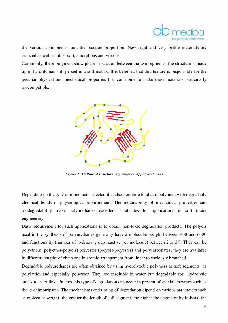
Commonly, these polymers show phase separation between the two segments: the structure is made
up of hard domains dispersed in a soft matrix. It is believed that this feature is responsible for the
peculiar physical and mechanical properties that contribute to make these materials particularly
biocompatible.
Figure 2. Outline of structural organization of polyurethanes
Depending on the type of monomers selected it is also possibile to obtain polymers with degradable
chemical bonds in physiological environment. The modulability of mechanical properties and
biodegradability make polyurethanes excellent candidates for applications in soft tissue
engineering.
Basic requirement for such applications is to obtain non-toxic degradation products. The polyols
used in the synthesis of polyurethanes generally have a molecular weight between 400 and 6000
and functionality (number of hydroxy group reactive per molecule) between 2 and 8. They can be
polyethers (polyether-polyols) polyester (polyols-polyester) and polycarbonates; they are available
in different lengths of chain and in atomic arrangement from linear to variously branched.
Degradable polyurethanes are often obtained by using hydrolyzeble polymers as soft segments as
polylattidi and especially polyester. They are insoluble in water but degradable for hydrolytic
attack to ester link . In vivo this type of degradation can occur in present of special enzymes such as
the 'α-chimotripsina. The mechanisms and timing of degradation dipend on various parameters such
as molecular weight (the greater the length of soft segment, the higher the degree of hydrolysis) the

7
degree of crystallinity, the glass transition temperature and the hydrophily of monomers.
Degrapol® is made of poly-hydroxybutyrate-diol (Hard Segment) and polycaprolactole-diglycole-
diol (Soft Segment).
2.2 Synthesis of hard-segment
The hard-segment is obtained from poly-hydroxybutyrate (PHB), which can be extracted and
isolated from the chemical degradation of a number of microorganisms.
The PHB undergoes a first transformation with ethylene glycol to reduce the molecular weight, to
cutting the long chains of polymer gaining as a result short-chain diols of the hydroxybutyric acid.
O
H
O
O
H HO
OH
+
H
O
O
O
O
O
O
H
m
n
x
x > 1000
1<m<50
1<n<50
Scheme 1. Synthesis of PHB-diol
The initiator is a metal complex, and if the polymer is designed for applications in vivo, preferably
the octanoate tin as initiator is used, since it has the approval of the FDA (Food and Drug
Administration) as a food preservative.
The reaction is considered completed when the average molecular weight (Mw) of polymer reaches
the value of 2600. The chlorobenzene is removed by extraction with dichloromethane. While
methanol, a non-solvent of the polymer, is used for the precipitation.
The polymer obtained, called PHB-diol, appears as a white powder and has the same mechanical
characteristics of PHB, but differs for the molecular weight and the thermal properties.

8
Mw T melting point [°C]
PHB 500000 190 PHB-diol 2600 145
Table 1: Comparison PHB and PHB-diol
The reaction of trans-esterification and depolymerization is conducted in a batch reactor at 140°C,
using chlorobenzene as a solvent, in which the PHB and ethylenglycol are dissolved, according to
the following reaction:
H
O
O
O
O
O
O
H
m
nO
O
O
O
+
H
O
O
O
O
O
O
x+y
n
O
O
O
O
H
dibutyltin laureate
H
O
O
O
O
O
O
n
O
O
O
O
O
y
O
H
1<x+y<501<m<501<n<50
Scheme 2. Synthesis of the hard segment from monomers units

9
2.2.1 Characteristics of PHB
The PHB belongs to polyhydroxyalkanoate family.
The polyhydroxyalkanoate (PIA) are macromolecules synthesized by more than 90 kinds of bacteria
gram + and gram-. In appropriate culture conditions and in particular shortage of some nutrient (eg
N, P, S), polyhydroxyalkanoate accumulate into the bacterium in the form of granules (φ = 0.5 µm)
to a concentration that can reach 90% of dry weight of bacterial mass. The main types of
polyhydroxyalkanoate identified by now are polyester linear head-tail, made up of monomers
belonging to the group of β/3 (R-) hydroxyacids (in much lesser extent also γ, δ, ε (R-)
hydroxyacids). The synthetic scheme of polymerization is shown in Diagram 3.
HO
R
OH
O
n
O
R
*
O
n
+ (n-1) H2O
Scheme 3. Synthetic scheme of polymerization
The lateral group R in position β (3) is an alkyl with C ÷ 13 = 1, which can be linear or branched,
saturated or unsaturated, epoxidised and with halogenated or aromatic substituents.
The exact composition of polyhydroxyalkanoate depends on the type of bacterium from which they
are synthesized and on the culture. For the polyhydroxyalkanoate synthesis, the different types of
bacteria use monomers from different metabolic pathways.
The extreme variability of the chemical nature of the chains side lays the basis of a wide range of
properties of polyhydroxyalkanoate (ranging from typical thermoplastic polymers, such as the
polyhydroxibutyrate, to tires as polyhydroxyottanoate) and of possibility of secondary chemical
interventions (eg cross-linkage ).
The molecular mass of polymers synthesized by bacteria varies from 5,104 to 1,106 Da.
The main polyhydroxyalkanoate are listed in Table 2.
10
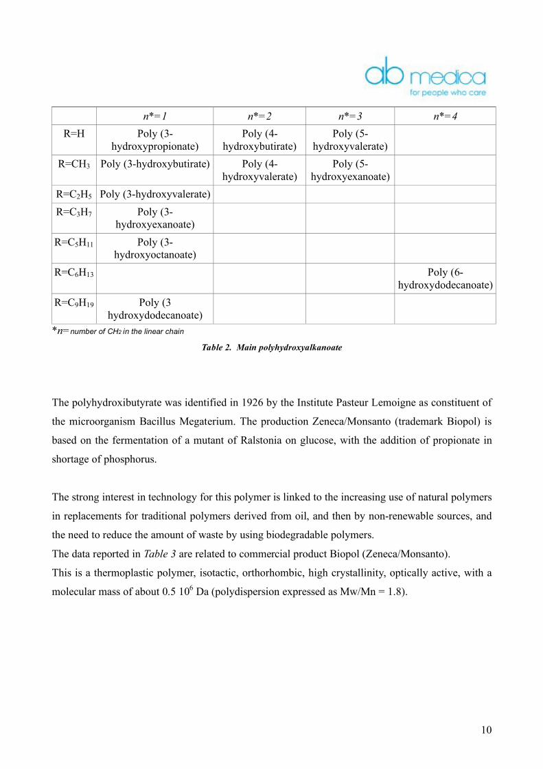
n*=1 n*=2 n*=3 n*=4 R=H Poly (3-
hydroxypropionate) Poly (4-
hydroxybutirate) Poly (5-
hydroxyvalerate)
R=CH3 Poly (3-hydroxybutirate) Poly (4-hydroxyvalerate)
Poly (5-hydroxyexanoate)
R=C2H5 Poly (3-hydroxyvalerate) R=C3H7 Poly (3-
hydroxyexanoate)
R=C5H11 Poly (3-hydroxyoctanoate)
R=C6H13 Poly (6-hydroxydodecanoate)
R=C9H19 Poly (3 hydroxydodecanoate)
*n=number of CH2 in the linear chain
Table 2. Main polyhydroxyalkanoate
The polyhydroxibutyrate was identified in 1926 by the Institute Pasteur Lemoigne as constituent of
the microorganism Bacillus Megaterium. The production Zeneca/Monsanto (trademark Biopol) is
based on the fermentation of a mutant of Ralstonia on glucose, with the addition of propionate in
shortage of phosphorus.
The strong interest in technology for this polymer is linked to the increasing use of natural polymers
in replacements for traditional polymers derived from oil, and then by non-renewable sources, and
the need to reduce the amount of waste by using biodegradable polymers.
The data reported in Table 3 are related to commercial product Biopol (Zeneca/Monsanto).
This is a thermoplastic polymer, isotactic, orthorhombic, high crystallinity, optically active, with a
molecular mass of about 0.5 106 Da (polydispersion expressed as Mw/Mn = 1.8).

11
Properties PHB Polypropilen Tg (°C) +15 -10 Tf (°C) 190 176
Cristallinity (%) 80 70 Resistance (Mpa) 40 38
Elongation at break (%) 8 40 Tensile (GPA) 3.5 -
Resistance to bending Izod-n (J/m) 60 100 Dielectric constant at 1 MHz 3 -
Resistivity (ohm•cm) ≥1016 ≥1016
Higher temperature od use(°C) 130 135 Chemical Resistance
Acid Alkali Alcool
Oils and fats
Low Low
Discreet Good
Excellent Excellent Excellent
Good UV Resistance Discreet Low
Table 3: comparison PHB e Polypropylen
The polyhydroxibutyrate is, for many reason, similar to polypropylene, but unlike this has a glass
transition temperature (Tg) too high and a resistance too low. In addition, the melting temperature is
very close to that of degradation, which makes it problematic, if not impossible, to process with
conventional techniques in use for thermoplastic polymers.
The polyhydroxibutyrate results to be a highly biodegradable polymer. In physiological conditions
ester link are split with an hydrolytic mechanism. The degree of biodegradation of these polymers is
determined primarily by molecular weight, exposed surface, crystallinity and in the case of
copolymers, by the chemical composition and distribution of monomer units. The bioresorption and
the ultimate degradation products may involve macrophages, neutrophils and lymphocytes,
different classes of white blood cells activated by the immune system in the presence of an antigen.
The final degradation product of PHB is the (R)-3-hydrxybutiryc acid, a constituent of human
blood, which is bioassimilable without any problem.
Many micro-organisms (bacteria, fungi) in soil, in urban and industrial discharges, in estuaries of
the rivers, can degrade the polyhydroxibutyrate and its copolymers out of the cells.
For this purpose, they are provided with appropriate enzymes secretion (depolymerasis) that adhere
to particles of polymer and catalyze scrapping to simple water-soluble molecules. In turn, these
molecules are used by microorganisms themselves in their metabolism. The final products of
biochemical demolition were found to be H2O and CO2 in aerobic environment and CO2 and CH4 in

12
anaerobic environment.
2.2.2 Characteristics of ethylenglycol
The ethylenglycol, IUPAC name 1,2-ethandiol, is
the most common diol. At room temperature looks
like a transparent liquid, miscible with water,
appearance and syrupy liquid with a sweet taste.
Figure 2. Chemical structure of ethylenglycol
Produced in small quantities during World War I as coolant and as an ingredient in explosives,
ethylenglycol is produced on a large scale since 1937, when its precursor, ethylene oxide, becomes
cheaply available. It is produced from ethylene, via the intermediate ethylene oxide. Ethylene oxide
reacts with water to produce ethylene glycol accordino to the following chemical equation:
C C
H
HH
H
+ O2
O
HO
OHH2O
Scheme 4. Synthesis of ethylenglycol
This reaction can be catalyzed by either acids or bases, or can occur at neutral pH at elevated
temperatures. The highest yields of ethylene glycol occur at acidic or neutral pH with a large excess
of water. Under these conditions, ethylene glycol yields of 90% can be achieved. The main
byproducts are the ethylene glycol oligomers: diethylene glycol, triethylene glycol and tetraethylene
glycol.
The presence of hydroxylic groups makes it very responsive and is widely used in polymerization
reactions for polyester.

13
Table 4: Ethylenglycol properties
2.3 Synthesis of Soft-segment
The soft segment is synthetized from polymerization process which involves the opening of the ring
of the ε-caprolactone, which is linked to diglycol units through glycol which plays the role of
initiator, with the task of controlling the molecular weight.
The synthesis takes place in the liquid phase at 135°C until it stabilizes the molecular weight. The
reaction is considered completed when the volume of retention, obtained by GPC, is constant over
the time. The value of molecular weight is determined by the operator and from the mass it’s
possible to determine the amount of monomers to be used.
Properties Molecular formula C2H6O2
Molar mass 62,07 Aspect Colorless liquid
CAS number 107-21-1 Chimical-fisic properties
Density (g/cm3, in c.s.) 1,11
Solubility in water 1000 g/l a 20°C Melting point (K) 260 (-13°C) Boiling point (K) 470,6 (197,6°C)
ΔebH0 (kJ·mol-1) 49,66
Termochemical properties
ΔfH0 (kJ·mol-1) -460
S0m(J·K-1mol-1) 163,2
C0p,m(J·K-1mol-1) 148,6
Hazards Flash point (K) 384 (111°C) Explosion limits 3,2 - 53% vol.
Autoignition temperature (K) 683 (410°C)
14
2.3.1 Characteristics of poly ε-caprolactone
The use of poly(ε-caprolactone) (PCL) for biomedical applications has become a real possibility
after its appearance on the market as degradable packaging material.
Later it was shown that the PCL may be degraded in physiological conditions through the same
hydrolityc mechanism of the other hydroxyacids. The bioerosion of the PCL is significantly slower
because of of more distinctly hydrophobic repeated units. The PCL is commonly considered a non-
toxic and biocompatible material.
The long aliphatic main chain of PCL gives to the polymeric material some properties uncommon
to other aliphatic polyesters such as an extremely low glass transition temperature (Tg ≈ -60°C), a
moderate melting temperature (Tf ≈ 60°C ), a high solubility in organic solvents, a significant
thermal stability (Td > 350°C) and the ability to form single-phase mixtures with many polymeric
materials. The high permeability of PCL matrices, which are always in rubbery state at
physiological temperature, has allowed to use it in systems controlled release of drugs.
2.3.2 Characteristics of copolymer ε-caprolactone-diglycolid
Polycaprolactone degradation is very long and therefor, in order to increase the degree of
degradation, a copolymer of caprolactone and diglycolid is used.
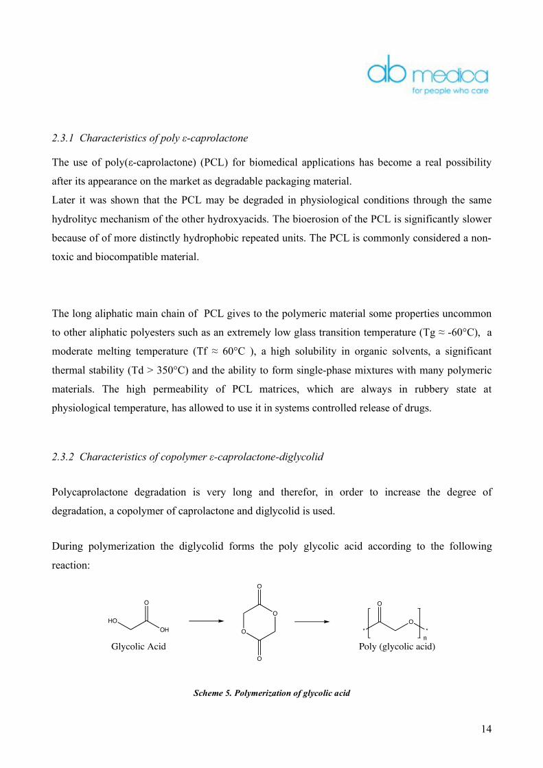
During polymerization the diglycolid forms the poly glycolic acid according to the following
reaction:
OH
HO
O
O
O
O
O
*
O
O
*
n
Glycolic Acid Poly (glycolic acid)
Scheme 5. Polymerization of glycolic acid
15
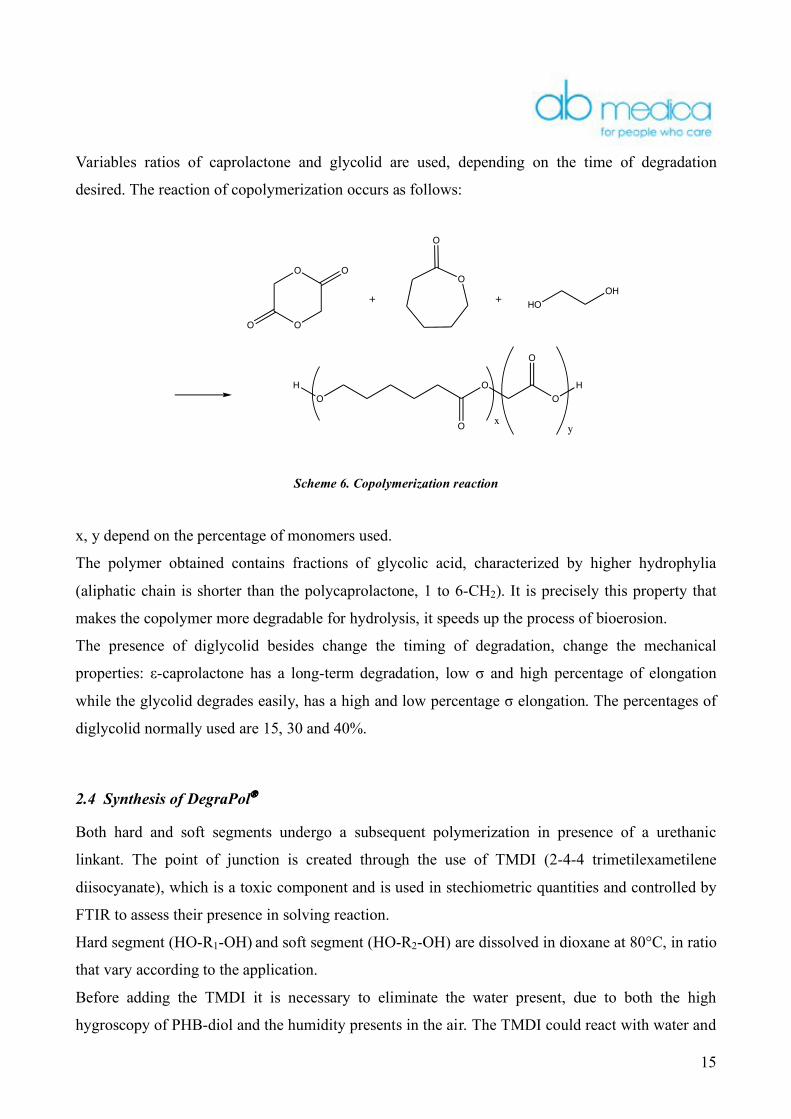
Variables ratios of caprolactone and glycolid are used, depending on the time of degradation
desired. The reaction of copolymerization occurs as follows:
O
O
O
O
O
O
+ +HO
OH
O
O
O
O
O
H H
xy
Scheme 6. Copolymerization reaction
x, y depend on the percentage of monomers used.
The polymer obtained contains fractions of glycolic acid, characterized by higher hydrophylia
(aliphatic chain is shorter than the polycaprolactone, 1 to 6-CH2). It is precisely this property that
makes the copolymer more degradable for hydrolysis, it speeds up the process of bioerosion.
The presence of diglycolid besides change the timing of degradation, change the mechanical
properties: ε-caprolactone has a long-term degradation, low σ and high percentage of elongation
while the glycolid degrades easily, has a high and low percentage σ elongation. The percentages of
diglycolid normally used are 15, 30 and 40%.
2.4 Synthesis of DegraPol®
Both hard and soft segments undergo a subsequent polymerization in presence of a urethanic
linkant. The point of junction is created through the use of TMDI (2-4-4 trimetilexametilene
diisocyanate), which is a toxic component and is used in stechiometric quantities and controlled by
FTIR to assess their presence in solving reaction.
Hard segment (HO-R1-OH) and soft segment (HO-R2-OH) are dissolved in dioxane at 80°C, in ratio
that vary according to the application.
Before adding the TMDI it is necessary to eliminate the water present, due to both the high
hygroscopy of PHB-diol and the humidity presents in the air. The TMDI could react with water and

16
form insoluble ureae as follows:
R
N
C
O+ H2O
R
HN
C
O
OH
R NH2 CO2+
Isocyanide Carbamic Acid Amine
1)
2)R
N
C
O+ R NH2
R
HN
HN
O
R
Isocyanide Amine Urea disubstitued
Scheme 7.
The removal of water from the reaction takes place by distillation of the solvent: dioxane and water
temperatures have very close boiling point (101°C and 100°C), this way it's possible to eliminate
50% H2O. Subsequently, the reaction mixture is anhydrificated in Soxhlet reflux on molecular
sieves.
The reaction between the diisocyanate and diol is a simple addition reaction with movement of a
hydrogen atom. The product of the reaction is a urethane, as follows:
R
N
C
O+ R' OH R
HN O
O
R' 1
Isocyanide Urethane (carbamate)Hydroxyl
Scheme 8.
The addition reaction is exothermic with H -105 kJ /-NCO and Energy activation 42 kJ / piers. The
diols used in the reaction have only primary hydroxyl group that react quickly with the urethanic
group.
In case of DegraPol® the reaction takes place by means of addition to the main chain. The
orientation of the groups R1 and R2 is random because it is not currently possible to establish a
regularity in the structure.

17
R1HO OH + R2HO OH +
N
C
O
N
C
O
R1
HO OHN
O
NH
O
R2
O
OH
Scheme 9. Synthesis of DegraPol®
The reaction is considered completed when all TMDI is consumed and the molecular weight of
polymer reaches the value of 100,000. The product, recovered by extraction with methanol,
contains traces of catalyst (Dilaurate of dibutyltin) and impurities found in solvents and in the
reaction.
A good purification of the polymer obtained is necessary, since the presence, even minimal, of
impurity limits its use in the biomedical field.
A solution of DegraPol® in chloroform is percolate through a bed of silica gel (silicon dioxide
hydrate), polymer of the orthosilicic acid. The gel has an amorphous structure, similar to that of
glass, which do not identify structural elements repetitive and ordered as crystal in the state. The
silica can be stratified into thin layers: up to 100 cm2 per gram of silica. The surface of the gel,
strongly polar for the presence of numerous groups OH-free, holds the polar compounds and
impurities solid, purifying the polymer which remains dissolved in chloroform.
18
3. Scaffold
A scaffold is a three-dimensional porous support made of biocompatible and bioerodible material
on which initial adhesion of cells and subsequent cell regrowth up to formation of tissue can take
place and having a degradation speed similar to cell regrowth.
The main features of these support are:
high porosity and three-dimensionality with the presence of a network of interconnected
pores to allow cell growth, transport of nutrients and elimination of waste substances
high biocompatibility so as not to generate any form of rejection by the host cells;
controlled biodegradability;
bioreabsorption with a degree of reabsorption that allows cell growth in vitro and /or in vivo;
surface chemically suitable for adhesion, proliferation and differentiation of cells;
mechanical properties similar to those of tissues that must play on the scaffold
reproducibility in various shapes and sizes
The cells to reproduce need a suitable environment, so when they are seeded on a polymer scaffold,
it is necessary that the substrate has the right characteristics of good biocompatibility, low
cytotoxicity, good biodegradability. Moreover, toh ave a correct cell growth, it is necessary that
there is similarity between the mechanical properties of regenerated tissues and those of the scaffold
on which these cells are seeded.
The mechanical properties essential to bear in mind is Young’s modulus, since for high values of
this quantity corrispond to hard and brittle materials that do not work as support for cell regrowth.
The first fundamental principle is that the Young's moduli of materials, used as support for
regrowth, have comparable values to the relative moduli from regenerate tissue; if this does not
apply, a wrong cell regrowth will accour with, possibly, death of cells. In fact, the tissue necrosis is
due to stress which is created at the interface between support and tissue, due to the movement that
generates between scaffold and tissue, caused by their difference in Young's modulus. Another
possible reason is the absence of effective transfer of stress through the interface support/tissue.
The second basic principle is the analysis of viscoelastic behaviour of support materials, because it
is necessary to analyse how the mechanical properties change in time (variable data) affecting the
process of tissue regrowth.
19
The surface topography of a material is usually classified according to the roughness, texture and
porosity; each of these aspects is important in monitoring the adhesion and cell functionality.
Indeed in tissues and organs, cells are organized in well-defined space relatively to specific
functional requirements. Indeed, during the development of a tissue or an organ, dividing cells
recognize gradients of biochemical signals that guide their polarization and final position. Many of
these signals are produced in relation to the topography of the environment surrounding the cells.
The techniques used to obtain scaffold in our laboratory are:
• the separation phase, it exploits the principles of thermodynamics to create two phases to
different concentrations within the polymer solution. Phase poor of polymer is removed,
leaving a highly porous polymer network. This technique allows to create microporose
sponges but show little interconnections;
• fibre production, through elettrospinning, involves the formation of polymeric networks
with large areas of cell attack that can ensure a rapid diffusion of nutrients.
While the technique of phase separation allows to obtain scaffold with controlled porosity, using
solutions with different concentrations of Degrapol®, the electrospinning allows to control the
topography of the scaffold, choosing appropriately the plot, and the alternation of layers of
Degrapol®.

20
4. Experimental Plan
The strength of the product is the ability to modify the property in a manner that is fully
independent on the application.
The properties of Degrapol® are according to:
operating conditions of the production process (temperature)
the chemical composition (ratio of copolymers)
productive processes
Production process Mechanical properties Composition
Mechanical properties
Degradation time Manufacturing process
Mechanical properties
Surface properties
The production process has been optimized at ETH Zurich; Ab Medica’s task is to make it
reproducible, controlling operating conditions. For example, the temperature must remain constant
throughout the process, it is noted that a temperature lower than 90°C results in a polymer low
molecular weight, and then with poor mechanical properties that do not make it suitable for
elettrospinning. The polymerizations must be conducted in a controlled environment to reduce
contamination and the thermal fluctuations, also the molecular weight of polymer needs to be
monitored in order to establish the reaction time needed to obtain Degrapol® molecular weights
known.
The chemical composition determines both the mechanical properties and the time degradation
because the individual blocks, as seen previously, have different mechanical properties. A different
ratio of hard and soft segments makes the material harder or softer and therefore the choice of
different percentage mixture is made according to the type of tissue to regenerate: for example, to
regenerate bones it is necessary to use a very strong material, while for the skin a soft material is
preferable. The different contents of poly glycolic acid adjust times of degradation. One of our
objectives is to identify the chemical and physical characteristics, suitable for different
compositions and establish standard formulations for each regenerating tissue.
As seen above, the polymer support must ensure the growth and cell differentiation. These features
are conferred by sponging techniques previously described. For each application should be

21
established:
the ratio empty / full such as to ensure adequate mechanical properties
the percentage of interconnected pores
the surface topography
To evaluate the properties of surface morphology of pores, their size and the presence of
interconnections, analytical technique of Microscopy scanning electron (SEM) has been used.
For the tensile tests (Young modulus and rupture elongation) and creep tests (visco-elastic
behaviour), a isotonic position transducer must be used (model 7006 of Ugo Basile Biological
Research Apparatus), designed to measure size and polymeric changes in muscle fiber.
All the mechanical tests should be performed on both scaffold and compact film of the same
material, in order to assess the influence of the microproduction on such properties. It is also
necessary to know the dimensions, the techniques to carry out the test and then regulations of
reference for the mechanical tests used in engineering tissue.
Determination of biotoxicity, biocompatibility and degradation time is carried out by institutes of
cell biology.
The biocompatibility is guaranteed by the use of biocompatible materials, while biotoxicity depends
on the presence of traces of catalysts and solvents, toxic substances that should be removed before
using the material for cell cultures. The timing of degradation varies from a few weeks to few years,
generally it assesses the changing of weight of the support over time under certain conditions that
are similar to those physiological.
It will be necessary to make a market investigation on the scaffold, to meet the needs of potential
buyers of the product and thus establish an experimental plan that takes this information into
account.
The diagram (Figure 4) shows the interconnection between the various disciplines involved in
tissue engineering. It is essential to cooperation with institutes involved in cell growth and
characterization of biomaterials in order to design the material depending on the application.

22

23
5. Current applications Studies in the literature of both in vitro and in vivo cultures of DegraPol® have demonstrated the
real possibility of using it as a scaffold, in fact apart from being a good support for cell growth, it
makes it possible to preserve the specific properties of cells.
The first study of compatibility between the material and biological tissue covered the culture of
osteoblasts, cells isolated from bone tibia rats [3]. After 8 days from seeding on highly porous
DegraPol® (pore size of between 100-150 µm), the osteoblasts showed both a multi-lamellar
structure and migration inside the pores of polymer, with high degree of cell growth. It was also
observed that the amount of collagen type I and osteocalcium produced, remains constant over the
time in cultures of osteoblasts, indicating that the osteoblasts maintain their phenotype.
These data are confirmed by another study[4]. In vitro the growth of osteoblasts of rats and a variety
of human cells (HF01, MC3T3) on sponges of DegraPol® (pore size of between 100-400 µm)
confirmed the previous data. The possibility of using the sponges of DegraPol® as carrier for BMP
(bone morphogenetic protein) was also assessed. The BMP are biologically active proteins that
induce the formation of bone in vivo. Discs of DegraPol® with BMP adsorbed were implanted under
the skin on the back of rats and after 2 weeks from the implant the histological activity showed
both the activity of the alkaline phosphatase enzyme (ALP), which leads to target substances for the
renewal of bone cells, and the presence of calcium, confirmed by the X-ray analysis, attesting its
calcification.
Another study of in vivo cell regeneration[5] refers to the possibility of using DegraPol®, as a guide
for the regrowth of nerves, nerve growth channel (NGC). The NGC are polymeric tubular structures
in which the ends of nerves are inserted and sutured and which are able to issue, within their lumen,
tropics factors that improve the regeneration device. Tubular structures of 3 different DegraPol®,
containing various percentages of PHB-diol were planted on 26 rats; after 4 weeks in 23 rats,
regardless of the type of DegraPol® used, it is noted that into the channel epineural tissue
surrounded by myelinic axons and Schwann cells is present. This study also shows how a low-
PHB-diol induces a faster degradation.
There have been inflammatory phenomena that affect only small fragments of polymers because of
the high air interface. These small fragments are caused by the splitting of soft segment, which are
however phagocytized by macrophages without interfering with the cell regeneration.
There were also studied of chondrocytes’s culture derived from human tracheal cartilage on porous

24
and three-dimensional structures of DegraPol®[6]. After 8 weeks from seeding it was observed that
large numbers of type II collagen is produced by chondrocytes while collagen type I was absent.
This indicates a reduced loss of cell phenotype because collagen type II, interconnected with the
type of collagen IX, is a typical component of cartilage.
Furthermore, SEM observations demonstrated that the cells assume a spherical configuration
similar to that of native cartilaginous tissue cells. It therefore has a cell-specific growth, reflecting
in terms of size and shape the phenotype cell. These data are confirmed by other studies in vitro
about the regeneration of chondrocytes isolated from xiphoid of rats and from costal area of
cattles.[7] [8]
Another recent study regards the culture of cells in the smooth muscle tissue on the DegraPol®
scaffold (pore size of between 100-300 µm)[9]. After 2 days from seeding, cells have adhered well to
the surface of DegraPol® and retain their characteristic fusiform shape confirming that the porosity
of the scaffold allows cell penetration and adherence.
After 8 days apoptosis (the programmed death of cells and the loss of morphology) is observed.
This event is due to the static conditions of the culture rather then the support; indeed this type of
cell is much more sensitive to culture conditions compared to chondrocytes primary used in
previous examples.
All studies confirm that the scaffolds of DegraPol® ensure both the growth and the maintenance of
cellular phenotype of many biological tissues and that it can be used for many applications.

25
6. Possible development
In the body, the interactions between cells and the surrounding environment are based on the
recognition of certain biological molecular structures through specific membrane receptors. Thus, a
convenient method to confer biological activity to a polymer surface is represented by blending
natural or covalently molecular species which promote the phenomena of adehesion and
proliferation. It's possible to functionalize diols with proteins, making the cell recognition faster
and more effective. This will reduce the time of adehesion on the scaffold and regeneration time of
the tissues.
This technique is necessary to assess compatibility with diols used to produce DegraPol® and the
feasibility of the process to industrial level.

26
References
[1] Saad B., Keiser O.M., Welti M., Uhlschmid G.K., Neuenschwander P., Suter U.W.,
Multiblock copolyesters as biomaterials: in vitro biocompatibility testing. Journal of
Materials Science: Materials in Medicine. 1997; 8: 497-505B.
[2] Saad , T.D. Hirt, M. Welti, G.K. Uhlschmid, P. Neuenschwander, U.W. Suter,
Development of degradable polyesterurethanes for medical applications:In vitro and in vivo
evalutions. Journal of Biomedical Materials Research. 1997; 36: 65-74
[3] B. Saad, S. Matter, G. Ciardelli, G. K: Uhlschmid, M. Welti, P. Neuenschwander, U.
W.Suter, Interaction of osteoblasts and macrophages with biodegradable and higly porous
polyesterurethane foam and its degration products. Journal of biomedical Materials
Reserch.1996; 32:355-366
[4] B. Saad, Y. Kuboki, M. Welti, G.K. Uhlschmid, P. Neuenschwander, U. W.Suter,
DegraPol-Foam: a degradable and highly porous polyesterurethane foam as a new
substrate for bone formation. Artificial Organs. 2000; 24: 936-945
[5] M. Borkenhage, R.C. Stoll, P. Neuenschwander,U.W: Suter, P. Aebischer, In vivo
performance of a new biodegradable polyester urethane system used as a nerve guidance
channel. Biomaterials.1998; 19:2155-2165
[6] Yang L., Korom S., Weltia M., Hoerstrup S.P., Zund G., Jung F.J., Neuenschwander P.,
Weder W., Tissue engineered cartilage generated from human trachea using DegraPol®
scaffold. European Journal of Cardio-thoracic Surgery. 2003; 24: 201-207
[7] B. Saad, M. Moro, A. Tun-Kyi, M. Welti, P. Schmutz, G.K. Uhlschmid, P.
Neuenschwander, U. W.Suter, Condrocyte-biocompatibility of DegraPol-Foam: in vitro
evaluations. Journal of Biomaterials Sci. Polymer. 1999; 10:1107-1119

27
[8] B. Saad, M. Moro, A. Tun-Kyi, M. Welti, P. Schmutz, G.K. Uhlschmid, P.
Neuenschwander, U. W.Suter, Hyghly porous and biodegrable DegraPol-foam as
sugstrate for the formation of neo-cartilage: in vitro evalution. 9th simposium-Materials
in clinical Application. 1999
[9] Danielsson C., Ruault S., Simonet M., Neuenschwander P., Frey P., Polyesterurethane foam
scaffold for smooth muscle cell tissue engineering. Biomaterials. 2006; 27: 1410-15