new: elisa-based method for fast, sensitive in this … cation of activated ras gtpase ... antibody...
TRANSCRIPT

Active Motif is pleased to introduce the new Ras GTPase Chemi ELISA Kit. It is the fi rst ELISA-based kit designed to detect and quantify activated Ras GTPase. As an ELISA, it offers a number of advantages over exist-ing methods. The Ras GTPase Chemi ELISA Kit is much more sensitive than pull-down/Western methods, so you can detect even low levels of protein, and use less sample. In addition, ELISAs are more quantitative than Westerns, and they eliminate the need to run and develop gels. This means you’ll get better results faster, and with less ef-fort. And, as the assay uses a 96-well plate made up of 12-well strips, it is convenient and economical to run anywhere from 1 to 96 samples in a single experiment.
Cell extract (µg/well)
UnstimulatedEGF stimulated
Activ
ated
Ras
Sig
nal (
RLU) 800000
600000
400000
200000
0502512.56.25
Figure 1: Quantifi cation of activated Ras in stimulated HeLa cells.Increasing amounts of whole-cell extract from HeLa cells that had been stimulated with 50 ng/ml of EGF for 2 minutes were assayed using the Ras GTPase Chemi ELISA Kit.
New: ELISA-based Method for Fast, Sensitive Quantifi cation of Activated Ras GTPase
www.activemotif.com
continued on page 6
Active Motif’s new TransAM™ Flexi Kits make it possible to effi ciently study the binding of transcription factors at any DNA-binding site you choose. These kits are the latest version of Active Motif’s original TransAM Kits, which have rapidly become the method of choice for studying transcription factor binding activity.
New version lets you choose your siteThe original TransAM Kits are DNA-binding ELISAs that provide a fast, non-radioac-tive alternative to classical methods such as Electrophoretic Mobility Shift Assays (EMSAs). Each kit includes a 96-well plate that is coated with a double-stranded oligonucleotide, which contains a spe-cifi c consensus-binding site. While this
format makes it simple and convenient to measure transcription factor binding at this consensus site, it does not allow you to study binding of the transcrip-tion factor at alternative sites, such as those which contain mutations or that are putative, unproven binding sites. The new TransAM Flexi Kits give you the fl exibility to immobilize any oligo or PCR product in the 96-well plate, while providing you with proven antibodies and all of the other components you’ll need to quantitatively measure transcription factor binding. With TransAM Flexi Kits, you can now study variant transcription factor-binding sites, analyze native promoters, confi rm chroma-tin immunoprecipitation (ChIP) results and determine isoform-binding affi nity.
NFκ
B Ac
tivat
ion
(OD 4
50 n
m)
0.8
1
0.2
0.4
0.6
0
TPA + CI withmutant oligos
Unstimulated TPA + CI stimulatedTPA + CI with wild-type oligos
Figure 2: Specifi city of TransAM Flexi assays.TransAM Flexi NFκB p65 assays are performed in the presence of wild-type and mutant competitor oligonucleotides using 5 µg/well of nuclear extract from Jurkat cells that were stimulated with TPA and calcium ionophore (CI).
continued on page 4
New: Measure Transcription Factor Binding at Any DNA Sequence You Choose
I N T H I S I S S U E1 New: ELISA-based Method for
Fast, Sensitive Quantifi cation of Activated Ras GTPase
1 New: Measure Transcription Factor Binding at Any DNA Sequence You Choose
2 New: Simplify Chromatin Shearing Using the Enzymatic Shearing Kit for ChIP
3 Chariot™ Delivers Functionally Active Proteins, Peptides and Antibodies into Live Cells
5 New: A Novel, Cell-based Method for Monitoring STAT Phosphorylation
7 A Complete Solution for Studying Nuclear Receptor Protein Activity
8 New Antibodies for Chromatin and DNA Methylation
THE NEWSLETTER OF ACTIVE MOTIF — APRIL 2005 • VOLUME 6 • NUMBER 1

Toll Free — 1 877 222 95432
A P R I L 2 0 0 5 • V O L U M E 6 • N U M B E R 1
www.activemotif.com 3
C h r o m a t i n I m m u n o p r e c i p i t a t i o n
New: Simplify Chromatin Shearing Using the Enzymatic Shearing Kit for ChIP
Active Motif is pleased to announce the release of another innovative prod-uct that simplifi es your chromatin immunoprecipitation (ChIP) experiments. The new Enzymatic Shearing Kit is your best bet for generating chromatin suitable for ChIP as well as other applications that study chromatin structure with little to no optimi-zation required – nothing could be simpler.
Successful shearing every timeIn order to perform a successful ChIP experiment, chromatin must fi rst be sheared to 200-1000 bp fragments. Traditionally, shearing has been performed by subjecting the isolated chromatin to different pulses of sonication. Although sonication can be
���� ��
��� ��
��� ��
� � � � �
Figure 1: Analysis of DNA sheared using the Enzymatic Shearing Kit.HeLa cells were fi xed for 10 minutes with 1% formaldehyde and then chromatin was prepared using the Enzymatic Shearing Kit protocol. Chromatin was sheared with the Enzymatic Shearing cocktail for 5, 10 & 15 minutes and the reaction was stopped. The sheared and unsheared chromatin samples were subjected to cross-link reversal, treated with Proteinase K, phenol/chloroform extracted and precipitated as described in the protocol. Samples were separated by electrophoresis through a 1% agarose gel to assess shearing results:
Lane 1: 100 to 1000 bp ladder.Lane 2: Unsheared HeLa DNA.Lane 3: HeLa DNA treated for 5 minutes.Lane 4: HeLa DNA treated for 10 minutes.Lane 5: HeLa DNA treated for 15 minutes.
an effective method for shearing DNA, it can also be time consuming and diffi cult to optimize due to complications arising from emulsifi cation and overheating during the sonication process. Also, because the quality of your sheared sample depends greatly upon the quality of your sonicator, it may be necessary to purchase an expensive, “high-end” sonicator to get reproducible shearing. Because of this, Active Motif has developed the Enzymatic Shearing Kit, which is a more robust and user-friendly method to shear chromatin for ChIP. The Enzymatic Shearing Kit uses a proprietary enzymatic shearing cocktail that quickly and easily shears DNA into 200-1000 bp fragments (Figure 1). And, because enzymatic shearing is dependent only on time and temperature, all of the problems associated with sonication are eliminated and ChIP results are improved.
How it worksShearing chromatin with the Enzymatic Shearing Kit is simple to perform. First, cells are grown to confl uency and then fi xed using formaldehyde, which cross-links and therefore preserves protein/DNA interac-tions. A Glycine “Stop-Fix” buffer is then added to stop the fi xation procedure and prevent over-crosslinking of the sample. Next, the cells are lysed and the nuclei are collected. A simple, 10-minute incuba-tion with the enzymatic cocktail is then performed, the reaction is stopped and the chromatin is ready to use.
How can I get oneThe Enzymatic Shearing Kit is sold sepa-rately and can be used in conjunction with Active Motif’s ChIP-IT™ Kit buffers and controls. Order an Enzymatic Shearing Kit today and simplify your shearing.
Product Format Catalog No.
Enzymatic Shearing Kit 10 rxns 53005
ChIP-IT™ Kit 25 rxns 53001
ChIP-IT™ w/o controls 25 rxns 53004
ChIP Product LineComplete ChIP-IT KitsActive Motif’s ChIP-IT Kit is the most effective product available for per-forming ChIP. The kit contains nearly everything you need to perform ChIP including a comprehensive protocol and optimized buffers, inhibitor cocktails, DNA purifi cation columns as well as positive control primers and antibodies, all of which have been validated in actual ChIP experiments.
Pre-validated ChIP antibodiesFinding an antibody that has been validated in ChIP experiments can be diffi cult. As specialists in transcription factor research, Active Motif offers a large number of antibodies, which are actively being validated for use in ChIP. Below is a list of antibodies that have been positively tested in ChIP, which are ideal when used with our ChIP-IT w/o controls kit. Be sure to keep up to date on new ChIP-validated antibodies by visiting our website.
Antibody Cat. No.
AML-1/Runx1 pAb 39000
AP-2 pAb 39304
c-Jun pAb 39309
C/EBPα pAb 39306
DNMT1 mAb 39204
DNMT3A mAb 39206
DNMT3B mAb 39207
E2F-1 pAb 39313
E2F-6 mAb 39509
GATA-1 pAb 39025
HBP-1 mAb 39511
HDAC3 pAb 40968
HDAC4 pAb 40969
HDAC5 pAb 40970
HDAC6 pAb 40971
IRF-3 pAb 39033
JunB pAb 39326
JunD pAb 39328
p53 pAb 39334
Pax-5 pAb 39336
PPARγ pAb 39338
RNA pol II mAb 39097
Sp1 pAb 39058
TRF2 pAb 39223
Toll Free — 1 877 222 95432
A P R I L 2 0 0 5 • V O L U M E 6 • N U M B E R 1
www.activemotif.com 3
P r o t e i n T r a n s f e c t i o n
Chariot™ is Active Motif’s patented* protein delivery reagent that effi ciently transports biologically active proteins, peptides and antibodies directly into cultured mamma-lian cells in less than two hours. The cells can be assayed immediately after delivery to determine the effects of the introduced material, without the need for fi xing. This makes Chariot ideal for a variety of func-tional studies in living cells.
? How does Chariot work?
Chariot is a peptide that forms a non-covalent complex when combined with a purifi ed protein, peptide or antibody. When added to cells, the complex is rapidly internalized. After delivery, the complex dissociates in a process called “decaging”, leaving the macromolecule biologically ac-tive and free to proceed to its cellular tar-get. One advantage of the complex is that it stabilizes the macromolecule, protecting it from degradation during internalization. It also eliminates the need to make fusion proteins or perform chemical coupling. Chariot delivery is also serum independent, which gives you the fl exibility to culture your cells in whichever type of media you prefer.
? Does Chariot work with primary cells or even in vivo?
Chariot effectively delivers into primary cells, something that most other delivery systems cannot do. Most notable is the use of Chariot on hard-to-transfect primary neurons (Jurney, W. et al. (2002) Journal of Neuroscience 22: 6019-602) without any signs of cytotoxicity or rejection. Chariot has also been shown to deliver proteins in vivo (Aoshiba, K et al. (2003) Am J Respira-tory Cell & Molecular Biology. 28: 555-562).
? What about protein functionality?
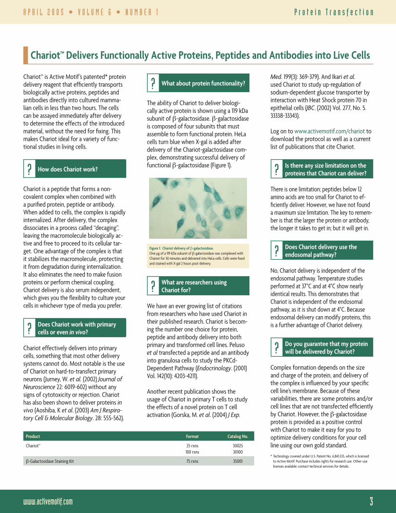
The ability of Chariot to deliver biologi-cally active protein is shown using a 119 kDa subunit of β-galactosidase. β-galactosidase is composed of four subunits that must assemble to form functional protein. HeLa cells turn blue when X-gal is added after delivery of the Chariot-galactosidase com-plex, demonstrating successful delivery of functional β-galactosidase (Figure 1).
Figure 1: Chariot delivery of β-galactosidase. One µg of a 119 kDa subunit of β-galactosidase was complexed with Chariot for 30 minutes and delivered into HeLa cells. Cells were fi xed and stained with X-gal 2 hours post-delivery.
? What are researchers using Chariot for?
We have an ever growing list of citations from researchers who have used Chariot in their published research. Chariot is becom-ing the number one choice for protein, peptide and antibody delivery into both primary and transformed cell lines. Peluso et al transfected a peptide and an antibody into granulosa cells to study the PKCd-Dependent Pathway (Endocrinology. (2001) Vol. 142(10): 4203-4211).
Another recent publication shows the usage of Chariot in primary T cells to study the effects of a novel protein on T cell activation (Gorska, M. et al. (2004) J Exp.
Product Format Catalog No.
Chariot™ 25 rxns100 rxns
3002530100
β-Galactosidase Staining Kit 75 rxns 35001
Chariot™ Delivers Functionally Active Proteins, Peptides and Antibodies into Live Cells
Med. 199(3): 369-379). And Ikari et al. used Chariot to study up-regulation of sodium-dependent glucose transporter by interaction with Heat Shock protein 70 in epithelial cells (JBC. (2002) Vol. 277, No. 5. 33338-33343).
Log on to www.activemotif.com/chariot to download the protocol as well as a current list of publications that cite Chariot.
? Is there any size limitation on the proteins that Chariot can deliver?
There is one limitation; peptides below 12 amino acids are too small for Chariot to ef-fi ciently deliver. However, we have not found a maximum size limitation. The key to remem-ber is that the larger the protein or antibody, the longer it takes to get in; but it will get in.
? Does Chariot delivery use the endosomal pathway?
No, Chariot delivery is independent of the endosomal pathway. Temperature studies performed at 37°C and at 4°C show nearly identical results. This demonstrates that Chariot is independent of the endosomal pathway, as it is shut down at 4°C. Because endosomal delivery can modify proteins, this is a further advantage of Chariot delivery.
? Do you guarantee that my protein will be delivered by Chariot?
Complex formation depends on the size and charge of the protein, and delivery of the complex is infl uenced by your specifi c cell line’s membrane. Because of these variabilities, there are some proteins and/or cell lines that are not transfected effi ciently by Chariot. However, the β-galactosidase protein is provided as a positive control with Chariot to make it easy for you to optimize delivery conditions for your cell line using our own gold standard.
* Technology covered under U.S. Patent No. 6,841,535, which is licensed to Active Motif. Purchase includes rights for research use. Other-use licenses available; contact technical services for details.

Toll Free — 1 877 222 95434
A P R I L 2 0 0 5 • V O L U M E 6 • N U M B E R 1
www.activemotif.com 5
D N A - b i n d i n g E L I S A s
continued from page 1 — New: Measure Transcription Factor Binding at Any DNA Sequence You Choose
The TransAM Flexi methodIn the TransAM Flexi method, a biotinylated oligo or PCR product, which contains the transcription factor-binding site of choice, is incubated with nuclear extracts that have been treated to activate the transcription factor. The samples are then transferred to a 96-well, streptavidin-coated plate, where the biotinylated oligonucleotide is captured. A primary antibody specifi c for the activated transcription factor is then added to the individual wells, followed by an HRP-conjugated antibody and devel-oping reagent. The plate is then read on a spectrophotometer, which provides a quantitative, colorimetric readout of transcription factor activation (Figure 3).
Assay a variety of sequences for more accurate disease researchHistorically, it has only been possible to measure the binding activity and affi nity of a transcription factor to non-consensus sequences, such as putative or polymorphic sites, using either reporter gene constructs or radioactive electrophoretic mobility shift assays (EMSAs). Unfortunately, these approaches are time consuming, lack sensitivity and require signifi cant validation before they can be used. The new TransAM Flexi Kits, however, make sequence-specifi c transcription factor studies more straight-forward than ever before. This is because every TransAM Flexi Kit is supplied as a complete solution of validated antibodies, controls and preparation & reaction buffers, so all you need to do is decide how many different sequences you want to test.
Proven antibodies to ensure your successAs in chromatin immunoprecipitation, transcription factor-binding assays require the use of highly specialized antibodies. Developing antibodies with the proper-ties needed for use in TransAM is no easy task. They must be able to recognize the target protein when it is bound to DNA, and they must bind both specifi cally and with high affi nity. To ensure that you get the highest quality results, every TransAM antibody has been extensively tested for sensitivity and specifi city (Figure 2, page 1).
Confi rm your ChIP resultsChIP is fast becoming an invaluable tool for the analysis of gene promoter regulation. However, there are currently no convenient tools that can be used to confi rm if the transcription factor of interest is capable of binding to the promoter being studied. Because TransAM Flexi Kits make it possible
to analyze the ability of a transcription factor to bind to any sequence, they offer a fast and convenient tool to confi rm that your ChIP results are valid.
TransAM Flexi advantages• Flexible assay – enables analysis of
binding at any DNA sequence• Reproducible – proven antibodies
and reagents ensure your results• Sensitive & quantitative – ELISAs
provide better results than Westerns• Fast – have results in less than 5 hours• Versatile – assay cell or tissue samples
Study your chosen sequences todayTransAM Flexi Kits make it fast and easy for you to study transcription factor binding at any DNA sequence you choose. Kits for NFκB p50 and p65 are now available, with more on the way. Try one today!
TransAM™ Product LineTransAM™ AP-1 Family New: TransAM™ Flexi NFκB p50 TransAM™ ATF-2 TransAM™ GR TransAM™ NFκB p50*
TransAM™ GATA Family New: TransAM™ Flexi NFκB p65 TransAM™ c-Myc TransAM™ HIF-1 TransAM™ NFκB p65*
TransAM™ HNF Family New: TransAM™ AML-1/Runx1 TransAM™ C/EBP α/β TransAM™ HNF-1 TransAM™ Oct-4
TransAM™ IRF Family TransAM™ AML-3/Runx2 TransAM™ CREB & pCREB TransAM™ IRF-3 TransAM™ p53
TransAM™ MAPK Family TransAM™ AP-1 c-Fos TransAM™ Elk-1 TransAM™ MEF2 TransAM™ PPARγ
TransAM™ NFκB Family TransAM™ AP-1 c-Jun TransAM™ ER TransAM™ MyoD TransAM™ Sp1 & Sp1/Sp3
TransAM™ STAT Family TransAM™ AP-1 FosB TransAM™ FKHR (FOXO1) TransAM™ NF-YA TransAM™ STAT3
TransAM™ AP-1 JunD TransAM™ GATA-4 TransAM™ NFATc1* The Original TransAM NFκB p50 & p65 Kits are offered in both colorimetric and chemiluminescent formats. TransAM Chemi Kits require the use of a luminometer.
1 hr.
1 hr.
1 hr.
Add the extract-DNA cocktail to plate
Streptavidincoated plate
Add primaryantibody
Add anti-IgG HRP conjugate
Add developingand stop solution
Incubate biotinylated DNA with cell extract containing activated transcription factor
Figure 3: Flowchart of the TransAM Flexi procedure.
Toll Free — 1 877 222 95434
A P R I L 2 0 0 5 • V O L U M E 6 • N U M B E R 1
www.activemotif.com 5
C e l l - b a s e d E L I S A s
New: A Novel, Cell-based Method for Monitoring STAT Phosphorylation
Fast Activated Cell-based ELISA (FACE™) Kits provide a simple, innovative alternative to classical methods for monitoring protein phosphorylation. With FACE, modifi cation-specifi c analysis can be performed without time-consuming cell extractions, gel elec-trophoresis or membrane blotting.
The importance of STATsThe new FACE STAT Kits make it easy to study phosphorylation of STAT proteins. STATs (signal transducers and activators of transcription) are latent transcription fac-tors that are activated by phosphorylation via tyrosine kinases. Because the disruption of STAT signaling blocks neoplastic trans-formation, inhibitors of STAT proteins have become important drug candidates for the treatment of cancer.
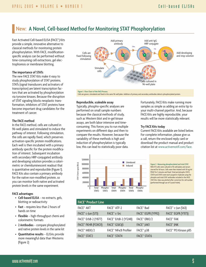
The FACE methodIn the FACE method, cells are cultured in 96-well plates and stimulated to induce the pathway of interest. Following stimulation, the cells are rapidly fi xed, which preserves activation-specifi c protein modifi cations. Each well is then incubated with a primary antibody specifi c for the protein modifi ca-tion of interest. Subsequent incubation with secondary HRP-conjugated antibody and developing solution provides a colori-metric or chemiluminescent readout that is quantitative and reproducible (Figure 1). FACE Kits also contain a primary antibody for the native non-modifi ed protein, so you can monitor both native and activated protein levels in the same experiment.
FACE advantages• Cell-based ELISA – no extracts, gels,
blotting or radioactivity• Fast – requires less than 2 hours of
hands on time• Flexible – high-throughput chemi and
colorimetric formats• 2 Antibodies – compare phosphorylated
and native protein levels in the same kit• Quantitative results – ELISAs provide
more meaningful data than Westerns (Figure 2)
Fortunately, FACE Kits make running more samples as simple as adding an extra tip to your multi-channel pipettor. And, because FACE Kits are highly reproducible, your results will be more statistically relevant.
Try FACE Kits todayCurrent FACE Kits available are listed below. For complete information, please give us a call, return the enclosed reply card or download the product manual and product citation list at www.activemotif.com/face.
Stimulate cells
3 hrs.1 hr.
Cells are fixed following
stimulation
Cells cultured in 96-well plate
Add primaryantibody
Add anti-IgG HRP conjugate
Add developingand stop solutionP P P
Figure 1: Flow Chart of the FACE Process.Cells are grown, stimulated and fi xed in the same 96-well plate. Addition of primary and secondary antibodies detects phosphorylated protein.
FACE™ Product LineFACE™ AKT FACE™ ATF-2 FACE™ Bad FACE™ c-Jun (S63)
FACE™ c-Jun (S73) FACE™ c-Src FACE™ EGFR (Y992) FACE™ EGFR (Y1173)
FACE™ ErbB-2 (Y877) FACE™ ErbB-2 (Y1248) FACE™ ERK1/2 FACE™ FAK
FACE™ FKHR (FOXO1) FACE™ GSK3β FACE™ JAK1 FACE™ JNK
FACE™ MEK1/2 FACE™ NFκB Profi ler FACE™ p38 FACE™ PI3 Kinase p85
FACE™ STAT2 FACE™ STAT4 FACE™ STAT6
1200000
1000000
800000
600000
400000
200000
0Total-STAT2
Phospho-STAT2
Total-STAT4
Phospho-STAT4
Total-STAT6
Phospho-STAT6
UninducedInduced
STAT
Che
mi s
igna
l (RL
U) Figure 2: Measuring phosphorylated and total STAT.NIH/3T3 cells were cultured in 96-well plates and serum-starved for 16 hours. Cells were then treated with 50 ng/ml PDGF for 5 minutes and fi xed. Total and phospho STAT2, STAT4 and STAT6 were each assayed in triplicate using the phospho and total STAT antibodies included in the FACE STAT Kits. Data was plotted after correction for cell number (performed through use of Crystal Violet).
Reproducible, scaleable assayTypically, phospho-specifi c analyses are performed on small sample numbers because the classical methods of study, such as Western blot and in-gel kinase assays, are both labor intensive and time-consuming. This forces you to run multiple experiments on different days and then to compare the results. However, because the variability of these methods is high and induction of phosphorylation is typically low, this can lead to statistically poor data.

Toll Free — 1 877 222 95436
A P R I L 2 0 0 5 • V O L U M E 6 • N U M B E R 1
www.activemotif.com 7
R a s A c t i v a t i o n A s s a y
45 min45 min45 min
Add cell extract to
plate
GST-Raf-RBD coated plate
Add primary antibody
Add anti-IgG HRP conjugate
Add developingand stop solutions,then quantify
Cell extract containing activated
Ras GTPase
Figure 3: Flowchart of the Ras GTPase Chemi Kit.Cell extract is added to a glutathione-coated plate that contains immobilized GST-Raf-RBD protein. Activated Ras in the extract binds to the Raf-RBD protein. Addition of primary & secondary antibodies and developing solution followed by reading on a luminometer enables sensitive quantifi cation of activated Ras.
GTPases are important regulatorsSmall GTPases, also known as GTP-bind-ing proteins, are a family of enzymes that serve as molecular switches in regulating a number of signal transduction pathways including cellular growth, apoptosis and dif-ferentiation. These proteins cycle between an inactive, GDP-bound state and an active, GTP-bound state. Activated GTPases exert their effects by activating a variety of downstream effector proteins such as Raf and PI3K. Activation of effector proteins in turn initiates phosphorylation cascades that modulate a number of different processes in the cell (Figure 2).
GTPPi
GDPH2O
Ras-GDP
Ras-GTP
Effectors
Cellular responses
+
Activating signal
Figure 2: The Ras activation pathway.Ras GTPase cycles between its inactive, GDP-bound form and its activated, GTP-bound form.
Aberrant Ras signaling causes many cancersRas GTPase is of particular interest because it is involved in many different pathways, and because aberrant regulation by Ras has been implicated in a number of disease states. Normally, GTPase-signaling cascades are only transiently activated because the intrinsic hydrolyzing activity of GTPases gradually converts GTP to GDP, leading to inactivation. However, a number of muta-tions in the ras gene have been identifi ed that cause Ras to remain constitutively in its active, GTP-bound form. This results in continuous stimulation of cellular prolifera-tion. Consequently, constitutively active, mutant forms of Ras GTPase are estimated to be present in ~30% of all human cancers.
Ras GTPase Chemi ELISA advantages• More sensitive – assay uses only 25 µg
of extract, which is 20-fold less than pull-down/Western methods
• Better results – quantitative readouts make it easier to compare results
• Less effort – no need to run gels or develop Western blots
• Save time – results in less than 5 hours• Versatile – assay activated extracts
from cells or tissue samples, or study recombinant Ras protein
The Ras GTPase Chemi ELISA methodBecause activated Ras binds specifi cally to the Ras-binding domain (RBD) of Raf effector protein, Raf-RBD can be used as a probe to isolate Ras-GTP. Active Motif’s new Ras GTPase Chemi ELISA Kit contains a Raf-RBD protein that is fused to GST and a 96-well, glutathione-coated assay plate. GST-Raf-RBD is fi rst incubated on the plate for one hour to immobilize the capture probe. Addition of sample to the plate re-sults in the binding of activated Ras to the
Raf-RBD. A primary antibody specifi c for Ras is then added to the individual wells, followed by an HRP-conjugated secondary antibody and developing reagent (Figure 3). The plate is then read on a luminometer, which provides a sensitive and quantitative chemiluminescent readout of activated Ras (Figure 1, page 1).
Try the quantitative, sensitive assayThe Ras GTPase Chemi ELISA Kit makes it fast and easy to detect and quantify activated Ras GTPase. The kit is ideal for the study of novel signaling pathways that activate Ras, as well as for determining if a particular malignancy is related to inappro-riately activated, oncogenic Ras. Additional assays to detect other GTPase proteins are in development. Please give us a call, return the enclosed reply card or log on to www.activemotif.com/gtpase for complete information, downloadable manuals and to fi nd out about new additions to this innovative product line. To get to the best Ras activation assay available for Ras, order the Ras GTPase Chemi ELISA Kit.
continued from page 1 — New: ELISA-based Method for Fast, Sensitive Quantifi cation of Activated Ras GTPase
Product Format Catalog No.
Ras GTPase Chemi ELISA Kit 1 x 96-well plate 52097

Toll Free — 1 877 222 95436
A P R I L 2 0 0 5 • V O L U M E 6 • N U M B E R 1
www.activemotif.com 7
N u c l e a r R e c e p t o r A s s a y s
Active Motif offers a variety of nuclear receptor analysis tools that make study-ing nuclear receptor proteins both faster and more accurate than using traditional methods. Whether you are interested in DNA-binding activity, activation state, protein level or agonist/antagonist effects, Active Motif has the product you need.
Monitor ligand activation with NR Peptide Studying the agonist/antagonist effects of potential drug targets is an important element of nuclear receptor-targeted drug discovery. Active Motif’s NR Peptide ELISAs are specifi cally designed to capture ligand-activated nuclear receptor and can be used with both cell extracts and proteins. Each NR Peptide ELISA Kit provides a 96-well plate that is coated with a Capture Peptide that includes the consensus-binding motif of the nuclear receptor’s co-activator. Addition of sample results in binding of ligand-activated nuclear receptor to the Capture Peptide. Each well is then incu-bated with a primary antibody specifi c for the nuclear receptor of interest, followed by an HRP-conjugated secondary antibody and developing solution to provide an easily quantifi ed readout. This enables you to quickly and quantitatively measure the agonist/antagonist effects of target com-pounds on the binding of ligand-activated nuclear receptors (Figure 1).
Figure 1: ERα agonism/antagonism dose-response curves.Nuclear extracts from the breast cancer cell line MCF-7 are incubated in wells of the NR Peptide ELISA ER plate in the presence of 100-fold serial dilutions (from 10-6 to 10-14 M) of the agonist compounds diethylstilbestrol (DES) and estradiol, and the antagonist compounds tamoxifen and clomiphene. Only ligand-activated ER protein can bind to the Capture Peptide immobilized in the plate. Bound ER is specifi -cally detected with ERα antibody and quantifi ed using a secondary antibody and Detection Solution.
96-well format is convenient and sensitive, with only a minimal amount of material required to give quantitative readout of nuclear receptor levels (Figure 2).
extract/well (µg)
AR d
etec
tion
(OD
450n
m)
21.8
0.60.4
0.81
1.21.41.6
0.20
LNCaP (AR+ve)PC-3 (AR-ve)DU 145 (AR-ve)
10 5 2.5 1.2 0.62
Figure 2: Monitoring expression levels of AR using NR Sandwich AR.Different amounts of nuclear extracts from three human prostate cancer cell lines, LNCaP, PC-3 and DU 145, were analyzed for levels of AR protein using the NR Sandwich AR Kit.
A collection of NR abs & proteins Active Motif’s extensive line of antibod-ies provide superior performance and reliable results. We offer over 200 highly characterized antibodies directed against transcription factors and nuclear receptors, including antibodies for GR, PPARα and γ, PXR, RAR-β, -β2 and -γ, RXR-α and -β, and VDR. In addition, Active Motif offers a number of recombinant nuclear receptor proteins that are ideal for use as positive controls, in in vitro screening studies and in many other applications.
Get the tools you need for NR researchTo get complete information on all of Active Motif’s tools for studying nuclear receptors, please give us a call, visit our website or send in the enclosed reply card.
Product Format Catalog No.
NR Peptide ERα 1 x 96-well plate5 x 96-well plates
4909649596
NR Peptide ERα Chemi 1 x 96-well plate5 x 96-well plates
4909749597
NR Sandwich AR 1 x 96-well plate5 x 96-well plates
4919649696
NR Sandwich ERα 1 x 96-well plate5 x 96-well plates
4929649796
TransAM™ ER 1 x 96-well plate5 x 96-well plates
4139641996
TransAM™ GR 1 x 96-well plate5 x 96-well plates
4549645996
TransAM™ PPARγ 1 x 96-well plate5 x 96-well plates
4019640696
A Complete Solution for Studying Nuclear Receptor Protein Activity
Assess DNA-binding activity of NRsInappropriate nuclear receptor signal-ing is associated with numerous diseases including cancer, asthma and arthritis, which makes NRs promising drug targets. Because the end point of nuclear recep-tor activation is DNA binding, monitoring changes in the DNA-binding activity of a target nuclear receptor can serve as an ideal biomarker. Classical methods such as radioactive gelshifts and time-consuming reporter gene assays are not well suited to this application. Active Motif’s TransAM™ Kits provide an innovative alternative to these classical assays. TransAM Kits use a combination of DNA binding and antibody detection to give a specifi c, quantitative readout of DNA-binding activity from all sample types. (See page 1 for more details.)
Quantify total NR with Sandwich ELISAsIn order to fully examine the activation of a given nuclear receptor, it is important to be able to quantify the total levels of a given nuclear receptor within a sample. The new NR Sandwich ELISAs offer a simple, rapid method to quantify the total amount of nuclear receptor protein present in both cell and tissue samples. NR Sandwich Kits utilize the Sandwich ELISA-based method that is an improvement over other methods used to study proteins, such as Western blotting. Using NR Sandwich means that there is no need for gels, blotting or long, tedious incubations. The

Active Motif has recently added a number of antibodies related to epigenetics and signal transduction to its already extensive
New Antibodies for Chromatin and DNA Methylation
A n t i b o d i e s
8
PRSRT STDUS POSTAGE
PAIDPERMIT 236
92054
line of antibodies. The tables below list these recent additions. Active Motif also offers numerous cell extracts that are
ideal positive controls for use with its kits and antibodies. For complete information, please visit www.activemotif.com.
Product Format Application React. Cat. No.
APE pAb 100 µg WB H 39200
APEXL2 pAb 200 µl WB H 39201
INHAT-1/TAF-1α/TAF-1β pAb 200 µl WB H 39202
CGBP pAb 200 µl WB H 39203
DNMT1 mAb 100 µg Ch, IHC, IP, WB H, M 39204
DNMT2 pAb 100 µg WB H, M 39205
DNMT3A mAb 100 µg Ch, IF, IHC, WB H, M 39206
DNMT3B mAb 100 µg Ch, IF, IP, WB H, M 39207
HDAC11 pAb 200 µl WB H, M 39208
Histone H2A pAb 100 µg WB H 39209
Histone H2B pAb 100 µg WB H 39210
Histone H3 (Phosphorylated) mAb 100 µg WB H 39211
Histone H4 pAb 100 µg WB H 39212
INHAT-2/pp32 pAb 200 µl WB H 39213
LIG1 pAb 200 µl WB H 39214
Product Format Application React. Cat. No.
MBD1 mAb 100 µg WB H 39215
MBD3 mAb 100 µg WB H 39216
MBD4 pAb 100 µg WB H 39217
MeCP2 pAb 100 µg WB H 39218
MLH-1 mAb 100 µg WB H 39219
MRE11 pAb 100 µg WB H 39220
PMS2 mAb 100 µg WB H, M 39221
Rad17 (Phosphorylated) pAb 100 µg IF, WB H 39222
TRF2 pAb 100 µg Ch, IP, WB H, M 39223
UBE2N pAb 100 µg IP, WB H 39224
UBE2V1a pAb 100 µg WB H 39225
UBE2V2 pAb 100 µg WB H 39226
Application Key: Ch = Chromatin Immunoprecipitation; IF = Immunofl uorescence; IHC = Immunohistochemistry;IP = Immunoprecipitation; WB = Western blot
Reactivity (React.) Key: H = Human; M = Mouse
CONT
ACT
INFO U.S.
1914 Palomar Oaks Way, Suite 150Carlsbad, CA 92008Toll Free: 877 222 9543Direct: 760 431 1263Fax: 760 431 1351Email: [email protected]
EUROPE104 Avenue Franklin Roosevelt–Box 25B-1330 Rixensart, BelgiumGermany Free Phone: 0800 181 99 10France Free Phone: 0800 90 99 79United Kingdom Free Phone: 0800 169 31 47Other countries, Direct: +32 (0)2 653 0001Fax: +32 (0)2 653 0050Email: [email protected]
JAPANAzuma Bldg, 7th Floor2-21 Ageba-Cho, Shinjuku-KuTokyo, 162-0824, JapanDirect: +81 (0)3 5225 3638Fax: +81 (0)3 5261 8733Email: [email protected]