neuropsychology of human body parts: exploring ...ilabs.uw.edu/sites/default/files/18shen et al...
TRANSCRIPT
Neuropsychology of Human Body Parts: ExploringCategorical Boundaries of Tactile PerceptionUsing Somatosensory Mismatch Responses
Guannan Shen1, Nathan J. Smyk1, Andrew N. Meltzoff 2, and Peter J. Marshall1
Abstract
■ The focus of the current study is on a particular aspect oftactile perception: categorical segmentation on the body sur-face into discrete body parts. The MMN has been shown tobe sensitive to categorical boundaries and language experiencein the auditory modality. Here we recorded the somatosensoryMMN (sMMN) using two tactile oddball protocols and comparedsMMN amplitudes elicited by within- and across-boundary odd-ball pairs. Both protocols employed the identity MMN methodthat controls for responsivity at each body location. In the firstprotocol, we investigated the categorical segmentation of tactilespace at the wrist by presenting pairs of tactile oddball stimuliacross equal spatial distances, either across the wrist or withinthe forearm. Amplitude of the sMMN elicited by stimuli pre-
sented across the wrist boundary was significantly greaterthan for stimuli presented within the forearm, suggesting acategorical effect at an early stage of somatosensory process-ing. The second protocol was designed to investigate thegenerality of this MMN effect, and involved three digits onone hand. Amplitude of the sMMN elicited by a contrast ofthe third digit and the thumb was significantly larger than acontrast between the third and fifth digits, suggesting a func-tional boundary effect that may derive from the way that ob-jects are typically grasped. These findings demonstrate thatthe sMMN is a useful index of processing of somatosensoryspatial discrimination that can be used to study body partcategories. ■
INTRODUCTION
The organization of a physically continuous range of stim-uli into discrete categories is a fundamental componentof human cognition. One prominent example of categoriza-tion that has been extensively studied is the phenomenonof categorical perception. For example, evidence for cate-gory boundary effects is apparent in the domains of speechperception (Harnad, 1987; Liberman, Harris, Hoffman, &Griffith, 1957) and color recognition (Franklin et al., 2008;Roberson, Davidoff, Davies, & Shapiro, 2005) using bothbehavioral measurements and neurophysiological indices.Behavioral studies using classical stimulus discriminationand identification tasks show that stimulus variance acrossa categorical boundary is more easily discriminated thanwithin-category variance across a stimulus continuum(e.g., Shen & Froud, 2016; Werker & Tees, 2005; Francis,Ciocca, & Ng, 2003). In neuroimaging studies, a nonlinear,discontinuous representation of auditory and visual stimulihas been found to occur in early stages of sensory process-ing (e.g., Bidelman, Hutka, & Moreno, 2013; Mo, Xu, Kay, &Tan, 2011; Liebenthal et al., 2010). A great deal of relatedresearch has added to the foundational notion that, dueto the categorical nature of perception, similar degrees of
physical deviance between pairs of stimuli do not alwayselicit similar differences in behavioral or neural responses(Shen & Froud, 2018; Kazanina, Phillips, & Idsardi, 2006;Kasai et al., 2001; Sharma & Dorman, 1999; Winkler et al.,1999; Dehaene-Lambertz, 1997).A classic case of the perceptual nonlinearities associ-
ated with categorical perception was first reported inthe domain of speech perception (Liberman et al.,1957). As originally described, it involved establishingboth an “identification” and a “discrimination” functionfor a series of stimuli separated by equal physical steps.The peak in the discrimination function occurred at theboundary between the two categories. The term cate-gorical perception is now commonly applied to a broaderset of cases beyond the domain of speech. In many testsof infants and adults, only the discrimination of pairs ofstimuli equally distant along a stimulus continuum is tested.In the current experiment, we adopt this latter approachand test for enhanced discrimination for across-categorycompared with within-category stimuli in the tactiledomain.Although categorical perception in the auditory modal-
ity has been studied intensively, this phenomenon is lesswell understood in the somatosensory domain. A fewstudies have used behavioral measures to examine thecategorical perception of body parts in relation to spatial1Temple University, 2University of Washington, Seattle
© 2018 Massachusetts Institute of Technology Journal of Cognitive Neuroscience 30:12, pp. 1858–1869doi:10.1162/jocn_a_01313
tactile perception (Knight, Cowie, & Bremner, 2017;Knight, Longo, & Bremner, 2014; de Vignemont, Majid,Jola, & Haggard, 2009). Similar to auditory and visual per-ception, tactile distance perception has been found to benonlinear (Miller, Longo, & Saygin, 2014), even thoughthe tactile receptor surface (i.e., the skin) forms a contin-uous sheet (de Vignemont et al., 2009). A focus of recentwork in this area has been on body part boundaries thatare established by the joints (e.g., the wrist joint). Al-though as a continuous sheet, the skin surface doesnot have natural boundaries, the joints act as specificlandmarks for segmenting the body (de Vignemont,Tsakiris, & Haggard, 2006; Bermúdez, 1998).The notion of joints as marking category boundaries is
supported by findings that tactile stimuli presented at eitherside of the wrist joint (i.e., one stimulus on the hand andone on the wrist) are perceived as further apart than pairsof stimuli presented within the forearm or within the hand,even when the physical distances between stimulationpoints are identical (Knight et al., 2014, 2017; de Vignemontet al., 2009). It has been argued that this enhanced tactilespatial acuity across body part boundaries is due to a cate-gorical segmentation effect and is not a result of increasedacuity and density of innervation of primary afferentfibers at the wrist and hand skin surface (Knight et al.,2014; Gibson & Craig, 2005).The categorical and nonlinear perception of distance
across joints reflects a part-based representation of bodystructure (Knight et al., 2017; Longo & Haggard, 2010),with touch being automatically referenced to this topolog-ically structured body representation (Mancini, Longo,Iannetti, & Haggard, 2011; de Vignemont, Ehrsson, &Haggard, 2005). This representation of body parts is pres-ent in childhood and may be sharpened by the functionalroles of body parts through action learning (de Vignemontet al., 2006; Berthier, Clifton, McCall, & Robin, 1999) aswell as by the acquisition of language for labeling distinctseparate parts of the body (Enfield, Majid, & Van Staden,2006).Complementing the influence of anatomical joint
bound- aries, categorization effects in tactile perceptionmay also arise from differences in the functional usage ofbody parts. How body parts are used in motor acts mod-ulates how they are represented and perceived (Milleret al., 2014; Braun, Schweizer, Elbert, Birbaumer, & Taub,2000; Hamilton & Pascual-Leone, 1998). However, be-cause functional categories usually overlap with ana-tomical, joint-based body part categories, the specificrelations between the functional use of body parts andthe categorical segmentation of tactile perception areunclear. We suggest that tactile stimulation of digits ofthe hand may provide a useful opportunity to study the ef-fects of functional categories on tactile perception, becausedigits are anatomically similar but functionally distinct.One salient functional distinction is between the first
digit (the thumb) and the second through fifth digits.The use of the first digit is of special interest to evolution-
ary biologists, because of the role it plays in tool use. Thethumb and the fingers are employed differently duringgrasping and picking up objects, with the thumb posi-tioned on one side of the object and fingers positionedon the other side (Wing & Fraser, 1983). In the humaninfant, the thumb–finger opposition grip “precision grip”develops from an earlier imprecise “power grip” in whichobjects are held between the fingers and the palm (e.g.,Butterworth, Verweij, & Hopkins, 1997; Newell, Scully,McDonald, & Baillargeon, 1989). Although the functionaldistinction between the thumb and the fingers in graspinghas been well studied, potential perceptual categorizationeffects based on this experience have not been investi-gated. This may be because of the challenges of performingbehavioral perceptual assessments such as tactile distancejudgments and 2-point discrimination tests on differentfingers. However, advances in neuroimaging methodshave provided tools to examine sensory–perceptual catego-rization that can be readily applied to this question.
The MMN is a well-documented aspect of the ERP as-sociated with involuntary deviance processing (Garrido,Kilner, Stephan, & Friston, 2009; Näätänen, Jacobsen, &Winkler, 2005; Pincze, Lakatos, Rajkai, Ulbert, & Karmos,2001). In the auditory modality, the MMN is commonlyelicited over frontocentral sites in response to deviantstimuli embedded in a train of standards (referred to asan “oddball paradigm”) and appears around 100–200 msecafter change onset. It is thought to reflect the neuralactivity associated with change detection mechanismsin primary and secondary auditory cortex, and elicitingthis component does not require listeners’ consciousattention (e.g., Näätänen, 2001). The amplitude of theauditory MMN increases as the perceived salience ofthe acoustic discrepancy between standard and deviantstimuli increases (Chandrasekaran, Krishnan, & Gandour,2009; Näätänen & Alho, 1997). This characteristic of theMMN allows it to be used to evaluate the electrophysio-logical correlates of categorical perception. As shown in anumber of studies in both the auditory and visualmodalities, larger MMN amplitudes are elicited by across-category deviants compared with within-category deviants.For example, discontinuous MMN responses reflectingadult categorical perception have been reported for pho-nemes (Kazanina et al., 2006; Kasai et al., 2001; Sharma &Dorman, 1999; Winkler et al., 1999; Dehaene-Lambertz,1997), Mandarin tones (Xi, Zhang, Shu, Zhang, & Li,2010), and colors (Mo et al., 2011). These findings suggestthat the enhanced MMN response to cross-category de-viants compared with within-category deviants withequal physical variance is a reliable indicator of categor-ical discrimination at a relatively early stage of perceptu-al processing. One further important observation is that,although the categorical effects indexed by MMN re-sponses occur in the absence of overt attention, otherfindings suggest that these effects are sensitive to expe-rience. For example, the categorical effect on Mandarintone perception indexed by MMN responses was only
Shen et al. 1859

observed in native Mandarin speakers and not in Englishspeakers or adult learners of Mandarin Chinese (Shen &Froud, 2018).
In the tactile domain, the somatosensory MMN (sMMN)can be elicited by deviance in various stimulus properties,such as duration (Butler et al., 2011; Spackman, Towell, &Boyd, 2010; Akatsuka et al., 2005), vibrotactile frequency(Spackman, Boyd, & Towell, 2007), and spatial location(Shen, Smyk, Meltzoff, & Marshall, 2018; Naeije et al.,2016; Restuccia et al., 2009; Akatsuka et al., 2007). Analo-gous to findings concerning the auditory MMN response,the sMMN is generated in primary somatosensory cortex(Akatsuka et al., 2007; Shinozaki, Yabe, Sutoh, Hiruma,& Kaneko, 1998), with additional generators in secondarysomatosensory cortex (Naeije et al., 2016; Butler et al.,2011) and frontal cortex (Kekoni et al., 1997). One studyreported that spatially larger deviance evoked greatersMMN, suggesting that sMMN amplitude is modulatedby the degree of tactile distance between points of stim-ulation (Akatsuka et al., 2007). A recent study showedthat sMMN amplitude is sensitive to the nature of thecortical body map representation in somatosensorycortex, as indicated by greater amplitude and shorterlatency of the sMMN for stimulation of body locationswith more separated representations in primary somato-sensory cortex (Shen et al., 2018).
The current study investigated the effect of categoricalboundaries and functional experience on tactile percep-tion using the sMMN, a tool that can provide a uniquewindow into the early stages of somatosensory pro-cessing and the neurophysiological correlates of tactileperception. In addition to the sMMN, the analyses alsoincluded an early obligatory somatosensory ERP compo-nent, the N80 (Sambo et al., 2012; Schubert et al., 2008),and a later attention orienting component, the P300,which often co-occurs with MMN in oddball paradigms(Lugo et al., 2014; Polich, 2007). The study consisted oftwo complementary experimental protocols. For theinvestigation of nonlinearity in body perception, we firstleveraged a categorical segmentation of tactile space atthe wrist and presented two pairs of tactile oddball stim-uli with equal spatial distances, either across the wrist orwithin the forearm. A second oddball paradigm wasbased on the functional distinction between the thumband other digits in goal-directed motor acts, such as pick-ing up and grasping objects. In this second protocol, theamplitude of the sMMN was compared for a contrast in-volving first versus third digit stimulation (hypothesizedto be cross-boundary) and a contrast involving stimula-tion of the fifth versus third digits (hypothesized to bewithin category). Because the sMMN response reflects arelatively early stage of tactile processing and becausetactile perceptual categorization is thought to involveautomatic reference to body part representations (Manciniet al., 2011, de Vignemont et al., 2005), we hypothesizedthat cross-boundary deviants should elicit greater sMMNresponses than within-boundary deviants.
METHODS
Thirty-five undergraduate students received course creditin return for participation. Data from four participantswere excluded from the analysis because of participantfatigue (n = 2) or insufficient numbers of artifact-freetrials (< 50 trials per condition; n= 2). The final analysesutilized data from a total of 32 participants (10 men;mean age = 20.47 years, SD= 2.01). All participants wereright-handed as assessed by the Edinburgh HandednessInventory (Oldfield, 1971), had normal-to-correctedvision, and reported no history of neurological illness orabnormality. This study was carried out with approval fromthe institutional review board at Temple University, withinformed consent obtained from each individual beforeparticipation.
Stimuli
Tactile stimuli were delivered using an inflatable mem-brane (10-mm diameter) mounted in a plastic casing.The membrane was inflated by a short burst of com-pressed air delivered via flexible polyurethane tubing(3-m length, 3.2-mm outer diameter). The compressedair delivery was controlled by STIM stimulus presentationsoftware in combination with a pneumatic stimulator unit(both from James Long Company) and an adjustable reg-ulator that restricted the airflow to 60 psi. The pneumaticstimulator and regulator were located in an adjacentroom to the participant. To generate each tactile stimu-lus, the STIM software delivered a TTL trigger (10-msecduration) that served to open and close a solenoid in thepneumatic stimulator. Expansion of the membranestarted 15 msec after trigger onset and peaked 20 mseclater (i.e., 35 msec after trigger onset). The total durationof membrane movement was around 100 msec. This stim-ulation method has been successfully used in a number ofprevious EEG and MEG studies (Meltzoff et al., 2018; Shenet al., 2018; Shen, Saby, Drew, & Marshall, 2017).During presentation of the tactile stimuli, participants
watched a video presented on a CRT monitor (40 cm view-able). Participants were seated approximately 70 cm fromthe monitor screen. The video consisted of around 30 minof footage of a wildlife documentary presented via DVD.No auditory soundtrack was presented, and subtitleswere displayed in English. To mask any subtle sounds as-sociated with delivery of the tactile stimuli, participantswore earplugs during data collection, and ambient whitenoise was broadcast in the testing room.
Design and Procedure
Six blocks of tactile stimuli were presented across twoprotocols: hand/forearm stimulation and digit stimula-tion. There were three blocks within each protocol, andthe order of protocol presentation was counterbalancedacross participants.
1860 Journal of Cognitive Neuroscience Volume 30, Number 12
Hand/Arm Stimulation
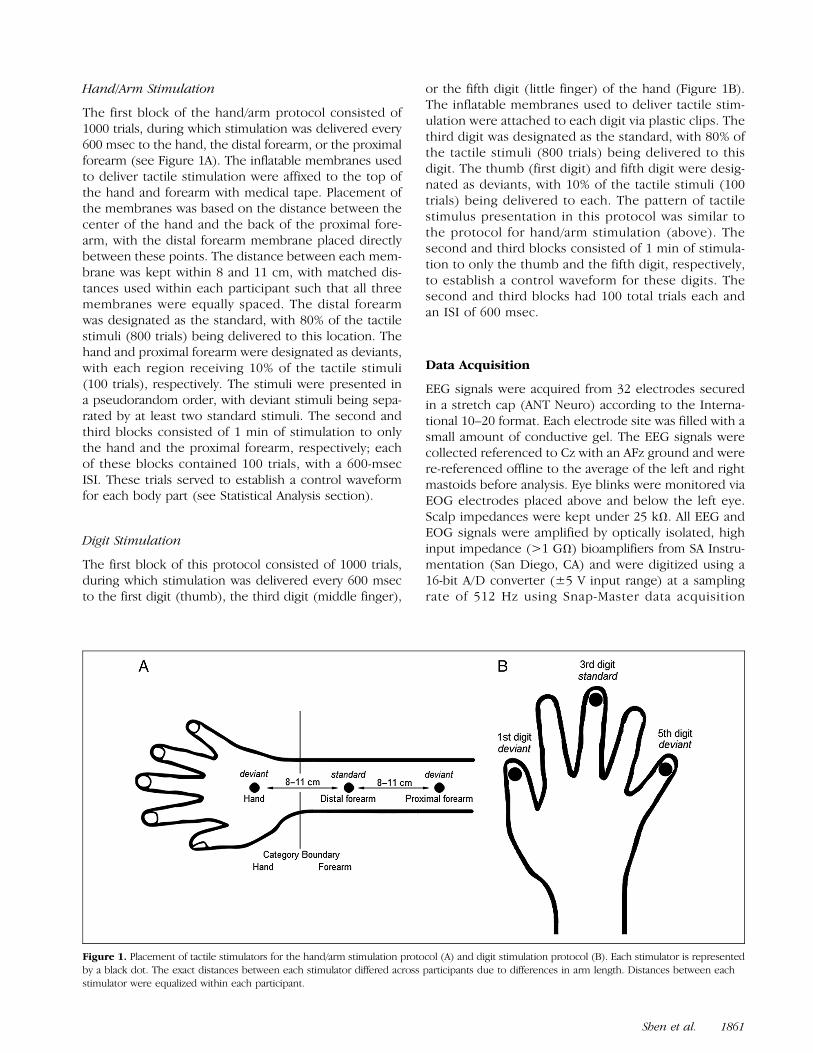
The first block of the hand/arm protocol consisted of1000 trials, during which stimulation was delivered every600 msec to the hand, the distal forearm, or the proximalforearm (see Figure 1A). The inflatable membranes usedto deliver tactile stimulation were affixed to the top ofthe hand and forearm with medical tape. Placement ofthe membranes was based on the distance between thecenter of the hand and the back of the proximal fore-arm, with the distal forearm membrane placed directlybetween these points. The distance between each mem-brane was kept within 8 and 11 cm, with matched dis-tances used within each participant such that all threemembranes were equally spaced. The distal forearmwas designated as the standard, with 80% of the tactilestimuli (800 trials) being delivered to this location. Thehand and proximal forearm were designated as deviants,with each region receiving 10% of the tactile stimuli(100 trials), respectively. The stimuli were presented ina pseudorandom order, with deviant stimuli being sepa-rated by at least two standard stimuli. The second andthird blocks consisted of 1 min of stimulation to onlythe hand and the proximal forearm, respectively; eachof these blocks contained 100 trials, with a 600-msecISI. These trials served to establish a control waveformfor each body part (see Statistical Analysis section).
Digit Stimulation
The first block of this protocol consisted of 1000 trials,during which stimulation was delivered every 600 msecto the first digit (thumb), the third digit (middle finger),
or the fifth digit (little finger) of the hand (Figure 1B).The inflatable membranes used to deliver tactile stim-ulation were attached to each digit via plastic clips. Thethird digit was designated as the standard, with 80% ofthe tactile stimuli (800 trials) being delivered to thisdigit. The thumb (first digit) and fifth digit were desig-nated as deviants, with 10% of the tactile stimuli (100trials) being delivered to each. The pattern of tactilestimulus presentation in this protocol was similar tothe protocol for hand/arm stimulation (above). Thesecond and third blocks consisted of 1 min of stimula-tion to only the thumb and the fifth digit, respectively,to establish a control waveform for these digits. Thesecond and third blocks had 100 total trials each andan ISI of 600 msec.
Data Acquisition
EEG signals were acquired from 32 electrodes securedin a stretch cap (ANT Neuro) according to the Interna-tional 10–20 format. Each electrode site was filled with asmall amount of conductive gel. The EEG signals werecollected referenced to Cz with an AFz ground and werere-referenced offline to the average of the left and rightmastoids before analysis. Eye blinks were monitored viaEOG electrodes placed above and below the left eye.Scalp impedances were kept under 25 kΩ. All EEG andEOG signals were amplified by optically isolated, highinput impedance (>1 GΩ) bioamplifiers from SA Instru-mentation (San Diego, CA) and were digitized using a16-bit A/D converter (±5 V input range) at a samplingrate of 512 Hz using Snap-Master data acquisition
Figure 1. Placement of tactile stimulators for the hand/arm stimulation protocol (A) and digit stimulation protocol (B). Each stimulator is representedby a black dot. The exact distances between each stimulator differed across participants due to differences in arm length. Distances between eachstimulator were equalized within each participant.
Shen et al. 1861
software (HEM Data Corp.). Hardware filter settings were0.1 Hz (high-pass) and 100 Hz (low-pass), with a 12-dB/octave roll-off; bioamplifier gain was 4000 for the EEGchannels and 1000 for the EOG channels.
Data Analysis
Preprocessing of EEG Data
Processing and initial analysis of the EEG signals were per-formed using the EEGLAB 13.5.4b toolbox (Delorme &Makeig, 2004) implemented in MATLAB (The Math-Works). Epochs of 600-msec duration were extracted fromthe continuous EEG data, with each epoch extending from−100 msec to 500 msec relative to stimulus onset. Inde-pendent component analysis was used to identify and re-move eye movement artifacts (Hoffmann & Falkenstein,2008). Visual inspection of the EEG signal was used toreject epochs containing other movement artifacts. Themean number of artifact-free trials per body part locationor digit was 86 (SD = 8). A one-way ANOVA showed thatthere was no significant difference between locations inthe number of usable trials across all standard and deviantconditions ( p = .572). To prepare the data for ERP anal-ysis, artifact-free epochs were low-pass filtered at 30 Hzbefore being averaged and baseline-corrected relativeto a 100-msec prestimulus baseline.
Statistical Analysis
As well as the sMMN, the ERP analyses included the N80,a mandatory tactile ERP component, as well as a laterP300 component. Because early somatosensory ERPcomponents typically have frontal and central scalpdistributions (e.g., Shen et al., 2017; Sambo et al., 2012;Wang, Mouraux, Liang, & Iannetti, 2008), analyses of theN80 and sMMN focused on 12 electrodes over frontal (F7,F3, F4, F8), frontocentral (FC5, FC1, FC2, FC6), and cen-trotemporal (T7, C3, C4, T8) regions. N80 amplitudeswere calculated by averaging the mean amplitude inthe 12-msec window, extending 6 msec before and afterthe most negative peak within the period of 60–100 msecfollowing stimulus onset. To compare N80 amplitudeelicited by different deviants and controls, a four-wayrepeated-measures ANOVA was conducted separately fordigit stimulation and arm/hand stimulation with factorsStimulus type (deviant/control), Category type (within-boundary deviants: arm, fifth finger; across-boundarydeviants: hand, thumb), Region (frontal/frontocentral/central), and Hemisphere (left/right).
The first step in computing sMMN amplitude was tosubtract the ERPs for one stimulus as the control fromthe ERP when the same stimulus was the deviant(Zheng, Minett, Peng, & Wang, 2012; Xi et al., 2010).The most negative peak in the deviant-minus-controldifference wave between 100 and 200 msec (Garrido
et al., 2009; Näätänen et al., 2005) was identified atthe selected electrodes for each participant. To com-pute sMMN amplitude, the difference wave amplitudewas then averaged over a 20-msec time window, ex-tending 10 msec before and 10 msec after this negativepeak. Three-way repeated-measures ANOVAs wereconducted separately for digit stimulation and arm/hand stimulation using factors Category type (within-boundary deviants: arm, fifth finger; across-boundarydeviants: hand, thumb), Region (frontal/frontocentral/central), and Hemisphere (left/right). Pairwise t testswith false discovery rate (FDR) correction were usedin all post hoc comparisons.The analysis of P300 amplitude followed a similar pro-
cedure as for the sMMN. In line with prior work on theP300 scalp distribution (Polich, 2007), three midline elec-trode sites were selected for statistical analysis: Fz, Cz,and Pz. Mean P300 amplitude was calculated by averagingthe amplitude of the deviant-minus-control waveformin a 100-msec window surrounding the most positivevalue between 180 and 400 msec. Two-way repeated-measures ANOVAs on P300 amplitude were conductedseparately for digit stimulation and arm/hand stim-ulation using factors category type (within-boundarydeviants: arm, fifth finger; across-boundary deviants:hand, thumb) and electrode (Fz/Cz/ Pz). Pairwiset tests with FDR correction were used in all post hoccomparisons.
RESULTS
Figures 2 and 3 show the grand-averaged waveforms tothe deviant and control stimuli at frontal and central sitesand the topographic plots for hand/arm stimulation anddigit stimulation, respectively. Visual inspection of theERP waveforms shows that, compared with controlstimuli, deviant stimuli evoked a larger early negative com-ponent, N80, a more negative-going deflection around100–150 msec for deviant stimuli (sMMN), followed by alarger positive response around 200–300 msec (P300).
N80
For arm/hand stimulation, the four-way repeated-measuresANOVA, with factors Stimulus type (deviant/control), Cate-gory type (within-/across-boundary deviants, arm/hand),Region (frontal/frontocentral/central), and Hemisphere(left/right) revealed a significant main effect of Hemisphere(F(1, 31) = 6.998, p = .013; left hemisphere > right hemi-sphere). There was also a significant main effect of Region(F(2, 62) = 35.597; p < .001), with greater N80 responsesat frontal and frontocentral regions than centrotemporal re-gions ( p < .001). There was no significant main effect ofCategory type (F(1, 31) = 2.343, p = .136). In addition,the results showed a significant interaction betweenStimulus type and Category type (F(1, 31) = 7.013,
1862 Journal of Cognitive Neuroscience Volume 30, Number 12

p = .013). Two separate three-way ANOVAs were con-ducted for arm and hand stimulation for post hoc analysis.Results showed a significant main effect of Stimulus typefor hand stimulation (F(1, 31) = 9.875, p = .004), withgreater N80 responses to deviant than control stimuli.No significant main effect of Stimulus type was foundfor arm stimulation (F(1, 31) = 0.321, p = .575).For digit stimulation, the four-way repeated-measures
ANOVA revealed a significant main effect of Stimulus type(F(1, 31) = 6.017, p = .019), with greater N80 amplitudefor deviant stimuli than for control stimuli. The results alsoshowed a significant main effect of Region (F(2, 62) =
16,441, p < .001), with greater N80 responses at frontaland frontocentral regions than centrotemporal regions( p < .001). In addition, there were two significant three-way interactions between Stimulus type, Category type,and Hemisphere (F(1, 31) = 8.712, p = .006), as well asbetween Stimulus type, Category type, and Region (F(2,62) = 4.102, p = .028). There was no significant maineffect of Category type (F(1, 31) < 0.001, p = .998). Tofurther examine the significant interactions, two separatethree-way ANOVAs were conducted on N80 amplitudefor the thumb and fifth digit stimulation. There was a sig-nificant main effect of Stimulus type for the thumb
Figure 2. Hand/arm sMMN. (A) Grand-averaged ERP waveforms in response to hand (A) and arm (C) stimulation presented as frequent controls (black)in the control blocks and as infrequent deviants (red) in the oddball block. (B, D) Topographic plots of mean N80 and sMMN amplitude.
Shen et al. 1863
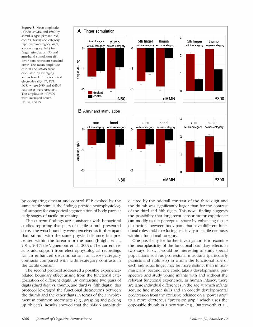
stimulation (F(1, 31) = 7.186, p = .012; deviant > con-trol), but not for the fifth digit stimulation (F(1, 31) =0.45, p = .507; Figure 5).
MMN
The ANOVA on sMMN amplitude showed a significantmain effect of Category type (F(1, 31) = 5.427,p = .026), with significantly greater sMMN amplitudefor hand than arm stimuli, as well as a significant maineffect of Hemisphere (F(1, 31) = 8.014, p = .008; left
> right hemisphere). The ANOVA also revealed a signif-icant main effect of Region (F(2, 62) = 5.147, p = .009),with greater sMMN responses over frontal electrodesthan frontocentral sites ( p = .001) and centrotemporalsites ( p = .22). There were no significant interactionsbetween any of the factors.For digit stimulation, an ANOVA on sMMN amplitude
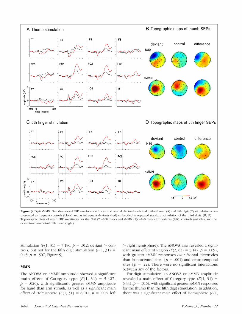
revealed a main effect of Category type (F(1, 31) =6.441, p= .016), with significant greater sMMN responsesfor the thumb than the fifth digit stimulation. In addition,there was a significant main effect of Hemisphere (F(1,
Figure 3. Digit sMMN. Grand-averaged ERP waveforms at frontal and central electrodes elicited to the thumb (A) and fifth digit (C) stimulation whenpresented as frequent controls (black) and as infrequent deviants (red) embedded in repeated standard stimulation of the third digit. (B, D)Topographic plots of mean ERP amplitudes for the N80 (70–100 msec) and sMMN (130–160 msec) for deviants (left), controls (middle), and thedeviant-minus-control difference (right).
1864 Journal of Cognitive Neuroscience Volume 30, Number 12
31) = 12.153, p = .001, left > right hemisphere). Therewere no significant main effect of Region and no signifi-cant interactions between factors.
P300
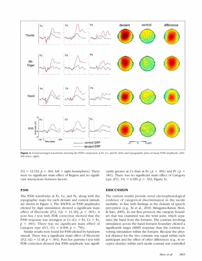
The P300 waveforms at Fz, Cz, and Pz, along with thetopographic maps for each deviant and control stimuli,are shown in Figure 4. The ANOVA on P300 amplitudeselicited by digit stimulation showed a significant maineffect of Electrode (F(2, 62) = 11.182, p < .001). Apost hoc t test with FDR correction showed that theP300 response was strongest at Cz (Cz > Pz, Cz > Fz,p < .001). There was no significant main effect ofCategory type (F(1, 31) = 0.068, p = .796).Similar results were found for P300 elicited by hand/arm
stimuli. There was a significant main effect of Electrode(F(2, 62) = 11.48, p < .001). Post hoc pairwise t test withFDR correction showed that P300 amplitude was signifi-
cantly greater at Cz than at Fz ( p < .001) and Pz ( p <.001). There was no significant main effect of Categorytype (F(1, 31) = 0.399, p = .532; Figure 5).
DISCUSSION
The current results provide novel electrophysiologicalevidence of categorical discrimination in the tactilemodality, in line with findings in the domain of speechperception (e.g., Xi et al., 2010; Minagawa-Kawai, Mori,& Sato, 2005). In our first protocol, the category bound-ary that was examined was the wrist joint, which sepa-rates the hand from the forearm. The contrast involvingstimulation across the hand–forearm boundary elicited asignificantly larger sMMN response than the contrast in-volving stimulation within the forearm. Because the phys-ical distance for the two contrasts was equal within eachparticipant and the effect of other differences (e.g., in re-ceptor density) within each tactile contrast was controlled
Figure 4. Grand-averaged waveforms showing the P300 component at Fz, Cz, and Pz (left) and topographic plots of mean P300 amplitude (200–300 msec; right).
Shen et al. 1865
by comparing deviant and control ERP evoked by thesame tactile stimuli, the findings provide neurophysiolog-ical support for categorical segmentation of body parts atearly stages of tactile processing.
The current findings are consistent with behavioralstudies reporting that pairs of tactile stimuli presentedacross the wrist boundary were perceived as further apartthan stimuli with the same physical distance but pre-sented within the forearm or the hand (Knight et al.,2014, 2017; de Vignemont et al., 2009). The current re-sults add support from electrophysiological recordingsfor an enhanced discrimination for across-categorycontrasts compared with within-category contrasts inthe tactile domain.
The second protocol addressed a possible experience-related boundary effect arising from the functional cate-gorization of different digits. By contrasting two pairs ofdigits (third digit vs. thumb, and third vs. fifth digits), thisprotocol leveraged the functional distinctions betweenthe thumb and the other digits in terms of their involve-ment in common motor acts (e.g., grasping and pickingup objects). Results showed that the sMMN amplitude
elicited by the oddball contrast of the third digit andthe thumb was significantly larger than for the contrastof the third and fifth digits. This novel finding suggeststhe possibility that long-term sensorimotor experiencecan modify tactile perceptual space by enhancing tactiledistinctions between body parts that have different func-tional roles and/or reducing sensitivity to tactile contrastswithin a functional category.One possibility for further investigation is to examine
the neuroplasticity of the functional boundary effects intwo ways. First, it would be interesting to study specialpopulations such as professional musicians (particularlypianists and violinists) in whom the functional role ofeach individual finger may be more distinct than in non-musicians. Second, one could take a developmental per-spective and study young infants with and without therelevant functional experience. In human infancy, thereare large individual differences in the age at which infantsacquire fine motor skills and an orderly developmentalprogression from the exclusive reliance on a “power grip”to a more dexterous “precision grip,” which uses theopposable thumb in a new way (e.g., Butterworth et al.,
Figure 5. Mean amplitudeof N80, sMMN, and P300 bystimulus type (deviant: red;control: black) and categorytype (within-category: right;across-category: left) forfinger stimulation (A) andarm/hand stimulation (B).Error bars represent standarderror. The mean amplitudeof N80 and sMMN werecalculated by averagingacross four left frontocentralelectrodes (F3, F7, FC1,FC5) where N80 and sMMNresponses were greatest.The amplitudes of P300were averaged acrossFz, Cz, and Pz.
1866 Journal of Cognitive Neuroscience Volume 30, Number 12
1997; Newell et al., 1989). If functional experience plays asignificant role, then individual infants differing in theirfine motor skills (and matched in age) will demonstratesignificantly different MMN responses in a thumb versusthird versus fifth digit protocol, and similarly, longitudinalstudies of the same infant over time would be expectedto show the neural responses after he or she has ac-quired functional experience with the precision grip.The N80—which occurs before the MMN and is gener-
ated in primary somatosensory cortex contralateral to thetactile stimulation (Sambo et al., 2012; Schubert et al.,2008)—also appears to show categorical effects. The cur-rent results show that across-boundary deviant stimulievoked significantly larger N80 amplitudes than the cor-responding control stimuli, whereas a difference in N80amplitude between within-category deviants and controlswas less apparent. The enhancement of the N80 by devi-ant tactile stimuli in an oddball paradigm has been ob-served in other sMMN studies (Strömmer, Tarkka, &Astikainen, 2014; Hötting & Röder, 2009; Akatsuka et al.,2007), although the meaning of this enhancement has notbeen thoroughly explored. The suggestion in these priorstudies was that this early SEP effect reflects obligatorysensory processing that precedes higher-order cognitiveprocessing (Strömmer et al., 2014) and that the N80 en-hancement may be due to physical differences in the tac-tile stimuli between deviant and standard stimuli (Hötting& Röder, 2009). Crucially, the current study employed theidentity MMN paradigm that involves comparing the SEPelicited by the same tactile stimuli presented as deviantsand as controls (Möttönen, Dutton, & Watkins, 2013),and thus, the N80 enhancement in the current study can-not be explained purely by bottom–up, featural differ-ences in the tactile stimuli.One potential explanation for the differences in N80
enhancement for across- versus within-boundary con-trasts may be that categorical effects on sensory process-ing start before the higher-order processing that occursaround 100–200 msec poststimulus onset and is typicallyindexed by MMN responses (e.g., Bidelman et al., 2013;Zheng et al., 2012; Mo et al., 2011; Xi et al., 2010). Recentstudies of somatosensory processing have revealed thatearly processing in primary somatosensory cortex is notonly influenced by bottom–up featural variation in sen-sory stimuli but is also modulated by cognitive factorssuch as sustained attention (Eimer & Forster, 2003), spa-tial attention (Schubert et al., 2008), and multisensorycongruency (Cardini & Longo, 2016). Consistent withthese views, the findings of the current study may indi-cate that initial stages of somatosensory processing in pri-mary somatosensory cortex, indexed by N80, is alreadyreflective of categorical representations of body partsand sensorimotor functions, echoing recent findings inthe auditory domain that emphasize a role of primarysensory cortex in early processing of language-relatedstimuli (Papanicolaou, Kilintari, Rezaie, Narayana, &Babajani-Feremi, 2017).
In addition to the early N80 and MMN responses, wealso observed an enhanced positive component at 200–350 msec elicited by deviant stimuli compared with con-trol stimuli. This component is known as the P300 or P3aand reflects an orienting response to the violation of ex-pected patterns of sensory stimulation (Light, Swerdlow,& Braff, 2007; Polich, 2007). The P300 is commonly elic-ited in oddball paradigms and often follows the MMN re-sponse in the form of an “MMN/P3a complex” (Hermenset al., 2010). In the current study, we found that P300 am-plitude was enhanced by all deviants compared with theircorresponding control stimuli, but in contrast to the pat-tern of sMMN responses, the degree of increase in P300amplitude was similar for cross- and within-boundary de-viants. This is in line with other recent studies reportingthat changes in MMN amplitude are often dissociatedfrom changes in P300 amplitude in both the auditory(Horváth, Winkler, & Bendixen, 2008) and somatosen-sory domains (Shen et al., 2017). The current findingmay lend support to previous work on somatosensorydeviance detection, which suggested that sMMN ampli-tude is modulated by somatotopic cortical representa-tions of body parts, whereas the P300 response may bemore related to tactile processing referenced to theactual 3-D human body in space (Shen et al., 2017). Be-cause P300 responses can be enhanced by attentionaldeployment and behavioral tasks involving noveltydetection (Polich, 2007), future studies could shed lighton the differences between the sMMN and P300 byasking participants to attend to the tactile stimuli andto respond to deviant stimuli.
Parallel to perceptual categorization of speech in theauditory modality (e.g., Shen & Froud, 2018; Dehaene-Lambertz, 1997) and color perception in the visual do-main (e.g., Mo et al., 2011), our findings suggest thatbody part categorization can modulate early stages ofsomatosensory processing, without overt attentional de-ployment. Further questions for investigation can furtheraddress the neural mechanisms of body categorization.For instance, are receptive fields in the primary somato-sensory cortex reflective of the cognitive categorizationof body perception and related functional segmentations?Or, is this knowledge stored in areas associated with fur-ther processing, such as secondary somatosensory cortex.Future studies using high-resolution neuroimaging couldshed light on the neural mechanisms of categorical per-ception in the tactile domain by examining differentialactivations to across- and within-category deviance in cor-tical areas that contribute to the sMMN response, specifi-cally SI and SII (Akatsuka et al., 2007), as well as frontalcortex (Garrido et al., 2009).
In conclusion, these novel findings provide evidencethat relatively early stages of neural processing of tactilestimulation are influenced by the categorical segmentationof the body into discrete body parts. Perceptual effects re-lated to categorization are apparently ubiquitous in humancognition and have been observed in a number of sensory
Shen et al. 1867
modalities. Future research on the relations between bodypart categorization and sensorimotor experience in adultsas well as developmental studies examining changes intactile perception as a function of specific motor experi-ences (e.g., with grasping and picking up objects) willfurther illuminate this phenomenon.
Acknowledgments
The authors thank Staci Weiss, Rebecca Laconi, and JebediahTaylor for their help with data collection. The writing of thisar t ic le was supported in part by awards from NIH(1R21HD083756) and NSF (BCS-1460889 and SMA-1540619).
Reprint requests should be sent to Guannan Shen, Department ofPsychology, Temple University, 1701 N 13th Street, Philadelphia,PA 19122, or via e-mail: [email protected].
REFERENCES
Akatsuka, K., Wasaka, T., Nakata, H., Inui, K., Hoshiyama, M., &Kakigi, R. (2005). Mismatch responses related to temporaldiscrimination of somatosensory stimulation. ClinicalNeurophysiology, 116, 1930–1937.
Akatsuka, K., Wasaka, T., Nakata, H., Kida, T., Hoshiyama, M.,Tamura, Y., et al. (2007). Objective examination for two-pointstimulation using a somatosensory oddball paradigm: AnMEG study. Clinical Neurophysiology, 118, 403–411.
Bermúdez, J. L. (1998). The paradox of self-consciousness.Cambridge, MA: MIT Press.
Berthier, N. E., Clifton, R. K., McCall, D. D., & Robin, D. J.(1999). Proximodistal structure of early reaching in humaninfants. Experimental Brain Research, 127, 259–269.
Bidelman, G. M., Hutka, S., & Moreno, S. (2013). Tone languagespeakers and musicians share enhanced perceptual andcognitive abilities for musical pitch: Evidence forbidirectionality between the domains of language and music.PLoS One, 8, e60676.
Braun, C., Schweizer, R., Elbert, T., Birbaumer, N., & Taub, E. (2000).Differential activation in somatosensory cortex for differentdiscrimination tasks. Journal of Neuroscience, 20, 446–450.
Butler, J. S., Molholm, S., Fiebelkorn, I. C., Mercier, M. R.,Schwartz, T. H., & Foxe, J. J. (2011). Common or redundantneural circuits for duration processing across audition andtouch. Journal of Neuroscience, 31, 3400–3406.
Butterworth, G., Verweij, E., & Hopkins, B. (1997). Thedevelopment of prehension in infants: Halverson revisited.British Journal of Developmental Psychology, 15, 223–236.
Cardini, F., & Longo, M. R. (2016). Congruency of body-relatedinformation induces somatosensory reorganization.Neuropsychologia, 84, 213–221.
Chandrasekaran, B., Krishnan, A., & Gandour, J. T. (2009).Sensory processing of linguistic pitch as reflected by themismatch negativity. Ear and Hearing, 30, 552–558.
Dehaene-Lambertz, G. (1997). Electrophysiological correlatesof categorical phoneme perception in adults. NeuroReport, 8,919–924.
Delorme, A., & Makeig, S. (2004). EEGLAB: An open sourcetoolbox for analysis of single-trial EEG dynamics includingindependent component analysis. Journal of NeuroscienceMethods, 134, 9–21.
de Vignemont, F., Ehrsson, H. H., & Haggard, P. (2005). Bodilyillusions modulate tactile perception. Current Biology, 15,1286–1290.
de Vignemont, F., Majid, A., Jola, C., & Haggard, P. (2009).Segmenting the body into parts: Evidence from biases in
tactile perception. Quarterly Journal of ExperimentalPsychology, 62, 500–512.
de Vignemont, F., Tsakiris, M., & Haggard, P. (2006). Bodymereology. In G. Knoblich, I. M. Thornton, M. Grosjean, & M.Shiffrar (Eds.), Human body perception from the inside out(pp. 147–170). New York: Oxford University Press.
Eimer, M., & Forster, B. (2003). Modulations of earlysomatosensory ERP components by transient and sustainedspatial attention. Experimental Brain Research, 151, 24–31.
Enfield, N. J., Majid, A., & Van Staden, M. (2006). Cross-linguisticcategorization of the body: Introduction. Language Sciences,28, 137–147.
Francis, A. L., Ciocca, V., & Ng, B. K. C. (2003). On the (non)categorical perception of lexical tones. Perception &Psychophysics, 65, 1029–1044.
Franklin, A., Drivonikou, G. V., Clifford, A., Kay, P., Regier, T.,& Davies, I. R. L. (2008). Lateralization of categoricalperception of color changes with color term acquisition.Proceedings of the National Academy of Sciences, U.S.A.,105, 18221–18225.
Garrido, M. I., Kilner, J. M., Stephan, K. E., & Friston, K. J.(2009). The mismatch negativity: A review of underlyingmechanisms. Clinical Neurophysiology, 120, 453–463.
Gibson, G. O., & Craig, J. C. (2005). Tactile spatialsensitivity and anisotropy. Perception & Psychophysics, 67,1061–1079.
Hamilton, R. H., & Pascual-Leone, A. (1998). Cortical plasticityassociated with Braille learning. Trends in CognitiveSciences, 2, 168–174.
Harnad, S. (1987). Psychophysical and cognitive aspects ofcategorical perception: A critical overview. In Categoricalperception: The groundwork of cognition (pp. 1–52).New York: Cambridge University Press.
Hermens, D. F., Ward, P. B., Hodge, M. A. R., Kaur, M., Naismith,S. L., & Hickie, I. B. (2010). Impaired MMN/P3a complex infirst-episode psychosis: Cognitive and psychosocialassociations. Progress in Neuro-Psychopharmacology andBiological Psychiatry, 34, 822–829.
Hoffmann, S., & Falkenstein, M. (2008). The correction of eyeblink artefacts in the EEG: A comparison of two prominentmethods. PLoS One, 3, e3004.
Horváth, J., Winkler, I., & Bendixen, A. (2008). Do N1/MMN,P3a, and RON form a strongly coupled chain reflecting thethree stages of auditory distraction? Biological Psychology,79, 139–147.
Hötting, K., & Röder, B. (2009). Auditory and auditory-tactileprocessing in congenitally blind humans. Hearing Research,258, 165–174.
Kasai, K., Yamada, H., Kamio, S., Nakagome, K., Iwanami, A.,Fukuda, M., et al. (2001). Brain lateralization for mismatchresponse to across- and within-category change of vowels.NeuroReport, 12, 2467–2471.
Kazanina, N., Phillips, C., & Idsardi, W. (2006). The influenceof meaning on the perception of speech sounds.Proceedings of the National Academy of Sciences, U.S.A.,103, 11381–11386.
Kekoni, J., Hämäläinen, H., Saarinen, M., Gröhn, J., Reinikainen,K., Lehtokoski, A., et al. (1997). Rate effect and mismatchresponses in the somatosensory system: ERP-recordings inhumans. Biological Psychology, 46, 125–142.
Knight, F. L. C., Cowie, D., & Bremner, A. J. (2017). Part basedrepresentations of the body in early childhood: Evidencefrom perceived distortions of tactile space across limbboundaries. Developmental Science, 20, e12439.
Knight, F. L. C., Longo, M. R., & Bremner, A. J. (2014). Categoricalperception of tactile distance. Cognition, 131, 254–262.
Liberman, A. M., Harris, K. S., Hoffman, H. S., & Griffith, B. C.(1957). The discrimination of speech sounds within and
1868 Journal of Cognitive Neuroscience Volume 30, Number 12

across phoneme boundaries. Journal of ExperimentalPsychology, 54, 358–368.
Liebenthal, E., Desai, R., Ellingson, M. M., Ramachandran, B.,Desai, A., & Binder, J. R. (2010). Specialization along the leftsuperior temporal sulcus for auditory categorization.Cerebral Cortex, 20, 2958–2970.
Light, G. A., Swerdlow, N. R., & Braff, D. L. (2007). Preattentivesensory processing as indexed by the MMN and P3a brainresponses is associated with cognitive and psychosocialfunctioning in healthy adults. Journal of CognitiveNeuroscience, 19, 1624–1632.
Longo, M. R., & Haggard, P. (2010). An implicit bodyrepresentation underlying human position sense.Proceedings of the National Academy of Sciences, U.S.A.,107, 11727–11732.
Lugo, Z. R., Rodriguez, J., Lechner, A., Ortner, R., Gantner, I. S.,Laureys, S., et al. (2014). A vibrotactile P300-based brain–computer interface for consciousness detection andcommunication. Clinical EEG and Neuroscience, 45, 14–21.
Mancini, F., Longo, M. R., Iannetti, G. D., & Haggard, P. (2011).A supramodal representation of the body surface.Neuropsychologia, 49, 1194–1201.
Meltzoff, A. N., Ramírez, R. R., Saby, J. N., Larson, E., Taulu, S., &Marshall, P. J. (2018). Infant brain responses to felt andobserved touch of hands and feet: An MEG study.Developmental Science. https://doi.org/10.1111/desc.12651.
Miller, L. E., Longo, M. R., & Saygin, A. P. (2014). Toolmorphology constrains the effects of tool use on bodyrepresentations. Journal of Experimental Psychology:Human Perception and Performance, 40, 2143–2153.
Minagawa-Kawai, Y., Mori, K., & Sato, Y. (2005). Differentbrain strategies underlie the categorical perception offoreign and native phonemes. Journal of CognitiveNeuroscience, 17, 1376–1385.
Mo, L., Xu, G., Kay, P., & Tan, L.-H. (2011). Electrophysiologicalevidence for the left-lateralized effect of language onpreattentive categorical perception of color. Proceedings of theNational Academy of Sciences, U.S.A., 108, 14026–14030.
Möttönen, R., Dutton, R., & Watkins, K. E. (2013). Auditory-motorprocessing of speech sounds. Cerebral Cortex, 23, 1190–1197.
Näätänen, R. (2001). The perception of speech sounds by thehuman brain as reflected by themismatch negativity (MMN) andits magnetic equivalent (MMNm). Psychophysiology, 38, 1–21.
Näätänen, R., & Alho, K. (1997). Mismatch negativity—Themeasure for central sound representation accuracy.Audiology and Neurotology, 2, 341–353.
Näätänen, R., Jacobsen, T., & Winkler, I. (2005). Memory-basedor afferent processes in mismatch negativity (MMN): Areview of the evidence. Psychophysiology, 42, 25–32.
Naeije, G., Vaulet, T., Wens, V., Marty, B., Goldman, S., &De Tiège, X. (2016). Multilevel cortical processing ofsomatosensory novelty: A magnetoencephalography study.Frontiers in Human Neuroscience, 10, 259.
Newell, K. M., Scully, D. M., McDonald, P. V., & Baillargeon, R.(1989). Task constraints and infant grip configurations.Developmental Psychobiology, 22, 817–831.
Oldfield, R. C. (1971). The assessment and analysis of handedness:The Edinburgh inventory. Neuropsychologia, 9, 97–113.
Papanicolaou, A. C., Kilintari, M., Rezaie, R., Narayana, S., &Babajani-Feremi, A. (2017). The role of the primary sensorycortices in early language processing. Journal of CognitiveNeuroscience, 29, 1755–1765.
Pincze, Z., Lakatos, P., Rajkai, C., Ulbert, I., & Karmos, G. (2001).Separation of mismatch negativity and the N1 wave in theauditory cortex of the cat: A topographic study. ClinicalNeurophysiology, 112, 778–784.
Polich, J. (2007). Updating P300: An integrative theory of P3aand P3b. Clinical Neurophysiology, 118, 2128–2148.
Restuccia, D., Zanini, S., Cazzagon, M., Del Piero, I., Martucci, L.,& Della Marca, G. (2009). Somatosensory mismatchnegativity in healthy children. Developmental Medicine andChild Neurology, 51, 991–998.
Roberson, D., Davidoff, J., Davies, I. R. L., & Shapiro, L. R.(2005). Color categories: Evidence for the cultural relativityhypothesis. Cognitive Psychology, 50, 378–411.
Sambo, C. F., Vallar, G., Fortis, P., Ronchi, R., Posteraro, L.,Forster, B., et al. (2012). Visual and spatial modulation oftactile extinction: Behavioural and electrophysiologicalevidence. Frontiers in Human Neuroscience, 6, 217.
Schubert, R., Ritter, P., Wüstenberg, T., Preuschhof, C., Curio, G.,Sommer, W., et al. (2008). Spatial attention related SEPamplitude modulations covary with BOLD signal in S1—Asimultaneous EEG–fMRI study. Cerebral Cortex, 18, 2686–2700.
Sharma, A., & Dorman, M. F. (1999). Cortical auditory evokedpotential correlates of categorical perception of voice-onsettime. Journal of the Acoustical Society of America, 106,1078–1083.
Shen, G., & Froud, K. (2016). Categorical perception of lexicaltones by English learners of Mandarin Chinese. Journal of theAcoustical Society of America, 140, 4396–4403.
Shen, G., & Froud, K. (2018). Electrophysiological correlatesof categorical perception of lexical tones by Englishlearners of Mandarin Chinese: An ERP study. Bilingualism:Language and Cognition. https://doi.org/10.1017/S136672891800038X.
Shen, G., Saby, J. N., Drew, A. R., & Marshall, P. J. (2017). Exploringpotential social influences on brain potentials duringanticipation of tactile stimulation. Brain Research, 1659, 8–18.
Shen, G., Smyk, N. J., Meltzoff, A. N., & Marshall, P. J. (2018).Using somatosensory mismatch responses as a window intosomatotopic processing of tactile stimulation.Psychophysiology, 55, e13030.
Shinozaki, N., Yabe, H., Sutoh, T., Hiruma, T., & Kaneko, S.(1998). Somatosensory automatic responses to deviantstimuli. Cognitive Brain Research, 7, 165–171.
Spackman, L. A., Boyd, S. G., & Towell, A. (2007). Effects of stimulusfrequency and duration on somatosensory discriminationresponses. Experimental Brain Research, 177, 21.
Spackman, L. A., Towell, A., & Boyd, S. G. (2010).Somatosensory discrimination: An intracranial event-relatedpotential study of children with refractory epilepsy. BrainResearch, 1310, 68–76.
Strömmer, J. M., Tarkka, I. M., & Astikainen, P. (2014).Somatosensory mismatch response in young and elderlyadults. Frontiers in Aging Neuroscience, 6, 293.
Wang, A. L., Mouraux, A., Liang, M., & Iannetti, G. D. (2008).The enhancement of the N1 wave elicited by sensory stimulipresented at very short inter-stimulus intervals is a generalfeature across sensory systems. PLoS One, 3, e3929.
Werker, J. F., & Tees, R. C. (2005). Speech perception as awindow for understanding plasticity and commitment inlanguage systems of the brain. DevelopmentalPsychobiology, 46, 233–251.
Wing, A. M., & Fraser, C. (1983). The contribution of the thumbto reaching movements. Quarterly Journal of ExperimentalPsychology Section A, 35, 297–309.
Winkler, I., Kujala, T., Tiitinen, H., Sivonen, P., Alku, P., Lehtokoski,A., et al. (1999). Brain responses reveal the learning offoreign language phonemes. Psychophysiology, 36, 638–642.
Xi, J., Zhang, L., Shu, H., Zhang, Y., & Li, P. (2010). Categoricalperception of lexical tones in Chinese revealed by mismatchnegativity. Neuroscience, 170, 223–231.
Zheng, H.-Y., Minett, J. W., Peng, G., & Wang, W. S.-Y. (2012).The impact of tone systems on the categorical perception oflexical tones: An event-related potentials study. Languageand Cognitive Processes, 27, 184–209.
Shen et al. 1869