neuromodulation in experimental animal models of...
TRANSCRIPT

Neuromodulation in Experimental Animal Models of
Epilepsy
Stefanie Dedeurwaerdere
Promoter: Prof. Dr. Paul Boon Co-promoter: Prof. Dr. Walter Verraes

Ghent University
Neuromodulation in Experimental Animal Models of Epilepsy
Stefanie Dedeurwaerdere
Thesis submitted to fulfill the requirements for the degree of Doctor in Medical Sciences
Promoter: Prof. Dr. Paul Boon Co-promoter: Prof. Dr. Walter Verraes
Laboratory for Clinical and Experimental Neurophysiology,
Department of Neurology

Nothing in life is to be feared It is only to be understood
Marie Curie

Table of contents
Chapter 1: General introduction 5
Chapter 2: General methodology 23
Chapter 3: Efficacy of levetiracetam Dedeurwaerdere,S., Boon,P., De Smedt,T., Claeys,P., Raedt,R., Bosman,T., Van Hese,P., Van Maele,G. and Vonck,K. (2005) Chronic levetiracetam treatment early in life decreases epileptiform events in GAERS, but does not prevent the expression of spike and wave discharges during adulthood. Seizure 14(6), 403-411.
43
Chapter 4: Efficacy of vagus nerve stimulation Dedeurwaerdere,S., Vonck,K., Claeys,P., Van Hese,P., D'Have,M., Grisar,T., Naritoku,D. and Boon,P. (2004). Acute vagus nerve stimulation does not suppress spike and wave discharges in "Genetic Absence Epilepsy Rats from Strasbourg". Epilepsy Res 59, 191-198.
Dedeurwaerdere,S., Vonck,K., Van Hese,P., Wadman,W., Boon,P. (2005) The acute and chronic effect of vagus nerve stimulation in "Genetic Absence Epilepsy Rats from Strasbourg" (GAERS). Epilepsia 46(Suppl 5), 94-97.
Dedeurwaerdere,S., Gilby,K., Vonck,K., Delbeke,J., Boon,P. and McIntyre,D.C. Vagus nerve stimulation does affect memory in Fast rats, but has both pro-convulsive and anti-convulsive effects on amygdala kindled seizures. Submitted.
61
Chapter 5: Mechanism of action of vagus nerve stimulation Vonck,K., Van Laere,K., Dedeurwaerdere,S., Caemaert,J., De Reuck,J. and Boon,P. (2001) The mechanism of action of vagus nerve stimulation for refractory epilepsy: the current status. J Clin Neurophysiol 18, 394-401.
Dedeurwaerdere,.S., Cornelissen,B., Van Laere,K., Vonck,K., Achten,E., Slegers,G. and Boon,P. Small animal positron emission tomography during vagus nerve stimulation in rats: a pilot study. Epilepsy Res In Press.
103
Chapter 6: General discussion Levetiracetam
Vagus nerve stimulation
131
Summary-Samenvatting-Résumé 175
Abbreviations 187
Dankwoord 198
Curriculum vitae 193


Chapter 1: General introduction


General introduction Chapter 1
7
General introduction Epilepsy is a neurological disorder consisting of recurrent seizures, resulting from
excessive, uncontrolled electrical activity in the brain. Despite the pharmacological
development of new treatments, still one third of the epilepsy patients does not respond
sufficiently to anti-epileptic drugs (AEDs). Those patients are called refractory patients.
Hence, there is a constant impetus to search for other treatment strategies like epilepsy
surgery, gamma knife surgery, vagus nerve stimulation and deep brain stimulation. Besides
the ongoing research on the efficacy of anti-epileptic treatments in suppressing seizures (anti-
seizure effect), we want to seek therapies that can lead to plastic changes in the epileptic
network and in this way have a modulating effect. The impact of such therapies cannot be
overlooked, because they may slow down processes underlying epilepsy, might prevent or
even cure epilepsy.
This thesis deals with the potential of neuromodulatory treatments for patients with
refractory epilepsy. The treatments that are considered include the pharmacological therapy
with levetiracetam and vagus nerve stimulation. The effect of these therapies was
investigated in chronic models of epilepsy, which imitate human epilepsy.
1.1 A short introduction to epilepsy The word ‘epilepsy’ derives from the Greek verb έπιλαμβαυειυ (epilambanein),
meaning ‘to be seized, to be overwhelmed by surprise’. Epilepsy is the most common serious
brain disorder affecting 0.5-1% of the general population (Hauser, 1998), but still there is a lot
of prejudice and misunderstanding about the disease. Epileptic seizures are characterized by a
paroxysmal manifestation of highly synchronized abnormal neuronal activity of a part of the
brain (partial) or the whole brain (generalized) resulting in various clinical symptoms which
can manifest as motor, sensory, emotional or mixed phenomena possibly with alteration or
loss of consciousness. Although 9% of the population experiences a seizure once in a lifetime,
epilepsy is only diagnosed when seizures are recurring. The conversion from a normal
neuronal network into a hyperexcitable epileptic network is called epileptogenesis, which
consists of complex and dynamic processes.
Several epilepsy syndromes exist, but they are all characterized by repetitive seizures.
There are multiple causes for epilepsy and seizures can be classified according to their
etiology into three categories: idiopathic (primary, without a known or suggested genetic
origin), symptomatic (secondary, resulting from known origins e.g. tumors, lesions,
infections, vascular causes) or cryptogenic (presumably symptomatic but currently of
unknown specific etiology) (Engel and Pedley, 1999). Furthermore, the International League

General introduction Chapter 1
8
Against Epilepsy has classified epilepsies according to their seizures onset zone: focal
(partial) or generalized.
1.2 Treatment of epilepsy Epilepsy treatment is indicated following two or more unprovoked epileptic seizures
and is successful in the majority of the cases. Despite the pharmacological development of
new treatments still, one third of the epilepsy patients do not respond sufficiently to AEDs
and are called ‘refractory patients’ (Kwan and Brodie, 2000). This leads to a constant search
for other treatment strategies such as epilepsy surgery, gamma knife surgery, ketogenic diet,
deep brain stimulation, vagus nerve stimulation and transcranial magnetic stimulation but also
the development of new AEDs with novel mechanisms of action.
Pharmacological treatment is still the most important way to treat epilepsy and newly
developed AEDs (e.g. levetiracetam) can provide seizure freedom in up to 7% of refractory
patients (Fisher, 1993). AEDs are mainly administered as a long-term treatment to prevent the
occurrence of seizures. During the last two or three decades, considerable advances have been
made in our understanding of the mechanism of action (MOA) of AEDs (Table 1). This
allowed a more rational approach to drug treatment. There are different ways by which AEDs
can suppress epileptic seizures: i) decrease in neuronal excitation (glutamatergic system), ii)
increase in neuronal inhibition (GABAergic system) or iii) by modulation of ion channels
(modification of cellular excitability) (Bradford, 1995). The difficulty of treating epilepsy is
that it can involve multifactorial causes and that the use of one approach is often insufficient.
The original goal of pharmacological therapy for patients with epilepsy is to suppress
seizures without side effects. In seizure free patients, seizures will mostly remain suppressed
as long as the treatment continues, but may reoccur once therapy stops. Anti-epileptogenic
Table 1: Different AEDs with their mechanisms of action (Brodie and French, 2003).

General introduction Chapter 1
9
treatment is therefore an attractive idea. However, for the rational development of anti-
epileptogenic therapies there is need for a better understanding of epileptogenesis.
Levetiracetam (LEV) is believed to belong to a novel class of AEDs having anti-
epileptogenic properties and it was discovered by unconventional drug screening. LEV has a
favorable pharmacokinetic profile with rapid absorption following oral administration,
excellent bioavailability, quick achievement of steady-state concentrations, linear kinetics and
a minimal plasma binding (Patsalos, 2000). The MOA of LEV differs from other AEDs and is
as yet not fully elucidated.
Epilepsy surgery is an invasive but often curative treatment option that aims at
removing the ictal onset zone believed to be responsible for seizure occurrence (Figure 1).
Depending on the underlying epilepsy syndrome,
approximately 70% of the patients are rendered
seizure free after resective epilepsy surgery (Engel,
1996). The risk-benefit-analysis for surgery must be
individualized using a presurgical evaluation protocol
and eventually a substantial number of patients have
to be rejected (Boon et al., 1999).
A promising alternative is gamma knife surgery (GKS), which is based on the
convergence of 201 gamma ray beams. Cortical structures can be targeted with GKS with a
stereotaxic precision, without opening the skull and with a limited risk. A recent multicenter
study demonstrates that GKS has positive outcomes in patients with epilepsy (Regis et al.,
2004).
The ketogenic diet is a high-fat, adequate protein, low carbohydrate diet that mimics
the biochemical changes associated with starvation, which create ketosis. It appears to be a
very effective treatment for epilepsy, particularly during childhood, but has many side effects.
It must be prescribed thoughtfully, implemented carefully and monitored closely (Nordli,
2002).
Different sites in the nervous system have been stimulated electrically in an attempt to
treat seizures resulting in several types of neurostimulation (Figure 2). Deep brain stimulation
(DBS) consists of continuous stimulation of specific deep brain regions at the seizure onset
zone e.g. amygdala, hippocampus, thalamus (Vonck et al., 2002). On the other hand, electrical
stimulation of relay nuclei may have the capacity to influence electrical activity in widespread
regions of the brain, useful for multifocal seizure types. DBS is still far from being an
established therapeutic technique. Basic research to date has been insufficient to answer
questions needed prior to large-scale use of this technique.
Figure 1: resection of epileptic tissue.

General introduction Chapter 1
10
Vagus nerve stimulation (VNS) is a less invasive extracranial form of stimulation. The
left vagus nerve is stimulated intermittently by means of a pulse generator to reduce frequency
and severity of epileptic seizures. Controlled randomized studies showed a 50% decrease in
overall seizure frequency in approximately one third of the patients, between 30-50% decrease
in seizure frequency in another third op patients and, finally, one third of the patients has less
than 30% decrease in seizure frequency and are consider to be non-responders (Salinsky,
2003). Through diffuse projections of the vagus nerve in the cerebral hemispheres, VNS can
have a broad effect on neuronal excitability (Rutecki, 1990). However, the precise MOA of
VNS remains unknown. Several studies in humans describe a cumulative effect of VNS
resulting in an increased efficacy over a longer period of time (Vonck et al., 1999; Boon et al.,
2001).
Transcranial magnetic stimulation (TMS) with either a hand-held magnet or a frame
is used widely in clinical neurophysiology to measure motor-cortex excitability. Recently,
low frequency repetitive TMS (RTMS) has been used to reduce motor-cortex excitability
(Ziemann et al., 1998). However, clinical experience is limited and only a few epilepsy
patients have been treated with RTMS.
Figure 2: Overview of the different targets for brain stimulation adapted from Vonck (2003). Deep brain stimulation (DBS) is a form of intracranial stimulation, whereas vagus nerve stimulation (VNS) and transcranial magnetic stimulation (TMS) are both forms of extracranial stimulation. Abbreviations: STN, subthalamic nucleus; MCS, motor cortex stimulation.

General introduction Chapter 1
11
1.3 Animal models of epilepsy The development and characterization of existing and future animal models will lead
to greater insight into the pathophysiology underlying epilepsy and will contribute to the
evaluation and the development of new anti-epileptic and anti-epileptogenic therapies.
A number of characteristics can be suggested for an ideal animal model imitating
human epilepsy such as similar pathology and spontaneous seizures after a period of
epileptogenesis. Therefore, chronic models (animals made epileptic or with inborn epilepsy)
are preferable to acute models (seizure induction in naïve, healthy (non-epileptic) animals). At
present, there are two distinct groups of chronic models: models for idiopathic (genetic)
epilepsy and models for symptomatic (acquired) epilepsy.
A discussion of all possible animal models of epilepsy is beyond the scope of the
present thesis. Subsequently, we will be focusing on two chronic models of absence epilepsy
(WAG/Rij and genetic absence epilepsy rats from Strasbourg- GAERS) and two chronic
models of temporal lobe epilepsy (status epilepticus model and kindling model).
1.3.1 Models of absence epilepsy (idiopathic epilepsy)
It has become increasingly obvious during recent decades that genetic factors play a
main role in the idiopathic generalized epilepsies, including absence epilepsy. Absence
seizures are characterized by paroxysmal unresponsiveness to environmental stimuli and
cessation of ongoing activity. In humans, absence seizures are associated with the appearance
of bilaterally synchronous 3 Hz spike and wave discharges (SWDs) on the EEG. They occur
mainly during quiet wakefulness, inattention and in the transition between sleep and waking
(Guey et al., 1969). Absence epilepsy has a genetic predisposition without evidence of any
structural lesion as its substrate (Niedermeyer, 1996). A thalamocortical dysfunction is
assumed to play a major role in the underlying pathophysiology (Danober et al., 1998).
Research on absence epilepsy is greatly facilitated by the use of animal models.
Both models discussed are validated models for spontaneous absence seizures in
humans. The hallmark is the appearance of bilateral generalized SWDs on the electro-
encephalogram (EEG) (Figure 3), which are of higher frequency than in man (7-12 Hz versus
3 Hz, respectively). In addition, absence seizures in rats appear later during life than in
humans (during adulthood in rats versus childhood with tendency to disappear with adulthood
in humans). However, in both animals and humans, absence seizures are believed to have the
same pathophysiological background. The involvement of a thalamocortical circuit,
particularly the contribution of the ventrobasal thalamus (VB) and the nucleus reticularis of
the thalamus (NRT), in the propagation of absence seizures has been established for several
years (Marescaux et al., 1992; Danober et al., 1998). Aberrant corticothalamic rhythms are
believed to be the substrate of SWDs. Recently, it has been demonstrated that SWDs have a

General introduction Chapter 1
12
focus in the peri-oral region of the somatosensory cortex from where they spread over the rest
of the cortex (Meeren et al., 2002). This does not inevitably mean that the focus is also the
source of the defect underlying SWDs (Crunelli and Leresche, 2002).
The WAG/Rij strain has been developed in Rijswijk (the Netherlands). The main
difference between GAERS and the WAG/Rij strain is that the latter has two types of SWDs
on the EEG. The type 2 SWD is not accompanied by behavioral signs and seems to be a more
localized phenomenon (Midzianovskaia et al., 2001). The discharges in WAG/Rij rats are also
less frequent, shorter in duration than in GAERS and can be accompanied with mild clinical
manifestations (facial myoclonic jerks) (Coenen and Van Luijtelaar, 2003). The WAG/Rij rat
will not be discussed any further as we chose to work with GAERS.
We preferred to utilize the GAERS model because they do not display the type 2
SWDs and GAERS have longer and more frequent SWDs, which makes them favorable to
work with. The absences are characterized by behavioral arrest and are sometimes
accompanied by rhythmic twitching of the vibrissae. Seizures develop gradually and the first
SWDs appear around postnatal day 30. They are rare (1 or 2/hour), short lasting (0.5 to 3 s),
with a low frequency (4 to 7 Hz) and variable morphology (Marescaux et al., 1992;
Dedeurwaerdere et al., 2005). At the age of four months, 100% of the GAERS have
developed SWDs on the EEG. Adult animals typically have 60 SWDs per hour, 0.5-75 s in
duration and with a frequency of 7-12 Hz (Marescaux et al., 1992).
Figure 3: Bilateral generalized SWDs on the EEG in GAERS. Abbreviations: F1, frontal left; P2, parietal right; Ref, reference.

General introduction Chapter 1
13
Drugs effective against absence seizures in humans (ethosuximide, valproate,
trimethadione, benzodiazepines and topiramate) suppress SWDs dose-dependently, whereas
drugs for convulsive or focal seizures (carbamazepine, phenytoin) are ineffective or aggravate
absence seizures (Marescaux et al., 1992).
The frequent spontaneous seizures, the similarity with human absence epilepsy and
the gradual development of epilepsy make this an attractive model to study epileptogenesis
and treatments that can interfere with epilepsy. Another strong point is that SWDs appearing
on the cortical EEG strictly correlate with the occurrence of the numerous clinical absences.
Therefore, EEG recordings can be used to quantify the appearance of SWDs and absences.
1.3.2 Models of temporal lobe epilepsy (symptomatic epilepsy)
Of the various epilepsies and epilepsy syndromes the acquired epilepsies account for
approximately 30-49% of the new cases (Herman, 2002). Acquired epilepsy is a consequence
of brain-damaging insults such as: head trauma, stroke, brain infection, brain surgery or status
epilepticus. Epileptic processes consist of three phases: the initial insult followed by the latent
period (epileptogenesis) resulting in recurrent seizures (symptomatic epilepsy).
The models we are focusing on are most closely approximating human temporal lobe epilepsy
(TLE). Temporal lobe epilepsy with complex partial seizures is in 70% of cases associated
with hippocampal sclerosis as a pathologic substrate (Engel et al., 1999).
Ten percent of all acquired epilepsies in humans evolve after status epilepticus (SE)
and the risk of developing epilepsy as a consequence of SE ranges between 30 and 80%
(Shorvon, 2002; Herman, 2002). SE can be induced in animals by a number of different
stimuli. In rodents, it is typically induced by kainic acid, pilocarpine or by sustained electrical
stimulation of the amygdala, perforant path or hippocampus. Unlike kindling and other
models of acquired epilepsy, a high proportion of the animals that survive the initial status
develop spontaneous seizures after a latent period. Disadvantage is the high mortality during
the SE and the aggressiveness of the survivors caused by destruction of parts of the limbic
system. For the purpose of drug or other treatment efficacy studies, the status epilepticus
model is laborious and time consuming, because the rats have to be continuously recorded for
spontaneous seizures during several weeks or even months after the status (Loscher, 2002).
In the current study we used the electrical amygdala kindling model. Kindling is
currently the most widely used animal model for epilepsy and can be performed in many
species like rats, mice, cats, dogs, primates and even amphibians. Different brain structures
(e.g. amygdala, hippocampus, piriform cortex) can be targeted electrically or chemically (e.g.
glutamate, kainate). During the kindling process seizure severity and duration gradually
progress. Its relatively slow onset due to daily or more frequent brief stimulation, allows
detailed study of the events associated with the epileptogenic process. In addition, it offers the

General introduction Chapter 1
14
advantage that seizures can be elicited at will. On the other hand these animals will not
develop spontaneous seizures with traditional kindling protocols.
Because of the growing need for an animal model of complex partial seizures based
on a genetic predisposition, two new lines of rats have been developed that are kindling-prone
(Fast rats) or kindling-resistant (Slow rats) (Racine et al., 1999). These Fast-kindling and
Slow-kindling rats are a parent mixture of two outbred strains that showed strong genetic
control in the rate of amygdala kindling, with faster kindling rates in the Fast rats (Racine et
al., 1999). Obvious differences between the two strains in cholinergic, monoaminergic and
glutamatergic systems are not found (McIntyre et al., 2002). However, there are large strain
differences in their response to GABA modulators. The Fast rats are very sensitive to negative
modulators like bicuculline and picrotoxin and experience convulsive seizures to those drugs
at much lower concentrations than the Slow rats (Racine et al., 2003). By contrast, the Slow
rats are more sensitive to positive GABA modulators like pentobarbital and diazepam and
experience sedation at doses that do not significantly affect the Fast rats (McIntyre et al.,
1999). In agreement with this differential sensitivity to GABAergic agents, the pattern of
GABAA subunit expression in the two strains is importantly different. Besides differences in
excitability and epileptogenesis, the Fast strain also shows other natural differences with the
Slow strain such as behavioral comorbidities including impulsivity, learning impairment, an
attention deficit disorder and increased body weight during development (Anisman and
McIntyre, 2002). Therefore, these Fast rats are of great interest to determine the therapeutic
effects of VNS on memory and body weight next to kindling development and seizures.
1.4 Rationale and outline of the thesis As outlined before, there is a continuous impetus to search for new and innovative
treatment strategies. Neuromodulation is the science of how electrical, chemical and
mechanical interventions can modulate or change central and peripheral nervous system
functioning by initiating and influencing neurophysiological signals. Modulatory synapses in
the central nervous system transmit information that will have long-lasting effects on the
postsynaptic neuron’s metabolic activity and on its response to subsequent input. These
effects are fundamental to the development and adaptation of the nervous system and are
believed to underlie higher functions such as learning and memory. Lately, neuromodulation
has been defined by the Journal of the International Neuromodulation Society as “the
therapeutic alteration of activity in the central, peripheral or autonomic nervous systems,
electrically or pharmacologically, by means of implanted devices. Since, many
neuromodulation therapies have been developed in several research fields, e.g. heart rate
disorders, breathing disorders, movement disorders, pain, incontinence, spasticity, paralysis,
depression and epilepsy. However, the definition of neuromodulation as “a form of therapy in

General introduction Chapter 1
15
which neurophysiological signals are initiated or influenced with the intention of changing the
function and performance of the nervous system to achieve therapeutic effects” covers this
dissertation probably best.
The potential of such modulating and maybe even anti-epileptogenic therapies could
be of great significance. They could slow down, alter processes underlying epilepsy or might
prevent and even cure epilepsy. The current research project investigated the modulation
capability of treatments (LEV and VNS) for patients with refractory epilepsy. Studies in man
are hampered by the heterogeneity of patient populations (age, course of the epilepsy, type of
epilepsy, AED regime and genetic background) and the difficulty to study therapy-related
effects in a systematic way. Therefore, investigation was performed utilizing two models
mimicking epilepsy in humans. They are both chronic models with seizures evolving from
true and genetically-driven epileptogenesis. GAERS have inborn epilepsy and Fast rats have a
genetically determined sensitivity for electrical amygdala kindling.
Levetiracetam (LEV) is a novel well tolerated AED approved as an adjunctive therapy
for epilepsy patients with refractory partial seizures with or without secondary generalization.
Anti-epileptogenic effects of LEV in addition to anti-epileptic effects have been reported in
the rat amygdala kindling model for temporal lobe epilepsy (Loscher et al., 1998; Stratton et
al., 2003) and the spontaneously epileptic rat (SER), a model of primary generalized epilepsy
characterized by spontaneous tonic convulsions and absence seizures (Sasa et al., 2003). LEV
suppresses kindling development at doses devoid of adverse effects with persistent reduction
in afterdischarge duration after termination of treatment (Loscher et al., 1998). In the SER
model study, only reported as an abstract, LEV was administered before the appearance of
spontaneous seizures and was terminated at the expected age for seizure expression, which
resulted in a lower seizure number in pre-treated animals (Sasa et al., 2003). These exciting
findings have motivated us to evaluate the effect of chronic LEV administration early in life
on the development of spontaneous SWDs in GAERS.
Vagus nerve stimulation (VNS) has been used since 1988 and at present
approximately 30 000 patients are being treated with VNS worldwide. Experience and
knowledge about VNS is rapidly increasing, however several questions remain unclear. VNS
is used in generalized and partial epilepsy (Ben-Menachem, 2002), although responder groups
have not been clearly identified. Currently 30% of patients treated with VNS will not benefit
from VNS. Understanding VNS could improve seizure outcome by identifying specific
epilepsy syndromes or types of epilepsy that respond well to VNS or by optimizing
stimulation parameters. The precise MOA by which VNS exerts its anti-epileptic effect has
not been elucidated yet. Through stimulation of the vagal afferent fibers in the neck, a large
number of intracerebral structures are potentially affected. Initial animal studies with VNS
showed promising results in reducing both ictal and interictal EEG abnormalities (Lockard et

General introduction Chapter 1
16
al., 1990; Woodbury and Woodbury, 1990; Woodbury and Woodbury, 1991; Zabara, 1992;
McLachlan, 1993; Takaya et al., 1996). These findings laid the foundation for further
development of VNS as a treatment for human epilepsy. However, VNS efficacy in animals
has primarily been assessed in acute models (3-mercaptopropionate, pentylenetetrazole,
maximal electroshock, penicillin or strychnine application) utilizing application protocols
immediately before and/or after seizure provocation. Only a few studies have assessed the
effect of VNS in chronic animal models of epilepsy (Lockard et al., 1990; Munana et al.,
2002), which is probably related to practical issues of chronic VNS in animals. It is clear that
additional research is needed using chronic animal models and using both acute and chronic
VNS protocols. Moreover, such fundamental data for idiopathic epilepsy are presently
completely lacking. Information about the potential efficacy of VNS in GAERS, a validated
animal model of absence epilepsy, could help to clarify the general principles that underlie
VNS, although extensive therapeutic use of VNS in absence epilepsy is unlikely. In addition,
we have investigated the effect of VNS on epileptogenesis and seizures in a model of partial
epilepsy. We examined whether VNS could interfere with the development of amygdala
kindling and whether seizures could be aborted in Fast rats.
Recently, VNS has been explored as a treatment for cognitive disorders and obesity
(Sjogren et al., 2002; Kneedy-Cajem et al., 2002). Therefore, we also have performed more
detailed research concerning effects on memory and body weight in the Fast rat strain, which
has natural abnormalities like impaired learning and increased body weight.
Several functional imaging studies have been conducted to investigate the activation
or inhibition of brain structures by VNS. These studies found changes on both sides of the
brain by unilateral left VNS and pointed out a key role for the thalamus and medial temporal
lobe structures in the MOA of VNS (Vonck et al., 2000; Van Laere et al., 2002; Chae et al.,
2003). However, there is no consensus on other activated structures neither on the type of
changes (inhibition or excitation). This discrepancy can be attributed to a number of
confounding factors such as the differences between the imaging techniques used (PET,
SPECT, fMRI), the contrast agents, scanning protocols, stimulation parameters, medication
regimes, course of the illness and treatment response. Heterogeneity of the patient samples is
difficult to avoid. In addition, data gathering from healthy subjects is impossible for ethical
reasons as VNS is a relatively invasive procedure. Therefore, we have explored the possibility
to investigate the effects of VNS on brain metabolism in rats using small animal PET.

General introduction Chapter 1
17
The aim of this thesis was to investigate the effect of chemical (chronic treatment
with LEV) and electrical (acute and chronic VNS) neuromodulation in chronic animal models
of epilepsy. The following questions were raised:
1. Effects of chronic LEV treatment on seizure development
a) Do young GAERS have comparable SWDs (morphology, number, duration) as adult
GAERS?
b) What is the effect of chronic LEV administration on SWDs in young GAERS?
c) Are SWDs in young GAERS altered after discontinuing chronic LEV treatment?
d) If SWDs are altered after discontinuing chronic LEV treatment, is this still the case when
these animals reach adulthood?
2. Efficacy and spectrum of vagus nerve stimulation
a) Is it practically feasible to stimulate the vagus nerve of rats acutely and chronically?
b) Can home-made cuff-electrodes activate different vagal nerve fibers?
c) Does chronic VNS induce nerve damage?
d) Does VNS interrupt ongoing SWDs when applied immediately after their onset in a
model of absence epilepsy?
e) Does acute VNS decrease SWDs in number and duration?
f) Does chronic VNS decrease SWDs in number and duration?
g) Does acute VNS interrupt generalized convulsions in rats?
h) Does chronic VNS have a prophylactic effect on generalized convulsions in rats?
i) Can chronic VNS interfere with epileptogenesis in rats?
j) Does VNS cause permanent changes in the epileptic network?
k) Does chronic VNS improve spatial memory in rats?
l) Does chronic VNS reduce body weight in rodent models of epilepsy?
m) What are the side-effects of chronic VNS in rats?
3. Mechanism of action of vagus nerve stimulation
a) Can small animal PET be used to detect changes in brain glucose metabolism induced by
VNS?
b) Does acute VNS induce changes in glucose metabolism in rats?
c) Does chronic VNS induce changes in glucose metabolism in rats?
d) Do acute and chronic VNS induce different changes in glucose metabolism in rats?

General introduction Chapter 1
18
Chapter 1 provides a general introduction on epilepsy, treatments for epilepsy and animal
models for epilepsy. Also the rationale and the aim of this thesis have been outlined. Specific
questions regarding neuromodulation and epilepsy are formulated.
Chapter 2 gives a general description of the methods and materials used in this animal study
with emphasis on the cuff-electrode constructed for VNS in rats, including an
electrophysiological and morphological evaluation.
Chapter 3 deals with the effect of chronic LEV treatment in GAERS. We have investigated
the effect of LEV on the age-related SWDs in GAERS by chronic administration of LEV
starting before the age of occurrence of SWDs. The effect of early chronic LEV
administration on development of SWDs was evaluated in young GAERS during treatment,
immediately after treatment termination and two months after the last LEV injection (age four
months), when brain maturation was achieved and SWDs recorded on cortical EEG were
numerous.
Chapter 4 describes the efficacy of acute and chronic vagus nerve stimulation in two animal
models of epilepsy reported in three manuscripts. First, we investigated whether VNS applied
at seizure onset can interrupt SWDs in GAERS. Subsequently, we determined whether SWD
are suppressed or shortened in duration when VNS is applied several hours per day. Chronic
VNS for one week was also evaluated in this validated model of absence epilepsy. Finally, we
studied the effects of vagus nerve stimulation (VNS) on spatial memory, development of
amygdala kindling, generalized convulsions and body weight using a Fast-kindling rat strain
known to be prone to seizures and exhibiting several associated comorbid behaviors.
Chapter 5 focuses on the mechanism of action of VNS. The first manuscript is a review of
the historical basis of VNS underlying the development of VNS for human use. In addition,
animal and human research data on the mechanism of action of VNS are presented. The
second manuscript was a pilot study to explore the potential of 2-[18F]-fluoro-2-deoxy-D-
glucose (FDG) PET to investigate the effect of acute and chronic VNS on glucose metabolism
in the brain of normal rats.
Chapter 6 discusses the results obtained from this thesis. The different observations from the
experiments are related to each other and are discussed with regard to the current literature
and the main questions and objectives. Future perspectives for further research within the
field of neuromodulation for epilepsy are provided.
Finally a summary of the thesis is presented in English, Dutch and French.

General introduction Chapter 1
19
References 1. Anisman,H. and McIntyre,D.C. (2002). Conceptual, Spatial and Cue Learning in the Morris Water
Maze in Fast or Slow Kindling Rats: Attention Deficit Comorbidity. J Neurosci 22, 7809-7817.
2. Ben-Menachem,E. (2002). Vagus-nerve stimulation for the treatment of epilepsy. Lancet Neurol 1, 477-482.
3. Boon,P., Vandekerckhove,T., Achten,E., Thiery,E., Goossens,L., Vonck,K., D'Have,M., Van Hoey,G., Vanrumste,B., Legros,B., Defreyne,L. and De Reuck,J. (1999). Epilepsy surgery in Belgium, the experience in Gent. Acta Neurol Belg 99, 256-265.
4. Boon,P., Vonck,K., De Reuck,J. and Caemaert,J. (2001). Vagus nerve stimulation for refractory epilepsy. Seizure 10, 448-455.
5. Bradford,H.F. (1995). Glutamate, GABA and epilepsy. Prog Neurobiol 47, 477-511.
6. Brodie,M.J., French,J.A. (2003). Role of levetiracetam in the treatment of epilepsy. Epileptic Disord 5 (Suppl 1), 65-72.
7. Chae,J.H., Nahas,Z., Lomarev,M., Denslow,S., Lorberbaum,J.P., Bohning,D.E. and George,M.S. (2003). A review of functional neuroimaging studies of vagus nerve stimulation (VNS). J Psychiatr Res 37, 443-455.
8. Coenen,A.M. and Van Luijtelaar,E.L. (2003). Genetic animal models for absence epilepsy: a review of the WAG/Rij strain of rats. Behav Genet 33, 635-655.
9. Crunelli,V. and Leresche,N. (2002). Childhood absence epilepsy: genes, channels, neurons and networks. Nat Rev Neurosci 3, 371-382.
10. Danober,L., Deransart,C., Depaulis,A., Vergnes,M. and Marescaux,C. (1998). Pathophysiological mechanisms of genetic absence epilepsy in the rat. Prog Neurobiol 55, 27-57.
11. Dedeurwaerdere,S., Boon,P., De Smedt,T., Claeys,P., Raedt,R., Bosman,T., Van Hese,P., Van Maele,G. and Vonck,K. (2005). Chronic levetiracetam treatment early in life decreases epileptiform events in GAERS, but does not prevent the expression of spike and wave discharges during adulthood. Seizure 14(6), 403-411.
12. Engel,J. (1996). Surgery for Seizures. The New England Journal of Medicine 334, 647-653.
13. Engel,J. and Pedley,T.A. (1999). What is Epilepsy? In Epilepsy, A comprehensive Textbook, J.Engel and T.A.Pedley, eds. (Philadelphia: Lippincott-Raven), 1-10.
14. Fisher,R.S. (1993). Emerging antiepileptic drugs. Neurology 43, 12-20.
15. Guey,J., Bureau,M., Dravet,C. and Roger,J. (1969). A study of the rhythm of petit mal absences in children in relation to prevailing situations. The use of EEG telemetry during psychological examinations, school exercises and periods of inactivity. Epilepsia 10, 441-451.
16. Hauser,W.A. (1998). Incidence and Prevalence. In Epilepsy, a comprehensive textbook, J.Engel and T.A.Pedley, eds. (Philadelphia: Lippincot-Raven), 47-58.
17. Herman,S.T. (2002). Epilepsy after brain insult: targeting epileptogenesis. Neurology 59, 21-26.
18. Kneedy-Cayem,K., Shu,R., Huf,R. and Reiger,R. (2002). Positive effects of VNS on weight regulation. Epilepsia 43(Suppl. 7), 342.
19. Kwan,P. and Brodie,M.J. (2000). Early identification of refractory epilepsy. N Engl J Med 342, 314-319.

General introduction Chapter 1
20
20. Lockard,J.S., Congdon,W.C. and DuCharme,L.L. (1990). Feasibility and safety of vagal stimulation in monkey model. Epilepsia 31, 20-26.
21. Loscher,W., Honack,D. and Rundfeldt,C. (1998). Antiepileptogenic effects of the novel anticonvulsant levetiracetam (ucb L059) in the kindling model of temporal lobe epilepsy. J Pharmacol Exp Ther 284, 474-479.
22. Loscher,W. (2002). Animal models for the development of disease-modifying drugs. A comparison of the pharmacology of kindling and post-status epilepticus models of temporal lobe epilepsy. Epilepsy Res 50, 105-123.
23. Marescaux,C., Vergnes,M. and Depaulis,A. (1992). Genetic absence epilepsy in rats from Strasbourg--a review. J Neural Transm (Suppl 35), 37-69.
24. McIntyre,D.C., Poulter,M.O. and Gilby,K. (2002). Kindling: some old and some new. Epilepsy Res 50(1-2), 79-92.
25. McLachlan,R.S. (1993). Suppression of interictal spikes and seizures by stimulation of the vagus nerve. Epilepsia 34, 918-923.
26. Meeren,H.K., Pijn,J.P., Van Luijtelaar,E.L., Coenen,A.M. and Lopes da Silva,F.H. (2002). Cortical focus drives widespread corticothalamic networks during spontaneous absence seizures in rats. J Neurosci 22, 1480-1495.
27. Midzianovskaia,I.S., Kuznetsova,G.D., Coenen,A.M.L., Spiridonov,A.M. and Van Luijtelaar,E.L. (2001). Electrophysiological and pharmacological characteristics of two types of spike-wave discharges in WAG/Rij rats. Brain Res 911, 62-70.
28. Munana,K.R., Vitek,S.M., Tarver,W.B., Saito,M., Skeen,T.M., Sharp,N.J., Olby,NJ. and Haglund,M.M. (2002). Use of vagal nerve stimulation as a treatment for refractory epilepsy in dogs. J Am Vet Med Assoc 221(7), 977-983.
29. Niedermeyer,E. (1996). Primary (idiopathic) generalized epilepsy and underlying mechanisms. Clin Electroencephalogr 27, 1-21.
30. Nordli,D. (2002). The ketogenic diet: Uses and abuses. Neurology 58, 21-24.
31. Patsalos,P.N. (2000) Pharmacokinetic profile of levetiracetam: toward ideal characteristics. Pharmacol Ther 85, 77-85.
32. Racine,R.J., Steingart,M., Bureau,Y. and McIntyre,D.C. (2003). Differential sensitivity of genetically Fast vs. Slow kindling rats strains to GABAergic convulsive agents. Neuropharmacology 45, 918-924.
33. Racine,R.J., Steingart,M. and McIntyre,D.C. (1999). Development of kindling-prone and kindling-resistant rats: selective breeding and electrophysiological studies. Epilepsy Res 35, 183-195.
34. Regis,J., Rey,M., Bartolomei,F., Vladyka,V., Liscak,R., Schrottner,O. and Pendl,G. (2004). Gamma Knife Surgery in Mesial Temporal Lobe Epilepsy: A Prospective Multicenter Study. Epilepsia 45, 504-515.
35. Rutecki,P. (1990). Anatomical, physiological and theoretical basis for the antiepileptic effect of vagus nerve stimulation. Epilepsia 31, 1-6.
36. Salinsky,M.C. (2003). Vagus Nerve Stimulation As Treatment for Epileptic Seizures. Curr Treat Options Neurol 5, 111-120.
37. Sasa,M., Yan,H., Nagayama,T. and Seriwaka,T. (2003). Anti-epileptogenic Properties of Levetiracetam in the Spontaneously Epileptic Rat (SER). Epilepsia 44(Suppl. 8), 175-176.

General introduction Chapter 1
21
38. Shorvon,S. (2002). Does convulsive status epilepticus (SE) result in cerebral damage or affect the course of epilepsy-the epidemiological and clinical evidence? Prog Brain Res 135, 85-93.
39. Sjogren,M.J.C., Hellstrom,P.T.O., Jonsson,M.A.G., Runnerstam,M., Silander,H.C.S. and Ben Menachem,E. (2002). Cognition-enhancing effect of vagus nerve stimulation in patients with Alzheimer's disease: a pilot study. J Clin Psychiatry 63, 972-980.
40. Stratton,S.C., Large,C.H., Cox,B., Davies,G. and Hagan,R.M. (2003). Effects of lamotrigine and levetiracetam on seizure development in a rat amygdala kindling model. Epilepsy Res 53, 95-106.
41. Takaya,M., Terry,W.J. and Naritoku,D.K. (1996). Vagus nerve stimulation induces a sustained anticonvulsant effect. Epilepsia 37, 1111-1116.
42. Van Laere,K., Vonck,K., Boon,P., Versijpt,J. and Dierckx,R. (2002). Perfusion SPECT Changes After Acute and Chronic Vagus Nerve Stimulation in Relation to Prestimulus Condition and Long-Term Clinical Efficacy. J Nucl Med 43, 733-744.
43. Vonck,K. (2003). Neurostimulation for refractory epilepsy, clinical efficacy and mechanism of action. Thesis submitted for the degree of Doctor in Medical Sciences at Ghent University.
44. Vonck,K., Boon,P., Achten,E., De Reuck,J. and Caemaert,J. (2002). Long-term amygdalohippocampal stimulation for refractory temporal lobe epilepsy. Ann Neurol 52, 556-565.
45. Vonck,K., Boon,P., D'Have,M., Vandekerckhove,T., O'Connor,S. and De Reuck,J. (1999). Long-term results of vagus nerve stimulation in refractory epilepsy. Seizure 8, 328-334.
46. Vonck,K., Boon,P., Van Laere,K., D'Have,M., Vandekerckhove,T., O'Connor,S., Brans,B., Dierckx,R. and De Reuck,J. (2000). Acute single photon emission computed tomographic study of vagus nerve stimulation in refractory epilepsy. Epilepsia 41, 601-609.
47. Woodbury,D.M. and Woodbury,J.W. (1990). Effects of vagal stimulation on experimentally induced seizures in rats. Epilepsia 31, 7-19.
48. Woodbury,J.W. and Woodbury,D.M. (1991). Vagal stimulation reduces the severity of maximal electroshock seizures in intact rats: use of a cuff electrode for stimulating and recording. Pacing Clin Electrophysiol 14, 94-107.
49. Zabara,J. (1992). Inhibition of experimental seizures in canines by repetitive vagal stimulation. Epilepsia 33, 1005-1012.
50. Ziemann,U., Steinhoff,B.J., Tergau,F. and Paulus,W. (1998). Transcranial magnetic stimulation: its current role in epilepsy research. Epilepsy Res 30, 11-30.


Chapter 2: General methodology


General methodology Chapter 2
25
General methodology This chapter consists of the general description of the methods used in the different
experiments. For the purpose of the VNS experiments in rats we developed specific novel
hardware such as simple cuff-electrodes and spiral silicone embedded cuff-electrodes. In the
second part of this chapter, we describe an electrophysiological and morphological evaluation
of the use of these electrodes.
2.1 Overview of the materials and methods
2.1.1 Animals
Wistar rats were purchased from a recognized breeder (Harlan, Netherlands). Genetic
absence epilepsy rats from Strasbourg (GAERS) were bred and raised at the animal facility of
Ghent University Hospital. All animals were treated according to guidelines approved by the
European Ethics Committee (decree 86/609/CEE). The study protocols were approved by the
Animal Experimental Ethical Committee of Ghent University Hospital. The Fast rats were
bred and raised in the Life Science Animal Facility of Carleton University (Ottawa, CA). The
experiments at Carleton University with the Fast rats were performed in accordance with the
guidelines of the Canadian Council on Animal Care and a protocol approved by the Carleton
University Animal Care Committee.
2.1.2 Housing
The animals were kept under environmentally controlled conditions (12 h light/dark
cycles, lights on at 7 a.m., 20-23 °C, ± 50 % relative humidity) with standard food pellets
(Pavan service PVBA, Carfil quality) and tap water ad libitum. Cages were provided with
bedding and with additional nesting material for pregnant females or nursing mothers. The
animals were housed in pairs in type III standard opaque plastic cages (22 cm x 32 cm x 19
cm, type III, Macrolon® Technilab-BMI). Operated adults were single housed in taller cages
(Type III).
When acute experiments (short-term experiments)
were performed, the animals were moved to square
glass cages (30 cm x 30 cm x 30 cm, Figure 1). The
animals stayed at maximum 8 h in this acute setup.
The electrodes on their heads were connected with a
flexible lead to the EEG or the stimulation device
(for vagus nerve stimulation or kindling). EEG was Figure 1: Experimental setup with glass square cages and flexible leads for acute experiments in rats.

General methodology Chapter 2
26
acquired using a H2O™ portable digital 32-channel EEG-recorder (Telefactor Corporation,
USA). Data were saved digitally on a dedicated PC.
In our laboratory, we also have a long-term video-monitoring unit for 16 rats (Figure
2). The cages were equipped with spring covered flexible leads (to avoid gnawing at the leads
by the rat; Plastics One INC, USA), a spring (allows up and down movements of the rat) and
a communicator or swivel (allows the rat to walk around in the cage without cables getting
mixed up, Plastics One INC, USA). Leads (flat cables) were connecting the swivel with the
EEG recorder. If required, video images could be simultaneously recorded with the EEG.
2.1.3 Surgical techniques
Before surgery, the surgical area was cleaned and disinfected with H.A.C. (Hospital
antiseptic concentrate, chlorhexidine: 150 mg/l and cetrimidine: 1500 mg/l). Surgical
instruments were sterilized by dry heat (30 min at 180 °C).
After a presurgical evaluation (vivid reaction to stimuli, healthy fur, bright eyes, not
too skinny) to insure that the animal was not ill, the rat was anesthetized and shaved (Figure
3). The rats were anesthetized with ketamine/xylazine (80 mg/kg and 7.5 mg/kg respectively,
i.p.). The ketamine/xylazine mix was diluted with saline (0.9 % NaCl) till an injection volume
of 2 ml/kg was reached. Ketamine is a dissociative anesthetic with a probable action on
NMDA receptors which results in analgesia. Xylazine is an α-2-adrenergic agonist, which
stimulates α-2 adrenoceptors and causes sedation and analgesia by a decreased release in
norepinephrine in the central nervous system (negative feedback system). During the
operation, additional ketamine (5 mg/kg) was given when sensorial pain stimuli, by squeezing
Figure 2: Video-EEG monitoring unit. A. Cage with flexible lead (a), a spring (b) and a swivel (c). B. Rats in video-EEG monitoring unit; a, flat cables to EEG recorder.

General methodology Chapter 2
27
the foot pad, were felt. An electric heat pad was provided to preserve the rat’s body
temperature during surgery. Then the surgical site (neck and head) was disinfected with
Hibitane, ethanol and Iso-betadine (10% polyvidon iodine) from the centre of the site to the
periphery.
Implantation of the vagus nerve stimulation-cuff-electrode
• Simple cuff-electrode
The assembly consisted of two isolated stainless steel wires
(bare/coated diameter= 0.2 mm/ 0.4 mm) and silk thread was
used to hold the assembly together (Figure 4). The isolated
wires were stripped on one side over a length of 3 mm and
were fold in the shape of a cuff to hold the nerve. The
stimulation poles were spaced out 1.5 mm from each other.
• Self-sizing spiral silicone cuff-electrode
The manufacture of the spiral silicone embedded cuff-electrode was adapted for small
diameter cuffs (inner diameter= 0.8 mm), as previously described (Naples and Mortimer,
1988) and was also made in house (Figure 5). Two 1 x 3 mm dot contacts were cut in a 25-μm
platinum foil (Alfa Aesar, USA) and the inclusion of contacts and leads was carried out
according to Veraart et al. (1993). Electrode contacts were
glued with a spiral silicone elastomer (MED-4210, Nusil
Technology, CA) on a silicone rubber sheet (MED-4750
from Statice Santé, France with NuSil components). A
second stretched sheet of silicone was then pressed on top of
the first one. After curing of the silicone elastomer,
rectangular windows (0.7 mm x 2 mm) were opened in the
inner layer using the sharp section of a hypodermic needle.
The electrodes were finally trimmed at 5 mm cuff-width.
Figure 3: Presurgical preparations: weighing, anesthetizing, shaving and surgical equipment.
Figure 4: Simple cuff-electrode with lead wires.
Figure 5: Fabrication of the spiral cuff-electrodes. Upper panel: three electrodes are manufactured on one silicone sheet and are cut above and below the electrode contacts at 5 mm cuff-width. Middle panel: magnification of the electrode contacts with indication of the measurements; the centers of the electrode contacts (rectangular window of 0.7 mm x 2 mm) are spaced out 3mm. Lower panel: finished spiral cuff-electrode with flexible wires.

General methodology Chapter 2
28
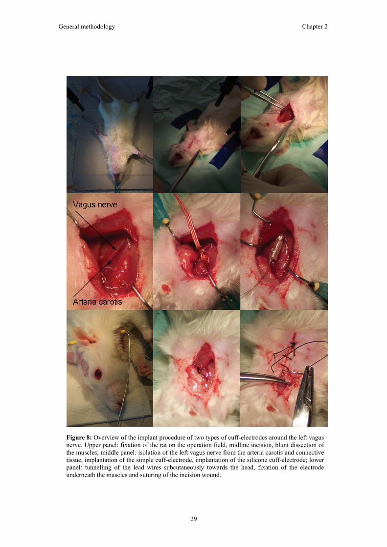
• Cuff-electrode implantation
An overview of the procedure to implant a cuff-
electrode around the vagus nerve is presented in
figure 8.
The rat was fixated with hooks or tape on the
sterile operating field. An incision (1.5 cm) was
made over the left ventral region of the neck to
allow free dissection of the left vagus nerve. The
subcutaneous tissues were incised and blunt
dissection was extended through the muscle
layers. The left vagus nerve was isolated from
connective tissue and the arteria carotis.
Subsequently, the cuff-electrode was placed
around the vagus nerve (Figure 6). The cuff-
electrode was sutured in place to prevent
displacement by animal movement and to secure
the connection between the nerve and electrode.
The wires were bended in a strain relief loop and
sutured to the surrounding tissue (muscles). In this way, potential tension on the cuff-
electrode due to animal movements (e.g. neck stretching) is avoided. The ends of the cuff-
electrode were tunneled subcutaneously using a shunt-passing tool from the ventral cervical
incision via the dorsal cervical region to the head, where they were fixated with acrylic
cement together with the EEG electrodes.
Implantation of the EEG electrodes The animal was fixated in the stereotaxic frame by means of ear bars (Figure 9 and
11). A midline incision was made on the head of the rat. After exposure of the skull,
dependent on the number of epidural EEG electrodes small holes were bored for the
positioning of the screws (Figure 11).
Epidural electrodes (Figure 7), consisting of a stainless steel screw attached
to an isolated silver wire, were positioned on the skull opposite to the
frontal cortex (2 mm anterior to bregma and 1.2 mm lateral to the midline)
and were placed on the parietal cortex (2 mm posterior to bregma and 1.2
mm lateral to the midline). An epidural reference electrode was screwed at
lambda.
Figure 6: Positioning of the VNS cuff-electrode. The cuff-electrode is implanted inthe cervical region around the left vagusnerve, which is located next to the arteriacarotis. The cathode and anode are indicatedon the picture as ‘−’ and ‘+’, respectively.The electrode wires are later onsubcutaneously tunneled towards the head ofthe rat.
Figure 7: Epidural electrode made of a stainless steel screw, a silver wire and a connection piece. Magnification 2x.
5 mm
General methodology Chapter 2
29
Figure 8: Overview of the implant procedure of two types of cuff-electrodes around the left vagus nerve. Upper panel: fixation of the rat on the operation field, midline incision, blunt dissection ofthe muscles; middle panel: isolation of the left vagus nerve from the arteria carotis and connectivetissue, implantation of the simple cuff-electrode, implantation of the silicone cuff-electrode; lower panel: tunnelling of the lead wires subcutaneously towards the head, fixation of the electrodeunderneath the muscles and suturing of the incision wound.

General methodology Chapter 2
30
In case depth electrodes were required for amygdala kindling, bipolar
stimulating/recording electrodes were implanted in both amygdalae (Figure 10 and 11). These
home-made electrodes consisted of two twisted strands of 0.127 mm diameter Diamel-
insulated Nichrome wire and were cut at approximately 1 cm. Coordinates for the
implantation were 2 mm
posterior to bregma, 4.5 mm
lateral to the midline and 8.4
mm below the skull surface
(Paxinos and Watson, 1998).
In addition, five stainless steel
screws, embedded in the skull
were used to anchor the
electrodes in place. The
electrodes and screws were
secured in place by dental
acrylic (Figure 11).
Following surgery, the rats were given acetaminophen rectal gel, a topical analgesic
(xylocaine gel, 0.2%) at the wound edges and a subcutaneous injection of saline (NaCl 0.9%,
2 ml/kg) and were then placed in plastic cages under warming lamps to maintain normal body
temperature until they became behaviorally active again. The recovering animals were closely
observed until they were breathing normally and were able to move around easily. The rats
were then returned to the colony room and allowed up to two weeks of recovery before the
beginning of the recordings.
Figure 10: Amygdala placement of the bipolar electrodes. Redlines encircle the amygdalair nuclei.
Figure 9: Operation field with the rat fixated in the stereotaxic frame.

General methodology Chapter 2
31
2.1.4 Kindling procedures
The kindling model is currently the most widely used animal model for temporal lobe
epilepsy. During the kindling process seizure severity and duration gradually progress. It
offers the advantage that seizures can be elicited at will and that it allows detailed study of the
events associated with the epileptogenic process.
Afterdischarge threshold determination
Two weeks after surgery, initial (prekindling) afterdischarge thresholds (ADTs) were
determined for both amygdalae. The ADT was defined as the minimum stimulus intensity
necessary to provoke a clearly discriminable, high-voltage, electrographic seizure event (an
AD, Figure 12) that outlasted the stimulus by two or more seconds (Kelly et al., 1999). To
assess the threshold a 1 s, 500 µs pulse duration, 60 Hz square wave stimulus of progressively
increasing intensity (15, 25, 35, 50, 75, 100, 150, 200, 300, 400, 500, 600 µA) was delivered
using a constant current generator until an AD was triggered. The interval between
stimulations was 1 min. Post-kindling ADTs were determined the day after kindling was
completed.
Figure 11: Implantation of EEG electrodes. Upper panel: fixation of the rat with ear bars in the stereotaxic frame and preparation of the skull, placement of the epidural EEG electrodes; lower panel: position of epidural screw electrodes, implantation of the depth stimulating/recording electrodes and fixation of the electrodes with acrylic cement.

General methodology Chapter 2
32
Kindling
Twenty-four hours after the ADT determination, kindling
began. All rats were stimulated in the amygdala once daily at
their individual ADT intensity until five stage-5 generalized
convulsive seizures, characterized by forelimb clonus with
rearing and falling, had occurred (Racine, 1972). During the
course of kindling, if the rat failed to respond with an AD, the
rat was stimulated at a higher intensity until an AD was evoked
(using the increment technique described at ADT
determination).
The typical behavior associated with the initial amygdala ADs
is behavioral arrest. With repeated stimulations, duration and
severity of seizures gradually progress. Seizures are usually
classified according to Racine (1972): stage 1, immobility, eye
closure, twitching of vibrissae, sniffing, facial clonus; stage
2, head nodding associated with more severe facial clonus;
stage 3, clonus of one forelimb; stage 4, rearing, often
accompanied by bilateral forelimb clonus; stage 5, rearing with
loss of balance and falling accompanied by generalized clonic
seizures. Stage-5 seizures represent the final seizure stage
(Figure 12). Typically between 10 and 15 stimulation trials are
required before a stage-5 seizure is successfully triggered from an amygdala focus. The exact
rate of amygdala kindling is dependent not only on the strain of rat, but also on the particular
amygdala nucleus in which the stimulation electrode is situated.
2.1.5 Vagus nerve stimulation
Vagus nerve stimulation (VNS) was performed by connecting the cuff-electrode to an
external NeuroCybernetic Prosthesis device (NCP, model 100; Cyberonics Inc., USA). As in
patients, output current was ramped up to a just tolerable level of stimulation (Handforth et
al., 1998). When a stimulus of higher intensity was given, animals behaviorally reacted by
flattening the ears (Dedeurwaerdere et al., 2004) and a fixed posture. The following
stimulation parameters were used: pulse duration 500 µs and frequency 30 Hz. Output current
was dependent on the type of cuff-electrode and the tolerable level was 1.5 mA for the simple
cuff-electrode and 0.5 mA for the spiral silicone cuff-electrode.
A short test stimulus was delivered daily in treated animals to confirm that true
stimulation had been delivered by evoking a behavioral response and to check the impedance
and integrity of the electrode-to-vagus nerve interface (using the Lead Test of the NCP
Figure 12: AD on the EEG and behavioral stage-5 seizure (characterized by generalizedconvulsions with rearing andfalling) evoked by kindling.

General methodology Chapter 2
33
500 µV
1 s
A
B
Figure 13: Different types of SWDs. A. Typicaladult SWD (amplitude: >3 times baselineamplitude; peak frequency: 7-12 Hz; duration >0.5 s); B. SWD-like events i.e. SISWDs in youngGAERS (PN57-PN64) consist of irregular shortdischarges, with clear spike and wave aspectscharacterized by lower amplitude (< 3 timesbaseline amplitude) or lower peak frequency (5-7 Hz).
programming software). The impedance values were always within the attainable range to
deliver the desired output current by the pulse generator. However, the simple cuff-electrode
had lower impedances than the spiral cuff-electrode. Factors that can influence the impedance
are measuring aspects such as frequency, pulse duration or difference in voltage applied. As
these parameters were the same during the impedance measurements of both electrodes
numerous other factors which can influence impedance should be considered. The simple
cuff-electrode has stainless steel contacts whereas the spiral cuff has platinum contacts.
Stainless steel can initially result in lower impedance. Indeed, these electrodes first have
lower impedance values, however, after prolonged stimulation of a few days up to 2 weeks
impedance increased dramatically. In addition, a closer contact of the simple cuff with
surrounding fluid could also contribute to the initial lower impedance.
2.1.6 EEG analysis
For the analysis of the spike and wave discharges (SWDs) (Figure 13, A) on the EEG
of adult GAERS, a previously described and validated detection software package was used
(Van Hese et al., 2003). Once a list of marked time instances was obtained by the detection
method, a number of post-processing steps were performed by the software package:
detections lasting less than 0.5 s were discarded and two detections less than 1 s apart were
taken together. SWDs detected by the software package were visually confirmed. In these
animals, both the number per hour and mean
duration of the SWDs during the recording
hours were calculated as well as the
cumulative duration of the SWDs per hour
(summation of the duration of all SWDs per
hour) as a quantitative measure for seizure
evaluation.
GAERS (under the age of 4 months) may not
all have developed typical SWDs yet or have
infrequent SWDs (Marescaux et al., 1992).
For quantification purposes, epileptiform
events were classified into two closely related
types, because the morphology of SWDs in
young GAERS was found to be less
pronounced and more variable when
compared with SWD patterns in adult
GAERS (Figure 13): i) typical SWDs also

General methodology Chapter 2
34
present in adult GAERS (amplitude: >3 times baseline; peak frequency: 7-12 Hz; duration >
0.5 s) and ii) SWD-like activity, which we have termed ‘short irregular spike and wave
discharges’ (SISWDs), with clear spike and wave aspects characterized by lower amplitude (<
3 times baseline amplitude) or lower peak frequency (5-7 Hz) and shorter in duration (< 2 s).
The software package specifically developed for the detection of SWDs was not
appropriate for the scoring of the more irregular variant of the SWDs (SISWDs). Hence,
epileptiform events in young GAERS (PN57-PN64) were quantified by means of visual
inspection of the EEG using TWIN™ off-line EEG analysis (Telefactor Corporation, USA)
by an experienced electroencephalographer (SD).
2.1.7 Histology
In the electrical kindling experiments, brains were collected for electrode placement
verification. The rats were perfused 24 h after the experiments. Each rat was deeply
anesthetized with 65 mg/kg sodium pentobarbital and was then perfused intracardially with
saline followed by 10% formalin. One day later, the electrodes were removed from the
cranium and the brains were excised and stored in 30% sucrose for at least three days before
sectioning. Frozen sections of 40 µm were taken through the electrode tract tips and stained
with cresyl violet to identify the kindled sites (Figure 14).
Figure 14: A. Serial coronal slices (40 µm) of the rat brain stained withcresyl violet. The electrode tract tips can be followed on the differentslices. B. Magnification of coronal slice with electrode placement in leftamygdala. Electrode placements are indicated with an arrow.
A
B

General methodology Chapter 2
35
2.2 Electrophysiological and morphological evaluation of the use of two cuff-electrodes for vagus nerve stimulation in rats.
This study has been presented as a poster at the 26th International Epilepsy
Conference (Paris, France, 2005) and was published in abstract form in Epilepsia. It was
beyond the scope of this Ph.D. thesis to develop a new cuff-electrode stimulation interface or
to perform a detailed physiological and morphometrical study on cuff-electrodes. Hence, the
aim was to evaluate two types of ready-designed cuff-electrodes fabricated in house on an
electrophysiological and morphological basis for their use in stimulating the vagus nerve of
rats on an acute and chronic basis.
2.2.1 Abstract
Purpose: A better understanding of the mechanism of action of vagus nerve stimulation
(VNS) could lead to stimulation parameter optimization and identification of responder
groups. Animal research is therefore crucial. The aim of this study was to evaluate two types
of stimulation cuff-electrodes for rat nerves on electrophysiological and morphological basis.
Methods: Two cuff-electrodes were evaluated: i) a simple cuff-type electrode and ii) a self-
sizing spiral silicone cuff-electrode.
Electrophysiological measurements of compound nerve potentials induced by the two cuff-
electrodes were performed under deep xylazine/ketamine anesthesia. Subsequently, several
combinations of stimulation parameters (amplitude: 50-1500 µA, pulse duration: 50-500 µs)
were tested.
For the evaluation of morphological modifications, vagus nerves were dissected from non-
implanted control animals (n= 6). The simple cuff and spiral silicone cuff-electrodes were
implanted around the left vagus nerve of respectively 27 and 10 rats and vagus nerves were
dissected after 34 to 377 days of implantation. Nerve tissue was fixated with formalin,
embedded in paraffin and subsequently stained.
Results: Both cuff-electrodes were able to elicit A, B and C fiber compound action potentials.
The spiral silicone cuff-electrode required lower stimulus currents to trigger the different
fibers.
Long-term simple cuff-electrode implantation caused substantial nerve damage, which was
mainly mechanical in nature in contrast to the spiral silicone cuff-electrode, which induced
significantly less morphological changes (p< 0.05).
Conclusion: Both cuff-electrodes were able to induce vagus nerve compound action
potentials, but the spiral silicone cuff-electrode required lower output current. For long-term
implantation, the spiral silicone cuff-electrode induced less morphological changes than the
simple cuff-type electrode and was therefore recommended.

General methodology Chapter 2
36
2.2.2 Electrophysiological evaluation of two cuff-electrodes
During our animal studies on VNS, we have used two types of cuff-electrodes, which
were fabricated in house. The first electrode was a simple cuff-type electrode for acute
experiments, which was easy to manufacture and inexpensive (Figure 4). The second
electrode was a more sophisticated self-sizing spiral silicone cuff-electrode developed for
long-term use, which took more time and specific equipment to manufacture and was more
expensive (Figure 5).
We have tested the capability of both electrodes to induce compound action potentials (CAPs)
in the vagus nerve. Electrophysiological measurements of compound nerve potentials induced
by the two cuff-electrodes were performed under ketamine/xylazine anesthesia (80 mg/kg and
7.5 mg/kg respectively, i.p.). Firstly, the measurements of nerve potentials evoked by the
simple cuff-electrode were performed at the Center of Neuroscience, University of
Amsterdam (Amsterdam, the Netherlands). We have recently started to use the more
sophisticated spiral silicone embedded cuff-electrode. Testing of this electrode took place at
the Neural Rehabilitation Engineering Laboratory, Université Catholique de Louvain,
(Brussels, Belgium) were this electrode was developed. During this electrophysiological
evaluation, two spiral silicone cuff-electrodes were placed around the vagus nerve, the caudal
one was used to stimulate and the more rostrally positioned electrode to record the evoked
CAPs. The experimental setup is shown in figure 15. It was possible to measure the three
fiber components (A, B and C) of the vagus nerve (Figure 15). Both cuff-electrodes were able
to induce CAPs in the vagus nerve.
Figure 15: Overview of the setup for vagus nerve stimulation and recording with the self-sizing spiral silicone cuff-electrode. CAPs of A, B and C fibers of the vagus nerve evoked at output current 1000 µA and pulse duration 50 µs using the silicone cuff-electrode. The response of the A fibers appeared first and was partially enclosed by the stimulation artifact. Subsequently, the CAPs of the slower B and C fibers could be measured. Distance between cathode and registration electrode was 3.6 mm.
5 mm5 mm5 mm5 mm
25 µV 1 ms
25 µV 1 ms

General methodology Chapter 2
37
The amplitude of the CAPs increased when stimulation intensity was augmented
(Figure 16). Threshold current values to excite large myelinated fibers (A and B fibers) are
often low (15-70 µA; 100-200 µs) (Fenik et al., 2001). We found relative low stimulation
thresholds for myelinated A and B fibers with both electrode types. To activate the C fibers
higher current was needed. However, it has been argued that the activation of these C fibers is
not required for the anti-epileptic effect of VNS (Krahl et al., 2001). The spiral silicone cuff-
electrode required lower stimulus currents to trigger the different fibers than the simple cuff-
electrode. Indeed, thresholds to elicit side effects were lower in the spiral silicone electrode
than in the simple cuff (0.5 mA versus 1.5 mA). Most likely, the nerve is better isolated in the
spiral silicone cuff, which prevents current to leak to the surrounding tissue.
Figure 16: CAPs recorded after stimulation of the vagus nerve with the simple cuff. A. Input/outputcurve of CAPs recorded from the vagus nerve evoked by increasing current (0-950 µA) at pulse duration 200 µs using the simple cuff-electrode. B. The amplitude of the CAPs increased as a function of the stimulation intensity. A fibers (1) have a lower threshold than B fibers (2) and the two responsesare clearly discriminable. C fibres activation was not present at these stimulation parameters. Distance between cathode and registration electrode (Teflon coated wire with isolation removed from the tip, 80 µm diameter, California Fine Wire, Canada) was 10 mm. a.u., arbitrary unit.
A
B

General methodology Chapter 2
38
2.2.3 Morphological evaluation of two cuff-electrodes
The left vagus nerve was sampled in 35 implanted animals and six control non-
implanted animals for histological evaluation. At the conclusion of the implant period, the
animals are brought under a deep level of anesthesia (overdose pentobarbital; 180 mg/kg,
i.p.); this is a pain-free and stress-free death. Nerve tissue were resected and subsequently
fixated with formalin and embedded in paraffin. Longitudinal sections were taken for
examination of the nerve tissue and external epineurium. Thick sections (10 µm) were cut,
mounted on slides, stained with hematoxylin and eosin (cells and nuclei), s100 (calcium
binding proteins present in nerves) or myelin basic protein (mbp, staining of myelin) and
coverslipped (Figure 17).
Long-term implantation of simple cuff-electrode caused substantial nerve damage,
which was mainly mechanical in nature in contrast to the spiral silicone cuff-electrode, which
induced less morphological changes (Dedeurwaerdere et al., 2005).
The thickness of the external epineurium was
significantly increased in the stimulated simple
cuff group when compared to the right intact
(non-implanted) nerve of the animals (p< 0.01,
Wilcoxon signed rank test) (Figure 18). Such a
trend was also noticeable in the non-stimulated
simple cuff group, but not in the spiral silicone
cuff group. This is not surprising because the
simple cuff-electrode consists of rigid leads,
which may have caused friction with the nerve.
Relative movement between cuff or cuff-leads
and underlying nerve may have induced a
continued proliferation of connective tissue,
which could have led to the increase in
thickness of the epineurium.
0
0.5
1
1.5
2
2.5
3
3.5
Epineurium Nerve
Thic
knes
s (m
m) .
control
non-stim simple cuff
stim simple cuff
non-stim silicone cuff
stim silicone cuff
*
*
*
*
Figure 18: Effect of electrical stimulation andcuff implantation on epineurium and nervethickness of the vagus nerve in rats.Abbreviations: non-stim, non-stimulated; stim,stimulated; data are expressed as mean ± SEM,significance is set at P< 0.05, * significantlyhigher than control nerve.
Figure 17: Staining of nerve tissue with hematoxylin/eosin, s100 and mbp (left to right respectively) of a normal nerve. Abbreviations: e, external epineurium; n, nerve.

General methodology Chapter 2
39
The thickness of the vagus nerve was significantly higher in the stimulated spiral
silicone-cuff group when compared to the control group (p< 0.05, Mann-Whitney U test) and
to the non-stimulated spiral silicone cuff group (p< 0.05, Mann-Whitney U test) (Figure 18).
This was not the case for nerves stimulated through the simple cuff-electrode. Increase in
thickness of the internal epineurium might be a possible cause. However, it is difficult to
explain why this is only present in the stimulated spiral silicone group and not in the simple
electrode group.
The epineurial sheath included blood vessels, sometimes foreign object reaction and
fibrosis. Blood vessels were significantly increased in the non-stimulated (p< 0.05, Wilcoxon
signed rank test) and stimulated (p< 0.01, Wilcoxon signed rank test) simple cuff group when
compared to their corresponding intact right nerves. Fibrosis was increased significantly in
the stimulated simple cuff group only. Electrical stimulation is considered in itself to be a
conspicuous element of potential nerve injury. Prolonged (> 8h), high-frequency (> 50 Hz)
electrical stimulation of a peripheral nerve induces neural injury (Agnew et al., 1999). Neural
damage due to electrical stimulation is decreased or abolished by reduction of the frequency
of stimulation, by stimulating at 20 Hz (versus 50 Hz) or by the use of an intermittent duty
cycle (Agnew and McCreery, 1990; McCreery et al., 1995). In addition, it has been suggested
that continuous electrical stimulation of peripheral nerves at a low frequency induces little or
no neural damage, even if the stimulus amplitude is very high (McCreery et al., 1995). Based
on these findings, it has been established that VNS should not exceed 30 Hz and continuous
stimulation protocols should be avoided (Agnew and McCreery, 1990). In our studies, we
bore these guidelines in mind and VNS was applied at 30 Hz. In the spiral silicone cuff-
electrode there was no stimulation induced fibrosis. Hence, the increased fibrosis in the
stimulation simple cuff-group is likely due to a combination of the rigid simple cuff and
stimulation rather than to stimulation alone. Also inflammation was significantly increased
(p< 0.025, Wilcoxon signed rank test) in the epineurium of the simple cuff groups. Improving
the quality of the fabrication environment should reduce the chronic inflammation element of
this immunological response.
In the spiral silicone cuff groups, components of the epineurial sheath were not
altered. This is in line with a previous morphometrical study stating that the effects produced
by the chronic implant of this spiral silicone cuff-electrode were negligible (Romero et al.,
2001). The spiral silicone cuff-electrode has been manufactured under a laminar flow bench
in a clean room. This is possibly the reason why significant inflammation of the vagus nerve
was not present in the animals implanted with the spiral silicone cuff-electrode.
Transverse sections of samples from the spiral silicone cuff-electrode were made for
electron microscopy (Figure 19). Appearance of the myelinated axons was not different
between non-stimulated and stimulated groups. However, in both groups, nerves were found

General methodology Chapter 2
40
with decreased myelination of axons. Previous studies were unable to establish a link between
morphological abnormalities caused by chronic cuff implantation and functional deficit (Grill
and Mortimer, 2000). Epineurial electrodes have been found to induce axonal degeneration,
however, the pathology had no apparent effect on the functional response to electrical
stimulation (Koller et al., 1992).
The simple cuff-electrode was a handy electrode for short term experiments. It was
easy to fabricate, to implant and was assembled with inexpensive materials. For long-term
implantation, the spiral silicone cuff-electrode was recommended, because of its more flexible
cabling and contacts it induced less morphological changes than the simple cuff-type. In
addition, less current is required to activate the different fibers, which is preferred for the
battery life.
Figure 19: Electron microscopy of the myelination of the vagus nerve. Upper panel: non-implanted nerves, lower panel: non-stimulated implanted nerve with decreased myelination (left) and stimulated nerve with almost normal myelination (left). Abbreviations: A, A fiber; B, B fiber and C, C fiber.

General methodology Chapter 2
41
References 1. Agnew,W.F. and McCreery,D.B. (1990). Considerations for safety with chronically implanted
nerve electrodes. Epilepsia 31, 27-32.
2. Agnew,W.F., McCreery,D.B., Yuen,T.G. and Bullara,L.A. (1999). Evolution and resolution of stimulation-induced axonal injury in peripheral nerve. Muscle Nerve 22(10), 1393-1402.
3. Dedeurwaerdere,S., Vonck,K., Claeys,P., Van Hese,P., D'Have,M., Grisar,T., Naritoku,D. and Boon,P. (2004). Acute vagus nerve stimulation does not suppress spike and wave discharges in "Genetic Absence Epilepsy Rats from Strasbourg". Epilepsy Res 59, 191-198.
4. Dedeurwaerdere,S., Vonck,K., Van den Broecke,C., Delbeke,J., Wadman,W. and Boon,P. (2005). Electrophysiological and morphological evaluation of two cuff-electrodes for vagus nerve stimulation in rats. Epilepsia 46(Suppl 6), 205.
5. Fenik,V., Fenik,P. and Kubin,L. (2001). A simple cuff electrode for nerve recording and stimulation in acute experiments on small animals. J Neurosci Methods 106, 147-151.
6. Grill,W.M. and Mortimer,J.T. (2000). Neural and connective tissue response to long-term implantation of multiple contact nerve cuff electrodes. J Biomed Mater Res 50(2), 215-226.
7. Handforth,A., DeGiorgio,C.M., Schachter,S.C., Uthman,B.M., Naritoku,D.K., Tecoma,E.S., Henry,T.R., Collins,S.D., Vaughn,B.V., Gilmartin,R.C., Labar,D.R., Morris,G.L.3., Salinsky,M.C., Osorio,I., Ristanovic,R.K., Labiner,D.M., Jones,J.C., Murphy,J.V., Ney,G.C. and Wheless,J.W. (1998). Vagus nerve stimulation therapy for partial-onset seizures: a randomized active-control trial. Neurology 51, 48-55.
8. Kelly,M.E., Battye,R.A. and McIntyre,D.C. (1999). Cortical spreading depression reversibly disrupts convulsive motor seizure expression in amygdala-kindled rats. Neuroscience 91, 305-313.
9. Koller,R., Girsch,W., Liegl,C., Gruber,H., Holle,J., Losert,U., Mayr,W. and Thoma,H. (1992). Long-term results of nervous tissue alterations caused by epineurial electrode application: an experimental study in rat sciatic nerve. Pacing Clin Electrophysiol 15, 108-115.
10. Krahl,S.E., Senanayake,S.S. and Handforth,A. (2001). Destruction of peripheral C-fibers does not alter subsequent vagus nerve stimulation-induced seizure suppression in rats. Epilepsia 42, 586-589.
11. Marescaux,C., Vergnes,M. and Depaulis,A. (1992). Genetic absence epilepsy in rats from Strasbourg--a review. J Neural Transm (Suppl 35), 37-69.
12. McCreery,D.B., Agnew,W.F., Yuen,T.G. and Bullara,L.A. (1995). Relationship between stimulus amplitude, stimulus frequency and neural damage during electrical stimulation of sciatic nerve of cat. Med Biol Eng Comput 33, 426-429.
13. Naples,G.G., Mortimer,J.T., Scheiner,A., Sweeney,J.D. (1988). A spiral nerve cuff electrode for peripheral nerve stimulation. IEEE Trans Biomed Eng 35(11), 905-916.
14. Paxinos,G. and Watson,C. (1998). The Rat Brain in stereotaxic coordinates. (San Diego: Academic Press).
15. Racine,R.J. (1972). Modification of seizure activity by electrical stimulation. II. Motor seizure. Electroencephalogr Clin Neurophysiol 32, 281-294.
16. Romero,E., Denef,J.F., Delbeke,J., Robert,A. and Veraart,C. (2001). Neural morphological effects of long-term implantation of the self-sizing spiral cuff nerve electrode. Medical & Biological Engineering & Computing 39, 90-100.
General methodology Chapter 2
42
17. Van Hese,P., Martens,J.P., Boon,P., Dedeurwaerdere,S., Lemahieu,I.. and Van de Walle,R. (2003). Detection of spike and wave discharges in the cortical EEG of genetic absence epilepsy rats from Strasbourg. Phys Med Biol 48(12), 1685-1700.
18. Veraart,C., Grill,W.M. and Mortimer,J.T. (1993). Selective control of muscle activation with a multipolar nerve cuff electrode. IEEE Trans Biomed Eng 40, 640-653.

Chapter 3: Efficacy of levetiracetam


Efficacy of levetiracetam Chapter 3
45
Purpose: In genetic absence epilepsy rats from Strasbourg (GAERS) age-related absence seizures start to appear from postnatal day (PN) 30 concomitant with ‘spike and wave discharges’ (SWDs) appearing on cortical EEG recordings. The aim of this study was to investigate the effect of early chronic levetiracetam (LEV) treatment on the development of SWDs in young and adult GAERS. Methods: From PN23 until PN60, LEV (54 mg/kg, i.p.) was administered once daily to GAERS (n= 8), while control GAERS (n= 7) received saline (0.9% NaCl, i.p.). All animals were implanted with four epidural EEG electrodes at PN51. EEG was recorded for 3 h daily, during the last four days of the treatment (PN57-PN60) and during four additional days after treatment had been terminated (PN61-PN64). The animals were monitored again at the age of four months (PN120-PN124), about two months after the last administration of LEV. Results: During treatment, epileptiform events in the LEV group were significantly reduced (62%, p< 0.05) in comparison with the control group. During the following four days, epileptiform events were reduced in the LEV group, with an average difference of 53% (p= 0.064). Once the animals had reached adult age, there was no difference in epileptiform events between the LEV group and controls. Conclusion: In this study, chronic LEV administration induced a reduction in epileptiform events in young GAERS. This effect persisted in some extent after treatment cessation (PN61-PN64), which might indicate a slowing down of epileptogenic processes. However, at the age of four months all animals revealed a similar expression of epileptiform discharges. Key words: levetiracetam, anti-epileptogenic, absence epilepsy, GAERS, spike and wave discharges (SWDs)

Efficacy of levetiracetam Chapter 3
46
1. Introduction
Every year the incidence of new epilepsy cases is between 26 and 70 per 100 000
(Hauser, 1998). Despite the development of new pharmacological treatments, approximately
one third of epilepsy patients does not respond to anti-epileptic drugs (AEDs), and are
referred to as refractory patients (Kwan and Brodie, 2000). Treatments with anti-
epileptogenic potential could modulate the processes underlying the development of epilepsy.
Anti-epileptogenic properties have been attributed to some AEDs like valproate (Silver et al.,
1991), lamotrigine (Stratton et al., 2003), and pregabalin (Andre et al., 2003) as well as to
vagus nerve stimulation (Fernandez-Guardiola et al., 1999). Anti-epileptogenic effects of
levetiracetam (LEV) in addition to anti-epileptic effects have been reported to appear in the
rat amygdala kindling model for temporal lobe epilepsy (Loscher et al., 1998; Stratton et al.,
2003) and the spontaneously epileptic rat (SER), a model of primary generalized epilepsy
(Sasa et al., 2003).
LEV is a novel well tolerated AED approved as an adjunctive therapy for epilepsy
patients with refractory partial seizures with or without secondary generalization (Cereghino
et al., 2000). LEV has an anti-epileptic effect in a broad range of animal models mimicking
both partial (Loscher and Honack, 1993; Loscher et al., 2000; Glien et al., 2002) and
generalized epilepsy in man (Loscher and Honack, 1993) including genetic absence epilepsy
rats from Strasbourg (GAERS) (Gower et al., 1995). In contrast, LEV revealed no effect in
the two conventional screening tests, namely the maximal electroshock and pentylenetetrazole
models (Loscher and Honack, 1993). The mechanism of action of LEV differs from other
AEDs and is as yet not fully elucidated.
Studies conducted on AEDs administered during the process of epileptogenesis have
so far focused on animal models of acquired symptomatic epilepsy (Pitkanen, 2002; Andre et
al., 2003). In contrast, animal models of genetic idiopathic epilepsy have only been used
scarcely. This study was stimulated by the ability of LEV to suppress seizure activity in
animal models mimicking absence epilepsy in man (Gower et al., 1995; Boon et al., 2002;
Bouwman and van Rijn, 2004). Moreover, in a previous pilot study of chronic LEV
administration to young GAERS, a preliminary indication of an anti-epileptogenic effect was
found (Boon et al., 2002). Indeed, it was felt that investigating the effect of chronic LEV
treatment in young GAERS could provide new insights and strategies for the treatment of
epilepsy.
GAERS is an isomorphic and predictive model of absence epilepsy. The hallmark is
the appearance of bilateral generalized ‘spike and wave discharges’ (SWDs) in the cortical
EEG recordings during spontaneous absence seizures characterized by behavioral arrest. The
first SWDs appear around postnatal day (PN) 30-PN40 and at the age of four months 100% of

Efficacy of levetiracetam Chapter 3
47
the GAERS have SWDs, which persist throughout the lifespan of the animal (Danober et al.,
1998).
In the present study, we have investigated the effect of LEV on the age-related
development of SWDs in GAERS by chronic administration of LEV (PN23-PN60) starting
before the age of occurrence of SWDs. The effect of early chronic LEV administration on the
development of SWDs was evaluated in young GAERS during treatment (PN57-PN60),
immediately after treatment termination (PN61-PN64) and two months after the last LEV
injection (age four months), when brain maturation was achieved and SWDs recorded on
cortical EEG were numerous.
2. Methods
2.1 Animals
Fifteen GAERS were used in this study. The animals were born and raised under
environmentally controlled conditions (12 h light/dark cycles, lights on at 7 a.m., 20-22°C)
with food and water ad libitum at the animal facility of the Ghent University Hospital. All
animals were treated according to the guidelines approved by the European Ethics Committee
(decree 86/609/CEE). The study protocol was approved by the Animal Experimental Ethical
Committee of Ghent University Hospital.
2.2 Surgery
The pups (n= 15) were randomly assigned to the two treatment groups: LEV and
control group. LEV (54 mg/kg, provided by UCB Pharma, Belgium) was dissolved in saline
(0.9% NaCl) and injected intraperitoneally (i.p.) in a dose-volume of 2 ml/kg body weight,
whereas control rats received an equivalent dose-volume of saline. From PN23 until PN60,
LEV was administered once daily to eight GAERS, while the control GAERS (n= 7) received
saline. All animals were weighed daily.
At age PN51, four epidural EEG stainless steel screw electrodes were bilaterally
implanted over the fronto-parietal cortex of all 15 animals. The rats were anesthetized with
ketamine/xylazine (80 mg/kg and 7.5 mg/kg respectively, i.p.) and additional ketamine (5
mg/kg, i.p.) was given when sensorial pain stimuli, by squeezing the foot pad, elicited motor
reflexes. One epidural electrode was positioned on the left side of the skull at height of the
frontal cortex (AP= +2 mm, ML= +1.2 mm, referred to bregma) and one was placed on the
right sided parietal cortex (AP= -2 mm, ML= -1.2 mm, referred to bregma). The reference
electrode was placed at lambda with the ground electrode on the left side of lambda (ML=
+1.2 mm, referred to lambda). The EEG electrodes were fixed on the skull of the rat with

Efficacy of levetiracetam Chapter 3
48
acrylic cement. Lidocaine gel (2%) was applied at the incision wound and the rats were
placed under a heating lamp until they had recovered from anesthesia.
2.3 EEG recording
EEG was recorded by means of a H2O™ portable digital 32-channel EEG-recorder
(Telefactor Corporation, U.S.A.). To avoid any interference with the circadian cycle, all
recordings were performed during daytime (light period) starting at 10 a.m. in awake and
freely moving animals (Rigoulot et al., 2003). Every 10 min a standardized sound stimulus
was provided, to prevent the rats from falling asleep (Gower et al., 1995; Manning et al.,
2004). During the recording sessions, the rats were continuously observed.
2.4 Effect of early chronic LEV administration on epileptiform events in young
GAERS (PN57-PN64)
One week after surgery, the animals were placed in the EEG recording chambers and
stayed there from PN56 (one day before the start of the EEG recordings) until PN64 with food
and water ad libitum. During the last four days of the treatment (PN57-PN60), EEG was
recorded for 3 h daily. In this period, LEV or saline was injected 1 h after the start of the EEG
recording. From PN61 on, treatment was halted, animals (controls and LEV group) received
no longer injections and EEG was recorded during four additional days (PN61-PN64), 3 h
daily (Figure 1). The EEG was recorded on several days to have a more robust dataset on the
effect of LEV in these young animals.
Epileptiform events in young GAERS (PN57-PN64) were quantified by means of
visual inspection of the EEG using TWIN™ off-line EEG analysis (Telefactor Corporation,
U.S.A.) by an experienced electroencephalographer (SD). Young GAERS (under the age of 4
Figure 4: Study design. LEV (45 mg/kg, i.p.) was administered once daily at 11 a.m. from PN23 till PN60. During the last four days of the treatment (PN57-PN60) EEG was recorded for 3 h starting at 10 a.m.; the following four days (PN61-PN64), treatment was terminated and EEG was again recorded for 3 h a day.
LEV treatment EEG recording
PN0 PN20 PN40PN30 PN50
PN60
1 h
2 h
3 h
Birth Weaning LEV injectionOperation
LEV treatment EEG recordingLEV treatment EEG recording
PN0 PN20 PN40PN30 PN50
PN60
1 h
2 h
3 h
Birth Weaning LEV injectionOperation

Efficacy of levetiracetam Chapter 3
49
500 µV1 s
A
B
months) may not all have developed typical SWDs yet or have infrequent SWDs (Danober et
al., 1998). For quantification purposes, epileptiform events were classified into two closely
related types, because the morphology of SWDs in young GAERS was found to be less
pronounced and more variable when compared with SWD patterns in adult GAERS (Figure
2): i) typical SWDs also present in adult GAERS (amplitude: > 3 times baseline; peak
frequency: 7-12 Hz; duration > 0.5 s) and ii)
SWD-like activity, which we have termed
‘short irregular spike and wave discharges’
(SISWDs), with clear spike and wave
aspects characterized by lower amplitude (<
3 times baseline amplitude) or lower peak
frequency (5-7 Hz) and shorter in duration
(< 2 s). Only the numbers of different SWD
types (SWDs and SISWDs) per hour were
used as dependent values. The durations of
SWDs and SISWDs were still very short in
these young animals; therefore relevant
differences in discharge duration between
treated and control animals could not be
detected.
2.5 Evaluation of SWDs in adult GAERS (age four months) after chronic LEV
treatment during early life
About the age of four months (range PN120-PN124), EEG was recorded for one day
(duration of recording between 6 and 8 h) in 15 animals. For the analysis of the SWDs on the
EEG of the adult GAERS a previously described and validated detection software package
was used (Van Hese et al., 2003). Once a list of marked time instances was obtained by the
detection method, a number of post-processing steps were performed by the software
package: detections lasting less than 0.5 s were discarded and two detections less than 1 s
apart were taken together. SWDs detected by the software package were visually confirmed.
In these animals, both the number per hour and mean duration of the SWDs during the
recording hours were calculated as well as the cumulative duration of the SWDs per hour
(summation of the duration of all SWDs per hour).
The software package specifically developed for the detection of SWDs was not
appropriate for the scoring of the more irregular variant of the SWDs (SISWDs). Hence, the
Figure 5: Different types of SWDs. A. Typicaladult SWD (amplitude: > 3 times baselineamplitude; peak frequency: 7-12 Hz; duration > 0.5s); B. SWD-like events i.e. SISWDs in youngGAERS (PN57-PN64) consist of irregular shortdischarges, with clear spike and wave aspectscharacterized by lower amplitude (< 3 timesbaseline amplitude) or lower peak frequency (5-7 Hz).

Efficacy of levetiracetam Chapter 3
50
EEG was visually inspected for SISWDs, fulfilling the conditions described above, and the
number per hour was calculated.
2.6 Statistical analysis
In young GAERS, statistical analysis was performed on both pooled and unpooled
data. Data could be pooled after exclusion of interactions (hour x treatment and day x
treatment) by ANOVA with repeated measurements. The number of SWDs and SISWDs per
3 h was taken together; this was termed the number of epileptiform events. Statistical analysis
was also performed on unpooled data, which consisted of the number of typical SWDs and
SISWDs per hour at the different time points during a total recording time of 24 hours (3 h a
day, during 8 consecutive days).
For the inter-group comparison between the LEV and control group the Mann-
Whitney U-test was used. For the intra-group comparisons within the LEV and control group,
a Friedman test was utilized. For the intra-group comparison between the mean number of
epileptiform events on the two time points: during the last four days of treatment (PN57-
PN60) and during the following four days after treatment had been terminated (PN61-PN64),
a Wilcoxon signed rank test was used.
Through the further course of this paper (results and discussion), the results obtained
in young GAERS will be presented and discussed according to the recording periods: a) the
last four days of the treatment (PN57-PN60) and b) the following four days after treatment
cessation (PN61-PN64).
In adult rats, the Mann-Whitney U-test was used to compare the number per hour, the
mean duration and the cumulative duration/hour of the SWDs between the LEV and control
group. For SISWDs, the number per hour was compared between the LEV and control group
using the Mann-Whitney U-test.
All statistical tests were performed utilizing the statistical software package SPSS 12.
Data are presented as the mean ± the standard error of the mean (SEM), and the significance
level has been set at α= 0.05.

Efficacy of levetiracetam Chapter 3
51
3. Results Chronic administration of LEV during 38 days in young GAERS (PN23-PN60) did
not induce growth changes. Normal body weight in LEV treated GAERS (157 ± 8 g for males
and 144 ± 10 g for females at PN60) was achieved in comparison with the control group (140
± 15 g for males and 121 ± 6 g for females at PN60) treated with saline (NS, Student’s t-test).
3.1 Effect of chronic LEV administration on epileptiform events in young GAERS
(PN57-PN64)
LEV treated rats (n= 8) showed less SWDs and SISWDs than control rats (n= 7)
during treatment and also during the four days after the discontinuation of the treatment.
During this 8-day recording period, typical SWDs were observed in three control rats. Only
one of these rats had frequent (12 ± 2 per hour) SWDs on the EEG, which were short in
duration (1.6 ± 0.2 s). None of the LEV treated rats had typical SWDs; hence, there were too
few SWDs to detect significant differences in SWDs between LEV and control group.
SISWDs were observed in all rats. These SWD-like events were short lasting (< 1.5 s) in the
LEV and in the control group.
a) Effect of chronic LEV administration on epileptiform events during the last four days of
the treatment period (PN57-PN60)
During LEV treatment (PN57-PN60), the mean number of epileptiform events
(pooled data) was 62% lower (p< 0.05, Mann-Whitney U-test) in the LEV group. The control
group had a mean number of 86 ± 18 epileptiform events per 3 h, whereas this figure in the
LEV group was 33 ± 11. Significant differences could also be found in the number of
SISWDs (up to 81%) between LEV and control group on different time points during
treatment (Figure 3). Also during the pre-injection hours (PN57-PN60), SISWDs were
significantly lower in the LEV group (8 ± 2; 6 ± 2; 4 ± 2 and 7 ± 3) than in the control group
(28 ± 8; 32 ± 6; 17 ± 6 and 20 ± 4) (Figure 3).
Pre- and post-injection hours differed significantly in treated animals on PN59 and
PN60 (Friedman test). Surprisingly, the number of SISWDs in the post-injections hours was
higher than in the pre-injection hour in the LEV treated group (Figure 3). However, a trend of
a higher number of SISWDs in the post-injection hours was also present in the control group
on PN59 (p= 0.066; Friedman test).
No differences were found between the four individual days during treatment (PN57-PN60)
within LEV and control group (data not shown, Friedman test).

Efficacy of levetiracetam Chapter 3
52
b) Effect of chronic LEV administration on epileptiform events after treatment cessation
(PN61-PN64)
When treatment was discontinued (PN61-PN64), there was a borderline missed
reduction of 53% in the mean number of epileptiform events (p= 0.064, pooled data, Mann-
Whitney U-test) in the LEV group compared with the control group. The control group had a
mean number of 73 ± 14 epileptiform events per 3 h, whereas this figure in the LEV group
was 34 ± 8. However, significant differences could be found in SISWDs (up to 66%) between
LEV and control group at different separate time points (unpooled data, Figure 3).
After treatment had been stopped, there were no differences between the recording
hours within the LEV and the control group (Friedman test).
Within LEV and control group, there was a significant difference between the four
days after treatment cessation (pooled data, Friedman test); this was manifested by an increase
in the number of epileptiform events on the last two days of the recording period. In the LEV
group these number of epileptiform events on PN61, 62, 63 and 64 were respectively 25 ± 8;
23 ± 7; 40 ± 10 and 48 ± 9. In the control group these figures were 52 ± 14; 57 ± 10; 94 ± 19
and 88 ± 19.
The Wilcoxon signed rank test did not reveal differences between the mean number
of epileptiform events during treatment period and after treatment cessation in both groups
(86 ± 13 versus 73 ± 14 for the control group and 33 ± 11 versus 34 ± 6 for the LEV group).
05
101520253035404550
Num
ber o
f SIS
WD
s/h
. Saline LEV*
****
**
*
*
* *
* * *
***
*
*
Last four days of treatment (PN57-PN60) Following four days after treatment cessation (PN61-PN64)
1 2 3 1 2 3 1 2 3 1 2 3 1 2 3 1 2 3 1 2 3 1 2 3 PN57 PN58 PN59 PN60 PN62 PN63 PN64 PN61
Control group LEV group
Figure 6: Effect of chronic LEV administration (PN23-PN60) on SISWDs in young GAERS (PN57-PN64). Number of SISWDs in the LEV group treated with LEV (solid bars, n= 8) and in the controlgroup treated with saline (open bars, n= 7) during treatment (PN57-PN60) and after treatment discontinuation (PN61-PN64). Data are expressed as mean ± SEM; SISWDs= short irregular spike andwave discharges; PN= post natal day; 1 h= pre-injection hour or first recording hour; 2 h= first post-injection hour or second recording hour; 3 h= second post-injection hour or third recording hour. Differences between control and LEV group are assessed using the Mann-Whitney U-test and differences between the three different hours within the groups is assessed using the Friedman test; * indicates p< 0.05, ** indicates p< 0.01.

Efficacy of levetiracetam Chapter 3
53
3.2 Evaluation of SWDs in adult GAERS (age four months) after chronic LEV
treatment during early life
At the age of four month all animals had typical SWDs. EEG recordings showed no
significant difference between the LEV and control group in number, mean duration and
cumulative duration of the SWDs (Mann-
Whitney U-test). These numbers were only
slightly lower in the LEV group (Figure 4).
Also in SISWDs no differences could be found
between both groups (Figure 4). However,
SISWDs were decreased substantially in favor of
typical SWDs in comparison with young
GAERS (PN57-PN64), which had more SISWDs
than SWDs.
4. Discussion
A new approach to drug screening, including the process of epileptogenesis, may
identify new classes of drugs (Kupferberg, 2001; Klitgaard and Pitkanen, 2003). LEV is a
novel AED with potential anti-epileptogenic properties, which was discovered using non-
conventional drug screening. It suppresses kindling development at doses devoid of adverse
effects with persistent reduction in afterdischarge duration after termination of the treatment
(Loscher et al., 1998). In the SER-model, LEV was administered before the appearance of
spontaneous seizures and was terminated at the expected age for seizure expression, which
resulted in a lower seizure number in pre-treated animals (Sasa et al., 2003). In the present
study, we found that chronic LEV administration induced a decrease in epileptiform events
(SWDs and SISWDs) in young GAERS (PN57-PN60), which persisted immediately
following cessation of treatment (PN61-PN64), but not until adulthood.
In GAERS, SWDs strictly correlate with the occurrence of clinical absences (Danober
et al., 1998), which was also observed in this study. However, behavioral absence seizures
can be subtle as they mostly appear during quiet wakefulness (Danober et al., 1998).
Therefore, EEG recordings were used to quantify the appearance of SWDs. In our study, we
0
50
100
150
200
250
Cum
ulat
ive
dura
tion
of S
WD
s/h
.
0
5
10
15
20
25
30
35
40
SWDs SISWDs
Num
ber o
f SW
Ds a
nd S
ISW
Ds/h
.
Control groupLEV group
0
1
2
3
4
5
6
7
8
9
10
Mea
n du
ratio
n of
SW
Ds .
A
CB
Figure 7: Evaluation of epileptiform events in adult GAERS (age 4 months) after chronic LEV treatment during early life. A. Number of SWDs and SISWDs per hour, B. mean duration of SWDs (s) and C.cumulative duration of SWDs per hour (s/h) in the LEV group (solid bars, n= 8) and in the control group (open bars, n= 7). Data are expressed as mean ± SEM.

Efficacy of levetiracetam Chapter 3
54
found that typical SWDs are rare in young GAERS. Only three out of seven young control
GAERS showed typical SWDs on the EEG and none of the LEV treated animals (n= 8).
SISWDs, however, were the most prevalent type of discharge on the EEG during quiet
wakefulness. Hence, these more irregular discharges were taken into account to quantify the
effect of LEV on epileptiform events. While it is difficult to correlate SISWDs with
behavioral arrest because of their short duration (< 1.5 s), it can be assumed that these
discharges are closely related to SWDs and reflect premature SWDs given their
morphological resemblance with typical SWDs and their strong decrease in favor of typical
SWDs as the rats grow older.
In this study, epileptiform events (SWDs + SISWDs) were significantly suppressed
(difference of 62%) during chronic LEV treatment in young GAERS (PN57-PN60) in
comparison with control animals. This is in line with a previous acute study of Bouwman and
van Rijn (2004), which found a similar difference in SWDs between control and LEV treated
adult rats in the WAG/RIJ model of absence epilepsy.
The LEV treated animals displayed less SISWDs (up to 81%) than the control group
even in the pre-injection hours during PN57-PN60. This could suggest that there was a
slowing down of the epileptogenic processes by early chronic treatment (PN23-PN60) with
LEV. Yet, the lower number of SISWDs in the pre-injection hours might also be caused by
retention of LEV in the brain from previous injections, although LEV was already eliminated
from the blood plasma.
Interestingly, there was no acute decrease in SISWDs after injection of LEV (PN57-
PN60) and even the opposite could be observed. This is in contrast with previous acute
experiments in adult GAERS, in which a clear decrease in SWDs could be observed after a
single LEV injection (Gower et al., 1995; Boon et al., 2002; Bouwman and van Rijn, 2004).
These studies, which were performed in adult GAERS, report the effect of a first and single
LEV injection, while the effect of chronic LEV administration was not investigated.
Moreover, several studies dealing with the chronic administration of LEV in animal models of
temporal lobe epilepsy report increased functional tolerance towards LEV and some loss of
efficacy after prolonged administration (Loscher et al., 1998; Loscher and Honack, 2000;
Glien et al., 2002). This could be a possible reason for the lack of an acute decrease in
SISWDs in rats that were already pretreated.
The acute increase in SISWDs on PN59 and PN60 after LEV administration could
represent a flaring up which stabilized again towards the next day. A trend for this
phenomenon was also observed in the control group on PN59 (p= 0.066). There was no
difference between recording hours during the last four days, when the rats were no longer
injected with saline or LEV (PN61-PN60). Hence, stress after injection could be a responsible
Efficacy of levetiracetam Chapter 3
55
factor for this increase in SISWDs, although the animals were accustomed to the handling
procedures.
When treatment was stopped, there was no sudden increase in epileptiform events
(SWDs and SISWDs). This is in line with previously performed rat and mice experiments,
where chronic treatment with LEV did not lead to withdrawal hyperexcitability (Klitgaard et
al., 1998; Loscher and Honack, 2000). During different time points, epileptiform events were
still significantly lower (up to 66%) in young LEV treated GAERS. This difference in number
of absence seizures between controls and LEV treated rats is comparable to the study in the
SER model (Sasa et al., 2003). In the SER-study, absence seizures were still decreased two
weeks after treatment cessation during the 30 min recording period. However, the next week
there was no longer a significant difference between the control and LEV pretreated group. In
the study of Glien et al. (2002), spontaneous partial seizures in the pilocarpine model of
temporal lobe epilepsy were also still decreased one week after termination of chronic LEV
administration (two weeks) in certain rats, this was described as a carryover effect.
In both LEV and control group, epileptiform events started to increase on the last
days of recording, which can be due to the progressive nature of absence epilepsy in young
GAERS. Still, significant lower SISWDs (up to 66%) in the LEV group could be detected on
these last days of EEG recording (PN63 and PN64) in comparison with the control group.
Early chronic treatment with LEV in young GAERS might have caused an initial
slowing down of the epileptogenic processes, which had persisting effects after treatment
termination. The major metabolite of LEV is pharmacologically inactive, and other
metabolites are produced at levels below those that are relevant for pharmacological activities
(Loscher et al., 1998). LEV does not interfere with enzyme systems (Patsalos, 2000;
Benedetti, 2000) and is washed out rapidly (half-live ~2-3 h) after stopping the treatment
(Loscher et al., 1998). As we used a relatively high dose to inject and LEV is already effective
in low doses in GAERS (Gower et al., 1995), we expect that relevant therapeutic
concentrations are present in the blood plasma for 8 h. Hence, the prolonged decrease in
epileptiform events on PN61-PN64 is probably not due to a prolonged acute effect caused by
accumulated LEV or other metabolites.
Early chronic administration of LEV had no effect on the later expression of SWDs at
the age of four months. SISWDs have diminished considerably in both groups; this supports
its role as premature SWDs. Most likely, after discontinuation of the treatment,
epileptogenesis could further progress. To our knowledge, there is only one other study where
an AED i.e. remacemide was chronically administered in young GAERS (PN7-PN25) to
establish its effect on SWD development (Nehlig and Boehrer, 2003). This study failed to
demonstrate an effect of early remacemide treatment on the expression of SWDs in adult
GAERS (Nehlig and Boehrer, 2003). In the present study, adult GAERS displayed fewer

Efficacy of levetiracetam Chapter 3
56
seizures and shorter mean SWD durations compared to previously published experiments in
GAERS, which were monitored only for a short period of time (Danober et al., 1998).
However, these values were similar to a previous chronic experiment performed by our group
(Dedeurwaerdere et al., 2004). This phenomenon of a decrease in seizures during longer
recording periods is correlated with the decrease of vigilance resulting in more sleep episodes
and less and shorter seizures (Dufour et al., 2001; Rigoulot et al., 2003).
Until now, it is not described in what manner LEV exerts its effect on absence
seizures in GAERS. LEV has no effect on the T-type voltage-activated Ca2+ current (Zona et
al., 2001), a common target for absence epilepsy drugs. A possible effect of LEV that might
contribute to its action against SWDs in GAERS could be by progressively decreased firing of
the substantia nigra pars reticulata (SNR) neurons (Loscher et al., 1996). The SNR is not part
of the epileptic circuit underlying absence seizures, but it may belong to a system
participating in a remote control over seizures (Danober et al., 1998). Indeed, high frequency
stimulation of the SNR has been shown to interrupt SWDs in GAERS (Feddersen et al., 2004)
and intracerebral microinjections of GABA mimetics in the SNR suppress absence seizures
(Danober et al., 1998). A part of LEV’s ability to suppress seizures may origin in its effect on
neuronal firing in the SNR, but the anatomical network and molecular mechanisms by which
this is mediated remains to be demonstrated (Klitgaard et al., 1998). By this way LEV could
suppress SWDs in GAERS, but it is not known if this might interfere with epileptogenesis. In
GAERS pups, metabolic increases in SNR compared to non-epileptic control rats were
observed, which was suggested to reflect a mechanism of prevention of epileptogenesis in
GAERS (Nehlig et al., 1998).
4. Conclusion
In this study, chronic LEV administration induced a decrease in epileptiform events
in young GAERS (PN57-PN60), although this was not obtained by a decrease immediately
after injection. This effect persisted partly after cessation of treatment (PN61-PN64), which
might reflect a slowing of epileptogenic process due to previous chronic treatment with LEV.
However, when LEV and saline treated animals grew up, they all developed a similar
expression of spontaneous absence seizures.

Efficacy of levetiracetam Chapter 3
57
Acknowledgements We would like to acknowledge Dr. H. Klitgaard (UCB Pharma, Braine-L’ Alleud,
Belgium) for the critical reading of the manuscript and UCB-Pharma for their generous gift of
LEV.
Lic. S. Dedeurwaerdere is supported by Grant 011D9601 from the Ghent University
Research Fund (B.O.F.). Robrecht Raedt and Tim De Smedt are supported by grants from the
Institute for encouragement of Innovation through Science and Technology in Flanders
(IWT). Dr. K. Vonck is supported by a Junior Research (“Aspirant”) Grant from the Fund for
Scientific Research-Flanders (F.W.O.). Dr. P. Boon is a Senior Clinical Investigator of the
Fund for Scientific Research-Flanders and supported by grants 1.5236.99 and 6.0324.02 from
the Fund for Scientific Research-Flanders; by grant 01105399 from Ghent University
Research Fund (B.O.F.) and by the Clinical Epilepsy Grant Ghent University Hospital 2000-
2004.

Efficacy of levetiracetam Chapter 3
58
References
1. Andre,V., Rigoulot,M.A., Koning,E., Ferrandon,A. and Nehlig,A. (2003). Long-term Pregabalin Treatment Protects Basal Cortices and Delays the Occurrence of Spontaneous Seizures in the Lithium-Pilocarpine Model in the Rat. Epilepsia 44, 893-903.
2. Benedetti,M.S. (2000). Enzyme induction and inhibition by new antiepileptic drugs: a review of human studies. Fundam Clin Pharmacol 14, 301-19.
3. Boon,P., Seys,L., Vonck,K., Dedeurwaerdere,S., D'Havé,M., Grisar,T., Claeys,P. and De Reuck,J. (2002). The effect of levetiracetam in genetic absence epilepsy Strasbourg rats. Epilepsia 43(Suppl 8), 60.
4. Bouwman,B.M. and van Rijn,C.M. (2004). Effects of levetiracetam on spike and wave discharges in WAG/Rij rats. Seizure 13(8), 591-594.
5. Cereghino,J.J., Biton,V., Abou-Khalil,B., Dreifuss,F., Gauer,L.J. and Leppik,I. (2000). Levetiracetam for partial seizures: results of a double-blind, randomized clinical trial. Neurology 55, 236-242.
6. Danober,L., Deransart,C., Depaulis,A., Vergnes,M. and Marescaux,C. (1998). Pathophysiological mechanisms of genetic absence epilepsy in the rat. Prog Neurobiol 55, 27-57.
7. Dedeurwaerdere,S., Vonck,K., Claeys,P., Van Hese,P., D'Have,M., Grisar,T., Naritoku,D. and Boon,P. (2004). Acute vagus nerve stimulation does not suppress spike and wave discharges in "Genetic Absence Epilepsy Rats from Strasbourg". Epilepsy Res 59, 191-198.
8. Dufour,F., Nalecz,K.A., Nalecz,M.J. and Nehlig,A. (2001). Modulation of absence seizures by branched-chain amino acids: correlation with brain amino acid concentrations. Neurosci Res 40, 255-263.
9. Feddersen,B., Deransart,C., Vercueil,L., Noachtar,S. and Depaulis,A. (2004). High-frequency stimulation of the substantia nigra pars reticulata suppresses absence seizures in a genetic model of absence epilepsy in the rat. Epilepsia 45(Suppl 3), 118.
10. Fernandez-Guardiola,A., Martinez,A., Valdes-Cruz,A., Magdaleno-Madrigal,V.M., Martinez,D. and Fernandez-Mas,R. (1999). Vagus nerve prolonged stimulation in cats: effects on epileptogenesis (amygdala electrical kindling): behavioral and electrographic changes. Epilepsia 40, 822-829.
11. Glien,M., Brandt,C., Potschka,H. and Loscher,W. (2002). Effects of the novel antiepileptic drug levetiracetam on spontaneous recurrent seizures in the rat pilocarpine model of temporal lobe epilepsy. Epilepsia 43, 350-357.
12. Gower,A.J., Hirsch,E., Boehrer,A., Noyer,M. and Marescaux,C. (1995). Effects of levetiracetam, a novel antiepileptic drug, on convulsant activity in two genetic rat models of epilepsy. Epilepsy Res 22, 207-213.
13. Hauser,W.A. (1998). Incidence and Prevalence. In Epilepsy, a comprehensive textbook, J.Engel and T.A.Pedley, eds. (Philadelphia: Lippincot-Raven), 47-58.
14. Klitgaard,H., Matagne,A., Gobert,J. and Wulfert,E. (1998). Evidence for a unique profile of levetiracetam in rodent models of seizures and epilepsy. Eur J Pharmacol 353, 191-206.
15. Klitgaard,H. and Pitkanen,A. (2003). Antiepileptogenesis, neuroprotection, and disease modification in the treatment of epilepsy: focus on levetiracetam. Epileptic Disord 5(Suppl 1), 9-16.

Efficacy of levetiracetam Chapter 3
59
16. Kupferberg,H. (2001). Animal models used in the screening of antiepileptic drugs. Epilepsia 42, 7-12.
17. Kwan,P. and Brodie,M.J. (2000). Early identification of refractory epilepsy. N Engl J Med 342, 314-319
18. Loscher,W. and Honack,D. (1993). Profile of ucb L059, a novel anticonvulsant drug, in models of partial and generalized epilepsy in mice and rats. Eur J Pharmacol 232, 147-158.
19. Loscher,W. and Honack,D. (2000). Development of tolerance during chronic treatment of kindled rats with the novel antiepileptic drug levetiracetam. Epilepsia 41, 1499-1506.
20. Loscher,W., Honack,D. and Bloms-Funke,P. (1996). The novel antiepileptic drug levetiracetam (ucb L059) induces alterations in GABA metabolism and turnover in discrete areas of rat brain and reduces neuronal activity in substantia nigra pars reticulata. Brain Res 735, 208-216.
21. Loscher,W., Honack,D. and Rundfeldt,C. (1998). Antiepileptogenic effects of the novel anticonvulsant levetiracetam (ucb L059) in the kindling model of temporal lobe epilepsy. J Pharmacol Exp Ther 284, 474-479.
22. Loscher,W., Reissmuller,E. and Ebert,U. (2000). Anticonvulsant efficacy of gabapentin and levetiracetam in phenytoin- resistant kindled rats. Epilepsy Res 40, 63-77.
23. Manning,J.-P.A., Richards,D.A., Leresche,N., Crunelli,V. and Bowery,N.G. (2004). Cortical-area specific block of genetically determined absence seizures by ethosuximide. Neuroscience 123, 5-9.
24. Nehlig,A., Vergnes,M., Boyet,S. and Marescaux,C. (1998). Metabolic activity is increased in discrete brain regions before the occurrence of spike-and-wave discharges in weanling rats with genetic absence epilepsy. Brain Res Dev Brain Res 108, 69-75.
25. Nehlig,A. and Boehrer,A. (2003). Effects of remacemide in two models of genetically determined generalized epilepsy, the GAERS and the audiogenic Wistar AS. Epilepsy Res 52, 253-261.
26. Patsalos,P.N. (2000). Pharmacokinetic profile of levetiracetam: toward ideal characteristics. Pharmacol Ther 85, 77-85.
27. Pitkanen,A. (2002). Drug-mediated neuroprotection and antiepileptogenesis: animal data. Neurology 59, 27-33.
28. Rigoulot,M.A., Boehrer,A. and Nehlig,A. (2003). Effects of Topiramate in Two Models of Genetically Determined Generalized Epilepsy, the GAERS and the Audiogenic Wistar AS. Epilepsia 44, 14-19.
29. Sasa,M., Yan,H., Nagayama,T. and Seriwaka,T. (2003). Anti-epileptogenic Properties of Levetiracetam in the Spontaneously Epileptic Rat (SER). Epilepsia 44(Suppl 8), 175-176.
30. Silver,J.M., Shin,C. and McNamara,J.O. (1991). Antiepileptogenic effects of conventional anticonvulsants in the kindling model of epilepsy. Ann Neurol 29(4), 356-363.
31. Stratton,S.C., Large,C.H., Cox,B., Davies,G. and Hagan,R.M. (2003). Effects of lamotrigine and levetiracetam on seizure development in a rat amygdala kindling model. Epilepsy Res 53, 95-106.
32. Van Hese,P., Martens,J.P., Boon,P., Dedeurwaerdere,S., Lemahieu,I. and Van de Walle,R. (2003). Detection of spike and wave discharges in the cortical EEG of genetic absence epilepsy rats from Strasbourg. Phys Med Biol 48, 1685-1700.
33. Zona,C., Niespodziany,I., Marchetti,C., Klitgaard,H., Bernardi,G. and Margineanu,D.G. (2001). Levetiracetam does not modulate neuronal voltage-gated Na+ and T-type Ca2+ currents. Seizure 10, 279-286.


Chapter 4: Efficacy of vagus nerve stimulation


Efficacy of vagus nerve stimulation Chapter 4
63
We evaluated the efficacy of vagus nerve stimulation (VNS) in genetic absence epilepsy rats from Strasbourg (GAERS), a validated model for absence epilepsy. In the first experiment, we investigated whether VNS applied at seizure onset can interrupt spike and wave discharges (SWDs). In the second experiment, we investigated whether SWDs are suppressed or shortened in duration when VNS is applied several hours per day. Both control and VNS groups underwent EEG and VNS electrode implantation. For the first experiment, a randomized crossover design was used. Stimuli (amplitude: 3 V; frequency: 30 Hz; pulse duration: 500 µs) were given when an SWD occurred on the EEG. The experiment was repeated the next day. In the second experiment, treated animals were stimulated (amplitude: 1.5 mA; frequency: 30 Hz; pulse duration: 500 µs; on/off time cycle: 30 s/ 5 min) for three hours per day, during five consecutive days. In the first experiment, the duration of the SWDs was increased on day 1, (p< 0.05). There was no difference in SWD duration on day 2. In the second experiment, no significant differences could be found in number, duration and EEG frequency of SWDs. VNS applied at the onset of an SWD can prolong the duration of SWDs in GAERS. As a five-day stimulation protocol had no effect, long-term VNS might be necessary to affect SWDs. Key words: vagus nerve stimulation, absence epilepsy, GAERS, EEG
1. Introduction
Vagus nerve stimulation (VNS) is an efficacious broad-spectrum add-on treatment for
patients with medically or surgically refractory epilepsy (Ben-Menachem, 2002). The left
vagus nerve is stimulated intermittently by means of an implanted pulse generator and reduces
the frequency and severity of epileptic seizures. Controlled randomized trials showed a 50%
decrease in overall seizure frequency in approximately 30% of the patients (Salinsky, 2003).

Efficacy of vagus nerve stimulation Chapter 4
64
The precise mechanism of action of VNS is not known. Mechanistic VNS research
generally presumes that VNS exerts its effect by inducing action potentials in the afferent
fibres of the left vagus nerve (Zagon et al., 1999; Henry, 2002). Through diffuse projections
of the vagus nerve in the cerebral hemispheres, VNS can have a broad effect on neuronal
excitability (Rutecki, 1990).
VNS is being used to treat generalized and partial epilepsy, but responder groups are
not clearly identified. The efficacy profile of VNS in non-convulsive generalized epilepsy,
such as absence epilepsy is limited. Fundamental research of VNS in convulsive generalized
and partial animal models preceded and supports the use of VNS in epilepsy patients
(Lockard et al., 1990; Woodbury and Woodbury, 1990; Woodbury and Woodbury, 1991;
Zabara, 1992; McLachlan, 1993; Takaya et al., 1996; Fernandez-Guardiola et al., 1999). Such
fundamental research of VNS in absence epilepsy is presently lacking.
Absence seizures are characterized by paroxysmal unresponsiveness to environmental
stimuli and cessation of ongoing activity. Absence seizures are associated with the appearance
of bilaterally synchronous 3 Hz spike and wave discharges (SWDs) on the EEG. They occur
mainly during quiet wakefulness, inattention and in the transitions between sleep and waking
(Guey et al., 1969). Human absence epilepsy has a genetic predisposition, without evidence of
any structural lesion as its substrate (Niedermeyer, 1996). A thalamocortical dysfunction is
assumed to play a major role in the underlying pathophysiology (Danober et al., 1998).
Research on absence epilepsy is greatly facilitated by the use of animal models.
Genetic absence epilepsy rats from Strasbourg (GAERS) constitute an experimental animal
model and fulfill the requirements for an isomorphic and predictive model of absence
epilepsy (Marescaux, 1992). The absence seizures in GAERS are accompanied by typical
SWDs (7-12 Hz) on the EEG. The absences are characterized by behavioral arrest and are
sometimes accompanied by rhythmic twitching of the vibrissae. Unexpected environmental
stimuli can interrupt the seizures. Absence seizures in GAERS differ from human absences in
SWD frequency (7-9 Hz versus 3 Hz) and the occurrence during lifetime (adulthood versus
childhood with tendency to disappear with adulthood). But both GAERS and human absence
seizures are believed to arise from the same pathophysiological background.
In this short-term study, it was our intention to investigate the efficacy of VNS in
GAERS. In the first experiment, we investigated whether SWDs can be interrupted by acutely
applying VNS at a SWD onset. In the second experiment we investigated whether SWDs are
suppressed or shortened in duration when VNS is applied during several hours/day during
five consecutive days.

Efficacy of vagus nerve stimulation Chapter 4
65
2. Methods
2.1 Animals
Twenty-one GAERS (male and female), weighing 200-300 g and aged four to six
months, were used in this study. The animals were born and raised under environmentally
controlled conditions (12 h light/dark cycles, 20-22°C) in the animal facility of the Ghent
University Hospital with food and water ad libitum. All animals were treated according to
guidelines approved by the European Ethics Committee (decree 86/609/EEC). The study
protocol was approved by the Animal Experimental Ethical Committee of Ghent University
Hospital (ECP 01/26).
2.2 Surgical and technical procedures
The animals were implanted with a cuff-electrode around the left vagal nerve and five
epidural EEG electrodes. The GAERS were anasthetized with pentobarbital (40 mg/kg, i.p.).
For implantation of the stimulation cuff-electrode an incision was made over the left anterior
cervical region to allow free dissection of the left vagus nerve over 1 cm. The ends of the
cuff-electrode were tunnelled under the skin over the back of the neck to the head, where they
were fixated with acrylic cement.
Stimulation was performed by connecting the cuff-electrode to an external Tektronix
stimulator (Tektronix, Inc., USA) (experiment 1) or to an external NeuroCybernetic
Prosthesis device (NCP, model 100; Cyberonics Inc., USA) (experiment 2). The impedance of
the electrode-to-vagus nerve interface was measured at the beginning and end of each
experiment; impedance values were the same before and after the experiments for all animals
and were always between 1 and 3 kΩ. In the first experiment, output voltage was set at 3 V
(~1.5 ± 0.75 mA). Behaviourally, animals did not react to the stimulus given. In the second
experiment, a current source (an external NeuroCybernetic Prosthesis device) was used, to
assure that a constant current (1.5 mA) was provided. When a stimulus of higher intensity was
given, animals behaviourally reacted by turning away of the head and sometimes by
scratching toward the neck. A short test stimulus (duration: 1s; amplitude: 5 V or 2.5 mA,
frequency: 30 Hz and pulse duration: 500 µs) was delivered after each experiment to confirm
that true stimulation had been delivered.
Epidural EEG stainless steel screw electrodes were bilaterally implanted over the
fronto-parietal cortex. Two epidural electrodes were positioned left and right on the skull
opposite to frontal cortex (AP= +2 mm, ML= ±1.2 mm, DV= 0 mm, referred to bregma) and
two were placed left and right on the parietal cortex (AP= -2 mm, ML= ±1.2 mm, DV= 0 mm,
referred to bregma). The reference electrode was placed at lambda. The EEG electrodes were
fixed together with the cuff-electrode leads on the skull of the rat with acrylic cement. For the

Efficacy of vagus nerve stimulation Chapter 4
66
EEG recording a H2O™ portable digital 32-channel EEG recorder (Telefactor Corporation,
USA) was used. The data were quantified by visual inspection of the EEG using TWIN™ off-
line EEG-analysis (Telefactor Corporation, USA). In GAERS, during the awake state EEG
amplitude is lower then 300 µV. During sleep, there is a high voltage synchronized activity,
often accompanied with sleep spindles, but the appearance of SWDs on the EEG of sleeping
GAERS is extremely rare (Pinault et al., 2001). Therefore, the following criteria were used for
the detection of SWDs: duration: ≥ 1 s; amplitude: > 300 µV; EEG frequency: 7-12 Hz.
The animals were allowed to recover for three to seven days before the experiments
were performed. They were placed in the recording chambers one day before the experiments
began and stayed there during the whole experimental period. The experiments were carried
out during morning and early afternoon hours (light period). During the experiments, the
animals were not receiving anesthetics and were freely moving.
2.3 Experimental design
2.3.1 Experiment 1: acute VNS at the onset of a SWD
Stimulation parameters were set at 3 V, 30 Hz frequency and 500 µs pulses. The EEG
was visually inspected and VNS was manually initiated every time a SWD occurred on the
EEG and was stopped when the SWD ended. An automated detection system for SWDs in
GAERS triggering VNS online when a SWD appeared on the EEG, was not available. Stimuli
were given with an external Tektronix stimulator (Tektronix, Inc., USA).
A randomized crossover design was used. Thirteen animals were implanted as
described above. During two consecutive days, 2 h of EEG were recorded. During one hour,
VNS was applied (VNS condition) and during the other hour, the animals were not stimulated
(baseline condition). In the VNS group, baseline and VNS conditions were randomly
assigned. The stimulation protocol of the stimulated animals (n= 8) is described in figure 1.
To provide baseline values and to allow inter-group comparison a control group (n= 5) was
included.
Following the experiments, the duration of the SWDs (start and end of the SWD),
was manually marked on the EEG recordings. The average latency to initiate VNS when a
SWD appeared on the screen was 2.5 ± 1.0 s. Consequently, all SWDs with duration shorter
than 2.5 s were left out of the analysis during baseline and VNS conditions, in both the
control and stimulated group.
For the statistical analysis of the intra-group comparison, we used the non parametric
Wilcoxon signed rank test for paired samples. In the VNS group, paired comparisons were
made for baseline conditions and VNS conditions in the same rat, on day 1 and day 2. Inter-
group comparison between the baseline values of the control group and the VNS condition
values of the VNS group was performed by using a Mann-Whitney U test for day 1 and day 2.

Efficacy of vagus nerve stimulation Chapter 4
67
2.3.2 Experiment 2: continuous application of VNS during ongoing SWD recording
For the purpose of this protocol, VNS was delivered by means of a
programmable stimulating device (NeuroCybernetic Prosthesis, model 100; Cyberonics Inc.,
USA) using the following parameters: amplitude: 1.5 mA; frequency: 30 Hz; pulse duration:
500 µs; on/off cycle: 30 s/ 5 min.
On day 1, baseline EEG was recorded during four hours. For a period of five
consecutive days, six rats were stimulated for 3 h/day. On day 2, 3 and 6, EEG was recorded
during 4 h starting 1 h before stimulation (baseline). Two control GAERS underwent
implantation of the recording and stimulation electrodes but were not stimulated, EEG was
recorded during 4 h on day 1, 2, 3 and 6.
On day 1, 2, 3 and 6, the number, the mean duration and EEG frequency of SWDs
were compared between the first hour (hour 1, baseline) and the last hour of stimulation (hour
4) in the control and the VNS group. For this purpose, a Wilcoxon signed rank test was used
for paired samples. The Fisher’s exact test was used to compare the last hour of stimulation
(hour 4) of day 1 with the last hour of stimulation (hour 4) of day 2, day 3 and day 6 in the
control and the VNS group.
3. Results
3.1 Experiment 1: acute application of VNS at the onset of a SWD (Figure 1)
On the first day of the experiment, the mean duration of the SWDs increased
significantly (p< 0.05) in the stimulated hour versus the non-stimulated hour of the VNS
group. The mean duration of SWDs without VNS was 13.3 s (SD= 4.1) and 19.4 s (SD= 4.7)
with VNS in the stimulated group.
When the experiment was repeated, there was no change in SWD duration on the
second day of the study (day 2). When VNS conditions of the VNS group were compared
with the baseline values of the control group, there were no significant differences on day 1
and day 2.
3.2 Experiment 2: continuous application of VNS during ongoing SWD recording
(Figure 2)
When baseline conditions (hour 1 of day 2, 3 and 6 and hour 4 of day 1) were
compared to VNS conditions (hour 4 of day 2, 3 and 6) in the stimulated GAERS (n= 6) and
in the control group (n= 2), no statistically significant differences could be found in number,
duration and EEG frequency (Figure 2) of the SWDs.

Efficacy of vagus nerve stimulation Chapter 4
68
On day 2, in both controls and VNS group, the average number of SWDs in all
recording hours was lower than on the other days.
4. Discussion
The precise mechanism of action by which VNS exerts its anti-epileptic effect has not
been elucidated. A better insight in the mechanism of action may help to identify specific
epilepsy syndromes or types of epilepsy that respond well to VNS, a major issue that has not
been resolved. Absence seizures in humans are usually a benign form of epilepsy (Wolf and
Inoue, 1984). Despite the good pharmacological response in this type of epilepsy,
investigating the efficacy of VNS in GAERS aims at identifying clues on the mechanism of
action of VNS.
DAY 1 DAY 2 Group A (n=4) Group B (n=4) Group A (n=4) Group B (n=4)
1st hour VNS conditions Baseline conditions
Baseline conditions VNS conditions
2nd hour Baseline conditions VNS conditions VNS conditions Baseline
conditions
0
5
10
15
20
25
control group VNS group control group VNS group
dura
tion
of S
WD
(s)
DAY 1 DAY 2
Figure 8: Acute effect of VNS on SWD duration in GAERS, experiment 1. Upper panel: study design (cross over); lower panel: X-axis: treatment group and Y-axis: mean duration of SWD per hour (s). Data are expressed as mean ± SD, * indicates significance, which is set at p< 0.05.

Efficacy of vagus nerve stimulation Chapter 4
69
0
50
100
150
200
day 1 day 2 day 3 day 6
num
ber o
f SW
D/h
our .
0
5
10
15
20
25
30
day 1 day 2 day 3 day 6
dura
tion
of S
WD
(s) .
contr ol gr oup, hour 1 contr ol gr oup, hour 4 VNS gr oup, hour 1 VNS gr oup, hour 4
0
5
10
15
day 1 day 2 day 3 day 6
frequ
ency
of S
WD
(Hz)
.
control group, hour 1 control group, hour 4VNS group, hour1 VNS group, hour 4
Initial animal studies with stimulation of vagal afferents showed promising results in
reducing both ictal and interictal EEG abnormalities (Lockard et al., 1990; Woodbury and
Woodbury, 1990; Woodbury and Woodbury, 1991; Zabara, 1992; McLachan, 1993; Takaya
et al., 1996; Fernandez-Guardiola et al., 1999). These findings laid the foundation for the
further development of VNS as a treatment for human epilepsy. So far, VNS has not been
investigated in GAERS. As in this model the SWDs correlate strictly with the occurrence of
clinical absences, we can state that this study investigated the effect of VNS both on ictal
Figure 9: Effect of VNS on the number, duration and EEG frequency of SWD/hour in GAERS,experiment 2. X-axis: day of the experimentation period; Y1-axis: mean number of SWD per hour;Y2-axis: mean duration of SWD (s) and Y3-axis: mean frequency of SWD (Hz). Data are presented asmean ± SD.

Efficacy of vagus nerve stimulation Chapter 4
70
EEG and seizures. In the present study we were not able to suppress or shorten the typical
EEG pattern associated with absence seizures in these animals when VNS was applied acutely
or in a prolonged pattern over several days. On day 2 of experiment 2, there was a reduction
in number of SWDs both in the VNS group and in the control group. It is unlikely that this
reduction is due to the treatment. It could be related to external factors, as the occurrence of
absence seizures in GAERS is sensible for changes to environmental conditions.
VNS decreases interictal epileptiform spikes and spike and wave activity on the EEG
over time, but may induce an initial synchronization of this epileptiform activity (Koo, 2001).
This previous finding in man could correlate with our finding of an initial prolongation of the
SWDs in GAERS during acute VNS in the first experiment. This prolongation was only seen
on the first day of experiment 1. Precautions were taken to assure true stimulation during the
whole experiment. This included the measurement of the impedance and a test stimulus at the
end of the day. In a previous study, utilizing the same stimulation protocol, a decrease in body
weight was observed after two weeks of VNS (Dedeurwaerdere et al., 2003). There remains
however a need for a more direct and accurate measuring of the efficacy of the stimulation,
both in animals and humans. Adapting the electrode to include measuring capability of action
potentials of the different nerve fibres induced by VNS, could provide an answer to this
question.
VNS efficacy in animals has mainly been assessed using acute models. In contrast,
GAERS are a genetic model in which it takes four months before full epileptogenesis is
established and can be considered as a chronic model with spontaneous seizures. Anti-
epileptic and even anti-epileptogenic effects of VNS were found in a chronic kindling model
(Fernandez-Guardiola, 1999). But when VNS was tested for its efficacy in dogs with
refractory epilepsy, seizure reduction was found only after nine weeks of stimulation
(Munana et al., 2002). These animals studies, investigated VNS efficacy in convulsive
seizures. Absence seizures, which are non-convulsive, use neuronal mechanisms that are
different from convulsive events (Eskazan et al., 2002). The effect of VNS on convulsive
seizures may be different from the effect on non-convulsive seizures. This differential effect
has also been shown for certain anti-epileptic drugs (Marescaux et al., 1992).
There are some reports of the use of VNS in human absence epilepsy (Labar et al.,
1998; Frost et al., 2001; Helmers et al., 2001; Labar et al., 2001; Farrag et al., 2002). Ben-
Menachem et al. (1999) described a positive effect of VNS in a small number of patients such
as primary generalized epilepsy with absence seizures and generalized tonic clonic seizures.
As these patients were treated with VNS, the absence seizures were most likely medically
refractory. Typical absences and more refractory absence seizures may be distinct clinical
entities. It is unknown to what extent GAERS are also a valuable model for refractory absence
seizures. Apart from these reports in the literature, suggesting that generalized epilepsy

Efficacy of vagus nerve stimulation Chapter 4
71
responds equally well to VNS as partial epilepsy, it appears that in several patients with
absence epilepsy a significant decrease in seizure frequency was found only after six to
twelve months of treatment with VNS (Parain et al., 2003).
Some imaging studies show a differential effect of acute and chronic VNS on cerebral
blood flow (Henry, 2000; Vonck et al., 2003). It could be that acute and chronic VNS have a
different mechanism of action. Whereas acute VNS likely reflects a purely anti-convulsive
effect, the major therapeutic effect of VNS has been assigned to chronic VNS, which is
probably related to an anti-epileptic effect (Boon et al., 2002).
Finally, the role of different stimulation parameters needs to be further investigated. It
may be that different stimulation parameters are more efficacious in different types of
epilepsy. Our experimental setup used stimulation parameters comparable to the ones used in
humans.
5. Conclusion
Acute application of VNS at the onset of SWDs and continuous application of VNS
during ongoing SWD recording in GAERS failed to show seizure suppressing effects. VNS
applied at the onset of SWDs can prolong the duration of SWDs in GAERS. It could be of
great value to stimulate GAERS over an extended period to observe chronic effects.
Epileptogenesis in GAERS is a process that takes months so it is not unlikely that a longer
stimulation period is required to make a difference and to affect an established pathology.
This reasoning is supported by several studies in humans that describe a cumulative effect of
VNS resulting in increased efficacy over a longer period of time. Further research using
different stimulation parameters and/or a longer stimulation period is necessary to elucidate
the real nature of the effect of VNS on SWDs in GAERS.
Acknowledgements
Lic. S. Dedeurwaerdere is supported by Grant 011D9601 from the Ghent University
Research Fund (B.O.F.). Dr. K. Vonck is supported by a Junior Researcher (“Aspirant”)
Grant from the Fund for Scientific Research-Flanders (F.W.O.). Ir. P. Van Hese is supported
by Grant G.0324.02 from the Fund for Scientific Research-Flanders (F.W.O.). Prof. Dr. P.
Boon is a Senior Clinical Investigator of the Fund for Scientific Research-Flanders and he is
supported by Grants 1.5236.99 and 6.0324.02 from the Fund for Scientific Research-Flanders;
by Grant 01105399 from Ghent University Research Fund (B.O.F.) and by the Clinical
Epilepsy Grant Ghent University Hospital 2000-2004.

Efficacy of vagus nerve stimulation Chapter 4
72
References
1. Ben-Menachem,E., Hellstrom,K., Waldton,C. and Augustinsson,L.E. (1999). Evaluation of refractory epilepsy treated with vagus nerve stimulation for up to 5 years. Neurology 52(6), 1265-1267.
2. Ben-Menachem,E. (2002). Vagus-nerve stimulation for the treatment of epilepsy. The lancet Neurology 1, 477-482.
3. Boon,P., Vonck,K., De Reuck,J. and Caemaert,J. (2002). Vagus nerve stimulation for refractory epilepsy. Seizure 11(Suppl A), 448-455.
4. Danober,L., Deransart,C., Depaulis,A., Vergnes,M. and Marescaux,C. (1998). Pathophysiological mechanisms of genetic absence epilepsy in the rat. Prog Neurobiol 55(1), 27-57.
5. Dedeurwaerdere,S., Raedt,R., Vonck,K., Claeys,P. and Boon,P. (2003). Vagus nerve stimulation reduces body weight in Genetic Absence Epilepsy Ras from Strasbourg (GAERS). Epilepsia 44(Suppl 9), 327.
6. Eskazan,E., Onat,F.Y., Aker,R. and Oner,G. (2002). Resistance to propagation of amygdaloid kindling seizures in rats with genetic absence epilepsy. Epilepsia 43(10), 1115-1119.
7. Farrag,B., Pestana,M. and Kotagal,P. (2002). Vagus nerve stimulation for refractory absence seizures. Epilepsia 43(Suppl 7), 79.
8. Fernandez-Guardiola,A., Martinez,A., Valdeze-Cruz,A., Magdaleno-Madrigal,V., Martinez,D. and Fernandez-Mas,R. (1999). Vagus nerve prolonged stimulation in cats: effects on epileptogenesis (amygdala electrical kindling): behavioral and electrographic changes. Epilepsia 40(7), 822-829.
9. Frost,M., Gates,J., Helmers,S.L., Wheless,J.W., Levisohn,P., Tardo,C. and Conry,J.A. (2001). Vagus nerve stimulation in children with refractory seizures associated with Lennox-Gastaut syndrome. Epilepsia 42(9), 1148-1152.
10. Guey,J., Bureau,M., Dravet,C. and Roger,J. (1969). A study of the rhythm of petit mal absences in children in relation to prevailing situations. The use of EEG telemetry during psychological examination, school exercises and periods of inactivity. Epilepsia 10, 441-451.
11. Helmers,S.L., Wheless,J.W., Frost,M., Gates,J., Levisohn,P., Tardo,C., Conry,J.A., Yalnizoglu,D. and Madsen,J.R. (2001). Vagus nerve stimulation therapy in pediatric patients with refractory epilepsy: retrospective study. J Child Neurol 16(11), 843-848.
12. Henry,T. (2000). Functional imaging studies of epilepsy therapies. Adv Neurol 83, 305-317.
13. Henry,T. (2002). Therapeutic mechanisms of vagus nerve stimulation. Neurology 59(6 Suppl 4), 3-14.
14. Koo,B. (2001). EEG changes with vagus nerve stimulation. J Clin Neurophysiol 18(5), 434-441.
15. Labar,D., Nikolov,B., Tarver,B. and Fraser,R. (1998). Vagus nerve stimulation for symptomatic generalized epilepsy: pilot study. Epilepsia 39(2), 201-205.
16. Labar,D., Fraser,R., Li,M., Nikolav,B. and Ponticello,L. (2001). Vagus Nerve Stimulation for Typical Absence Seizures. Epilepsia 42(Suppl 7), 205.
17. Lockard,J.S., Congdon,W.C. and DuCharme,L.L. (1990). Feasibility and safety of vagal stimulation in monkey model. Epilepsia 31(Suppl 2), 20-26.
18. Marescaux,C., Vergnes,M. and Depaulis,A. (1992). Genetic absence epilepsy in rats from Strasbourg--a review. J Neural Transm(Suppl 35), 37-69.

Efficacy of vagus nerve stimulation Chapter 4
73
19. McLachlan,R.S. (1993). Suppression of interictal spikes and seizures by stimulation of the vagus nerve. Epilepsia 34(5), 918-923.
20. Munana,K.R., Vitek,S.M., Tarver,W.B., Saito,M., Skeen,T.M., Sharp,N.J., Olby,NJ. and Haglund,M.M. (2002). Use of vagal nerve stimulation as a treatment for refractory epilepsy in dogs. J Am Vet Med Assoc 221(7), 977-983.
21. Niedermeyer,E. (1996). Primary (idiopathic) generalized epilepsy and underlying mechanisms. Clinical Electroencephalogr 27, 1-21.
22. Parain,D., Blondeau,C., Peudenier,S. and Delangre,T. (2003). Vagus nerve stimulation in refractory childhood absence epilepsy. Epilepsia 44(Suppl 9), 326.
23. Pinault,D., Vergnes,M. and Marescaux,C. (2001). Medium-voltage 5-9-Hz oscillations give rise to spike-and-wave discharges in a genetic model of absence epilepsy: in vivo dual extracellular recording of thalamic relay and reticular neurons. Neuroscience 105(1), 181-201.
24. Rutecki,P. (1990). Anatomical, physiological, and theoretical basis for the antiepileptic effect of vagus nerve stimulation. Epilepsia 31, 1-6.
25. Salinsky,M.C. (2003). Vagus nerve stimulation as treatment for epileptic seizures. Curr Treat Options Neurol 5(2), 111-120.
26. Takaya,M., Terry,W.J. and Naritoku,D.K. (1996). Vagus nerve stimulation induces a sustained anticonvulsant effect. Epilepsia 37(11), 1111-1116.
27. Vonck,K., Van Laere,K., De Reuck,J., Caemaert,J. and Boon,P. (2003). Cerebral bloodflow changes in patients treated with vagus nerve stimulation for refractory epilepsy. Fundam Clin Pharmacol 17, 271.
28. Wolf,P. and Inoue,Y. (1984). Therapeutic response of absence seizures in patients of an epilepsy clinic for adolescents and adults. J Neurol 231, 225-229.
29. Woodbury,D.M. and Woodbury,J.W. (1990). Effects of vagal stimulation on experimentally induced seizures in rats. Epilepsia 31(Suppl 2), 7-19.
30. Woodbury,J.W. and Woodbury,D.M. (1991). Vagal stimulation reduces the severity of maximal electroshock seizures in intact rats: use of a cuff electrode for stimulating and recording. Pacing Clin Electrophysiol 14(1), 94-107.
31. Zabara,J. (1992). Inhibition of experimental seizures in canines by repetitive vagal stimulation. Epilepsia 33, 1005-1012.
32. Zagon,A., Ishizuka,K., Rocha,I. and Spyer,K.M. (1999). Late vagal inhibition in neurons of the ventrolateral medulla oblongata in the rat. Neuroscience 92(3), 877-888.

Efficacy of vagus nerve stimulation Chapter 4
74
PURPOSE: The aim of this study is to evaluate the efficacy of acute and chronic vagus nerve stimulation (VNS) in genetic absence epilepsy rats from Strasbourg (GAERS). This is a validated model for absence epilepsy, characterized by frequent spontaneous absences concomitant with spike and wave discharges (SWDs) on the EEG. Although absences are a benign form of seizures, it is conceptually important to investigate the efficacy of VNS in a controlled study by using this chronic epilepsy model. METHODS: Both control and stimulated GAERS were implanted with five epidural EEG electrodes and a stimulation electrode around the left vagus nerve. In the first experiment, VNS was given when SWDs occurred on the EEG; this was repeated the next day. A randomized cross-over design (n= 8) was used. In the chronic experiment, GAERS underwent EEG monitoring during a first baseline week. During the second week, the treated group (n= 18) received VNS, controls (n= 13) on the other hand only underwent EEG recordings. RESULTS: On day 1 of the acute VNS experiment, the mean duration of the SWDs when VNS was applied was higher than in baseline conditions (p< 0.05). However, on day 2, there was no difference in mean duration of the SWDs. In the chronic VNS experiment no statistically significant differences were found between control and stimulated GAERS. CONCLUSION: Acute VNS applied shortly after the onset of SWDs prolonged the mean duration of SWDs in GAERS at least during the first day of VNS. Chronic stimulation hardly affected SWDs in GAERS. Key words: vagus nerve stimulation (VNS), GAERS, absence epilepsy
1. Introduction
Vagus nerve stimulation (VNS) is an adjunctive treatment for refractory epilepsy. It is
used in generalized and partial epilepsy (Ben-Menachem, 2002), although responder groups
have not been clearly identified. Knowledge about the efficacy profile of VNS in non-
convulsive generalized epilepsy, i.e. absence epilepsy, is limited because patients with this
type of epilepsy generally have a good pharmacological response. Extensive therapeutic use
of VNS in absence epilepsy is therefore unlikely, but information about the potential efficacy
of VNS in this type of epilepsy could help to clarify the general principles that underlie VNS.

Efficacy of vagus nerve stimulation Chapter 4
75
GAERS are a validated genetic model for absence epilepsy characterized by typical
spike and wave discharges (SWDs) on the EEG during absence seizures (Danober et al.,
1998).
This work evaluates the efficacy of acute and chronic VNS in this model of primary
generalized epilepsy. We investigated if acute VNS can abort SWDs and if chronic
stimulation suppresses absence seizures.
2. Methods
Thirty-nine GAERS (male and female), weighing 200-300 g and between four to six
months of age, were used in this study. All animals were treated according to guidelines
approved by the European Ethics Committee (decree 86/609/EEC), confirmed by an approval
of the local ethical committee for animal studies.
The animals were implanted with a cuff-electrode around the left vagal nerve and
with five epidural EEG electrodes as described previously (Dedeurwaerdere et al., 2004). This
cuff-electrode has been successfully used in suppressing pentylenetetrazole (PTZ) evoked
seizures during experiments of Takaya et al. (1996).
After one week of recovery, the animals were placed in a recording cage one day
before EEG recordings began and stayed there for the whole experimental period. Stimulation
parameters used in this study were based upon previous experiments (Woodbury and
Woodbury, 1990; Woodbury and Woodbury, 1991; McLachlan et al., 1993; Takaya et al.,
1996). The impedance of the electrode-to-vagus nerve interface was measured at the
beginning and end of each stimulation session. A short test stimulus of higher intensity (2.5
mA, 500 µs pulses at 30 Hz for 1 s), which provoked a behavioral reaction of the rat (turning
away of the head or flattening of the ears and sometimes scratching toward the neck), was
used to check proper functioning of the stimulation configuration. Strong and unexpected
sensory stimulations immediately interrupt SWDs in GAERS (Danober et al., 1998). This was
also the case for the high intensity test stimulus and we therefore selected a subthreshold
stimulation of 1.5 mA in this study.
2.1 Experiment 1: acute VNS
Because the response time of the stimulator normally used in VNS (see below) was
too long (> 1 s) for the acute interference experiments, stimuli (500 µs pulses at 30 Hz) had to
be applied by a standard voltage source (Tektronix, Inc., USA). Cuff-impedance was
measured before and after each experiment (between 1 and 3 kΩ) and stimulation voltage was
adjusted to deliver a current of 1.5 mA. EEG was visually inspected by an experienced
electroencephalographer (SD) and VNS was manually switched on every time a SWD

Efficacy of vagus nerve stimulation Chapter 4
76
occurred; it was stopped once the SWD had ended. A randomized crossover design (n= 8)
was used: two hours of EEG were recorded on two consecutive days. During one hour, VNS
was applied (VNS condition) and during the other hour, the animals were not stimulated
(baseline condition).
Following the experiments, the start and end of the SWDs and VNS were marked on
the EEG recordings. The average latency between the start of the SWD and the activation of
VNS was 2.5 ± 0.4 s. Consequently, all SWDs with duration shorter than 2.5 s were left out of
the analysis during baseline and VNS conditions.
Averaged data are presented as mean ± standard error of the mean (SEM). For the
statistical analysis, we used the non-parametric Wilcoxon signed rank test for paired samples
to compare mean duration in baseline conditions and VNS conditions in the same rat, on day
1 and on day 2. P< 0.05 is assumed to indicate a significant difference.
2.2 Experiment 2: chronic VNS
In the chronic VNS experiment, stimuli were delivered by a constant current
stimulator, normally used in VNS (NeuroCybernetic Prosthesis system, model 100,
Cyberonics Inc., USA). Following settings were used: amplitude: 1.5 mA; frequency: 30 Hz;
pulse duration: 500 µs; on/off cycle: 60 s/ 12 s. During a first week (baseline period), animals
(n= 31) were not stimulated. During a second week, 18 GAERS were stimulated 24/24 h and
control GAERS (n= 13) were only recorded. EEG was recorded for 3 h on day 7 and again on
day 14. The detection of the SWDs on the EEG was performed using a custom designed
software package in Labview (Van Hese et al., 2003).
The number of SWDs per hour, their mean duration, cumulative duration per hour
and peak frequency is compared. Data are presented as the mean and the SEM. Groups are
compared using the Mann-Whitney U-test (controls and VNS group) or the Wilcoxon signed
rank test (paired observations: day 7 and day 14). P< 0.05 was assumed to indicate a
significant difference.
3. Results
3.1 Experiment 1: acute VNS
On the first day of the acute VNS experiment animals (n= 8) received 23 ± 5
stimulations, on the second day 29 ± 7 stimulations were given (NS). When the experiment
was performed for the first time, the mean duration of the SWDs was higher when VNS was
switched on (19.4 ± 1.7 s) compared to the baseline condition (13.3 ± 1.4 s) (p< 0.05, Figure
1). The second day of the experiment, the small difference in mean duration of the SWDs:

Efficacy of vagus nerve stimulation Chapter 4
77
15.7 ± 2.8 s under baseline conditions versus 16.8 ± 1.8 s with VNS switched on, did not
reach significance.
3.2 Experiment 2: chronic VNS
SWD characteristics (number per hour, mean duration, cumulative duration per hour
and peak frequency) of controls (n= 13) were compared with those of stimulated GAERS (n=
18). No differences in any of these parameters between control and treated animals could be
detected (Table 1, Figure 2).
A decrease in cumulative duration of SWDs between baseline condition (day 7)
versus stimulation condition (day 14) was observed in both the control group (n= 13) and the
VNS group (n= 18) (Figure 2). This cumulative duration of SWDs/hour in the VNS group
was significantly higher before (day 7): 472 ± 94 s, than after (day 14) one week of VNS: 300
± 61 s (Figure 2). The other SWD characteristics were not significantly different between
both conditions (Table 1).
4. Discussion
Basic research of VNS in convulsive generalized and partial epilepsy models
(Lockard et al., 1990; Woodbury and Woodbury, 1990; Woodbury and Woodbury, 1991;
Zabara, 1992; McLachlan et al., 1993; Takaya et al., 1996; Fernandez-Guardiola et al., 1999)
0
5
10
15
20
25
Day 1 Day 2
Mea
n du
ratio
n of
SW
D (s
)
Baseline conditions VNS conditions
0
5
10
15
20
25
Day 1 Day 2
Mea
n du
ratio
n of
SW
D (s
)
Baseline conditions VNS conditions
Figure 1: Experiment 1: Acute vagus nervestimulation (VNS). Mean duration of SWD inGAERS (n= 8), without (open bars) and with(filled bars) the initiation of acute VNS oncethe SWD was detected. Identical experimentswere performed on day 1 and day 2. Verticallines on the bars indicate SEM. * indicates p<0.05.
Figure 2: Experiment 2: Chronic vagus nerve stimulation (VNS). Cumulative duration of SWD in seconds over one hour of recording for GAERS without ( n= 13) and with (n= 18) the application of the chronic VNS protocol (0.5 ms, 1.5 mA, pulses at 30 Hz, 60 s on, 12 s off). Vertical lines on the bars indicate SEM. * indicates p< 0.05.
0
100
200
300
400
500
600
700
Day 7 Day 14Cum
ulat
ive
dura
tion
of S
WD
/hou
r (s)
Control group VNS group
0
100
200
300
400
500
600
700
Day 7 Day 14Cum
ulat
ive
dura
tion
of S
WD
/hou
r (s)
Control group VNS groupControl group VNS group

Efficacy of vagus nerve stimulation Chapter 4
78
predicted positive results of VNS in epilepsy patients, but has so far failed to clearly identify
good responders. Such fundamental data are presently completely lacking for absence
epilepsy, a benign form of epilepsy. Human data are limited because VNS is only applicable
in refractory absence epilepsy or when unacceptable side effects of anti-epileptic drugs
(AEDs) occur. Investigating the efficacy of VNS in GAERS, a chronic epilepsy model with
spontaneous seizures, could help to identify clues that predict the outcome of VNS. In this
study we investigated the efficacy of VNS in a chronic animal model of epilepsy (seizures
evolving from true and genetically-driven epileptogenesis) different from the common acute
seizure models used so far, such as the PTZ model and the maximal electroshock model.
In experiment 1, a prolongation of the SWDs was found when VNS was started about
2.5 seconds after the initiation of the SWDs. An initial synchronization of epileptiform spikes
and spike-wave activity on the EEG during VNS has been reported in man (Koo, 2001),
which might resemble the initial increase in mean SWD duration observed here. We do not
have a simple explanation for the aggravating effect of VNS on SWDs. At least it indicates
that VNS stimulation can quickly exert an effect on local neuronal circuitry. On the other
hand it also shows that SWDs are not easily aborted by VNS once they are ongoing. Absence
seizures are non-convulsive and they most likely use different neuronal mechanisms than do
convulsive events (Eskazan et al., 2002). Besides anecdotal reports of seizure aggravation by
acute VNS, this is the first situation where VNS systematically prolongs an epileptic event, a
phenomenon not uncommon for anti-epileptic drugs such as carbamazepine, phenytoin and
GABAA mimetics (Danober et al., 1998).
The prolongation seems related to an acute effect of VNS; it disappears in a subacute
experimental setting with several hours of VNS per day (Dedeurwaerdere et al., 2004) and
also in the chronic setting of the second experiment. A difference between the effect of acute
and continuous subthalamic nucleus (STN) stimulation on SWDs in GAERS has also been
reported (Verceuil et al., 1998). SWDs could be aborted by acute STN stimulation, while
there was no effect during a 10-min stimulation period.
VNS in man is typically delivered on the left vagus nerve. Krahl et al. (2003) have
stated that right VNS is as effective as left VNS and should not induce significant cardiac
effects when using clinically relevant stimulation parameters and a careful surgical technique.
Stimulating both vagus nerves may affect a larger part of the brain and have a stronger effect
on neuroexcitability, but this hypothesis has not yet been investigated. In the case of
trigeminal nerve stimulation (TNS), bilateral TNS was more effective at reducing PTZ
induced seizures than unilateral stimulation (Fanselow et al., 2000).

Efficacy of vagus nerve stimulation Chapter 4
79
Table 2: Experiment 2: Chronic vagus nerve stimulation (VNS).
Number over one hour of recording, mean duration and peak frequency of SWDs in control (n= 13) and chronic VNS group (n= 18) on day 7 and day 14.
In the chronic VNS experiment, cumulative SWD duration was significantly reduced
in the VNS group, but the effect was very similar in the control group where it just did not
reach significance. A previously reported spontaneous reduction of seizures in GAERS may
be an interfering factor (Dufour et al., 2001; Rigoulot et al., 2003). This phenomenon is
correlated with the decrease of vigilance resulting in more sleep episodes and less and shorter
seizures (Dufour et al., 2001; Rigoulot et al., 2003). It could also explain the fewer seizures
and shorter mean SWD durations that we found in our chronic experiment (Table 1)
compared to previously published experiments in GAERS, which were monitored only for a
short period of time (Danober et al., 1998).
Epileptogenesis in GAERS is a process that takes months, so it cannot be excluded
that a longer VNS period is required to affect an already established absence epilepsy
syndrome. This assumption is supported by several studies in humans describing a cumulative
effect of VNS resulting in increased efficacy over a longer period of time (Vonck et al.,
1999).
From this study we can conclude that acute and chronic (during one week) VNS have
no suppressing effect on SWDs in GAERS. Moreover, acute VNS applied shortly after the
onset of SWDs prolongs the mean duration of SWDs in GAERS at least during the first day
of VNS.
Acknowledgments
Lic. S. Dedeurwaerdere is supported by Grant 011D9601 from the Ghent University
Research Fund (B.O.F.). Dr. K. Vonck is supported by a Junior Research (“Aspirant”) Grant
from the Fund for Scientific Research-Flanders (F.W.O.). Prof. Dr. P. Boon is a Senior
Clinical Investigator of the Fund for Scientific Research-Flanders and supported by grants
1.5236.99 and 6.0324.02 from the Fund for Scientific Research-Flanders; by grant 01105399
from Ghent University Research Fund (B.O.F.) and by the Clinical Epilepsy Grant Ghent
University Hospital 2000-2004.
Number of SWDs/h Mean duration of
SWDs (s) Peak frequency of
SWDs (Hz) Day 7 37.0 ± 8.4 11.6 ± 1.7 8.1 ± 0.2 Control group
(n= 13) Day 14 27.5 ± 6.4 9.6 ± 1.6 7.7 ± 0.2 Day 7 35.7 ± 5.3 11.4 ± 1.2 8.0 ± 0.1 VNS group
(n= 18) Day 14 33.2 ± 5.0 8.5 ± 0.9 7.9 ± 0.1

Efficacy of vagus nerve stimulation Chapter 4
80
References
1. Ben-Menachem,E. (2002). Vagus-nerve stimulation for the treatment of epilepsy. Lancet Neurol 1(8), 477-82.
2. Danober,L., Deransart,C., Depaulis,A., Vergnes,M. and Marescaux,C. (1998). Pathophysiological mechanisms of genetic absence epilepsy in the rat. Prog Neurobiol 55, 27-57.
3. Dedeurwaerdere,S., Vonck,K., Claeys,P., Van Hese,P., D'Have,M., Grisar,T., Naritoku,D. and Boon,P. (2004). Acute vagus nerve stimulation does not suppress spike and wave discharges in "Genetic Absence Epilepsy Rats from Strasbourg". Epilepsy Res 59, 191-198.
4. Dufour,F., Nalecz,K.A., Nalecz,M.J. and Nehlig,A. (2001). Modulation of absence seizures by branched-chain amino acids: correlation with brain amino acid concentrations. Neurosci Res 40, 255-263.
5. Eskazan,E., Onat,F.Y., Aker,R. and Oner,G. (2002). Resistance to propagation of amygdaloid kindling seizures in rats with genetic absence epilepsy. Epilepsia 43, 1115-1119.
6. Fanselow,E.E., Reid,A.P. and Nicolelis,M.A. (2000). Reduction of pentylenetetrazole-induced seizure activity in awake rats by seizure-triggered trigeminal nerve stimulation. J Neurosci 20, 8160-8168.
7. Fernandez-Guardiola,A., Martinez,A., Valdeze-Cruz,A., Magdaleno-Madrigal,V., Martinez,D. and Fernandez-Mas,R. (1999). Vagus nerve prolonged stimulation in cats: effects on epileptogenesis (amygdala electrical kindling): behavioral and electrographic changes. Epilepsia 40(7), 822-829.
8. Koo,B. (2001). EEG changes with vagus nerve stimulation. J Clin Neurophysiol 18(5), 434-441.
9. Krahl,S.E., Senanayake,S.S. and Handforth,A. (2003). Right-sided vagus nerve stimulation reduces generalized seizure severity in rats as effectively as left-sided. Epilepsy Res 56, 1-4.
10. Lockard,J.S., Congdon,W.C. and DuCharme,L.L. (1990). Feasibility and safety of vagal stimulation in monkey model. Epilepsia 31(Suppl 2), 20-26.
11. McLachlan,R.S. (1993) Suppression of interictal spikes and seizures by stimulation of the vagus nerve. Epilepsia 34(5), 918-923.
12. Rigoulot,M.A., Boehrer,A. and Nehlig,A. (2003). Effects of Topiramate in Two Models of Genetically Determined Generalized Epilepsy, the GAERS and the Audiogenic Wistar AS. Epilepsia 44, 14-19.
13. Takaya,M., Terry,W.J. and Naritoku,D.K. (1996). Vagus nerve stimulation induces a sustained anticonvulsant effect. Epilepsia 37(11), 1111-1116.
14. Van Hese,P., Martens,J.P., Boon,P., Dedeurwaerdere,S., Lemahieu,I. and Van de Walle,R. (2003). Detection of spike and wave discharges in the cortical EEG of genetic absence epilepsy rats from Strasbourg. Phys Med Biol 48(12), 1685-1700.
15. Vercueil,L., Benazzouz,A., Deransart,C., Bressand,K., Marescaux,C., Depaulis,A. and Benabid,A.L. (1998). High-frequency stimulation of the subthalamic nucleus suppresses absence seizures in the rat: comparison with neurotoxic lesions. Epilepsy Res 31, 39-46.
16. Vonck,K., Boon,P., D'Have,M., Vandekerckhove,T., O'Connor,S. and De Reuck,J. (1999). Long-term results of vagus nerve stimulation in refractory patients. Seizure 8(6), 328-334.
17. Woodbury,D.M. and Woodbury,J.W. (1990). Effects of vagal stimulation on experimentally induced seizures in rats. Epilepsia 31(Suppl 2), 7-19.

Efficacy of vagus nerve stimulation Chapter 4
81
18. Woodbury,J.W. and Woodbury,D.M. (1991). Vagal stimulation reduces the severity of maximal electroshock seizures in intact rats: use of a cuff electrode for stimulating and recording. Pacing Clin Electrophysiol 14(1), 94-107.
19. Zabara,J. (1992). Inhibition of experimental seizures in canines by repetitive vagal stimulation. Epilepsia 33, 1005-1012.

Efficacy of vagus nerve stimulation Chapter 4
82
Vagus nerve stimulation does not affect memory in Fast rats, but has both pro-convulsive and anti-convulsive effects on amygdala
kindled seizures. Stefanie Dedeurwaerdere1, Krista Gilby2, Kristl Vonck1, Jean Delbeke3,
Paul Boon1 and Dan McIntyre2
1. Laboratory for Clinical and Experimental Neurophysiology and Reference Centre for Refractory
Epilepsy , Department of Neurology, Ghent University Hospital, Ghent, Belgium;
2. Department of Psychology, Carleton University, Ottawa, Canada K1S 5B6;
3. Neural Rehabilitation Engineering Laboratory, Department of Physiology and Pharmacology,
Université Catholique de Louvain, Brussels, Belgium.
Submitted
Rationale: Using a seizure-prone Fast-kindling rat strain with known comorbid behavioral features, we investigated the effects of vagus nerve stimulation (VNS) on spatial memory, epileptogenesis, kindled seizures and body weight. Methods: Electrodes were implanted in both amygdalae and around the left vagus nerve of 17 rats. After recovery, rats were first tested in the Morris water-maze (MWM) utilizing a fixed platform paradigm. The VNS group received 2 h of stimulation prior to entering the MWM. To investigate the effects of VNS on the kindling process, the VNS group received 2 h of stimulation prior to daily kindling stimulation. The abortive effects of acute VNS were then determined in fully kindled rats by applying VNS immediately after the kindling pulse. Body weight, water consumption and food intake were measured throughout. Results: Memory performance in the MWM was not different between control and VNS rats. Also kindling rate was unaffected by antecedent VNS; besides, pro-convulsive effects were noted during the stage-5 convulsions in the VNS group (p< 0.01). Still, VNS showed anti-convulsant effects (p< 0.01) in fully kindled rats when applied immediately after the kindling stimulus. Body weight was significantly lower during kindling (p< 0.01) in VNS-treated rats compared to controls, which was associated with reduced food intake, but without difference in water consumption. Conclusion: VNS stimulation appears to be devoid of significant cognitive side effects in the MWM. Although VNS exhibited no prophylactic anti-epileptic effect on epileptogenesis or seizure severity when applied prior to the kindling stimulus, it showed significant anti-convulsant effects in fully kindled rats when applied after seizure initiation. Lastly, VNS slowed the weight gain associated with kindling by reducing food intake. Key words: vagus nerve stimulation (VNS), amygdala kindling, fast rats, memory, body weight
1. Introduction
Vagus nerve stimulation (VNS) is an efficacious broad-spectrum add-on treatment for
patients with medically or surgically refractory epilepsy (Ben-Menachem, 2002). Treatment
involves stimulation of the left vagus nerve via an implanted pulse generator in an attempt to
reduce the frequency and severity of epileptic seizures. Controlled randomized trials with VNS
have shown a 50% reduction in overall seizure frequency in approximately 30% of patients

Efficacy of vagus nerve stimulation Chapter 4
83
(Salinsky, 2003). However, the mechanism of action remains unclear and basic research using
chronic animal models of epilepsy is limited.
Kindling is considered to be one of the best animal models of temporal lobe epilepsy.
Using a Fast-kindling rat strain, with known behavioral comorbidities, we investigated the
therapeutic profile of VNS. The original Fast-kindling and Slow-kindling rat strains were
created through selective breeding of two outbred parent strains (Long Evans Hooded and
Wistar rats) (McIntyre et al., 1999). The selected strains demonstrate strong genetic control
over amygdala epileptogenesis with faster rates of kindling in the Fast-kindling strain (Racine
et al., 1999). In addition to faster kindling rates, the Fast strain shows behavioral
comorbidities, including relatively greater impulsivity, hyperactivity and learning impairments
compared to Slow rats (Anisman and McIntyre, 2002). Fast rats also exhibit greater body
weights throughout development than the Slow strain.
Several studies have observed improved alertness, behavior and mood following VNS
treatment in patients with refractory epilepsy, independent of changes in seizure frequency
(Elger et al., 2000; Harden et al., 2000; Harden, 2001; Kossoff and Pyzik, 2004). In rats, as
well as in humans, Clark and colleagues found that VNS was able to enhance memory storage
when applied acutely during memory consolidation, which was dependent on an inverted-U
shaped function of the stimulation current (Clark et al., 1995; Clark et al., 1998; Clark et al.,
1999). In contrast, chronic VNS application in a clinical setting did not affect cognitive
performance in patients with epilepsy using standardized tests (Hoppe et al., 2001; Dodrill and
Morris, 2001). As a further test of the effects of VNS on memory processes, we wanted to
investigate the impact of VNS on spatial learning in the Morris water-maze (MWM), when
applied prior to each learning session in seizure-prone Fast rats, which have natural learning
problems.
Initial animal studies showed promising results for VNS, which reduced both seizures
(Lockard et al., 1990; Woodbury and Woodbury, 1990; Woodbury and Woodbury, 1991;
Zabara, 1992; Takaya et al., 1996) and interictal EEG abnormalities (McLachlan, 1993).
Consequently, VNS was further explored as a treatment for human epilepsy. VNS efficacy in
animals has primarily been assessed in acute models (3-mercaptopropionate,
pentylenetetrazole, maximal electroshock, penicillin or strychnine application) utilizing
stimulation protocols immediately before and/or after seizure induction. However, only a few
studies have assessed the effect of VNS in chronic animal models of epilepsy (Lockard et al.,
1990; Munana et al., 2002; Dedeurwaerdere et al., 2004; Dedeurwaerdere et al., 2005). For
instance, Fernandez-Guardiola et al. (1999) demonstrated that VNS inhibited epileptogenic
processes during kindling in cats. Further investigation using such chronic epilepsy models
may help to elucidate the mechanism of action of VNS, which should ultimately lead to a
more hypothesis-driven approach to VNS clinical trials and guide the search for optimal

Efficacy of vagus nerve stimulation Chapter 4
84
stimulation parameters and appropriate patient populations. This study was designed to
investigate the effect of VNS on the progression of daily amygdala kindling in the Fast-
kindling rat strain and to then examine its anti-convulsant properties in fully kindled Fast rats.
In addition, since weight reduction has been reported in patients treated with VNS
(Burneo et al., 2002; Kneedy-Cayem et al., 2002), we wanted to validate this observation in
Fast rats. In a previous study using genetic absence epilepsy rats from Strasbourg (GAERS),
we observed a significant weight reduction after two weeks of chronic VNS (Dedeurwaerdere
et al., 2003). Because seizure-prone Fast rats exhibit increased body weight compared to
seizure-resistant Slow rats and amygdale kindling is known to increase body weight (Loscher
et al., 2003; Bhatt et al., 2004), we aimed to study the effects of VNS on body weight
throughout this study.
2. Methods
2.1 Animals
Seventeen Fast-kindling rats were bred and raised in the Life Science Animal Facility
of Carleton University and treated in accordance with the guidelines of the Canadian Council
on Animal Care and a protocol approved by the Carleton University Animal Care Committee.
After weaning, rats were housed in pairs in standard opaque plastic cages (32 cm x 22 cm x
20 cm) with free access to food and water. They were maintained on a 12 h light/dark cycle
(lights on at 8 a.m.) at 23°C with 60% relative humidity. At the time of the experiments, rats
weighed between 300 and 450 g and were singly housed.
2.2. Surgery
After 4-7 days of daily handling, rats were anesthetized with sodium pentobarbital
(Somnotol, 65 mg/kg) via intraperitoneal (i.p.) injection. A deep level of anesthesia during the
surgical procedures was maintained by means of inhalation anesthesia utilizing Isoflurane
(1%).
2.2.1. Implantation of the vagus nerve cuff-electrode
A self-sizing spiral silicone cuff-electrode (inner diameter: 0.7 mm) with recessed
platinum contacts (surface: 1 x 2 mm, stimulation poles separated by 2 mm) and with flexible
leads was used to deliver VNS. We have previously assessed the electrophysiological
properties of this electrode and it was able to activate different nerve fibers at intensities
lower than when using a simple wire cuff-electrode, likely due to minimized shunting of
current to surrounding tissue.
The stimulation cuff-electrode was implanted as described previously (Takaya et al.,
1996; Dedeurwaerdere et al., 2004). The cuff-electrode leads, attached to male Amphenol

Efficacy of vagus nerve stimulation Chapter 4
85
pins, were tunneled subcutaneously towards the head and were inserted together with the EEG
electrodes in a plastic head cap (Molino and McIntyre 1972).
2.2.2. Implantation of the depth electrodes
Bipolar stimulating/recording electrodes, consisting of two twisted strands of 0.127
mm diameter Diamel-insulated Nichrome wire attached to male Amphenol pins and cut to
approximately 9 mm, were implanted in both amygdalae. Stereotaxic coordinates for the
implantation were 0.2 mm posterior to bregma, 4.5 mm lateral to the midline and 8.2 mm
below the skull surface (Paxinos and Watson, 1998). Five stainless steel screws, embedded in
the skull and secured with dental acrylic, were used to anchor the electrodes in place. The
Amphenol pins were inserted in the plastic head cap and secured in place by dental acrylic
according to standard procedures (Molino and McIntyre, 1972). Following surgery, rats were
given acetaminophen rectal gel, a topical analgesic (bupivacaine) and a subcutaneous
injection of saline (NaCl 0.9%, 2 ml/kg) and then placed in plastic cages under warming
lamps to maintain normal body temperature until behaviorally active. Rats were then returned
to the colony room and allowed a two week recovery period before the initiation of kindling.
2.3. Vagus nerve stimulation
VNS was performed by connecting the cuff-electrode to an external NeuroCybernetic
Prosthesis device (NCP, model 100; Cyberonics Inc., USA). Similar to VNS practice, the
output current was ramped up to a just tolerable level of stimulation (Handforth et al., 1998).
The following stimulation parameters were used for chronic stimulation: output current: 0.25-
0.5 mA; pulse duration: 500 µs; frequency: 30 Hz; and on/off duty cycle: 30 s/ 1.1 min. When
a stimulus of higher intensity was given, animals behaviorally reacted by flattening the ears,
taking a fixed posture and sometimes vocalizing. A short test stimulus (output current: 1 mA;
pulse duration: 500 µs; frequency: 30 Hz and duration: 1s), which evoked a behavioral
response, was delivered daily in treated animals to check the integrity and impedance of the
electrode-to-vagus nerve interface. The impedance values were always between 10 and 20
kΩ, which was within the range to deliver output currents of 0.5 mA.
2.4. Experiments
2.4.1. Effect of VNS on spatial memory in the Morris water-maze
The Morris water maze (MWM) is frequently employed to assess working memory.
Much is known about the behavior of Fast rats in that context (Anisman and McIntyre, 2002).
The MWM we used was a white, circular polypropylene pool (158 cm in diameter, 60 cm in
height) that was filled with water (21°C and 38 cm deep) made opaque by the addition of
powdered milk. A clear Plexiglas platform (14 cm in diameter) was submerged 2 cm below

Efficacy of vagus nerve stimulation Chapter 4
86
the surface. The laboratory contained many extra maze cues for spatial orientation and the
experimenter remained in the same position throughout the training.
The MWM test requires that an animal, placed in different start positions in the maze,
locates and ascends a submerged platform. Acquisition performances were compared between
control (n= 10) and VNS (n= 7) groups. All rats were placed in white plexiglass cages for 2 h
before training began in the MWM, but only the VNS group received 2 h of stimulation, using
the parameters described above. There were four training days during which the rats received
four trials per day in the MWM to learn the location of the submerged platform and latency to
reach the platform was measured. The platform remained in the same location throughout the
entire experiment, while the starting location for the rats on the four daily trials varied
between the four maze quadrants.
Cumulative latency to ascend the platform per day (sum of the four trials on each
day) and per trial (sum of each trial over the four days) was calculated. Differences between
the control and VNS groups in cumulative latencies were assessed using ANOVA with
repeated measures. Data are presented as mean ± standard error of the mean (SEM) and
significance is set at p< 0.05.
2.4.2. Effect of VNS on amygdala kindling
- Effect of VNS on afterdischarge threshold (ADT)
On the first day before daily kindling, initial (prekindling) ADTs and associated
afterdischarges (AD) were determined for both amygdalae. The ADT was defined as the
minimum stimulus intensity required to trigger a clearly discriminable, high-voltage,
electrographic seizure event (an AD) that outlasted the stimulus by two or more seconds
(Kelly et al., 1999). To assess the threshold, a 1 s, 60 Hz sine wave stimulus of progressively
increasing intensity (15, 25, 35, 50, 75, 100, 150, 200, 300, 400, 500, 600 µA) was delivered
until an AD was observed using a constant current generator. The interval between
stimulations was 1 min. The interval between amygdala threshold determinations in the
structure to be kindled (primary site) and contralateral amygdala was 10 min. During the ADT
determinations, none of the animals received VNS. In one rat an AD could not be elicited and
it was excluded from further kindling experiments. Post-kindling ADTs and associated
behavioral profiles were also determined in both amygdalae 24 h after the last stage-5
behavioral seizure was elicited from the primary site.
- Effect of VNS on kindling development
Fast rats (n= 16) were stimulated in the amygdala once daily at their individual ADT
intensity (Racine, 1972). During the course of kindling, if the rat failed to respond with an
AD, it was stimulated at a higher intensity until an AD was evoked (using the increment
technique described for ADT determination). Kindling continued until five stage-5 seizures

Efficacy of vagus nerve stimulation Chapter 4
87
were accumulated. Kindling rate was defined as the number of AD-evoking stimulations
necessary to develop the first stage-5 convulsion (Racine, 1972). Cumulative AD duration
(ADD) was defined as the sum of ADDs from the primary site elicited prior to reaching the
first stage-5 seizure. The stages of behavioral seizures were recorded and seizure severity was
classified according to Racine (1972): Stage 1, immobility, eye closure, twitching of vibrissae,
sniffing, facial clonus; Stage 2, head nodding associated with more severe facial clonus; Stage
3, clonus of one forelimb; Stage 4, rearing, often accompanied by bilateral forelimb clonus;
Stage 5, rearing with loss of balance and falling accompanied by generalized clonic seizures.
The convulsion profile of the five stage-5 seizures was determined by measuring latency to
rearing and bilateral forelimb clonus, the duration of the stage-5 clonus and ipsilateral and
contralateral ADDs .
During the kindling process, the vagus nerve was stimulated for 2 h prior to the
kindling stimulus in the VNS group (n= 7), because it has been previously demonstrated that
VNS has prophylactic action after prolonged stimulation (Takaya et al., 1996). In one VNS
rat, electrical lead fracture occurred after four kindling days with VNS; hence this rat was
excluded from the analysis. The control group (n= 8) was kindled in the same way as the VNS
group, but did not receive VNS.
- Anti-convulsive effect of VNS on stage-5 seizures
In six animals with intact cuff-electrodes, the effects of acute VNS (for 60 s initiated
using a magnet to trigger the device) on their stage-5 seizure profile was assessed. This VNS
stimulation was applied approximately 2 s after induction of the kindling stimulus and the
profile of the seizure was determined. Ten ‘control’ rats received the same kindling
stimulation, without VNS. The next day, all rats received an additional kindling stimulation,
which was also applied without VNS, as a further control observation. To assess the effect of
acute VNS on amygdala kindled seizures, the following parameters were determined for all
seizures: latency to bilateral forelimb clonus, the duration of clonus and the associated AD
duration in the ipsilateral and contralateral amygdalae. In fully kindled rats, a stage-5
generalized convulsive seizure, characterized by forelimb clonus with rearing and falling, is
generally evoked at the ADT. Thus, we also compared post-kindling ADTs and associated
behavioral profiles between VNS-treated and control rats.
- Statistical analysis
Differences between control and VNS groups were determined using the Mann-
Whitney U test for seizure severity (behavioral score) or by the Student's t-test for all other
dependent values. ADTs and their primary ADDs at both the primary and contralateral sites
were also compared before and after kindling using the paired Student's t-test. To determine
differences in the convulsion profile between both groups, ANOVA with repeated measures

Efficacy of vagus nerve stimulation Chapter 4
88
was used. Data are presented as mean ± standard error of the mean (SEM) and significance is
set at p< 0.05.
2.4.3. Effect of VNS on body weight
During the kindling experiments, all rats were weighed daily. On four consecutive
days, food and water consumption were also measured in control and VNS groups by
weighing the water bottles and the food. Differences in weight gain during the kindling
process (body weight on the day of prekindling ADT determination was used as reference),
water consumption and food intake were assessed using ANOVA with repeated
measurements.
2.5. Histology
Rats were perfused 24 h following the final kindling stimulus for histological
assessment of stimulating/recording electrode placements. Each rat was deeply anesthetized
with 65 mg/kg sodium pentobarbital and was then perfused intracardially with saline followed
by 10% formalin. One day later, the electrodes were removed from the cranium and the brains
were excised and stored in 30% sucrose for at least three days before sectioning. Frozen
sections of 40 µm were taken through the electrode tract tips and stained with cresyl violet to
identify the kindled site.
3. Results
3.1. Effect of VNS on spatial memory in the Morris water-maze test
Spatial memory was not altered in VNS Fast rats compared to control Fast rats in the
MWM. Specifically, cumulative latencies to reach the platform over trials or days were not
different between control and VNS groups (Figure 1). As expected, cumulative latencies to
reach the fixed platform declined over days indicating a learning curve for both groups
(Figure 1). Clearly, VNS stimulation had no negative impact on acquisition of learning in
naïve seizure-prone Fast rats in the MWM.

Efficacy of vagus nerve stimulation Chapter 4
89
3.2. Effect of VNS on amygdala kindling
In all animals, histological examination confirmed placement of the kindling
electrodes in the amygdala.
3.2.1. Effect of VNS on afterdischarge threshold (ADT)
Before kindling, no significant differences were observed between VNS and control
groups with respect to ADTs or evoked ADDs from either the ipsilateral or contralateral
amygdala kindling sites (Table 1). After kindling, ADTs from the kindled and contralateral
site were again not significantly different between the groups, yet ADT stimulation evoked a
stage-5 seizure at the primary site in all previously VNS-treated rats, whereas the mean
seizure stage evoked in control rats surprisingly was only 3 ± 1 (p< 0.05). This might suggest
that previous VNS treatment can render the amygdala more excitable when VNS is not
provided shortly before the ADT testing procedure.
As expected, ADDs were significantly prolonged in the amygdalae after kindling
when ADTs were re-determined from both the primary (ipsilateral) and the contralateral site
within the VNS group (p< 0.01, Table 1). This was also noted as a trend in the control group,
but was however not significant (p< 0.085).
Figure 1: Effect of VNS on spatial memory as tested with a fixed platform position. Cumulative acquisition latencies to reach the platform for the control (n= 10) and VNS group (n= 7), A: on the fourconsecutive days and B: on the four daily trials. VNS was applied during 2 h before the Morris-water-maze test in the VNS group. Data are expressed as mean ± SEM.
0
50
100
150
200
250
1 2 3 4Days
Cum
ulat
ive
late
ncy
(s)
. VNS groupcontrol group
0
50
100
150
200
250
1 2 3 4Trials
Cum
ulat
ive
late
ncy
(s) .
A B

Effic
acy
of v
agus
ner
ve st
imul
atio
n
C
hapt
er 4
Tab
le 1
: Eff
ect o
f chr
onic
VN
S on
AD
thre
shol
d (A
DT)
and
AD
dur
atio
n (A
DD
) in
the
ipsi
late
ral (
= ki
ndlin
g si
te) a
nd c
ontra
late
ral a
myg
dala
.
Bef
ore
kind
ling
Afte
r kin
dlin
g
Ipsi
late
ral s
ite
Con
trala
tera
l site
Ipsi
late
ral s
ite
Con
trala
tera
l site
G
roup
N
AD
T (µ
A)
AD
D
(s)
AD
T (µ
A)
AD
D (s
)
AD
T (µ
A)
AD
D (s
) St
age
AD
T (µ
A)
AD
D (s
) St
age
Con
trol g
roup
8
95 ±
19
27 ±
5
69 ±
20
16 ±
5
11
3 ±
16
54 ±
15
3 ±
1 75
± 1
6 54
± 1
1 0
± 0
VN
S gr
oup
7 10
1 ±
36
24 ±
3
62 ±
14
22 ±
5
89
± 1
2 66
± 1
0a 5
± 0*
54
± 1
2 74
± 9
a 1
± 1
The
mea
n (±
SEM
) A
DT
(µA
) an
d its
AD
D (
s) a
re p
rese
nted
bef
ore
and
afte
r ki
ndlin
g in
the
cont
rol a
nd V
NS
grou
p. B
efor
e ki
ndlin
g, a
nim
als
did
not
disp
lay
beha
vior
al se
izur
es a
t AD
T an
d th
e be
havi
oral
stag
es (±
SEM
) afte
r kin
dlin
g ar
e pr
esen
ted
in th
e ta
ble.
Non
e of
the
anim
als r
ecei
ved
VN
S du
ring
the
AD
T de
term
inat
ions
. * Si
gnifi
cant
ly d
iffer
ent t
han
the
corr
espo
ndin
g co
ntro
l gro
up, p
< 0.
05.
a Sign
ifica
ntly
long
er th
an th
e co
rres
pond
ing
AD
D b
efor
e ki
ndlin
g, p
< 0.
01.

Efficacy of vagus nerve stimulation Chapter 4
91
3.2.2. Effect of VNS on kindling development
Kindling rate and cumulative ADD to the first stage-5 seizure were not different
between control (n= 8) and VNS treated (n= 7) rats (Table 2). Notably, one rat in the VNS
group exhibited a kindling rate (23 stimuli) that was remarkably delayed in comparison to the
control group (5.9 ± 0.5 stimuli).
Table 3: Effect of chronic VNS on kindling rate and cumulative afterdischarge duration (ADD) to develop stage-5 seizures in controls and VNS treated fast rats.
Group N Kindling rate Cumulative ADD (s) to stage-5
Control group 8 5.9 ± 0.5 275.1 ± 36.7
VNS group 7 8.6 ± 2.5 502.0 ± 210.1 Control and VNS group were stimulated once daily at afterdischarge threshold (ADT). The VNS group received daily VNS for 2 h before kindling. The table shows the average number of stimulation sessions to the first stage-5 seizure and the cumulative ADD to the first stage-5 seizure (all data with SEM). Cumulative ADD at the primary site was calculated by summing up the individual ADDs until the first stage-5 seizure.
With respect to convulsion profile, no significant differences in latency to rearing
with bilateral forelimb clonus were apparent between VNS and control groups (Table 3).
Surprisingly, convulsion duration of the stage-5 seizures was actually longer in the VNS
group than in the control group (p< 0.01, Table 3). Related to this, the mean ADDs of the
ipsilateral and contralateral amygdala tended to be longer in the VNS group (p< 0.09),
however not significantly.
Table 4: Effect of VNS on the convulsive profile (five stage-5 seizures) in amygdala kindled fast rats.
Group N Mean latency (s) Mean convulsion duration (s)
Mean ADD (s) (ipsilateral)
Mean ADD (s) (contralateral)
Control group 8 20.3 ± 3.8 36.4 ± 4.1 76.6 ± 8.8 74.8 ± 9.2
VNS group 7 29.0 ± 7.2 56.9 ± 5.3** 103.6 ± 12.2 102.0 ± 12.6 All stimulations were carried out at ADT. Data are means and SEM of the five stage-5 seizures. Highly significant (p< 0.01) differences between both groups are indicated by **.
3.2.3. Anti-convulsive effect of VNS on stage-5 seizures
Statistically, when all VNS rats were treated equally, the VNS group showed
significantly reduced ADDs in the ipsilateral and contralateral amygdalae in the anti-
convulsive test (p< 0.01, Figure 2). Specifically, in two out of six VNS-treated rats, the stage-
5 convulsion was completely suppressed when VNS was administered immediately after the
kindling stimulus. Behavioral suppression in those two rats was associated with significantly

Efficacy of vagus nerve stimulation Chapter 4
92
reduced ADDs in the ipsilateral and contralateral amygdalae. However, the other four VNS
treated rats and all control rats (n= 10) displayed very similar generalized seizure profiles with
respect to behavioral latencies, convulsion durations and ADDs. The next day, when VNS
was withheld in the VNS group, no differences in behavioral stage, latency, convulsion
duration or ADDs were evident between the two groups of rats (data not shown).
3.2.4. VNS side effects
During the kindling experiments, several side effects of daily VNS were observed,
including increased urination and defecation, drooling and tearing (mostly left eye and
sometimes both eyes).
3.3. Effect of VNS on body weight
Control Fast rats showed a gradual weight gain while the VNS group remained at a
relatively stable weight throughout the kindling process (Figure 3). Hence, body weight was
significantly (p< 0.05) lower in the VNS group throughout the majority of the kindling
process. Accordingly, food consumption was significantly reduced in the VNS group
compared to control Fast rats (p< 0.05, Figure 3). On the other hand, no difference in water
consumption was observed between the two groups (data not shown).
Figure 2: Anti-convulsive effect of acute VNS on stage-5 seizures in amygdala kindled Fast rats. After all animals had displayed five stage-5 seizures (fully kindled), anti-convulsive effects of VNS were tested by applying a 60 s VNS train immediately after the kindling stimulus was initiated in the VNS group (n=6). Only the kindling pulse was administered to the control group (n= 10). Data are expressed as mean ± SEM, significance is set at p< 0.05, ** p< 0.01 and a p= 0.059 (borderline missed significance).
0
5
10
15
20
Late
ncy
(s)
0
1
2
3
4
5
Stag
e
0
10
20
30
40
50
Con
vuls
ion
dura
tion
(s) .
0
20
40
60
80
100
120
140
Ipsilateral Contralateral
AD
dur
atio
n (s
)
Control group VNS group
**
a
**

Efficacy of vagus nerve stimulation Chapter 4
93
4. Discussion
The present study investigated the effect of VNS on spatial memory, development of
amygdala kindling, the profile of generalized convulsions and weight gain in a highly seizure-
prone rat strain (Fast rats) with natural comorbid learning impairments.
4.1. Effect of VNS on spatial memory in the Morris water-maze test
We found that applying VNS before daily training in the MWM did not alter
acquisition of spatial memory of otherwise naïve Fast rats. Clearly, VNS did not negatively
impact learning in this paradigm. While other therapeutic agents, especially AEDs, have been
shown to exhibit mild to serious effects on cognition (Ortinski and Meador, 2004;
Aldenkamp, 2001; Motamedi and Meador, 2004), multiple human studies have shown that
VNS, using stimulation parameters typical of clinical settings, does not affect
learning/memory (Hoppe et al., 2001). Similarly, we did not find changes in learning
performance in the MWM following VNS treatment, which was in contrast to the results of
Clark et al. (1995, 1998) who investigated rats in an avoidance test. Several factors may
contribute to this discrepancy. We have applied VNS prior to the learning test, while Clark et
Figure 10: Effect of chronic VNS on body weight and food intake. The figure shows changes inbody weight during the kindling experiments starting from the ADT determination until the 10th
stimulation session. Body weight on the day of ADT determination (1st stimulation) was used as a reference point for each group. During four consecutive days water (not shown) and food intake (g)were measured. Data are expressed as mean ± SEM, significance is set at p< 0.05 and is indicated by an asterisk, * for p< 0.05 and ** for p< 0.01.
0
10
20
30
40
50
60
70
80
90
100
Mea
n fo
od in
take
(g) .
-10
0
10
20
30
40
50
60
1 2 3 4 5 6 7 8 9 10
Stimulations
Wei
ght c
hang
e (g
) .
VNS groupcontrol group
* ** *
* * ** ** **
*

Efficacy of vagus nerve stimulation Chapter 4
94
al. (1995, 1998), administered VNS after the test during memory consolidation. In addition,
the stimulus current used in the various studies may also be relevant, since learning
improvements in Clark’s study were dependent on an inverted-U shaped function of the
output current, with only intermediate currents being effective. The stimulation parameters
used in our study, correspond to a strong rather than intermediate stimulation regimen. As
described in the methods, just as is done in patients (Handforth et al., 1998), the output
current was ramped up to the tolerance limit of the rat, which is the intensity believed to exert
anti-epileptic effects.
4.2. Effect of VNS on amygdala kindling
An acute VNS train administered after the kindling stimulus in fully kindled Fast rats
completely suppressed seizure severity in two of six rats and collectively shortened ADDs in
our chronic amygdala kindling model of temporal lobe epilepsy. Other animal studies using
acute seizure models have also observed this anti-convulsant effect of acute VNS (Woodbury
and Woodbury, 1990; Woodbury and Woodbury, 1991; Zabara, 1992; McLachlan, 1993). In
humans, 50-60% of patients who use the magnet to activate the VNS pulse generator benefit
from magnet-induced VNS (Boon et al., 2001; Morris III, 2003). Our findings demonstrate
VNS benefit in only a subpopulation of our Fast rats while other rats appeared relatively
unaffected. Further research directed towards identification of critical criteria that leads to
success for VNS application is warranted.
The mechanism through which acute VNS produces its anti-convulsant effect is not
yet understood. However, some authors hypothesize possible involvement of non-specific
arousal (Rutecki, 1990; McLachlan, 1993; Fanselow et al., 2000). For instance, McLachlan et
al. (1993) observed that similar reductions in spike frequency were obtained by VNS
application and by an arousing sensory stimulation treatment involving thermal stimulation of
the tail. When acute VNS was applied at high intensity during absence seizures in GAERS,
spike and wave discharges (SWDs) were stopped (Dedeurwaerdere et al. 2005). Indeed,
absence seizures are often aborted by unexpected sensory stimuli (e.g. noise, touching the
animal) in GAERS (Danober et al., 1998). On the other hand, when VNS was applied at a
lower intensity, presumably not perceptible by the animal (indicated by no evidence of
recognition), seizures were not interrupted by the stimulation and were even prolonged
(Dedeurwaerdere et al., 2004, Dedeurwaerdere et al., 2005). Perhaps in a similar way, kindled
seizures can be blocked by an antecedent footshock (Pinel et al., 1973). It has been
demonstrated, in humans, that sensory stimulation can suppress focal spikes and absence
seizures (Ricci et al., 1972; Rajna and Lona, 1989). Thus, beneficial effects from acute VNS
could be mediated via nonspecific arousal, although other possible mechanisms of action
cannot be excluded.

Efficacy of vagus nerve stimulation Chapter 4
95
VNS did not affect kindling epileptogenesis in seizure-prone rats as evidenced by
similar kindling rates and cumulative ADDs to the first stage-5 seizure in control and VNS
groups. In apparent contrast, Fernandez-Guardiola et al. (1999) demonstrated that daily
amygdala kindling (AK) was delayed in VNS treated cats. Stimulation parameters used in our
study and the one of Fernandez-Guardiola et al. (1999) were similar, with the exception of the
administration protocol. In the cat study, a VNS train of 60 s was applied before kindling and
four times thereafter with a 60 min interval. Alternatively, a more likely cause for the
discrepant findings would be the use of initially ‘normal’ cats versus our seizure-prone rats. It
is conceivable that VNS cannot interfere with epileptogenesis in animals with a strong genetic
predisposition to develop epilepsy, like the Fast rats in our study. Support for this hypothesis
comes from a previous study in GAERS, a genetic model of absence epilepsy, wherein one
week of VNS was not enough to decrease seizures significantly from control rats
(Dedeurwaerdere et al., 2005). However, it is noted that in the present study we saw that some
VNS-treated rats (n= 3) showed delayed kindling rates compared to controls (n= 3) when the
piriform cortex, rather than the amygdala, served as the kindling focus (S. Dedeurwaerdere,
personal observations, 2004). While kindling rate in the piriform cortex is normally much
faster than in the amygdala (McIntyre et al., 1999), VNS rats did not display stage-5 seizures
until VNS was terminated when kindled from the piriform cortex. Therefore, the origin of
epileptogenesis could also be an important factor in the efficacy of VNS against kindling
processes in Fast rats.
Two hours of VNS treatment administered prior to amygdala kindling stimulation in
fully kindled Fast rats did not affect the latency to the stage-5 seizure between the two
treatment groups. Surprisingly, however, convulsion duration was actually significantly
longer in VNS-treated fully kindled animals. Similarly, the associated ADDs were also
longer in the VNS group when VNS was applied for two hours before the kindling stimulus.
Seizure aggravation is rarely reported in the clinical VNS literature (Koutroumanidis et al.,
2000; Scherrmann et al., 2001; Connor et al., 2003); however increased synchronization of
the EEG by VNS has been described (Koo, 2001). Further, Stoica and Tudor (1968) noted an
increase in interictal spike frequency with high-voltage stimulation of the vagus nerve. In a
previous study using GAERS, we observed a transient prolongation of SWDs associated with
VNS treatment (Dedeurwaerdere et al., 2004; Dedeurwaerdere et al. 2005). The reports of
seizure aggravation in humans have been anecdotally associated with higher output current
(Koutroumanidis et al., 2000) and rapid cycling (Connor et al., 2003). However, it is generally
assumed that increasing output current and duty cycle will improve efficacy (DeGiorgio et al.,
2001). Nevertheless, evidence is available that VNS can have both inhibitory and excitatory
effects in the brain.

Efficacy of vagus nerve stimulation Chapter 4
96
Pre- and post-kindling ADTs were not significantly different between control and
VNS rats. Ipsilaterally and contralaterally triggered ADDs were only significantly prolonged
in the VNS group after kindling. After kindling, ipsilateral ADTs evoked stage-5 seizures in
all VNS rats, which was not the case in the control group. Paralleling the human condition, it
might be that if VNS therapy is discontinued after a prolonged period of treatment, there is a
decreased capability to shut down or to control seizures. Accordingly, some patients with
batteries reaching end of life, indicate increased seizure severity in comparison to periods
before VNS treatment (K. Vonck pers. com.). Besides relapse of seizures, altered seizure
patterns have also been reported (Tatum IV et al., 2004). In addition, it has been stated that
postponing generator replacement may result in permanent loss of seizure control with VNS
(Vonck et al., 2005).
4.2. Effect of VNS on body weight
Vagal afferents play a predominant role in the regulation of food intake (Schwartz,
2000). They carry signals from the stomach that pertain to the size and chemical composition
of a meal to nucleus tractus solitarius, information that is then transmitted to satiety centers in
the hypothalamus (Laskiewicz et al., 2004). Several studies performed in healthy animals
found that vagal neuromodulation results in central and peripheral effects altering food intake
and body weight down regulation (Roslin and Kurlan, 2001; Sobocki et al., 2001; Sobocki et
al., 2002; Laskiewicz et al., 2004).
A basic association between amygdala kindled seizures and weight gain in rats has
been documented (Loscher et al., 2003; Bhatt et al., 2004). In the present study we found that
VNS treatment prevented the gradual increase in body weight observed in control rats
throughout the kindling process. Interestingly, the amygdala has been suggested as a region
where interactions between gustatory and vagal signs take place (Han et al., 2003). This
reduction in amygdala kindling-induced weight gain in VNS rats was associated with reduced
food intake compared to control kindled rats. In a previous study from our laboratory using
GAERS, weight reduction was observed after two weeks of chronic VNS (Dedeurwaerdere et
al., 2003). As in the kindling study the Fast rats only received two hours of VNS a day, it is
possible that more continuous VNS would result not only in reduced weight gain, but also in
reduced body weight as in GAERS. We are the first to report altered body weight in animal
studies evaluating the efficacy of VNS on epilepsy likely because previous studies have
primarily applied VNS acutely over very short time spans.
The effects of VNS on satiety and food intake could suggest an additional anti-
convulsant mechanism of action of VNS by caloric restriction. Diets have been used for the
treatment of intractable childhood epilepsy since the 1920s and are re-emerging as a treatment
option (Kossoff, 2004), although the mechanism of action of these diets may be related to

Efficacy of vagus nerve stimulation Chapter 4
97
induced ketosis. It has also been quoted that acute VNS could result in hemodynamically-
induced hypoperfusion creating a relative deficit of energetic substrates (Sunderam et al.,
2001).
5. Conclusion In the present study we found that VNS with high-intensity stimulation parameters
used in clinical settings does not affect spatial mapping memory in seizure-prone rats. When a
VNS train was applied after the kindling pulse with the same stimulation parameters, stage-5
seizures could successfully be altered in some animals. However, VNS applied prior to the
kindling stimulus was not able to prevent kindling epileptogenesis and had no prophylactic
anti-convulsive effect on stage-5 seizures. In fact, seizure profiles in fully kindled Fast rats
were worsened in the VNS-treated group. Finally, VNS treatment prevented weight gain
associated with the kindling process presumably via the observed reduction in food intake.
Clearly, the complexities of VNS treatment should be further investigated in order to optimize
treatment in patients with refractory epilepsy.
Acknowledgement We would like to acknowledge Teresa Fortin for her technical support. Lic. S.
Dedeurwaerdere is supported by a travel allowance from the Boerhinger Ingelheim Fonds and
a Grant 011D9601 from the Ghent University Research Fund (B.O.F.). Prof. Dr. P. Boon is a
Senior Clinical Investigator of the Fund for Scientific Research-Flanders and supported by
grants 1.5236.99 and 6.0324.02 from the Fund for Scientific Research-Flanders; by grant
01105399 from Ghent University Research Fund (B.O.F.).

Efficacy of vagus nerve stimulation Chapter 4
98
References 1. Aldenkamp,A.P. (2001). Effects of antiepileptic drugs on cognition. Epilepsia 42(Suppl 1), 46-9.
2. Anisman,H. and McIntyre,D.C. (2002). Conceptual, Spatial, and Cue Learning in the Morris Water Maze in Fast or Slow Kindling Rats: Attention Deficit Comorbidity. J Neurosci 22, 7809-7817.
3. Ben-Menachem,E. (2002). Vagus-nerve stimulation for the treatment of epilepsy. Lancet Neurol 1, 477-82.
4. Boon,P., Vonck,K., Van Walleghem,P., D'Have,M., Goossens,L., Vandekerckhove,T., Caemaert,J. and De Reuck,J. (2001). Programmed and magnet-induced vagus nerve stimulation for refractory epilepsy. J Clin Neurophysiol 18, 402-407.
5. Bhatt,R., Bhatt,S., Rameshwar,P. and Siegel,A. (2004). Amygdaloid kindled seizures induce weight gain that reflects left hemisphere dominance in rats. Physiol Behav 82(2-3), 581-587.
6. Burneo,J.G., Faught,E., Knowlton,R., Morawetz,R. and Kuzniecky,R. (2002). Weight loss associated with vagus nerve stimulation. Neurology 59, 463-464.
7. Clark,K.B., Krahl,S.E., Smith,D.C. and Jensen,R.A. (1995). Post-training Unilateral Vagal Stimulation Enhances Retention Performance in the Rat. Neurobiology of Learn and Mem 63, 213-216.
8. Clark,K.B., Naritoku,D.K., Smith,D.C., Browning,R.A. and Jensen,R.A. (1999). Enhanced recognition memory following vagus nerve stimulation in human subjects. Nat Neurosci 2, 94-98.
9. Clark,K.B., Smith,D.C., Hassert,D.L., Browning,R.A., Naritoku,D.K. and Jensen,R.A. (1998). Posttraining Electrical Stimulation of Vagal Afferents with Concomitant Vagal Efferent Inactivation Enhances Memory Storage Processes in the Rat. Neurobiology Learn Mem 70, 364-373.
10. Connor,S., Chico,M., Song,A., Mitchell,L., Marcuccilli,C., Kohrman,M., van Drongelen,W. and Hecox,K. (2003). Rapid cycling of vagus nerve stimulators (VNS) may worsen seizure control. Epilepsia 44(Suppl 9), 325.
11. Danober,L., Deransart,C., Depaulis,A., Vergnes,M. and Marescaux,C. (1998). Pathophysiological mechanisms of genetic absence epilepsy in the rat. Prog Neurobiol 55, 27-57.
12. Dedeurwaerdere,S., Raedt,R., Vonck,K., Claeys,P. and Boon,P. (2003). Vagus nerve stimulation reduces body weight in Genetic Absence Epilepsy Rats from Strasbourg (GAERS). Epilepsia 44(Suppl 9), 327.
13. Dedeurwaerdere,S., Vonck,K., Claeys,P., Van Hese,P., D'Have,M., Grisar,T., Naritoku,D. and Boon,P. (2004). Acute vagus nerve stimulation does not suppress spike and wave discharges in "Genetic Absence Epilepsy Rats from Strasbourg". Epilepsy Res 59, 191-198.
14. Dedeurwaerdere,S., Vonck,K., Van Hese,P., Wadman,W. and Boon,P. (2005). The acute and chronic effect of vagus nerve stimulation in "Genetic Absence Epilepsy Rats from Strasbourg" (GAERS). Epilepsia 46(Suppl 5), 94-97.
15. DeGiorgio,C.M., Thompson,J., Lewis,P., Arrambide,S., Naritoku,D., Handforth,A., Labar,D., Mullin,P. and Heck,C. (2001). Vagus nerve stimulation: analysis of device parameters in 154 patients during the long-term XE5 study. Epilepsia 42, 1017-1020.
16. Dodrill,C.B. and Morris,G.L. (2001). Effects of Vagal Nerve Stimulation on Cognition and Quality of Life in Epilepsy. Epilepsy Behav 2, 46-53.

Efficacy of vagus nerve stimulation Chapter 4
99
17. Elger,G., Hoppe,C., Falkai,P., Rush,A.J. and Elger,C.E. (2000). Vagus nerve stimulation is associated with mood improvements in epilepsy patients. Epilepsy Res 42, 203-210.
18. Fanselow,E.E., Reid,A.P. and Nicolelis,M.A. (2000). Reduction of pentylenetetrazole-induced seizure activity in awake rats by seizure-triggered trigeminal nerve stimulation. J Neurosci 20, 8160-8168.
19. Fernandez-Guardiola,A., Martinez,A., Valdes-Cruz,A., Magdaleno-Madrigal,V.M., Martinez,D. and Fernandez-Mas,R. (1999). Vagus nerve prolonged stimulation in cats: effects on epileptogenesis (amygdala electrical kindling): behavioral and electrographic changes. Epilepsia 40, 822-829.
20. Han,Z., Yan,J.Q., Luo,G.G., Liu,Y. and Wang,Y.L. (2003). Leptin receptor expression in the basolateral nucleus of amygdala of conditioned taste aversion rats. World J Gastroenterol 9(5), 1034-1037.
21. Handforth,A., DeGiorgio,C.M., Schachter,S.C., Uthman,B.M., Naritoku,D.K., Tecoma,E.S., Henry,T.R., Collins,S.D., Vaughn,B.V., Gilmartin,R.C., Labar,D.R., Morris,G.L.3., Salinsky,M.C., Osorio,I., Ristanovic,R.K., Labiner,D.M., Jones,J.C., Murphy,J.V., Ney,G.C. and Wheless,J.W. (1998). Vagus nerve stimulation therapy for partial-onset seizures: a randomized active-control trial. Neurology 51, 48-55.
22. Harden,C.L. (2001). Mood changes in epilepsy patients treated with vagus nerve stimulation. Epilepsy Behav 2, 17-20.
23. Harden,C.L., Pulver,M.C., Ravdin,L.D., Nikolov,B., Halper,J.P. and Labar,D.R. (2000). A Pilot Study of Mood in Epilepsy Patients Treated with Vagus Nerve Stimulation. Epilepsy Behav 1, 93-99.
24. Hoppe,C., Helmstaedter,C., Scherrmann,J. and Elger,C.E. (2001). No Evidence for Cognitive Side Effects after 6 Months of Vagus Nerve Stimulation in Epilepsy Patients. Epilepsy Behav 2, 351-356.
25. Kelly,M.E., Battye,R.A. and McIntyre,D.C. (1999). Cortical spreading depression reversibly disrupts convulsive motor seizure expression in amygdala-kindled rats. Neuroscience 91, 305-313.
26. Kneedy-Cayem,K., Shu,R., Huf,R. and Reiger,R. (2002). Possitive effects of VNS on weight regulation. Epilepsia 43(Suppl 7), 342.
27. Koo,B. (2001). EEG changes with vagus nerve stimulation. J Clin Neurophysiol 18, 434-441.
28. Kossoff,E.H. (2004). More fat and fewer seizures: dietary therapies for epilepsy. Lancet Neurol 3, 415-420.
29. Kossoff,E.H. and Pyzik,P.L. (2004). Improvement in alertness and behavior in children treated with combination topiramate and vagus nerve stimulation. Epilepsy Behav 5, 256-259.
30. Koutroumanidis,M., Hennessy,M.J., Binnie,C.D. and Polkey,C.E. (2000). Aggravation of partial epilepsy and emergence of new seizure type during treatment with VNS. Neurology 55, 892-893.
31. Laskiewicz,J., Krolczyk,G., Zurowski,D., Enck,P. and Thor,P.J. (2004). Capasaicin induced deafferentation enhances the effect of electrical vagal nerve stimulation on food intake and body mass. J Physiol Pharmacol 55, 155-163.
32. Lockard,J.S., Congdon,W.C. and DuCharme,L.L. (1990). Feasibility and safety of vagal stimulation in monkey model. Epilepsia 31, 20-26.
33. Loscher,W., Brandt,C. and Ebert,U. (2003). Excessive weight gain in rats over extended kindling of the basolateral amygdala. Neuroreport 14, 1829-1832.

Efficacy of vagus nerve stimulation Chapter 4
100
34. McIntyre,D.C., Kelly,M.E. and Dufresne,C. (1999). FAST and SLOW amygdala kindling rat strains: comparison of amygdala, hippocampal, piriform and perirhinal cortex kindling. Epilepsy Res 35, 197-209.
35. McLachlan,R.S. (1993). Suppression of interictal spikes and seizures by stimulation of the vagus nerve. Epilepsia 34, 918-923.
36. Molino,A. and McIntyre,D.C. (1972) Another inexpensive headplug for the electrical recording and or stimulation of rats. Physiol Behav 9(2), 273-275.
37. Morris III,G.L. (2003). A retrospective analysis of the effects of magnet-activated stimulation in conjunction with vagus nerve stimulation therapy. Epilepsy Behav 4, 740-745.
38. Motamedi,G.K. and Meador,K.J. (2004). Antiepileptic drugs and memory. Epilepsy Behav 5, 435-439.
39. Munana,K.R., Vitek,S.M., Tarver,W.B., Saito,M., Skeen,T.M., Sharp,N.J., Olby,NJ. and Haglund,M.M. (2002). Use of vagal nerve stimulation as a treatment for refractory epilepsy in dogs. J Am Vet Med Assoc 221(7), 977-983.
40. Ortinski,P. and Meador,K.J. (2004). Cognitive side effects of antiepileptic drugs. Epilepsy Behav 5, 60-65.
41. Paxinos,G. and Watson,C. (1998). The Rat Brain in stereotaxic coordinates. (San Diego: Academic Press).
42. Pinel,J.P.J., Phillips,A.G. and MacNeill,B. (1973). Blockage of highly stable 'kindled' seizures in rats by antecedent footshock. Epilepsia 14, 29-37.
43. Racine,R.J. (1972). Modification of seizure activity by electrical stimulation. II. Motor seizure. Electroencephalogr Clin Neurophysiol 32, 281-294.
44. Racine,R.J., Steingart,M. and McIntyre,D.C. (1999). Development of kindling-prone and kindling-resistant rats: selective breeding and electrophysiological studies. Epilepsy Res 35, 183-195.
45. Rajna,P. and Lona,C. (1989). Sensory stimulation for inhibition of epileptic seizures. Epilepsia 30, 168-174.
46. Ricci,G., Berti,G. and Cherubini,E. (1972). Changes in interictal focal activity and spike-wave paroxysms during motor and mental activity. Epilepsia 13, 785-794.
47. Roslin,M. and Kurlan,M. (2001). The use of electrical stimulation of the vagus nerve to treat morbid obesity. epilepsy Behav 2, 11-16.
48. Rutecki,P. (1990). Anatomical, physiological, and theoretical basis for the antiepileptic effect of vagus nerve stimulation. Epilepsia 31, 1-6.
49. Salinsky,M.C. (2003). Vagus Nerve Stimulation As Treatment for Epileptic Seizures. Curr Treat Options Neurol 5, 111-120.
50. Scherrmann,J., Hoppe,C., Kral,T., Schramm,J. and Elger,C.E. (2001). Vagus nerve stimulation: clinical experience in a large patient series. J Clin Neurophysiol 18, 408-414.
51. Schwartz,G.J. (2000). The role of gastrointestinal vagal afferents in the control of food intake: current prospects. Nutrition 16, 866-873.
52. Sobocki,J., Thor,P., Krolczyk,G., Uson,J., Diaz-Guemes,I. and Lipinski,M. (2002). The cybergut. An experimental study on permanent microchip neuromodulation for control of gut function. Acta Chir Belg 102, 68-70.

Efficacy of vagus nerve stimulation Chapter 4
101
53. Sobocki,J., Thor,P.J., Uson,J., Diaz-Guemes,I., Lipinski,M., Calles,C. and Pascual,S. (2001). Microchip vagal pacing reduces food intake and body mass. Hepatogastroenterology 48, 1783-1787.
54. Stoica,.I. and Tudor,I. (1968). Vagal trunk stimulation influences on epileptic spiking focus activity. Rev Roum Neurol 5, 203-210.
55. Sunderam,S., Osorio,I., Watkins,J.F.3., Wilkinson,S.B., Frei,M.G. and Davis,R.E. (2001). Vagal and sciatic nerve stimulation have complex, time-dependent effects on chemically-induced seizures: a controlled study. Brain Res 918, 60-66.
56. Takaya,M., Terry,W.J. and Naritoku,D.K. (1996). Vagus nerve stimulation induces a sustained anticonvulsant effect. Epilepsia 37, 1111-1116.
57. Tatum IV,W.O., Ferreira,J.A., Benbadis,S.R., Heriaud,L.S., Gieron,M., Rodgers-Neame,N.T. and Vale,F.L. (2004). Vagus nerve stimulation for pharmacoresistant epilepsy: clinical symptoms with end of service. Epilepsy Behav 5, 128-132.
58. Vonck,K., Dedeurwaerdere,S., Groote,L.D., Thadani,V., Claeys,P., Gossiaux,F., Roost,D.V. and Boon,P. (2005). Generator replacement in epilepsy patients treated with vagus nerve stimulation. Seizure 14, 89-99.
59. Woodbury,D.M. and Woodbury,J.W. (1990). Effects of vagal stimulation on experimentally induced seizures in rats. Epilepsia 31, 7-19.
60. Woodbury,J.W. and Woodbury,D.M. (1991). Vagal stimulation reduces the severity of maximal electroshock seizures in intact rats: use of a cuff electrode for stimulating and recording. Pacing Clin Electrophysiol 14, 94-107.
61. Zabara,J. (1992). Inhibition of experimental seizures in canines by repetitive vagal stimulation. Epilepsia 33, 1005-1012.


Chapter 5: Mechanism of action of vagus nerve stimulation


Mechanism of action of vagus nerve stimulation Chapter 5
105
Vagus nerve stimulation (VNS) is a neurophysiological treatment for patients with medically or surgically refractory epilepsy. Since the first human implant in 1989, more than 10 000 patients have been treated with VNS. The precise mechanism of action remains to be elucidated. Animal experiments with VNS were initially performed to demonstrate efficacy and safety preceding the clinical trials in human patients. Mechanism of action research involving animal experiments can provide essential clues. Animal experiments are often labor-intensive even in the hands of experienced researchers, however and the results remain only a reflection of the complicated pathophysiological systems of the human brain. Mechanism of action research in human patients treated with VNS is particularly challenging because of safety concerns, the large number of patients required and the heterogeneous nature of various small patient series. This study provides an overview of the progress that has been made in the past 10 years through neurophysiological, neuroanatomic, neurochemical and cerebral blood flow studies in animals and patients treated with VNS. Further elucidation of the mechanism of action of VNS may increase its clinical efficacy. It may also provide inspiration for the development of new therapeutic modalities for refractory epilepsy. Key Words: Vagus nerve stimulation—Mechanism of action— Refractory epilepsy. 1. Introduction When epileptic seizures are uncontrolled, diagnostic and therapeutic evaluation in a
specialized setting is warranted. Alternative treatment modalities for patients with refractory
epilepsy include trials with newly developed anti-epileptic drugs, epilepsy surgery and
neurostimulation.
The introduction of a new anti-epileptic drug in the medication regimen of refractory
patients results in seizure freedom in only a small percentage of patients (Fisher, 1993).
Repeated inclusion in trials with novel anti-epileptic drugs may be associated with a low
quality of life.
Epilepsy surgery is an alternative treatment for patients with medically refractory
epilepsy in whom the “epileptogenic zone” can be identified and resected. Only 30% to 50%

Mechanism of action of vagus nerve stimulation Chapter 5
106
of surgical candidates who are enrolled in the presurgical evaluation protocol eventually
undergo surgery (Boon et al., 1999). For the remaining patients, there are few therapeutic
options left.
The inability to provide adequate treatment for refractory patients with multifocal
epilepsy provides an impetus to develop novel therapies. One such type of treatment is the
electrical stimulation of nervous tissue to suppress seizure generation. Electrical stimulation
of the vagus nerve is a neurophysiological treatment that was developed in the late 1980s and
is currently available in specialized epilepsy centers worldwide (Handforth et al., 1998). Deep
brain stimulation for epilepsy using electrical stimulation of areas within the brain is currently
in the pilot study phase and is beyond the scope of this review.
The precise mechanism of action (MOA) of vagus nerve stimulation (VNS) and how
it suppresses seizures remains to be elucidated. Most specialists in the field believe that
greater insight in the MOA might help to identify certain seizure types or epileptic syndromes
that respond better to VNS. It may also guide the search for optimal stimulation parameters
leading to an increase in clinical efficacy. It may even lead to the development of new
therapies (e.g., the identification of optimal stimulation targets for peripheral stimulation or
deep brain stimulation). The purpose of this review is to provide an overview of the currently
available data on MOA research in VNS.
2. History The first vagus nerve stimulator was implanted in a human subject in 1989; however,
the historical basis of peripheral stimulation for treating seizures dates back centuries. In the
sixteenth and seventeenth centuries, physicians described the use of a ligature around the limb
from which a seizure had started so as to arrest its progress. The method had been described
many centuries before by Pelops, an ancient Greek author. He believed this observation was
proof that epileptic fits originated from the limb itself. The hypothesis was reviewed in the
beginning of the nineteenth century when Odier (1811) showed that ligatures were equally
efficacious in arresting seizures caused by organic brain disease (e.g., a brain tumor). At the
end of that century, Gowers (1881) attributed these findings to raised resistance in the sensory
and motor nerve cells in the brain corresponding to the limb involved. This change of
resistance, in turn, would arrest the spread of the epileptic discharge.
Gowers (1881) also reported several other ways by which sensory stimulation could
prevent seizures from spreading (e.g., pinching of the skin and inhalation of ammonia).
Almost a hundred years later, Rajna and Lona (1989) demonstrated that afferent sensory
stimuli could abort epileptic paroxysms in human beings.

Mechanism of action of vagus nerve stimulation Chapter 5
107
3. Anatomic basis The vagus nerve is a mixed cranial nerve that consists of ~80% afferent fibers
originating from the heart, aorta, lungs and gastrointestinal tract. Approximately 20% of these
efferent fibers provide parasympathetic innervation of these structures and also innervate the
voluntary striated muscles of the larynx and the pharynx (Agostini et al., 1957; Foley and
DuBois, 1937; Paintal 1973). Cell bodies of the efferent fibers are located in the dorsal motor
nucleus and nucleus ambiguous respectively. Afferent fibers have their origin in the nodose
ganglion and primarily project to the nucleus of the solitary tract (NST). At the cervical level,
the vagus nerve mainly consists of small-diameter unmyelinated C fibers (65% to 80%) and a
smaller portion of intermediate-diameter myelinated B fibers and large-diameter myelinated
A fibers. The NST has widespread projections to numerous areas in the forebrain as well as in
the brainstem, including important areas for epileptogenesis such as the amygdala and
thalamus. There are direct neural projections into the raphe nucleus, which is a major source
of serotonergic neurons and indirect projections to the locus coeruleus and A5 nuclei which
contain noradrenergic neurons. Finally, there are numerous diffuse cortical connections.
The diffuse pathways of the vagus nerve mediate important visceral reflexes such as
coughing, vomiting, swallowing, control of blood pressure and heart rate. Heart rate is mostly
influenced by the right vagus nerve, which has dense projections primarily to the atria of the
heart (Saper et al., 1990).
4. Animal experiments Early animal experiments investigated the effect of stimulation of the cervical vagus
nerve on EEG. Depending on the level of anesthesia and the stimulus parameters used, VNS
can induce EEG synchronization, EEG desynchronization, rapid eye movement and sleep or
slow wave sleep in animals (Chase et al., 1966, 1967; Zanchetti et al., 1952). This was
explained by the fact that different types of nerve fibers in the vagus nerve are activated when
different stimulus parameters (frequency and intensity) are used. Desynchronization results
from high-intensity and high-frequency (> 70 Hz) stimuli activating unmyelinated C fibers.
Lower intensity high-frequency (> 70 Hz) stimulation induces synchronization because of
activation of myelinated A and B fibers. Desynchronization may also be caused by high-
intensity slower stimulation in the range of 20 to 50 Hz. Magnes et al. (1961) evaluated EEG
changes induced by stimulation of the NST and found desynchronization when using
frequencies above 30 Hz and synchronization when using a lower stimulation frequency in
the range of 1 to 17 Hz. Because epileptic seizures are characterized by a paroxysmal
abnormal synchronicity of EEG, it was believed that VNS could suppress seizures by
desynchronizing the EEG.

Mechanism of action of vagus nerve stimulation Chapter 5
108
Following up on this idea, the effect of VNS on clinical behavior and EEG epileptic
activity in different animal models was studied. Vagus nerve stimulation blocks interictal
spike activity induced by strychnine applied to the cortex of the cat (Zanchetti et al., 1952).
Zabara (1992) found that generalized seizures in dogs induced by pentylenetetrazole and
strychnine were inhibited by VNS and he made estimation about optimal stimulation
parameters. These were found to be 20 to 30 Hz in frequency, 3.5 to 7 mA in output current
and 0.2 ms in stimulus duration (Zabara, 1992). Woodbury and Woodbury (1990) established
the anti-convulsant efficacy of VNS using frequencies greater than 4 Hz in rats after induction
of seizures with pentylenetetrazole, 3-mercaptoproprionate and maximal electroshock.
According to their research, the anti-convulsant effect is directly related to the fraction of
unmyelinated C fibers stimulated. Chronic VNS also reduced the frequency of recurrent
spontaneous seizures in monkeys with alumina gel foci (Lockard et al., 1990).
The results of these animal studies led to the development of an implantable device
for human use and the first human trials for the treatment of epilepsy. Since 1989, more than
10 000 patients have been treated with VNS. This growing number of patients has aroused
further interest in MOA research.
Other work has concentrated on the identification of anatomic brain structures that are
potential mediators of the anti-seizure effect of VNS. Using c-fos, a nuclear protein that is
expressed in neurons as a result of high neuronal activity, Naritoku et al. (1995) were the first
to identify some key structures in the neuronal network between the brainstem and forebrain
that are activated during VNS. Vagus nerve stimulation induced severe staining in limbic
structures such as the amygdala, a highly epileptogenic region that plays a role in the
generalization of seizures. The habenula and posteromedian nucleus of the thalamus,
structures implicated in seizure regulation, also showed intense c-fos immunoreactivity.
Support for a role of monoamines in the MOA of VNS was found through VNS-induced c-fos
activation in the locus coeruleus and A5 nuclei of the brainstem. These two noradrenergic
nuclei receive indirect projections from the nucleus tractus solitarius, the main efferent
nucleus for the vagus nerve. Vagus nerve stimulation was shown to produce bilateral
symmetric labeling in the previously mentioned brain structures, supporting the existence of a
bilateral anti-convulsant effect despite unilateral left-sided stimulation. Because c-fos is a
protein that signals transcription of other genes, VNS efficacy may be based on long-term
neuronal changes. This hypothesis was confirmed by Takaya et al. (1996), who showed that
the anti-seizure effect of VNS outlasts the duration of the stimulation train and that repetition
of stimuli increases VNS efficacy. Inspired by the results from earlier studies on the circuitry
involved in the anti-nociceptive effect and seizure-modulating effect of the locus coeruleus,
Krahl et al. (1998) performed bilateral lesioning of the locus coeruleus in rats. As a result,
seizure-suppressing effects of VNS in these animals were lost when treated with convulsant

Mechanism of action of vagus nerve stimulation Chapter 5
109
chemicals. The authors claimed that lesioning of the locus coeruleus blocks the anti-
convulsant effects of VNS by preventing VNS-induced norepinephrine release either globally
or in some specific brain site.
Further investigation of neurotransmission on a neurochemical level was performed
by Walker et al. (1999). These authors tried to establish the seizure-regulating influence of
medullary nuclei by further investigating the role of gamma-aminobutyric acid (GABA) and
glutamate synaptic transmission within the NST with respect to regulation of susceptibility to
seizures of forebrain origin (Walker et al., 1999). They concluded that an increase in GABA
or a decrease in glutamate transmission in the rat NST reduces the severity of limbic seizures.
Vagus nerve stimulation may therefore exert its anti-seizure effect by inhibition of ascending
outputs from the NST that project via various anatomic connections to the forebrain.
The early hypothesis of cortical desynchronization induced by activation of
unmyelinated afferent vagal C fibers through the reticular activating system was further
investigated by three different research groups. In human beings, effective therapeutic
stimulation parameters are subthreshold for C fibers. Krahl et al. (2001) found strong
evidence that vagal C fibers are neither responsible nor necessary for the seizure-suppressing
effect of VNS. According to their experiments in awake and freely moving rats, activation of
myelinated A and B fibers was shown to be responsible for seizure suppression.
Zagon and Kemeny (2000) performed animal studies at a cellular level. They
particularly looked at the effects of the “weak stimuli” that predominantly activate the
myelinated vagal afferents and are produced in cortical pyramidal neurons of the parietal
cortex. They confirmed the findings by Krahl et al. (2001) on the role of myelinated afferent
vagal fibers. Moreover, they showed that trains of vagal stimuli lead to prominent slow
hyperpolarization of pyramidal cell membranes, reducing excitability. According to these
findings, VNS is more likely to suppress neuronal excitability in cortical neurons, which may
contribute to epileptic discharges, than to interfere with the synchronization of ongoing
neuronal activity. Vagus nerve stimulation at low intensity repeated at such a rate that a
second inhibition is initiated before the end of the previous inhibitory response may achieve
sustained inhibition of the activity of involved cortical neurons.
Fanselow et al. (2000) investigated the effect of trigeminal nerve stimulation on
seizures. They showed that stimulation of the infraorbital branch of the trigeminal nerve
effectively reduces pentylenetetrazole-induced seizures in awake rats. Effective stimulation
frequency (100 to 333 Hz) and current (9 to 11 mA) were higher when compared with
parameters used in VNS. They believe that stimulation of cranial nerves suppresses seizures
by activating the midbrain reticular formation and that this activation results in general
arousal via the reticular activating system.
The efficacy and mode of action of VNS have mainly been studied in acute animal

Mechanism of action of vagus nerve stimulation Chapter 5
110
models of epilepsy. Fernandez-Guardiola et al. (1999) performed VNS studies in electrically
kindled cats, a model for chronic epilepsy. Their results showed that VNS delays the
development of seizures induced by electrical kindling in the amygdala, suggesting a possible
preventative effect of VNS on epileptogenesis. These results were confirmed by Naritoku and
Mikels (1996, 1997), who evaluated the effects of VNS in electrically kindled rats.
To date, no results of studies in animal models with spontaneous primary generalized
epilepsy have been published. Preliminary results from our group at Ghent University
Hospital, who studied VNS efficacy in genetic absence epilepsy rats from Strasbourg, suggest
that short-term VNS (3 h of stimulation, 1.5 mA, 30 Hz, 500 µs, 30 s on/5 min off during five
consecutive days) does not reduce the frequency or duration of absence seizures
(Dedeurwaerdere et al., 2004). Further studies using chronic stimulation need to be
undertaken.
5. Research in human beings The first experimental work in human beings aimed to reproduce EEG findings from
animal experiments. Vagus nerve stimulation–induced suppression of interictal epileptic
activity recorded by scalp EEG could not be demonstrated (Hammond et al., 1992a). In one
patient, ictal EEG activity was abruptly terminated by acute VNS. In another patient, bilateral
rhythmic δ activity during an aura was also interrupted by acute VNS. In three other patients,
acute VNS delivered well into an ongoing seizure did not have any influence on the EEG or
behavioral symptoms. Using fast-Fourier EEG analysis, no changes of normal background
EEG during wakefulness, sleep or anesthesia were found.
Another study assessed the acute effects of VNS on background EEG activity using
magnet mode activation (Salinsky and Burchiel, 1993). Epochs of 3 min of EEG were
analyzed: 60 s before and after a VNS stimulation train and 60 s between during stimulation.
Quantitative analysis did not reveal any changes in EEG background activity.
Other neurophysiological studies investigated VNS-induced vagal nerve evoked
potentials (EPs) and the interference of VNS with clinically used EPs for diagnostic reasons.
Vagus nerve stimulation induces a high-amplitude EP with a widespread scalp distribution
(Hammond et al., 1992b). In one patient who was given a muscle-paralyzing agent during
anesthesia, the vagus nerve EP was abolished. This suggested the presence of a myogenic EP
rather than a true cortical EP, which was believed to be generated through activation of the
recurrent laryngeal nerve. In this study, acute or chronic VNS did not have any influence on
the different diagnostic EPs (visual, auditory brainstem, auditory 40 Hz and long-latency
cognitive).
Naritoku et al. (1992) performed brainstem auditory EPs and somatosensory EPs to
determine if chronic VNS results in electrophysiologically measurable changes. There was no

Mechanism of action of vagus nerve stimulation Chapter 5
111
influence on brainstem auditory EPs. There was a significant increase in the interval between
cervicomedullary and thalamocortical potentials (N13 to N20 interval) when three VNS
patients were compared after one month of stimulation with three normal individuals and with
baseline findings before VNS. An increase of somatosensory EP interpeak latencies has also
been described in patients treated with phenytoin and carbamazepine. The prolongation of the
interpeak latency may be related to the anti-epileptic effect of VNS. According to these
authors, their findings support the hypothesis that VNS has effects on neuronal mechanisms
that extend beyond structures immediately associated with the vagus nerve. The number of
responders was too small (two responders) to allow meaningful conclusions on the prognostic
value of these EP findings. The findings by Naritoku et al. (1992) were confirmed in another
study that showed delays in latencies of evoked responses induced by direct esophageal
stimulation in VNS-treated patients (Tougas et al., 1993).
Neurochemical studies quantified amino acid and neurotransmitter metabolite
concentrations in cerebrospinal fluid (CSF) samples before and after VNS to clarify the
hypothesis that VNS might act through the release of neurotransmitters and other compounds
at the projection site of the vagus nerve (Ben-Menachem et al., 1995; Hammond et al.,
1992c). The first study revealed selective increases in 5-hydroxyindoleacetic acid and ho-
movanillic acid, metabolites of serotonin and dopamine, respectively. Also, a significant
decrease in the levels of aspartate was found, correlating with better seizure control. The
second study revealed increased GABA and ethanolamine levels. Although serotoninergic as
well as dopaminergic systems have been found to have anti-convulsant effects in animal and
human studies of various types of epilepsy, it remains to be clarified whether these findings
are epiphenomena or findings directly related to VNS.
Finally, the effect of VNS on human central nervous system structures has been
studied through cerebral blood flow (CBF) studies. Various changes in supratentorial and
cerebellar CSF caused by acute and chronic VNS have been reported.
The first study was published by Garnett et al. (1992), who found ipsilateral
activation in the thalamus and cingulate gyrus in five patients. A study by Ko et al. (1996) in
three patients reported increased CBF in the left posterior cerebellum and putamen and in the
right medial temporal gyrus and thalamus. The main conclusion was that VNS induces
measurable CBF changes in anatomic structures that are part of the diffuse vagus nerve
pathways. Occurrence of (subclinical) seizures during scanning and previous resective
surgery in these early studies may have influenced the results and may account for
discrepancies in the reported findings. Ko and his colleagues (1998) extended their original
study and finally examined nine individuals in whom changes in CBF were correlated with
seizure control. A reduction in seizure frequency best correlated with decreased CBF in the

Mechanism of action of vagus nerve stimulation Chapter 5
112
right fusiform gyrus. Vagus nerve stimulation was also shown to exert an effect on CBF
longer than the duration of the stimulation train.
The first positron emission tomography study of acute VNS showed diffuse CBF
changes (Henry et al., 1998). Acute CBF changes were correlated with seizure control after
three months of VNS (three responders versus 11 non-responders). Bilateral thalamic
hyperperfusion correlated most significantly with a decrease in seizure frequency, suggesting
that alterations in thalamic activity may contribute to anti-seizure effects of VNS. Repeated
positron emission tomography studies in the same patients after three months showed that
decreased CBF in bilateral hippocampi, the amygdala and the cingulate gyrus and increased
bilateral insular CBF found during acute VNS was no longer present (Henry, 2000).
Our group performed an acute 99mtechnetium single-photon emission computed
tomography activation study in 11 patients receiving an initial stimulation train (0.25 to 0.50
mA, 30 Hz, 500 µs, 30 seconds) that revealed ipsilateral thalamic hypoperfusion as the most
significant finding (Vonck et al., 2000). No significant CBF increases were found. In a
follow-up study, 23 patients underwent an acute activation paradigm and a subgroup of 10
patients also underwent a single-photon emission computed tomography activation study after
six months of chronic VNS. Cerebral blood flow changes were compared with clinical
outcome (Van Laere et al., 2002). Chronic VNS (as compared to baseline CBF before
stimulation) resulted in decreased CBF in bilateral thalami and the left caudate head. During
the activation paradigm in a chronic situation in which the generator was activated with an
additional 0.25 mA during 30 s, a significant left thalamic activation was found. An acute
positron emission tomography study by the same group during the first stimulation train (0.25
to 0.50 mA, 30 Hz, 500 µs, 30 s) in six patients (Vandekerckhove et al., 2000) showed
significantly increased CBF in the right thalamus, somatosensory cortex and left inferior
cerebellum. Significantly decreased CBF was found in the left fusiform gyrus and in bilateral
dorsolateral parietal cortex. In a recent study in eight patients, Ring et al. (2000) found
bilateral thalamic perfusion decrease after chronic intermittent stimulation.

Mechanism of action of vagus nerve stimulation Chapter 5
113
6. Discussion and conclusion In the past 10 years, a large body of experimental literature on the MOA of VNS has
been published. Research has focused mainly on four topics: neurophysiological (EEG and
EPs), neuroanatomic, neurochemical and CBF studies. When comparing animal and human
EEG studies, there is some concern about the discrepancies that were found. Although it was
originally believed that desynchronization of the EEG induced by C fiber activation played a
key role in the MOA of VNS, there are now strong arguments that this is not the case. The
finding that VNS did not change interictal EEG activity in human beings as had been
observed in animals was quite surprising. Moreover, human efficacy data from clinical trials
show that stimulation at levels sub-threshold for activating C fibers is efficacious. Animal
experiments further exploring this issue confirmed that C fibers most likely did not play a
critical role. EEG recordings in animals were performed using epidural electrodes or depth
electrodes. Such EEG recordings in human subjects are much less available. A single-case
study with intracranial EEG during VNS was reported. Spectral analysis showed clear
changes in α, β and γ power (Thompson et al., 1999). It is possible that visually undetectable
subtle changes in scalp EEG occur during VNS. Quantitative techniques such as nonlinear
dynamic analysis may help to clarify this matter.

Mechanism of action of vagus nerve stimulation Chapter 5
114
The different level of consciousness of experimental animals in different studies also
confounds interpretation of results. The earliest animal experiments were performed in
anesthetized animals and differed from the studies by Krahl et al. (2001) in awake and freely
moving animals.
Another point is that in the animal experiments, the effect of VNS was always
evaluated when stimulation was performed in close relation to time of seizure onset, testing
the anti-seizure effect of VNS. In human beings, VNS is used in an intermittent mode.
Seizures are also suppressed when the stimulator is in the “off” mode, suggesting an anti-
epileptic effect rather than an anti-seizure effect only. This finding is supported in the animal
studies done by Takaya et al. (1996), who demonstrated that the efficacy of VNS outlasts the
duration of the stimulation train. There may be different pathways involved in anti-seizure
and anti-epileptic effects. Zagon and Kemeni (2000) suggested that chronic VNS suppresses
neuronal excitability and induces a sustained hyperpolarization in cell membranes of cortical
neurons.
Findings concerning the anti-seizure effect of VNS are concordant in animals and
human subjects. When stimulated early enough in the course of an ictal EEG discharge
(accompanied by behavioral symptoms), the seizure can be interrupted. This is also
concordant with data from a magnet stimulation study that is reported elsewhere in this issue.
Some authors suggest that this anti-seizure effect is strong enough in some patients to treat
epilepsy, assuming that seizures might be predicted early enough to trigger stimulation of
cranial nerves (Fanselow et al., 2000).
Concerning neuroanatomic structures that are important in the MOA, several authors
have shown through different kind of studies that the locus coeruleus and the NST play a
crucial role. Neurochemical studies support the hypothesis that these structures, when
activated through electrical stimulation of the vagus nerve, may cause release of seizure-
suppressing neurotransmitters.
Although CBF studies have contributed to the identification of brain structures that
are located on the pathway from the cervical level of the vagus nerve to the cortex, their
spatial resolution is not high enough to visualize these structures on an anatomic basis. A
difference has to be made between structures on this pathway that may be activated during
CBF activation studies and structures that truly play a role in the MOA of VNS. Varying
results were found in different CBF studies, but the thalamus is most consistently involved.
The VNS-induced CBF changes that were found suggest suppression of seizures by threshold
modulation in structures known to play an important role in epileptogenicity. It is unclear
whether activation of inhibitory neurons or an increase in inhibitory neurotransmitters is
responsible for this.

Mechanism of action of vagus nerve stimulation Chapter 5
115
Further studies in different animal models may reveal important clues about the
MOA. Some initial interesting work has been performed in kindling models during the acute
phase of kindling itself. Exploring the effect of long-term VNS in chronic seizures after
kindling procedures have been performed may reveal even more interesting findings.
Vagus nerve stimulation is a patient-friendly treatment in the sense that it requires a
minimally invasive procedure, has few side effects and requires minimum cooperation of the
patient, which can be an advantage in mentally retarded patients. If proven not to be
efficacious, the generator and lead can be removed successfully (Espinosa et al., 1999).
Conversely, the palliative character and low efficacy of VNS compared with epilepsy surgery
have to be considered. Further elucidation of the MOA may enhance clinical efficacy or
identify structures within the brain that are potential targets for electrical stimulation.
Acknowledgement Partially supported by grant BOZF 01105399 from Ghent University. Dr. K. Vonck is
sponsored by a junior research (“Aspirant”) grant from the Fund for Scientific Research-
Flanders. Prof. Dr. P. Boon is sponsored by grants 1.5.236.99 and 6.0324.02 from the Fund
for Scientific Research-Flanders.

Mechanism of action of vagus nerve stimulation Chapter 5
116
References 1. Agostini,E., Chinnock,J.E., Daly,M.D. and Murray,J.G. (1957). Functional and histological studies of
the vagus nerve and its branches to the heart, lungs and abdominal viscera in the cat. J Physiol 135, 182–205.
2. Ben-Menachem,E., Hamberger,A., Hedner,T., Hammond,E.J., Uthman,B.M., Slater,J., Treig,T., Stefan,H., Ramsay,R.E., Wernicke,J.F. and Wilder,B.J. (1995). Effects of vagus nerve stimulation on amino acids and other metabolites in the CSF of patients with partial seizures. Epilepsy Res 20, 221–227.
3. Boon,P., Vandekerckhove,T., Achten,E., Thiery,E., Goossens,L., Vonck,K., D'Have,M., Van Hoey,G., Vanrumste,B., Legros,B., Defreyne,L. and De Reuck,J. (1999). Epilepsy surgery in Belgium, the experience in Gent. Acta Neurol Belg 99, 256– 265.
4. Chase,M., Nakamura,Y. and Clemente,C. (1966). Cortical and subcortical patterns of response to afferent vagal stimulation. Exp Neurol 16, 36–49.
5. Chase,M., Nakamura,Y., Clemente,C. and Sterman,M. (1967). Afferent vagal stimulation: neurographic correlates of induced EEG synchronization and desynchronization. Brain Res 5, 236–249.
6. Dedeurwaerdere,S., Vonck,K., Claeys,P., Van Hese,P., D'Have,M., Grisar,T., Naritoku,D. and Boon,P. (2004). Acute vagus nerve stimulation does not suppress spike and wave discharges in "Genetic Absence Epilepsy Rats from Strasbourg". Epilepsy Res 59, 191-198.
7. Espinosa,J., Aiello,M. and Naritoku,D. (1999). Revision and removal of stimulating electrodes following long-term therapy with the vagus nerve stimulator. Surg Neurol 51, 659–664.
8. Fanselow,E., Reid,A. and Nicolelis,M. (2000). Reduction of pentylenetetrazol-induced seizure activity in awake rats by seizure-triggered trigeminal nerve stimulation. J Neurosc 20, 8160–8168.
9. Fernandez-Guardiola,A., Martinez,A., Valdez-Cruz,A., Magdaleno-Madrigal,V., Martinez,D. and Fernandez-Mas,R. (1999). Vagus nerve prolonged stimulation in cats: effects on epileptogenesis; behavioral and electrocorticographic changes. Epilepsia 40, 822–829.
10. Fisher,RS. (1993). Emerging antiepileptic drugs. Neurology 43(Suppl 5), 12–20.
11. Foley,J. and DuBois,F. (1937). Quantitative studies of the vagus nerve in the cat. The ratio of sensory and motor fibres. J Comp Neurol 67, 49–97.
12. Garnett,E., Nahmias,C., Scheffel,A., Firnau,G. and Upton,A.R.M. (1992). Regional cerebral blood flow in man manipulated by direct vagal stimulation. Pacing Clin Electrophysiol 15, 1579–1580.
13. Gowers,W.R. (1881). Epilepsy and other convulsive diseases, their causes, symptoms and treatment. London: ChurchillJ&A.
14. Hammond,E., Uthman,B., Reid,S. and Wilder,B. (1992a). Electrophysiological studies of cervical vagus nerve stimulation in humans: I. EEG effects. Epilepsia 33, 1013–1020.
15. Hammond,E., Uthman,B., Reid,S. and Wilder,B. (1992b). Electrophysiological studies of cervical vagus nerve stimulation in humans: II. Evoked potentials. Epilepsia 33, 1021–1028.
16. Hammond,E., Uthman,B., Wilder,B., Ben-Menachem,E., Hamberger,A., Hedner,T. and Ekman,R. (1992c). Neurochemical effects of vagus nerve stimulation in humans. Brain Res 583, 300–303.
17. Handforth,A., De Giorgio,C., Schachter,S., Uthman,.B.M., Naritoku,D.K., Tecoma,E.S., Henry,T.R., Collins,S.D., Vaughn,B.V., Gilmartin,R.C., Labar,D.R., Morris,G.L., Salinsky,M.C., Osorio,I., Ristanovic,R.K., Labiner,D.M., Jones,J.C., Murphy,J.V., Ney,G.C. and Wheless,J.W. (1998). Vagus nerve stimulation for partial onset seizures. A randomized, active control trial. Neurology 51, 48–55.

Mechanism of action of vagus nerve stimulation Chapter 5
117
18. Henry,T.R., Bakay,R.A.E., Votaw,J.R., Pennell,P.B., Epstein,C.M., Faber,T.L., Grafton,S.T. and Hoffman,J.M. (1998). Brain blood flow alterations induced by therapeutic vagus nerve stimulation in partial epilepsy: Acute effects at high and low levels of stimulation. Epilepsia 39, 983–990.
19. Henry,T. (2000). Functional imaging studies of epilepsy therapies. In: Henry T, Duncan J, Berkovic S, eds. Functional imaging in the epilepsies. Philadelphia: Lippincott Williams & Wilkins, 305–317.
20. Ko,D., Heck,C., Grafton,S., Apuzzo,M.L., Couldwell,W.T., Chen,T., Day,J.D., Zelman,V., Smith,T. and DeGiorgio,C.M. (1996). Vagus nerve stimulation activates central nervous system structures in epileptic patients during PET H2
15O blood flow imaging. Neurosurgery 39, 426–431.
21. Ko,D., Grafton,S., Gott,P., Heck,C. and De Giorgio,C. (1998). PET 15
O cerebral blood flow study of vagus nerve stimulation: progressive changes over time and correlation with efficacy. Epilepsia 39(Suppl 6), 101.
22. Krahl,S., Clark,K., Smith,D. and Browning,R. (1998). Locus coeruleus lesions suppress the seizure-attenuating effects of vagus nerve stimulation. Epilepsia 39, 709–714.
23. Krahl,S., Senanayake,S. and Handforth,A. (2001). Destruction of peripheral C-fibers does not alter subsequent vagus nerve stimulation-induced seizure suppression in rats. Epilepsia 42, 586–589.
24. Lockard,J., Congdon,W. and DuCharme,L. (1990). Feasibility and safety of vagal stimulation in monkey model. Epilepsia 31(Suppl 2), 20–26.
25. Magnes,J., Moruzzi,G. and Pompeiano,O. (1961). Synchronization of the EEG produced by low frequency electrical stimulation of the region of the solitary tract. Arch Ital Biol 99, 33–67.
26. Naritoku,D., Morales,A., Pencek,T. and Winkler,D. (1992). Chronic vagus nerve stimulation increases the latency of the thalamocortical somatosensory evoked potential. Pacing Clin Electrophysiol 15, 1572–1578.
27. Naritoku,D., Terry,W. and Helfert,R. (1995). Regional induction of fos immunoreactivity in the brain by anticonvulsant stimulation of the vagus nerve. Epilepsy Res 22, 53–62.
28. Naritoku,D. and Mikels,J. (1996). Vagus nerve stimulation attenuates electrically kindled seizures. Epilepsia 37(Suppl 5), 75.
29. Naritoku,D. and Mikels,J.A. (1997). Vagus nerve stimulation is antiepileptogenic in the electrical kindling model. Epilepsia 38(Suppl 3), 220.
30. Odier. (1811). Manuel de Médecine Pratique. Geneva.
31. Paintal,A.S. (1973). Vagal sensory receptors and their reflex effects. Physiol Rev 53, 159–227.
32. Rajna,P. and Lona,C. (1989). Sensory stimulation for inhibition of epileptic seizures. Epilepsia 30, 168–174.
33. Ring,H., White,S., Costa,D., Pottinger,R., Dick,J., Koeze,T. and Sutcliff,J. (2000). A SPECT study of the effect of vagal nerve stimulation on thalamic activity in patients with epilepsy. Seizure 9, 380–384.
34. Salinsky,M.C. and Burchiel,K.J. (1993). Vagus nerve stimulation has no effect on awake EEG rhythms in humans. Epilepsia 34(2), 299–304.
35. Saper,C., Kibbe,M., Hurley,K., Spencer,S., Holmes,H.R., Leahy,K.M. and Needleman,P. (1990). Brain natriuretic peptide-like immunoreactive innervation of the cardiovascular and cerebrovascular systems in the rat. Circ Res 67, 1345–1354.
36. Takaya,M., Terry,W. and Naritoku,D. (1996). Vagus nerve stimulation induces a sustained anticonvulsant effect. Epilepsia 37, 1111–1116.
Mechanism of action of vagus nerve stimulation Chapter 5
118
37. Thompson,J., Zaveri,H., McCarthy,K., Carpentier,A., Spencer,S. and Spencer,D. (1999). Vagus nerve stimulation effects on intracranial EEG spectra recorded from cortex and thalamus. Epilepsia 40(Suppl 7), 138.
38. Tougas,G., Hudoba,P., Fitzpatrick,D., Hunt,R. and Upton,A. (1993). Cerebral-evoked potential responses following direct vagal and oesophagal electrical stimulation in humans. Am J Physiol 264, 486–491.
39. Vandekerckhove,T., Boon,P., Van Laere,K., Vonck,K., Dierckx,R. and De Reuck,J. (2000). C15
O2-Positron emission tomography activation study in patients treated with vagus nerve stimulation for refractory epilepsy. Epilepsia 41(Suppl 7), 38.
40. Van Laere,K., Vonck,K., Boon,P., De Reuck,J. and Dierckx,R. (2002). Perfusion changes after acute and chronic vagus nerve stimulation related to pre-stimulus condition and long-term clinical efficacy. J Nucl Med 43, 733-744.
41. Vonck,K., Boon,P., Van Laere,K., D'Have,M., Vandekerckhove,T., O'Connor,S., Brans,B., Dierckx,R. and De Reuck,J. (2000). Acute single photon emission computed tomographic study of vagus nerve stimulation in refractory epilepsy. Epilepsia 41, 601–609.
42. Walker,B., Easton,A. and Gale,K. (1999). Regulation of limbic motor seizures by GABA and glutamate transmission in nucleus tractus solitarius. Epilepsia 40, 1051–1057.
43. Woodbury,D. and Woodbury,J. (1990). Effects of vagal stimulation on experimentally induced seizures in rats. Epilepsia 31(Suppl 2), 7–19.
44. Zabara,J. (1992). Inhibition of experimental seizures in canines by repetitive stimulation. Epilepsia 33, 1005–1012.
45. Zagon,A. and Kemeny,A. (2000). Slow hyperpolarization in cortical neurons: a possible mechanism behind vagus nerve stimulation therapy for refractory epilepsy? Epilepsia 41, 1382–1389.
46. Zanchetti,A., Wang,S.C. and Moruzzi,G. (1952). The effect of vagal afferent stimulation on the EEG pattern of the cat. Electroencephalogr Clin Neurophysiol 4, 357–361.

Mechanism of action of vagus nerve stimulation Chapter 5
119
Small animal positron emission tomography during vagus nerve stimulation in rats:
a pilot study
Stefanie Dedeurwaerdere1, Bart Cornelissen2,3, Koen Van Laere4, Kristl Vonck2,5, Eric Achten6, Guido Slegers2 and Paul Boon1,5
1Laboratory for Clinical and Experimental Neurophysiology, Ghent University Hospital, Ghent,
Belgium; 2Laboratory of Radiopharmacy, Ghent University, Ghent, Belgium;
3Laboratory for Molecular Imaging and Targeted Radiotherapy, University of Toronto, Toronto, ONT,
Canada; 4Division of Nuclear Medicine, UZ Gasthuisberg, KU Leuven, Belgium; 5Department of Neurology, Ghent University Hospital, Ghent, Belgium;
6Department of Radiology, Ghent University Hospital, Belgium.
Epilepsy Research In Press
Purpose: Vagus nerve stimulation (VNS) is an effective neurophysiological treatment for patients with refractory epilepsy, however, the mechanism of action remains unclear. Small animal positron emission tomography (PET) permits the monitoring of biochemical processes during multiple scans in the same animal. The aim of this pilot-study was to explore the potential of 2-[18F]-fluoro-2-deoxy-D-glucose (FDG) PET to investigate the effect of acute and chronic VNS on glucose metabolism in the rat brain. Methods: One week after surgery, a baseline FDG-PET scan was acquired during which animals were not stimulated. Secondly, scans were taken after first activation of the VNS electrode (acute VNS) and after one week of continuous VNS (chronic VNS). Images were obtained in a control group at the same time as the acute and chronic VNS scans. After acquisition, PET images were manually fused with MRI-data. Normalized brain activities and left/right activity ratios of different brain structures were compared between control measurements and VNS group. Results: During acute VNS, glucose metabolism was significantly decreased in the left hippocampus (p< 0.05). Significant increases were found in both olfactory bulbs (p< 0.05). A significant decrease in left/right ratio in the striatum (p< 0.05) was found which corresponded with a decrease in the left striatum, which was not significant (p< 0.07). Conclusion: Acute and chronic VNS induced changes in glucose metabolism in regions important for seizure control (hippocampus, striatum). Our results are consistent with previous findings in patients with epilepsy using indirect perfusion markers. Key words: small animal positron emission tomography (PET), vagus nerve stimulation (VNS), micro-PET, mechanism of action, rat, epilepsy. 1. Introduction
Vagus nerve stimulation (VNS) is used to reduce the frequency and severity of
epileptic seizures in refractory patients, although the precise mechanism of action (MOA) still
remains to be elucidated (Vonck et al., 2001). Understanding the MOA is an important aspect

Mechanism of action of vagus nerve stimulation Chapter 5
120
of clinical VNS, as identification of responder groups and optimization of stimulation
parameters is of great interest.
It is generally assumed that VNS exerts its effect by inducing action potentials in the afferents
of the left vagus nerve, which has diffuse projections in the central nervous system. Several
functional imaging studies have been conducted to investigate the activation or inhibition of
brain structures by VNS. These studies found changes on both sides of the brain by unilateral
left VNS and pointed out a key role for the thalamus and medial temporal lobe structures in
the MOA of VNS (Vonck et al., 2001; Chae et al., 2003). However, there is no consensus on
other activated structures neither on the type of changes (inhibition or excitation). This
discrepancy can be attributed to a number of confounding factors such as imaging techniques
used (PET, SPECT, fMRI), tracer and contrast agents, scanning protocols, stimulation
parameters, medication regimes, course of the illness and treatment response. Heterogeneity
of relatively small patient samples is difficult to avoid and, in addition, data gathering from
healthy subjects is impossible for ethical reasons due to the invasiveness of VNS.
Small animal positron emission tomography (PET) is a technique used for in vivo,
quantitative and high-resolution spatial determination of positron emitting isotopes in small
animal tissues. It permits the monitoring of biochemical processes over time during a dynamic
scan period but also longitudinally across multiple scans of the same subject. This technique
is of increasing importance as fewer animals have to be used in comparison with post-mortem
techniques like autoradiographic methods (Phelps, 2000). Small animal PET is widely used in
all fields of pathophysiological research such as experimental oncology, gene expression and
CNS research and is presently also emerging in animal research on epilepsy (Kornblum, et al.,
2000). This technique could give the unique possibility to assess the effect of VNS on brain
metabolism in healthy subjects, avoiding many of the disease-related confounding factors
listed above.
Some of our previous imaging studies reported a different effect of acute and chronic
VNS on cerebral blood flow, which is ascribed to a combination of an anti-seizure and anti-
epileptic effect (Vonck et al., 2000; Van Laere et al., 2002). Therefore the aim of this study
was to explore whether it is feasible to investigate the effect of acute and chronic effect of
VNS on relative brain glucose metabolism in rats using micro-PET.
2. Materials and Methods
2.1 Animal preparation Eight male Wistar rats, weighing 300-350 g, were used in this study. The animals
were housed under environmentally controlled conditions (12 h light/dark cycles, 20-22°C) in
the animal facility of the Ghent University Hospital with food and water ad libitum. All

Mechanism of action of vagus nerve stimulation Chapter 5
121
animals were treated according to guidelines approved by the European Ethics Committee
(decree 86/609/EEC). The study protocol was approved by the Animal Ethical Committee of
Ghent University Hospital (ECP 01/26).
All animals were implanted with five epidural EEG electrodes and a stimulation cuff-
electrode around the left vagus nerve under deep ketamine/xylazine anesthesia (80 mg/kg and
7.5 mg/kg respectively, i.p.) as previously described (Dedeurwaerdere et al., 2004). During
surgery, additional ketamine (5 mg/kg) was given when sensorial pain prickles, by squeezing
the foot pad, were felt. Following surgery, xylocaine (0.2 %) was applied at the incision
wound and animals were placed under a heating lamp in their home cages to maintain normal
body temperature until behaviorally active. The experimental period started at least one week
after surgery and consisted of EEG recordings and repetitive scans.
2.2 EEG recordings
EEG was recorded during 3 h at the beginning and at the end of the experimental
period to ensure that no spontaneous epileptic activity was present. For the EEG recording, a
H2O™ portable digital 32-channel EEG recorder (Telefactor Corporation, USA) was used.
The data were quantified by visual inspection of the EEG using TWIN™ off-line EEG
analysis (Telefactor Corporation, USA).
2.3 Small animal PET imaging
The PET detector used for this study was the VUB-PET, developed at the University
of Brussels (Belgium), which utilizes BaF2 scintillation crystals coupled to a position and
photo-sensitive wire chamber, containing Tetrakis-dimethylamine-ethylene (TMAE) vapor to
detect annihilation photons (Bruyndonckx et al., 2001). The transaxial resolution at the centre
was 2.6 mm and at 2.5 cm from the centre the radial and tangential resolution were
respectively 4.3 mm and 3.8 mm. Details of the VUB-PET system have been discussed at
length previously (Bruyndonckx et al., 2001; Tavernier et al., 1992) .
For the acquisition of the PET images, we used 2-deoxy-2[18
F]fluoro-D-glucose
(FDG) as a tracer, which reflects regional glucose metabolism correlating with the degree of
neuronal activity (Kornblum et al., 2000). FDG was synthesized by a previously reported
method that is routinely used in our facility (Hamacher et al., 1968). FDG (11.1-14.8 MBq,
200 µl in volume) was administered i.v. through the dorsal penile vein under halothane gas
anesthesia (5% induction and 1-1.5% for maintenance); thereafter the animals were placed
fully awake in a dark, quiet room for 40 min. During this period, FDG is trapped in cells after
phosphorylation by hexokinase, in a distribution reflective for conscious metabolic activity
(Jacobs and Cherry, 2001). Brain activity initially rises rapidly; the rate of increase then
progressively slows until activity is approximately constant between 30 and 60 min (Shimoji

Mechanism of action of vagus nerve stimulation Chapter 5
122
et al., 2004). Images, acquired 45 min after FDG-injection, predominately reflect glucose
metabolism of the first 15 min of FDG uptake. Subsequently, animals were brought under
light anesthesia (medetomidine: 0.1 mg/kg and ketamine: 5 mg/kg, i.p.) to immobilize them in
a fixed prone position in the centre of the scanner. Then, static PET scans in the fast and
rotationary VUB-PET mode were performed for 10 min. No absolute quantification using
blood sampling was carried out. Thus, we have not calculated the absolute cerebral metabolic
rates for glucose (CMRGlc).
Three scans were acquired in the VNS group; a first baseline scan (n= 5) was
obtained when animals were not stimulated. Secondly, a scan was taken (n= 4), when VNS
was activated immediately after FDG injection, to investigate the acute effect of VNS.
Finally, to measure the chronic effect of VNS, animals were scanned after one week of VNS
(n= 3). During the acute and chronic VNS scan, the animals were receiving VNS during the
45 min of FDG uptake postinjection and during the scan. To compare acute and chronic VNS
scans with a control group scans were also taken in non-stimulated control animals (n= 3) at
the same time as the acute and chronic scan in the VNS animals.
2.4 Vagus nerve stimulation
Stimulation was performed by connecting the cuff-electrode to an external
NeuroCybernetic Prosthesis device (NCP, model 100; Cyberonics Inc., USA). The impedance
of the electrode-to-vagus nerve interface was measured at the beginning and end of each
experiment; impedance values were the same before and after the experiments for all animals
and were always between 1 and 8 kΩ. The following stimulation parameters based on
previous studies (Dedeurwaerdere et al., 2004) were used: amplitude: 1.5 mA; frequency: 30
Hz; pulse duration: 500 µs; on/off cycle: 30 s/ 5 min.
2.5 MRI-scan
A single male Wistar Rat (300 g) was anesthetized with medetomidine (0.1 mg/kg)
and ketamine (5 mg/kg), i.p. for immobilization. MRI was performed on a clinical 1.5 T
whole body system (Symphony, Siemens, Germany) and the rat was placed in a wrist coil.
After scout images were obtained, a 3D MPRAGE (Magnetization Prepared Rapid
Acquisition Gradient Echo) T1-weighted image of the rat brain was acquired (repetition time
(TR)= 1100 ms, echo time (TE)= 4.45 ms, inversion time (TI)= 640, 176 x 256 matrix, slice
thickness 0.9 mm, field of view (FOV)= 52 mm x 75 mm x 29 mm, taking 38 min 34 s). The
obtained DICOM images were extended with empty voxels in order to yield square image
slices before fusion to the micro-PET images.

Mechanism of action of vagus nerve stimulation Chapter 5
123
2.6 Analysis of PET-data
Data were collected in list mode and reconstructed using 2-dimensional ordered-
subset expectation maximization (2D-OSEM: 4 subsets; 2 iterations; 19 coronal slices; 128 x
128 matrix) utilizing in house software (Bruyndonckx et al., 2001). No postsmoothing filter
was applied. Axial (coronal) MRI-slices
of a rat brain were used to assign
regions of interest (ROIs) per slice (14
slices) by means of a rat brain atlas
(Paxinos and Watson, 1998). Eight
areas were pointed out on each
hemisphere: olfactory bulb, cerebral
cortex, striatum, hippocampus,
thalamus and hypothalamus,
cerebellum, medulla oblongata and
brainstem. PET and MRI images were
manually co-registered (Figure 1) using
the multimodality package of the
HERMES software (HERMES, Nuclear
Diagnostics, Sweden). ROIs were
matched on the corresponding PET
slices. In this way activity was
measured for all ROIs in 14 different
slices and a mean value of activity per
pixel was calculated.
The Mann-Whitney U test was used to compare whole brain normalized activities (%)
and left/right activity ratios of the different brain structures between control measurements
and VNS group. As control measurements, normal data were taken together from both the
direct control group as well as the baseline measurements from each set of animals (acute
versus chronic VNS). Statistics were calculated with SPSS (v12 for windows, SPPS Inc,
Belgium). Significance was set at p< 0.05 (two-tailed) and data are presented as mean ± SEM
Figure 11: Co-registration of PET and MRI images.Coronal, axial and sagittal view of the rat brain. ROIsindicated in red: cerebral cortices, in blue: hippocampi,in white: thalami and hypothalami.

Mechanism of action of vagus nerve stimulation Chapter 5
124
3. Results
Ratios of glucose metabolism have been obtained in the regions of interest as
mentioned above.
During acute VNS, significant changes in glucose metabolism were present in both
olfactory bulbs (approximately 45% increased glucose uptake, p< 0.05) and in the left
hippocampus (8% decreased glucose uptake, p< 0.05) (Figure 2). Metabolic values in the
right hippocampus also tended to be decreased (11% decreased glucose uptake, p< 0.09)
(Figure 2). No significant changes in left/right ratios were observed in the studied brain
structures after acute VNS (data not shown).
During chronic VNS, there was a tendency for an increased glucose uptake of the left
part of the medulla oblongata (p< 0.07). There was also a trend towards a decrease of the left
striatum (p< 0.07), which corresponded with a significant (p< 0.05) decreased left/right ratio
(1.00 ± 0.01) compared to control measurements (1.11 ± 0.01) (Figure 3).
Figure 12: Effect of acute and chronic VNS on glucose metabolism in rats. Control measurements (whitebars) were obtained without VNS. A. VNS was activated immediately after FDG-injection in the VNS group (black bars) and was on during the whole period of uptake and image acquisition. B. After oneweek of chronic VNS, additional images were acquired in the VNS group (black bars). Whole brainnormalizations are shown for the eight brain regions. Plain bars are representing the left structure, dotted bars the right structure. Olf. Bulbs, olfactory bulbs; Th. + H-th., thalamus and hypothalamus; Hippoc., hippocampus; Cerebell., Cerebellum; MO, medulla oblongata. Data are expresses as mean ± SEM, significance is set at p< 0.05 and is indicated with an asterisk, a= p< 0.07 and b= p< 0.09.

Mechanism of action of vagus nerve stimulation Chapter 5
125
4. Discussion
We showed that it was feasible to measure changes in glucose metabolism during
VNS using a small animal PET-detector and a limited number of rats. Acute VNS induced a
significant decrease in left hippocampus and an increase of glucose metabolism in both
olfactory bulbs. Chronic VNS significantly decreased left/right striatal ratios.
In this animal study, the results observed are in agreement with several previous
imaging studies in epileptic patients (Henry et al., 1998; Henry, 2000; Henry et al., 2004; Van
Laere et al., 2002; Vonck et al., 2000). We found a decrease in the left hippocampus (trend in
the right hippocampus), a structure known to be often involved in the generation of complex
partial seizures in temporal lobe epilepsy. A decreased glucose metabolism in this region may
reflect the anti-convulsant activity of acute VNS. More specifically, in patients, acute VNS
can be triggered at seizure onset by a magnet to interfere with the seizure. In line with the
long-term study of Henry et al. (2004), these changes in hippocampal CBF were absent after
chronic VNS. It is believed that these differences between acute and chronic VNS reflect
cerebral adaptation to chronic VNS (Henry, 2000). This adaptation might underlie the finding
that therapeutic VNS often causes gradual improved seizure control over several months of
treatment (Henry, 2000). In the current study, acute VNS bilaterally increased glucose uptake in the olfactory
bulbs. Evoked potential and unit activity recording techniques have revealed the existence of
a vagus nerve-olfactory bulb pathway (Garcia-Diaz et al., 1984). Also in patients, VNS has
been found to influence and modulate the processing of olfactory information (Kirchner et al.,
2004). This last neurophysiological study reported even a positive correlation between
activation of the olfactory bulb and therapeutic benefit. As the olfactory bulb is on the
Figure 13: Effect of chronic VNS on left/right ratio of glucose uptake in rats. Control measurements(white bars) were obtained without VNS. After one week of chronic VNS, additional images were acquired in the VNS group (black bars). During the FDG uptake and image acquisition, VNS was stillturned on. Left/right ratios are shown for eight brain regions. Abbreviations: Olf. Bulbs, olfactory bulbs; Th. + H-th., thalamus and hypothalamus; Hippoc., hippocampus; Cerebell., Cerebellum; MO, medullaoblongata. Data are expresses as mean ± SEM, significance is set at p< 0.05 and is indicated with an asterisk.

Mechanism of action of vagus nerve stimulation Chapter 5
126
contrary very small in humans, it is conceivable that metabolic changes in this structure were
not measurable in human imaging studies.
The increase of glucose metabolism in the left medulla oblongata after chronic VNS
was not significant (P= 0.07) and a higher number of animals might be necessary to
demonstrate a difference in this structure. Changes in metabolism in dorsal-rostral medulla
during acute and chronic VNS have also been reported in CBF studies of Henry (2000). These
findings correspond to the known anatomical connections and projections of the vagus nerve
to the brainstem tracti and nuclei.
The striatum is part of the basal ganglia and consists of the caudate nucleus and
putamen. It is fairly large (around 5 mm3) and well separated in the rat brain; because of this,
changes can be successfully measured even at moderate 3-4 mm spatial resolution (Hume et
al., 1996). During baseline measurements, glucose metabolism in the striatum was slightly
higher on the left side than on the right side (ratio: 1.1). In one human study, this has also
been observed in healthy males (Kawachi et al., 2002). After chronic VNS, we found a
significant decreased left/right ratio in striatum in comparison with baseline and a tendency
towards a decrease in the left striatum. This brain structure has been acknowledged to play an
important role in the control of seizures (Deransart and Depaulis, 2002). Ictal hyperperfusion
of the striatum was shown using single photon emission computed tomography (SPECT) in
patients with temporal lobe epilepsy (Semah, 2002). The striatum receives indirect
innervation from the vagus nerve via the medial reticular formation of the medulla which
projects to the intralaminar nuclei of the thalamus (having extremely widespread projections
to cerebral cortex and the striatum) (Henry, 2002). Moreover, in PET imaging studies in
humans, changes in CBF have been found in the putamen due to VNS (Ko et al., 1996).
Our study and brain imaging studies in humans have reported changes in both
hemispheres (Chae et al., 2003) confirming that unilateral left VNS has bilateral functional
projections into the brain.
In this pilot-study we compared relative differences in glucose metabolism (whole
brain normalizations and left/right ratios of brain structures) after acute and chronic VNS with
control measurements. Comparison with specific reference structure such as the cerebellum
was judged as impossible, because it is presently unknown how glucose metabolism in such a
structure will be altered by VNS. An alternative approach to whole brain normalization could
include the scanning of a phantom with known radioactivity together with the rat and to
utilize brain structure/phantom ratios as dependent values. The use of relative quantitative
values was preferred for several reasons. The performance of absolute quantitative
longitudinal studies would require the placement of a chronic, indwelling arterial catheter,
which would increase potential morbidity and mortality in the experimental animals
(Kornblum et al., 2000). Recently, several studies are endeavoring to acquire absolute

Mechanism of action of vagus nerve stimulation Chapter 5
127
quantification of CMRGlc necessitating measurement of plasma glucose levels and the setup
of automated microsampling systems (Lapointe et al., 1999). Nevertheless, the blood
sampling procedures during the FDG uptake period could affect the animals’ behavior and
thus the results (Kornblum et al., 2000). Moreover, the comparative ease of performing
studies using relative (as opposed to absolute) quantization can increase small animal PET’s
accessibility to many investigators (Kornblum et al., 2000).
For more precise anatomical localization, autoradiography may provide functional
data at higher spatial resolution than can be achieved with PET. Although much more animals
are needed and comparison within the same animal is impossible, autoradiography is still the
paramount method to detect submillimetre FDG changes in the rat brain. The resolution of the
current VUB-PET system is limited, and therefore more subtle changes or changes in small
structures can be missed due to the partial volume effect. Indeed, previously reported changes
in both directions in thalamus (Henry et al., 1998; Vonck et al., 2000; Henry et al., 2004) and
hypothalamus (Henry et al., 1998; Henry et al., 2004) (for review see Chae et al., 2003) were
not observed in this study. The negative findings in these regions therefore do not exclude the
presence of smaller or more localized changes and will need confirmation in a higher-
resolution acquisition or analysis. Secondly, due to the presence of Harderian glands near the
frontal cortex, significant inscatter effects cannot be excluded, although, no changes were
measured in glucose uptake in the Harderian glands due to acute or chronic VNS. One way to
optimize the analysis is to incorporate partial volume correction schemes based upon high-
resolution anatomical information, which can be performed post-hoc (but then MRI scans for
all individual animals are needed) or even partial-volume corrections during reconstruction
(Baete et al., 2004).
In this study we have co-registered a single MRI template with PET images, which
greatly improves the interpretation of the PET images. Instead of software fusion, integrated
multimodality imaging systems e.g. combining PET with CT or MRI can obtain most
accurate hardware fusion. Currently, hybrid systems like small animal PET/CT are being
marketed and research is focusing on integrated (small animal) PET/MRI devices as well
(Gaa et al., 2004).
In conclusion, we showed that small animal PET is a useful and promising technique
for imaging of cerebral activation in long-term studies in rats. In this feasibility study only a
limited group of animals were studied. In the future, it would be appropriate to further extend
the study towards the effect of VNS on brain metabolism by using higher-resolution
acquisition, individual MRI scans and increased numbers of animals. In addition, it would be
of great interest to investigate VNS its properties in different animal models of epilepsy (e.g.
genetic absence epilepsy models, status epilepticus models). These studies could be focusing
on different stimulation parameters (high or low intensity stimulation), prolonged stimulation

Mechanism of action of vagus nerve stimulation Chapter 5
128
over months, different tracers (e.g. [3H]-Flumazenil a cBZP/GABAA) or absolute CMRGlc.
Briefly, this pilot study demonstrated with restricted means the potential of small animal PET
as an explorative tool for studying rat brain metabolism and responses to treatments during
several stages of VNS therapy.
Acknowledgement
We would like to acknowledge Dr. P. Bruyndonck (Medical Instrumentation Group,
Vrije Universiteit Brussel , Brussels, Belgium) and Ir. E. Nolf (Department of Nuclear
Medicine, Ghent University Hospital, Ghent, Belgium) for their technical support.
Lic. S. Dedeurwaerdere is supported by Grant 011D9601 from the Ghent University
Research Fund (B.O.F.). Prof. Dr. P. Boon is a Senior Clinical Investigator of the Fund for
Scientific Research-Flanders and supported by grants 1.5236.99 and 6.0324.02 from the Fund
for Scientific Research-Flanders; by grant 01105399 from Ghent University Research Fund
(B.O.F.) and by the Clinical Epilepsy Grant Ghent University Hospital 2000-2004.

Mechanism of action of vagus nerve stimulation Chapter 5
129
References
1. Baete,K., Nuyts,J., Laere,K.V., Van Paesschen,W., Ceyssens,S., De Ceuninck,L., Kelles,A., Van den Eynden,J., Suetens,P. and Dupont,P. (2004). Evaluation of anatomy based reconstruction for partial volume correction in brain FDG-PET. NeuroImage 23, 305-317.
2. Bruyndonckx,P., Wang,Y., Tavernier,S. and Carnochan,P. (2001). Design and Performance of a Data Acquisition System for VUB-PET. IEEE Transactions on Nuclear Science 48, 150-156.
3. Chae,J.H., Nahas,Z., Lomarev,M., Denslow,S., Lorberbaum,J.P., Bohning,D.E. and George,M.S. (2003). A review of functional neuroimaging studies of vagus nerve stimulation (VNS). J Psychiatr Res 37, 443-455.
4. Dedeurwaerdere,S., Vonck,K., Claeys,P., Van Hese,P., D'Have,M., Grisar,T., Naritoku,D. and Boon,P. (2004). Acute vagus nerve stimulation does not suppress spike and wave discharges in "Genetic Absence Epilepsy Rats from Strasbourg". Epilepsy Res 59, 191-198.
5. Deransart,C. and Depaulis,A. (2002). The control of seizures by the basal ganglia? A review of experimental data. Epileptic Disord 4(Suppl 3), 61-72.
6. Gaa,J., Rummeny,E.J. and SeemannM.D. (2004). Whole-body imaging with PET/MRI. Eur J Med Res 9, 309-312.
7. Garcia-Diaz,D.E., Aguilar-Baturoni,H.U., Guevara-Aguilar,R. and Wayner,M.J. (1984). Vagus nerve stimulation modifies the electrical activity of the olfactory bulb. Brain Res Bull 12, 529-537.
8. Hamacher,K., Coenen,H.H. and Stocklin,G. (1986). Efficient stereospecific synthesis of no-carrier-added 2-[18F]-fluoro-2-deoxy-D-glucose using aminopolyether supported nucleophilic substitution. J Nucl Med 27, 235-238.
9. Henry,T.R.(2000). Functional imaging studies of epilepsy therapies. Adv Neurol 83, 305-317.
10. Henry,T.R. (2002). Therapeutic mechanisms of vagus nerve stimulation. Neurology 59, 3-14.
11. Henry,T.R., Bakay,R.A.E., Pennell,P.B., Epstein,C.M. and Votaw,J.R. (2004). Brain Blood-flow Alterations Induced by Therapeutic Vagus Nerve Stimulation in Partial Epilepsy: II. Prolonged Effects at High and Low Levels of Stimulation. Epilepsia 45, 1064-1070.
12. Henry,T.R., Bakay,R.A.E., Votaw,J.R., Pennell,P.B., Epstein,C.M., Faber,T.L., Grafton,S.T. and Hoffman,J.M. (1998). Brain blood flow alterations induced by therapeutic vagus nerve stimulation in partial epilepsy: Acute effects at high and low levels of stimulation. Epilepsia 39, 983–990.
13. Hume,S.P., Lammertsma,A.A., Myers,R., Rajeswaran,S., Bloomfield,P.M., Ashworth,S., Fricker,R.A., Torres,E.M., Watson,I. and Jones,T. (1996). The potential of high-resolution positron emission tomography to monitor striatal dopaminergic function in rat models of disease. J Neurosci Methods 67, 103-112.
14. Jacobs,R.E. and Cherry,S.R. (2001). Complementary emerging techniques: high-resolution PET and MRI. Curr Opin Neurobiol 11, 621-629.
15. Kawachi,T., Ishii,K., Sakamoto,S., Matsui,M., Mori,T. and Sasaki,M. (2002). Gender differences in cerebral glucose metabolism: a PET study. J Neurol Sci 199, 79-83.
16. Kirchner,A., Landis,B.N., Haslbeck,M., Stefan,H., Renner,B. and Hummel,T. (2004). Chemosensory function in patients with vagal nerve stimulators. J Clin Neurophysiol 21, 418-425.
17. Ko.D., Heck,C., Grafton,S., Apuzzo,M.L., Couldwell,W.T., Chen,T., Day,J.D., Zelman,V., Smith,T. and DeGiorgio,C.M. (1996). Vagus nerve stimulation activates central nervous system

Mechanism of action of vagus nerve stimulation Chapter 5
130
structures in epileptic patients during PET H2(15)O blood flow imaging. Neurosurgery 39, 426-430.
18. Kornblum,H.I., Araujo,D.M., Annala,A.J., Tatsukawa,K.J., Phelps,M.E. and Cherry,S.R. (2000). In vivo imaging of neuronal activation and plasticity in the rat brain by high resolution positron emission tomography (microPET). Nat Biotechnol 18, 655-660.
19. Lapointe,D., Brasseur,N., Cadorette,J., La Madeleine,C., Rodrigue,S., van Lier,J.E. and Lecomte,R. (1999). High-resolution PET imaging for in vivo monitoring of tumor response after photodynamic therapy in mice. J Nucl Med 40, 876-882.
20. Paxinos,G. and Watson,C. (1998). The Rat Brain in stereotaxic coordinates. 4th ed. San Diego: Academic Press.
21. Phelps,M.E. (2000). Inaugural article: positron emission tomography provides molecular imaging of biological processes. Proc Natl Acad Sci USA 97, 9226-9233.
22. Semah,F. (2002). PET imaging in epilepsy: basal ganglia and thalamic involvement. Epileptic Disord 4(Suppl 3), 55-60.
23. Shimoji,K., Ravasi,L., Schmidt,K., Soto-Montenegro,M.L., Esaki,T., Seidel,J., Jagoda,E., Sokoloff,L., Green,M.V., Eckelman,W.C. (2004). Measurement of cerebral glucose metabolic rates in the anesthetized rat by dynamic scanning with 18F-FDG, the ATLAS small animal PET scanner, and arterial blood sampling. J Nucl Med 45(4), 665-672.
24. Tavernier,S., Bruyndonckx,P. and Shuping,Z. (1992). A fully 3D small PET scanner. Phys Med Biol 37(3), 635-643.
25. Van Laere,K., Vonck,K., Boon,P., Versijpt,J. and Dierckx,R. (2002). Perfusion SPECT Changes After Acute and Chronic Vagus Nerve Stimulation in Relation to Prestimulus Condition and Long-Term Clinical Efficacy. J Nucl Med 43, 733-744.
26. Vonck,K., Boon,P., Van Laere,K., D'Have,M., Vandekerckhove,T., O'Connor,S., Brans,B., Dierckx,R. and De Reuck,J. (2000). Acute single photon emission computed tomographic study of vagus nerve stimulation in refractory epilepsy. Epilepsia 41, 601–609.
27. Vonck,K., Van Laere,K., Dedeurwaerdere,S., Caemaert,J., De Reuck,J. and Boon,P. (2001). The mechanism of action of vagus nerve stimulation for refractory epilepsy: the current status. J Clin Neurophysiol 18, 394-401.

Chapter 6: General discussion


General discussion Chapter 6
133
General discussion
Neuropharmacological therapy with levetiracetam (LEV) and vagus nerve stimulation
(VNS) are two novel treatments for refractory epilepsy. Acute application of both treatment
options can be very effective. LEV can act rapidly on seizures in both animals and humans. In
addition, preclinical studies suggest that LEV may have anti-epileptogenic and
neuroprotective effects, with the potential to slow or arrest disease progression. VNS too can
have an immediate effect on seizures in animals and patients with in addition an important
cumulative effect after prolonged treatment. These treatments can be considered as
neuromodulatory, since there is evidence that changes in central nervous system function or
organization are induced by influencing and initiating neurophysiological signals.
In this general discussion, we will deal with different aspects of these treatments with
regard to the results found in this Ph.D. project as well as in the light of the current literature.
Also future perspectives will be presented for both types of treatment.

General discussion Chapter 6
134
6.1 Levetiracetam
Lately, several novel anti-epileptic drugs (AEDs) were added to the
pharmacotherapeutic armamentarium. One of these drugs is LEV (ucb L059, “Keppra”) with
a wide spectrum of anti-convulsive effects in animal models for different types of epileptic
seizures (Loscher and Honack, 1993; Klitgaard et al., 1998; Loscher et al., 2000). Apart from
its anti-seizure effect, there is evidence that LEV also exhibits anti-epileptogenic properties in
the amygdala kindling model of epilepsy (Loscher et al., 1998) and the spontaneously
epileptic rat (SER), a model of primary generalized epilepsy (Sasa et al., 2003).
Administration of LEV suppresses the occurrence of absence seizures in genetic
absence epilepsy rats from Strasbourg (GAERS), a validated model of absence epilepsy
(Gower et al., 1995). The effect of LEV on the development of epilepsy in a model of primary
generalized absence epilepsy has not been studied so far. In a pilot study in GAERS, the
robust anti-seizure effect of LEV was confirmed and a trend towards an anti-epileptogenic
effect was found (Boon et al., 2002). This encouraged us to further investigate the
neuromodulatory properties of LEV in GAERS; it was felt that investigating the effect of
chronic LEV treatment in young GAERS could provide new insights and strategies for the
treatment of epilepsy. During this Ph.D. research, we have investigated the effect of LEV on
the age-related development of spike and wave discharges (SWDs) in GAERS by chronic
administration of LEV (postnatal day (PN) 23-PN60) starting before the age of occurrence of
SWDs. The effect of early chronic LEV administration on the development of SWDs was
evaluated in young GAERS during treatment (PN57-PN60), immediately after treatment
termination (PN61-PN64) and two months after the last LEV injection (PN120, age four
months), when brain maturation was achieved and SWDs recorded on cortical EEG were
numerous.
We found that chronic LEV administration induced a decrease in epileptiform events
(SWDs and “short irregular SWDs”= SISWDs) in young GAERS (PN57-PN60), which
persisted to some extent immediately following cessation of treatment (PN61-PN64), but not
until adulthood (PN120).
6.1.1 Clinical relevance
The treatment of epilepsy focuses exclusively on suppressing seizures, which is one of the
end products of epilepsy. The question is whether epileptogenesis can be prevented or
modulated by therapeutic intervention (anti-epileptogenesis). There are several possible
clinical roles for anti-epileptogenic compounds (Schachter, 2002):
1. An anti-epileptogenic drug could prevent seizures in patients at high risk for developing
epilepsy. This could include patients undergoing craniotomy, patients with stroke or

General discussion Chapter 6
135
severe brain injury and patients with a family history of a genetically determined epilepsy
syndrome.
2. An anti-epileptogenic therapy could induce permanent seizure remission in patients with
epilepsy that would persist after the drug was discontinued.
3. An anti-epileptogenic drug could halt or reverse an increasing seizure frequency or
severity in patients with progressive epilepsy syndromes.
4. If epileptogenesis could not be prevented or stopped, an anti-epileptogenic drug could
modify the disease process in such way that epilepsy would become easier to treat.
As the underlying mechanisms of epileptogenesis are still largely unknown,
pharmacological prophylaxis has no solid theoretical basis, but is predicted on the assumption
that what is good for seizure control will also help to prevent the development of an epileptic
focus. On the other hand, anti-epileptogenic drugs do not necessarily have an effect on the
seizures themselves.
At present, human studies have not revealed an ideal strategy for anti-epileptogenesis. A
number of AEDs have been evaluated in clinical trials to test whether they prevent
epileptogenesis in humans, but to date no drug has been shown to be effective in such trials.
But we also have to keep in mind that none of the new AEDs have yet been adequately tested
for anti-epileptogenic effects in humans (Temkin, 2001). However, animal experiments could
give some clues on anti-epileptogenic properties of AEDs.
6.1.2 Animal research on neuromodulatory and anti-epileptogenic properties of AEDs
A number of AEDs have been tested in the kindling model of epilepsy for a
pharmacological prophylactic effect (Table 1). In this model, drugs can either be tested in
fully kindled rats for their effect on elicited seizures (anti-convulsant effect) or drugs can be
administered during the kindling process to test whether they suppress kindling development
(thought to reflect an anti-epileptogenic effect). Various compounds have been found to
interfere with the kindling process (Table 1) (Loscher, 2002). Several studies have been performed in the status epilepticus (SE) models (Table 2).
There are different treatment strategies in the timescale from the initial insults through the
latent period followed by the actual onset of the seizures (Figure 1).

General discussion Chapter 6
136
Figure 1: Epileptic processes as a treatment target during the development of acquired epilepsy (Pitkanen, 2002). Various etiologic factors, such as genetically programmed lesions, head trauma, stroke, infection and SE, can cause neuronal damage in the brain. Brain injury triggers epileptogenesis in a subpopulation of patients during which time different neurobiological changes occur. These include acute and delayed neuronal loss, neurogenesis, gliosis, axonal and dendritic plasticity, angiogenesis, inflammation and molecular reorganization of receptors and channels. This period provides a window of opportunity for anti-epileptogenic compounds that can either completely prevent the development of epilepsy or at least have disease-modifying effect. After the epileptogenic latency period, spontaneous recurrent seizures appear and epilepsy is diagnosed. In parallel to initial insult-induced epileptogenesis, cognitive performance might also decline. Good seizure control can be obtained in approximately 70% of the patients. In the remaining patients, molecular, cellular and network reorganization can continue even after the diagnosis of epilepsy because of recurrent seizures. This might have two functional consequences: worsening of epilepsy and further decline in cognitive functions.
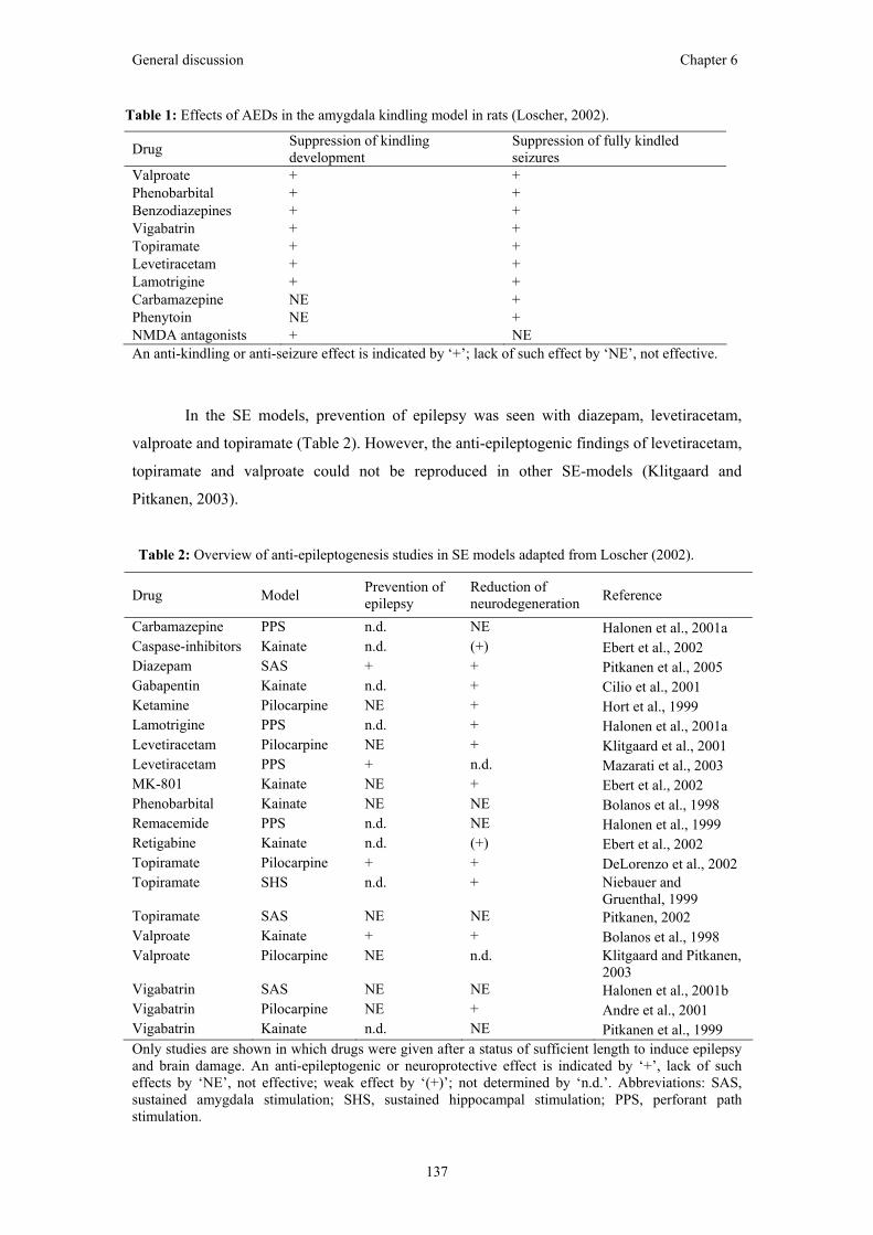
General discussion Chapter 6
137
An anti-kindling or anti-seizure effect is indicated by ‘+’; lack of such effect by ‘NE’, not effective.
In the SE models, prevention of epilepsy was seen with diazepam, levetiracetam,
valproate and topiramate (Table 2). However, the anti-epileptogenic findings of levetiracetam,
topiramate and valproate could not be reproduced in other SE-models (Klitgaard and
Pitkanen, 2003).
Drug Model Prevention of epilepsy
Reduction of neurodegeneration Reference
Carbamazepine PPS n.d. NE Halonen et al., 2001a Caspase-inhibitors Kainate n.d. (+) Ebert et al., 2002 Diazepam SAS + + Pitkanen et al., 2005 Gabapentin Kainate n.d. + Cilio et al., 2001 Ketamine Pilocarpine NE + Hort et al., 1999 Lamotrigine PPS n.d. + Halonen et al., 2001a Levetiracetam Pilocarpine NE + Klitgaard et al., 2001 Levetiracetam PPS + n.d. Mazarati et al., 2003 MK-801 Kainate NE + Ebert et al., 2002 Phenobarbital Kainate NE NE Bolanos et al., 1998 Remacemide PPS n.d. NE Halonen et al., 1999 Retigabine Kainate n.d. (+) Ebert et al., 2002 Topiramate Pilocarpine + + DeLorenzo et al., 2002 Topiramate SHS n.d. + Niebauer and
Gruenthal, 1999 Topiramate SAS NE NE Pitkanen, 2002 Valproate Kainate + + Bolanos et al., 1998 Valproate Pilocarpine NE n.d. Klitgaard and Pitkanen,
2003 Vigabatrin SAS NE NE Halonen et al., 2001b Vigabatrin Pilocarpine NE + Andre et al., 2001 Vigabatrin Kainate n.d. NE Pitkanen et al., 1999 Only studies are shown in which drugs were given after a status of sufficient length to induce epilepsy and brain damage. An anti-epileptogenic or neuroprotective effect is indicated by ‘+’, lack of such effects by ‘NE’, not effective; weak effect by ‘(+)’; not determined by ‘n.d.’. Abbreviations: SAS, sustained amygdala stimulation; SHS, sustained hippocampal stimulation; PPS, perforant path stimulation.
Drug Suppression of kindling development
Suppression of fully kindled seizures
Valproate + + Phenobarbital + + Benzodiazepines + + Vigabatrin + + Topiramate + + Levetiracetam + + Lamotrigine + + Carbamazepine NE + Phenytoin NE + NMDA antagonists + NE
Table 2: Overview of anti-epileptogenesis studies in SE models adapted from Loscher (2002).
Table 1: Effects of AEDs in the amygdala kindling model in rats (Loscher, 2002).

General discussion Chapter 6
138
Discrepancies between studies using the kindling model and studies using the SE
model indicate that mechanisms underlying development of epilepsy in different animal
models might not be the same (Loscher, 2002). In addition, there is a lack of data from other
models of epileptogenesis and it is difficult to extrapolate the data of kindling and SE models
to other epileptogenic etiologies such as stroke- or head trauma-induced epileptogenesis.
Animal models of genetic idiopathic epilepsy have only been used scarcely. Absence
seizures, a type of idiopathic epilepsy in humans, are usually a benign form of epilepsy (Wolf,
1984). Despite the good pharmacological response in this form of epilepsy, investigating the
anti-epileptogenic properties of AEDs in GAERS could provide new insights and strategies
for epilepsy treatment. In GAERS, spontaneous absence seizures develop progressively.
Therefore, it is an excellent model to study epileptogenesis and interference with
epileptogenesis by neuromodulation. To our knowledge, there is only one study that
chronically administered an AED i.e. remacemide in young GAERS (PN7-PN25) to establish
its effect on SWD development (Nehlig and Boehrer, 2003). This study failed to demonstrate
an effect of early remacemide treatment on the expression of SWDs in adult GAERS.
6.1.3 Animal research on levetiracetam
LEV has an anti-epileptic effect in a broad range of animal models mimicking both
partial (Loscher and Honack, 1993; Klitgaard et al., 1998; Loscher et al., 2000; Glien et al.,
2002) and generalized epilepsy in man (Loscher and Honack, 1993; Klitgaard et al., 1998)
including GAERS (Gower et al., 1995). In contrast, LEV revealed no effect in the two
conventional screening tests, namely the maximal electroshock and pentylenetetrazole models
(Klitgaard et al., 1998; Loscher and Honack, 1993).
The anti-seizure effect of LEV has mainly been established after a single injection of
LEV. However, in several studies LEV was administered during prolonged periods (Loscher
et al., 1998; Loscher and Honack, 2000; Glien et al., 2002; Stratton et al., 2003). We found
that chronic LEV treatment significantly suppressed (average difference of 62%) epileptiform
events in young GAERS (PN57-PN60) in comparison with control animals (Dedeurwaerdere
et al., 2005). LEV was administered daily by i.p. injection and its effect was investigated
during treatment (3 h a day for four consecutive days) and immediately after termination of
the treatment (3 h a day for four consecutive days) in young animals. Young GAERS have
few SWDs; therefore the more frequent SWD-like events were also taken into account. These
were defined as short irregular SWDs (SISWDs) with clear spike and wave aspects
characterized by lower amplitude (< 3 times baseline amplitude) or lower peak frequency (5-7
Hz) and shorter in duration (< 2 s) (Dedeurwaerdere et al., 2005). The LEV treated animals
displayed less SWD-like events (up to 81%) than the control group even in the pre-injection
hours. This could suggest that there was a slowing down of the epileptogenic processes by

General discussion Chapter 6
139
early chronic treatment (PN23-PN60) with LEV. Yet, the lower number in the pre-injection
hours might also be caused by retention of LEV in the brain from previous injections,
although LEV was already eliminated from the blood plasma.
Interestingly, there was no acute decrease in SISWDs after injection of LEV (PN57-
PN60) (Dedeurwaerdere et al., 2005). This is in contrast with previous acute experiments in
adult GAERS, in which a clear decrease in SWDs could be observed after a single LEV
injection (Gower et al., 1995; Boon et al., 2002; Bouwman and van Rijn, 2004). These
studies, which were performed in adult GAERS, report the effect of a first and single LEV
injection, while the effect of chronic LEV administration was not investigated. Moreover,
several studies dealing with the chronic administration of LEV in animal models of temporal
lobe epilepsy report increased functional tolerance towards LEV and some loss of efficacy
after prolonged administration (Loscher et al., 1998; Loscher and Honack, 2000; Glien et al.,
2002). This increased tolerance could provide a possible reason for the lack of an acute
decrease in SISWDs in GAERS that were already pretreated.
When treatment was stopped, there was no sudden increase in epileptiform events
(SWDs and SISWDs) (Dedeurwaerdere et al., 2005). This is in line with previously
performed rat and mouse experiments in which chronic treatment with LEV did not lead to
withdrawal hyperexcitability (Klitgaard et al., 1998; Loscher and Honack, 2000). Moreover,
there was a trend (p= 0.064) towards a lower number of epileptiform events (an average
difference of 53%) in the young LEV pretreated GAERS. In the study of Glien et al. (2002)
LEV was administered during two weeks in the pilocarpine model of temporal lobe epilepsy.
Some animals still had less spontaneous partial seizures one week after terminating LEV
treatment and this was described as a carryover effect (Glien et al., 2002).
Neuroprotective and anti-epileptogenic properties of LEV have been reported in
several animal models of epilepsy. LEV induces neuroprotection in a rat model of focal
cerebral ischemia (Hanon and Klitgaard, 2001). Recent observations showed that LEV
attenuates both hippocampal cell death and enhancement in hippocampal excitability
following a pilocarpine induced SE (Klitgaard et al., 2001). Another study demonstrated that
chronic levetiracetam treatment has a mild neuroprotective effect on hippocampal principal
cells and interneurons after pilocarpine-induced SE (Pitkanen and Kubova, 2004).
These neuroprotective properties may be relevant to its anti-epileptogenic action.
Klitgaard et al. (2001) induced pilocarpine-induced SE in rats and terminated it with
diazepam 30 min after the beginning of SE. Thereafter, levetiracetam treatment was initiated
and continued for the next 21 days. Continuous video-EEG monitoring (72 h) three days after
drug cessation revealed, however, no differences in the percentage of pretreated rats with
spontaneous seizures compared to a vehicle group. Mazarati et al. (2003) started
levetiracetam treatment 1, 3 and 6 h after PPS and continued it for 29 days via subcutaneous

General discussion Chapter 6
140
osmotic minipumps. Two weeks after treatment discontinuation, seizures were analyzed using
video-EEG monitoring. Seizure frequency was reduced and the number of seizure free days
was greater in the levetiracetam group compared to the vehicle group.
In the amygdala kindling model, LEV suppressed kindling development at doses
devoid of adverse effects with persistent reduction in afterdischarge duration after termination
of treatment (Loscher et al., 1998). These findings have recently been confirmed (Stratton et
al., 2003; Zhang et al., 2003). The ability of LEV to delay the development of kindling
suggests that it has the potential to interfere with the epileptic circuitry modifying seizure
recruitment and projection properties of the epileptic network (Loscher et al., 1998).
However, LEV has been administered before the kindling stimulus was given. There are some
controversies whether or not its anti-convulsant action might have prevented the development
of kindling by reducing the expression of the evoked seizures rather than affecting the
processes underlying the hyperexcitibility of the fully kindled state. We have put up a pilot
study in which LEV (54 mg/kg i.p.) and/or valproate (200 mg/kg i.p.) was administered daily
for 24 consecutive days before kindling (De Smedt et al., 2003). One week after the last
injection, kindling procedures were applied according to the “rapid kindling with recurrent
hippocampal seizures (RKRHS) model”. Preliminary results did not show differences in
kindling rate or seizures expression from the vehicle group.
The anti-epileptogenic potential of LEV has also been supported in the genetic SER
model characterized by spontaneous tonic convulsions and absence seizures (Sasa et al.,
2003). LEV was administered during weeks 5–9 after birth before the appearance of
spontaneous seizures and was terminated at the expected age for seizure expression, which
resulted in a decrease in the frequency of both tonic and absence seizures in pre-treated
animals. Also, the duration of both tonic convulsions and absence seizures was shorter than in
controls.
In a previous study, we found an indication for such an anti-epileptogenic effect in
the GAERS model (Boon et al., 2002). Recently, we found that epileptiform events tended
(p= 0.064) to be less frequent (up to 60%) in LEV pretreated (PN23-PN60) GAERS during
the four observation days after treatment cessation in comparison with the control group
(Dedeurwaerdere et al., 2005). The major metabolite of LEV is pharmacologically inactive
and other metabolites are produced at levels below those that are relevant for pharmacological
activities (Loscher et al., 1998). LEV does not interfere with enzyme systems (Patsalos, 2000;
Benedetti, 2000) and is washed out rapidly (half-live ~2-3 h) after stopping the treatment
(Loscher et al., 1998). The decrease in epileptiform events on the first two recording days
after treatment cessation might be due to retention of LEV or other metabolites in the brain;
however, this is unlikely for the last two recording days. Early chronic treatment with LEV in

General discussion Chapter 6
141
young GAERS might have caused an initial slowing down of the epileptogenic processes, but
had no effect on the later expression of SWDs (age of four months).
It is clear that LEV can have a lasting and sometimes even permanent
neuromodulatory effect on the epileptic central nervous system. Chronic treatment is probably
not just a summation of several single administrations of LEV. Additional phenomena appear
like prolonged effect after treatment for days (Dedeurwaerdere et al., 2005), a week (Glien et
al., 2002) and even permanent modifications of the epileptic network occur (Loscher et al.,
1998). Nevertheless, undesired effects like functional tolerance have also been described after
chronic treatment (Loscher and Honack, 2000; Zhang et al., 2003).
6.1.4 Mechanisms of action of levetiracetam
Until now, it is not described in what manner LEV exerts its effect on absence
seizures in GAERS. A possible effect of LEV that might contribute to its action against
SWDs in GAERS could be by progressively decreased firing of neurons of the substantia
nigra pars reticulata (Loscher et al., 1996). A reduction in the firing rate of these neurons has
been shown to result in anti-convulsant activity (Loscher and Ebert, 1996). In addition, direct
micro-injection of LEV into the SNR induces seizure protection in the pilocarpine model of
temporal lobe epilepsy (Klitgaard et al., 2003). The SNR is not part of the epileptic circuit
underlying absence seizures, but it may belong to a system participating in a remote control
over seizures (Danober et al., 1998). Indeed, high frequency stimulation of the SNR has been
shown to interrupt SWDs in GAERS (Feddersen et al., 2004) and intracerebral micro-
injections of GABA mimetics in the SNR suppress absence seizures (Danober et al., 1998). In
line with this, Klitgaard et al. (1998, 2003) stresses that a part of LEV’s ability to suppress
seizures may originate from its effect on neuronal firing in the SNR, but the anatomical
network and molecular mechanisms by which this is mediated remains to be demonstrated.
We hypothesize that LEV’s anti-epileptic action on SWDs in GAERS is mediated through an
effect on the SNR, but it is not known if this might interfere with epileptogenesis. In GAERS
pups, metabolic increases in the nigral inhibitory system such as SNR compared to non-
epileptic control rats were observed (Nehlig et al., 1998). Increase in these structures involved
in the control of seizure activity was suggested to reflect a mechanism of prevention of
epileptogenesis in GAERS.
6.1.5 Future perspectives
As the incidence of new epilepsy cases is between 26 and 70 per 100 000 (Hauser,
1998), it would be favorable to not only search for treatments that suppress seizures, but also
to seek for treatments that tackle the underlying pathophysiology of epilepsy itself. A new

General discussion Chapter 6
142
approach to drug screening, including the process of epileptogenesis, may identify new
classes of drugs (Kupferberg, 2001; Klitgaard and Pitkanen, 2003). LEV is a novel AED with
potential anti-epileptogenic properties, which was discovered using non-conventional drug
screening. Not only the properties of the drug itself, but also the time frame during which it is
applied is of great importance. More research is required to determine the optimal time
windows for preventing epileptogenesis in several animal models of epilepsy.
In the SE model, it has been shown by several investigators that the optimal time
window to pharmacologically intervene with the initial insult (SE) is within 2 h (Brandt et al.,
2003; Pitkanen et al., 2005). Pretreatment with LEV by i.v. injection via the tail vein reduced
or prevented (higher doses) the development of self-sustaining seizures (Mazarati et al.,
2004). Treatment during the maintenance phase of self-sustaining status epilepticus
diminished or aborted seizures (higher doses) (Mazarati et al., 2004). The authors, however,
did not mention whether these animals were further monitored to investigate the development
of seizures and to assess the neural damage, which would have been of interest. In some
studies this initial interference was extended by continuing treatment during the latent period
(Klitgaard et al., 2001; Mazarati et al., 2003; Pitkanen and Kubova, 2004). This approach has
been shown to induce mild neuroprotection and to interfere with epileptogenesis in the PSS
induced SE model, but not in the pilocarpine SE model. It has not been assessed whether
seizures are easier to treat when epilepsy was not prevented, but delayed or reduced in
severity.
In the amygdala kindling model, LEV had anti-epileptogenic properties when applied
before the kindling stimulus. In a pilot-study we performed, epileptogenesis was not altered
when chronic LEV treatment was applied before the kindling procedure was started (De
Smedt et al., 2003). Another interesting strategy would be to apply the AED immediately
after the evoked seizure to interfere with processes underlying epilepsy development during
kindling. In our laboratory, a new experimental design has been set up to further investigate
the anti-epileptogenic properties of LEV on kindling development. An alternative of the
RKRHS model, which does not lead to permanent hyperexcitibility (Lothman and
Williamson, 1994) is the RKRHS model with alternate day kindling. In this model, kindling is
performed every two days during eight days. During the subsequent week, epileptogenic
processes are indelibly imprinted into the brain resulting in a permanent hyperexcitibility
(Lothman and Williamson, 1994). The week of epileptogenesis can be regarded as a “latent
period”, enabling to interact with epileptogenesis without interfering with the kindling
seizures themselves (pers. com. T. De Smedt). If permanent hyperexcitibility is affected by
treatment during this week, this would provide evidence of an anti-epileptogenic effect of the
tested compound.

General discussion Chapter 6
143
Animal models of genetic idiopathic epilepsy have only just been used in anti-
epileptogenesis research. In GAERS, therapy was started (PN23) before seizures became
apparent to interfere directly or indirectly with early pathological changes. Although there
was a temporal effect of LEV on seizures immediately after treatment discontinuation, seizure
expression was not altered during adulthood. It is known that important pathophysiological
changes (T-type calcium conductance of reticular thalamic neurons, glucose metabolism,
astrocytic modifications and glutamate transporters) in GAERS emerge early in life, before
the appearance of spontaneous absence seizures (Dutuit et al., 2000; Dutuit et al., 2002;
Tsakiridou et al., 1995; Vergnes et al., 2003). Some of these neuropathological and
neurophysiological changes take place before we started the treatment in this study (Vergnes
et al., 2003). Therefore, it could be possible that earlier initiation of LEV treatment, e.g.
beginning at birth, could lead to a more sustained effect. The chronic delivery of LEV did not
interfere with normal development and body weight of the animals. Clinical findings suggest
that LEV is a well-tolerated and safe treatment option for pediatric patients with epilepsy
(Patsalos, 2000; Coppola et al., 2004). Importantly, Bittigau et al. (2003) has, however,
demonstrated that the same AEDs, including diazepam, clonazepam, phenytoin,
phenobarbital, valproate and vigabatrin, can induce apoptotic neurodegeneration in the
developing brain in clinically relevant doses. Pitkanen et al. (2004) commented that
extrapolation of data obtained in mature brain to immature brain should be done with caution,
hence, neuroprotection in the immature brain remains a challenge.
As we used a relatively high dose to inject and LEV is already effective in low doses
in GAERS (Gower et al., 1995), we expect that relevant therapeutic concentrations are
present in the blood plasma for 8 h. However, more continuous administration of the drug
should be considered. This could be done by the implantation of an osmotic mini-pump that
provides a continuous release and thereby more steady plasma exposure with LEV.
An interesting feature not investigated in the present GAERS study, is whether
prolonged LEV treatment early in life would have modified the response to AED treatment
during adulthood. In addition, also the effect of chronic LEV in adult animals could be of
interest to investigate whether LEV has disease modifying properties in adult GAERS.
More recently, Krupp et al. (2000) raised the question whether treatment with AEDs
during epileptogenesis induces development of drug-refractory seizures if the prevention of
epileptogenesis fails. Although LEV shows potent anti-epileptogenic and anti-convulsant
effects in amygdala-kindled rats, repeated treatment induces anti-convulsant tolerance and
unidirectional cross-tolerance to carbamazepine administered subsequently (Zhang et al.,
2003). The consequences of anti-epileptogenic treatment on the later response to AEDs in
animals in which the treatment fails present a challenge that needs to be addressed in anti-
epileptogenesis studies (Pitkanen and Kubova, 2004).

General discussion Chapter 6
144
We hypothesized that the effect of LEV on absence seizures in GAERS could be
manifested through an effect on the SNR. However, this hypothesis is based on indirect
evidence from related studies. Therefore, it would be interesting to set up an experiment
which determines the anatomical network and molecular mechanisms by which this effect of
LEV on SNR neurons is mediated in GAERS.
Finally, future perspectives must not be limited to LEV, several other AEDs on the
market should be considered. For instance valproate, lamotrigine, topiramate and zonisamide
have been shown to possess interesting features potentially being anti-epileptogenic (Bolanos
et al., 1998; Halonen et al., 2001a; DeLorenzo et al., 2002; Hashimoto et al., 2003). However,
at present, none of these compounds has been shown to completely protect against
epileptogenesis. Of course, these drugs have been selected for their ability to block seizures. It
is not known whether there is a pivotal correlation between anti-seizure capability and the
effect on epileptogenesis. Therefore new screening procedures, testing for effects on the
processes of epileptogenesis, could reveal new classes of drugs.
Clearly, serious efforts have already been made to understand epileptogenesis and
anti-epileptogenesis; however, we still have a long way to go to find the ultimate compounds
and treatment strategies.

General discussion Chapter 6
145
References 1. Andre,V., Ferrandon,A., Marescaux,C. and Nehlig,A. (2001). Vigabatrin protects against
hippocampal damage but is not antiepileptogenic in the lithium-pilocarpine model of temporal lobe epilepsy. Epilepsy Res 47(1-2), 99-117.
2. Benedetti,M.S. (2000). Enzyme induction and inhibition by new antiepileptic drugs: a review of human studies. Fundam Clin Pharmacol 14, 301-319.
3. Bittigau,P., Sifringer,M. and Ikonomidou,C. (2003). Antiepileptic Drugs and Apoptosis in the Developing Brain. Ann NY Acad Sci 993, 103-114.
4. Bolanos,A.R., Sarkisian,M., Yang,Y., Hori,A., Helmer,.S.L., Mikati,M., Tandon,P., Stafstrom,C.E. and Holmes,G.L. (1998). Comparison of valproate and phenobarbital treatment after status epilepticus in rats. Neurology 51(1), 41-48.
5. Boon,P., Seys,L., Vonck,K., Dedeurwaerdere,S., D'Havé,M., Grisar,T., Claeys,P. and De Reuck,J. (2002). The effect of levetiracetam in genetic absence epilepsy Strasbourg rats. Epilepsia 43(Suppl 8), 60.
6. Bouwman,B.M. and van Rijn,C.M. (2004). Effects of levetiracetam on spike and wave discharges in WAG/Rij rats. Seizure 13(8), 591-594.
7. Brandt,C., Glien,M., Potschka,H., Volk,H. and Loscher,W. (2003). Epileptogenesis and neuropathology after different types of status epilepticus induced by prolonged electrical stimulation of the basolateral amygdala in rats. Epilepsy Res 55, 83-103.
8. Cilio,M.R., Bolanos,A.R., Liu,Z., Schmid,R., Yang,Y., Stafstrom,C.E., Mikati,M.A. and Holmes,G.L. (2001). Anticonvulsant action and long-term effects of gabapentin in the immature brain. Neuropharmacology 40(1), 139-47.
9. Coppola,G., Mangano,S., Tortorella,G., Pelliccia,A., Fels,A., Romano,A., Nardello,R., Habetswallner,F., Licciardi,F., Operto,F.F. and Pascotto,A. (2004). Levetiracetam during 1-year follow-up in children, adolescents, and young adults with refractory epilepsy. Epilepsy Res 59, 35-42.
10. Danober,L., Deransart,C., Depaulis,A., Vergnes,M. and Marescaux,C. (1998). Pathophysiological mechanisms of genetic absence epilepsy in the rat. Prog Neurobiol 55, 27-57.
11. Dedeurwaerdere,S., Boon,P., De Smedt,T., Claeys,P., Raedt,R., Bosman,T., Van Hese,P., Van Maele,G. and Vonck,K. (2005). Chronic levetiracetam treatment early in life decreases epileptiform events in young GAERS, but does not prevent the expression of spike and wave discharges during adulthood. Seizure 14(6):403-411.
12. DeLorenzo,R.J., Morris,A.T., Blair,R.E., Wallace,M. and Razvi,T. (2002). Topiramate is both neuroprotective and antiepileptogenic in the pilocarpine model of status epilepticus. Epilepsia 43(Suppl 7), 15.
13. De Smedt,T., Raedt,R., Dedeurwaerdere,S., Vonck,K. and Boon,P. (2003). The effect of levetiracetam and valproate in rapid kindling. Epilepsia 44(Suppl 9), 337.
14. Doheny,H.C., Ratnaraj,N., Whittington,M.A., Jefferys,J.G. and Patsalos,P.N. (1999). Blood and cerebrospinal fluid pharmacokinetics of the novel anticonvulsant levetiracetam (ucb L059) in the rat. Epilepsy Res 34, 161-168.
15. Dutuit,M., Didier-Bazes,M., Vergnes,M., Mutin,M., Conjard,A., Akaoka,H. and Belin,M.F., Touret,M. (2000). Specific alteration in the expression of glial fibrillary acidic protein, glutamate dehydrogenase, and glutamine synthetase in rats with genetic absence epilepsy. Glia 32, 15-24.

General discussion Chapter 6
146
16. Dutuit,M., Touret,M., Szymocha,R., Nehlig,A., Belin,M.F. and Didier-Bazes,M. (2002). Decreased expression of glutamate transporters in genetic absence epilepsy rats before seizure occurrence. J Neurochem 80, 1029-1038.
17. Ebert,U., Brandt,C. and Loscher,W. (2002). Delayed sclerosis, neuroprotection, and limbic epileptogenesis after status epilepticus in the rat. Epilepsia 43(Suppl 5), 86-95.
18. Feddersen,B., Deransart,C., Vercueil,L., Noachtar,S. and Depaulis,A. (2004). High-frequency stimulation of the substantia nigra pars reticulata suppresses absence seizures in a genetic model of absence epilepsy in the rat. Epilepsia 45(Suppl 3), 118.
19. Glien,M., Brandt,C., Potschka,H. and Loscher,W. (2002). Effects of the novel antiepileptic drug levetiracetam on spontaneous recurrent seizures in the rat pilocarpine model of temporal lobe epilepsy. Epilepsia 43, 350-357.
20. Gower,A.J., Hirsch,E., Boehrer,A., Noyer,M. and Marescaux,C. (1995). Effects of levetiracetam, a novel antiepileptic drug, on convulsant activity in two genetic rat models of epilepsy. Epilepsy Res 22, 207-213.
21. Halonen,T., Nissinen,J. and Pitkanen,A. (1999). Neuroprotective effect of remacemide hydrochloride in a perforant pathway stimulation model of status epilepticus in the rat. Epilepsy Res 34(2-3), 251-269.
22. Halonen,T., Nissinen,J. and Pitkanen,A. (2001a). Effect of lamotrigine treatment on status epilepticus-induced neuronal damage and memory impairment in rat. Epilepsy Res 46(3), 205-223.
23. Halonen,T., Nissinen,J. and Pitkanen,A. (2001b). Chronic elevation of brain GABA levels beginning two days after status epilepticus does not prevent epileptogenesis in rats. Neuropharmacology 40(4), 536-550.
24. Hanon,E. and Klitgaard,H. (2001). Neuroprotective properties of the novel antiepileptic drug levetiracetam in the rat middle cerebral artery occlusion model of focal cerebral ischemia. Seizure 10, 287-293.
25. Hashimoto,Y., Araki,H., Futagami,K., Kawasaki,H., Gomita,Y. (2003). Effects of valproate, phenytoin, and zonisamide on clonic and tonic seizures induced by acute and repeated exposure of mice to flurothyl. Physiol Behav 78(3):465-469.
26. Hauser,W.A. (1998). Incidence and Prevalence. In: Epilepsy, a comprehensive textbook (Engel J, Pedley TA, eds), Philadelphia: Lippincot-Raven, 47-48.
27. Hort,J., Brozek,G., Mares,P., Langmeier,M. and Komarek,V. (1999). Cognitive functions after pilocarpine-induced status epilepticus: changes during silent period precede appearance of spontaneous recurrent seizures. Epilepsia 40(9), 1177-1183.
28. Klitgaard,H., Matagne,A., Gobert,J. and Wulfert,E. (1998). Evidence for a unique profile of levetiracetam in rodent models of seizures and epilepsy. Eur J Pharmacol 353, 191-206.
29. Klitgaard,H., Matagne,A., Vanneste-Goemaere,J. and Margineanu,D.G. (2001). Effect of chronic administration of levetiracetam on Pilocarpine-induced epileptogenesis in rat. Epilepsia 42(Suppl 7), 114.
30. Klitgaard,H., Matagne,A., Grimee,R., Vanneste-Goemaere,J. and Margineanu,D.G. (2003). Electrophysiological, neurochemical and regional effects of levetiracetam in the rat pilocarpine model of temporal lobe epilepsy. Seizure 12, 92-100.
31. Klitgaard,H. and Pitkanen,A. (2003). Antiepileptogenesis, neuroprotection, and disease modification in the treatment of epilepsy: focus on levetiracetam. Epileptic Disord 5(Suppl 1), 9-16.

General discussion Chapter 6
147
32. Krupp,E., Heynen,T., Li,X.L., Post,R.M. and Weiss,S.R.B. (2000). Tolerance to the Anticonvulsant Effects of Lamotrigine on Amygdala Kindled Seizures: Cross-Tolerance to Carbamazepine But Not Valproate or Diazepam. Exp Neurol 162, 278-289.
33. Kupferberg,H. (2001). Animal models used in the screening of antiepileptic drugs. Epilepsia 42, 7-12.
34. Lemos,T. and Cavalheiro,E.A. (1995). Suppression of pilocarpine-induced status epilepticus and the late development of epilepsy in rats. Exp Brain Res 102, 423-428.
35. Loscher,W. and Ebert,U. (1996). Basic mechanisms of seizure propagation: targets for rational drug design and rational polypharmacy. Epilepsy Res(Suppl 11), 17-43.
36. Loscher,W. and Honack,D. (1993). Profile of ucb L059, a novel anticonvulsant drug, in models of partial and generalized epilepsy in mice and rats. Eur J Pharmacol 232, 147-158.
37. Loscher,W., Honack,D. and Bloms-Funke,P. (1996). The novel antiepileptic drug levetiracetam (ucb L059) induces alterations in GABA metabolism and turnover in discrete areas of rat brain and reduces neuronal activity in substantia nigra pars reticulata. Brain Res 735, 208-216.
38. Loscher,W., Honack,D. and Rundfeldt,C. (1998). Antiepileptogenic effects of the novel anticonvulsant levetiracetam (ucb L059) in the kindling model of temporal lobe epilepsy. J Pharmacol Exp Ther 284, 474-479.
39. Loscher,W. and Honack,D. (2000). Development of tolerance during chronic treatment of kindled rats with the novel antiepileptic drug levetiracetam. Epilepsia 41, 1499-1506.
40. Loscher,W., Reissmuller,E. and Ebert,U. (2000). Anticonvulsant efficacy of gabapentin and levetiracetam in phenytoin- resistant kindled rats. Epilepsy Res 40, 63-77.
41. Loscher,W. (2002). Animal models of epilepsy for the development of antiepileptogenic and disease-modifying drugs. A comparison of the pharmacology of kindling and post-status epilepticus models of temporal lobe epilepsy. Epilepsy Res 50(1), 105-123
42. Lothman,E.W. and Williamson,J.M. (1994). Closely spaced recurrent hippocampal seizures elicit two types of heightened epileptogenesis: a rapidly developing, transient kindling and a slowly developing, enduring kindling. Brain Res 649, 71-84.
43. Mazarati,A.M., Baldwin,R.A., Klitgaard,H., Matagne,A. and Waisterlain,C.G. (2003). Treatment with levetiracetam during the latent period following experimental status epilepticus reduces chronic spontaneous recurrent seizures. Epilepsia 44(Suppl 9), 223.
44. Mazarati,A.M., Baldwin,R., Klitgaard,H., Matagne,A. and Wasterlain,C.G. (2004). Anticonvulsant effects of levetiracetam and levetiracetam-diazepam combinations in experimental status epilepticus. Epilepsy Res 58, 167-174.
45. Nehlig,A., Vergnes,M., Boyet,S. and Marescaux,C. (1998). Metabolic activity is increased in discrete brain regions before the occurrence of spike-and-wave discharges in weanling rats with genetic absence epilepsy. Brain Res Dev Brain Res 108, 69-75.
46. Nehlig,A. and Boehrer,A. (2003). Effects of remacemide in two models of genetically determined generalized epilepsy, the GAERS and the audiogenic Wistar AS. Epilepsy Res 52, 253-261.
47. Niebauer,M. and Gruenthal,M. (1999). Topiramate reduces neuronal injury after experimental status epilepticus. Brain Res 837(1-2), 263-269.
48. Patsalos,P.N. (2000). Pharmacokinetic profile of levetiracetam: toward ideal characteristics. Pharmacol Ther 85, 77-85.

General discussion Chapter 6
148
49. Pitkanen,A., Nissinen,J., Jolkkonen,E., Tuunanen,J. and Halonen,T. (1999). Effects of vigabatrin treatment on status epilepticus-induced neuronal damage and mossy fiber sprouting in the rat hippocampus. Epilepsy Res 33(1), 67-85.
50. Pitkanen,A. (2002). Drug-mediated neuroprotection and antiepileptogenesis: animal data. Neurology 59, 27-33.
51. Pitkanen,A. and Kubova,H. (2004). Antiepileptic drugs in neuroprotection. Expert Opin Pharmacother 5, 777-798.
52. Pitkanen,A., Kharatishvili,I., Narkilahti,S., Lukasiuk,K. and Nissinen,J. (2005). Administration of diazepam during status epilepticus reduces development and severity of epilepsy in rat. Epilepsy Res 63, 27-42.
53. Sasa,M., Yan,H., Nagayama,T. and Seriwaka,T. (2003). Anti-epileptogenic Properties of Levetiracetam in the Spontaneously Epileptic Rat (SER). Epilepsia 44(Suppl 8), 175-176.
54. Schachter,S.C. (2002). Drug-mediated antiepileptogenesis in humans. Neurology 59, 34-35.
55. Stratton,S.C., Large,C.H., Cox,B., Davies,G. and Hagan,R.M. (2003). Effects of lamotrigine and levetiracetam on seizure development in a rat amygdala kindling model . Epilepsy Res 53, 95-106.
56. Temkin,N.R. (2001). Antiepileptogenesis and seizure prevention trials with antiepileptic drugs: meta-analysis of controlled trials. Epilepsia. 42(4), 515-524.
57. Tsakiridou,E., Bertollini,L., de Curtis,M., Avanzini,G. and Pape,H.C. (1995). Selective increase in T-type calcium conductance of reticular thalamic neurons in a rat model of absence epilepsy. J Neurosci 15, 3110-3117.
58. Vergnes,M., Boehrer,A. and Nehlig,A. (2003). Developmental characteristics of picrotoxin-induced convulsions in rats with genetic absence epilepsy. Exp Neurol 184, 549-551.
59. Wolf,P. and Inoue,Y. (1984). Therapeutic response of absence seizures in patients of an epilepsy clinic for adolescents and adults. J Neurol 231, 225-229.
60. Zhang,Z.J., Xing,G.Q., Russell,S., Obeng,K. and Post,R.M. (2003). Unidirectional cross-tolerance from levetiracetam to carbamazepine in amygdala-kindled seizures. Epilepsia 44, 1487-1493.

General discussion Chapter 6
149
6.2. Vagus nerve stimulation
During the current research, several aspects of VNS have been approached using
animal models of epilepsy. In a chronic model of primary generalized epilepsy, namely
GAERS, we investigated the effect of both acute and chronic VNS. In a chronic model of
partial epilepsy (kindling in Fast rats), we investigated several therapeutic properties of VNS
like the acute abortive effect of VNS on generalized convulsions, chronic effects of VNS on
the development of epilepsy, cognitive effects and effects on body weight regulation. In
addition, we were able to detect changes in glucose metabolism after acute and chronic VNS
in healthy rats, providing a tool to further elucidate the mechanism of action (MOA) of VNS.
6.2.1 Efficacy of VNS in animal models of epilepsy
Jacob Zabara published two abstracts in Epilepsia in 1985 on the efficacy of VNS,
which unleashed further research on VNS as a new therapeutic approach to treat epilepsy. He
found a reduction in seizure duration and severity when stimulating the vagus nerve in dogs.
One year later, Joan Lockard (1986) showed that is was feasible to implant a VNS cuff-
electrode and stimulating device in a monkey model of epilepsy on a long-term basis.
At present, VNS is an efficacious broad-spectrum add-on treatment for patients with
medically or surgically refractory epilepsy (Ben-Menachem, 2002). Treatment involves
stimulation of the left vagus nerve via an implanted pulse generator in an attempt to reduce
the frequency and severity of epileptic seizures. Controlled randomized trials using this
treatment strategy have shown a 50% reduction in overall seizure frequency in approximately
30% of patients (Salinsky, 2003). In addition, VNS seems to have effects on interictal
epileptiform discharges (IED). Koo (2001) observed a decrease in interictal spikes, which was
later on confirmed (Kuba et al., 2002).
The benefits of VNS as a form of seizure control are numerous. Firstly, adverse
effects are minimal and not additive with those frequently encountered with AEDs such as
sedation and ataxia. Problems of drug toxicity, inherent to chronic medical therapy and
rapidly increasing with polytherapy are avoided with VNS. Secondly, because the device is
programmed to deliver continuous intermittent stimulation, there is never an issue with
respect to treatment compliance. Thirdly, unlike many AEDs, the effectiveness of VNS is
maintained during prolonged treatment without development of tolerance or becoming
refractory to treatment. Lastly, VNS does not seem to oppose the action of additional AEDs.
Initially, animal studies on VNS preceded the use of VNS in humans, but currently,
animal experiments are still ongoing trying to optimize VNS therapy and to elucidate the
MOA. A chronological overview of these studies is presented in table 1. Mostly, acute VNS

General discussion Chapter 6
150
studies were performed utilizing application protocols immediately before, during and/or after
seizure provocation.
Table 5: Chronological overview of the efficacy of VNS in animal models of epilepsy.
Date Researcher Model Animal Result 1952 Zanchetti et al. Strychnine Cats Blocking of IED and desynchronization of
the cortical EEG.
1967, 1968
Stoica and Tudor Strychnine Cats Decrease of IED and prevention of spreading to the contralateral side by VNS. Increase of IED by high-intensity VNS.
1990, 1991
Woodbury and Woodbury
3-MPA/ PTZ/ MES
Rats Interruption of seizures, abortive effect.
1990 Lockard et al. Alumina gel Rhesus monkeys
Reduction of the number of seizures, prophylactic effect by chronic high-intensity VNS.
1992 Zabara Strychnine/ PTZ
Dogs Inhibition of generalized seizures by high-intensity VNS.
1993 McLachlan Penicillin/ PTZ
Rats Decrease of IED and seizure duration, abortive effect.
1996 Takaya et al. MES Rats Dose response and prophylactic effect.
1997 Naritoku and Mikels
Kindling Rats Delay of kindling development.
1999 Fernandez-Guardiola et al.
Kindling Cats Delay of kindling development.
2001 Sunderam et al. 3-MPA Rats Moderate anti-seizure effect.
2002 Munana et al. Refractory epilepsy
Dog Cumulative efficacy of chronic VNS after long-term treatment.
2004 Dedeurwaerdere et al.
GAERS Rats VNS can prolong SWDs. Sub-acute VNS had no effect.
2005a Dedeurwaerdere et al.
GAERS Rats High-intensity (perceptible) VNS aborts SWDs. One week of VNS does not suppress SWDs.
Submitted(a)
Dedeurwaerdere et al.
Kindling in Fast rats
Rats Prevention of seizures and reduced ADD when VNS is applied after the kindling stimulus. No delay of kindling development and prolongation of convulsions when VNS is applied for 2 h before the kindling stimulus.
Abstracts are only included if they are not published later on in full article format. Abbreviations: IED, interictal epileptiform discharges; VNS, vagus nerve stimulation; 3-MPA, 3-mercaptoproprionate; PTZ, pentylenetetrazol; MES, maximal electroshock seizure; SWDs, spike and wave discharges; ADD, afterdischarge duration.
Zanchetti et al. (1952) were among the first to experiment with the anti-convulsive
properties of VNS. They showed that interictal spikes, produced by topical application of
strychnine to the cerebral cortex of the cat, were blocked by low-intensity VNS (output

General discussion Chapter 6
151
current: 1-2 V, pulse duration: 0.5 ms and frequency 50 Hz). Similarly, Stoica and Tudor
(1967, 1968) found that low-intensity VNS (output current: 1-4 V, pulse duration: 0.3 ms and
frequency: 30 Hz) decreased the frequency of spiking activity by about 34% induced by
strychnine applied topically to the coronal, anterior sigmoid, suprasylvian or medial marginal
gyri of the cat. Spread to the contralateral side was also inhibited by VNS. Stoica and Tudor
(1968) stated that the vagal afferent projections to these areas are diffuse and non-specific and
that a different number of fibers project to each area. On the contrary, they also noted an
increase in interictal spike frequency when high-intensity stimulation of the vagus nerve was
performed (Stoica and Tudor, 1968). Two decades later, Woodbury and Woodbury (1990,
1991) reported that acute high-intensity stimulation at frequencies greater than 4 Hz
prevented or reduced chemically (3-mercaptoproprionate, 3-MPA and pentylenetetrazole,
PTZ) induced clonic convulsions in anesthetized rats and electrically (maximal electroshock
seizure, MES) induced clonic and tonic clonic seizures in awake rats. This effect was
attributed directly to the fraction of C fibers stimulated. It was observed that VNS shortened,
but did not shut down a chemically induced seizure once it had begun. Optimal stimulation
parameters were described as: output current 0.2-0.5 mA/mm² of nerve cross-section, pulse
duration 0.5-1 ms and frequency 10-20 Hz. In 1992, Zabara published his work in manuscript
form on the anti-convulsant effect of VNS in epileptic dogs. He found that high-intensity
VNS could interrupt or terminate strychnine-induced seizures and PTZ-induced tremors in
anesthetized dogs. Inhibitory effects of VNS persisted for a considerable time after
termination of stimulation. It was suggested that small unmyelinated nerve fibers must be
stimulated and that the optimum parameters were: output current 10-20 V (~3-20 mA), pulse
duration 0.2 ms and frequency 20-30 Hz. McLachlan (1993) found that low-intensity VNS
not only suppressed PTZ-induced seizures (within 3 s after seizure onset), but also reduced
(by 33%) focal interictal spikes produced by penicillin in anesthetized rats. Interictal spikes
could already been suppressed at 0.2 mA; however, higher currents > 0.8 mA (pulse duration:
0.5 ms and frequencies 20-50 Hz) produced a more consistent response and persisted up to 20
s after stimulation. Vagal stimulation caused no change in seizure duration in high-dose PTZ
induced seizures, which was explained by a more intense ictal involvement of the brainstem
reticular system. Takaya et al. (1996) found that VNS pretreatment (output current: 1 mA,
pulse duration: 0.5 ms and frequency: 30 Hz continuously for 0.1 or 60 min or intermittently -
30 s/5 min on/off - for 60 min) induced a sustained anti-convulsive effect on PTZ-induced
seizures in awake animals, which efficacy was dependent on the cumulative stimulus
duration. Sunderam et al., (2001) reported the effect of left VNS (output current: 1.4 mA,
pulse duration: 0.5 ms, frequency: 20 Hz and duty cycle: 30 s/3 min on/off) and left sciatic
nerve stimulation on 3-MPA-induced seizures using a highly complicated study design. VNS
only had a small anti-seizure effect, which the authors ascribed to a hemodynamically-

General discussion Chapter 6
152
induced deficit in energy substrates. In a chronic model of spontaneous absence epilepsy
namely GAERS, we showed a transient increase in seizure duration following acute VNS
(output current: 3 V, pulse duration: 0.5 ms and frequency: 30 Hz), which disappeared in a
sub-acute setting (Dedeurwaerdere et al., 2004). However, when VNS was applied at higher
intensities (5-10 V) perceptible for the animals, the typical spike and wave discharges
(SWDs) were shut down immediately (Dedeurwaerdere et al., 2005a). In amygdala kindled
Fast rats (genetic strain of seizure prone rats), acute VNS (output current: 0.5 mA, pulse
duration: 0.5 ms, frequency: 30 Hz for 60 s) showed clear anti-convulsant effects in a subset
of animals when applied immediately after the kindling stimulus. However, when VNS (duty
cycle: 30 s/1.1 min on/off) was applied for two hours before the kindling pulse, generalized
convulsions were prolonged.
Only a few studies have assessed the effect of chronic VNS in animal models of
epilepsy (Lockard et al., 1990; Munana et al., 2002; Dedeurwaerdere et al., 2005a). A
possible reason for this is that long-term research on VNS in laboratory animals like rats has
some practical implications. Firstly, chronic studies are laborious and time consuming,
because they require long-term monitoring of the animal and subsequent analysis of all data.
Secondly, there are also technical aspects, which make chronic VNS stimulation difficult to
apply in small animals. To deliver chronic VNS, a pulse generator has to be implanted or
VNS has to be delivered via flexible leads connected to an external stimulator. Because the
Cyberonics stimulating device is rather big to implant in a rat, we chose for the latter option.
We have constructed a long-term video EEG monitoring unit equipped with spring covered
flexible leads (to avoid gnawing at the leads by the rat), a spring (allows up and down
movements of the rat) and a communicator or swivel (allows the rat to walk around freely in
the cage). The lead ends of the cuff-electrode have to be easily accessible, to connect the rat
to the VNS pulse generator without difficulties. In addition, these lead ends have to be safely
secured in a way that the animals cannot pull or move the leads. Therefore, we have fixated
the lead ends together with the EEG electrodes with dental acrylic on the head of the rat.
Finally, VNS necessitates implantation of a cuff-electrode, which is well tolerated by the
animals, does not induce nerve damage and stays functional over prolonged periods. In our
laboratory, such a stimulation electrode is available now (Dedeurwaerdere et al., 2005b).
A study in rhesus monkeys showed that chronic VNS was feasible and that epileptic
processes are influenced (Lockard et al., 1990). However, at that time, the authors stated that
safety and efficacy of the procedure were still in question. Munana et al. (2002) evaluated the
use of VNS (output current: 0.25-1 mA, pulse duration: 0.5 ms, frequency: 30 Hz and on/off
cycle: 30 s/ 5 min) as a treatment for refractory epilepsy in dogs. No significant difference in
seizure frequency, duration or severity was detected between overall 13-week treatment and
control periods. However, during the final four weeks of the treatment period, a significant

General discussion Chapter 6
153
decrease in mean seizure frequency (34.4%) was found, compared with the control group.
This may reflect an increase in the efficacy of VNS over time. When chronic VNS (output
current: 1.5 mA, pulse duration: 0.5 ms, frequency: 30 Hz and duty cycle: 60 s/12 s on/off)
was applied during one week in GAERS, the decrease in SWDs did not significantly differ
from the control group (Dedeurwaerdere et al., 2005a). It can be hypothesized that a longer
period of VNS or earlier intervention during life might be required to affect an already
established absence epilepsy syndrome.
The effect of VNS on epileptogenesis has been investigated in the amygdala kindling
model of temporal lobe epilepsy. During the kindling process seizure severity and duration
gradually progress. It offers the advantage that seizures can be elicited at will and that it
allows detailed study of the events associated with the epileptogenic process. In cats, it was
found that VNS trains (output current: 1.2-2 mA, pulse duration: 0.5 ms and frequency: 30 Hz
for 1 min) before and four times after the kindling stimulus interfered with epileptogenesis
(Fernandez-Guardiola et al., 1999). These findings were confirmed in a previous abstract of
Naritoku and Mikels (1997) in rats, who showed that VNS before each kindling stimulus
delays the completion of kindling. However, we could not reproduce these observations using
a genetic seizure prone rat strain. Kindling rate was not delayed by VNS (output current: 0.5
mA, pulse duration: 0.5 ms, frequency: 30 Hz and duty cycle: 30 s/1.1 min on/off) and during
the stage-5 convulsions evidence of hyperexcitability was even noted in the VNS group
(Dedeurwaerdere et al, submitted). It is conceivable that VNS cannot interfere with
epileptogenesis in individuals with a strong genetic predisposition to develop epilepsy, like
the Fast rats in our study. Moreover, kindling might involve different mechanisms in Fast rats
compared to healthy animals. It is noted that in this study we saw that some VNS-treated rats
(n= 3) showed delayed kindling rates compared to controls (n= 3) when the piriform cortex,
rather than the amygdala, served as the kindling focus (S. Dedeurwaerdere, personal
observations, 2004). Although, kindling rate in the piriform cortex is faster than in the
amygdala (McIntyre et al., 1999), VNS rats did not display stage-5 seizures unless VNS was
terminated (by lead fracture). Therefore, the origin of epileptogenesis and seizure generation
could also be an influencing factor whether VNS can act on kindling development in Fast
rats.
It is impossible to extract an ideal set of stimulation parameters from these studies.
Most studies use frequencies between 20 and 30 Hz and a pulse duration of 0.5 ms. However,
the probably most crucial parameter namely the output current is difficult to evaluate. The
actual current delivered to the nerve is depending on the construction of the electrode.
Different electrode types have been used and in addition, studies have been performed in
anesthetized animals as well as in awake animals. In human, output current is increased till
tolerability level.

General discussion Chapter 6
154
Zabara (1992) observed that right or left vagal stimulation is equally effective in
controlling motor seizures, but bilateral vagal stimulation produced no measurable greater
effect than did unilateral stimulation. Krahl et al. (2003) confirmed that right-sided VNS was
just as effective as left-sided VNS in reducing the severity of PTZ-induced seizures. They,
however, speculated that stimulation of both vagus nerves may affect a larger portion of the
brain than unilateral VNS. This was the case for trigeminal nerve stimulation (Fanselow et al.,
2000), though was not observed by Zabara in earlier VNS studies using anesthetized dogs.
Stimulation of the right or left nerve of Hering (ninth cranial nerve) can also successfully
control focal seizure activity, whereas stimulation of the twelfth cranial nerve fails to suppress
seizure activity (Patwardhan et al., 2002).
6.2.2 Emerging indications for VNS treatment
Vagal stimulation may affect numerous brain structures involved in the regulation of
mood and cognition (Schachter, 2004). Several studies report improved alertness, behavior
and mood following VNS independent of changes in seizure frequency (Elger et al., 2000;
Harden et al., 2000; Harden, 2001; Kossoff and Pyzik, 2004). Accordingly, VNS has been
explored as a treatment for depression (Rush et al., 2000; Sackeim et al., 2001a; Sackeim et
al., 2001b; Sjogren et al., 2002). Also in animal studies, VNS has shown to be an effective
antidepressant in the forced-swim test in rats (Krahl et al., 2004).
An open-label pilot study suggested a positive effect on cognition of VNS treatment
for patients with Alzheimer’s disease (Sjogren et al., 2002). Clark et al. (1995) hypothesized
that vagal afferents affect central operations involved in the modulation of memory
consolidation processes. In rats and humans, they reported that VNS was able to enhance
memory storage when applied during memory consolidation, which was dependent on an
inverted-U shaped function of output current (Clark et al., 1995; Clark et al., 1998; Clark et
al., 1999). In contrast, chronic VNS application in a clinical setting has not been found to
affect cognitive performance in patients with epilepsy since standardized tests have not
identified systematic positive or negative changes in attention, motor function, short-term
memory, learning and memory or executive functions following chronic VNS (Hoppe et al.,
2001; Dodrill and Morris, 2001). Therapeutic agents such as AEDs, on the contrary, often
exhibit mild to serious effects on cognition (Ortinski and Meador, 2004). In Fast rats, a
genetic strain with learning impairment, we found that VNS stimulation was devoid of
cognitive side effects in the Morris water maze, which is used to investigate spatial memory
in rats. In the Alzheimer study and the studies of Clark et al. (1995, 1998, 1999) intermediate
stimulation parameters were used, whereas our study and those performed in patients with
epilepsy (Hoppe et al., 2001; Dodrill and Morris, 2001) used high stimulation parameters (up

General discussion Chapter 6
155
to tolerability level), which are believed to have anti-epileptic effects. Therefore, different
stimulation parameters may result in a different therapeutic effect.
Obesity is the most significant health problem facing westernized societies being the
primary risk factor for diabetes and obstructive sleep apnea (Roslin and Kurlan, 2001). In
addition, it increases the risks for heart disease, pulmonary disease, infertility, osteoarthritis,
cholecystitis and several major cancers, including breast and colon cancers (Roslin and
Kurlan, 2001). It is estimated that 50 to 60% of the population is obese or overweight of
whom 5 to 6% are considered morbidly obese (Roslin and Kurlan, 2001). Surgical procedures
for morbid obesity are becoming more common because of long-term successful results
(Mason, 1992). However, optimal treatment for morbid obesity is not yet at hand (Roslin and
Kurlan, 2001).
Weight reduction has been reported in patients treated with VNS (Burneo et al., 2002;
Kneedy-Cayem et al., 2002). Vagal afferents play a predominant role in the regulation of food
intake (Schwartz, 2000). They conduct signals from the stomach to the nucleus of the solitary
tract (NST) carrying information about the size and chemical composition of a meal, which is
transmitted by other specific connections to the satiety center in the hypothalamus and the
collateral ventro-medial nucleus (Laskiewicz et al., 2004). A pilot-study in humans has been
set up with subdiaphragmatic stimulation of the vagus nerve. This study was based on the
finding in dogs that chronic bilateral VNS induced a decrease in weight, which was suggested
to result from changes in the central nervous system and a secondary alteration of food intake
by VNS.
In a study in GAERS, we found a significant weight reduction after two weeks of
chronic VNS (Dedeurwaerdere et al., 2003). In addition, we found that VNS treatment
prevented the gradual increase in body weight observed in control rats throughout the
kindling process. A basic association between amygdala kindled seizures and weight gain in
rats has been documented (Loscher et al., 2003; Bhatt et al., 2004). Interestingly, the
amygdala has been suggested as a region where interactions between gustatory and vagal
input take place (Han et al., 2003). This reduction in kindling-induced weight gain in VNS
rats was associated with reduced food intake compared to control kindled rats. As in the
kindling study the Fast rats only received two hours of VNS a day, it is possible that more
continuous VNS would result not only in reduced weight gain, but also in reduced body
weight as in GAERS. We are the first to report altered body weight in animal studies
evaluating the efficacy of VNS on epilepsy likely because previous studies have primarily
applied VNS acutely over very short time spans. In addition, our studies showed that in
animals, weight can be regulated by chronic unilateral left VNS applied through the vagus
nerve in the neck.

General discussion Chapter 6
156
6.2.3 Mechanism of action of VNS
Desynchronization of the background EEG by stimulation of vagal C fibers
The idea of using VNS to suppress epileptic seizures arose from early animal
experiments in which background EEG was desynchronized by VNS. As seizures result from
hypersynchronous firing of a group of neurons, it was hypothesized that stimuli that produced
desynchronization of the EEG may have anti-epileptic properties.
Experiments on “encéphale isolé” cats showed that stimulation of the vagus reduced
the amplitude of the EEG background, produced EEG desynchronization and blocked sleep
spindles during slow wave sleep (Zanchetti et al., 1952). However, these effects were not
obtained in animals without spindling and the effect was similar to that of other alerting
stimuli (nose rubbing, whistling). Forty years later, unilateral and bilateral VNS in cats
induced, within less than 5 s, changes in the pattern and periodicity of EEG spindles,
associated with depressed background rhythms or rhythmic EEG activities (Balzamo et al.,
1990).
In apparent contrast, several studies observed VNS induced synchronization of the
EEG. As early as 1938, Bailey and Bremer reported that VNS in the cat elicited synchronized
activity on the orbital-frontal cortex EEG. Rojas (1964) showed that direct current stimulation
(15-60 s) of the vagus nerve first produced EEG desynchronization with subsequent
synchronization. Also Puizillout and Foutz (1974) reported that VNS produced
synchronization of the EEG. McLachlan (1993) observed that background rhythms were not
altered by VNS in the majority of the animals, however, in a few animals a slight increase in
synchrony was noted after low-intensity and low-frequency (2-3 Hz) stimulation (McLachlan,
1993). Stimulation of the solitary tract, which receives projections of the vagus nerve, at low
frequencies (1-16 Hz) produced EEG synchronization, whereas high-frequency (> 30 Hz)
stimulation results in EEG desynchronization from a sleep spindle like background also
comparable to that observed with tactile or auditory stimuli (Magnes et al., 1961). Neither
synchronization nor desynchronization could be elicited when the EEG background was that
of arousal. Chase and colleagues (1966) performed more detailed experiments and showed a
complex relation between VNS and EEG rhythmicity associated with the activation of
specific nerve fiber types of the vagus. In encéphale isolé cat preparations, stimulation of the
vagus at frequencies above 20 Hz and at intensities greater than 3 V produced EEG
desynchronization or rather a blockage from spindle like activity. At frequencies above 70 Hz
and at intensities less than 3 V, assuming that these parameters only activate myelinated
fibers, vagal stimulation produced EEG synchronization. However, the changes elicited were
often inconsistent and varied with the background activity. In a study in humans, both

General discussion Chapter 6
157
synchronization and desynchronization of the EEG was present during long-term VNS
utilizing standard stimulation parameters, which was most prominent in patients with much
epileptiform activity (Koo, 2001).
The vagus nerve mainly consists of unmyelinated C fibers (65-80%) and a smaller
portion myelinated A and B fibers (Rutecki, 1990). It was assumed that high-intensity and
high-frequency (20-70 Hz) stimuli induce desynchronization resulting from the activation of
unmyelinated C fibers, whereas, low-intensity high-frequency stimulation induces
synchronization by activating myelinated A and B fibers only. However, this supposition is
most likely oversimplified, because several studies contradict and the distinction between
low- versus high-intensity and low- versus high-frequency stimulation is often vague.
In the study of Takaya et al. (1996), using awake and freely moving animals, no
obvious VNS-induced changes in background EEG activity were observed. In addition, early
studies in humans revealed that VNS induces little if any effect on EEG background rhythms.
(Hammond et al., 1992a; Salinsky and Burchiel, 1993). Hence, it was hypothesized that acute
desynchronization of background EEG activity is not a prominent feature of VNS when
administered during physiologic wakefulness and sleep, nor does it explain the anti-
convulsant effect of VNS. In these studies C fibers were probably not affected and this could
be a reason why desynchronization of the EEG was not observed. Indeed, effective
therapeutic stimulation parameters appear to be subthreshold for vagal C fibers in humans as
there are no clinical reports of autonomic side effects (significant gastrointestinal, cardiac or
respiratory effects), which would arise from C fiber stimulation (Krahl et al., 2001). These
findings were supported by studies in humans who performed intraoperative nerve potential
recordings (Koo et al., 2001; Evans et al., 2004). Their results showed that predominantly A
and B fibers and not C fibers are activated by the stimulation output currents used in humans.
Therefore, C fiber activation cannot fully explain the MOA of VNS. Moreover, Krahl et al.
(2001) showed that selective destruction of capsaicin-sensitive C fibers did not affect the anti-
convulsive effects of VNS on PTZ-induced seizures in rats.
General discussion Chapter 6
158
Anatomical and functional projections of the vagus nerve into the cerebral hemispheres
The vagus nerve is a mixed cranial nerve that consists of ~80% afferent fibers and
20% efferent fibers parasympathically innervating heart, lungs and gastrointestinal tract
(Figure 1). Mechanistic VNS research generally presumes that VNS exerts its effect through
the induction of action potentials in the afferent fibers of the left vagus nerve (Zagon et al.,
1999; Henry, 2002). Indeed, the anatomical diffuse projections of the vagus nerve in the
cerebral hemispheres would support a broad effect for VNS on neural excitability (Rutecki,
1990) (Figure 1).
Moreover, vagal stimulation produces evoked potentials (EPs) which have been
recorded from several brain regions. In the parietal association cortex, VNS evoked slow
hyperpolarization in the pyramidal neurons (Zagon and Kemeny, 2000). This
hyperpolarization of the membrane could reduce the excitability of these neurons. In these
anesthetized rats, stimulus intensities that predominantly activated myelinated fibers (less than
200 µA) were more effective in inducing long-lasting inhibitory effects than higher stimulus
intensities that activated unmyelinated vagal afferents. Furthermore, stimulation of the cervical
vagus produced EPs in the cerebral cortex of several animal models (O’Brien et al., 1971; Car
et al., 1975) and human (Tougas et al. 1993). In the cat thalamus, low-intensity vagal nerve
stimuli facilitated cellular firing in the reticular nuclei and depressed cellular firing in the
ventro-postero-medial nucleus of the thalamus (Car et al., 1975). Higher intensity vagal
stimulation increased firing frequency and duration of discharges in both nuclei. EPs have also
been measured in the brainstem (Bennett et al., 1988), cerebellum (Dell and Olsen, 1951;
Hennemann and Rubia, 1978) and the hippocampus (Serkov and Bratus, 1970) of animals.
Functional imaging of the effect of VNS
Naritoku et al. (1995) demonstrated expression of c-fos (reflecting high neuronal
activity) immunoreactivity in several brain regions of anesthetized rats after VNS. These
brain regios included limbic structures, thalamic nuclei and brainstem noradrenergic nuclei
important in the control of seizures (Naritoku et al., 1995) and supports the idea that the anti-
epileptic action of VNS involves pathways that project from brainstem to forebrain.
Non-invasive mapping of anatomical sites of increased or decreased synaptic activity
can be performed with functional imaging. The influence of VNS on brain activity has been
demonstrated by imaging studies using positron emission tomography (PET), single photon
emission computed tomography (SPECT) or functional magnetic resonance imaging (fMRI)
techniques (Henry et al., 1998; Vonck et al., 2000; Narayanan et al., 2002; Sucholeiki et al.,
2002).

General discussion Chapter 6
159
Figure 14: Efferent and afferent projections of the vagus nerve. A. The vagus nerve had efferent projections towards the pharynx, lungs, heart and gastrointestinal tract. B.Anatomical diffuse projections of the vagus nerve to cerebral hemisphere structures adaptedfrom Rutecki et al. (1990).

General discussion Chapter 6
160
These imaging studies found changes on both sides of the brain by unilateral left VNS
and pointed out a key role for the thalamus and medial temporal lobe structures in the MOA
of VNS. However, there is no consensus on other activated structures neither on the type of
changes (inhibition or excitation). This discrepancy can be attributed to a number of
confounding factors such as imaging techniques used (PET, SPECT, fMRI), tracer and
contrast agents, scanning protocols, stimulation parameters, medication regimes, course of the
illness and treatment response. Heterogeneity of relatively small patient samples is difficult to
avoid. In addition, data gathering from healthy subjects is impossible for ethical reasons due
to the invasiveness of VNS.
We found that changes in glucose metabolism due to VNS in healthy rats can be
monitored using small animal PET, avoiding confounding factors as described above
(Dedeurwaerdere et al., 2005c). In this animal study, results have been observed consistent
with several previous imaging studies in epileptic patients (Vonck et al., 2000; Van Laere et
al., 2002; Henry et al., 1998; Henry et al., 2004). During acute VNS, we found bilateral
decreases in the glucose metabolism of the hippocampi (tendency in the right hippocampus),
which are known to be often involved in the generation of complex partial seizures in
temporal lobe epilepsy. A decreased metabolism in this region may reflect the anti-convulsant
activity of acute VNS. More specifically, in patients, acute VNS can be triggered at seizure
onset by a magnet to interfere with the seizure. In line with the long-term study of Henry et al.
(2004), these changes in hippocampi were absent after chronic VNS. It is believed that these
differences between acute and chronic VNS reflect the brains adaptation to chronic VNS. At
this moment we cannot explain these changes after chronic VNS treatment observed in
imaging studies. Further study will be required to determine why the acute effects of VNS on
brain blood flow differ from VNS effects after months or years of stimulation. Henry (2002)
suggested that intrasubject differences between acute and chronic VNS activation scans may
reveal processes of adaptation to chronic VNS, and that some of them may have an anti-
epileptic effect. These adaptation processes might underlie the finding that therapeutic VNS
often causes gradual improved seizure control over several months of treatment (Vonck,
2003). In this case, ‘plasticity’ instead of ‘adaptation’ could be a more suitable term as
'adaptation' would in fact be expected to suppress the effect of VNS and therefore reduce the
anti-convulsive effect with time
We also observed that acute VNS bilaterally increased glucose uptake in the olfactory
bulbs. Evoked potential and unit activity recording techniques have revealed the existence of
a vagus nerve-olfactory bulb pathway (Garcia-Diaz et al., 1984). Also in patients, VNS has
been found to influence and modulate the processing of olfactory information (Kirchner et al.,
2004). This neurophysiological study reported even a positive correlation between activation
of the olfactory bulb and therapeutic benefit. As the olfactory bulb is on the contrary very

General discussion Chapter 6
161
small in humans, it is conceivable that changes in metabolism of this structure were not
measurable in human imaging studies.
Chronic VNS induced an increase in metabolism in the left medulla oblongata
(Dedeurwaerdere et al., 2005c). Changes in dorsal-rostral medulla during chronic VNS have
also been reported in an initial study of Henry et al. (2000). These findings correspond to the
known anatomical connections and projections of the vagus nerve to the brainstem tracti and
nuclei.
In addition, we found a significantly decreased left/right ratio in striatum and a
tendency towards a decrease in glucose uptake in the left striatum after chronic VNS
(Dedeurwaerdere et al., 2005c). This brain structure has been acknowledged to play an
important role in the control of seizures (Deransart and Depaulis, 2002). Ictal hyperperfusion
of the striatum was shown using single photon emission computed tomography (SPECT) in
patients with temporal lobe epilepsy (Semah, 2002). The striatum receives indirect
innervation from the vagus nerve via the medial reticular formation of the medulla which
projects to the intralaminar nuclei of thalamus (having extremely widespread projections to
cerebral cortex and the striatum) (Henry, 2002). Moreover, in a PET imaging study in
humans, changes in glucose metabolism have been found in the putamen (Ko et al., 1996) and
caudate nucleus (Vonck, 2003) due to VNS.
Finally, we were not able to detect previously reported changes in thalamus (Henry et
al., 1998; Vonck et al., 2000; Chae et al., 2003; Henry et al., 2004) and hypothalamus (Henry
et al., 1998; Henry et al., 2004) (for review see Chae et al., 2003). The negative findings in
these regions therefore do not exclude the presence of smaller or more localized changes.
Further study will require higher-resolution acquisition and more advanced analysis. Imaging
studies in patients of our group showed a decreased tracer uptake in the left thalamus during
chronic VNS (Vonck et al., 2000; Van Laere et al., 2002; Vonck, 2003). This may
demonstrate that VNS induces a chronic inhibitory state in this key structure for seizure
spread, which is manifested by a decrease in blood flow. In line with this, it has been quoted
that VNS could result in hemodynamically-induced hypoperfusion creating a relative deficit
of energetic substrates (Sunderam et al., 2001).
Changes in neurotransmission induced by VNS
Naritoku et al. (1992) found that chronic VNS prolongs the cervicomedullary to
thalamocortical potential interval during somatosensory evoked potential studies. It was
concluded that chronic VNS does alter neuronal networks outside of the brain stem vagus
system and that alteration of forebrain neurotransmission might be a primary mechanism of
VNS.

General discussion Chapter 6
162
Woodbury and Woodbury hypothesized that VNS appears to act via release of large
quantities of the inhibitory mediators GABA and glycine throughout large volumes of the
brain, because it counteracts PTZ, 3-MPA (decreases efficacy of all inhibitory synapses) and
strychnine induced seizures (Woodbury and Woodbury, 1991). In addition, GABA-mediated
inhibition is implicated in the anti-epileptic effect of NST stimulation in rats, which is the
principal target of VNS (Walker et al., 1999).
Despite these experimental observations, a direct or indirect role of GABA in the
anti-epileptic effect of VNS in humans remains to be demonstrated. In humans, the effect of
VNS has been examined on the concentration of amino acids and neurotransmitters in
cerebrospinal fluid (CSF) samples and no clear effect on GABA concentrations was
determined (Hammond et al., 1992b; Ben-Menachem et al., 1995). Nevertheless, significant
increases were seen in homovanillic acid (metabolite of dopamine), 5-hydroxyindoleacetic
acid (metabolite of serotonin) and ethanolamine levels. Decreases were found in the level of
aspartate. Although serotoninergic as well as dopaminergic systems have been found to have
anti-convulsant effects in animal and human studies of various types of epilepsies, it remains
to be clarified whether these findings are epiphenomena or findings directly related to VNS
(Hammond et al., 1992b).
The integrity of an additional neurotransmitter system, namely norepinephrine
released from the locus coeruleus, appears to play an important role in seizure suppression
(Krahl et al., 1998). VNS results in a significant increase in the discharge rate of locus
coeruleus neurons in rats (Groves et al., 2005). Bilateral lesions of the locus coeruleus in rats
prevented the seizure-suppressing effects of VNS (Krahl et al., 1998). The authors claimed
that lesioning the locus coeruleus blocks the anti-convulsant effects of VNS by preventing
VNS-induced norepinephrine release either globally or in some specific brain sites. VNS also
results in a long-lasting (greater than 80 min) increase in norepinephrine efflux in the
basolateral amygdala (Hassert et al., 2004). Noradrenergic projections to the amygdala arise
from the locus coeruleus, which is the largest population of noradrenergic neurons in the brain
and which receives projections from the NST, thus could be modulated by the vagus (Van
Bockstaele et al., 1999). It is hypothesized that part of the mechanism of kindling may be a
decrease in the effects of norepinephrine (Loscher et al., 2003). Interestingly, we found that
VNS could suppress generalized convulsions evoked by amygdala kindling in some animals
(Dedeurwaerdere et al., submitted). Moreover, electrical stimulation of the locus coeruleus
suppresses epileptiform activity produced by stimulation of the amygdala (Jimenez-Rivera et
al., 1987). Taken together, these data suggest that activation of the locus coeruleus by VNS
might be a significant factor for the attenuation of seizures.
The decrease of norepinephrine in various forebrain regions (including the stimulated
amygdala) due to kindling could, besides the induction of seizures, also be a possible reason

General discussion Chapter 6
163
for kindling-induced weight gain (Loscher et al., 2003). Drugs to treat obesity include
noradrenergic drugs that reduce food intake (Bray, 2000). This could be of possible relevance
to the finding that VNS reduced kindling-induced weight gain in Fast rats, which was
associated with a reduced food intake compared to control kindled rats (Dedeurwaerdere et
al., submitted). In conclusion, an additional role of VNS-induced changes in norepinephrine
in the amygdala could include the regulation body weight and decreased food intake.
The effects of chronic VNS on satiety and food intake could suggest an additional
anti-convulsant MOA of VNS by caloric restriction. Diets have been used for the treatment of
intractable childhood epilepsy since the 1920s and are re-emerging as a treatment option
(Kossoff, 2004), although the MOA of these diets may be related to induced ketosis.
Acute and chronic VNS
Most likely there is not one MOA of VNS, but rather a summation of several
physiological phenomena attributing to the overall effect of VNS. This is also apparent in the
differences between acute and chronic VNS. Acute and chronic VNS may act via two distinct
mechanisms of action which is supported by imaging studies in humans (Vonck et al., 2000;
Van Laere et al., 2002).
Acute VNS could be mediated by a possible mechanism of non-specific arousal by
stimulation of somatic sensory pathways (Rutecki, 1990; McLachlan, 1993; Fanselow et al.,
2000). McLachlan et al. (1993) observed that similar reduction in spike frequency by VNS
was obtained by thermal stimulation of the tail. He hypothesized that VNS influences
epileptiform activity by a nonspecific mechanism mediated through the reticular activating
system. When acute VNS was applied during absence seizures in GAERS at high intensity,
noticeable for the animals, SWDs were stopped (Dedeurwaerdere et al. 2005a). Indeed
absence seizures are aborted by unexpected sensory stimuli (e.g. noise, touching the animal)
in GAERS (Danober et al., 1998). On the other hand, when VNS was applied at lower
intensity, not perceptible by the animal, seizures were not interrupted and even prolonged
(Dedeurwaerdere et al., 2004, 2005a). We also found that VNS reduced amygdala evoked
seizures (Dedeurwaerdere et al. submitted). However, it has been observed that kindling-
induced seizures can be stopped by sensory stimulation (K. Gilby, pers. com.). Moreover,
kindled seizures can be blocked by an antecedent footshock (Pinel et al., 1973). PTZ-induced
seizures can also be reduced by trigeminal and Hering nerve stimulation (Fanselow et al.,
2000; Patwardhan et al., 2002). In humans, it has been demonstrated that sensory stimulation
can suppress focal spikes and absence seizures (Ricci et al., 1972; Rajna and Lona, 1989). In
patients with VNS, output current is typically programmed higher in the magnet mode to
obtain this arousal effect. Acute VNS could likely be mediated by nonspecific arousal,
although other or additional mechanisms of action cannot be excluded.

General discussion Chapter 6
164
Chronic VNS is administered in an intermittent way and it appears that seizures
occurring during the VNS off-time are also affected. This intermittent way of stimulation is
insufficient to explain the reduction of seizures on the basis of abortive effects alone and
suggests a true preventative or so-called anti-epileptic effect of VNS (Vonck, 2003).
Processes that mediate VNS-induced sustained changes are unknown, but the persistence of
the anti-convulsant effects suggests that VNS induces long-term changes in neuronal activity
(Takaya et al., 1996). Naritoku et al. (1995) demonstrated expression of c-fos
immunoreactivity in several brain regions important in epileptogenesis by VNS. C-fos is a
protein that signals transcription of other genes. Therefore, the anti-epileptic effect of VNS
may be mediated through transcriptional events resulting into intermediate or long-term
changes. It is reasonable that chronic VNS induces permanent changes in network
connections as the efficacy of VNS generally increases after long-term treatment.
In relation to the improving long-term effect of VNS it has been reasoned out that if
epileptic fits can induce more epileptic fits as is the case in kindling, then reducing the
number of epileptic events (and perhaps also subclinical ones) by VNS could reduce the risk
for more epileptic events and thus 'cure' the epilepsy. This mechanism could be seen as the
reverse of kindling. Firstly, Gowers’ (1881) famous quotation that “seizures beget seizures” is
still under debate (Theodore and Wasterlain, 1999; McIntosh and Berkovic, 2005). Indeed,
experimental work in animals provided support that even single seizures are harmful
(Bengzon et al., 1997), which may initiate or facilitate a cascade of events leading to an
epileptogenic lesion (Briellmann et al., 2005). However, in patients inconsistent data exist.
Moreover, human studies suggest that epilepsy is usually not a progressive disorder although
some epilepsy syndromes (e.g. progressive myoclonic epilepsies, epileptic encephalopathies)
do have a progressive intractable course (Berg and Shinnar, 1997). Anyway, it is probably not
by preventing the seizures themselves that epilepsy could be cured, but rather by treating the
underlying cause of the seizures (Berg and Shinnar, 1997). Also our own study in young
GAERS may be relevant to this discussion. Although GAERS displayed fewer seizures
during development of the absence epilepsy because of the chronic LEV treatment, they all
developed a similar epilepsy profile as the control group after terminating the treatment
(Dedeurwaerdere et al., 2005d). Nevertheless, the hypothesis that VNS unkindles epilepsy has
been tested by Dasheiff et al., (2001). They hypothesized that the longer and more frequent a
person has seizures, the longer it will take to unkindle them. Therefore, they have accessed
the database of two large clinical trials and multiple types of analyses could not support their
hypothesis. Hence they stated that VNS does not unkindle seizures. Moreover, responders to
VNS treatment do have seizure relapse after battery failure. Hence, at this moment it does not
seem that VNS is curing epilepsy.

General discussion Chapter 6
165
6.2.4 Conclusion
Left-sided VNS is an effective treatment for patients with complex partial seizures
with or without secondary generalization who show insufficient response to anti-convulsant
drugs and are unsuited for neurosurgical treatment. In addition it is a well-tolerated and safe
form of neurostimulation. Indeed, we found that VNS does not induce cognitive side effects
in Fast rats.
Remarkably, VNS could as well prolong as shorten SWDs in GAERS depending on
the output current used. This phenomenon was also observed on amygdala kindling induced
seizures in Fast rats. In this study, VNS application before the induction of a seizure
prolonged seizures while VNS applied after the kindling pulse could prevent seizures. We
should also note that the anti-epileptic effect of VNS on amygdala kindled seizures was only
present in a small subgroup of animals. Briefly, VNS can induce both synchronization and
desynchronization and can result in both excitation and inhibition as supported by our studies
in a model of generalized primary epilepsy (GAERS) and a model of partial epilepsy
(amygdala kindling).
Despite several discrepancies in the VNS literature, we can state that VNS activates
and inhibits multiple brain regions thereby affecting several neurotransmitter systems. More
specifically, in our micro-PET study we found decreased metabolism in the left hippocampus
and left striatum and increases in glucose metabolism in olfactory bulbs and left medulla
oblongata.
Most likely there is not one MOA of VNS, but rather a summation of several
physiological phenomena attributing to the overall effect of VNS. The major MOAs
underlying the desired anti-seizure actions of VNS may be mediated by its diffuse projections
in the cerebral hemispheres inducing i) a transient decrease in synaptic activities in the
amygdala, hippocampus and other components of the limbic system, ii) an intermittent
increase in release of norepinephrine in the cerebrum or iii) a change in synaptic activities in
the thalamus and thalamocortical projection pathways bilaterally, leading to increased arousal
and possibly to decreased synchrony of synaptic activities between and within cortical
regions. Perhaps, these separate mechanisms of VNS can be fine tuned by combining specific
stimulation parameters and stimulation protocols resulting in an optimal outcome.
There is clear evidence that acute and chronic VNS differ in MOA. Whereas the anti-
convulsive effect of acute VNS could be due to an arousal effect, results from different
studies in humans and animals are increasingly supporting the idea that the anti-epileptic
effect of chronic VNS is based on long-term modulatory changes in synaptic transmission
within certain neuronal network. Hence, it takes several months for these changes to be fully
installed and expressed.

General discussion Chapter 6
166
Besides its anti-epileptic effect, it is not surprising that VNS, which has numerous
projections to the brain, could influence several other functions like mood, memory, feeding
behavior. In our studies, effect on body weight induced by VNS was indicated in two animal
models of epilepsy namely GAERS and amygdala kindled Fast rats.
6.2.5 Future perspectives
Vagus nerve stimulation has been used since 1988 and at present almost 30 000
patients are being treated with VNS worldwide. Experience and knowledge about VNS is
rapidly increasing, however several questions remain unclear. VNS is used in generalized and
partial epilepsy (Ben-Menachem, 2002), but still responder groups have not been clearly
identified. Currently 30% of patients treated with VNS will not benefit from VNS. A better
understanding of VNS could improve seizure outcome by identifying specific epilepsy
syndromes or types of epilepsy that respond well to VNS on the one hand or by optimizing
stimulation parameters on the other hand. Future animal research is therefore crucial.
VNS efficacy in animals has primarily been assessed in acute models utilizing
stimulation protocols in close relation to the time of seizure onset. These studies have pointed
out a series of stimulation parameters, although there is no agreement whether high- or low-
stimulation parameters should be used and which fiber groups are affected by such
stimulation. Unfortunately, an adapted electrode including the measuring capability of action
potentials of the different nerve fibers induced by VNS, is not yet available for the vagus
nerve of rats and previous attempts were faced with lots of difficulties. However, the
capability of the VNS electrode to selectively elicit different fibers can be first tested in a
setup using anesthetized animals, avoiding the practical difficulties of using freely moving
animals. After distinguishing sets of stimulation parameters (output current, pulse duration,
frequency) that are activating different fibers, their anti-epileptic properties can be tested in a
chronic model of epilepsy, like the kindling model in which seizures can be induced at will.
The efficacy of chronic VNS (several weeks of stimulation) should be investigated in
different animal models of epilepsy that are closely imitating human epilepsy such as
GAERS, kindling model, SE model, concussion model and stroke models. This may provide
clues on the identification of responder groups. However, responders to VNS therapy are
perhaps not associated with a specific type of epilepsy. In a model of temporal lobe epilepsy,
VNS benefited only a subpopulation of the amygdala kindled rats while other rats appeared
relatively unaffected. Further research directed towards identification of essential criteria that
leads to success for VNS application is warranted. Maybe specific genes can be identified
which correlate with the degree of benefit from VNS.
Presently, there are only a few guesses of which neurotransmitters contribute to the
action of VNS. Further research should determine which neurotransmitters (e.g. GABA,

General discussion Chapter 6
167
glutamate, norepinephrine, serotonin, dopamine) are involved in the acute and chronic effects
of VNS. This could be realized by means of microdialysis investigating changes in
neurotransmitters in several brain structures. Also small animal PET could be performed
using specific tracers during different stimulation protocols. As the resolution of small animal
PET is limited, several structures like the nucleus coeruleus cannot be visualized. Therefore,
refining research using autoradiography and fMRI can be performed to investigate inhibition
and excitation of anatomically small regions.
Besides its anti-epileptic effect, VNS could also influence several other functions like
mood, memory, feeding behavior. However, it is conceivable that different stimulation
parameters can result in a different effect. Hence, stimulation parameters probably must be
optimized for each target group.
Clearly, the complexities of VNS treatment should be further investigated in order to
optimize treatment in patients with refractory epilepsy.

General discussion Chapter 6
168
References
1. Bailey,P. and Bremer,F. (1938). A sensory cortical representation of the vagus nerve. J Neurophysiol 405-412.
2. Balzamo,E., Gayan-Ramirez,G. and Jammes,Y. (1990). Pulmonary vagal sensory afferents and spontaneous EEG rhythms in the cat sensorimotor cortex. J Auton Nerv Syst 30(2), 149-57.
3. Bengzon,J., Kokaia,Z., Elmer,E., Nanobashvili,A., Kokaia,M., Lindvall,O. (1997). Apoptosis and proliferation of dentate gyrus neurons after single and intermittent limbic seizures. Proc Natl Acad Sci USA 94(19):10432-10437.
4. Ben-Menachem,E. (2002). Vagus-nerve stimulation for the treatment of epilepsy. Lancet Neurol 1, 477-482.
5. Ben-Menachem,E., Hamberger,A., Hedner,T., Hammond,E.J., Uthman,B.M., Slater,J., Treig,T., Stefan,H., Ramsay,R.E., Wernicke,J.F. and Wilder,B.J. (1995). Effects of vagus nerve stimulation on amino acids and other metabolites in the CSF of patients with partial seizures. Epilepsy Res 20, 221-227.
6. Bennett,J.A., Goodchild,C.S., Kidd,C. and McWilliam.P,N. (1988). Inhibition of brainstem neuronal activity by cardiac and pulmonary vagal afferent fibres in the cat. Q J Exp Physiol 73(6), 959-972.
7. Berg,A.T., Shinnar,S. (1997). Do seizures beget seizures? An assessment of the clinical evidence in humans. J Clin Neurophysiol 14(2):102-110.
8. Bhatt,R., Bhatt,S., Rameshwar,P. and Siegel,A. (2004). Amygdaloid kindled seizures induce weight gain that reflects left hemisphere dominance in rats. Physiol Behav 82(2-3), 581-587.
9. Bray,G.A. (2000). A concise review on the therapeutics of obesity. Nutrition 16, 953-960.
10. Briellmann,R.S., Wellard,R.M., Jackson,G.D. (2005). Seizure-associated abnormalities in epilepsy: evidence from MR imaging. Epilepsia 46(5):760-766.
11. Burneo,J.G., Faught,E., Knowlton,R., Morawetz,R. and Kuzniecky,R. (2002). Weight loss associated with vagus nerve stimulation. Neurology 59, 463-464.
12. Car,A., Jean,A. and Roman,C. (1975). A pontine primary relay for ascending projections of the superior laryngeal nerve. Exp Brain Res 22(2), 197-210.
13. Chae,J.H., Nahas,Z., Lomarev,M., Denslow,S., Lorberbaum,J.P., Bohning,D.E. and George,M.S. (2003). A review of functional neuroimaging studies of vagus nerve stimulation (VNS). J Psychiatr Res 37, 443-455.
14. Chase,M.H., Sterman,M. and Clemente,C.D. (1966). Cortical and subcortical patterns of response to afferent vagal stimulation. Exp Neurol 16, 36-49.
15. Clark,K.B., Krahl,S.E., Smith,D.C. and Jensen,R.A. (1995). Post-training Unilateral Vagal Stimulation Enhances Retention Performance in the Rat. Neurobiology Learn Mem 63, 213-216.
16. Clark,K.B., Naritoku,D.K., Smith,D.C., Browning,R.A. and Jensen,R.A. (1999). Enhanced recognition memory following vagus nerve stimulation in human subjects. Nat Neurosci 2, 94-98.
17. Clark,K.B., Smith,D.C., Hassert,D.L., Browning,R.A., Naritoku,D.K. and Jensen,R.A. (1998). Posttraining Electrical Stimulation of Vagal Afferents with Concomitant Vagal Efferent Inactivation Enhances Memory Storage Processes in the Rat. Neurobiology Learn Mem 70, 364-373.
General discussion Chapter 6
169
18. Danober,L., Deransart,C., Depaulis,A., Vergnes,M. and Marescaux,C. (1998). Pathophysiological mechanisms of genetic absence epilepsy in the rat. Prog Neurobiol 55, 27-57.
19. Dasheiff,R.M., Sandberg,T., Thompson,J., Arrambide,S., E03 and E05 Cooperative Study Group. (2001) Vagal nerve stimulation does not unkindle seizures. J Clin Neurophysiol 18(1):68-74.
20. Dedeurwaerdere,S., Boon,P., De Smedt,T., Claeys,P., Raedt,R., Bosman,T., Van Hese,P., Van Maele,G., Vonck,K. (2005d). Chronic levetiracetam treatment early in life decreases epileptiform events in young GAERS, but does not prevent the expression of spike and wave discharges during adulthood. Seizure 14(6):403-411.
21. Dedeurwaerdere,.S., Cornelissen,B., Van Laere,K., Vonck,K., Achten,E., Slegers,G. and Boon, P. (2005c) Small animal positron emission tomography during vagus nerve stimulation in rats: a pilot study. Epilepsy Research In Press.
22. Dedeurwaerdere,S., Gilby,K., Vonck,K., Delbeke,J., Boon,P. and McIntyre,D.C. Vagus nerve stimulation does not cause memory deficits in Fast rats and decreases amygdala kindled seizure expression without prophylactic effects on epileptogenesis. Neuroscience Submitted.
23. Dedeurwaerdere,S., Raedt,R., Vonck,K., Claeys,P. and Boon,P. (2003). Vagus nerve stimulation reduces body weight in Genetic Absence Epilepsy Rats from Strasbourg (GAERS). Epilepsia 44(Suppl 9), 327.
24. Dedeurwaerdere,S., Vonck,K., Claeys,P., Van Hese,P., D'Have,M., Grisar,T., Naritoku,D. and Boon,P. (2004). Acute vagus nerve stimulation does not suppress spike and wave discharges in "Genetic Absence Epilepsy Rats from Strasbourg". Epilepsy Res 59, 191-198.
25. Dedeurwaerdere,S., Vonck,K., Van den Broecke,C., Delbeke,J., Wadman,W. and Boon,P. (2005b). Electrophysiological and morphological evaluation of two cuff-electrodes for vagus nerve stimulation in rats. Epilepsia 46(Suppl 6), 198.
26. Dedeurwaerdere,S., Vonck,K., Van Hese,P., Wadman,W. and Boon,P. (2005a). The acute and chronic effect of vagus nerve stimulation in "Genetic Absence Epilepsy Rats from Strasbourg" (GAERS).Epilepsia 46(Suppl 5), 94-97.
27. Dell,P. and Olson,R. (1951). Projections “secondaire” mesencephaliques, diencephalique et amygdaliennes des afferences visceral vagales. C R Soc Biol 145, 1088-1091.
28. Deransart,C. and Depaulis,A. (2002). The control of seizures by the basal ganglia? A review of experimental data. Epileptic Disord 4(Suppl 3), 61-72.
29. Dodrill,C.B. and Morris,G.L. (2001). Effects of Vagal Nerve Stimulation on Cognition and Quality of Life in Epilepsy. Epilepsy Behav 2, 46-53.
30. Elger,G., Hoppe,C., Falkai,P., Rush,A.J. and Elger,C.E. (2000). Vagus nerve stimulation is associated with mood improvements in epilepsy patients. Epilepsy Res 42, 203-210.
31. Evans,M.S., Verma-Ahuja,S., Naritoku,D.K. and Espinosa,J.A. (2004). Intraoperative human vagus nerve compound action potentials. Acta Neurol Scand 110, 232-238.
32. Fanselow,E.E., Reid,A.P. and Nicolelis,M.A. (2000). Reduction of pentylenetetrazole-induced seizure activity in awake rats by seizure-triggered trigeminal nerve stimulation. J Neurosci 20, 8160-8168.
33. Fernandez-Guardiola,A., Martinez,A., Valdes-Cruz,A., Magdaleno-Madrigal,V.M., Martinez,D. and Fernandez-Mas,R. (1999). Vagus nerve prolonged stimulation in cats: effects on epileptogenesis (amygdala electrical kindling): behavioral and electrographic changes. Epilepsia 40, 822-829.

General discussion Chapter 6
170
34. Garcia-Diaz,D.E., Aguilar-Baturoni,H.U., Guevara-Aguilar,R. and Wayner,M.J. (1984). Vagus nerve stimulation modifies the electrical activity of the olfactory bulb. Brain Res Bull 12, 529-537.
35. Groves,D.A., Bowman,E.M. and Brown,V.J. (2005). Recordings from the rat locus coeruleus during acute vagal nerve stimulation in the anaesthetised rat. Neurosci Lett 379, 174-179.
36. Gowers,W.R. (1881) Epilepsy and other chronic convulsive diseases: their causes, symptoms and treatment. London: JA Churchill.
37. Hammond,E.J., Uthman,B.M., Reid,S.A. and Wilder,B.J. (1992a). Electrophysiological studies of cervical vagus nerve stimulation in humans: I. EEG effects. Epilepsia 33, 1013-1020.
38. Hammond,E.J., Uthman,B.M., Wilder,B.J., Ben-Menachem,E., Hamberger,A., Hedner,T. and Ekman,R. (1992b). Neurochemical effects of vagus nerve stimulation in humans. Brain Res 583, 300-303.
39. Han,Z., Yan,J.Q., Luo,G.G., Liu,Y. and Wang,Y.L. (2003). Leptin receptor expression in the basolateral nucleus of amygdala of conditioned taste aversion rats. World J Gastroenterol 9(5), 1034-1037.
40. Harden,C.L., Pulver,M.C., Ravdin,L.D., Nikolov,B., Halper,J.P. and Labar,D.R. (2000). A Pilot Study of Mood in Epilepsy Patients Treated with Vagus Nerve Stimulation. Epilepsy Behav 1, 93-99.
41. Harden,C.L. (2001). Mood changes in epilepsy patients treated with vagus nerve stimulation. Epilepsy Behav 2, 17-20.
42. Hassert,D.L., Miyashita,T. and Williams,C.L. (2004). The effects of peripheral vagal nerve stimulation at a memory-modulating intensity on norepinephrine output in the basolateral amygdala. Behav Neurosci 118, 79-88.
43. Hennemann,H.E. and Rubia,F.J. (1978). Vagal representation in the cerebellum of the cat. Pflugers Arch 375(2), 119-23.
44. Henry,T.R., Bakay,R.A., Votaw,J.R., Pennell,P.B., Epstein,C.M., Faber,T.L., Grafton,S.T. and Hoffman,J.M. (1998). Brain blood flow alterations induced by therapeutic vagus nerve stimulation in partial epilepsy: I. Acute effects at high and low levels of stimulation. Epilepsia 39, 983-990.
45. Henry,T.R. (2002). Therapeutic mechanisms of vagus nerve stimulation. Neurology 59, 3-14.
46. Henry,T.R., Bakay,R.A.E., Pennell,P.B., Epstein,C.M. and Votaw,J.R. (2004). Brain Blood-flow Alterations Induced by Therapeutic Vagus Nerve Stimulation in Partial Epilepsy: II. Prolonged Effects at High and Low Levels of Stimulation. Epilepsia 45, 1064-1070.
47. Hoppe,C., Helmstaedter,C., Scherrmann,J. and Elger,C.E. (2001). No Evidence for Cognitive Side Effects after 6 Months of Vagus Nerve Stimulation in Epilepsy Patients. Epilepsy Behav 2, 351-356.
48. Jimenez-Rivera,C., Voltura,A. and Weiss,G.K. (1987). Effect of locus ceruleus stimulation on the development of kindled seizures. Exp Neurol 95, 13-20.
49. Kirchner,A., Landis,B.N., Haslbeck,M., Stefan,H., Renner,B. and Hummel,T. (2004). Chemosensory function in patients with vagal nerve stimulators. J Clin Neurophysiol 21, 418-425.
50. Kneedy-Cayem,K., Shu,R., Huf,R. and Reiger,R. (2002). Possitive effects of VNS on weight regulation. Epilepsia 43(Suppl 7), 342.
51. Ko,D., Heck,C., Grafton,S., Apuzzo,M.L., Couldwell,W.T., Chen,T., Day,J.D. , Zelman,V., Smith,T. and DeGiorgio,C.M. (1996). Vagus nerve stimulation activates central nervous system

General discussion Chapter 6
171
structures in epileptic patients during PET H2(15)O blood flow imaging. Neurosurgery 39, 426-430.
52. Koo,B. (2001). EEG changes with vagus nerve stimulation. J Clin Neurophysiol 18, 434-441.
53. Koo,B., Ham,S.D., Sood,S. and Tarver,B. (2001). Human vagus nerve electrophysiology: a guide to vagus nerve stimulation parameters. J Clin Neurophysiol 18, 429-433.
54. Kossoff,E.H. (2004). More fat and fewer seizures: dietary therapies for epilepsy. Lancet Neurol 3, 415-420.
55. Kossoff,E.H. and Pyzik,P.L. (2004). Improvement in alertness and behavior in children treated with combination topiramate and vagus nerve stimulation. Epilepsy Behav 5, 256-259.
56. Krahl,S.E., Clark,K.B., Smith,D.C. and Browning,R.A. (1998). Locus coeruleus lesions suppress the seizure-attenuating effects of vagus nerve stimulation. Epilepsia 39, 709-714.
57. Krahl,S.E., Senanayake,S.S. and Handforth,A. (2001). Destruction of peripheral C-fibers does not alter subsequent vagus nerve stimulation-induced seizure suppression in rats. Epilepsia 42, 586-589.
58. Krahl,S.E., Senanayake,S.S. and Handforth,A. (2003). Right-sided vagus nerve stimulation reduces generalized seizure severity in rats as effectively as left-sided. Epilepsy Res 56(1), 1-4.
59. Krahl,S.E., Senanayake,S.S., Pekary,A.E. and Sattin,A. (2004) Vagus nerve stimulation (VNS) is effective in a rat model of antidepressant action. J Psychiatr Res 38(3), 237-240.
60. Kuba,R., Guzaninova,M., Brazdil,M., Novak,Z., Chrastina,J. and Rektor,I. (2002). Effect of vagal nerve stimulation on interictal epileptiform discharges: a scalp EEG study. Epilepsia 43(10), 1181-1188.
61. Laskiewicz,J., Krolczyk,G., Zurowski,D., Enck,P. and Thor,P.J. (2004). Capasaicin induced deafferentation enhances the effect of electrical vagal nerve stimulation on food intake and body mass. J Physiol Pharmacol 55, 155-163.
62. Lockard,J.S. and Congdon,W.C. (1986). Effects of vagal stimulation on seizure rate in monkey model. Epilepsia 27, 626.
63. Lockard,J.S., Congdon,W.C. and DuCharme,L.L. (1990). Feasibility and safety of vagal stimulation in monkey model. Epilepsia 31, 20-26.
64. Loscher,W., Brandt,C. and Ebert,U. (2003). Excessive weight gain in rats over extended kindling of the basolateral amygdala. Neuroreport 14, 1829-1832.
65. Magnes,J., Moruzzi,G. and Pompeiano,O. (1961). Synchronization of the EEG produced by low frequency electrical stimulation of the region of the solitary tract. Arch Ital Biol 99, 33-67.
66. Mason,E.E. (1992). Gastric surgery for morbid obesity. Surg Clin North Am 72, 501-513.
67. McIntosh,A.M., Berkovic,S.F. (2005). Treatment of new-onset epilepsy: seizures beget discussion. Lancet 365(9476):1985-1986.
68. McIntyre,D.C., Kelly,M.E. and Dufresne,C. (1999). FAST and SLOW amygdala kindling rat strains: comparison of amygdala, hippocampal, piriform and perirhinal cortex kindling. Epilepsy Res 35, 197-209.
69. McLachlan,R.S. (1993). Suppression of interictal spikes and seizures by stimulation of the vagus nerve. Epilepsia 34, 918-923.

General discussion Chapter 6
172
70. Munana,K.R., Vitek,S.M., Tarver,W.B., Saito,M., Skeen,T.M., Sharp,N.J., Olby,N.J. and Haglund,M.M. (2002). Use of vagal nerve stimulation as a treatment for refractory epilepsy in dogs. J Am Vet Med Assoc 221, 977-983.
71. Narayanan,J.T., Watts,R., Haddad,N., Labar,D.R., Li,P.M. and Filippi,C.G. (2002). Cerebral activation during vagus nerve stimulation: a functional MR study. Epilepsia 43, 1509-1514.
72. Naritoku,D.K., Morales,A., Pencek,T.L. and Winkler,D. (1992). Chronic vagus nerve stimulation increases the latency of the thalamocortical somatosensory evoked potential. Pacing Clin Electrophysiol 15(10), 1572-1578.
73. Naritoku,D.K., Terry,W.J. and Helfert,R.H. (1995). Regional induction of fos immunoreactivity in the brain by anticonvulsant stimulation of the vagus nerve. Epilepsy Res 22, 53-62.
74. Naritoku,D.L. and Mikels,J.A. (1997). Vagus nerve stimulation (VNS) is antiepileptogenic in the electrical kindling model. Epilepsia 38(Suppl 3), 220.
75. O'Brien,J.H., Pimpaneau,A. and Albe-Fessard,D. (1971). Evoked cortical responses to vagal, laryngeal and facial afferents in monkeys under chloralose anaesthesia. Electroencephalogr Clin Neurophysiol 31(1), 7-20.
76. Ortinski,P. and Meador,K.J. (2004). Cognitive side effects of antiepileptic drugs. Epilepsy Behav 5, 60-65.
77. Patwardhan,R.V., Tubbs,R.S., Killingsworth,C.R., Rollins,D.L., Smith,W.M. and Ideker,R.E. (2002). Ninth cranial nerve stimulation for epilepsy control. Part 1: efficacy in an animal model. Pediatr Neurosurg 36(5), 236-243.
78. Pinel,J.P.J., Phillips,A.G. and MacNeill,B. (1973). Blockage of highly stable 'kindled' seizures in rats by antecedent footshock. Epilepsia 14, 29-37.
79. Puizillout,J.J. and Foutz,A.S. (1976). Vago-aortic nerves stimulation and REM sleep: evidence for a REM-triggering and a REM-maintenance factor. Brain Res 111(1), 181-184.
80. Rajna,P. and Lona,C. (1989). Sensory stimulation for inhibition of epileptic seizures. Epilepsia 30, 168-174.
81. Ricci,G., Berti,G. and Cherubini,E. (1972). Changes in interictal focal activity and spike-wave paroxysms during motor and mental activity. Epilepsia 13, 785-794.
82. Rojas,J.H.P. (1964). Electrographic synchronization resulting from direct current application to the vagus nerve. Exp Neurol 9, 367-371.
83. Roslin,M. and Kurlan,M. (2001). The use of electrical stimulation of the vagus nerve to treat morbid obesity. Epilepsy Behav 2, 11-16.
84. Rush,A.J., George,M.S., Sackeim,H.A., Marangell,L.B., Husain,M.M., Giller,C., Nahas,Z., Haines,S., Richard,K. and Goodman,R. (2000). Vagus nerve stimulation (VNS) for treatment-resistant depressions: a multicenter study. Biological Psychiatry 47, 276-286.
85. Rutecki,P. (1990). Anatomical, physiological, and theoretical basis for the antiepileptic effect of vagus nerve stimulation. Epilepsia 31, 1-6.
86. Sackeim,H.A., Keilp,J.G., Rush,A.J., George,M.S., Marangell,L.B., Dormer,J.S., Burt,T., Lisanby,S.H., Husain,M., Cullum,C.M., Oliver,N. and Zboyan,H. (2001a). The effects of vagus nerve stimulation on cognitive performance in patients with treatment-resistant depression. Neuropsychiatry Neuropsychol Behav Neurol 14, 53-62.
87. Sackeim,H.A., Rush,A.J., George,M.S., Marangell,L.B., Husain,M.M., Nahas,Z., Johnson,C.R., Seidman,S., Giller,C., Haines,S., Simpson,R.K., Jr. and Goodman,R.R. (2001b). Vagus nerve

General discussion Chapter 6
173
stimulation (VNS) for treatment-resistant depression: efficacy, side effects, and predictors of outcome. Neuropsychopharmacology 25, 713-728.
88. Salinsky,M.C. (2003). Vagus Nerve Stimulation As Treatment for Epileptic Seizures. Curr Treat Options Neurol 5, 111-120.
89. Salinsky,M.C. and Burchiel,K.J. (1993). Vagus nerve stimulation has no effect on awake EEG rhythms in humans. Epilepsia 34, 299-304.
90. Schachter,S.C. (2004). Vagus nerve stimulation: mood and cognitive effects. Epilepsy Behav 5, 56-59.
91. Schwartz,G.J. (2000). The role of gastrointestinal vagal afferents in the control of food intake: current prospects. Nutrition 16, 866-873.
92. Semah,F. (2002). PET imaging in epilepsy: basal ganglia and thalamic involvement. Epileptic Disord 4(Suppl 3), 55-60.
93. Serkov,F.N. and Bratus,N.V. (1970). Electrical responses of the hippocampus to stimulation of the vagus nerve. In: Rusinov VS, ed. Electrophysiology of the central nervous system. New York: Plenum, 391-402.
94. Sjogren,M.J.C., Hellstrom,P.T.O., Jonsson,M.A.G., Runnerstam,M., Silander,H.C.S. and Ben Menachem,E. (2002). Cognition-enhancing effect of vagus nerve stimulation in patients with Alzheimer's disease: a pilot study. J Clin Psychiatry 63, 972-980.
95. Stoica,.I. and Tudor,I. (1967). Effects of vagus afferents on strychninic focus of coronal gyrus. Rev Roum Neurol 4, 287-295.
96. Stoica,.I. and Tudor,I. (1968). Vagal trunk stimulation influences on epileptic spiking focus activity. Rev Roum Neurol 5, 203-210.
97. Sucholeiki,R., Alsaadi,T.M., Morris,G.L.3., Ulmer,J.L., Biswal,B. and Mueller,W.M. (2002). fMRI in patients implanted with a vagal nerve stimulator. Seizure 11, 157-162.
98. Sunderam,S., Osorio,I., Watkins,J.F.3., Wilkinson,S.B., Frei,M.G. and Davis,R.E. (2001). Vagal and sciatic nerve stimulation have complex, time-dependent effects on chemically-induced seizures: a controlled study. Brain Res 918, 60-66.
99. Takaya,M., Terry,W.J. and Naritoku,D.K. (1996). Vagus nerve stimulation induces a sustained anticonvulsant effect. Epilepsia 37, 1111-1116.
100. Theodore,W., Wasterlain,C.G. (1999) Do early seizures beget epilepsy? Neurology 53(5):898-899.
101. Tougas,G., Hudoba,P., Fitzpatrick,D., Hunt,R. and Upton,A. (1993). Cerebral-evoked potential responses following direct vagal and oesophagal electrical stimulation in humans. Am J Physiol 264, 486–491.
102. Van Bockstaele,E.J., Peoples,J. and Telegan,P. (1999). Efferent projections of the nucleus of the solitary tract to peri-locus coeruleus dendrites in rat brain: evidence for a monosynaptic pathway. J Comp Neurol 412, 410-428.
103. Van Laere,K., Vonck,K., Boon,P., Versijpt,J. and Dierckx,R. (2002). Perfusion SPECT Changes After Acute and Chronic Vagus Nerve Stimulation in Relation to Prestimulus Condition and Long-Term Clinical Efficacy. Journal Nucl Med 43, 733-744.
104. Vonck,K., Boon,P., Van Laere,K., D'Have,M., Vandekerckhove,T., O'Connor,S., Brans,B., Dierckx,R. and De Reuck,J. (2000). Acute single photon emission computed tomographic study of vagus nerve stimulation in refractory epilepsy. Epilepsia 41, 601-609.

General discussion Chapter 6
174
105. Vonck,K. (2003). Neurostimulation for refractory epilepsy, clinical efficacy and mechanism of action. Thesis submitted for the degree of Doctor in Medical Sciences at Ghent University.
106. Walker,B., Easton,A. and Gale,K. (1999). Regulation of limbic motor seizures by GABA and glutamate transmission in nucleus tractus solitarius. Epilepsia 40, 1051–1057.
107. Woodbury,D.M. and Woodbury,J.W.. (1990). Effects of vagal stimulation on experimentally induced seizures in rats. Epilepsia 31(Suppl 2), 7-19.
108. Woodbury,J.W. and Woodbury,D.M. (1991). Vagal stimulation reduces the severity of maximal electroshock seizures in intact rats: use of a cuff electrode for stimulating and recording. Pacing Clin Electrophysiol 14, 94-107.
109. Zabara,J. (1985a). Peripheral control of hypersynchronous discharge in epilepsy. Electroencephalogr Clin Neurophysiol 61, 162.
110. Zabara,J. (1985b). Time course of seizure control to brief, repetitive stimuli. Epilepsia 26, 518.
111. Zabara,J. (1992). Inhibition of experimental seizures in canines by repetitive vagal stimulation. Epilepsia 33(6), 1005-12.
112. Zagon,A., Ishizuka,K., Rocha,I. and Spyer,K.M. (1999). Late vagal inhibition in neurons of the ventrolateral medulla oblongata in the rat. Neuroscience 92, 877-888.
113. Zagon,A. and Kemeny,A.A. (2000). Slow hyperpolarization in cortical neurons: a possible mechanism behind vagus nerve stimulation therapy for refractory epilepsy? Epilepsia 41, 1382-1389.
114. Zanchetti,A., Wang,S.C. and Moruzzi,G. (1952). The effect of vagal afferent stimulation on the EEG pattern of the cat. Electroencephalogr Clin Neurophysiol 4, 357-361.

Summary

Summary
176
Summary
Epilepsy is the most common serious brain disorder affecting 0.5-1% of the general
population. This neurological disorder consists of recurrent seizures, resulting from excessive,
uncontrolled electrical activity in the brain. Despite the pharmacological development of new
treatments, still one third of the epilepsy patients does not respond sufficiently to anti-
epileptic drugs (AED) and are called refractory patients. Hence, there is a constant impetus to
search for other treatment strategies like epilepsy surgery, vagus nerve stimulation and deep
brain stimulation. Besides the ongoing research on the efficacy of anti-epileptic treatments in
suppressing seizures (anti-seizure effect), we want to seek for therapies that can lead to plastic
changes in the epileptic network and in this way have a modulating effect. The impact of
therapies which may slow down processes underlying epilepsy and which might prevent or
even cure epilepsy cannot be overlooked.
Neuropharmacological therapy with levetiracetam (LEV) and vagus nerve stimulation
(VNS) are two novel treatments for refractory epilepsy. Acute application of both treatment
options can be very effective. LEV can act rapidly on seizures in both animals and humans. In
addition, preclinical studies suggest that LEV may have anti-epileptogenic and
neuroprotective effects, with the potential to slow or arrest disease progression. VNS as well
can have an immediate effect on seizures in animals and patients with in addition a
cumulative effect after prolonged treatment. These treatments can be considered as
neuromodulatory since there is evidence that changes in central nervous system function or
organization are induced by influencing and initiating neurophysiological signals.
Studies in man are hampered by the heterogeneity of patient populations (age, course
of the epilepsy, type of epilepsy, AED regime and genetic background) and the difficulty to
study therapy-related effects in a systematic way. Therefore, investigation was performed
utilizing two models mimicking epilepsy in humans. They are both chronic models with
seizures evolving from true, genetically-driven epileptogenesis. Genetic absence epilepsy rats
from Strasbourg (GAERS) have inborn absence epilepsy and Fast rats have a genetically
determined sensitivity for electrical amygdala kindling, which is an excellent model of
temporal lobe epilepsy.
Anti-epileptogenic effects of LEV in addition to anti-epileptic effects have been
reported in the rat amygdala kindling model and the spontaneously epileptic rat (SER), a
model of primary generalized epilepsy characterized by spontaneous tonic convulsions and
absence seizures. Administration of LEV strongly suppresses the occurrence of absence
seizures in GAERS. In a pilot study in GAERS, the robust anti-seizure effect of LEV was
confirmed and a trend towards an anti-epileptogenic effect was found. This encouraged us to
further investigate the neuromodulatory properties of LEV in GAERS; it was felt that

Summary
177
investigating the effect of chronic LEV treatment in young GAERS could provide new
insights and strategies for the treatment of epilepsy. During the present research, we have
investigated the effect of LEV on the age-related development of spike and wave discharges
(SWDs) in GAERS by chronic administration of LEV (postnatal day (PN) 23-PN60) starting
before the age of occurrence of SWDs. We found that chronic LEV administration induced a
reduction in epileptiform events in young GAERS. This effect persisted to some extent after
treatment cessation (PN61-PN64), which might indicate a slowing down of epileptogenic
processes. However, this effect was only temporary, because at the age of four months all
animals revealed a similar expression of epileptiform discharges. Further studies should
determine the optimal time window to interfere more permanently with epileptogenesis in
GAERS.
VNS has been used since 1988 and at present approximately 30 000 patients are being
treated with VNS worldwide. Experience and knowledge about VNS is rapidly increasing,
however several questions remain unclear. VNS is used in generalized and partial epilepsy,
although responder groups have not been clearly identified. Currently 30% of patients treated
with VNS will not benefit from VNS. VNS is believed to induce its effect by affecting a large
number of intracerebral structures through stimulation of the vagal fibers in the neck. The
precise mechanism of action by which VNS exerts its anti-epileptic effect has not been
elucidated yet. Understanding VNS could improve seizure outcome by identifying specific
epilepsy syndromes or types of epilepsy that respond well to VNS or by optimizing
stimulation parameters.
Initial animal studies with VNS showed promising results in reducing both ictal and
interictal EEG abnormalities. These findings laid the foundation for further development of
VNS as a treatment for human epilepsy. However, VNS efficacy in animals has primarily
been assessed in acute models (3-mercaptopropionate, pentylenetetrazole, maximal
electroshock, penicillin or strychnine application) utilizing application protocols immediately
before and/or after seizure provocation. Only a few studies have evaluated the effect of VNS
in chronic animal models of epilepsy. It is clear that additional research is needed using
chronic animal models and using both acute and chronic VNS protocols. Moreover, such
fundamental data for idiopathic epilepsy are presently completely lacking. Information about
the potential efficacy of VNS in GAERS, a validated animal model of absence epilepsy, could
help to clarify the general principles that underlie VNS, although extensive therapeutic use of
VNS in absence epilepsy is unlikely. In this chronic model of spontaneous absence epilepsy,
we observed a transient increase in seizure duration following acute VNS, which disappeared
in a sub-acute setting. However, when VNS was applied at higher intensities perceptible for
the animals, the typical SWDs were shut down immediately. When chronic VNS was applied
during one week in GAERS, the decrease in SWDs did not significantly differ from the

Summary
178
control group. It can be hypothesized that a longer period of VNS or earlier intervention
during life might be required to affect an already established and genetically driven epilepsy
syndrome. In a genetic seizure-prone kindling-strain, epileptogenesis was not prevented by
two hours of daily VNS. Again both excitatory and inhibitory effects of VNS were observed.
Seizure profiles in these fully kindled Fast rats were worsened when VNS was applied before
the kindling pulse, whereas VNS applied immediately after the kindling pulse could
completely prevent stage-5 seizures in a subset of animals. In addition, VNS stimulation
appeared to be devoid of significant cognitive side effects in the Morris water maze. Finally,
VNS reduced body weight after two weeks of treatment in GAERS and prevented weight gain
associated with the kindling process in Fast rats presumably via the observed reduction in
food intake. Clearly, the complexities of VNS treatment should be further investigated in
order to optimize treatment in patients with refractory epilepsy.
Several functional imaging studies have been conducted to investigate the activation
or inhibition of brain structures by VNS. These studies found changes on both sides of the
brain by unilateral left VNS and pointed out a key role for the thalamus and medial temporal
lobe structures in the MOA of VNS. However, there is no consensus on the type of changes
(inhibition or excitation) neither on other potentially activated structures. This discrepancy can
be attributed to a number of confounding factors such as the differences between the imaging
techniques used (PET, SPECT, fMRI), the contrast agents, scanning protocols, stimulation
parameters, medication regimes, course of the illness and treatment response. Heterogeneity of
the patient samples is difficult to avoid. In addition, data gathering from healthy subjects is
impossible for ethical reasons as VNS is a relatively invasive procedure. Therefore, we
explored whether it is feasible to investigate the effect of acute and chronic effect of VNS on
brain glucose metabolism in rats using micro-PET. We showed that acute and chronic VNS
induced changes in glucose metabolism in regions important for seizure control such as
hippocampus and striatum. Our pilot study demonstrated that small animal PET is a useful and
promising technique for imaging cerebral activation in long-term studies in rats.

Samenvatting

Samenvatting
180
Samenvatting
Epilepsie is een ernstige neurologische aandoening die voorkomt bij 0,5 tot 1% van
de bevolking en die gekenmerkt wordt door het herhaald en spontaan optreden van
epileptische aanvallen. Een epileptische aanval bestaat uit een paroxysmale gedrags- en/of
bewustzijnsverandering ten gevolge van een excessieve neuronale ontlading ter hoogte van
een deel of de gehele hersenschors. Ondanks de farmacologische ontwikkeling van nieuwe
anti-epileptica, reageert een derde van de patiënten onvoldoende op medicatie (refractaire
patiënten). Er is dus een continue zoektocht naar alternatieve behandelingsmodaliteiten zoals
epilepsie chirurgie, nervus vagus stimulatie en diepe hersenstimulatie. Naast het bestaande
onderzoek naar de doeltreffendheid van behandelingen om aanvallen te onderdrukken, willen
we zoeken naar therapieën die plastische veranderingen in het epileptische netwerk kunnen
aanbrengen en hierbij een modulerend effect hebben. De impact van therapieën, die
epileptogene processen zouden kunnen vertragen, voorkomen en eventueel zelfs epilepsie
zouden kunnen genezen, is niet te onderschatten.
Twee nieuwe behandelingen voor refractaire epilepsie zijn het anti-epilepticum
levetiracetam (LEV) en nervus vagus stimulatie (NVS). LEV onderdrukt snel aanvallen zowel
in dierstudies als in patiënten. Daarenboven suggereren preklinische studies dat LEV anti-
epileptogene en neuroprotective eigenschappen bezit. Dit houdt in dat LEV de progressie van
epilepsie zou kunnen vertragen of zelfs de ziekte zou kunnen stoppen. Ook NVS kan een
onmiddellijk effect hebben op aanvallen in mens en dier met bovendien een verhoogde
doeltreffendheid op langere termijn. Deze behandelingsmodaliteiten kunnen worden
beschouwd als neuromodulatorisch omdat er evidentie is dat ze veranderingen teweeg
brengen in de werking en organisatie van het centrale zenuwstelsel door neurofysiologische
signalen te beïnvloeden en te initiëren.
Studies bij de mens worden belemmerd door de heterogeniteit van de patiënten
populaties (leeftijd, duur van de epilepsie, type van epilepsie, medicatie en genetische
achtergrond) en de moeilijkheid om therapie gerelateerde effecten op een systematische
manier te onderzoeken. Daarom werd voor het huidige onderzoek gebruik gemaakt van twee
diermodellen die dicht aansluiten bij de pathofysiologie van de mens. Het zijn beide
chronische modellen met aanvallen die resulteren uit een ware, genetisch gedreven
epileptogenesis. De “genetic absence epilepsy rats from Strasbourg” (GAERS) hebben een
overerfbare vorm van absence-epilepsie en de “Fast rat strain” heeft een genetisch bepaalde
predispositie voor het ontwikkelen van epilepsie door kindling van de amygdala, een
uitstekend model voor temporale kwabepilepsie.
Naast anti-epileptische effecten van LEV zijn ook anti-epileptogene eigenschappen
geobserveerd in het amygdala kindling model en in de “spontaneously epileptic rat” (SER),

Samenvatting
181
een model voor primair gegeneraliseerde epilepsie gekarakteriseerd door spontane, tonische
convulsies en absences. Toediening van LEV onderdrukt krachtig het voorkomen van
absences in GAERS. In een pilootstudie in GAERS, werd het robuuste anti-epileptische effect
van LEV bevestigd en werd tevens een aanwijzing voor een anti-epileptogeen effect
gevonden. Dit moedigde ons aan om de neuromodulatorische eigenschappen van LEV in
GAERS verder te onderzoeken. Tijdens het huidige onderzoek werd het effect van LEV
onderzocht op de leeftijdsafhankelijke ontwikkeling van “spike and wave discharges”
(SWDs) in GAERS. Chronische administratie van LEV werd gestart vóór de leeftijd waarop
SWDs verschijnen op het corticale EEG. Dit onderzoek toonde aan dat chronische LEV
administratie epileptiforme hersenactiviteit in jonge GAERS onderdrukt. Daarenboven was
dit anti-epileptische effect nog steeds aanwezig na onderbreking van de behandeling (PN61-
PN64), wat op een remming van epileptogene processen zou kunnen wijzen. Dit effect was
slechts tijdelijk, want op de leeftijd van vier maanden (volwassenheid) vertoonden zowel de
controle als de voordien behandelde dieren SWDs op het EEG, die niet verschilden in aantal
of duur. Verdere studies zijn vereist om na te gaan of een optimaal tijdvenster kan bepaald
worden waarbij een meer permanent effect op epileptogenesis in GAERS bekomen wordt.
NVS wordt toegepast sinds 1988 en momenteel worden ongeveer 30.000
epilepsiepatiënten over de hele wereld behandeld met NVS. De ervaring en de kennis over
NVS groeit snel, maar verscheidene vragen blijven onbeantwoord. NVS wordt gebruikt in
gegeneraliseerde en partiële epilepsie, hoewel “responders” niet duidelijk zijn geïdentificeerd.
Momenteel heeft dertig procent van de patiënten behandeld met NVS geen baat bij de
therapie. Het precieze werkingsmechanisme van NVS is nog niet volledig ontrafeld. Een beter
begrip van het werkingsmechanisme van NVS kan de efficiëntie van de behandeling
verbeteren door identificatie van specifieke epilepsiesyndromen of vormen van epilepsie die
goed reageren op NVS of door optimalisatie van stimulatieparameters. De aanvankelijke
dierstudies met NVS toonden veelbelovende resultaten met vermindering van zowel ictale als
interictale abnormaliteiten op het EEG. Deze bevindingen legden de fundamenten voor de
verdere ontwikkeling van NVS als behandeling voor epilepsie bij de mens. Nochtans is de
doeltreffendheid van NVS in dieren hoofdzakelijk beoordeeld in acute modellen (3-
mercaptopropionate, pentylenetetrazol, maximale elektroshock aanvalsmodel en applicatie
van penicilline of strychnine), waarbij stimulatieprotocollen gebruikt werden onmiddellijk
vóór en/of na de uitlokking van aanvallen. Slechts een paar studies hebben het effect van NVS
in chronische diermodellen voor epilepsie geëvalueerd. Het is duidelijk dat bijkomend
proefdieronderzoek, gebruikmakend van chronische modellen en zowel acute als chronische
protocollen, noodzakelijk is. Voorts ontbreken fundamentele gegevens over de
doeltreffendheid van NVS bij idiopathische epilepsie. Onderzoek naar de efficaciteit van NVS
in GAERS, een model voor idiopathische absence epilepsie, kan helpen om de algemene

Samenvatting
182
principes te verduidelijken die aan de grondslag liggen van NVS. In dit model namen wij een
tijdelijke verlenging van de aanvalsduur na acute NVS waar. Wanneer echter met een hogere,
voor de dieren voelbare intensiteit gestimuleerd werd, konden de typische SWDs onmiddellijk
worden onderbroken. Na één week van chronische NVS waren de aanvallen niet beduidend
lager dan in de controlegroep. Men kan de hypothese vooropstellen dat een langere periode
van NVS of vroegere interventie tijdens het leven vereist is om een gevestigd en genetisch
gedreven epilepsiesyndroom te beïnvloeden. In Fast ratten, met een genetische vatbaarheid
voor amygdala kindling, werd epileptogenesis niet verhinderd door dagelijks twee uren NVS
toe te dienen voor de kindling stimulus. Opnieuw werden zowel excitatorische als
inhibitorische effecten van NVS waargenomen in dit model. Aanvalsprofielen bij deze
volledig gekindelde ratten werden erger wanneer NVS voor de kindling puls werd gegeven,
terwijl NVS onmiddellijk na de kindling puls gegeneraliseerde convulsies in een subpopulatie
van de NVS-groep volledig kon verhinderen. Bovendien scheen NVS geen significante
cognitieve bijwerkingen te hebben in de “Morris water maze” in deze Fast ratten. Tot slot
verminderde NVS het lichaamsgewicht in GAERS na twee weken van behandeling.
Daarenboven werd gewichtsaanwinst, geassocieerd met het kindling proces, verhinderd bij de
Fast ratten, wat gekoppeld was aan een verlaagde voedselopname. Het is duidelijk dat de
complexiteit van NVS verder moet worden onderzocht om de behandeling van patiënten met
refractaire epilepsie te optimaliseren.
Verscheidene functionele beeldvormingstudies zijn uitgevoerd om de activering of de
inhibitie van hersenenstructuren te onderzoeken door NVS. Deze studies vonden
veranderingen aan beide zijden van de hersenen door unilaterale linker NVS en wezen op een
belangrijke rol voor de thalamus en de temporale kwabstructuren in het werkingsmechanisme
van NVS. Er is echter geen consensus betreffende de aard van de veranderingen (activatie of
inhibitie) noch over andere hersenstructuren die eventueel aangesproken worden door NVS.
Deze discrepantie kan aan een aantal factoren worden toegeschreven zoals verschillen tussen
de gebruikte beeldvormingtechnieken (PET, SPECT, fMRI), de contrastagenten, de
scanningprotocollen, de stimulatieparameters, de medicatie, de duur van de ziekte en respons
op de behandeling. Heterogeniteit van de patiëntensamples is moeilijk te vermijden.
Bovendien zijn gegevens afkomstig van gezonde individuen onmogelijk te verzamelen
omwille van ethische redenen aangezien NVS een vrij invasieve procedure is. Daarom
onderzochten wij of het haalbaar is om het effect van acute en chronische NVS op het
glucosemetabolisme van de hersenen van de rat na te gaan met micro-PET. Wij konden
aantonen dat acute en chronische NVS veranderingen veroorzaakten in het
glucosemetabolisme in gebieden belangrijk voor aanvalscontrole zoals hippocampus en
striatum. Onze pilootstudie toonde aan dat “small animal PET” een nuttige en veelbelovende
techniek is voor de weergave van hersenactivering in lange termijn studies bij de rat.

Résumé

Résumé
184
Résumé L’épilepsie est une maladie grave qui se manifeste chez 0,5 à 1 % de la population et
qui se caractérise par l’apparition répétée et spontanée de crises épileptiques. Une crise est associée à un changement de l’état de conscience et/ou du comportement dû à une décharge neuronale excessive dans une ou plusieurs structures corticales. Malgré le développement pharmaceutique de nouveaux anti-épileptiques, un tiers des patients réagit insuffisamment aux médicaments (épilepsie réfractaire). Il en résulte une recherche continuelle de nouveaux traitements comme la chirurgie de l’épilepsie (résection de la zone épileptogène), la stimulation du nerf vague et la stimulation cérébrale profonde. En plus de la recherche existante sur l’efficacité des traitements suppressifs, nous voulons explorer les thérapies capables non seulement d’entraîner des modifications plastiques dans le réseau épileptique mais également de présenter simultanément un effet modulateur. L’impact des thérapies qui pourraient ralentir les processus épileptogènes ne doit pas être sous-estimé. L’anti-épileptique levetiracetam (LEV) et la stimulation du nerf vague (SNV) constituent deux nouveaux traitements de l’épilepsie réfractaire. Le LEV supprime rapidement les crises aussi bien dans les études animales que chez les patients. En plus, les études précliniques suggèrent que le LEV possède des propriétés anti-épileptogènes et neuroprotectrices, ayant la possibilité de ralentir ou même d’arrêter la progression de la maladie. La SNV peut aussi avoir un effet immédiat sur les crises chez l’homme et l’animal avec en plus une efficacité élevée à long terme. Ces modalités de traitements peuvent être considérées comme neuromodulatrices puisqu’il est évident qu’elles provoquent des changements dans le fonctionnement et l’organisation du système nerveux central en influençant et en initiant des signaux neurophysiologiques. Les études chez l’homme sont difficiles en raison de l’hétérogénéité des populations de patients (âge, durée de l’épilepsie, type d’épilepsie, médicaments et terrain génétique) et à cause de la difficulté d’explorer les effets reliés à la thérapie d’une manière systématique. C’est pour cette raison que nous avons décidé de nous concentrer sur deux modèles animaux qui se rapprochent fortement de la pathophysiologie humaine. Ce sont des modèles chroniques avec des crises résultant d’une épileptogenèse liée à des modifications génétiques. Les « genetic absence epilepsy rats from Strasbourg (GAERS) » présentent une forme d’épilepsie de type absence héréditaire et le « Fast rat strain » est caractérisé par une prédisposition génétique pour le développement de l’épilepsie par « kindling » de l’amygdale, un modèle validé de l’épilepsie du lobe temporal. En plus des effets anti-épileptiques du LEV, des propriétés anti-épileptogènes furent observées dans le modèle de « kindling » de l’amygdale et chez le « rat épileptique spontané (RES) », un modèle d’épilepsie généralisée caractérisé par des convulsions toniques spontanées ainsi que des absences. L’administration du LEV réduit de manière importante l’apparition des crises de type absence chez les GAERS. Une étude pilote chez le GAERS a

Résumé
185
confirmé un effet anti-épileptique robuste du LEV et révélé l’effet anti-épileptogène. Ceci nous a encouragé à explorer les propriétés neuromodulatrices du LEV chez le GAERS plus en détail. Notre travail a consisté à examiner l’effet du LEV sur les décharges pointe-ondes (spike and wave discharges, SWDs ) chez le GAERS en fonction de l’âge. Le LEV a été administré de façon chronique avant l’âge d’apparition des SWDs dans l’EEG cortical. Notre travail montre que l’administration chronique du LEV supprime l’activité cérébrale épileptiforme chez les jeunes GAERS. De plus, l’effet anti-épileptique subsiste après la fin du traitement (PN61 – PN64), ce qui pourrait indiquer une inhibition des processus épileptogènes. Cet effet est cependant transitoire, car à l’âge de quatre mois (adulte) aussi bien les animaux non traités que les animaux traités présentent des SWDs sur l’EEG, qui ne diffèrent ni en durée ni en quantité. Des études plus poussées seront nécessaires pour définir une fenêtre temporelle optimale afin d’obtenir un effet plus permanent sur l’éptileptogenèse chez le GAERS. La SNV est utilisée depuis 1988, et environ 30 000 patients épileptiques à travers le
monde sont traités par cette méthode. L’expérience et la connaissance concernant la SNV
augmentent rapidement, cependant plusieurs questions restent sans réponse. La SNV est
utilisée pour les épilepsies généralisées et partielles, alors que les ‘répondeurs’ ne sont pas
identifiés clairement. Un tiers des patients actuellement traités avec la SNV ne présente pas
d’amélioration. Le mécanisme d’action de la SNV n’est pas encore parfaitement identifié.
Une meilleure compréhension de ce mécanisme pourrait augmenter l’efficacité du traitement
par l’identification des syndromes épileptiques spécifiques ou de formes d’épilepsie qui
réagissent de manière positive à la SNV ou à l’optimalisation des paramètres de stimulation.
Un grand nombre de structures intracérébrales sont potentiellement influencées par la
stimulation des fibres vagales afférentes dans le cou. Les études animales initiales avec la
SNV montraient des résultats prometteurs avec une diminution des anomalies critiques et
intercritiques de l’EEG. Ces résultats ont constitué la base du développement futur de la SNV
en tant que traitement de l’épilepsie chez l’homme. Néanmoins, l’efficacité de la SNV dans
les études animales est évaluée en grande partie sur la base des modèles aigus (3-
mercaptopropionate, pentylenetetrazol, électrochocs et application de pénicilline ou de
strychnine). Les protocoles correspondants utilisent une stimulation appliquée immédiatement
avant ou après l’induction des crises. Quelques études seulement ont évalué l’effet de la SNV
sur des modèles animaux chroniques. Il est évident que des recherches animales
supplémentaires, utilisant les modèles chroniques et des protocoles aussi bien aigus que
chroniques, sont nécessaires. En outre, il nous manque des données fondamentales à propos
de l’efficacité de la SNV sur l’épilepsie idiopathique. Des recherches sur l’efficacité de la
SNV chez le GAERS, un modèle d’épilepsie absence idiopathique, peut aider à clarifier les
principes généraux qui se trouvent à la base de la SNV. Dans ce modèle nous avons remarqué

Résumé
186
un allongement temporaire de la durée de la crise suite à la SNV aigue. Cependant, lorsqu’
une stimulation plus intense est utilisée, les SWDs typiques peuvent être interrompues
immédiatement. Après une semaine de SNV chronique, les crises n’étaient pas différentes par
rapport au groupe de contrôle. Nous émettons l’hypothèse qu’une longue période de SNV ou
une stimulation avant les premiers symptômes est nécessaire pour influencer l’apparition d’un
syndrome épileptique génétique. Deux heures d’administration quotidienne de SNV n’ont pas
suffit à empêcher l’épileptogenèse chez les rats Fast, qui ont une sensibilité génétique à
l’embrasement de l’amygdale. De même, des effets inhibiteurs aussi qu’excitateurs de la SNV
sont observés dans ce modèle. Des profils de crises chez les rats ayant subi un « kindling »
complet sont aggravés quand la SNV est donnée préalablement, tandis que la SNV
immédiatement après le « kindling » arrive à empêcher les convulsions généralisées dans une
sous-population du groupe SNV. De plus, la SNV ne paraît pas avoir d’effets secondaires
cognitifs démontrable par le labyrinthe de Morris chez les rats Fast. La SNV entraîne une
réduction du poids corporel des rats GAERS après deux semaines de traitement. Une hausse
de poids typiquement associée au « kindling », est évitée chez les rats Fast (par absorption
réduite de nourriture). Enfin, il est clair que la complexité de la SNV doit être étudiée en
profondeur afin d’optimiser le traitement de patients épileptiques réfractaires.
Plusieurs études en imagerie fonctionnelle ont été conduites afin de rechercher les
structures cérébrales activées ou inhibées par la SNV. Ces études montrent des changements
cérébraux bilatéraux à la SNV unilatérale gauche, et soulignent le rôle important du thalamus
et des structures temporales dans le mécanisme fonctionnel de la SNV. Il n’y a cependant
aucun consensus quant à la nature des changements (excitation ou inhibition) ni quant à
d’autres structures cérébrales qui pourraient être influencées par la SNV. Ces divergences
peuvent être attribuée à plusieurs facteurs comme les différences entre plusieurs techniques
d’imagerie (PET, SPECT, fMRI), les agents de contraste, les protocoles de scanning, les
paramètres de stimulation, les médicaments, la durée de la maladie et la réaction au
traitement. L’hétérogénéité des échantillons de patients est difficile à éviter. De plus, les
données provenant d’individus en bonne santé sont impossibles à obtenir pour des raisons
éthiques, la SNV étant une procédure assez invasive. C’est pour cela que nous avons examiné
la possibilité d’explorer les effets de la SNV chronique et aigue sur le métabolisme glucosé du
cerveau chez le rat en utilisant le micro-PET. Nous avons pu démontrer que la SNV chronique
et aigue induisent des changements dans le métabolisme glucosé des régions cérébrales
importantes pour le contrôle des crises comme l’hippocampe et le striatum. Notre étude pilote
démontre que le « small animal PET » est une technique utile et prometteuse pour refléter
l’activité du cerveau dans des études à long terme sur des rats.

Abbreviations
187
Abbreviations AD afterdischarge
ADD afterdischarge duration
ADT afterdischarge threshold
AEDs anti-epileptic drugs
CAP compound action potential
DBS deep brain stimulation
EEG electro-encephalogram
EP evoked potential
fMRI functional magnetic resonance imaging
GABA gamma-aminobutyric acid
GAERS genetic absence epilepsy rats from Strasbourg
LEV levetiracetam
MOA mechanism of action
n.d. not determined
NE not effective
NS not significant
NST nucleus of the solitary tract
PET positron emission tomography
PN post natal day
PPS perforant path stimulation
PTZ pentylenetetrazole
RKRHS rapid kindling with recurrent hippocampal seizures
SAS sustained amygdala stimulation
SE status epilepticus
SEM standard error of the mean
SER spontaneously epileptic rat
SD standard deviation
SHS sustained hippocampal stimulation
SNR substantia nigra pars reticulata
SISWD short irregular spike and wave discharge
SWD spike and wave discharge
SPECT single positron emission tomography
VNS vagus nerve stimulation


Dankwoord

Dankwoord
190
Dankwoord Het dankwoord komt tijdens het schrijven steeds het laatst aan bod, na het spreekwoordelijke
bloed, zweet en tranen … Maar eigenlijk is het het belangrijkst, want zonder de hulp van al
deze mensen was de thesis nooit tot stand gekomen.
Prof. Dr. Paul Boon, mijn promotor, verdient een heel bijzondere plaats in dit dankwoord. Je
hebt mij ingewijd in de wondere wereld van het brein. Je hebt vaak veel geduld moeten
oefenen bij het verbeteren van de teksten en bent soms snel moeten bijspringen als ik in
extremis nog met iets kwam aandraven. Ook heb je de bijzondere gave om van dingen die wat
minder meevallen iets positief te maken. Soms vraag ik me af of je geen twee paar handen
hebt en vier hersenhemisferen voor al de verschillende taken die je uitvoert.
Prof. Dr. Walter Verraes, mijn co-promotor, dank om de link te leggen tussen de Faculteit
Wetenschappen (vakgroep Biologie) en de Faculteit Geneeskunde. Meerderen zijn na mij ook
dit pad ingeslagen. Dank voor je goede raad en interesse in mijn onderzoek.
Prof. Dr. Romain Lefebvre, bedankt om ons een plaatsje te geven op de tweede verdieping
van het Heymans Instituut. Het is een beetje mijn tweede thuis geworden…
Prof. Dr. Wytse Wadman, alias de grote vriendelijk reus (GVR) en tevens een fantastisch
wetenschapper. Super bedankt Wytse, voor het bijstaan met raad en daad, voor je kritische
opmerkingen en de gastvrije ontvangst in Amsterdam. Een klein duimpje wordt nu stilaan
groot… (of toch een beetje).
Alle collega’s van het Laboratorium voor Klinische en Experimentele Neurofysiologie, een
jonge, dynamische ploeg die elk jaar een beetje uitbreidt. Kristl, die me enorm geholpen heeft
bij het maken van mijn licentiaatscriptie en waar ik ook later kon op rekenen. Corine, die
altijd nog wel een gaatje in de agenda van Prof. Boon kon vinden. Pieter (alias de computer
crack) en Robrecht, de eerste collega’s die erbij kwamen op het dierexperimentele lab. En
Veerle voor de leuke babbels die zich niet enkel beperkten tot NVS. Ook de ingenieurs Peter
(voor de zwarte-gaten-gesprekken: ik denk toch dat ze bestaan) en Hans (voor het
relativerende tegenwicht). De collega’s Tim en Tine: bedankt voor het nakijken van m’n
teksten, met jullie erbij is het er zeker niet saaier op geworden in het labo! Benjamin
natuurlijk, de Franstalige collega, waar we voornamelijk in het Engels mee
communiceerden… En ook alle studenten die over de vloer kwamen.
Ik wil ook alle mensen van het Heymans Instituut bedanken, Prof. Belpaire, Prof. Rosseel,
Prof. Peter Depaepe, Diederik Van Sassenbroeck, Germain… Roland voor het technische

Dankwoord
191
advies met oscilloscopen en stimulators. De mensen van de derde verdieping en hun
sympathieke secretaresse Annie De Smedt.
Ook de onderzoekers van de UCL te Woluwe met in het bijzonder Dr. Jean Delbeke: bedankt
voor jullie hartelijke ontvangst en het geduldig aanleren van de fabricatie van de cuff-
elektrode.
Krista and Dan, it was really great to come to Ottawa in Canada! Not only for research but
also for the personal experience… Thanx for all the efforts during the experiments and the
patience while writing the paper.
De dierverzorgers van het Animalarium, bedankt voor het goede contact en de zorgen voor de
ratjes.
Ook ik wil de volgende personen niet vergeten: Dr. Bart Cornelissen, Prof. Koen Van Laere,
Prof. Guido Slegers, Prof. Achtten, Prof. Georges Van Maele en iedereen die mij tijdens dit
onderzoek een helpende hand geboden heeft of gewoon wat tijd had voor een praatje.
Financieel, werd dit onderzoek mogelijk gemaakt door een doctoraatsbeurs (011D9601) van
het Bijzonder Onderzoeks Fonds (B.O.F.) van de Universiteit Gent. En een reisbeurs van het
Boerhinger Ingelheim Fonds heeft mijn onderzoek aan Carleton University ondersteund. Ook
UCB Pharma en Cyberonics wil ik bedanken voor de finciële middelen voor het afdrukken
van deze doctoraatsscriptie.
En natuurlijk de hele achterban vrienden! Zonder een sociaal leven was het natuurlijk ook niet
gelukt. Bedankt voor alle fantastische momenten en de bezorgdheid of ik me wel op tijd en
stond ontspande…Tineke, bedankt dat ik op het laatste moment toch weer op je kon rekenen
en ik beloof dat dit nu wel degelijk de laatste thesis zal geweest zijn.
Mijn ouders, mijn kleine zus en familie, bedankt voor jullie steun en vertrouwen gedurende de
voorbije jaren. Mede dankzij jullie heb ik dit doctoraat kunnen voltooien. En ook mijn oma,
die als de wanhoop nabij was nog snel enkele gebedjes richtte tot de Heilige Antonius, wat
blijkbaar toch voor enkele kleine wondertjes zorgde.
Liefste konijntje, jij vooral super bedankt! Voor je bezorgdheid, begrip en medewerking: ik
denk dat we een super team zijn! Dank vooral voor de toffe tijd tesamen, voor je steun, je bent
mijn toeverlaat. Veel kusjes!


Curriculum vitae

Curriculum vitae
194
Curriculum vitae 1. Personal details Stefanie Dedeurwaerdere Master in Science (biology) Master in Laboratory Animal Science PhD student Nationality: Belgian Date and place of birth, age: 18/04/1979, Bruges (Belgium), 26 years Marital status, number of children: not married, no children Private address and telephone: Wijngaardstraat 36 9000 Ghent Belgium +32-476-603282 Work address: Laboratory of Clinical and Experimental
Neurophysiology Ghent University Hospital, Building B De Pintelaan 185, 9000 Ghent Belgium Tel/Fax: +32-9-2403355 E-mail: [email protected] Position at work: PhD student
2. University education
Doctoral research in Medical Science Ghent University, Ghent, Belgium
01/10/2001- present
Thesis: Neuromodulation in experimental animal models for epilepsy. Faculty of Medicine and Health Sciences Promoter: Prof. Dr. P. Boon Co-Promoter: Prof. Dr. W. Verraes
Master in Laboratory Animal Science FELASA Category D great distinction
Ghent University, Ghent, Belgium
01/10/2002- 01/07/2004
Thesis: Anti-epileptogenic properties of levetiracetam in Genetic Absence Epilepsy Rats from Strasbourg Faculty of Veterinary Medicine Promoter: Prof. Dr. P. Boon Co-Promoter: Prof. Dr. L. Van Ham
Basic Course in Laboratory Animal Science FELASA Category C
Ghent University, Ghent, Belgium
01/10/2002- 06/02/2003
Master in Biology (option zoology) great distinction
Ghent University, Ghent, Belgium
07/07/2000- 03/07/2001
Thesis: Vagus nerve stimulation in experimental animal models for epilepsy. Faculty of Science, Department of Biology Promoter: Prof. Dr. P. Boon Co-Promoter: Prof. Dr. W. Verraes
Bachelor in Biology distinction
Ghent University, Ghent, Belgium
01/07/1998- 29/09/1999

Curriculum vitae
195
Practical training and projects performed at other research centers The effect of vagus nerve stimulation in Fast kindling rats on memory, body weight, feeding behavior, epileptogenesis and seizures. Prof. D. McIntyre and Dr. K. Gilby
Life Sciences Research Centre Carleton University, Ottawa, Canada
12/07/2004- 20/08/2004
Development of a new cuff-electrode for vagus nerve stimulation in rats and measurement of vagus nerve potentials induced by this spiral silicon-electrode in rats. Prof. C. Veraert and Dr. J. Delbeke
GREN (Unité de Génie de la réhabilitation Neurale) Université catholique de Louvain (UcLB), Brussels, Belgium
01/10/2003- 01/10/2004
Training on stereotaxic implantation of depth electrodes and measuring of vagus nerve potentials evoked by a simple cuff-electrode in rats. Prof. W. Wadman
Swammerdam Institute for Life Sciences University of Amsterdam (UA), Amsterdam, Netherlands
14/10/2002- 26/10/2002
3. Teaching experience
Practical course on reflexes, electromyography and nerve conduction Part of medical training
Ghent University, Ghent, Belgium 2002-2004
Supervise and guide master’s students thesis work Ghent University, Ghent, Belgium
Tim De Smedt (Biology), Anticonvulsive and anti-epileptogenic properties of levetiracetam en valproic acid in rapid hippocampal kindling of the rat. Awarded with the price Pierre Verkerck for best thesis
2002-2003
Liesbeth Waterschoot (Biomedical Sciences), Stimulation of the nucleus reticularis of the thalamus in Genetic Absence Epilepsy Rats from Strasbourg (GAERS) 2003-2005
Liselotte Hardy (Biomedical Sciences), Chronic vagus nerve stimulation in Genetic Absence Epilepsy Rats from Strasbourg (GAERS) 2004-2006
Supervise and guide individual practical work of undergraduate biology students
Ghent University, Ghent, Belgium March 2003
Correlation between Spike and Wave Discharges (SWD) and sleep/wake cycle in Genetic Absence Epilepsy Rats from Strasbourg (GAERS).
4. Awards and grants
- Predoctoral Research Grant 011D9601 from the Ghent University Research Fund (B.O.F.):
01/10/2001-30/09/2005. Neuromodulation in experimental animal models for epilepsy.
- Travel Grant: European Neurological Society 2002, Berlin (Germany): 22/06/2002-26/06/2002.
Vagus nerve stimulation in Genetic Absence Epilepsy Rats from Strasbourg (GAERS) (chairman
epilepsy session 2 and oral presentation).
- Young Investigators Travel Award: American Epilepsy Society 2003, Boston (US): 05/12/2003-
10/12/2003. Anti-epileptic properties of levetiracetam in GAERS (Platform presentation).
- Travel Bursary: 6th European Congress on Epileptology 2004, Vienna (Austria): 30/05/2004-
03/06/2004. Does levetiracetam has anti-epileptogenic properties in GAERS? (oral presentation).
- Boerhinger Ingelheim Fonds Travel Allowance: research project at Carleton University 2004,
Ottawa (Canada): 11/07/2004-20/07/2004. Vagus nerve stimulation in Fast kindling rats.

Curriculum vitae
196
- Travel Grant: European Neurological Society 2005, Vienna (Austria): 18/06/2002-22/06/2002. The
effect of chronic levetiracetam administration on the development of spontaneous spike and wave
discharges in Genetic Absence Epilepsy Rats from Strasbourg (oral presentation).
5. Professional profile
- Junior researcher with extensive training and expertise in epilepsy research e.g. animal models of
epilepsy (PTZ, GAERS, kindling models, status epilepticus model), anti-epileptic drugs, stereotaxic
surgery, animal care, histology, electrical stimulation, electrophysiology, EEG, small animal PET, MRI
and data analysis.
- Exceptional experience with vagus nerve stimulation in rats i.e. cuff-electrode fabrication,
implantation, evaluation of efficacy in different animal models and mechanism of action.
- Dedicated scientist with outstanding organizational, troubleshooting and problem-solving skills.
- Computer-proficient in Microsoft Word, Microsoft Excel, Microsoft PowerPoint, Microsoft Outlook,
SPSS, CorelDraw, Twin (Grass-Telefactor, Recording and Analysis Software, Hermes software
package (imaging software: coregistration MRI and PET images) and some Linux.
- Knowledge of Dutch (native speaker), English (very good), French (good) and German (passive
knowledge).
6. Conferences
26th International Epilepsy Congress: Electrophysiological and morphological evaluation of two cuff-electrodes for vagus nerve stimulation in rats.
Paris, France 28/08/2005- 01/09/2005
8th Workshop on the Neurobiology of Epilepsy – WONOEP VIII: Vagus nerve stimulation decreases seizures in Fast rats, but does not have prophylactic effects on epileptogenesis.
Villier-le-Mahieu, France 24/08/2005- 26/08/2005
6th European Congress on Epileptology: Does Levetiracetam have anti-epileptogenic properties in Genetic Absence Epilepsy Rats from Strasbourg?
Vienna, Austria 30/05/2004-03/06/2004
Science Day ( Department of Internal medicine): Acute and Chronic vagus nerve stimulation in Genetic Absence Epilepsy Rats from Strasbourg.
Ghent, Belgium 22/01/2004
VIIe Workshop On the Neurobiology Of Epilepsy (Wonoep): Acute and chronic vagus nerve stimulation in Genetic Absence Epilepsy Rats from Strasbourg.
Ericeira, Portugal 07/10/2003-11/10/2003
Science Day ( Department of Internal medicine): Vagus nerve stimulation in Genetic Absence Epilepsy Rats from Strasbourg. Ghent, Belgium 17/01/2003
Belgian Society for Fundamental and Clinical Physiology and Pharmacology: Vagus nerve stimulation in Genetic Absence Epilepsy Rats from Strasbourg (GAERS).
Ghent, Belgium 16/11/2002
From Brain Plasticity to Neuronavigation (EKN): Vagus nerve stimulation in Genetic Absence Epilepsy Rats from Strasbourg: results of a pilot study.
Ghent, Belgium 20/09/2002
55th Annual Meeting, American Epilepsy Society: Vagus nerve stimulation in GAERS: results of a pilot study. Philadelphia, USA 29/11/2001-
05/12/2001
Poster presentations (presenting author)

Curriculum vitae
197
Oral presentations 57th Annual Meeting, American Epilepsy Society: Antiepileptogenic properties of Levetiracetam in Genetic Absence Epilepsy Rats from Strasbourg.
Boston, USA 05/12/2003-10/12/2003
Conference on Neurology (EKN): Acute and Chronic vagus nerve stimulation in Genetic Absence Epilepsy Rats from Strasbourg.
Alden Biesen, Belgium
17/10/2003-18/10/2003
Belgian Society for Neuroscience: Acute vagus nerve stimulation in Genetic Absence Epilepsy Rats from Strasbourg. Brussels, Belgium 19/05/2003-
20/05/2003 Epilepsy and Sleep Update @Kempenhaeghe: The effect of VNS and Levetiracetam on GAERS, a model of human absence epilepsy.
Kempenhaeghe, Netherlands 28/03/2003
Belgian Society for Neuroscience: Vagus nerve stimulation in Genetic Absence Epilepsy Rats from Strasbourg. Liege, Belgium 14/12/2002
European Neurological Society 2002: Vagus nerve stimulation in Genetic Absence Epilepsy Rats from Strasbourg. Berlin, Germany 22/06/2002-
26/06/2002 Multidisciplinar Workshop on Disease Modification: Vagus nerve stimulation in Genetic Absence Epilepsy Rats from Strasbourg.
Flinckheuvel, Belgium 26/04/2002
7. Reviewer for International Journals
- Seizure
- Acta Neurologica Belgica
8. Publications
Full articles in International Journals
1. Dedeurwaerdere,.S., Cornelissen,B., Van Laere,K., Vonck,K., Achten,E., Slegers,G. and Boon, P. (2005). Small animal positron emission tomography during vagus nerve stimulation in rats: a pilot study. Epilepsy Res In Press.
2. De Smedt,T., Vonck,K., Raedt,R., Dedeurwaerdere,S., Claeys,P., Legros,B., Wyckhuys,T., Wadman,W., Boon,P. (2005). Rapid kindling in preclinical anti-epileptic drug development: the effect of levetiracetam. Epilepsy Res In Press.
3. Dedeurwaerdere,S., Boon,P., De Smedt,T., Claeys,P., Raedt,R., Bosman,T., Van Hese,P., Van Maele,G., Vonck,K. (2005). Chronic levetiracetam treatment early in life decreases epileptiform events in GAERS, but does not prevent the expression of spike and wave discharges during adulthood. Seizure 14(6), 403-411.
4. Dedeurwaerdere,S., Vonck,K., Van Hese,P., Wadman,W., Boon,P. (2005). The acute and chronic effect of vagus nerve stimulation in “genetic absence epilepsy rats from Strasbourg” (GAERS). Epilepsia 46(Suppl 5), 94-97.
5. Vonck,K., Boon,P., Claeys,P., Dedeurwaerdere,S., Achten,E., Van Roost,D. (2005). Long-term deep brain stimulation for refractory temporal lobe epilepsy. Epilepsia 46(Suppl 5), 98-99.
6. Vonck,K., Dedeurwaerdere,S., De Groote,L., Thadani,V., Claeys,P., Gossiaux,F., Van Roost,D., Boon,P. (2005). Generator replacement in epilepsy patients treated with vagus nerve stimulation. Seizure 14, 89-99.
7. Vonck,K., Thadani,V., Gilbert,K., Dedeurwaerdere,S., De Groote,L., De Herdt,V., Goossens,L., Gossiaux,F., Achten,E., Thiery,E., Vingerhoets,G., Van Roost,D., Caemaert,J., De Reuck,J., Roberts,D., Williamson,P., Boon,P. (2004). Vagus nerve stimulation for refractory epilepsy: a transatlantic experience. J Clin Neurophysiol 21(4), 283-289.

Curriculum vitae
198
8. Dedeurwaerdere,S., Vonck,K., Claeys,P., Van Hese,P., D’Havé,M., Grisar,T., Naritoku,D., Boon,P. (2004). Acute vagus nerve stimulation does not suppress spike and wave discharges in "genetic absence epilepsy rats from Strasbourg". Epilepsy Res 59(2-3), 191-198.
9. Vonck,K., Boon,P., Goossens,L., Dedeurwaerdere,S., Claeys,P., Gossiaux,F., Van Hese,P., De Smedt,T., Raedt,R., Achten,E., Deblaere,K., Thieleman,A., Vandemaele,P., Thiery,E., Vingerhoets,G., Miatton,M., Caemaert,J., Van Roost,D., Baert,E., Michielsen,G., Dewaele,F., Van Laere,K., Thadani,V., Robertson,D., Williamson,P. (2003). Neurostimulation for refractory epilepsy. Acta Neurol Belg 103(4), 213-217.
10. Lagae,L., Buyse,G., Ceulemans,B., Claeys,P., Dedeurwaerdere,S., de Meirleir,L., Hauman,R., Janssen,A., Schmedding,E., Verhelst,H. (2003). Vonck K. Anti-epileptogenesis research: the clinical relevance. Acta Neuro Belg 103(2), 78-82.
11. Van Hese,P., Martens,J.P., Boon,P., Dedeurwaerdere,S., Lemahieu,I., Van De Walle,R. (2003) Detection of SWD in the cortical EEG of GAERS. Phys Med Biol 48(12), 1685-1700.
12. Vonck,K., Van Laere,K., Dedeurwaerdere,S., Caemaert,J., De Reuck,J., Boon,P. (2001). The mechanism of action of vagus nerve stimulation for refractory epilepsy, the current status. J Clin Neurophysiol 18, 394-401.
Full articles in National Journals 13. Vonck,K., Dedeurwaerdere,S., Claeys,P., Goossens,L., Raedt,R., Vanhese,P., Van Roost,D.,
Caemaert,J., Achten,E., Vingerhoets,G., Thiery,E., De Reuck,J., Boon,P. (2003). Neurostimulatie voor refractaire epilepsie. De Agenda Psychiatrie 28, 11-12.
14. Dedeurwaerdere,S, Vonck,K., D’Havé,M., Grisar,T., Boon,P. (2002). Nervus vagus-stimulatie in “Genetic Absence Epilepsy Rats from Strasbourg”, Deel 2: Resultaten en discussie. Neuron 8, 229-233.
15. Dedeurwaerdere,S., Vonck,K., D’Havé,M., Grisar,T., Boon,P. (2002). Nervus vagus-stimulatie in “Genetic Absence Epilepsy Rats from Strasbourg”, Deel 1: Inleiding en methode. Neuron 6, 190-193.
Abstracts in International Journals
16. Dedeurwaerdere,S., Vonck,K., Van Den Broecke,C., Delbeke,J., Wadman,W., Boon,P. (2005) Electrophysiological and morphological evaluation of two cuff-electrodes for vagus nerve stimulation in rats. Epilepsia 46(Suppl 6), 205.
17. Legros,B., Hallez,H., Dedeurwaerdere,S., Raedt,R., De Smedt,T., Claeys,P., Pandolfo,M., Boon,P. (2005). Phototrombosis as a model for post-stroke epilepsy in rodents: results of chronic EEG recording. Epilepsia 46(Suppl 6), 198.
18. Wyckhuys,T.M.F., Raedt,R., Claeys,P., De Smedt,T., Pinxteren,J., Craeye,D., Smits,K., Vonck,K., Dedeurwaerdere,S., Waeytens,A., Van Den Broecke,C., Van De Kerckhove,B., Plum,J., Boon,P. (2005). Towards a cell therapy for temporal lobe epilepsy (TLE)? Epilepsia 46(Suppl 6), 355.
19. De Smedt,T., Raedt,R., Claeys,P., Dedeurwaerdere,S., Legros,B., Wyckhuys,T., Vonck,K., Boon,P. (2005). Antiepileptogenesis and antiepileptogenic properties of levetiracetam in rapid kindling. Epilepsia 46(Suppl 6), 369-370.
20. Dedeurwaerdere,S., Cornelissen,B., Vonck,K., Van Laere,K., Achten,E., Slegers,G., Boon,P. (2004). Micro-PET during vagus nerve stimulation in rats: a pilot study. Epilepsia 47(Suppl 7), 15.
21. Vonck,K., Dedeurwaerdere,S., Claeys,P., Achten,E., Van Roost,D., Boon,P. The mechanism of action of vagus nerve stimulation. Epilepsy Res 60(2-3), 150-152.
22. Dedeurwaerdere,S., Claeys,P., Raedt,R., De Smedt,T., Bosman,T., Van Hese,P., Van Maele,G., Vonck,K., Boon,P. (2004). Does levetiracetam have antiepileptogenic properties in genetic absence epilepsy rats from Strasbourg? Acta Neurol Belg 104(Suppl. 1), 58.

Curriculum vitae
199
23. Legros,B., Hallez,H., Dedeurwaerdere,S., Raedt,R., De Smedt,T., Pandolfo,M., Boon,P. (2004) Development of a rodent model of post-stroke epilepsy: preliminary results. Eur J Neurol 11(Suppl. 2), 79.
24. Dedeurwaerdere,S., Claeys,P., De Smedt,T., Raedt,R., Vonck,K., Grisar,T., Boon,P. (2004) Levetiracetam has antiepileptogenic properties in “Genetic absence epilepsy rats from Strasbourg”. Epilepsia 45 (Suppl. 3),112.
25. Dedeurwaerdere,S., Claeys,P., Raedt,R., De Smedt,T., Vonck,K., Grisar,T., Boon,P. (2004) Levetiracetam has antiepileptogenic properties in GAERS. Epilepsia 44(Suppl 9), 350.
26. Dedeurwaerdere,S., Raedt,R., Vonck,K., Claeys,P., Boon,P. (2003). Vagus nerve stimulation reduces body weight in Genetic Absence Epilepsy Rats from Strasbourg (GAERS). Epilepsia 44(Suppl 9), 327.
27. De Smedt,T., Raedt,R., Dedeurwaerdere,S., Vonck,K., Boon,P. (2003). The effect of levetiracetam and valproate in rapid kindling. Epilepsia 44(Suppl 9), 337.
28. Vonck,K., Boon,P., Claeys,P., Dedeurwaerdere,S., Achten,E., De Reuck,J., Caemaert,J. (2003). Amygdalohippocampal deep brain stimulation for refractory temporal lobe epilepsy. Epilepsia 44(Suppl 9), 329.
29. Vonck,K., Dedeurwaerdere,S., De Groote,L., Claeys,P., Baert,E., Caemaert,J., De Reuck,J., Boon,P. (2003). Indication and timing for pulse generator replacement in epilepsy patients treated with vagus nerve stimulation. J Neurol 250(Suppl.2), 17.
30. Dedeurwaerdere,S., Vonck,K., D’Havé,M., Naritoku,D., Grisar,T., Boon,P. (2003) Vagus nerve stimulation in genetic absence epilepsy rats from Strasbourg (GAERS). Fund Clin Pharmacol 17, 273.
31. Boon,P., Seys,L., Vonck,K., Dedeurwaerdere,S., D’Havé,M., Grisar,T., Claeys,P., De Reuck,J. (2003). The effect of levetiracetam in genetic absence epilepsy Strasbourg rats. Fund Clin Pharmacol 17, 271.
32. Dedeurwaerdere,S., Vonck,K., D’Havé,M., Naritoku,D., Grisar,T., Boon,P. (2003). Vagus nerve stimulation in GAERS. Acta Neurol Bel 1, 62.
33. Claeys,P., Vonck,K., Dedeurwaerdere,S., Boon,P. Reevaluation of intraperitoneal injection of pentylenetetrazol as a model for generalized epilepsy using digital video-EEG monitoring. Acta Neurol Bel 1, 62.
34. Dedeurwaerdere,S., Vonck,K., D’Havé,M., Naritoku,D., Grisar,T., Boon,P. (2002). Vagus nerve stimulation in genetic absence epilepsy rats from Strasbourg (GAERS). Acta Neurol Belg 4, 194.
35. Claeys,P., Vonck,K., Dedeurwaerdere,S., Boon,P. (2002). Reevaluation of intraperitoneal injection of pentylenetetrazol as a model for generalized epilepsy using digital video-EEG monitoring. Acta Neurol Belg 102, 192.
36. Dedeurwaerdere,S., Vonck,K., D’Havé,M., Naritoku,D., Grisar,T., Boon,P. (2002). Vagus nerve stimulation in genetic absence epilepsy rats from Strasbourg (GAERS). J Neurol 249(Suppl.1), 17.
37. Boon,P., Seys,L., Vonck,K., Dedeurwaerdere,S., D’Havé,M., Grisar,T., Claeys,P., De Reuck,J. (2002) The effect of levetiracetam in genetic absence epilepsy Strasbourg rats. Epilepsia 43(S8), 60.
38. Dedeurwaerdere,S., Vonck,K., D’Havé,M., Naritoku,D., Grisar,T., Boon,P. (2001). Vagus nerve stimulation in GAERS, results of a pilot study. Epilepsia 42(S7), 225-226.