neurology 2
DESCRIPTION
Neurology 2. Part 3. Assessing Motor System. Muscle Strength Tone Tension pressure when the muscle is at rest Spasticity Increase muscle tone Rigidity Resistance to passive stretch. Flaccidity Decreased muscle tone Atrophy Wasting away of muscle Hypotonia Lose of tone or strength - PowerPoint PPT PresentationTRANSCRIPT
Neurology 2
Part 3
Assessing Motor System
Muscle Strength• Tone– Tension pressure when the muscle is at rest
• Spasticity– Increase muscle tone
• Rigidity– Resistance to passive stretch
• Flaccidity– Decreased muscle tone
• Atrophy– Wasting away of muscle
• Hypotonia– Lose of tone or strength
• Atonia– No tone or strength
• Hypertonia– Increased tone or strength
• Gait– Manner / style of walking
• Ataxia– Failure of muscle coordination, irregular voluntary muscle
action• Akinesia– Abnormal absence of movement
• Bradykinesia– Slow movement
Balance & CoordinationCerebellum assessment
• RAM – Rapid Alternating
Movement
• Pronate / supinate

• Point to Point

• Heel to Shin
• Heel to toe walking• Hopping in place
• Have the patient walk across the room under observation.
• Next ask the patient to walk heel to toe across the room,
• Then on their toes only, • finally on their heels only.
• Romberg– Stand feet together
arms at side– Eyes open– Eyes closed 20-30
seconds– Slight sway is normal
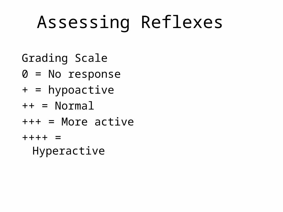
Assessing Reflexes
Grading Scale0 = No response+ = hypoactive++ = Normal+++ = More active++++ = Hyperactive
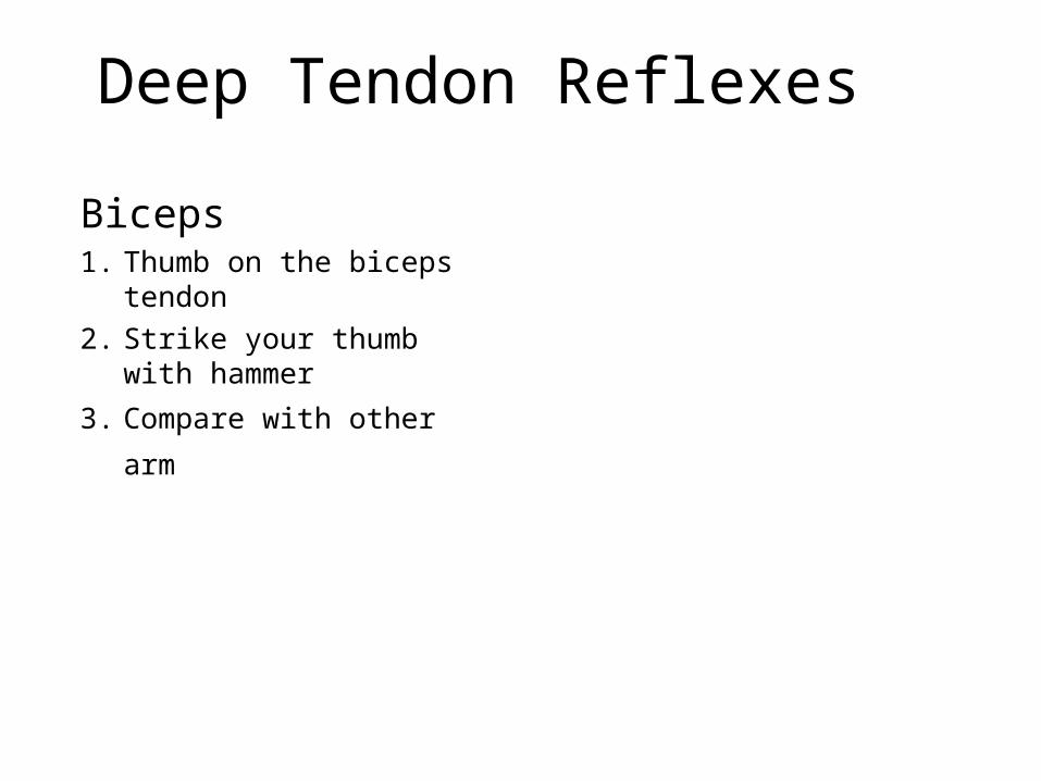
Deep Tendon Reflexes
Biceps1. Thumb on the biceps tendon2. Strike your thumb with hammer
3. Compare with other arm
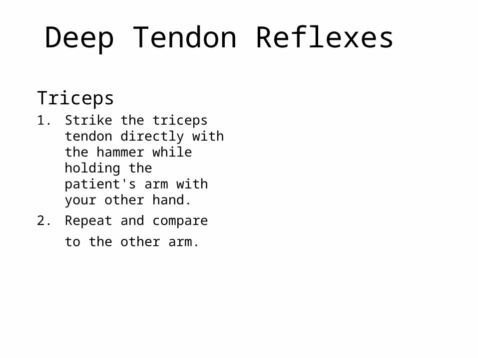
Deep Tendon Reflexes
Triceps1. Strike the triceps tendon
directly with the hammer while holding the patient's arm with your other hand.
2. Repeat and compare to the
other arm.
Deep Tendon Reflexes
Patellar

Deep Tendon Reflexes
Achilles
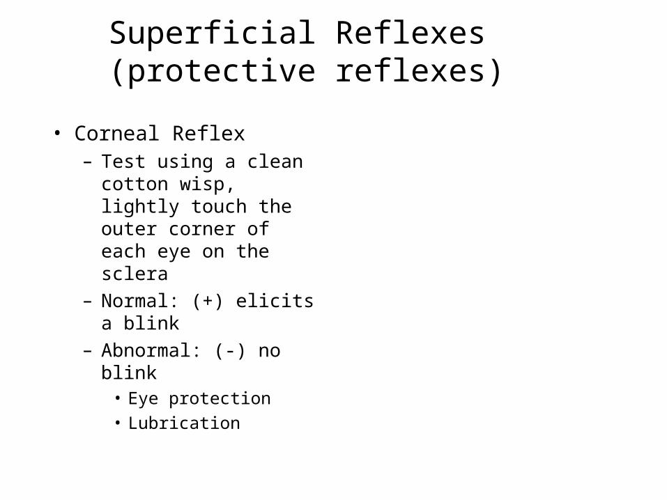
Superficial Reflexes (protective reflexes)
• Corneal Reflex– Test using a clean cotton
wisp, lightly touch the outer corner of each eye on the sclera
– Normal: (+) elicits a blink– Abnormal: (-) no blink
• Eye protection• Lubrication
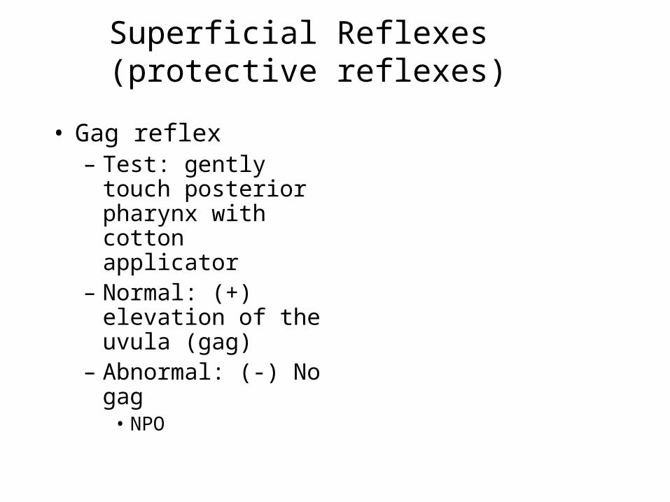
Superficial Reflexes (protective reflexes)
• Gag reflex– Test: gently touch
posterior pharynx with cotton applicator
– Normal: (+) elevation of the uvula (gag)
– Abnormal: (-) No gag• NPO
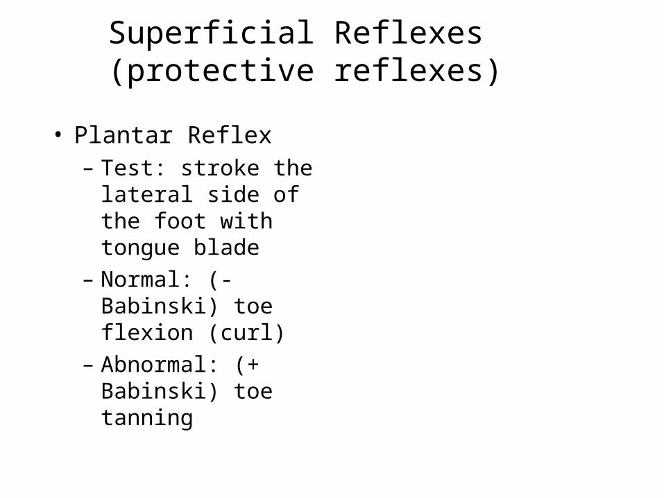
Superficial Reflexes (protective reflexes)
• Plantar Reflex– Test: stroke the
lateral side of the foot with tongue blade
– Normal: (- Babinski) toe flexion (curl)
– Abnormal: (+ Babinski) toe tanning
Superficial Reflexes (protective reflexes)
• - Babinski = Normal• + Babinski = Abnormal
Vital Signs
• Temperature– With head trauma
increased
Vital Signs
• Pulse– Strength, rate rhythm– Bradycardia
indicative of Increased ICP
Vital Signs
• Respirations– Depth, rate, rhythm,
effort– Ataxic
• Damage to medulla– Cheyne-stokes
• Lesion deep in both hemispheres, basal ganglia and upper brainstem
– Hyperventilation• Metabolic problems or
brainstem
Vital Signs
• Blood Pressure– Right verses left– Lying verses standing– Difference in systolic
by > 20mmHg potential cerebral ischemia
Vital Signs
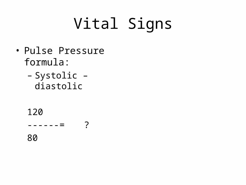
• Pulse Pressure formula:– Systolic – diastolic
120------ = ?80
Vital Signs
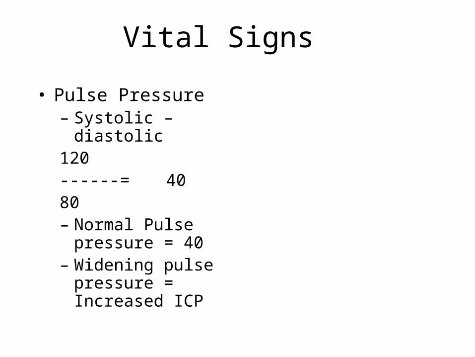
• Pulse Pressure– Systolic – diastolic120------ = 4080– Normal Pulse
pressure = 40– Widening pulse
pressure = Increased ICP
Neuro Checks
• LOC• Pupils– PERRLA
• Pupils• Equal• Round• Reactive to• Light• Accommodation
Neuro Check
• Pupils– Anisocoria
• Inequality in the size of the pupils
– Nystagmus– Progressive dilation
• Increase ICP
– Fixed & dilated• Injury at level of
midbrain
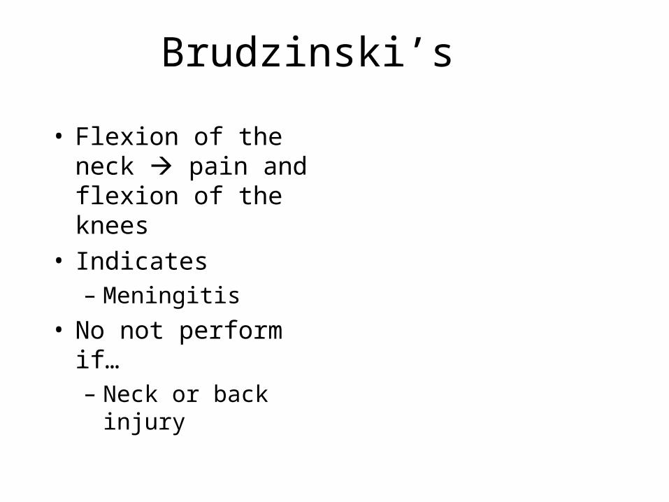
Brudzinski’s
• Flexion of the neck pain and flexion of the knees
• Indicates– Meningitis
• No not perform if…– Neck or back injury
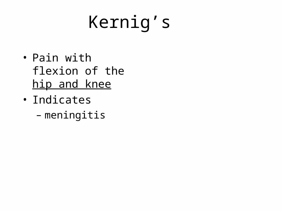
Kernig’s
• Pain with flexion of the hip and knee
• Indicates– meningitis
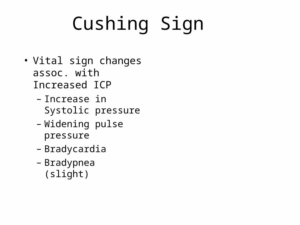
Cushing Sign
• Vital sign changes assoc. with Increased ICP– Increase in Systolic
pressure– Widening pulse
pressure– Bradycardia– Bradypnea (slight)