neurological and psychological applications of ... · neurological and psychological applications...
TRANSCRIPT

Biochemical Pharmacology 86 (2013) 447–457
Neurological and psychological applications of transcranial lasers andLEDs
Julio C. Rojas a,b, F. Gonzalez-Lima a,*a Departments of Psychology, Pharmacology and Toxicology, University of Texas at Austin, Austin, TX 78712, USAb Department of Neurology and Neurotherapeutics, University of Texas Southwestern Medical Center, Dallas, TX 75235, USA
A R T I C L E I N F O
Article history:
Received 8 May 2013
Accepted 15 June 2013
Available online 24 June 2013
Keywords:
Cognitive enhancement
Cytochrome oxidase
Low-level light therapy
Methylene blue
Neuroprotection
Photobiomodulation
A B S T R A C T
Transcranial brain stimulation with low-level light/laser therapy (LLLT) is the use of directional low-
power and high-fluency monochromatic or quasimonochromatic light from lasers or LEDs in the red-to-
near-infrared wavelengths to modulate a neurobiological function or induce a neurotherapeutic effect in
a nondestructive and non-thermal manner. The mechanism of action of LLLT is based on photon energy
absorption by cytochrome oxidase, the terminal enzyme in the mitochondrial respiratory chain.
Cytochrome oxidase has a key role in neuronal physiology, as it serves as an interface between oxidative
energy metabolism and cell survival signaling pathways. Cytochrome oxidase is an ideal target for
cognitive enhancement, as its expression reflects the changes in metabolic capacity underlying higher-
order brain functions. This review provides an update on new findings on the neurotherapeutic
applications of LLLT. The photochemical mechanisms supporting its cognitive-enhancing and brain-
stimulatory effects in animal models and humans are discussed. LLLT is a potential non-invasive
treatment for cognitive impairment and other deficits associated with chronic neurological conditions,
such as large vessel and lacunar hypoperfusion or neurodegeneration. Brain photobiomodulation with
LLLT is paralleled by pharmacological effects of low-dose USP methylene blue, a non-photic electron
donor with the ability to stimulate cytochrome oxidase activity, redox and free radical processes. Both
interventions provide neuroprotection and cognitive enhancement by facilitating mitochondrial
respiration, with hormetic dose–response effects and brain region activational specificity. This evidence
supports enhancement of mitochondrial respiratory function as a generalizable therapeutic principle
relevant to highly adaptable systems that are exquisitely sensitive to energy availability such as the
nervous system.
� 2013 Elsevier Inc. All rights reserved.
Contents lists available at SciVerse ScienceDirect
Biochemical Pharmacology
jo u rn al h om epag e: ww w.els evier .c o m/lo cat e/b io c hem p har m
1. Introduction
The use of transcranial low-level light/laser therapy (LLLT) tomodulate neurological and psychological functions is a paradigmthat has gained significant interest among researchers andclinicians in recent years. There is a need for an accurate reviewthat gives proper chronological attribution to the various groupsthat discovered the transcranial LLLT effects relevant to cognitiveenhancement and neuroprotection (listed in Table 1). Thefundamental observation that light can be used transcranially tomodulate brain function has derived into many significantcontributions to forward our understanding of the neurother-apeutic effects of light. Current research focuses on the elucidationof the neurochemical and photobiological mechanisms of action of
* Corresponding author. Tel.: +1 512 471 5895; fax: +1 512 471 5935.
E-mail address: [email protected] (F. Gonzalez-Lima).
0006-2952/$ – see front matter � 2013 Elsevier Inc. All rights reserved.
http://dx.doi.org/10.1016/j.bcp.2013.06.012
LLLT and ongoing pre-clinical and clinical investigations aim atdetermining the role of LLLT in the enhancement of normal brainfunction, neuroprotection and neural repair. Photobiomodulationwith LLLT has become one of the most dynamic and promisingfields of experimental neurotherapeutics. Its major appeal is asound mechanistic theory and the prospective to aid in thetreatment of neurological and psychological conditions in a non-invasive, non-expensive and safe manner. Prior reviews havediscussed the evidence and potential clinical applications of LLLT instroke [1] and chronic neurodegenerative conditions [2]. Impor-tant aspects of light sources and principles of dosimetry have alsobeen previously summarized. We recently provided an introduc-tory background to photobiology and an overview of the beneficialeffects of LLLT on the eye and brain [3]. The objective of the presentreview is to update on the benefits of transcranial LLLT and theneurochemical mechanisms supporting the cognitive-enhancingand brain-stimulatory effects of transcranial LLLT via low-levellasers and light emitting diodes (LEDs) in the red-to-near-infrared

Table 1Transcranial low-level light/laser therapy studies relevant to neuroprotection and cognitive enhancement.
Date Reference Relevance Source Parameters Effects
2004 Lapchak et al. [22] Embolic
stroke
Laser 808 nm, 25 mW/cm2, 15,000 J/cm2,
continuous
Improved motor function and reduction in effective clot dose
for stroke 3 h after clot injection (rabbit)
2006 De Taboada
et al. [23]
Atherothrombotic
stroke
Laser 808 nm, 7.5 mW/cm2, 0.9 J/cm2, 2 min
per point
Improved modified neurological score at 14, 21, and 28 after
MCAO (rat)
2006 Oron et al. [24] Atherothrombotic
stroke
Laser 808 nm, 7.5 mW/cm2, 0.9 J/cm2, 2 min
per point
Improved neurological scores 14 and 21 days after MCAO;
increased subventricular zone cell proliferation and migration
after (rat)
2007y Lampl et al. [15] Ischemic stroke Laser 808 nm, 1 J/cm2 per point Improved clinical outcome at 90 days after ischemic stroke
(human)
2007 Lapchak et al. [25] Embolic stroke Laser 808 nm, 25 mW/cm2, 15,000 J/cm2, pulsed
at 1 kHz
Improved motor function, decreased effective clot dose for
stroke 6 h after clot injection (rabbit)
2007 Oron et al. [26] Traumatic brain
injury
Laser 808 nm, 10 or 20 mW/cm2, 1.2–2.4 J/cm2,
single point for 2 min
Improved motor behavior 5 days after closed-head injury, and
decreased brain lesion size from 12.1% to 1.4% at 28 days after
injury (mouse)
2008* Michalikova
et al. [27]
Mild cognitive
impairment,
Alzheimer’s
disease
Laser 1072 nm, 6 min � 10 days Improved acquisition of working memory for spatial navigation
in middle-aged mice (mouse)
2008 Lapchak et al. [28] Embolic stroke Laser 808 nm, 25 mW/cm2, 15,000 J/cm2,
pulsed at 1 kHz
No worsening of hemorrhage incidence, volume or survival
after treatment with tPA (rabbit)
2008 Ahmed et al. [29] Epilepsy Laser 808 nm and 830 nm, 5.5 W/cm2, 3.1 W/cm2
and 2.8 W/cm2, 30 J/point, 11 J/point
and 5 J/point
Decrease in cortical aspartate, glutamate and taurine and
decreased hippocampal GABA (rat)
2009y Zivin et al. [16] Ischemic stroke 808 nm, 1 J/cm2 per point No improvement in mRS or NIHSS scores, no differences in
mortality or adverse events at 90 days (human)
2009 Moreira et al. [30] Traumatic brain
injury
Laser 660 nm and 780 nm, 952 mW/cm2, 3 J/cm2
and 5 J/cm2
Altered interleukin and tumor necrosis factor aplpha
concentrations in brain and plasma at 1 day after cryogenic
brain injury (rat)
2009*y Schiffer et al. [11] Depression,
prefrontal
functions
LED 810 nm, 250 mW/cm2, 60 J/cm2 Decreased depression scores, increased prefrontal blood flow
(human)
2010 Lapchak et al. [31] Embolic stroke Laser 808 nm, 25 mW/cm2, 15,000 J/cm2, pulsed
at 1 kHz
Increased cortical ATP (rabbit)
2010 Uozumi et al. [32] Anoxic brain injury Laser 808 nm, 1.6 W/cm2, 4320 J/cm2 Increased cerebral blood flow and decreased hippocampal and
cortical neuronal death after BCCAO (mouse)
2010*y Naeser et al. [14] Traumatic brain
injury
LED 633 nm and 870 nm, 22.2 mW/cm2,
13.3 J/cm2
Improved cognition of 2 patients with chronic mild traumatic
brain injury after 2–4 months of treatment (human)
2010 Shaw et al. [33] Parkinson’s
disease
Laser 670 nm, 40 mW/cm2, 2 J/cm2 in four
fractions
Reduction in substantia nigra dopaminergic cell loss after MPTP
toxicity (mouse)
2011 Yip et al. [34] Ischemic stroke Laser 660 nm, 8.8 mW, 2.6 J/cm2, 13.2 J/cm2
and 26.4 J/cm2, pulsed at 10 kHz
Increased expression of antiapopotic factors Akt, Bcl-2 and
pBAD and decreased expression of pro-apoptotic factors
caspase 3 and caspase 9 1 hr after ischemia and reperfusion
induced by transient unilateral MCAO (rat)
2011* Ando et al. [35] Traumatic brain
injury
Laser 810 nm, 50 mW/cm2, 36 J/cm2, continuous,
pulsed, 10 Hz or 100 Hz
Improved neurological severity score and body weight; smaller
lesion volumes, reduced helplessness at 4 weeks (mouse)
2011* De Taboada
et al. [20]
Alzheimer’s disease Laser 808 nm, 0.5 W/cm2, 2.8 W/cm2 and
5.6 W/cm2; 675 J/cm2, 336 J/cm2 and
672 J/cm2, continuous and pulsed,
three fractions per week for 6 months
Decreased escape latency in Morris water maze memory task,
decreased brain amyloid load and pro-inflammatory cytokines,
Decreased CSF and plasma b-amyloid, increased brain ATP
concentration and oxygen consumption (mouse)
2012 Quirk et al. [36] Traumatic brain
injury
LED 670 nm, 50 mW/cm2, 15 J/cm2, 3 or 10
daily fractions
Improved locomotor behavior, decreased pro-apoptotic and
increased anti-apoptotic gene expression, increased GSH (rat)
2012 Wu et al. [37] Traumatic brain
injury
Laser 665 nm, 730 nm, 810 nm and 980 nm,
150 mW/cm2, 36 J/cm2, one fraction
Improved neurological severity score and accelerated
neurological recovery with 665 nm and 810 nm, 4 weeks after
treatment (mouse)
2012 Oron et al. [38] Traumatic brain
injury
Laser 808 nm, pulsed at 100 Hz, one fraction Improved neurological severity score, increased survival,
smaller brain infarct volumes, from 5–28 days after trauma
(mouse)
2012 Khuman
et al. [39]
Traumatic
brain injury
Laser 800 nm, 500 mW/cm2, 60 J/cm2, one
fraction
Improved spatial memory, decreased microglial activation two
days after trauma (mouse)
2012* Rojas et al. [4] PTSD, specific
phobia
LED 660 nm, 9 mW/cm2, 5.4 J/cm2,
daily dosing after extinction for four days
Enhanced extinction of fear-conditioned memories, decreased
renewal of conditioned-fear, increase prefrontal oxygen
consumption and energy metabolism capacity (rat)
2013*y Barrett and
Gonzalez-Lima [13]
Prefrontal
cognitive
functions,
depression
Laser 1064 nm, 250 mW/cm2, 60 J/cm2 Improved sustained attention/psychomotor vigilance,
improved visual memory retrieval, improved affect (human)
2013 Xuan et al. [40] Traumatic
brain injury
Laser 810 nm, 25 mW/cm2, 18 J/cm2, 1, 3 or
14 doses
Improved neurological severity scores and wire grip and
motion test scores, smaller brain lesions sizes, decreased
degeneration, increased BrdU-positive cells at 14 days (mouse)
2013 Moro et al. [41] Parkinson’s
disease
LED 670 nm, 5.5 mW/cm2, 2 J/cm2 in four
fractions
Improved locomotor activity and preserved tyrosine
hydroxylase-positive cells in the substantia nigra pars
compacta (mouse)
Abbreviations: ATP = adenosine triphosphate, BCCAO = bilateral common carotid artery occlusion, GSH = reduced glutathione, LED = light-emitting diode, MCAO = medial
cerebral artery occlusion, MPTP = 1-Methyl-4-phenyl-1,2,3,6-tetrahydropyridine, mRS = modified Rankin scale, NIHSS = Neurological Institute of Health Stroke Scale,
tPA = tissue plasminogen activator, * = studies testing cognitive effects, y = studies with human subjects.
J.C. Rojas, F. Gonzalez-Lima / Biochemical Pharmacology 86 (2013) 447–457448

J.C. Rojas, F. Gonzalez-Lima / Biochemical Pharmacology 86 (2013) 447–457 449
wavelengths. The fundamental principle of transcranial LLLT is thedelivery of photons to brain cells that are primarily absorbed by themitochondrial respiratory enzyme cytochrome oxidase and up-regulate its enzymatic activity in vivo [3–5]. The proposedmechanistic rationale is that LLLT stimulation of cytochromeoxidase enhances brain oxygen utilization and metabolic capacity,which may enhance normal brain functions and protect againstneurological deficits caused by reduced cerebral blood perfusionand other insults to brain energy metabolism. It is important todiscuss the new data because they imply that transcranial LLLTmay become a novel intervention to enhance cognitive perfor-mance and treat neurological conditions linked to mitochondrialdysfunction. In addition, no neuroscience experts have properlyreviewed these findings in a detailed and integrated manner thatexplains how the mechanism of action of LLLT is related to bothcognitive enhancement and mitochondrial neuroprotection. Thecurrent review distinguishes itself from the existing literaturebecause it addresses the evidence of in vivo cognitive-enhancingeffects of LLLT in the normal brain as well as the in vivo
neuroprotective effects against neurometabolic energy failure.This review also highlights the existence of a common biochemicalmechanism of action for LLLT [3] and for the mechanism of actionof the metabolic enhancer and antioxidant methylene blue [6],focusing on the well-established central role of the mitochondrialenzyme cytochrome oxidase on brain function. Acknowledgmentof this common mitochondrial mechanism of action is expected toprovide important mechanistic insights to support the use of LLLTas a tool for the effective treatment of neurological andpsychological conditions.
Photobiomodulation is the use of radiant energy to modifybiological functions. LLLT is defined as the use of directional low-power and high-fluency monochromatic or quasimonochromaticlight from lasers or LEDs in the red-to-near-infrared wavelengthsto modulate a biological function or induce a therapeutic effect in anondestructive and nonthermal manner [3]. The fundamentalprinciple of photobiomodulation with LLLT is the presence ofchromophores, molecules capable of absorbing light in cells andtissues. The interaction of light-excited chromophores withdownstream molecules and pathways induces subsequent bio-chemical changes with potential pharmacological, physiologicaland clinical effects. LLLT with red-to-near-infrared light fromlasers and LEDs may be delivered transcranially to target the brainparenchyma. Transcranial LLLT is able to modify cognitive andneurological functions in animals and humans with effects that areindependent of visual pathway activation or heat [3].
2. Transcranial LLLT as a safe and novel neuromodulatoryintervention
As mentioned above, the fundamental principle of photobio-modulation with LLLT is the presence of chromophores capable ofabsorbing light in neurons. It is well-established that cytochromeoxidase is the major neuronal photoacceptor in the red-to-near-infrared range of radiant energy, and meaningful biologic effects ofLLLT in neural tissues have been documented in a number ofconditions ranging from cell cultures to human subjects [3]. Forexample, LLLT enhances both the activity and expression ofcytochrome oxidase in neurons in vitro [7]. Transcranial LLLT alsoaccelerates cell respiration and energy production in the brainparenchyma in vivo [4,8]. In addition, LLLT partially restoredenzyme activity blocked by potassium cyanide, a cytochromeoxidase inhibitor, and significantly reduced neuronal cell deathinduced by this mitochondrial toxin [9]. Prophylactic LLLT in vitro
has proved very effective at protecting neurons from neurode-generation induced by mitochondrial toxins [10]. Beneficialmitochondrial bioenergetics effects have also been demonstrated
in vivo as LLLT-induced up-regulation of cytochrome oxidase in thecortex, when delivered transcranially [4]. Transcranial LLLT hasalso been observed to augment prefrontal blood flow in humansubjects [11]. An encouraging common denominator of the effectsof transcranial LLLT on brain cytochrome oxidase is that it is a safeintervention with null deleterious effect on the structure andfunction of the brain at the doses observed to induce beneficialeffects [1–5]. Early investigations documented that the subunitexpression and assembly of cytochrome oxidase is tightlyregulated by energy consumption. Cytochrome oxidase is notonly a key enzyme in oxidative metabolism, but also has a limitingstep role in energy production. Cytochrome oxidase is a highlydynamic and autoinducible enzymatic complex, and it is notablefor its connection with activity-dependent gene expression path-ways relevant to energy metabolism, homeostasis and cell death[12]. Thus, photobiomodulation of brain cytochrome oxidase isexpected to provide beneficial effects primarily via the up-regulation of cytochrome oxidase itself. In turn, this is expectedto increase neuronal respiration and boost brain energy metaboliccapacity, which would constitute an adaptation with majorneuroprotective implications.
LLLT via commercial low-power lasers and LEDs constitutes anaffordable and safe alternative to current treatment options forcognitive impairment and brain dysfunction. Low-power LEDarrays and laser diode sources are compact, portable, and haveachieved non-significant risk status for human trials by the FDA.High bioavailability of LLLT to brain tissue in vivo is supported bypreclinical evidence of transcranially-induced increases in braincytochrome oxidase activity and improved behavioral outcome inrats with impaired mitochondrial function [5] and by improvedbrain cytochrome oxidase activity and memory retention innormal adult rats [4]. Further evidence from the first controlledhuman study demonstrated the beneficial effects of transcranialinfrared laser stimulation on cognitive functions [13]. Thus, LLLTtreatments could be cost-effective, safe, and non-invasive [14] andcould have broad impact and significance to improve the cognitivehealth of our growing aging population. Transcranial LLLT hasalready been successful at improving neurological outcome inhumans in some controlled clinical trials of stroke [15,16].However, early use of LLLT in people with compromised cerebralblood flow may prove to also be an effective strategy before strokebecause its beneficial effects would be based on metabolicneuroplasticity natural to the undamaged brain, as opposed tobe based on less physiologic and less generalizable processes of cellrepair. In other words, LLLT has a potential as a strategy for primaryor secondary stroke prevention in the specific setting of chronicbrain hypoperfusion (CBH) associated with cerebrovascularatherosclerosis. Likewise, LLLT given before the onset of cognitiveimpairment, either vascular or associated with primary neurode-generative processes, may induce neuroprotection by facilitating aneurochemical substrate for improved cognitive reserve. Thiswould seem more plausible and advantageous than interruption ofan advanced multifactorial neurodegenerative process in whichthe molecular machinery to support the secondary photobiologiceffects of LLLT has been damaged. In summary, the availableevidence indicates that LLLT may have the ability to enhancecognition and prevent neural dysfunction associated with CBH,stroke, traumatic brain injury, dementia and other neurodegener-ative processes when given before the onset of brain damage.
3. Methodological considerations for transcranial LLLT
Transcranial LLLT consists of applying monochromatic lightdirectly to the head, with wavelengths falling within an ‘‘opticalwindow’’ in the red-to-near-infrared optical region (�620–1150 nm). Wavelength is a major LLLT parameter as it greatly
J.C. Rojas, F. Gonzalez-Lima / Biochemical Pharmacology 86 (2013) 447–457450
determines the molecular target of light [17–19]. Cytochromeoxidase shows four major light absorption peaks within the red-to-near-infrared band. These are determined by CuA and CuB, two ofthe four metal centers within the enzyme. These peaks ofabsorption are 620 nm (CuA reduced), 680 nm (CuB oxidized),760 nm (CuB reduced) and 825 nm (CuA oxidized). In vitro, theseabsorption peaks correspond to peaks in DNA synthesis and cellattachment [17]. Within this band, light tissue penetration tends tobe higher with higher wavelengths; thus, wavelengths in the upperend are preferred in transcranial applications [20–26]. However,longer wavelengths do not provide linear improvement in tissuepenetration, since as the wavelengths get longer than 940 nm, lightabsorption by water increases [18]. Nevertheless, transcranial LLLTapplications have demonstrated relevant neural and behavioraleffects at wavelengths above 940 nm [13,27]. LLLT doses areexpressed as radiant exposure in Joules (J) per surface area (J/cm2,fluency or energy density) and radiant exposure is equivalent topower density or irradiance (W/cm2) per unit of time (s). Thus forachieving a desired dose, either power density or time of exposurecan be varied. However, wavelength and radiant exposure are notthe only parameters that are relevant for replicating a particularphotobiologic effect of LLLT, and the optimal LLLT dose may bedifficult to determine for a particular application (Table 1). Besideswavelength and radiant exposure, parameters that are expected toinfluence the efficacy, feasibility and safety of LLLT can be of threetypes: (a) device parameters, (b) irradiation parameters and (c)treatment parameters [18,19]. Until further research determineshow variation in such parameters will affect transcranial memory-enhancing and neuroprotective effects of LLLT, a thoroughmethodological description should be provided for any transcra-nial LLLT application. Device parameters include device manufac-turer, model identifier, number of emitters, emitter type or source(e.g. solid state, gas, laser diode, InGa AlP LED, GaAlAs laser, KTPlaser), spatial distribution of emitters and shape, size and type ofbeam delivery system (e.g. fiber optic, free air/scanned, hand-heldprobe). Irradiation parameters include center wavelength (nm),spectral bandwidth (nm), operating mode (e.g. continuouswavelength, switched continuous wavelength, pulsed), frequency(Hz), pulse duration (s), pulse off duration (s) or duty cycle (%),peak radiant power (mW), average radiant power (mW), aperturediameter (cm) and beam profile (e.g. Gaussian, Top Hat). Treatmentparameters include beam spot size at target (cm2), irradiance attarget (mW/cm2), exposure duration (s), radiant exposure (J/cm2),radiant energy (J), number of points irradiated, area irradiated(cm2), application technique (e.g. skin contact, contact withpressure, interstitial fiber optic), number and frequency oftreatment sessions (i.e. number of treatments per day or week)and cumulative radiant energy (i.e. individual doses multiplied bythe number of treatment sessions, in J) [19].
Factors such as dosing schedules and light sources have provento be extremely relevant parameters, besides wavelength anddose. For example, delivering large total doses of LLLT in a singlesession produces less favorable outcomes than giving the samedoses over several sessions. This dose fractionation has been testedin vitro and in vivo, and it has been shown to be highly effective atpreventing neuronal degeneration. In addition, it has been alsoshown that LLLT fractionation protocols including prophylacticdoses given before neurotoxic metabolic lesions are also effectiveat preventing neurodegeneration [5,10]. Similarly, light sources forLLLT may be obtained from lasers or LEDs. Laser sources produce100% of coherent light energy in a single wavelength. They allowhigh tissue penetration and they produce a constant beam widththat offers the advantage of energy delivery on circumscribedareas. The beam width of lasers can be modified by coupling theminto fiber optic, which allows delivering energy to areas of differentsizes. Yet, areas of tissues that can be treated with lasers could be
insufficient for some transcranial applications, and repeated singlebeam exposures are usually necessary. LEDs typically have abandwidth of 4 nm to 30 nm (at full width half maximum), andemitted light is not coherent and less collimated. Lasers are capableof delivering high amounts of energy with great efficiency, whichcan be high enough to rapidly produce tissue heating and damage.On the other hand, LEDs deliver energy with less efficiency, whichmay be advantageous in certain protocols of energy delivery.Although any light source can deliver comparable amounts ofenergy to a target surface, the photobiological effect will notnecessarily be the same. Substitution of an LED for a laser of thesame wavelength may deliver the same amount energy by varyingthe power or exposure time. However, this reciprocity rule hasbeen disproven in photobiology and photomedicine [19]. Inaddition, with irradiances used in LLLT, LEDs generate negligibleamounts of tissue heating at the site of light absorption. Thisreduces the risk of thermal injury [3]. In addition, LEDs can bemounted on arrays with ergonomic features that allow efficientenergy delivery, which is relevant when the target organ has alarge surface area, such as the brain. LED arrays and diode lasers arecompact and portable, which may facilitate their use in the clinicalsetting, and both have achieved nonsignificant risk status forhuman trials by the FDA (www.clinicaltrials.gov). Finally, theimportance of the operating mode of the light source has beendocumented in a number of studies. For example, pulsed-waveLLLT to rats is more effective at improving performance in theMorris water maze and decreasing hippocampal amyloid load thancontinuous wave LLLT [20].
For transcranial applications, tissue penetrance achieved with aspecific light source should be ideally described. Post-mortemanalyses in human specimens may provide an estimate of lasertransmittance through the skull. Incident light (I0) and transmittedlight (I) should be measured with the tissue directly overlying theaperture of the detector; average readings are then used tocalculate percent transmittance (k) as 100 � I/I0. Optical density(OD) is calculated as—log (k). The cross-sectional width of each setof tissues may be measured with calipers, and Beer’s law is thenapplied to calculate the absorption coefficient (a) = OD/width.Using this method, we have measured that approximately 2% of the1064 nm wavelength at 250 mW/cm2, 60 J/cm2 passed through theadult human supraorbital frontal bone, when the LLLT is deliveredby direct contact with bone. This yields an OD of 1.70 and anabsorption coefficient of a = 0.24. This is consistent with reportedvalues of transmittance of this wavelength through cranial bone ofa = 0.22 [21]. Consequently, the transcranial LLLT dose reaching thefrontal cortex surface was estimated to be 1.2 J/cm2 [13], which isconsistent with the most effective doses (around 1 J/cm2) found tostimulate cytochrome oxidase activity in neuron cultures(reviewed in Ref. [3]). Stimulation of cognitive functions inhumans has been achieved using a relatively high-wavelength1064 nm laser diode (Cell Gen Therapeutics, LLC, Model CG-5000laser, HD Laser Center, Dallas, TX). The wave type from this sourceis continuous, not pulsed. Marketing of Cell Gen lasers is FDA-cleared as safe for various indications (e.g. improving circulation,relief of muscle and joint pain, spasm, stiffness and relaxation).Pre-clinical studies support that the structure and function of theretina is not damaged by LLLT doses commonly used forexperimental applications. In fact, such LLLT doses have beenshown to exert neuroprotective effects [5]. However, scarce data isavailable on the potential retinotoxic effects of LLLT in humans.Thus it is advisable that subjects’ eyes be covered with protectiveeyewear during transcranial LLLT delivery. Enhancement ofcognitive and emotional functions in humans has been achievedwith irradiance of 250 mW/cm2 and fluency of 60 J/cm2. Beneficialeffects with these parameters have been observed by twoindependent groups [13,14]. These parameters allow transcranial
J.C. Rojas, F. Gonzalez-Lima / Biochemical Pharmacology 86 (2013) 447–457 451
light penetration of about 2%, which corresponds to a fluency of1.2 J/cm2 over the cortical surface. At these power levels the energyemitted is low, exposure to it is not harmful to tissue, and it causesnegligible tissue heating and no physical damage. Using theseparameters, we directed a 1064 nm laser diode at the right frontalpole of the cerebral cortex [13], which is the most anteriorprefrontal cortex (Brodmann’s areas 9 and 10). In reference to the10–20 system used for EEG electrode placement, the foreheadstimulation site was centered on the FP1 or FP2 (left or right frontalpole) point, and extended medially and laterally for a 4 cmdiameter area from this point. In animals such a rats, a powerdensity output of 9 mW/cm2 delivered at the 10.9 J/cm2 dose has atranscranial transmittance of 5.8%, with 0.63 J/cm2 reaching the ratcortical surface. With these parameters, LLLT enhanced prefrontalcortex oxygen consumption rate, increased cytochrome oxidaseexpression and facilitated fear-extinction memories [4].
4. Chronological overview of transcranial LLLT studies relevant tocognitive enhancement and neuroprotection
A sizable body of controlled studies assessing the effect of LLLTon human cognitive functions does not exist, but pioneer studieson the in vivo neuroprotective and cognitive-enhancing propertiesof LLLT started in the last decade (Table 1) [4,11,13–16,20,22–24,26–40]. Twenty-seven studies have assessed the effects oftranscranial LLLT targeting the brain in healthy animals, animalmodels of neurological disease, healthy human subjects or patientsaffected by neurological disease. Five of these studies have beendone with human subjects. These include one pilot case series inpatients with traumatic brain injury (n = 2) [14], one open label,non-controlled trial in patients with depression (n = 10) [11], twodouble-blind, randomized, sham-controlled studies in patientswith acute ischemic stroke (n = 780) [15,16] and one smallplacebo-controlled trial for effects on cognitive and emotionalfunctions in healthy volunteers (n = 40) [13]. Seven studies havespecifically tested the effects of LLLT on cognitive functions, threeof them in human subjects [4,11,13,14,20,27,35]. The firstpublished observation of LLLT’s memory effects in a mouse modelwas the Michalikova et al. study [27]; but other than thewavelength this paper did not report other relevant LLLTparameters, making it impossible to evaluate or replicate thisstudy. The work by De Taboada et al. [20] was the first publishedobservation of prevention of memory loss in a mouse model of AD.This study indicated the importance of LLLT’s treatment early in adisease process.
The first translational neuroprotective applications of tran-scranial LLLT were the NEST-1 and -2 clinical trials in stroke[15,16]. Until NEST-3 or a similar stroke clinical trial is publishedthere is still uncertainty in the use of LLLT in stroke. So far 10studies have assessed the effects of LLLT in ischemic, hemorrhagic,atherothrombotic, embolic or anoxic stroke. Beneficial neuropro-tective effects have been observed regardless of the strokemechanism. Functional neuroprotective effects in stroke havebeen correlated with changes including down-regulation of pro-apoptotic genes, up-regulation of anti-apoptotic genes, increasedenergy production and increased activation of cell proliferationand migration [9,42]. Nine studies have assessed the neuropro-tective effects of LLLT in traumatic brain injury, and this constitutesone of the most active areas of LLLT research. The scientificliterature on neuroprotective effects of LLLT in traumatic braininjury has provided vast evidence of functional, structural andcognitive effects at different time points [24,27,32,35–37,40,41]. Ithas also addressed the effects of wavelength, fluency, dose fractionand pulse width like no other field of in vivo brain photobiomo-dulation. Also, studies on the effects of LLLT in traumatic braininjury contain meaningful observations regarding the mechanisms
through which LLLT exerts its neuroprotective effects. Theavailable evidence shows that such neuroprotective effects maybe supported by induction of cell proliferation as well as anti-oxidant, anti-inflammatory and anti-apoptotic effects. Finallyexperimental transcranial applications of LLLT have also exploredits potential applications relevant to epilepsy, Parkinson’s disease,mild cognitive impairment, Alzheimer’s disease, depression andenhancement of normal cognitive function.
5. Authors’ studies of transcranial LLLT effects on braincytochrome oxidase activity, oxygen consumption andcognitive functions
Transcranial LLLT treatment and placebo effects on cytochromeoxidase and cognitive functions have been described in rats andhumans in our laboratory. In 2008, Rojas et al. [5] were the first toreport that upon transcranial delivery in vivo, LLLT induces brainmetabolic and antioxidant beneficial effects measured by increasesin cytochrome oxidase and superoxide dismutase. In 2011, weproposed LLLT as a novel paradigm to treat visual, neurological,and psychological conditions based on the stimulation of cyto-chrome oxidase activity in neurons [3]. In 2012, Rojas et al. [4]were the first to report that LLLT increased extinction memoryretention and oxygen consumption in the rat frontal cortex in vivo.
In 2013, Barrett and Gonzalez-Lima [13] reported the firstcontrolled study of transcranial laser stimulation of psychologicalfunctions in humans. Transcranial infrared laser stimulation to theforehead has been shown to produce beneficial effects on frontalcortex measures of attention, memory and mood. Our studies haveused different daily in vivo LLLT doses (1–60 J/cm2), fractionationprotocols (1–6 sessions), wavelengths in both the red (633 nm and660 nm) and in the near-infrared (1064 nm), and a range of powerdensities (2–250 mW/cm2). These variables allowed us to identifyeffective LLLT parameters for transcranial brain stimulation in ratsand humans, with findings that can be summarized as follows:
LLLT increases brain oxygen consumption in vivo. An increase incytochrome oxidase activity would be expected to facilitateoxygen consumption, as cytochrome oxidase is the enzyme thatcatalyzes the use of oxygen to form water in the mitochondrialelectron transport chain. Thus, we tested the hypothesis that LLLTstimulates brain oxygen consumption in vivo. Oxygen concentra-tion in the cortex of naı̈ve rats was measured immediatelyfollowing LLLT exposure at 9 mW/cm2 and l = 660 nm. The cortexoxygen concentration in control conditions (i.e. following no LLLTexposure) decreased only 1 � 0.7%. In contrast, LLLT induced a dose-dependent decrease in oxygen concentration of approximately 5 � 1%after LLLT 1 J/cm2 and 15.8 � 2% after LLLT 5 J/cm2. These data suggesta physiological effect of transcranial LLLT on the metabolic rate ofcortical oxygen consumption [4].
LLLT induces a hormetic dose–response on brain cytochrome
oxidase activity. LLLT has been shown to increase cytochromeoxidase expression in neuronal cultures [5]. It has been observedthat this secondary effect of LLLT also occurs in the brain in vivo.The effects of different doses of transcranial LLLT were delivered ina single fraction and levels of brain cytochrome oxidase activitywere measured. Unanesthetized rats were exposed to 660 nm ateither 10.9 J/cm2, 21.6 J/cm2, 32.9 J/cm2 or no LLLT in home cages.Treatments were delivered via four LED arrays with a powerdensity of 9 mW/cm2 for total treatment times of 20 min, 40 minand 60 min for each dose, respectively. Twenty-four hours after thesingle treatment session, animals were decapitated and theirbrains histochemically analyzed for cytochrome oxidase activity.LLLT showed enhancement of brain cytochrome oxidase followinga hormetic dose–response pattern. A single dose of 10.9 J/cm2 LLLTresulted in a 13.6% increase in cytochrome oxidase activity. In turn,a single dose of 21.6 J/cm2 resulted in an increase of only 10.3%,

Fig. 1. Primary and secondary effects of low-level light/laser therapy (LLLT). (A)
Primary effects occur with red-to-near-infrared light on and consist of direct
excitation of chromophores in the respiratory enzyme cytochrome oxidase
(yellow). Primary effects are fundamental for the in vivo beneficial effects of
light therapy, but they can also be observed in vitro in solutions of the purified
enzyme or in mitochondrial membrane isolates. The primary effects of cytochrome
oxidase excitation represent a boost in the activity of the respiratory chain and
consist of increases in transmembrane potential, oxidation of NADH+, oxygen
consumption and free radicals. (B) Secondary mechanisms may occur with light off.
Secondary effects are always preceded by primary effects and they occur only in the
presence of intact cellular metabolic machinery. Thus, secondary effects have been
observed only in living cells and in vivo and not in systems of membrane or enzyme
isolates. Secondary effects are pleiotropic and depend on activation of enzymatic
pathways that affect metabolic capacity, gene expression for mitogenic and repair
signaling, cytoskeleton processing and protein expression and translocation. Such
secondary effects are triggered due to the central role of mitochondria as integrators
of energy metabolism, cellular homeostasis and cell survival signaling.
J.C. Rojas, F. Gonzalez-Lima / Biochemical Pharmacology 86 (2013) 447–457452
whereas the highest dose induced no significant increase incytochrome oxidase activity (3%) [4]. A low dose given in a singleday had a stimulatory effect while higher doses were less effective.Hormetic dose–response effects, such as the one demonstrated onbrain cytochrome oxidase activity are not logarithmic, but LLLTrepeated daily can show improvements of up to 30–60% ascompared to control [3]. The available data support that althoughsmall, these effects are not negligible, but neurobiologicallymeaningful. It is expected that hormetic changes in brain metaboliccapacity may support neurotherapeutic cognitive improvements.
LLLT increases cognitive functions in humans. LLLT has been usednon-invasively in humans to stimulate the brain to improveneurological outcome after ischemic stroke [15], as an antidepres-sant treatment [11] as well as to alleviate muscle fatigue andenhance recovery [43]. We conducted the first controlled studydemonstrating that transcranial laser stimulation enhancescognitive functions in healthy humans [13]. These LLLT treatmentshave thus been proven to be not just safe but actually beneficial inhumans. In particular, Schiffer et al. [11] found that a single LLLTtreatment to the forehead resulted in a significant beneficial effectin patients with major depression and anxiety that correlated withincreased cerebral blood flow. No adverse side effects were foundin any of the patients, either immediately after the initialtreatment, or at 2 or 4 weeks post-treatment. We followed asimilar transcranial LLLT protocol to the forehead, targeting frontalcortex-based cognitive tasks such as a psychomotor vigilance task(PVT) and a delayed match-to-sample memory task (DMS) beforeand after LLLT vs. a placebo control. The PVT is a test that assessesan individual’s sustained attention. It involves the subjectmaintaining a vigilant state during a delay period, then respondingas fast as possible when a stimulus appears onscreen. Theseattentional processes are mediated by frontal cortical regions andPVT has been shown to be a reliable indicator of frontal function[44]. In turn, the DMS task has been shown to be mediated by afrontoparietal network [45]. This task involves the presentation ofa visual stimulus on a screen. Then the stimulus disappears, andthe participant must remember the stimulus through a delay. Thentwo choices appear, and the participant must decide which of thesetwo is identical to the previous stimulus (the ‘‘match’’).
The forehead of healthy volunteers was exposed to LLLT withcontinuous wave laser at l = 1064 nm. This wavelength maximizestissue penetration and intersects the absorption spectrum ofcytochrome oxidase. The irradiance and cumulative fluency were250 mW/cm2 and 60 J/cm2, respectively. These parameters are thesame that showed beneficial psychological effects in the study bySchiffer et al. [11]. At the power level described, the energy emittedby the laser is low, exposure to it is not harmful to tissue, and itcauses negligible heat and no physical damage. Similar settings areused clinically for treatment of chronic pain [46]. The treated groupshowed significant beneficial effects on the PVT. LLLT improvedreaction time in the sustained vigilance test. Performance in theDMS also showed a significant improvement in treated vs. placebocontrol groups as measured by memory retrieval latency andnumber of correct trials [13]. These data imply that transcraniallaser stimulation is effective as a noninvasive and efficaciousapproach to increase cognitive brain functions such as thoserelated to attention, memory and mood.
6. Mechanisms of action of LLLT and their implications formitochondrial neurotherapeutics
The remarkable modulatory effects of LLLT and its specificphotochemical mechanisms of action have major therapeuticpotential on their own, but their discovery has revealed a majorand broadly generalizable therapeutic principle. The photobiomo-dulation effects of LLLT indicate that support of mitochondrial
function is a very effective approach not only to facilitate normal cellfunctions, but also to preserve structural and physiological integrityin pathologic contexts. Maintenance and facilitation of optimalmitochondrial function is meaningful, since it represents a highlyspecialized version of a fundamental process in biological systems:assimilation and transfer of energy. Photobiomodulation is expectedto have major therapeutic relevance in highly adaptable systemsextremely sensitive to energy availability such as the brain.
The mechanism of action of LLLT consists of primary effects andsecondary effects (Fig. 1). Primary effects occur with light on anddepend on light absorption by mitochondria. The respiratoryenzyme cytochrome oxidase is regarded as the major acceptor oflight in the red-to-near-infrared wavelength range. Energeticimprovements are expected from LLLT because it acts as anexogenous source of highly energized electrons to the respiratorychain, otherwise provided by endogenous electron donors such asNADH and FADH2. This view is supported by evidence showing thatLLLT facilitates the catalytic activity of cytochrome oxidase,accelerates the electron transfer in the inner mitochondrialmembrane and boosts cell respiration and energy production[4,7,8,10,47]. In fact, LLLT may restore electron flow, when there isupstream blockade of electron entry into the respiratory chain [5].In addition, because cytochrome oxidase is sensitive to energydemands, a consequence of its activation by LLLT is an increase inits subunit expression and assembly, which leads to an increase in
J.C. Rojas, F. Gonzalez-Lima / Biochemical Pharmacology 86 (2013) 447–457 453
neuronal oxidative metabolic capacity and photoacceptor avail-ability [7]. In neural tissue, cytochrome oxidase is the mostabundant metalloprotein, and wavelengths in its absorptionspectra correlate well with its catalytic activity action spectraand with ATP content in vitro [5–8]. Cytochrome oxidase is acentral enzyme in neuronal bioenergetics, due to its role as a rate-limiting step in ATP synthesis and its exquisite functional responseto energy demands, changes in intermediate metabolism and celldamage. Cytochrome oxidase is in fact a reliable marker ofneuronal energy metabolism [7]. Due to its central role in oxidativemetabolism, the effects of LLLT on cytochrome oxidase are believedto be the origin of photosignal transduction from mitochondria toother neuronal compartments, including the cytoplasm, nucleusand cell membrane. These phototransduction processes beyondthe respiratory chain may occur at times after light exposure anddefine the secondary mechanisms of LLLT. The engagement of anumber of intracellular enzymatic and metabolic pathways isconsidered to be responsible for the pleiotropic effects of LLLT. Forexample, cell membrane functions, such as cell-adhesion, aresusceptible to modulation in vitro by LLLT and this is mediated bychanges in the cell surface integrin expression pattern and focaladhesion kinase activity [49]. Because no secondary LLLT effectsare observed in conditions where disruption of the plasmamembrane and cellular homeostasis occur, changes at the cell-membrane level are regarded as a secondary effect of LLLT,whereas the primary redox effects have been shown to occur inmitochondria. Similarly, only in conditions of cellular integrity,plasma membrane or nucleus functions are sensitive to secondaryeffects of LLLT [50]. Thus, the effects of LLLT ranging from lightabsorption to changes in neuronal function are highly dependenton the metabolic and signaling pathways available to support aphotobiological response.
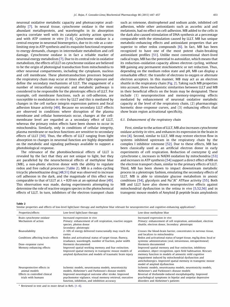
The relevance of the photobiochemical effects of LLLT isrevealed by the fact that they are not unique to light, but theyare paralleled by the neurochemical effects of methylene blue(MB), a non-photic electron donor with the ability to regulateredox and free radical processes (Table 2). MB is a redox-cyclingtricyclic phenothiazine drug [48,51] that was observed to increasecell adhesion in the dark, and the magnitude of this effect wascomparable to that of LLLT at l = 820 nm at an optimal dose [49].This observation was made, during experiments attempting todetermine the role of reactive oxygen species in the photochemicaleffects of LLLT. In turn, inhibitors of the electron transport chain
Table 2Similar properties and effects of low-level light/laser therapy and methylene blue rele
Properties/effects Low-level light/laser therapy
Brain cytochrome oxidase Increased expression in vivo
Mechanism of action Primary: enhancement of cell respiration, reacti
species, photon donor
Secondary: pleiotropic
Bioavailability 2–10% of energy delivered transcranially may r
cortex
Conditions affecting brain effects Redox and activational status of target tissue, fl
irradiance, wavelength, number of fraction, pul
Dose–response curve Hormesis documented
Memory enhancing effects Improved spatial working memory and fear ext
Improved spatial memory in transgenic mouse
amyloid dysfunction and models of traumatic b
Neuroprotective effects in
animal models
Ischemic models, neurotrauma models, neuroto
models, Alzheimer’s and Parkinson’s disease mo
Effects in controlled clinical
trials with humans
Improved neurological outcome after stroke. Im
psychomotor vigilance, visual memory retrieval
function, inhibition, and inhibition accuracy
a Reviewed in text and in more detail in Refs. [1–6].
such as rotenone, dinitrophenol and sodium azide, inhibited celladhesion, while other antioxidants such as ascorbic acid andmelatonin, had no effect on cell-adhesion. MB added to the cells inthe dark also caused stimulation of DNA synthesis at a percentagecomparable with the stimulation caused by LLLT. MB has uniquemetabolic-enhancing effects and antioxidant properties that aresuperior to other redox compounds [6]. In fact, MB has beenrecognized to have one of the most potent chain-breakingantioxidant profiles [51]. Unlike most conventional short-livedradical traps, MB has the potential to autoxidize, which means thatits reduction–oxidation capacity allows electron cycling, withoutMB gaining any permanent stoichiometric or net reduction. Thus,depending on the medium redox state and pH, MB can display aremarkable effect: the transfer of electrons to oxygen or alternateelectron acceptors. In this manner, MB may act as an electronshuttle in the respiratory chain (Fig. 2). Taking such MB propertiesinto account, three mechanistic similarities between LLLT and MBin their beneficial effects on the brain may be designated. Theseinclude (1) neuroprotection and memory-improving effectsmediated by enhancement of neuronal oxidative metaboliccapacity at the level of the respiratory chain, (2) pharmacologichormetic dose–response curves, and (3) enhancing effects thatshow brain region activational specificity.
6.1. Enhancement of the respiratory chain
First, similar to the action of LLLT, MB also increases cytochromeoxidase activity in vitro, and enhances its expression in the brain in
vivo [4]. Second, similar to LLLT, MB may restore electron flow insystems inhibited upstream in the respiratory chain by thecomplex I inhibitor rotenone [52]. Due to these effects, MB hasbeen classically used as an artificial electron donor in earlyexperiments of cell respiration. Reduction of coenzyme Q andcytochrome c, increases in NADH oxidation by mitochondria [53]and increases in ATP synthesis [54] support a direct effect of MB onthe electron transport chain, similar to the primary effects of LLLT.Third, MB has been shown to impact downstream metabolicprocess in a pleiotropic fashion, emulating the secondary effects ofLLLT. MB is able to stimulate glucose metabolism in anoxicconditions [54], glycolysis and Na+/K+ ATPase activity [55]. BothMB and LLLT have also shown neuroprotective effects againstmitochondrial dysfunction in the retina in vivo [5,52,56] and intransgenic mouse models of Amyloid b peptide brain amyloidosis
vant for neuroprotective and cognitive-enhancing applicationsa.
Low-dose methylene blue
Increased expression in vivo
ve oxygen Primary: enhancement of cell respiration, antioxidant, electron
shuttle, electron donor, Secondary: pleiotropic
each the Crosses the blood-brain barrier, concentrates in nervous tissue,
and localizes to mitochondria
uency,
se width
Redox and activational status of target tissue, mg/kg dose, local or
systemic administration (oral, intravenous, intraperitoneal)
Hormesis documented
inction.
models of
rain injury
Improved spatial memory and fear extinction, inhibitory
avoidance, object recognition, open field habituation. Rescues
memory function in models of amnestic mild cognitive
impairment induced by mitochondrial dysfunction and
anticholinergics. Improved spatial memory in transgenic mouse
model of amyloid dysfunction
xicity
dels
Ischemic models, neurotrauma models, neurotoxicity models,
Alzheimer’s and Parkinson’s disease models
proved
, executive
Reversal of ifosfamide-induced encephalopathy. Improved
psychological symptoms in bipolar and unipolar depressive
disorders and Alzheimer’s patients

Fig. 2. Enhancement of the mitochondrial respiratory chain as the basis for cognitive enhancement and neuroprotection. Two different strategies, low-level light/laser
therapy and methylene blue can achieve neuroprotective and cognitive enhancing effects by supporting and improving cell respiration. High-energy electrons are feed to the
mitochondrial respiratory chain by endogenous electron donors such as NADH+, which interacts with complex I or FADH2+, which interacts with complex II. Electrons flow to
ubiquinone, and subsequently to complex III, cytochrome c and finally complex IV (cytochrome oxidase). During this transfer, electrons release energy in a tightly regulated
fashion, which allows the pumping of protons into the mitochondrial inter-membrane space. This allows the storage of energy as an electrochemical gradient that is used in
the synthesis of ATP (top panel). Both low-level red-to-near-infrared light and methylene blue improve cell respiration. Low-level light directly stimulates cytochrome
oxidase, facilitating its catalytic activity and inducing an increase in holloenzyme subunit assembly, which improves neuronal metabolic capacity (mid panel). Similarly,
methylene blue acts as an exogenous electron shuttle, also boosting cell respiration and inducing changes that improve mitochondrial metabolic capacity (bottom panel).
Both interventions may have a higher facilitating effect of cell respiration in those neurons with increased energy demands, conditionally engaging and improving
mechanisms required in cognitive processing and neuroprotection.
J.C. Rojas, F. Gonzalez-Lima / Biochemical Pharmacology 86 (2013) 447–457454
[57,58]. Finally, the notable similarities between LLLT and MB arealso evident as their ability to enhance cognitive function. Asdiscussed above, LLLT has been used in rats to improve spatialworking memory [27], decrease helplessness scores [35] andfacilitate fear extinction [4]. In humans, LLLT decreases depression-related scores [13], and improves psychomotor vigilance, visualmemory retrieval, executive function, inhibition, and inhibitionaccuracy [13,14]. Animal studies have documented memory-enhancing properties in fear extinction using both LLLT and MB.MB has also shown memory-enhancing effects in a number oflearning and memory paradigms including inhibitory avoidance,spatial memory, fear extinction, object recognition, open-fieldhabituation and discrimination learning [4]. In addition, MBrescues memory function in models of amnestic mild cognitiveimpairment induced by mitochondrial dysfunction [59,60] oranticholinergics [61] and improves memory in transgenic mouse
models of amyloid-associated memory dysfunction [57,58]. Theseobservations and the evidence discussed above support that themechanistic similarities between LLLT and MB are generalizable,and that support of the electron transport chain may have broadpotential neuroprotective and cognitive-enhancing applications.
6.2. Hormesis
The hormetic response of both LLLT and MB consists of anincrease in the effect at a low dose, followed by a decrease in thesame effect with an intermediate dose, until the effect is equal to acontrol-type effect. With doses increasing beyond the hormeticzone, the effect decreases even further, until it is below the controleffect. Both interventions induce maximal pharmacologic effectsthat correspond to 30–60% increases compared to control, asopposed to several fold-increases typical of linear-non-threshold
J.C. Rojas, F. Gonzalez-Lima / Biochemical Pharmacology 86 (2013) 447–457 455
dose–response curves [19]. The magnitude of such effects is typicalof hormesis, and they have been considered rare and negligible byclassical pharmacology paradigms but it is known now that theyare very common, and biologically relevant [62]. Hormetic effectsfor both LLLT and MB at the neurochemical and behavioral levelshave been described [63,64]. In particular, LLLT and MB increasebrain cytochrome oxidase activity in a hormetic dose–responsemanner. If the principle of hormesis is generalizable in neurother-apeutic applications, lower doses of interventions that supportmitochondrial function will induce increased beneficial effects,compared to higher doses.
6.3. Brain region activational specificity
Experimental evidence that the effects of LLLT and MB showbrain region activational specificity has been provided by studies offacilitation of conditioned-fear extinction in rats [2]. LLLT givenduring the period of memory consolidation induced facilitation offear extinction memory, which is known to be mediated byincreased metabolic activity in the prefrontal cortex. When in situ
oxygen consumption and cytochrome oxidase activity weremeasured in the prefrontal cortex, subjects treated with LLLTshowed increases in both parameters compared to untreatedcontrols. Similarly, MB given during the memory consolidationphase of fear extinction was correlated with selective increases ofcytochrome oxidase activity in the prefrontal cortex. LLLT is moresusceptible to be absorbed by a mixed valence cytochrome oxidase(i.e. partially reduced or oxidized). The probability of finding amixed valence enzyme is higher with higher respiratory chainelectron flow, a state that is found in highly metabolic activetissues. Similarly, MB has been described as a ‘‘magic bullet’’, as itconcentrates in areas with high redox activity. Due to its affinity foractive oxidoreductases, MB has the greatest bioavailability tomitochondria with high rates of electron transfer. Thus, bothtreatments may reach the totality of the brain, but only those areasthat show higher metabolic rates will maximally benefit from theeffects of these interventions. These areas are likely to containneuronal networks engaged in a particular cognitive task. Thus, ifthe activational specificity principle is generalizable, it is expectedthat mitochondrial interventions will provide the greatest benefitwhen paired with physical therapy, cognitive rehabilitation or anyother strategy that would engage regional brain energy metabo-lism activation.
7. Future potential role of LLLT in the treatment ofneurodegeneration and cognitive impairment
There is a compelling public health need to develop interven-tions to prevent and effectively treat neuropsychological diseases.The burden of memory deficits in the aging population, includingthose at risk for developing mild cognitive impairment (MCI),Alzheimer’s disease (AD) and stroke is especially important, sinceit is expected to reach unparalleled endemic proportions. In the US,between 2.4 and 5.1 million people may have AD with enormouspersonal and societal costs, whereas stroke is the third leadingcause of death and the leading cause of long-term disability, with $43 billion cost on stroke patient care (NIH) [65,66]. There is a lackof disease-modifying treatments, and it is critical to intervene earlyin the natural history of neurodegeneration, ideally before theonset of cognitive impairment or severe neurological deficits. Forsuch reasons accessible strategies to stimulate the brain, enhanceits performance and prevent cognitive and neurological deficits areone of the most important research priorities of our times.Interventions that boost the cognitive reserve in healthy individu-als may play a major role in the effective management of chronicneurological dysfunction associated with AD and stroke. While
multiple mechanisms are likely responsible for MCI and AD, thereis no question that CBH secondary to cerebrovascular atheroscle-rotic steno-occlusive disease and inhibition of the mitochondrialenzyme cytochrome oxidase are metabolic risk factors for MCI andAD, as well as for vascular dementia and stroke [67]. Thus, it hasbeen hypothesized that the adverse cognitive consequences of CBHmay be modifiable to prevent or delay amnestic MCI andneurodegeneration [68]. The apparent link between age-relatedcognitive decline, CBH and mitochondrial dysfunction has beendeciphered by basic and clinical research in the last 30 years. Onone hand, a strong body of evidence supports a role ofmitochondrial dysfunction in memory-related neurodegenerativedisorders. In addition, mitochondrial dysfunction and the con-comitant oxidative stress and energy hypometabolism are believedto play a role in CBH-induced neuropathology [69]. For example, itis well-established that the brain, and in particular the aging brain,is vulnerable to hypoperfusion because it depends almostexclusively on electron transport-derived oxidative energy [70].Regional cytochrome oxidase dysfunction has been observed inbrains of patients affected by MCI and AD [67]. Cytochrome oxidasehas a key role in neuronal activity as the rate-limiting enzyme foroxidative energy production in the mitochondrial electrontransport and it also can catalyze the production of nitric oxideunder hypoxic conditions [71]. Since memory functions areextremely sensitive to oxidative energy deficits, cytochromeoxidase inhibition linked to aging and impairment in cerebralperfusion has been proposed as a major pathophysiologicalmechanism underlying memory dysfunction and neurodegenera-tion. Recent neuroimaging evidence, in particular with arterial spinlabeling fMRI techniques, have established that CBH in the elderlyis associated with cognitive decline [72], is present prior to ADonset [73] and can identify patients with high risk conversion fromhealthy aging to MCI to AD [74].
Surprisingly, the overwhelming evidence supporting cyto-chrome oxidase as an ideal molecular target to promoteneuroprotection and memory enhancement has not been utterlyexploited in translational medicine. Specifically, no preclinicalmodel or clinical protocol has ever investigated if improving braincytochrome oxidase activity may prevent memory impairment orneurological decline caused by CBH. In particular, more research isneeded to document in vivo LLLT effects on hypoxic CBH conditionsin which cytochrome oxidase may catalyze the synthesis of nitricoxide from nitrite, a biochemical process different from the classicnitric oxide synthase enzymes [75]. The cognitive decline thatunfolds in the general aging population, as well as in patients withMCI, AD and vascular dementia associated with CBH may beprevented by LLLT interventions that critically influence cognition,provide neuroprotection or enhance neural cell repair. The LLLTapproach is scientifically relevant because it will take the researchcommunity toward translational, noninvasive, accessible and earlyinterventions to modify the risk factors affecting the cognitive andneurological health of our growing aging population.
8. Concluding remarks
It is expected that research on transcranial applications of LLLTfor neuroprotection and cognitive enhancement, especially inhuman subjects, will increase in the forthcoming years. FurtherLLLT research should go beyond preclinical and clinical experimen-tal testing of LLLT effects. Specifically, there is a need to further testthe proposed mechanistic causality between stimulation of cyto-chrome oxidase with LLLT and its improvement of cognitivefunctions. The hypothesis that a primary molecular mechanism ofaction of LLLT on cognitive deficits is caused by up-regulation ofcytochrome oxidase needs further validation. This may be accom-plished in animal models using comparisons with LLLT-treated and

J.C. Rojas, F. Gonzalez-Lima / Biochemical Pharmacology 86 (2013) 447–457456
untreated control groups where cytochrome oxidase activity will bechronically stimulated or inhibited, as well as testing other lightwavelengths that are not absorbed by cytochrome oxidase. Inaddition, there is a need to evaluate the hypothesis that LLLT willinhibit the direct pathophysiologic consequences of CBH, throughproteomic quantification of markers of oxidative stress, fMRImeasures of cerebral blood flow, blood oxygen level-dependentsignaling and cerebral metabolic rate of oxygen consumption.Similarly, the effect of early LLLT in the pathophysiology of pre-symptomatic cognitive decline may be assessed by measuring itseffects on chemical and functional predictors of progression such asimaging-based glucose metabolism functional connectivity andblood biomarker levels, among others. Future research should alsofocus on a more extensive description of the neurotherapeuticeffects of LLLT based on its dosimetry-related parameters, as well ason further elucidation of the secondary mechanisms of action (i.e.
long-lasting cellular effects that occur once light is off) that arecritical for neuromodulation. Such studies are relevant to thesecondary mechanisms of action of LLLT given its documentedeffects on nitric oxide production and its relationship withcytochrome oxidase activity modulation [3]. Finally, it is anticipatedthat accelerated progress in the field of LLLT for neurotherapeuticapplications will derive from a better understanding of how suchtherapeutic photobiological effects can be modulated by concomi-tant pharmacotherapy, psychotherapy and physical and cognitiverehabilitation.
Non-invasive LLLT appears to be a safe and convenient tool formitochondrial enhancement and together with other strategies toaugment cell respiration may be part of a comprehensive approachfor treatment of neurological conditions featuring neurodegenera-tion and cognitive impairment. Support of energy metabolism atthe mitochondrial level may be a fundamental neurotherapeuticstrategy. The bioenergetic particularities of the brain demandconsideration of non-conventional strategies of neuroprotectionand enhancement, with attention to very specific neuropharma-cologic details to ensure maximal efficacy. Acknowledgment of thefundamental role of oxidative metabolism and its tremendouspotential as a neurotherapeutic target is desirable and may be thenecessary step to advance treatments in clinical neuroscience,which has traditionally lacked the benefit of disease modifyingtherapies. Targeted redox-mediated bioenergetic neuromodula-tion with LLLT is proposed as part of a holistic neurotherapeuticconstruct that focuses on optimizing both the neural context (e.g.
aerobic exercise, rehabilitation, cognitive therapy) and the redox-energy equilibrium through increases of energy availability (e.g.
cardiovascular risk factor reduction, ketogenic diet) and mito-chondrial respiration (e.g. LLLT, MB), as well as rationalizedreduction of the pro-oxidant tendencies of neurobiologicalsystems (e.g. MB, other exogenous or endogenous antioxidants).The crossroads between modern photobiology with lasers andLEDs and bioenergetics has the potential to lead a revolution in theway we treat brain dysfunction and enhance cognition.
Acknowledgments
We thank Dr. Douglas Barrett for helping with the graphicalabstract.
References
[1] Lampl Y. Laser treatment for stroke. Expert Rev Neurother 2007;7:961–5.[2] Hashmi JT, Huang YY, Osmani BZ, Sharma SK, Naeser MA, Hamblin MR. Role of
low-level laser therapy in neurorehabilitation. PM&R 2010;2:S292–305.[3] Rojas JC, Gonzalez-Lima F. Low-level light therapy of the eye and brain. Eye
Brain 2011;3:49–67.[4] Rojas JC, Bruchey AK, Gonzalez-Lima F. Low-level light therapy improves
cortical metabolic capacity and memory retention. J Alzheimers Dis2012;32:741–52.
[5] Rojas JC, Lee J, John JM, Gonzalez-Lima F. Neuroprotective effects of near-infrared light in an in vivo model of mitochondrial optic neuropathy. J Neurosci2008;28:13511–21.
[6] Rojas JC, Bruchey AK, Gonzalez-Lima F. Neurometabolic mechanisms formemory enhancement and neuroprotection of methylene blue. Prog Neuro-biol 2012;96:32–45.
[7] Wong-Riley MT, Liang HL, Eells JT, Chance B, Henry MM, Buchmann E, et al.Photobiomodulation directly benefits primary neurons functionally inacti-vated by toxins: role of cytochrome c oxidase. J Biol Chem 2005;280:4761–71.
[8] Mochizuki-Oda N, Kataoka Y, Cui Y, Yamada H, Heya M, Awazu K. Effects ofnear-infra-red laser irradiation on adenosine triphosphate and adenosinediphosphate contents of rat brain tissue. Neurosci Lett 2002;323:207–10.
[9] Liang HL, Whelan HT, Eells JT, Meng H, Buchmann E, Lerch-Gaggl A, et al.Photobiomodulation partially rescues visual cortical neurons from cyanide-induced apoptosis. Neuroscience 2006;139:639–49.
[10] Ying R, Liang HL, Whelan HT, Eells JT, Wong-Riley MT. Pretreatment with near-infrared light via light-emitting diode provides added benefit against rote-none- and MPP+-induced neurotoxicity. Brain Res 2008;1243:167–73.
[11] Schiffer F, Johnston AL, Ravichandran C, Polcari A, Teicher MH, Webb RH, et al.Psychological benefits 2 and 4 weeks after a single treatment with nearinfrared light to the forehead: a pilot study of 10 patients with majordepression and anxiety. Behav Brain Funct 2009;5:46.
[12] Wong-Riley MT, Nie F, Hevner RF, Liu S. Brain cytochrome oxidase: functionalsignificance and bigenomic regulation in the CNS. In: Gonzalez-Lima F, editor.Cytochrome oxidase in neuronal metabolism and Alzheimer’s disease. NewYork: Springer; 1998. p. 1–53.
[13] Barrett DW, Gonzalez-Lima F. Transcranial infrared laser stimulation producesbeneficial cognitive and emotional effects in humans. Neuroscience2013;230:13–23.
[14] Naeser MA, Saltmarche A, Krengel MH, Hamblin MR, Knight JA. Improvedcognitive function after transcranial, light-emitting diode treatments inchronic, traumatic brain injury: two case reports. Photomed Laser Surg2010;29:351–8.
[15] Lampl Y, Zivin JA, Fisher M, Lew R, Welin L, Dahlof B, et al. Infrared lasertherapy for ischemic stroke: a new treatment strategy: results of the Neu-roThera Effectiveness and Safety Trial-1 (NEST-1). Stroke 2007;38:1843–9.
[16] Zivin JA, Albers GW, Bornstein N, Chippendale T, Dahlof B, Devlin T, et al.Effectiveness and safety of transcranial laser therapy for acute ischemic stroke.Stroke 2009;40:1359–64.
[17] Karu TI, Pyatibrat LV, Kolyakov SF, Afanasyeva NI. Absorption measurementsof a cell monolayer relevant to phototherapy: reduction of cytochrome coxidase under near IR radiation. J Photochem Photobiol B 2005;81:98–106.
[18] Chung H, Dai T, Sharma SK, Huang YY, Carroll JD, Hamblin MR. The nuts andbolts of low-level laser (light) therapy. Ann Biomed Eng 2012;40:516–33.
[19] Jenkins PA, Carroll JD. How to report low-level laser therapy (LLLT)/photo-medicine dose and beam parameters in clinical and laboratory studies. Photo-med Laser Surg 2011;29:785–7.
[20] De Taboada L, Yu J, El-Amouri S, Gattoni-Celli S, Richieri S, McCarthy T, et al.Transcranial laser therapy attenuates amyloid-beta peptide neuropathology inamyloid-beta protein precursor transgenic mice. J Alzheimers Dis2011;23:521–35.
[21] Bashkatov AN, Genina EA. Optical properties of human cranial bone in thespectral range from 800 to 2000 nm. Proc SPIE 2006;6163:61630.
[22] Lapchak PA, Wei J, Zivin JA. Transcranial infrared laser therapy improvesclinical rating scores after embolic strokes in rabbits. Stroke 2004;35:1985–8.
[23] De Taboada L, Ilic S, Leichliter-Martha S, Oron U, Oron A, Streeter J. Tran-scranial application of low-energy laser irradiation improves neurologicaldeficits in rats following acute stroke. Lasers Surg Med 2006;38:70–3.
[24] Oron A, Oron U, Chen J, Eilam A, Zhang C, Sadeh M, et al. Low-level lasertherapy applied transcranially to rats after induction of stroke significantlyreduces long-term neurological deficits. Stroke 2006;37:2620–4.
[25] Lapchak PA, Salgado KF, Chao CH, Zivin JA. Transcranial near-infrared lighttherapy improves motor function following embolic strokes in rabbits: anextended therapeutic window study using continuous and pulse frequencydelivery modes. Neuroscience 2007;148:907–14.
[26] Oron A, Oron U, Streeter J, de Taboada L, Alexandrovich A, Trembovler V, et al.Low-level laser therapy applied transcranially to mice following traumaticbrain injury significantly reduces long-term neurological deficits. J Neuro-trauma 2007;24:651–6.
[27] Michalikova S, Ennaceur A, van Rensburg R, Chazot PL. Emotional responsesand memory performance of middle-aged CD1 mice in a 3D maze: effects oflow infrared light. Neurobiol Learn Mem 2008;89:480–8.
[28] Lapchak PA, Han MK, Salgado KF, Streeter J, Zivin JA. Safety profile of tran-scranial near-infrared laser therapy administered in combination with throm-bolytic therapy to embolized rabbits. Stroke 2008;39:3073–8.
[29] Ahmed NA, Radwan NM, Ibrahim KM, Khedr ME, El Aziz MA, Khadrawy YA.Effect of three different intensities of infrared laser energy on the levels ofamino acid neurotransmitters in the cortex and hippocampus of rat brain.Photomed Laser Surg 2008;26:479–88.
[30] Moreira MS, Velasco IT, Ferreira LS, Ariga SK, Barbeiro DF, Meneguzzo DT, et al.Effect of phototherapy with low intensity laser on local and systemic immu-nomodulation following focal brain damage in rat. J Photochem Photobiol B2009;97:145–51.
[31] Lapchak PA, De Taboada L. Transcranial near infrared laser treatment (NILT)increases cortical adenosine-50-triphosphate (ATP) content following embolicstrokes in rabbits. Brain Res 2010;1306:100–5.
J.C. Rojas, F. Gonzalez-Lima / Biochemical Pharmacology 86 (2013) 447–457 457
[32] Uozumi Y, Nawashiro H, Sato S, Kawauchi S, Shima K, Kikuchi M. Targetedincrease in cerebral blood flow by transcranial near-infrared laser irradiation.Lasers Surg Med 2010;42:566–76.
[33] Shaw VE, Spana S, Ashkan K, Benabid AL, Stone J, Baker GE, et al. Neuroprotec-tion of midbrain dopaminergic cells in MPTP-treated mice after near-infraredlight treatment. J Comp Neurol 2010;518:25–40.
[34] Yip KK, Lo SC, Leung MC, So KF, Tang CY, Poon DM. The effect of low-energylaser irradiation on apoptotic factors following experimentally induced tran-sient cerebral ischemia. Neuroscience 2011;190:301–6.
[35] Ando T, Xuan W, Xu T, Dai T, Sharma SK, Kharkwal GB, et al. Comparison oftherapeutic effects between pulsed and continuous wave 810 nm wavelengthlaser irradiation for traumatic brain injury in mice. PLoS One 2011;6:e26212.
[36] Quirk BJ, Torbey M, Buchmann E, Verma S, Whelan HT. Near-infrared photo-biomodulation in an animal model of traumatic brain injury: improvements atthe behavioral and biochemical levels. Photomed Laser Surg 2012;30:523–9.
[37] Wu Q, Xuan W, Ando T, Xu T, Huang L, Huang YY, et al. Low-level laser therapyfor closed-head traumatic brain injury in mice: effect of different wavelengths.Lasers Surg Med 2012;44:218–26.
[38] Oron A, Oron U, Streeter J, De Taboada L, Alexandrovich A, Trembovler V, et al.Near infrared transcranial laser therapy applied at various modes to micefollowing traumatic brain injury significantly reduces long-term neurologicaldeficits. J Neurotrauma 2012;29:401–7.
[39] Khuman J, Zhang J, Park J, Carroll JD, Donahue C, Whalen MJ. Low-level laserlight therapy improves cognitive deficits and inhibits microglial activationafter controlled cortical impact in mice. J Neurotrauma 2012;29:408–17.
[40] Xuan W, Vatansever F, Huang L, Wu Q, Xuan Y, Dai T, et al. Transcranial low-level laser therapy improves neurological performance in traumatic braininjury in mice: effect of treatment repetition regimen. PLoS One2013;8:e53454.
[41] Moro C, Torres N, El Massri N, Ratel D, Johnstone DM, Stone J, et al. Photo-biomodulation preserves behaviour and midbrain dopaminergic cells fromMPTP toxicity: evidence from two mouse strains. BMC Neurosci 2013;14:40.
[42] Byrnes KR, Wu X, Waynant RW, Ilev IK, Anders JJ. Low power laser irradiationalters gene expression of olfactory ensheathing cells in vitro. Lasers Surg Med2005;37:161–71.
[43] Leal Junior EC, Lopes-Martins RA, Frigo L, De Marchi T, Rossi RP, de Godoi V,et al. Effects of low-level laser therapy (LLLT) in the development of exercise-induced skeletal muscle fatigue and changes in biochemical markers related topostexercise recovery. J Orthop Sports Phys Ther 2010;40:524–32.
[44] Drummond SP, Bischoff-Grethe A, Dinges DF, Ayalon L, Mednick SC, Meloy MJ.The neural basis of the psychomotor vigilance task. Sleep 2005;28:1059–68.
[45] Nieder A, Miller EK. A parieto-frontal network for visual numerical informa-tion in the monkey. PNAS 2004;101:7457–62.
[46] Jang H, Lee H. Meta-analysis of pain relief effects by laser irradiation on jointareas. Photomed Laser Surg 2012;30:405–17.
[47] Pastore D, Greco M, Passarella S. Specific helium-neon laser sensitivity of thepurified cytochrome c oxidase. Int J Radiat Biol 2000;76:863–70.
[48] Schirmer RH, Adler H, Pickhardt M, Mandelkow E. Lest we forget you—methylene blue. . .. Neurobiol Aging 2011;32. 2325.e7–16.
[49] Karu TI, Pyatibrat LV, Kalendo GS. Cell attachment modulation by radiationfrom a pulsed light diode (lambda = 820 nm) and various chemicals. LasersSurg Med 2001;28:227–36.
[50] Karu TI, Pyatibrat LV, Kalendo GS, Esenaliev RO. Effects of monochromatic low-intensity light and laser irradiation on adhesion of HeLa cells in vitro. LasersSurg Med 1996;18:171–7.
[51] Ohlow MJ, Moosmann B. Phenothiazine: the seven lives of pharmacology’sfirst lead structure. Drug Discovery Today 2011;16:119–31.
[52] Zhang X, Rojas JC, Gonzalez-Lima F. Methylene blue prevents neurodegenera-tion caused by rotenone in the retina. Neurotoxicity Res 2006;9:47–57.
[53] Gabrielli D, Belisle E, Severino D, Kowaltowski AJ, Baptista MS. Binding,aggregation and photochemical properties of methylene blue in mitochondrialsuspensions. Photochem Photobiol 2004;79:227–32.
[54] Lee RB, Urban JP. Functional replacement of oxygen by other oxidants inarticular cartilage. Arthritis Rheum 2002;46:3190–200.
[55] Furian AF, Fighera MR, Oliveira MS, Ferreira AP, Fiorenza NG, de CarvalhoMyskiw J, et al. Methylene blue prevents methylmalonate-induced seizuresand oxidative damage in rat striatum. Neurochem Int 2007;50:164–71.
[56] Rojas JC, John JM, Lee J, Gonzalez-Lima F. Methylene blue provides behavioraland metabolic neuroprotection against optic neuropathy. Neurotoxicity Res2009;15:260–73.
[57] Medina DX, Caccamo A, Oddo S. Methylene blue reduces abeta levels andrescues early cognitive deficit by increasing proteasome activity. Brain Pathol2011;21:140–9.
[58] O’Leary 3rd JC, Li Q, Marinec P, Blair LJ, Congdon EE, Johnson AG, et al.Phenothiazine-mediated rescue of cognition in tau transgenic mice requiresneuroprotection and reduced soluble tau burden. Mol Neurodegener2010;5:45.
[59] Callaway NL, Riha PD, Wrubel KM, McCollum D, Gonzalez-Lima F. Methyleneblue restores spatial memory retention impaired by an inhibitor of cyto-chrome oxidase in rats. Neurosci Lett 2002;332:83–6.
[60] Riha PD, Rojas JC, Gonzalez-Lima F. Beneficial network effects of methyleneblue in an amnestic model. Neuroimage 2011;54:2623–34.
[61] Deiana S, Harrington CR, Wischik CM, Riedel G. Methylthioninium chloridereverses cognitive deficits induced by scopolamine: comparison with rivas-tigmine. Psychopharmacology 2009;202:53–65.
[62] Calabrese EJ. Hormesis: principles and applications for pharmacology andtoxicology. Am J Pharmacol Toxicol 2008;3:59–71.
[63] Bruchey AK, Behavioral Gonzalez-Lima F, Physiological. Biochemical hormeticresponses to the autoxidizable dye methylene blue. Am J Pharmacol Toxicol2008;3:72–9.
[64] Huang YY, Sharma SK, Carroll J, Hamblin MR. Biphasic dose response in lowlevel light therapy—an update. Dose–Response (a publication of InternationalHormesis Society) 2011;9:602–18.
[65] National Institute of Neurological Disorders and Stroke. Stroke: hope throughresearch. National Institutes of Health; 2004.
[66] National Institute of Aging. Alzheimer’s disease progress report: intensifyingthe research effort. U.S. Department of Health & Human Services, NIH; 2012.
[67] Arntzen KA, Schirmer H, Johnsen SH, Wilsgaard T, Mathiesen EB. Carotidatherosclerosis predicts lower cognitive test results: a 7-year follow-up studyof 4371 stroke-free subjects—the Tromso study. Cerebrovasc Dis2012;33:159–65.
[68] Valla J, Berndt JD, Gonzalez-Lima F. Energy hypometabolism in posteriorcingulate cortex of Alzheimer’s patients: superficial laminar cytochromeoxidase associated with disease duration. J Neurosci 2001;21:4923–30.
[69] de la Torre JC. Cardiovascular risk factors promote brain hypoperfusion leadingto cognitive decline and dementia. Cardiovasc Psychiatry Neurol2012;2012:367516.
[70] Sokoloff L, Siegel BW, Agranoff BW, Albers RW, Molinoff P. Circulation andenergy metabolism of the brain. Boston: Little Brown; 1989.
[71] Poyton RO, Castello PR, Ball KA, Woo DK, Pan N. Mitochondria and hypoxicsignaling: a new view. Ann NY Acad Sci 2009;1177:48–56.
[72] Bangen KJ, Restom K, Liu TT, Wierenga CE, Jak AJ, Salmon DP, et al. Assessmentof Alzheimer’s disease risk with functional magnetic resonance imaging: anarterial spin labeling study. J Alzheimers Dis 2012;31(Suppl 3):S59–74.
[73] Johnson NA, Jahng GH, Weiner MW, Miller BL, Chui HC, Jagust WJ, et al. Patternof cerebral hypoperfusion in Alzheimer disease and mild cognitive im-pairment measured with arterial spin-labeling MR imaging: initial experience.Radiology 2005;234:851–9.
[74] Mak HK, Chan Q, Zhang Z, Petersen ET, Qiu D, Zhang L, et al. Quantitativeassessment of cerebral hemodynamic parameters by QUASAR arterial spinlabeling in Alzheimer’s disease and cognitively normal elderly adults at 3-tesla. J Alzheimers Dis 2012;31:33–44.
[75] Poyton RO, Ball KA. Therapeutic photobiomodulation: nitric oxide and a novelfunction of mitochondrial cytochrome c oxidase. Discov Med 2011;11:154–9.