neurologic assessment - americanchildneurologyuae.ae · wilkins, 978-1-4160-5923-3...
TRANSCRIPT
95
Wilkins, 978-1-4160-5923-3
B978-1-4160-5923-3.00006-7, 00006
afferent anosognosia anisocoria apneustic ataxia ataxic breathing autotopagnosia Babinski’s sign botulism cerebral perfusion pressure Cheyne-Stokes respiration corneal reflex Cushing’s triad decerebrate posture decorticate posture
deep tendon reflexes doll’s eyes efferent gag reflex gait Glasgow Coma Scale graphesthesia intracranial pressure level of consciousness lower motor neuron Mini-Mental State Examination miosis myasthenia gravis mydriasis nystagmus
oculocephalic reflex oculovestibular reflex patellar reflex PERRLA persistent vegetative state phrenic nerves plantar reflex pneumotaxic pronator drift proprioception pupillary reflex sensory dissociation stereognosis upper motor neuron Westphal’s sign
KEY TERMS
LEARNING OBJECTIVES After reading this chapter, you will be able to:
Define key terms related to neurologic assessment. Describe the functional anatomy of the nervous system. Explain the cortical function of the different lobes of the brain. Describe the functions of the brainstem, the cerebellum, and 12 pair of cranial nerves. Describe common techniques used to assess the mental status. Identify the parameters necessary to obtain a Glasgow Coma Scale and be able to interpret the results. Describe the importance of assessing sedation and delirium in the intensive care unit. Describe common techniques used to assess the cranial nerves, the sensory system, the motor system, coordination, and gait. Describe common techniques used to assess deep, superficial, and brainstem reflexes. Explain the relationship between vital signs and neurologic status. Identify the importance of intracranial pressure monitoring and the value of assessing cerebral perfusion pressure.
��������
���
Functional Neuroanatomy Mental Status Examination and Level of
Consciousness Level of Consciousness Glasgow Coma Scale Mini-Mental State Examination Assessment of Sedation and Delirium in the
Intensive Care Unit Cranial Nerve Examination Sensory Examination
Motor Examination Deep Tendon, Superficial, and Brainstem
Reflexes Deep Tendon Reflexes Superficial Reflexes Brainstem Reflexes
Coordination, Balance, and Gait Examination
Vital Signs and the Neurologic System Intracranial Pressure Monitoring
CHAPTER OUTLINE
RUBEN D. RESTREPO
Neurologic Assessment
C h a p t e r 6
Wilkins, 978-1-4160-5923-3
B978-1-4160-5923-3.00006-7, 00006
96 CHAPTER 6 • Neurologic Assessment
N eurologic assessment is a method of obtaining specific data in relation to the function of a patient’s nervous system and is typically performed by the
attending physician. Since injuries that involve the nervous system often affect the patient’s respiratory system and the ability of the patient to cooperate with respiratory care pro-cedures, the respiratory therapist (RT) should become famil-iar with the key components of the neurologic assessment. The challenges of examining an intubated, restrained, and often-sedated patient in the intensive care unit (ICU) makes neurologic assessment difficult in many patients.
Proper clinical assessment of the nervous system empha-sizes the neurologic history and examination. Obtaining historical information from critically ill patients, particu-larly those with altered states of consciousness can be diffi-cult. 1 However, attempting to obtain a history by speaking with the patient or family members can provide extremely useful information in the ICU. Whereas the history usu-ally indicates the nature of the dysfunction, the neurologic examination localizes and quantifies its severity.
The neurologic examination is often brief if initial interac-tions with the patient are normal (e.g., the patient responds appropriately to verbal stimuli) and the patient has no symp-toms suggesting neurologic disease. This initial interaction with the patient could provide insights about the patient that could possibly impact adherence to respiratory care and coordination to use devices such as a pressurized metered-dose inhaler (pMDI). A more extensive examination is per-formed when abnormalities are suspected and may involve the expertise of a neurologist. The neurologic examination is a comprehensive evaluation that covers several areas: men-tal status, cranial nerve function, motor system, coordina-tion, sensory system, and muscle stretch reflexes. This initial examination establishes baseline data with which to com-pare subsequent assessment findings. Neurologic observa-tions allow monitoring and evaluation of changes in the nervous system that aid in the diagnosis and treatment that later impact patient prognosis and rehabilitation. 2 It also gauges the patient’s response to the clinician’s interven-tions. Once a thorough evaluation is done on admission or at the beginning of each shift, subsequent assessments should be tailored to the patient’s condition. The frequency of these assessments will depend on the patient’s diagnosis, acuity of condition, and on how rapidly changes are occur-ring or expected to occur. Neurologic assessment should start with minimal verbal stimulus and progress to maximal tactile stimulus as the condition warrants. A meaningful neurologic assessment requires adequate stimulation.
FUNCTIONAL NEUROANATOMY
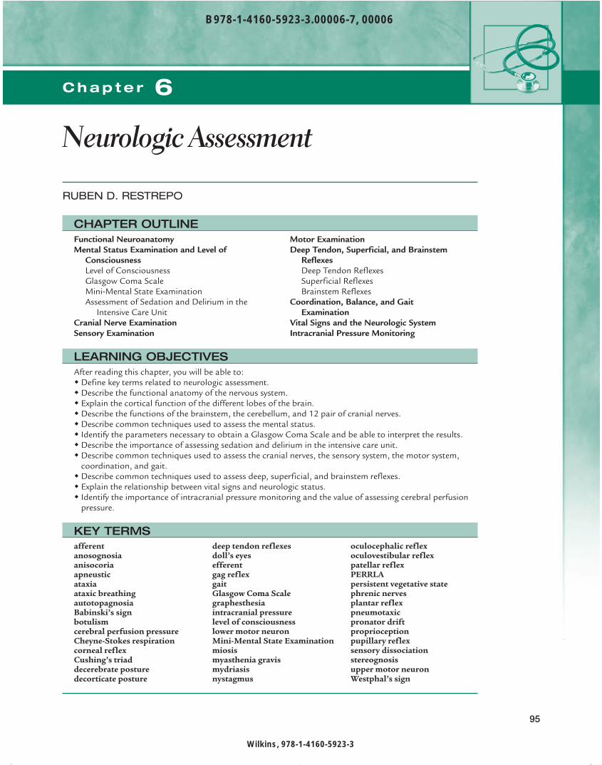
To perform or understand a neurologic assessment, the examiner needs a basic understanding of anatomy and func-tion of the nervous system. The neurologic system is made up of two major parts: the central and peripheral nervous systems. The central nervous system (CNS) contains the brain and spinal cord ( Figure 6-1 ), whereas the peripheral
FIGURE 6-1 The central nervous system: cerebrum, brainstem, cerebellum, and spinal cord.
SIMPLY STATED The frequency of neurologic assessments depends on the patient’s diagnosis, acuity of condition, and on how rapidly changes are occurring or expected to occur. A meaningful neurologic assessment requires adequate stimulation.
SIMPLY STATED Injuries that involve the nervous system often affect a patient’s respiratory system and the ability to cooperate with respiratory care procedures, thus the RT should become familiar with the key components of the neurologic assessment, especially when managing critically ill patients.
Wilkins, 978-1-4160-5923-3
B978-1-4160-5923-3.00006-7, 00006
Neurologic Assessment • CHAPTER 6 97
nervous system is composed of the 12 cranial nerves and the 31 spinal nerves. The brain consists of three parts: the cerebrum, which contains two hemispheres; the brainstem (midbrain, pons, and medulla); and the cerebellum.
The nervous system is also organized according to its function into sensory (afferent) and motor (efferent)
divisions. This functional organization allows the clinician to understand how signals are transmitted towards and from the CNS ( Figure 6-2 ).
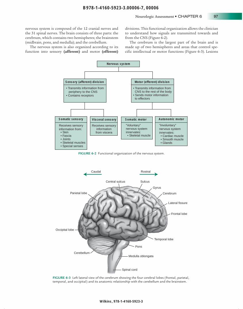
The cerebrum is the largest part of the brain and is made up of two hemispheres and areas that control spe-cific intellectual or motor functions ( Figure 6-3 ). Lesions
Nervous system
Sensory (afferent) division
Somatic sensory
• Skin • Fascia • Joints • Skeletal muscles • Special senses
Visceral sensory
Receives sensory informationfrom viscera
Somatic motor
“Voluntary”nervous systeminnervates:
Autonomic motor
• Cardiac muscle• Smooth muscle • Glands
Motor (efferent) division
• Transmits information from CNS to the rest of the body • Sends motor information to effectors
Receives sensory information from:
• Transmits information from periphery to the CNS• Contains receptors
“Involuntary” nervous system innervates:
• Skeletal muscle
FIGURE 6-2 Functional organization of the nervous system.
RostralCaudal
Frontal lobe
Parietal lobe
Occipital lobe
Gyrus
Sulcus
Temporal lobe
Cerebrum
Cerebellum
Pons
Medulla oblongata
Spinal cord
Lateral fissure
Central sulcus
FIGURE 6-3 Left lateral view of the cerebrum showing the four cerebral lobes (frontal, parietal, temporal, and occipital) and its anatomic relationship with the cerebellum and the brainstem.

Wilkins, 978-1-4160-5923-3
B978-1-4160-5923-3.00006-7, 00006
98 CHAPTER 6 • Neurologic Assessment
in the cerebrum can lead to abnormalities in functions such as movement, level of consciousness, ability to speak and write, emotions, or memory.
The brainstem is the lower part of the brain where it connects to the spinal cord. It consists of the midbrain, pons, and the medulla oblongata ( Figure 6-4 ). The brain-stem is the pathway for all fiber tracts passing up and down from peripheral nerves and spinal cord to the high-est parts of the brain. Most of the cranial nerves originate in the brainstem.
Many neurologic functions of particular importance to the RT, such as regulation of heart rate, blood pressure, and breathing, are located in the brainstem. In addition, the brainstem contains reflex centers for certain cranial nerve functions such as the pupillary reflex, which is dis-cussed later in this chapter. Lesions in the brainstem can cause a wide range of breathing problems from hyperven-tilation to apnea.
The cerebellum is located in the posterior part of the brain and is responsible for controlling equilibrium, mus-cle tone, and coordination of muscle movements. Lesions in the cerebellum cause characteristic symptoms such as loss of muscle coordination (ataxia) , tremors, and distur-bances in gait and balance.
The spinal cord lies within the center of the vertebral bodies and extends from the base of the brain down to the level of the first lumbar vertebra. It spans a distance of approximately 45 cm in the average adult. It serves the purpose of connecting the brain to the various parts of the
body for motor and sensory function. It is an oval cylinder that has two tapering bulges: one in the cervical region and one in the lumbar region.
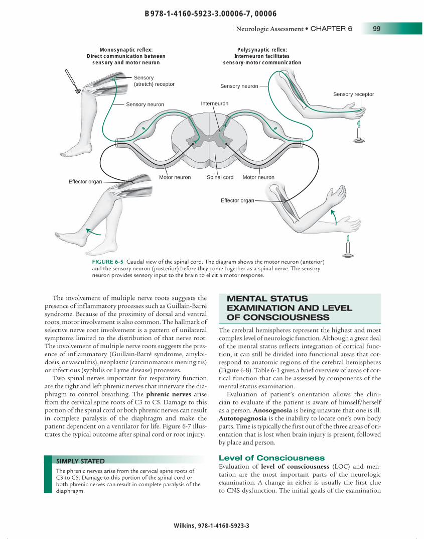
Two sets of nerve fibers called spinal nerves project from both sides of the spinal column at 31 locations along the spine. Posterior or dorsal (sensory) and anterior or ventral (motor) nerve roots separate as they exit the spinal cord until their fibers combine at the level of the dorsal root ganglion. The dorsal nerve root consists of posterior nerve fibers that carry sensory information into the spinal cord. The ventral nerve root consists of anterior nerve fibers that conduct motor impulses out of the spinal cord. Because all spinal nerves contain both motor and sensory fibers, they are called mixed nerves . Each has the ability to provide sensory input to the brain (e.g., feel pain) and the ability to cause muscle movement (e.g., extend the arm on com-mand) ( Figure 6-5 ).
These spinal nerves have no specific name but rather are numbered according to the level of the vertebral column at which they exit the spinal column. There are eight cervical (C1 to C8), twelve thoracic (T1 to T12), five lumbar (L1 to L5), five sacral (S1 to S5), and one coccygeal pair of spinal nerves.
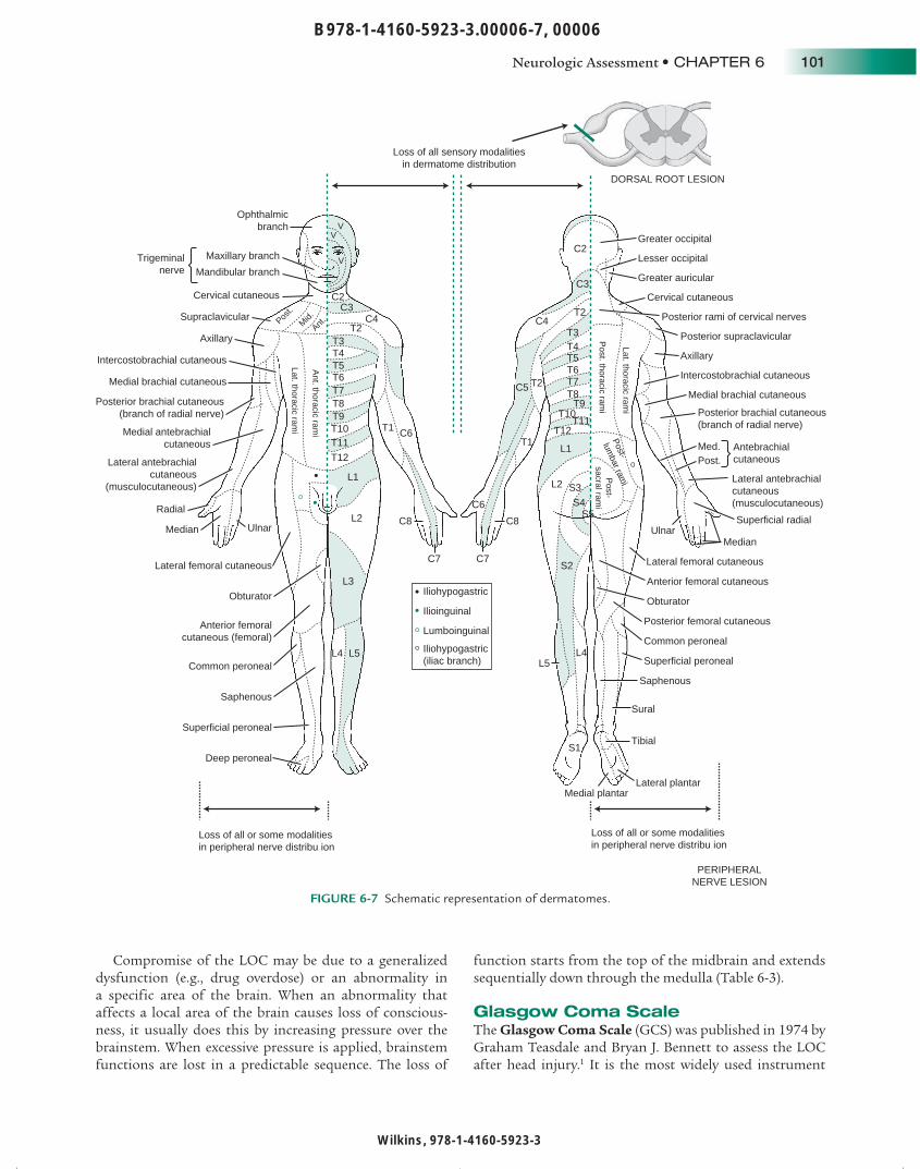
A herniated vertebral disk is the most common nerve root pathology that results in compression on the nerve roots. This usually results in pain with radiation into the affected area of skin (dermatome) supplied with afferent nerve fibers by a single posterior spinal root ( Figure 6-6 ).
Pons
Medulla oblongata
Mesencephalic aqueduct
Mammillary body
Fourth ventricle
Mesencephalon
Pineal gland
Corpora quadrigemina
White matter(arbor vitae)
Folia
Gray matter
FIGURE 6-4 Left lateral view of the cerebellum showing its anatomic relationship with the brainstem.
SIMPLY STATED Many neurologic functions of particular importance to the RT, such as regulation of heart rate, blood pressure, and breathing, are located in the brainstem.
SIMPLY STATED A herniated vertebral disk is the most common nerve root pathology, and it causes pain with radiation into the affected area of skin (dermatome) supplied with afferent nerve fibers by a single posterior spinal root.
Wilkins, 978-1-4160-5923-3
B978-1-4160-5923-3.00006-7, 00006
Neurologic Assessment • CHAPTER 6 99
The involvement of multiple nerve roots suggests the presence of inflammatory processes such as Guillain-Barré syndrome. Because of the proximity of dorsal and ventral roots, motor involvement is also common. The hallmark of selective nerve root involvement is a pattern of unilateral symptoms limited to the distribution of that nerve root. The involvement of multiple nerve roots suggests the pres-ence of inflammatory (Guillain-Barré syndrome, amyloi-dosis, or vasculitis), neoplastic (carcinomatous meningitis) or infectious (syphilis or Lyme disease) processes.
Two spinal nerves important for respiratory function are the right and left phrenic nerves that innervate the dia-phragm to control breathing. The phrenic nerves arise from the cervical spine roots of C3 to C5. Damage to this portion of the spinal cord or both phrenic nerves can result in complete paralysis of the diaphragm and make the patient dependent on a ventilator for life. Figure 6-7 illus-trates the typical outcome after spinal cord or root injury.
MENTAL STATUS EXAMINATION AND LEVEL OF CONSCIOUSNESS
The cerebral hemispheres represent the highest and most complex level of neurologic function. Although a great deal of the mental status reflects integration of cortical func-tion, it can still be divided into functional areas that cor-respond to anatomic regions of the cerebral hemispheres ( Figure 6-8 ). Table 6-1 gives a brief overview of areas of cor-tical function that can be assessed by components of the mental status examination.
Evaluation of patient’s orientation allows the clini-cian to evaluate if the patient is aware of himself/herself as a person. Anosognosia is being unaware that one is ill. Autotopagnosia is the inability to locate one’s own body parts. Time is typically the first out of the three areas of ori-entation that is lost when brain injury is present, followed by place and person.
Level of Consciousness Evaluation of level of consciousness (LOC) and men-tation are the most important parts of the neurologic examination. A change in either is usually the first clue to CNS dysfunction. The initial goals of the examination
Monosynaptic reflex:Direct communication between
sensory and motor neuron
Polysynaptic reflex:Interneuron facilitates
sensory-motor communication
Sensory(stretch) receptor
Effector organ
Effector organ
Motor neuron Motor neuron
Interneuron
Spinal cord
Sensory neuron
Sensory neuron
Sensory receptor
FIGURE 6-5 Caudal view of the spinal cord. The diagram shows the motor neuron (anterior) and the sensory neuron (posterior) before they come together as a spinal nerve. The sensory neuron provides sensory input to the brain to elicit a motor response.
SIMPLY STATED The phrenic nerves arise from the cervical spine roots of C3 to C5. Damage to this portion of the spinal cord or both phrenic nerves can result in complete paralysis of the diaphragm.

Wilkins, 978-1-4160-5923-3
B978-1-4160-5923-3.00006-7, 00006
100 CHAPTER 6 • Neurologic Assessment
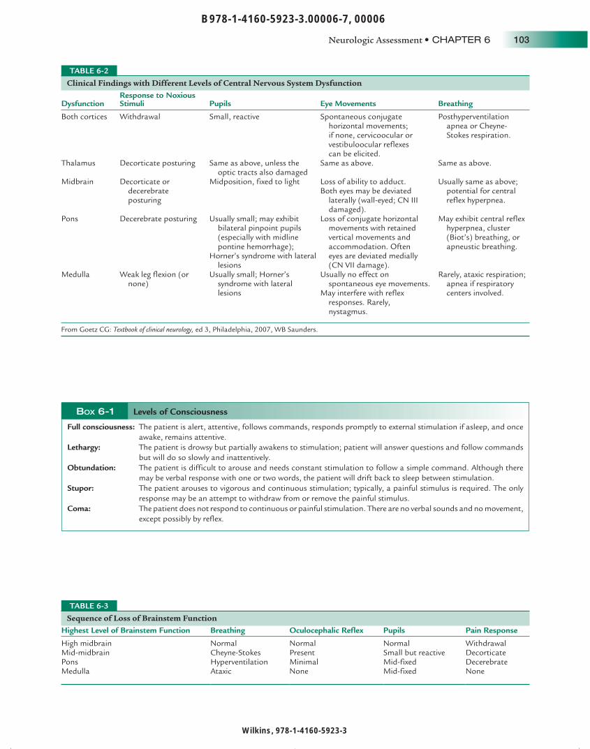
of a patient with altered mental status are to determine if the patient is conscious and then to determine awareness. Altered awareness is associated with either reticular system dysfunction or bilateral hemispheric dysfunction. Testing anatomic structures surrounding these structures provides the major clues regarding the etiology and level of dysfunc-tion. Table 6-2 shows the major findings on assessment of the LOC and their expected anatomic correlates.
Assessment of mentation begins when you first encoun-ter the patient. A neurologically healthy patient will be awake and interacting with those around. If asleep, the patient can be easily aroused to an awake, alert state. Different levels of consciousness from full alertness to coma have been defined ( Box 6-1 ). Since these and other terms used to categorize the LOC are frequently used
imprecisely, it is often recommended to avoid using them. Instead, a brief description of the applied stimulus and arousal pattern is preferred.
A condition in which a patient’s eyes may be open, but the patient cannot be aroused is known as a persistent vegetative state or prolonged postcoma unresponsiveness 3 and typically indicates irreversible brain damage. Breathing may not be affected if the brainstem is unaffected by the injury.
C1
C4
C7T1
T9
T10
L1
Spinal corddamage
Root damage
Patients with high cervical cordlesions seldom survive withoutimmediate ventilatory support.
Patients who survive a lesionabove C7 usually remaindependent on others for daily care.
Sparing of the C7 segment retainselbow and wrist extension andenables transfer from wheelchairto bed, providing a degreeof independence.
Patients with thoracolumbarinjuries usually regain fullindependence.
A mixed cord and lumbar rootlesion may occur at this level.Fortunately roots are moreresistant to injury––“rootescape”––and the outlookis more favorable.
FIGURE 6-6 Outcome after spinal cord or root injury.
SIMPLY STATED Evaluation of the LOC and mentation are the most important parts of the neurologic examination. A change in either is usually the first clue to CNS dysfunction.
Wilkins, 978-1-4160-5923-3
B978-1-4160-5923-3.00006-7, 00006
Neurologic Assessment • CHAPTER 6 101
Compromise of the LOC may be due to a generalized dysfunction (e.g., drug overdose) or an abnormality in a specific area of the brain. When an abnormality that affects a local area of the brain causes loss of conscious-ness, it usually does this by increasing pressure over the brainstem. When excessive pressure is applied, brainstem functions are lost in a predictable sequence. The loss of
function starts from the top of the midbrain and extends sequentially down through the medulla ( Table 6-3 ).
Glasgow Coma Scale The Glasgow Coma Scale (GCS) was published in 1974 by Graham Teasdale and Bryan J. Bennett to assess the LOC after head injury. 1 It is the most widely used instrument
Ophthalmicbranch
Trigeminalnerve
Maxillary branch
Mandibular branch
Cervical cutaneous
Supraclavicular
Axillary
Medial brachial cutaneous
Intercostobrachial cutaneous
Posterior brachial cutaneous(branch of radial nerve)
Medial antebrachialcutaneous
Lateral antebrachialcutaneous
(musculocutaneous)
Ulnar UlnarMedianMedian
Radial
Lateral femoral cutaneous Lateral femoral cutaneous
Obturator
Anterior femoralcutaneous (femoral)
Common peroneal
Saphenous
Superficial peroneal
Deep peroneal
Iliohypogastric
Ilioinguinal
Lumboinguinal
Iliohypogastric(iliac branch)
Loss of all sensory modalitiesin dermatome distribution
Loss of all or some modalitiesin peripheral nerve distribu ion
Loss of all or some modalitiesin peripheral nerve distribu ion
PERIPHERALNERVE LESION
Medial plantarLateral plantar
Tibial
Sural
Saphenous
Superficial peroneal
Common peroneal
Obturator
Posterior femoral cutaneous
Anterior femoral cutaneous
Superficial radial
Lateral antebrachialcutaneous(musculocutaneous)
Antebrachialcutaneous
Med.Post.
Posterior brachial cutaneous(branch of radial nerve)
Medial brachial cutaneous
Intercostobrachial cutaneous
Axillary
Posterior supraclavicular
Posterior rami of cervical nerves
Cervical cutaneous
Greater auricular
Lesser occipital
Greater occipitalC2
C2C3
C3C4C4
C5
C6
C6
C7C7
C8C8
T2
T2
T1T1
T2
T3T3 T4T4 T5T5 T6T6 T7T7 T8T8 T9T9 T10
T10T11
T11T12
T12L1
L1L2
L2
L4L4L5
L5
L3
S3
S2
S1
S4S5
Post-
sacral rami
Post-
lumbar ram
i
Post. thoracic ram
i
Ant. thoracic ram
i
Lat. thoracic rami
Lat. thoracic rami
Post.
Mid.
Ant.
VV
V
DORSAL ROOT LESION
FIGURE 6-7 Schematic representation of dermatomes.

Wilkins, 978-1-4160-5923-3
B978-1-4160-5923-3.00006-7, 00006
102 CHAPTER 6 • Neurologic Assessment
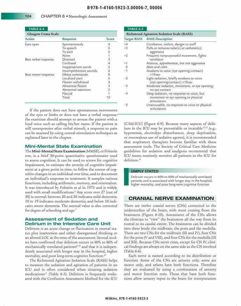
for quantifying neurologic impairment. The GCS is used to test best motor response, best verbal response, and eye opening. Although it is easy to perform and readily repro-ducible, it is poorly suited for patients who have impaired verbal responses caused by aphasia, hearing loss, or tracheal intubation. 4 A scale that goes from 3 (deep coma or death) to 15 (fully awake) is useful for rapid triage ( Table 6-4 ).
Patients with GCS scores of 12 to 15 often are admitted to a non-ICU observational unit unless neurologic exam-ination or a diagnostic test reveals a lesion or abnormal-ity that warrants ICU admission. Scores of 9 to 12 on the GCS indicate a significant insult with a moderate coma. 5
Patients with GCS scores less than 9 have a severe coma and typically require endotracheal intubation. Since endo-tracheal intubation makes it impossible to test the patient’s verbal response, the letter “T” is often attached to the GCS score to indicate the presence of the tube (e.g., GCS 5T).
Occipital lobeInterpretation of writtenlanguage (left hemisphere).Visual interpretation
Parietal lobeRecognition of right/leftdifferentiation, sensation,recognition of body parts
Frontal lobeJudgment, humor, social mores,affect, personality, motor movementexpressive speech (Broca, lefthemisphere), spatial and perceptualinformation (right hemisphere)
Temporal lobeHearing, comprehension ofspoken and written language(Wenecke, left hemisphere),long-term memory
FIGURE 6-8 Graphical representation of the cerebral lobes and their corresponding cortical function.
Assessment of Cerebral Cortical Function Cerebral Lobe Cortical Function Assessment
Frontal Attention: working memory
Judgment: abstract reasoningSet generation
Digit span, spelling backward, and naming months of the year backward
Problem solving, verbal similarities, and proverbsVerbal fluency and the ability to generate a set of items
Temporal Orientation, memory Questions about month, date, day of week and place Three-word recall (recent memory) Naming Presidents (remote memory)
Frontal-temporal Receptive languageExpressive language
Follow commands (spoken and written language)Fluency and correctness of content and grammar Reading comprehension
Parietal (dominant) Gnosis
Constructional
Identify objects placed in their hand and numbers written on their hand with eyes closed
Attending to the contralateral side of the body Drawing a face, clock, or geometric figures Right-left orientation, naming fingers, and calculations
Parietal (nondominant) Praxis Perform skilled motor tasks without any nonverbal promptingOccipitotemporal Visual recognition Recognition of colors and faces
TABLE 6-1
SIMPLY STATED The GCS is the most widely used instrument for quantifying neurologic impairment. Patients with GCS scores less than 8 have a severe coma and typically require endotracheal intubation.
Wilkins, 978-1-4160-5923-3
B978-1-4160-5923-3.00006-7, 00006
Neurologic Assessment • CHAPTER 6 103
Clinical Findings with Different Levels of Central Nervous System Dysfunction
DysfunctionResponse to Noxious Stimuli Pupils Eye Movements Breathing
Both cortices Withdrawal Small, reactive Spontaneous conjugate horizontal movements; if none, cervicoocular or vestibuloocular reflexes can be elicited.
Posthyperventilation apnea or Cheyne-Stokes respiration.
Thalamus Decorticate posturing Same as above, unless the optic tracts also damaged
Same as above. Same as above.
Midbrain Decorticate or decerebrate posturing
Midposition, fixed to light Loss of ability to adduct. Both eyes may be deviated
laterally (wall-eyed; CN III damaged).
Usually same as above; potential for central reflex hyperpnea.
Pons Decerebrate posturing Usually small; may exhibit bilateral pinpoint pupils (especially with midline pontine hemorrhage);
Horner’s syndrome with lateral lesions
Loss of conjugate horizontal movements with retained vertical movements and accommodation. Often eyes are deviated medially (CN VII damage).
May exhibit central reflex hyperpnea, cluster (Biot’s) breathing, or apneustic breathing.
Medulla Weak leg flexion (or none)
Usually small; Horner’s syndrome with lateral lesions
Usually no effect on spontaneous eye movements.
May interfere with reflex responses. Rarely, nystagmus.
Rarely, ataxic respiration; apnea if respiratory centers involved.
From Goetz CG: Textbook of clinical neurology, ed 3, Philadelphia, 2007, WB Saunders.
TABLE 6-2
Sequence of Loss of Brainstem Function Highest Level of Brainstem Function Breathing Oculocephalic Reflex Pupils Pain Response
High midbrain Normal Normal Normal WithdrawalMid-midbrain Cheyne-Stokes Present Small but reactive DecorticatePons Hyperventilation Minimal Mid-fixed DecerebrateMedulla Ataxic None Mid-fixed None
TABLE 6-3
BOX 6-1 Levels of Consciousness
Full consciousness: The patient is alert, attentive, follows commands, responds promptly to external stimulation if asleep, and once awake, remains attentive.
Lethargy: The patient is drowsy but partially awakens to stimulation; patient will answer questions and follow commands but will do so slowly and inattentively.
Obtundation: The patient is difficult to arouse and needs constant stimulation to follow a simple command. Although there may be verbal response with one or two words, the patient will drift back to sleep between stimulation.
Stupor: The patient arouses to vigorous and continuous stimulation; typically, a painful stimulus is required. The only response may be an attempt to withdraw from or remove the painful stimulus.
Coma: The patient does not respond to continuous or painful stimulation. There are no verbal sounds and no movement, except possibly by reflex.
Wilkins, 978-1-4160-5923-3
B978-1-4160-5923-3.00006-7, 00006
104 CHAPTER 6 • Neurologic Assessment
If the patient does not have spontaneous movements of the eyes or limbs or does not have a verbal response, the examiner should attempt to arouse the patient with a loud voice such as calling his/her name. If the patient is still unresponsive after verbal stimuli, a response to pain can be assessed by using central stimulation techniques as explained later in the chapter.
Mini-Mental State Examination The Mini-Mental State Examination (MMSE), or Folstein test, is a brief 30-point quantitative questionnaire used to assess cognition. It can be used to screen for cognitive impairment, to estimate the severity of cognitive impair-ment at a given point in time, to follow the course of cog-nitive changes in an individual over time, and to document an individual’s response to treatment. It samples various functions, including arithmetic, memory, and orientation. It was introduced by Folstein et al in 1975 and is widely used with small modifications. 6 Any score over 27 (out of 30) is normal; between 20 and 26 indicates mild dementia; 10 to 19 indicates moderate dementia, and below 10 indi-cates severe dementia. The normal value is also corrected for degree of schooling and age. 7
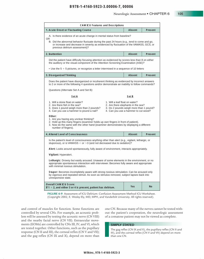
Assessment of Sedation and Delirium in the Intensive Care Unit Delirium is an acute change or fluctuation in mental sta-tus plus inattention and either disorganized thinking or an altered LOC at the time of the assessment. Several stud-ies have confirmed that delirium occurs in 60% to 80% of mechanically ventilated patients 8-10 and that it is indepen-dently associated with longer stay in the hospital, higher mortality, and poor long-term cognitive function. 10
The Richmond Agitation Sedation Scale (RASS) helps to measure the sedation and agitation of patients in an ICU and is often considered when titrating sedation medications 11 ( Table 6-5 ). Delirium is frequently evalu-ated with the Confusion Assessment Method for the ICU
(CAM-ICU) ( Figure 6-9 ). Because many aspects of delir-ium in the ICU may be preventable or treatable 12,13 (e.g., hypoxemia, electrolyte disturbances, sleep deprivation, or overzealous use of sedative agents), it is recommended that respiratory therapists become familiar with these assessment tools. The Society of Critical Care Medicine guidelines for sedation and analgesia recommend that ICU teams routinely monitor all patients in the ICU for delirium. 14
CRANIAL NERVE EXAMINATION
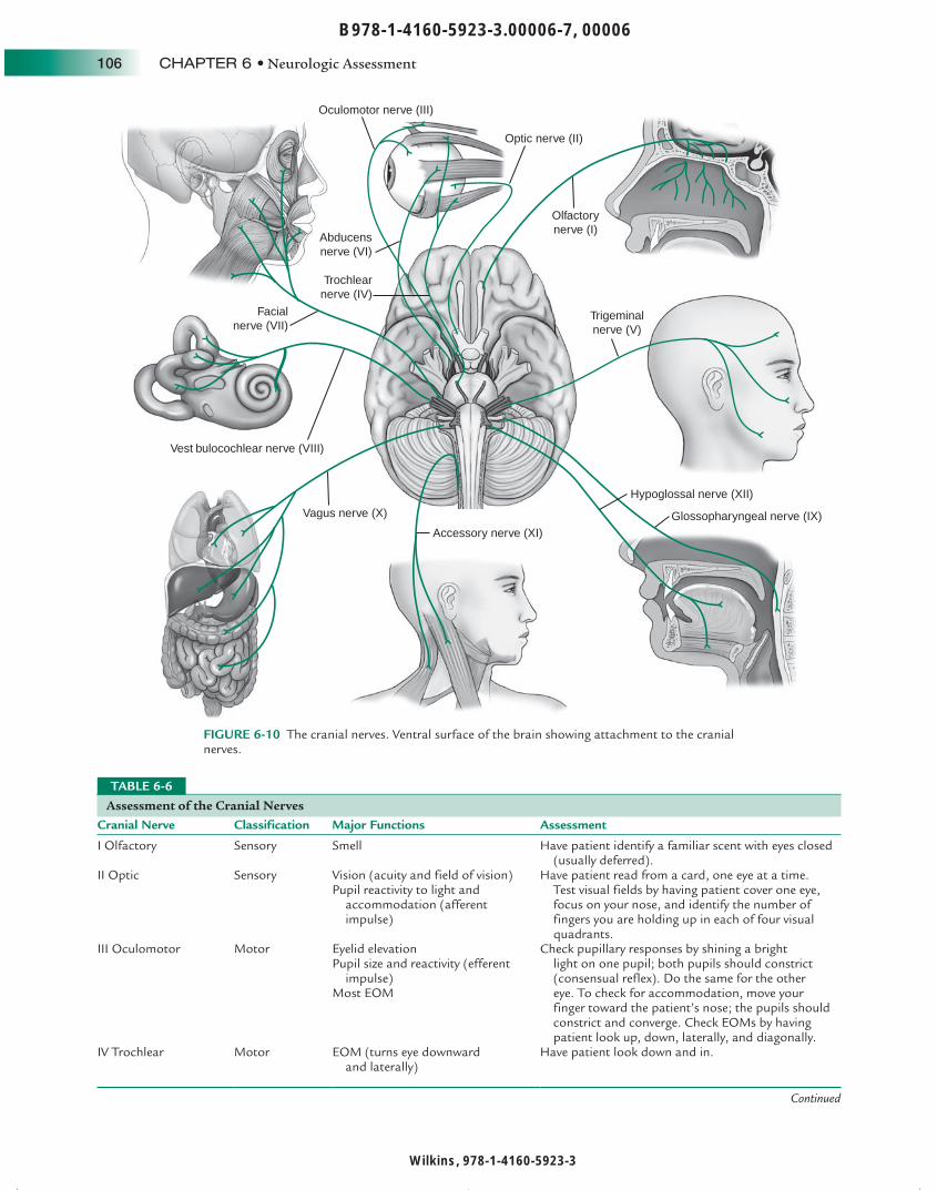
There are twelve cranial nerves (CNs) connected to the undersurface of the brain, with most coming from the brainstem ( Figure 6-10 ). Assessment of the CNs allows the clinician to “view” the brainstem all the way from its rostral to its caudal extent. The brainstem can be divided into three levels: the midbrain, the pons and the medulla. There are two CNs for the midbrain (III and IV), four CNs for the pons (V and VIII), and four CNs for the medulla (IX and XII). Because CNs never cross, except for CN IV, clini-cal findings are always on the same side as the CN involved ( Table 6-6 ).
Each nerve is named according to its distribution or function. Some of the CNs are sensory only, some are motor only, and others have both functions. Therefore they are evaluated by using a combination of sensory and motor function tests. Those that have both func-tions allow sensory input to the brain for interpretation
Glasgow Coma Scale Action Response Score
Eyes open SpontaneouslyTo speechTo painNone
4 3 2 1
Best verbal response OrientedConfusedInappropriate wordsIncomprehensive sounds
5 4 3 2
Best motor response Obeys commandsLocalized painFlexion withdrawalAbnormal flexionAbnormal extensionFlaccid
6 5 4 3 2 1
Total 15
TABLE 6-4
Richmond Agitation Sedation Scale (RASS) Target RASS RASS Description
+4 Combative, violent, danger to staff+3 Pulls or removes tube(s) or catheter(s);
aggressive+2 Frequent nonpurposeful movement, fights
ventilator+1 Anxious, apprehensive, but not aggressive 0 Alert and calm−1 Awakens to voice (eye opening/contact)
>10 sec−2 Light sedation, briefly awakens to voice
(eye opening/contact) <10 sec−3 Moderate sedation, movement, or eye opening;
no eye contact.−4 Deep sedation, no response to voice, but
movement or eye opening to physical stimulation
−5 Unarousable, no response to voice or physical stimulation
TABLE 6-5
SIMPLY STATED Delirium occurs in 60% to 80% of mechanically ventilated patients and is associated with longer stay in the hospital, higher mortality, and poor long-term cognitive function.
Wilkins, 978-1-4160-5923-3
B978-1-4160-5923-3.00006-7, 00006
Neurologic Assessment • CHAPTER 6 105
and control of muscles for function. Some functions are controlled by several CNs. For example, an acoustic prob-lem will be assessed by testing the acoustic nerve (CN VIII) and the nearby facial nerve (CN VII). Extraocular move-ments (EOMs) are controlled by CNs III, IV, and VI, which are tested together. Other functions, such as the pupillary response (CN II and III), the corneal reflex (CN V and VII); and the gag reflex (CN IX and X), depend on more than
one CN. Because many of the nerves cannot be tested with-out the patient’s cooperation, the neurologic assessment of a comatose patient may not be viewed as complete.
CAM ICU Features and Descriptions
1. Acute Onset or Fluctuating Course
A. Is there evidence of an acute change in mental status from baseline?
B. Did the abnormal behavior fluctuate during the past 24 hours (e.g., tend to come and go, or increase and decrease in severity as evidenced by fluctuation of the VAMASS, GCS, or previous delirium assessment)?
Did the patient have difficulty focusing attention as evidenced by scores less than 8 on either the auditory or the visual component of the Attention Screening Examination (ASE)?
• Use the 5 � 5 pictures, or recognize a letter intermixed in a sequence of 10 letters
Does the patient have disorganized or incoherent thinking as evidenced by incorrect answers to 2 or more of the following 4 questions and/or demonstrate an inability to follow commands?
Questions (Alternate Set A and Set B):
Is the patient’s level of consciousness anything other than alert (e.g., vigilant, lethargic, or stuporous), or is VAMASS � or �3 (and not decreased due to sedation)?
Alert: Looks around spontaneously, fully aware of environment, interacts appropriately.
Vigilant: Hyperalert.
Lethargic: Drowsy but easily aroused. Unaware of some elements in the environment, or no appropriate spontaneous interaction with interviewer. Becomes fully aware and appropriate with minimal noxious stimulation.
Stupor: Becomes incompletely aware with strong noxious stimulation. Can be aroused only by vigorous and repeated stimuli. As soon as stimulus removed, subject lapses back into unresponsive state.
Set A
1. Will a stone float on water?2. Are there fish in the sea?3. Does 1 pound weigh more than 2 pounds?4. Can you use a hammer to pound a nail?
Other:1. Are you having any unclear thinking?2. Hold up this many fingers (examiner holds up own fingers in front of patient).3. Now do the same with the other hand (examiner demonstrates by displaying a different number of fingers).
Set B
1. Will a leaf float on water?2. Are there elephants in the sea?3. Do 2 pounds weigh more than 1 pound?4. Can you use a hammer to cut wood?
Absent Present
2. Inattention Absent Present
3. Disorganized Thinking Absent Present
4. Altered Level of Consciousness Absent Present
Overall CAM ICU Score:If 1 � 2, and either 3 or 4 is present, patient has delirium. Yes No
OR
FIGURE 6-9 Assessment of ICU Delirium: Confusion Assessment Method ICU Worksheet. (Copyright 2002, E. Wesley Ely, MD, MPH, and Vanderbilt University. All rights reserved).
SIMPLY STATED The gag reflex (CN IX and X), the pupillary reflex (CN II and III), and the corneal reflex (CN V and VII) depend on more than one CN.
Wilkins, 978-1-4160-5923-3
B978-1-4160-5923-3.00006-7, 00006
106 CHAPTER 6 • Neurologic Assessment
Oculomotor nerve (III)
Optic nerve (II)
Abducensnerve (VI)
Facialnerve (VII)
Vest bulocochlear nerve (VIII)
Vagus nerve (X)
Accessory nerve (XI)
Hypoglossal nerve (XII)
Trochlearnerve (IV)
Glossopharyngeal nerve (IX)
Trigeminalnerve (V)
Olfactorynerve (I)
FIGURE 6-10 The cranial nerves. Ventral surface of the brain showing attachment to the cranial nerves.
Assessment of the Cranial Nerves Cranial Nerve Classification Major Functions Assessment
I Olfactory Sensory Smell Have patient identify a familiar scent with eyes closed (usually deferred).
II Optic Sensory Vision (acuity and field of vision) Pupil reactivity to light and
accommodation (afferent impulse)
Have patient read from a card, one eye at a time. Test visual fields by having patient cover one eye, focus on your nose, and identify the number of fingers you are holding up in each of four visual quadrants.
III Oculomotor Motor Eyelid elevation Pupil size and reactivity (efferent
impulse) Most EOM
Check pupillary responses by shining a bright light on one pupil; both pupils should constrict (consensual reflex). Do the same for the other eye. To check for accommodation, move your finger toward the patient’s nose; the pupils should constrict and converge. Check EOMs by having patient look up, down, laterally, and diagonally.
IV Trochlear Motor EOM (turns eye downward and laterally)
Have patient look down and in.
TABLE 6-6
Continued
Wilkins, 978-1-4160-5923-3
B978-1-4160-5923-3.00006-7, 00006
Neurologic Assessment • CHAPTER 6 107
Although a stroke is the most common cause of CN dysfunction, other abnormalities should be considered ( Table 6-7 ).
SENSORY EXAMINATION
Clinically, there are two major somatosensory pathways that are examined. The first is the spinothalamic (ST) part of the anterolateral system, and the second is the
dorsal column-medial lemniscus (DCML) system. The principle sensory modalities for the ST system are pain and temperature. The principle sensory modalities for DCML system are vibratory, position sense, and discrim-inatory or integrative sensation. Spinal cord and lower brainstem lesions can result in sensory dissociation , which means one sensory system is affected but the other is not.
Sensory evaluation is performed by having the patient respond to stimuli at a specific location. It evaluates the ability to perceive and identify specific sensations with the patient’s eyes closed. The patient must be able to cooper-ate with the examination by communicating whether or not the sensation is felt and whether both sides of the body feel it equally. The assessment of light touch, pinprick, and temperature sensation can be achieved by applying a cotton swab, clean pin, and a cold or warm object, respec-tively, to various parts of the body. The clinician should begin with the patient’s feet and move upward. Comparing one side with the other is valuable in localizing the spe-cific site of abnormality. To test vibratory sensation, use a low-frequency tuning fork.
To test proprioception , or position sense, have the patient with his eyes closed distinguish whether his finger or toe are moved up or down. Patients should be able to dis-criminate between two different points 2 to 10 mm apart on their fingers and hands and up to 75 mm on their thigh and back. Graphesthesia examines the patient’s ability to identify numbers written on their palm and stereognosis tests for recognition of objects placed in their hands with their eyes closed.
Cranial Nerve Classification Major Functions Assessment
V Trigeminal Both Chewing Facial and mouth sensation Corneal reflex (sensory)
Ask patient to hold the mouth open while you try to close it and to move the jaw laterally against your hand. With patient’s eyes closed, touch her face with cotton and have her identify the area touched. In comatose patients, brush the cornea with a wisp of cotton; the patient should blink.
VI Abducens Motor EOM (turns eye laterally) Have patient move the eyes from side to side.VII Facial Both Facial expression
Taste Corneal reflex (motor) Eyelid and lip closure
Ask patient to smile, raise eyebrows, and keep eyes and lips closed while you try to open them.
Have patient identify salt or sugar placed on the tongue.
VIII Acoustic Sensory Hearing Equilibrium
To test hearing, use tuning fork, rub your fingers, or whisper near each ear.
IX Glossopharyngeal Both Gagging and swallowing (sensory) Taste
Touch back of throat with sterile tongue depressor or cotton-tipped applicator. Have patient swallow.
X Vagus Both Gagging and swallowing (motor) Speech (phonation)
Assess gag and swallowing with CN IX. Assess vocal quality.
XI Spinal accessory Motor Shoulder movement Head rotation
Have patient shrug shoulders and turn head from side to side.
XII Hypoglossal Motor Tongue movement Speech (articulation)
Have patient stick out tongue and move it internally from cheek to cheek.
Assess articulation.
EOM, Extraocular movements; CN, cranial nerve.
TABLE 6-6
Assessment of the Cranial Nerves—cont’d
Etiology of Cranial Nerve Malfunction Cranial Nerve Cause of Impairment
I Trauma to the cribriform plate, frontal lobe mass or stroke, and nasal problems (e.g., allergic or viral)
II Eye disease or injury, diabetic retinopathy and glaucoma are major causes, occipital lobe mass or stroke.
III, IV, and VI Brainstem injury or compression (e.g., tumor, stroke, intracranial bleeding, diabetic neuropathy [can cause temporary palsies]).
V Stroke in the contralateral sensory cortex.VII Stroke-induced (central palsy).VIII Sensorineural hearing loss as a result
of age or noise exposure, tumors at cerebellopontine angle, acoustic neuroma, earwax or middle ear disease can cause temporary hearing loss.
IX and X Stroke.XI Neck injury.XII Stroke.
TABLE 6-7
Wilkins, 978-1-4160-5923-3
B978-1-4160-5923-3.00006-7, 00006
108 CHAPTER 6 • Neurologic Assessment
Motor Examination A bedside neurologic assessment almost always includes an evaluation of motor function. Because the clinician assesses the patient’s ability to move on command, they must be awake, willing to cooperate, and able to under-stand what the examiner is asking.
Motor strength is assessed bilaterally by having the patient flex and extend his/her arm against your hand, squeezing your fingers, lifting his/her leg while you press down on the thigh, holding his/her leg straight and lift-ing it against gravity, and flexing and extending his/her foot against your hand. Each extremity is graded by using a motor scale from 0 (no movement) to +5 (full range of motion with full strength). In an unconscious patient, the assessment of motor response is performed by applying a noxious stimulus and observing the patient’s response to it. Central stimulation, such as sternal pressure, produces an overall body response and is more reliable than periph-eral stimulation. In an unconscious patient, peripheral stimulation, such as nail bed pressure, can elicit a reflex response, which is not a true indicator of motor activity. If central stimulation is necessary, it should be performed judiciously because deep sternal pressure can easily bruise the soft tissue above the sternum. A less traumatic alterna-tive to sternal pressure is to squeeze the trapezius muscle. Supraorbital pressure should not be used for central stim-ulation on patients with facial fractures or vagal nerve sen-sitivity. The response to pain varies depending on the level of neurologic function. Normally, pain causes the patient to attempt to remove the source of the pain or to withdraw from the painful stimulation. If the cerebral cortex is func-tioning, there is a withdrawal from painful stimuli in a pre-dictable and reflexive manner. The symmetry and pattern of the motor response to noxious stimuli, as well as associ-ated neurologic symptoms, should be documented for all patients suspected of having a neurologic disease.
Subtle central weakness (such as with early CNS malig-nancy) can be tested via pronator drift . Ask your patient to hold his/her arms forward with palms up. In mild corti-cal weakness, the patient’s hand on the weak side pronates and drifts down.
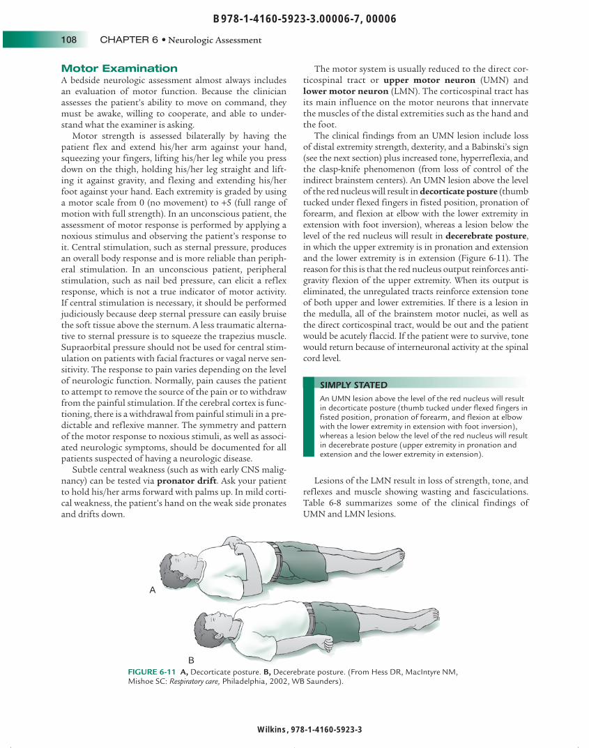
The motor system is usually reduced to the direct cor-ticospinal tract or upper motor neuron (UMN) and lower motor neuron (LMN). The corticospinal tract has its main influence on the motor neurons that innervate the muscles of the distal extremities such as the hand and the foot.
The clinical findings from an UMN lesion include loss of distal extremity strength, dexterity, and a Babinski’s sign (see the next section) plus increased tone, hyperreflexia, and the clasp-knife phenomenon (from loss of control of the indirect brainstem centers). An UMN lesion above the level of the red nucleus will result in decorticate posture (thumb tucked under flexed fingers in fisted position, pronation of forearm, and flexion at elbow with the lower extremity in extension with foot inversion), whereas a lesion below the level of the red nucleus will result in decerebrate posture , in which the upper extremity is in pronation and extension and the lower extremity is in extension ( Figure 6-11 ). The reason for this is that the red nucleus output reinforces anti-gravity flexion of the upper extremity. When its output is eliminated, the unregulated tracts reinforce extension tone of both upper and lower extremities. If there is a lesion in the medulla, all of the brainstem motor nuclei, as well as the direct corticospinal tract, would be out and the patient would be acutely flaccid. If the patient were to survive, tone would return because of interneuronal activity at the spinal cord level.
Lesions of the LMN result in loss of strength, tone, and reflexes and muscle showing wasting and fasciculations. Table 6-8 summarizes some of the clinical findings of UMN and LMN lesions.
A
B FIGURE 6-11 A, Decorticate posture. B, Decerebrate posture. (From Hess DR, MacIntyre NM, Mishoe SC: Respiratory care, Philadelphia, 2002, WB Saunders).
SIMPLY STATED An UMN lesion above the level of the red nucleus will result in decorticate posture (thumb tucked under flexed fingers in fisted position, pronation of forearm, and flexion at elbow with the lower extremity in extension with foot inversion), whereas a lesion below the level of the red nucleus will result in decerebrate posture (upper extremity in pronation and extension and the lower extremity in extension).

Wilkins, 978-1-4160-5923-3
B978-1-4160-5923-3.00006-7, 00006
Neurologic Assessment • CHAPTER 6 109
DEEP TENDON, SUPERFICIAL, AND BRAINSTEM REFLEXES
Reflex assessment encompasses deep tendon, superficial, and brainstem reflexes.
Deep Tendon Reflexes Deep tendon reflexes evaluate spinal nerves and include the triceps, biceps, brachioradialis, patellar, and the Achilles tendon. Although deep tendon reflexes are not routinely assessed, they should be tested in any patient with a spinal cord injury or symptoms consistent with a neurologic problem. The patellar reflex or “knee-jerk” is
tested by tapping on the patellar tendon with a reflex ham-mer while the patient’s leg hangs loosely at a right angle with the thigh. Normally, the lower leg jerks forward when this reflex is intact ( Figure 6-12 ). The absence of this reflex is known as Westphal’s sign .
The reflexes are graded on a scale from 0 to 5+, with 0 being no reflex, 2+ being normal, and 5+ being hyperreflexia with clonus (repeated rhythmic contractions). Abnormal or absent deep tendon reflexes indicate abnormalities in anatomic components required for the reflex arc to occur. These structures include the muscle, the nerve fibers going from the tendon to the spinal cord, and the nerve fibers returning from the spinal cord to the muscle. Myasthenia gravis and botulism are diseases characterized by abnor-mal deep tendon reflexes caused by abnormalities of the neuromuscular junction that impair the normal impulse transmission. Table 6-9 illustrates the spinal nerve level assessed with each reflex. Absent deep tendon reflexes may be a sign that the patient is at risk for respiratory failure.
Clinical Signs of UMN and LMN Lesions Clinical Sign UMN Lesion LMN Lesion
Weakness + +Atrophy − +Fasciculation − +Reflexes ↑ ↓Tone ↑ ↓Babinski + −
UMN, Upper motor neuron; LMN, lower motor neuron.
TABLE 6-8
1a afferent
Quadriceps
Muscle spindle
Tendon of quadriceps
Alpha motor neuron
FIGURE 6-12 Patellar reflex arc. Forward knee jerk is elicited by tapping of the tendon of the quadriceps.
SIMPLY STATED Myasthenia gravis and botulism are diseases characterized by abnormal deep tendon reflexes caused by abnormalities of the neuromuscular junction that impair the normal impulse transmission.
Wilkins, 978-1-4160-5923-3
B978-1-4160-5923-3.00006-7, 00006
110 CHAPTER 6 • Neurologic Assessment
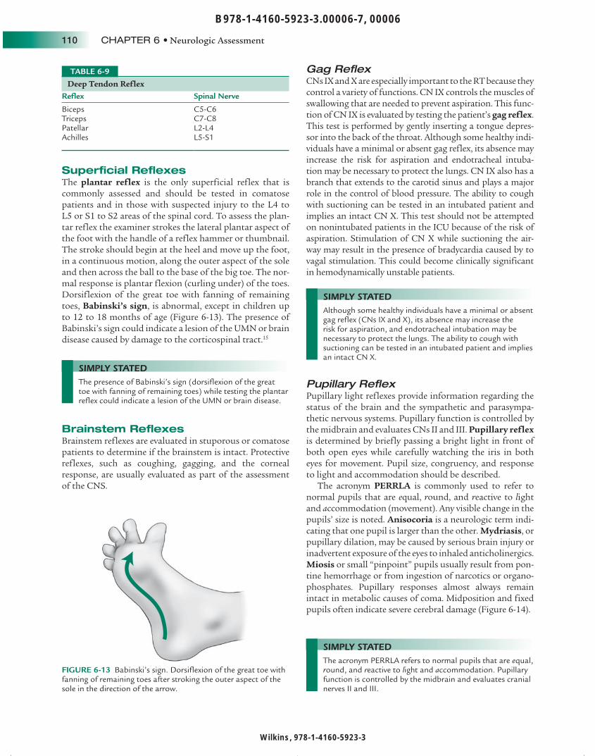
Superficial Reflexes The plantar reflex is the only superficial reflex that is commonly assessed and should be tested in comatose patients and in those with suspected injury to the L4 to L5 or S1 to S2 areas of the spinal cord. To assess the plan-tar reflex the examiner strokes the lateral plantar aspect of the foot with the handle of a reflex hammer or thumbnail. The stroke should begin at the heel and move up the foot, in a continuous motion, along the outer aspect of the sole and then across the ball to the base of the big toe. The nor-mal response is plantar flexion (curling under) of the toes. Dorsiflexion of the great toe with fanning of remaining toes, Babinski’s sign , is abnormal, except in children up to 12 to 18 months of age ( Figure 6-13 ). The presence of Babinski’s sign could indicate a lesion of the UMN or brain disease caused by damage to the corticospinal tract. 15
Brainstem Reflexes Brainstem reflexes are evaluated in stuporous or comatose patients to determine if the brainstem is intact. Protective reflexes, such as coughing, gagging, and the corneal response, are usually evaluated as part of the assessment of the CNS.
Gag Reflex CNs IX and X are especially important to the RT because they control a variety of functions. CN IX controls the muscles of swallowing that are needed to prevent aspiration. This func-tion of CN IX is evaluated by testing the patient’s gag reflex . This test is performed by gently inserting a tongue depres-sor into the back of the throat. Although some healthy indi-viduals have a minimal or absent gag reflex, its absence may increase the risk for aspiration and endotracheal intuba-tion may be necessary to protect the lungs. CN IX also has a branch that extends to the carotid sinus and plays a major role in the control of blood pressure. The ability to cough with suctioning can be tested in an intubated patient and implies an intact CN X. This test should not be attempted on nonintubated patients in the ICU because of the risk of aspiration. Stimulation of CN X while suctioning the air-way may result in the presence of bradycardia caused by to vagal stimulation. This could become clinically significant in hemodynamically unstable patients.
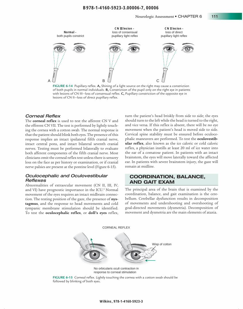
Pupillary Reflex Pupillary light reflexes provide information regarding the status of the brain and the sympathetic and parasympa-thetic nervous systems. Pupillary function is controlled by the midbrain and evaluates CNs II and III. Pupillary reflex is determined by briefly passing a bright light in front of both open eyes while carefully watching the iris in both eyes for movement. Pupil size, congruency, and response to light and accommodation should be described.
The acronym PERRLA is commonly used to refer to normal p upils that are e qual, r ound, and r eactive to l ight and a ccommodation (movement). Any visible change in the pupils’ size is noted. Anisocoria is a neurologic term indi-cating that one pupil is larger than the other. Mydriasis , or pupillary dilation, may be caused by serious brain injury or inadvertent exposure of the eyes to inhaled anticholinergics. Miosis or small “pinpoint” pupils usually result from pon-tine hemorrhage or from ingestion of narcotics or organo-phosphates. Pupillary responses almost always remain intact in metabolic causes of coma. Midposition and fixed pupils often indicate severe cerebral damage ( Figure 6-14 ).
Deep Tendon Reflex Reflex Spinal Nerve
Biceps C5-C6Triceps C7-C8Patellar L2-L4Achilles L5-S1
TABLE 6-9
FIGURE 6-13 Babinski’s sign. Dorsiflexion of the great toe with fanning of remaining toes after stroking the outer aspect of the sole in the direction of the arrow.
SIMPLY STATED The presence of Babinski’s sign (dorsiflexion of the great toe with fanning of remaining toes) while testing the plantar reflex could indicate a lesion of the UMN or brain disease.
SIMPLY STATED Although some healthy individuals have a minimal or absent gag reflex (CNs IX and X), its absence may increase the risk for aspiration, and endotracheal intubation may be necessary to protect the lungs. The ability to cough with suctioning can be tested in an intubated patient and implies an intact CN X.
SIMPLY STATED The acronym PERRLA refers to normal pupils that are e qual, r ound, and r eactive to l ight and a ccommodation. Pupillary function is controlled by the midbrain and evaluates cranial nerves II and III.
Wilkins, 978-1-4160-5923-3
B978-1-4160-5923-3.00006-7, 00006
Neurologic Assessment • CHAPTER 6 111
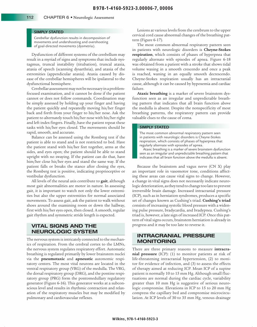
Corneal Reflex The corneal reflex is used to test the afferent CN V and the efferent CN VII. The test is performed by lightly touch-ing the cornea with a cotton swab. The normal response is that the patient should blink both eyes. The presence of this response implies an intact ipsilateral fifth cranial nerve, intact central pons, and intact bilateral seventh cranial nerves. Testing must be performed bilaterally to evaluate both afferent components of the fifth cranial nerve. Most clinicians omit the corneal reflex test unless there is sensory loss on the face as per history or examination, or if cranial nerve palsies are present at the pontine level ( Figure 6-15 ).
Oculocephalic and Oculovestibular Reflexes Abnormalities of extraocular movement (CN II, III, IV, and VI) have prognostic importance in the ICU. 3 Normal movement of the eyes requires an intact midbrain connec-tion. The resting position of the gaze, the presence of nys-tagmus , and the response to head movements and cold tympanic membrane stimulation should be identified. To test the oculocephalic reflex , or doll’s eyes reflex,
turn the patient’s head briskly from side to side; the eyes should turn to the left while the head is turned to the right, and vice versa. If this reflex is absent, there will be no eye movement when the patient’s head is moved side to side. Cervical spine stability must be ensured before oculoce-phalic maneuvers are performed. To test the oculovestib-ular reflex , also known as the ice caloric or cold caloric reflex, a physician instills at least 20 ml of ice water into the ear of a comatose patient. In patients with an intact brainstem, the eyes will move laterally toward the affected ear. In patients with severe brainstem injury, the gaze will remain at midline.
COORDINATION, BALANCE, AND GAIT EXAM
The principal area of the brain that is examined by the coordination, balance, and gait examination is the cere-bellum. Cerebellar dysfunction results in decomposition of movements and undershooting and overshooting of goal-directed movements (dysmetria). Decomposition of movement and dysmetria are the main elements of ataxia.
CORNEAL REFLEX
No orbicularis oculi contraction inresponse to corneal stimulation
Wisp of cotton
FIGURE 6-15 Corneal reflex. Lightly touching the cornea with a cotton swab should be followed by blinking of both eyes.
Normal - both pupils constrict
A B C
CN III lesion - loss of consensualpupillary light reflex
CN II lesion -loss of direct
pupillary light reflex
FIGURE 6-14 Pupillary reflex. A, Shining of a light source on the right may cause a constriction of both pupils in normal individuals. B, Constriction of the pupil only on the right eye in patients with lesions of CN III—loss of consensual reflex. C, Pupillary constriction of the opposite eye in lesions of CN II—loss of direct pupillary reflex.
Wilkins, 978-1-4160-5923-3
B978-1-4160-5923-3.00006-7, 00006
112 CHAPTER 6 • Neurologic Assessment
Dysfunction of different systems of the cerebellum may result in a myriad of signs and symptoms that include nys-tagmus, truncal instability (titubation), truncal ataxia, ataxia of speech (scanning dysarthria), and ataxia of the extremities (appendicular ataxia). Ataxia caused by dis-ease of the cerebellar hemispheres will be ipsilateral to the dysfunctional hemisphere.
Cerebellar assessment may not be necessary in a problem-focused examination, and it cannot be done if the patient cannot or does not follow commands. Coordination may be simply assessed by holding up your finger and having the patient quickly and repeatedly moving his/her finger back and forth from your finger to his/her nose. Ask the patient to alternately touch his/her nose with his/her right and left index fingers. Finally, have the patient repeat these tasks with his/her eyes closed. The movements should be rapid, smooth, and accurate.
Balance can be assessed using the Romberg test if the patient is able to stand and is not restricted to bed. Have the patient stand with his/her feet together, arms at the sides, and eyes open; the patient should be able to stand upright with no swaying. If the patient can do that, have him/her close his/her eyes and stand the same way. If the patient falls or breaks the stance after closing the eyes, the Romberg test is positive, indicating proprioceptive or vestibular dysfunction.
All levels of the neural axis contribute to gait , although most gait abnormalities are motor in nature. In assessing gait, it is important to watch not only the lower extremi-ties but also the upper extremities for normal associated movements. To assess gait, ask the patient to walk without shoes around the examining room or down the hallway, first with his/her eyes open, then closed. A smooth, regular gait rhythm and symmetric stride length is expected.
VITAL SIGNS AND THE NEUROLOGIC SYSTEM
The nervous system is intricately connected to the mechan-ics of respiration. From the cerebral cortex to the LMNs, the nervous system regulates respiratory effort. Automatic breathing is regulated primarily by lower brainstem nuclei via the pneumotaxic and apneustic autonomic respi-ratory centers. The most vital neurons are located in the ventral respiratory group (VRG) of the medulla. The VRG, the dorsal respiratory group (DRG), and the pontine respi-ratory group (PRG) form the pontomedullary regulatory generator ( Figure 6-16 ). This generator works at a subcon-scious level and results in rhythmic contraction and relax-ation of the respiratory muscles but may be modified by pulmonary and cardiovascular reflexes.
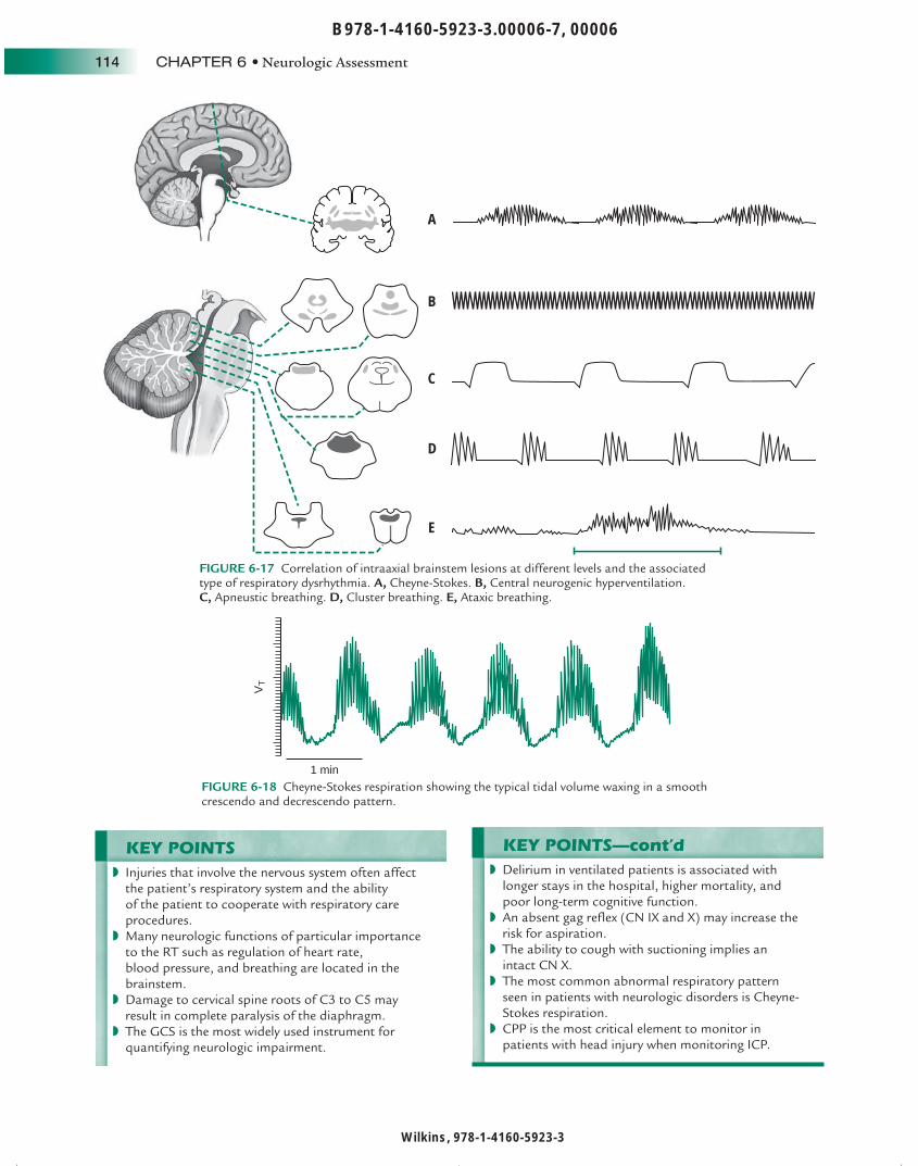
Lesions at various levels from the cerebrum to the upper cervical cord cause abnormal changes of the breathing pat-tern ( Figure 6-17 ).
The most common abnormal respiratory pattern seen in patients with neurologic disorders is Cheyne-Stokes respiration , which consists of phases of hyperpnea that regularly alternate with episodes of apnea. Figure 6-18 was obtained from a patient with a stroke that shows tidal volume waxing in a smooth crescendo and once a peak is reached, waning in an equally smooth decrescendo. Cheyne-Stokes respiration usually has an intracranial cause, although it can be caused by hypoxemia and cardiac failure.
Ataxic breathing is a marker of severe brainstem dys-function seen as an irregular and unpredictable breath-ing pattern that indicates that all brain function above the medulla is absent. Despite the nonspecificity of most breathing patterns, the respiratory pattern can provide valuable clues to the cause of coma.
Because the brainstem and vagus nerve (CN X) play an important role in vasomotor tone, conditions affect-ing these areas can cause vital signs to change. However, a change in vital signs does not necessarily indicate neuro-logic deterioration, as they tend to change too late to prevent irreversible brain damage. Increased intracranial pressure (ICP), such as in herniation syndromes, produces a specific set of changes known as Cushing’s triad. Cushing’s triad consists of increasing systolic blood pressure with a widen-ing pulse pressure, bradycardia, and bradypnea. Cushing’s triad is, however, a late sign of increased ICP. Once this pat-tern of vital signs occurs, brainstem herniation is already in progress and it may be too late to reverse it.
INTRACRANIAL PRESSURE MONITORING
There are three primary reasons to measure intracra-nial pressure (ICP): (1) to monitor patients at risk of life-threatening intracranial hypertension, (2) to moni-tor for evidence of infection, and (3) to assess the effects of therapy aimed at reducing ICP. Mean ICP of a supine patient is normally 10 to 15 mm Hg. Although small fluc-tuations are normal during the cardiac cycle, variability greater than 10 mm Hg is suggestive of serious neuro-logic compromise. Elevations in ICP to 15 to 20 mm Hg compress the capillary bed and compromise microcircu-lation. At ICP levels of 30 to 35 mm Hg, venous drainage
SIMPLY STATED Cerebellar dysfunction results in decomposition of movements and undershooting and overshooting of goal-directed movements (dysmetria).
SIMPLY STATED The most common abnormal respiratory pattern seen in patients with neurologic disorders is Cheyne-Stokes respiration, which consists of phases of hyperpnea that regularly alternate with episodes of apnea. Ataxic breathing is a marker of severe brainstem dysfunction seen as an irregular and unpredictable breathing pattern that indicates that all brain function above the medulla is absent.

Wilkins, 978-1-4160-5923-3
B978-1-4160-5923-3.00006-7, 00006
Neurologic Assessment • CHAPTER 6 113
is impeded and edema develops in uninjured tissue. Even when autoregulatory mechanisms are intact, cerebral per-fusion cannot be maintained if ICP increases to within 40 to 50 mm Hg of the mean arterial pressure (MAP). When ICP approximates MAP, perfusion stops and the brain dies.
Currently available ICP monitoring techniques fall into two categories: fluid-filled systems with external transduc-ers, such as intraventricular catheter and subarachnoid bolts, and solid-state systems with miniature pressure transducers that can be inserted in the lateral ventricle, brain parenchyma, or subarachnoid or epidural space. It is extremely important for the RT to remember that
although hyperventilation is associated with lower ICP val-ues because of the vasoconstriction of brain blood vessels associated with hypocapnia, the cerebral perfusion pres-sure (CPP) is the most critical element to monitor. 16 CPP is a result of the MAP minus the ICP.
Supplementary motor area
Motor cortex
Sensory cortex(sensation ofbreathlessness)
Voluntarybreathing
Limbic system
PRG
V
NA
VRG
DRG
IX nerve
X nerve
Involuntarybreathing
Peripheral chemoreceptors(arterial blood gases)
Mechanicalreceptors
Respiratory muscles
Spinal respiratorymuscles
Reticulospinalpathway
Ventilation
Lateralcorticospinal
tract
XII
FIGURE 6-16 Schematic representation of the neural control of breathing. CC, Central chemoreceptors; DRG, dorsal respiratory group; NA, nucleus ambiguous; PRG, pontine respiratory group; V, sensory nucleus of V; VRG, ventral respiratory group.
SIMPLY STATED Although hyperventilation is associated with lower ICP values because of the vasoconstriction of brain blood vessels associated with hypocapnia, CPP is the most critical element to monitor. CPP is a result of the MAP minus the ICP.
Wilkins, 978-1-4160-5923-3
B978-1-4160-5923-3.00006-7, 00006
114 CHAPTER 6 • Neurologic Assessment
A
B
C
D
E
FIGURE 6-17 Correlation of intraaxial brainstem lesions at different levels and the associated type of respiratory dysrhythmia. A, Cheyne-Stokes. B, Central neurogenic hyperventilation. C, Apneustic breathing. D, Cluster breathing. E, Ataxic breathing.
1 min
VT
FIGURE 6-18 Cheyne-Stokes respiration showing the typical tidal volume waxing in a smooth crescendo and decrescendo pattern.
KEY POINTS Injuries that involve the nervous system often affect the patient’s respiratory system and the ability of the patient to cooperate with respiratory care procedures. Many neurologic functions of particular importance to the RT such as regulation of heart rate, blood pressure, and breathing are located in the brainstem. Damage to cervical spine roots of C3 to C5 may result in complete paralysis of the diaphragm. The GCS is the most widely used instrument for quantifying neurologic impairment.
◗
◗
◗
◗
KEY POINTS—cont’d Delirium in ventilated patients is associated with longer stays in the hospital, higher mortality, and poor long-term cognitive function. An absent gag reflex (CN IX and X) may increase the risk for aspiration. The ability to cough with suctioning implies an intact CN X. The most common abnormal respiratory pattern seen in patients with neurologic disorders is Cheyne-Stokes respiration. CPP is the most critical element to monitor in patients with head injury when monitoring ICP.
◗
◗
◗
◗
◗

Wilkins, 978-1-4160-5923-3
B978-1-4160-5923-3.00006-7, 00006
Neurologic Assessment • CHAPTER 6 115
ASSESSMENT QUESTIONS
See Evolve Resources for answers. 1 . Which of the following is not an important factor
when determining the frequency of the neurologic assessment? a . Patient’s diagnosis b . Acuity of the condition c . How rapidly changes are occurring or expected to
occur d . Shift change
2 . Which of the following is not part of the brain? a . Cerebrum b . Cranial nerves c . Brainstem d . Cerebellum
3 . Where in the nervous system is regulation of breathing located? a . Brain cortex b . Medulla c . Cerebellum d . Brainstem
4 . Which of the following is the most common cause of nerve root pathology caused by compression? a . Herniated vertebral disc b . Spinal tumor c . Spinal cord injury d . Infection
5 . Which of the following diseases may be suggested by the involvement of multiple nerve roots? a . Myasthenia gravis b . Guillain-Barré syndrome c . Brain tumor d . Intracranial hypertension
6 . Injury to the cervical spine roots C2 to C4 is associated with which of the following abnormalities? a . Absence of deep tendon reflexes b . Babinski’s sign c . Paralysis of the diaphragm d . Doll’s eyes
7 . Which of the following is the most important examination in the neurologic examination? a . Sensory examination b . Motor examination c . Level of consciousness d . Gait examination
8 . What is the most widely used instrument to quantify neurologic impairment? a . The Merck Gait Evaluation b . Glasgow Coma Scale c . APACHE d . Mini-Mental State Examination
9 . Which Glasgow coma scale score is typically an indication for endotracheal intubation? a . <9 b . <10
c . <12 d . <14
10 . Which of the following cranial nerves are evaluated with the gag reflex? a . II and III b . V and VII c . I and II d . IX and X
11 . Which of the following diseases are characterized by abnormal deep tendon reflexes caused by abnormalities of the neuromuscular junction? a . Syphilis b . Myasthenia gravis c . Cerebritis d . Multiple sclerosis
12 . The presence of dorsiflexion of the great toe with fanning of remaining toes while testing the plantar reflex is known as which of the following? a . Babinski’s sign b . “Jerk” reflex c . Patellar reflex d . Quadriceps reflex
13 . Which of the following cranial nerves is intact if cough is present while suctioning the airway? a . I b . IX c . X d . XII
14 . Which of the following does not describe the acronym PERRLA? a . Positioned b . Accommodation c . Round d . Reactive
15 . Which of the following anatomic areas is compromised if the patient has dysmetria? a . Cerebrum b . Pons c . Cerebellum d . Brainstem
16 . Which of the following respiratory patterns consists of phases of hyperpnea that regularly alternate with episodes of apnea? a . Biot’s b . Ataxic c . Apneustic d . Cheyne-Stokes
17 . Which of the following is the most critical parameter to keep in mind when managing a patient with intracranial hypertension? a . Mean arterial pressure b . Cerebral perfusion pressure c . Intracranial pressure d . Pulse pressure
Wilkins, 978-1-4160-5923-3
B978-1-4160-5923-3.00006-7, 00006
116 CHAPTER 6 • Neurologic Assessment
References
1 . Bleck TP , Smith MC , Pierre-Louis SJ , et al : Neurologic complications of critical medical illnesses , Crit Care Med 21 : 98 - 103 , 1993 .
2 . Teasdale G , Jennett B : Assessment of coma and impaired con-sciousness. A practical scale , Lancet 2 : 81 - 84 , 1974 .
3 . Booth CM , Boone RH , Tomlinson G , et al : Is this patient dead, vegetative, or severely neurologically impaired? Assessing out-come for comatose survivors of cardiac arrest , JAMA 291 : 870 - 879 , 2004 .
4 . Marion DW , Carlier PM : Problems with initial Glasgow Coma Scale assessment caused by prehospital treatment of patients with head injuries: results of a national survey , J Trauma 36 (1) : 89 - 95 , 1994 .
5 . Gómez PA , Lobato RD , Ortega JM , et al : Mild head injury: differences in prognosis among patients with a Glasgow Coma Scale score of 13 to 15 and analysis of factors associated with abnormal CT findings , Br J Neurosurgery 10 (5) : 453 - 460 , 1996 .
6 . Folstein MF , Folstein SE , McHugh PR : “Mini-mental state.” A practical method for grading the cognitive state of patients for the clinician , J Psych Res 12 (3) : 189 - 198 , 1975 .
7 . Crum RM , Anthony JC , Bassett SS , et al : Population-based norms for the Mini-Mental State Examination by age and educational level , JAMA 269 (18) : 2386 - 2391 , 1993 .
8 . McNicoll L , Pisani MA , Zhang Y , et al : Delirium in the inten-sive care unit: occurrence and clinical course in older patients , J Am Geriatr Soc 51 : 591 - 598 , 2003 .
9 . Ely EW , Gautam S , Margolin R , et al : The impact of delirium in the intensive care unit on hospital length of stay , Intensive Care Med 27 : 1892 - 1900 , 2001 .
10 . Ely EW , Shintani A , Truman B , et al : Delirium as a predictor of mortality in mechanically ventilated patients in the inten-sive care unit , J Am Med Assoc 291 : 1753 - 1762 , 2004 .
11 . Sessler CN , Gosnell MS , Grap MJ , et al : The Richmond Agitation-Sedation Scale: Validity and reliability in adult intensive care unit patients , Am J Respir Crit Care Med 166 : 1338 - 1344 , 2002 .
12 . American Psychiatric Association : Diagnostic and statistical manual of mental disorders , Washington, DC , 1987 , American Psychiatric Association .
13 . Ely EW , Inouye SK , Bernard GR , et al : Delirium in mechan-ically ventilated patients: validity and reliability of the confusion assessment method for the intensive care unit (CAM-ICU) , J Am Med Assoc 286 : 2703 - 2710 , 2001 .
14 . Pun BT , Gordon SM , Peterson JF , et al : Large-scale implemen-tation of delirium and sedation monitoring in the intensive care unit: a report from two medical centers , Crit Care Med 33 : 1199 - 1205 , 2005 .
15 . van Gijn J : The Babinski sign: the first hundred years , J Neurol 243 (10) : 675 - 683 , 1996 .
16 . Robertson CS : Management of cerebral perfusion pressure after traumatic brain injury , Anesthesiology 95 (6) : 1513 - 1517 , 2001 .
Bibl iography
Adams AB , Lim S : Monitoring in management of the patient in the intensive care unit . In Wilkins RL , Stoller JK , Kackmarek RM , editors: Egan’s fundamentals of respiratory care , ed 9 , St Louis , 2008 , Mosby .
DeMyer WE : Technique of the neurologic examination , ed 5 , New York , 2004 , McGraw Hill .
Bleck TP : Levels of consciousness and attention . In Goetz CG : Textbook of clinical neurology , ed 3 , Philadelphia , 2007 , WB Saunders .
Griggs RC , Josefowics RE , Aminoff MJ . Approach to the patient with neurologic disease . In Goldman L , Ausiello D , editors: Cecil textbook of medicine , ed 22 , Philadelphia , 2004 , WB Saunders .
Kerr ME : Intracranial problems . In Lewis SM , Heitkemper MM , Dirksen SR , editors: Medical surgical nursing , ed 5 , St Louis , 2000 , Mosby .
Marshall RS , Mayer SA : On-call neurology , ed 2 , New York , 2001 , WB Saunders .
Vos H : The neurologic assessment . In Barker E , editor: Neuroscience nursing: spectrum of care , ed 2 , St Louis , 2002 , Mosby .
0000860659.INDD 1160000860659.INDD 116 9/10/2008 4:20:22 PM9/10/2008 4:20:22 PM