neurobiologyofdisease ... · environmentalenrichmentincreasesthegfap stemcell...
TRANSCRIPT

Neurobiology of Disease
Environmental Enrichment Increases the GFAP� Stem CellPool and Reverses Hypoxia-Induced Cognitive Deficits inJuvenile Mice
Natalina Salmaso,1 John Silbereis,1 Mila Komitova,1 Patrick Mitchell,1 Katherine Chapman,1 Laura R. Ment,2
Michael L. Schwartz,3 and Flora M. Vaccarino1,3
1Child Study Center and Departments of 2Pediatrics and 3Neurobiology, Yale University School of Medicine, New Haven, Connecticut 06520
Premature children born with very low birth weight (VLBW) can suffer chronic hypoxic injury as a consequence of abnormal lungdevelopment and cardiovascular abnormalities, often leading to grave neurological and behavioral consequences. Emerging evidencesuggests that environmental enrichment improves outcome in animal models of adult brain injury and disease; however, little is knownabout the impact of environmental enrichment following developmental brain injury. Intriguingly, data on socio-demographic factorsfrom longitudinal studies that examined a number of VLBW cohorts suggest that early environment has a substantial impact on neuro-logical and behavioral outcomes. In the current study, we demonstrate that environmental enrichment significantly enhances behavioraland neurobiological recovery from perinatal hypoxic injury. Using a genetic fate-mapping model that allows us to trace the progeny ofGFAP� astroglial cells, we show that hypoxic injury increases the proportion of astroglial cells that attain a neuronal fate. In contrast,environmental enrichment increases the stem cell pool, both through increased stem cell proliferation and stem cell survival. In micesubjected to hypoxia and subsequent enrichment there is an additive effect of both conditions on hippocampal neurogenesis fromastroglia, resulting in a robust increase in the number of neurons arising from GFAP� cells by the time these mice reach full adulthood.
IntroductionApproximately 1% of children born in the United States are pre-mature and born at a very low birth weight (VLBW). Clinically,VLBW children exhibit a range of neurological and behavioraldisturbances, including decreased brain volume, white matterabnormalities, and developmental delays. These perturbationsare thought to be a consequence of chronic hypoxic injury due toimmature lung development. Remarkably, a substantial portionof VLBW children are able to recover from these abnormalitiesover time, and some of them attain normal cognitive function bythe time they reach early adulthood (Saigal and Doyle, 2008; Luuet al., 2011). However, the factors that contribute to heterogene-ity in long-term outcomes are not known. The current studyattempts to elucidate the underlying cellular mechanisms by us-ing a mouse model of perinatal hypoxia that mimics the injuryand recovery observed in VLBW children.
Emerging evidence suggests that various types of injury, suchas hypoxic and/or ischemic insults, induce strong proliferativeand neurogenic/gliogenic responses in the subgranular zone ofthe hippocampal dentate gyrus (DG), a postnatal neurogenicniche (Kuhn et al., 2001; Sharp et al., 2002; Parent, 2003; Fagel etal., 2006; Yang and Levison, 2006; Kernie and Parent, 2010). Inthe postnatal brain, neural stem cells (NSCs) are a subset of glialfibrillary acidic protein (GFAP)-expressing astrocytes (Doetschet al., 1999; Seki and Arai, 1999; Seri et al., 2001; Alvarez-Buylla etal., 2002; Filippov et al., 2003; Imura et al., 2003; Garcia et al.,2004). Neural stem cells give rise to transient-amplifying progen-itors that express neuronal lineage makers and generate new neu-rons throughout life (Belachew et al., 2003; Hack et al., 2005;Hevner et al., 2006; Brill et al., 2008; Rivers et al., 2008).
To study the cellular and molecular mechanisms that conferhigher inherent plasticity to the DG, allowing for a more adaptiveresponse to brain injury, we investigated the fate of GFAP� as-troglia in the DG by tagging these cells with heritable reportergenes via tamoxifen-inducible Cre recombinase in GFAP-CreER T2 (GCE) transgenic mice. In addition, because environ-mental enrichment with exercise has been demonstrated toincrease hippocampal neurogenesis in adulthood (Kempermannet al., 1997; van Praag et al., 1999a; Olson et al., 2006), we assessedthe influence of enriched environment on the recovery processafter perinatal hypoxic injury. Number and self-renewal of hip-pocampal GFAP� neural stem cells, as well as phenotype andlong-term survival of the new neurons arising from these cells,were examined in both hypoxic and hypoxic enriched mice. Fi-nally, we examined the functional outcome of hypoxic injury and
Received March 19, 2012; revised April 26, 2012; accepted May 7, 2012.Author contributions: N.S., J.S., M.K., L.R.M., M.L.S., and F.M.V. designed research; N.S., J.S., M.K., P.M., and K.C.
performed research; N.S., J.S., and F.M.V. analyzed data; N.S. and F.M.V. wrote the paper.This work was funded by NIH Grants P01 NS062686 and R21 AG034495 to F.M.V., a PSD2 Fellowship from the
Fonds de recherche en sante du Quebec to N.S, and a Swedish Brain Foundation (Hjarnfonden) and a SwedishMedical Association fellowship to M.K. We acknowledge Teresa Sandoval-Minero, Eylem Ocal, Ellen Hoffman, Lau-ren Provini, Devon Fagel, and Allyson Vermaak for technical assistance and useful discussions. We thank Dr. DavidMumby for his advice on alternatives to testing a traditional Morris water maze paradigm. We thank Drs. CharlesStiles and John Alberta, Dana-Farber Cancer Institute, Boston, MA, for their gift of the Olig2 antibody.
The authors declare no competing financial interests.Correspondence should be addressed to Flora Vaccarino, Child Study Center, 230 South Frontage Road, New
Haven, CT 06520. E-mail: [email protected]:10.1523/JNEUROSCI.1398-12.2012
Copyright © 2012 the authors 0270-6474/12/328930-10$15.00/0
8930 • The Journal of Neuroscience, June 27, 2012 • 32(26):8930 – 8939

subsequent enriched rearing on cognitive and emotive behavioraltasks.
We show that perinatal hypoxia enhances the proportion ofnew neurons arising from GFAP� astroglial cells and corre-spondingly decreases the proportion of GFAP� astrocytesamong fate-mapped cells. The newly generated neurons persistlong-term in the DG. Rearing the animals in enriched environ-ment induces a large, additive increase in neurons derived fromGFAP� cell within the DG. We suggest that the effect of enrich-ment is attributable to a potent increase in proliferation and sur-vival of neural stem cells.
Materials and MethodsGeneration of mice, genotyping, and breeding strategy. The GCE mice weregenerated as previously described and back-crossed to C57/B6 mice 10generations (Ganat et al., 2006; Bi et al., 2011). GCE transgenic mice carrya Cre recombinase-estrogen receptor type 2 fusion protein, CreErT2,placed under control of the human GFAP promoter, which is active inradial glia, astrocytes, and adult neural stem cells (Brenner, 1994; Ganatet al., 2006). Genotyping was done by PCR using primers to the Cre gene(5�-GCAACGAGTGATGAGGTTCGCAAG-3�) (forward) and (5�-TCCGCCGCATAACCAGTGAAACAG-3�) (reverse) to generate aband of 307 bp (Ganat et al., 2006; Bi et al., 2011). GCE mice were crossedwith either the R26R LacZ reporter mice (Soriano, 1999) (available fromThe Jackson Laboratory) or the CAG-eGFP reporter mice (where eGFP isenhanced green fluorescent protein) (Nakamura et al., 2006). Numbersof animals used in experiments ranged between three and six per group,as indicated in Results. Littermates negative for the CAG-eGFP gene wereused for behavioral experiments. All animal experiments comply withinstitutional and national policies and guidelines.
Induction of Cre recombination via tamoxifen treatment. To induce Crerecombination in GFAP promoter-expressing cells, GCE mice crossedwith reporter lines were injected daily from postnatal day 12 (P12) to P14intraperitoneally with tamoxifen at a dosage of 60 mg/kg from a 2 mg/mlstock solution prepared in autoclaved sunflower seed oil and stored at�20°C.
Hypoxic rearing. Mice (males and females) were placed in a chambermaintaining a 9.5–10.5% O2 concentration by displacement with N2 asdescribed previously (Fagel et al., 2006). Hypoxia began at P3 for 8 d untilP11. A separate group of control (normoxic) mice were matched forstrain and age. Mice were sacrificed at P15, P16, P35, or P90. Mice wereperfused transcardially with 20 ml of PBS followed by 25 ml of 4%paraformaldehyde (PFA). Brains were post-fixed overnight in 4%PFA, followed by equilibration in a 30% sucrose solution overnightfor cryoprotection, and stored at �80°C.
Enriched environment. Following hypoxic or normoxic rearing condi-tions, male and female mice were placed under either enriched or stan-dard housing until sacrifice. Enrichment was maintained for 2 weeksfrom P21 to P35 or for 10 weeks from P21 to P90. All groups werecounterbalanced for previous hypoxic experience; therefore four groupsof animals were generated: normoxic standard environment (NSE), nor-moxic enriched (NEn), hypoxic standard environment (HSE) and hy-poxic enriched (HEn). Environmental enrichment consisted of a largercage (10” � 19” � 8” h), a running wheel, and several plastic toys,including hanging objects, balls, and modular plastic tunnels, that werechanged and reconfigured weekly during routine cage cleaning. Animalswere group housed in both the standard and enriched paradigms.
Behavioral testing. To assess the functional outcome of both hypoxiaand enriched environment, GCE;CAG-negative littermates of mice usedin the fate-mapping experiments were tested on the open field and Mor-ris water maze tests at P35.
Mice were brought to the behavioral testing room in their home cagesat least 30 min before testing to acclimatize them to the testing environ-ment. Mice were then placed in an open field (26 � 48 cm) under brightoverhead lights and allowed to explore freely for 20 min. Distance andtime spent in the periphery and center of the open field as well as freezingbouts and immobility were all assessed using the AnyMaze softwaretracking system (Stoelting). Following this, mice were tested on a short-
ened Morris water maze protocol adapted from J. Nunez, Michigan StateUniversity, East Lansing, MI (Nunez, 2008). Briefly, mice were placed ineach of the four quadrants of the water maze in random order. Theplatform remained in a fixed location until the probe trial. Mice receivedtwo sets of six trials separated by a 1 h break. Each trial (intertrial intervalwas 10 min) ended either when they found the platform or after 60 s hadelapsed. Mice that did not find the platform during the first trial weregently guided to the platform and removed from the platform after 5 s.Ten minutes following the 12th trial, the platform was removed andmemory retention for the platform location was tested with a 60 s probetrial. We assessed both the number of trials it took for �50% of theanimals in a group to find the platform (the rate of acquisition) and thequadrant preference during the probe trial (memory test—presented asboth percentage of time spent in the target quadrant and a probe prefer-ence score).
2-Bromodeoxyuridine, chlorodeoxyuridine, and iododeoxyuridine treat-ment. To assess the effect of environmental enrichment on cell survival,normoxic mice were injected with the thymidine analog chlorodeoxyu-ridine (CldU) (100 mg/kg) twice on P15 and P16, before the onset ofenrichment. Cell proliferation was assessed by injecting the thymidineanalog iododeoxyuridine (IdU) (100 mg/kg) 1 h before sacrifice at P35.
Tissue preparation and immunohistochemistry. Serial 20 �m cryosec-tions were obtained as described previously (Ganat et al., 2006). Forimmunohistochemistry, sections were blocked in PBS containing 0.3%Triton (PBS-T) containing 10% goat serum (10% GS/PBS-T) or 10%donkey serum (10% DS/PBS-T), and then incubated in primary anti-body in 10% GS/PBS-T or DS/PBS-T. For a list of primary antibodies, seeTable 1. Sections were washed thoroughly and then reacted to the sec-ondary antibody of the appropriate species. The secondary antibodiesused were as follows: Alexa Fluor 488-, Alexa Fluor 594-, Alexa Fluor350-, Alexa Fluor 546-, and Alexa Fluor 632-conjugated species directedIgGs (Invitrogen) and FITC-, DyLight 549- and DyLight 649-conjugatedspecies directed IgGs (Jackson Laboratories), all at 1:500 dilution. ForBrdU/CldU/IdU immunostaining, sections were incubated for 45 min in2 N HCl, followed by washes and immunohistochemistry as describedabove.
Cell counting and microscopic analysis. Unbiased estimates of cell num-ber were obtained via a Zeiss Axioskope 2 Mot Plus (Carl Zeiss) attachedto a motorized stage and connected to a computer running the Stereo-Investigator software (MicroBrightfield). Serial sagittal sections (one ev-ery 600 �m) were used for all counts. Contours of the dentate gyrus weredrawn at 10� magnification. Nuclear profiles of stained cells werecounted using the optical fractionator probe with a 40� oil immersionobjective. Sampling grids sized 300 � 135 �m were used to obtain arelatively constant number of cells sampled and obtain a coefficient of
Table 1. Primary antibodies
Antibody Manufacturer Species Dilution
�-Galactosidase Cappel Rabbit 1:10,000�-Galactosidase Abcam Chicken 1:500BrdU/CldU Accurate Chemical Rat 1:250BrdU/IdU Becton-Dickenson Mouse 1:100Doublecortin Abcam Rabbit 1:2000Doublecortin Santa Cruz Goat 1:500GFAP Sigma Mouse 1:500GFAP Dako Rabbit 1:1000GFP Molecular Probes Rabbit 1:500GFP Abcam Chicken 1:1000Ki67 Vector Laboratories Rabbit 1:500Neuronal Nuclei Chemicon Mouse 1:500Ng2 Millipore Mouse 1:500Olig2 Kindly provided by Drs. Charles Stiles and John
Alberta, Dana-Farber Cancer Institute, Boston,MA
Rabbit 1:20,000
Sox2 Chemicon Goat 1:200Ng2 Chemicon Rabbit 1:500Sox2 Chemicon Goat 1:200
Salmaso et al. • Hypoxia and Environmental Enrichment J. Neurosci., June 27, 2012 • 32(26):8930 – 8939 • 8931
error �0.5. This systematic yet unbiasedmethod provides an estimate of cell densityand number that is independent from cell size,shape, orientation, tissue shrinkage, and spatialdistribution of the cells (Schmitz and Hof,2005). Tri-dimensional sampling boxes or(100 � 100 � 10 �m) with three of six exclu-sion borders (Gundersen et al., 1988; West,1993) were automatically placed by StereoIn-vestigator at each grid intersection point. Thedensity for each cell type was calculated by di-viding the total number of cells by the totalvolume sampled. Image acquisition andZ-stack analysis was performed on anApoTome-equipped Axiovert 200M with Ax-iovision 4.5 software (Carl Zeiss).
Statistical analyses. All data were analyzedusing Statview 4.51 or SPSS 16.0.2 software. Ttests or factorial ANOVA were used as neces-sary, with level of enrichment (enriched or not)and/or hypoxia (hypoxic or normoxic) as inde-pendent variables. The density of cells/region,total overall number of cells, and, where appro-priate, the percentage colocalization with�-galactosidase (�gal) or eGFP reporter wereanalyzed. F values were considered significantwhen p � 0.05. Post hoc analyses using Fisher’sleast significant difference were conductedwhen p values reached significance. Plannedcomparisons were conducted to confirm pre-viously published results when omnibus Fscores were not significant.
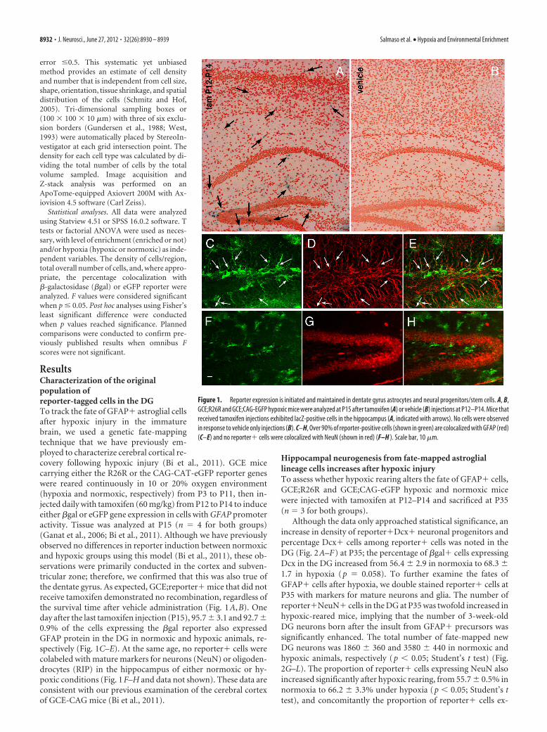
ResultsCharacterization of the originalpopulation ofreporter-tagged cells in the DGTo track the fate of GFAP� astroglial cellsafter hypoxic injury in the immaturebrain, we used a genetic fate-mappingtechnique that we have previously em-ployed to characterize cerebral cortical re-covery following hypoxic injury (Bi et al., 2011). GCE micecarrying either the R26R or the CAG-CAT-eGFP reporter geneswere reared continuously in 10 or 20% oxygen environment(hypoxia and normoxic, respectively) from P3 to P11, then in-jected daily with tamoxifen (60 mg/kg) from P12 to P14 to induceeither �gal or eGFP gene expression in cells with GFAP promoteractivity. Tissue was analyzed at P15 (n � 4 for both groups)(Ganat et al., 2006; Bi et al., 2011). Although we have previouslyobserved no differences in reporter induction between normoxicand hypoxic groups using this model (Bi et al., 2011), these ob-servations were primarily conducted in the cortex and subven-tricular zone; therefore, we confirmed that this was also true ofthe dentate gyrus. As expected, GCE;reporter� mice that did notreceive tamoxifen demonstrated no recombination, regardless ofthe survival time after vehicle administration (Fig. 1A,B). Oneday after the last tamoxifen injection (P15), 95.7 � 3.1 and 92.7 �0.9% of the cells expressing the �gal reporter also expressedGFAP protein in the DG in normoxic and hypoxic animals, re-spectively (Fig. 1C–E). At the same age, no reporter� cells werecolabeled with mature markers for neurons (NeuN) or oligoden-drocytes (RIP) in the hippocampus of either normoxic or hy-poxic conditions (Fig. 1F–H and data not shown). These data areconsistent with our previous examination of the cerebral cortexof GCE-CAG mice (Bi et al., 2011).
Hippocampal neurogenesis from fate-mapped astrogliallineage cells increases after hypoxic injuryTo assess whether hypoxic rearing alters the fate of GFAP� cells,GCE;R26R and GCE;CAG-eGFP hypoxic and normoxic micewere injected with tamoxifen at P12–P14 and sacrificed at P35(n � 3 for both groups).
Although the data only approached statistical significance, anincrease in density of reporter�Dcx� neuronal progenitors andpercentage Dcx� cells among reporter� cells was noted in theDG (Fig. 2A–F) at P35; the percentage of �gal� cells expressingDcx in the DG increased from 56.4 � 2.9 in normoxia to 68.3 �1.7 in hypoxia (p � 0.058). To further examine the fates ofGFAP� cells after hypoxia, we double stained reporter� cells atP35 with markers for mature neurons and glia. The number ofreporter�NeuN� cells in the DG at P35 was twofold increased inhypoxic-reared mice, implying that the number of 3-week-oldDG neurons born after the insult from GFAP� precursors wassignificantly enhanced. The total number of fate-mapped newDG neurons was 1860 � 360 and 3580 � 440 in normoxic andhypoxic animals, respectively (p � 0.05; Student’s t test) (Fig.2G–L). The proportion of reporter� cells expressing NeuN alsoincreased significantly after hypoxic rearing, from 55.7 � 0.5% innormoxia to 66.2 � 3.3% under hypoxia (p � 0.05; Student’s ttest), and concomitantly the proportion of reporter� cells ex-
Figure 1. Reporter expression is initiated and maintained in dentate gyrus astrocytes and neural progenitors/stem cells. A, B,GCE;R26R and GCE;CAG-EGFP hypoxic mice were analyzed at P15 after tamoxifen (A) or vehicle (B) injections at P12–P14. Mice thatreceived tamoxifen injections exhibited lacZ-positive cells in the hippocampus (A, indicated with arrows). No cells were observedin response to vehicle only injections (B). C–H, Over 90% of reporter-positive cells (shown in green) are colocalized with GFAP (red)(C–E) and no reporter� cells were colocalized with NeuN (shown in red) (F–H ). Scale bar, 10 �m.
8932 • J. Neurosci., June 27, 2012 • 32(26):8930 – 8939 Salmaso et al. • Hypoxia and Environmental Enrichment

pressing GFAP decreased from 49.1 � 2.2 to 35.1 � 4.8. Thesedata suggest that hypoxic rearing increases the proportion of fate-mapped astroglial cells that differentiate toward a neuronal fate.
Environmental enrichment increases neurogenesis fromGFAP� cellsWe next assessed both the ability of newly generated neurons tosurvive long term and the effect of environmental enrichment ontheir development. For these experiments, GCE mice carrying areporter gene were reared under normoxic or hypoxic condi-tions until P11 and subsequently returned to standard environ-ment or placed in enriched environment (En) upon weaning(P21) and then sacrificed at a juvenile/young adult stage (P35) orfull adulthood (P90) (see Fig. 3A for a schematic outline of theexperiment). For each of the four groups (normoxic standardenvironment, NSE; normoxic enriched environment, NEn;hypoxic standard environment, HSE; hypoxic enriched environ-ment, HEn) we used the following number of mice: P35, �galreporter: n � 4 for all groups; eGFP reporter: NSE, n � 3; NEn,n � 4; HSE, n � 6; Hen, n � 4; P90, n � 3 for all groups). Weassessed the effect of enrichment on the number of newly gener-ated neuronal cells arising from the GFAP lineage by analyzingDcx and NeuN double staining of reporter� cells in the DG atP35. We found that hypoxia and environmental enrichmentindependently increased neurogenesis from GFAP� astroglialcells by about fourfold, and that in hypoxic enriched mice
neurogenesis (both DCX� and NeuN�cells) from astroglial cells increased by�6-fold with respect to standard rear-ing (Fig. 3B– I, N,O). These changeswere also reflected in the total numberof eGFP� cells, where we observed asimilar pattern of results (Fig. 3Q). Inaddition, hypoxic enriched miceshowed a mild increase in the propor-tion of total granule neurons that ex-pressed GFP from 2.2% in NSE to 3.1%in NEn, and from 3.4% in HSE to 5.6%in Hen mice at P35. Finally, a nearly ad-ditive pattern of hypoxia and enrich-ment on NeuN� cells was also observedat P90 (Fig. 3J– M, P).
To assess whether there were popula-tion fate shifts in response to hypoxia andenvironmental enrichment, we also ex-amined the proportion of reporter� cellsthat coexpressed each of the lineage mark-ers, NeuN, Olig2, and GFAP in the fourgroups at P35 and found an increase in theproportion of reporter� cells that ex-pressed NeuN in both hypoxic and en-riched mice. This increase in proportionof GFAP� NSCs attaining neuronal fateseemed to be at the expense of the astro-glial population, as there was a concomi-tant decrease in the proportion ofreporter� cells expressing GFAP (Fig.3R). In contrast to our previous observa-tions in the cortex (Bi et al., 2011), veryfew GFAP� cells attained an oligoden-droglial fate in the DG; only one or tworeporter�Olig2� cells were observed inthe DG per animal, and these were only
seen in the enriched conditions.
Effect of hypoxia and subsequent enrichment on NSCsHypoxia and environmental enrichment increase neurogenesisfrom GFAP� astroglial cells in a nearly additive fashion. Wewanted to understand whether this prolonged increase in neuro-nal production depletes the DG stem cell pool or whether thispopulation was replenished and the number sustained. To assessthis, we counted the number of reporter� cells that expressedSox2, a transcription factor expressed by neural stem cells and, ina minor part, by neuronal progenitors (Avilion et al., 2003;Komitova and Eriksson, 2004; Episkopou, 2005). We found thatat 2 weeks after the onset of enrichment (P35), there was a drasticincrease in the total number of fate-mapped Sox2� cells in bothnormoxic and hypoxic enriched groups (Fig. 4, compare A–J withK–T, quantified in U). By contrast, the total number of fatemapped Sox2� neural stem cell population did not significantlychange due to hypoxia alone (Fig. 4, compare A–E with F–J,quantified in U).
To understand whether there are fate shifts within the neuralstem cell population, we also examined the proportion of eGFP�cells that expressed neural stem cell and progenitor markers. Wefound a decrease in the proportion of eGFP� cells expressingSox2� in both hypoxic, standard environment-reared mice andin hypoxic enriched mice. The decrease in the proportion ofeGFP�/Sox2� cells was concomitant to an increase in the pro-
Figure 2. Increased neurogenesis from fate mapped astroglial lineage cells following hypoxia. GCE;R26R and GCE;eGFP micewere tamoxifen-injected at P12–P14 and analyzed at P35. Hypoxic reared mice had increased numbers of reporter�/DCX�immature neurons (D–F ) and reporter�/NeuN� mature neurons (J–L) in the dentate gyrus as compared to normoxic mice(A–C, G–I ). Scale bar, 40 �m.
Salmaso et al. • Hypoxia and Environmental Enrichment J. Neurosci., June 27, 2012 • 32(26):8930 – 8939 • 8933

Figure 3. Hypoxia and subsequent environmental enrichment increase the number of neurons arising from GFAP� cells in an additive way. A, Schematic of the time course of hypoxia andenrichment experiment. B–I, Representative images showing the increase in reporter�/DCX�neurons in hypoxic, enriched mice (F–I ) as compared to hypoxic, standard reared mice (B–E) at P35;stereological quantification of the reporter�/DCX� and reporter�/NeuN� cell numbers is shown in N and O, respectively. J–M, Long-term increase in mature reporter�/NeuN� neurons inresponse to hypoxia and enrichment at P90, with stereological quantification of reporter�/NeuN� cell numbers at P90 shown in P. Q, The total number of eGFP� cells at P35 increases in hypoxicand enriched mice. R, Proportion of reporter� cells that attain neuronal (NeuN�), astroglial (GFAP�) and oligodendroglial (Olig2) fate are shown across groups. * denote significant increases fromNSE; # denotes significant difference from all other groups.
8934 • J. Neurosci., June 27, 2012 • 32(26):8930 – 8939 Salmaso et al. • Hypoxia and Environmental Enrichment

portion of eGFP cells expressing Dcx following hypoxia, withand without enrichment (Fig. 4V ). In summary, hypoxia leadsto an expansion of the DCX� neuroblast pool but has noeffects on the number of Sox2� cells.
To examine whether the shift in Sox2� cells toward neuronalfate is also true of the total Sox2� cell population (including bothreporter� and reporter� cells), we counted the proportion oftotal Sox2� cells that were colabeled with GFAP or Dcx (Fig.4W,X, and scheme in Y). We found that the percentage of Sox2�
cells that were also Dcx� increased signif-icantly in hypoxic, standard environment-reared mice, as well as in hypoxic enrichedmice, although post hoc analysis showedthat this last group remained significantlylower than the hypoxic, standard rearedmice (Fig. 4W). In addition, enrichmentincreased the proportion of Sox2� cellsthat were colabeled with GFAP, suggestingan increase in the stem cell pool (Fig. 4X).Together, it would seem that hypoxiainduces a shift of stem cells toward a neuro-genic fate that is not specific to the fate-mapped population.
Mechanisms of increased stem cellnumber in environmental enrichmentThe increased neural stem cell numberobserved in response to environmentalenrichment in the current study could beattained through increases in stem cellproliferation, stem cell survival, or a com-bination of both. To understand whetherthe increase in the stem cell pool in re-sponse to enrichment was due to changesin stem cell proliferation, we assessed thetotal number and proportion of Sox2�cells that coexpress Ki67, a marker of cellproliferation at P35. As compared to ani-mals reared in standard conditions, bothenriched groups exhibited almost fourtimes the total number of Sox2�Ki67�cells (Fig. 5A–G), as well as a significantincrease in the proportion of proliferatingstem cells (Fig. 5H).
To further understand whether in-creased stem cell proliferation and sur-vival contributed to our observations, weinjected normoxic mice with two thymi-dine analogues, CldU given at P15 andP16 and IdU given 1 h before sacrifice.CldU, when incorporated in cells duringtheir last mitotic division, allows us to ex-amine the long-term survival of cells la-beled before enrichment, and IdU,injected 1 h before sacrifice at P35, wasgiven to examine cell proliferation (NSE,n � 4; NEn, n � 3). Coexpression of bothof these markers in Sox2� cells(Sox2�CldU� and Sox2�IdU�) was in-creased 3-fold in enriched mice at P35,two weeks after the onset of enrichment(Fig. 6). This suggests that both stem cellsurvival and cell proliferation contribute
to the increased neurogenesis observed in response to environ-mental enrichment and that enhanced neurogenesis did not de-plete the Sox2� progenitor pool.
Behavioral testingThe four groups were tested for spatial memory using the Morriswater maze task (NSE, n � 9; NEn, n � 10; HSE, n � 6; HEn, n �9). Results showed that the rate of acquisition of new spatialinformation on the Morris water maze was significantly slower
Figure 4. Hypoxia pushes stem cells to a neuronal fate, whereas enrichment increases the total stem cell pool. A–T, Represen-tative Sox2/GFAP/eGFP immunostaining for all four groups; note the increase in number of Sox2� cells expressing GFAP withenvironmental enrichment. U, Environmental enrichment also increased the number of reporter�/Sox2� cells in both normoxicand hypoxic reared mice. V, Proportion of reporter� cells that attain an immature neuronal (DCX�) or stem cell (Sox2�) fate isshown across groups. W, The percentage of Sox2� stem cells that were committed to a neuronal lineage (colocalized with DCX)increased with hypoxic rearing, and this effect was attenuated with enrichment. X, The percentage of Sox2� stem cells thatremained in the NSC lineage (colocalized with GFAP) increased with enrichment. Y, Schematic representation of cell lineagesarising from fate mapped GFAP� astroglia. * denote significant increases from NSE; ** denotes significant increase from both NSEand HSE groups; — denotes significance from all other groups.
Salmaso et al. • Hypoxia and Environmental Enrichment J. Neurosci., June 27, 2012 • 32(26):8930 – 8939 • 8935
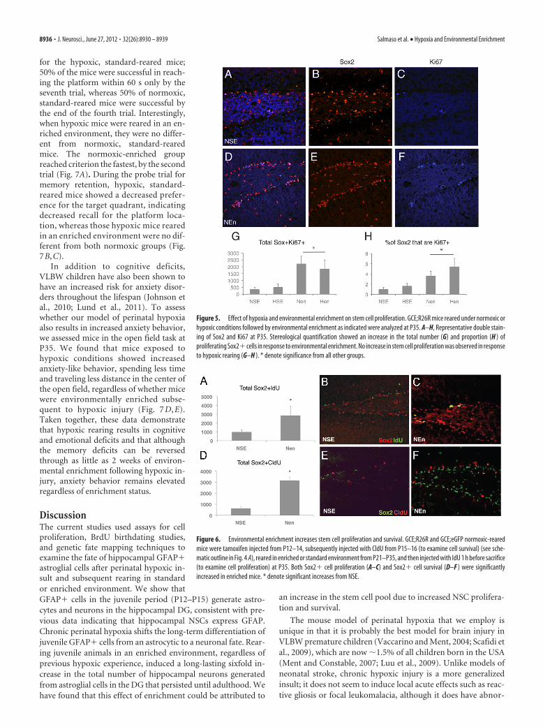
for the hypoxic, standard-reared mice;50% of the mice were successful in reach-ing the platform within 60 s only by theseventh trial, whereas 50% of normoxic,standard-reared mice were successful bythe end of the fourth trial. Interestingly,when hypoxic mice were reared in an en-riched environment, they were no differ-ent from normoxic, standard-rearedmice. The normoxic-enriched groupreached criterion the fastest, by the secondtrial (Fig. 7A). During the probe trial formemory retention, hypoxic, standard-reared mice showed a decreased prefer-ence for the target quadrant, indicatingdecreased recall for the platform loca-tion, whereas those hypoxic mice rearedin an enriched environment were no dif-ferent from both normoxic groups (Fig.7B,C).
In addition to cognitive deficits,VLBW children have also been shown tohave an increased risk for anxiety disor-ders throughout the lifespan (Johnson etal., 2010; Lund et al., 2011). To assesswhether our model of perinatal hypoxiaalso results in increased anxiety behavior,we assessed mice in the open field task atP35. We found that mice exposed tohypoxic conditions showed increasedanxiety-like behavior, spending less timeand traveling less distance in the center ofthe open field, regardless of whether micewere environmentally enriched subse-quent to hypoxic injury (Fig. 7 D, E).Taken together, these data demonstratethat hypoxic rearing results in cognitiveand emotional deficits and that althoughthe memory deficits can be reversedthrough as little as 2 weeks of environ-mental enrichment following hypoxic in-jury, anxiety behavior remains elevatedregardless of enrichment status.
DiscussionThe current studies used assays for cellproliferation, BrdU birthdating studies,and genetic fate mapping techniques toexamine the fate of hippocampal GFAP�astroglial cells after perinatal hypoxic in-sult and subsequent rearing in standardor enriched environment. We show thatGFAP� cells in the juvenile period (P12–P15) generate astro-cytes and neurons in the hippocampal DG, consistent with pre-vious data indicating that hippocampal NSCs express GFAP.Chronic perinatal hypoxia shifts the long-term differentiation ofjuvenile GFAP� cells from an astrocytic to a neuronal fate. Rear-ing juvenile animals in an enriched environment, regardless ofprevious hypoxic experience, induced a long-lasting sixfold in-crease in the total number of hippocampal neurons generatedfrom astroglial cells in the DG that persisted until adulthood. Wehave found that this effect of enrichment could be attributed to
an increase in the stem cell pool due to increased NSC prolifera-tion and survival.
The mouse model of perinatal hypoxia that we employ isunique in that it is probably the best model for brain injury inVLBW premature children (Vaccarino and Ment, 2004; Scafidi etal., 2009), which are now 1.5% of all children born in the USA(Ment and Constable, 2007; Luu et al., 2009). Unlike models ofneonatal stroke, chronic hypoxic injury is a more generalizedinsult; it does not seem to induce local acute effects such as reac-tive gliosis or focal leukomalacia, although it does have abnor-
Figure 5. Effect of hypoxia and environmental enrichment on stem cell proliferation. GCE;R26R mice reared under normoxic orhypoxic conditions followed by environmental enrichment as indicated were analyzed at P35. A–H, Representative double stain-ing of Sox2 and Ki67 at P35. Stereological quantification showed an increase in the total number (G) and proportion (H ) ofproliferating Sox2� cells in response to environmental enrichment. No increase in stem cell proliferation was observed in responseto hypoxic rearing (G–H ). * denote significance from all other groups.
Figure 6. Environmental enrichment increases stem cell proliferation and survival. GCE;R26R and GCE;eGFP normoxic-rearedmice were tamoxifen injected from P12–14, subsequently injected with CldU from P15–16 (to examine cell survival) (see sche-matic outline in Fig. 4 A), reared in enriched or standard environment from P21–P35, and then injected with IdU 1 h before sacrifice(to examine cell proliferation) at P35. Both Sox2� cell proliferation (A–C) and Sox2� cell survival (D–F ) were significantlyincreased in enriched mice. * denote significant increases from NSE.
8936 • J. Neurosci., June 27, 2012 • 32(26):8930 – 8939 Salmaso et al. • Hypoxia and Environmental Enrichment

malities in white matter development, including delayedmaturation of oligodendrocytes and abnormal myelination (B.Jablonska, J. Scafildi, A. Aguirre, F. Vaccarino, E. Borok, T. Hor-vath, V. Gallo, unpublished observation). Interestingly, amongthe best predictors of positive outcome in VLBW children are atwo-parent household and an increased level of maternal educa-
tion (Ment et al., 2003). This raises the important question of themechanisms by which increased environmental stimulation ofVLBW children months and years after the perinatal insultimproves recovery (Ment et al., 2003). It has been well docu-mented in adult rodents that environmental enrichment notonly increases cell proliferation and neurogenesis, but in-creases the ability of new neurons to differentiate, integrate,and mature in the DG (Kempermann et al., 2004). However, thecognitive and neurobiological effect of environmental enrich-ment during development and, in particular, its ability to interactwith prior hypoxic injury has not been investigated. Our fatemapping studies show that environmental enrichment for 2weeks increased the number of neurons generated from astroglialcells in the DG at P35 but did not augment the neurogenic effectof hypoxia. Interestingly, extending the enrichment until fulladulthood (P90) increased neurogenesis in both normoxic andhypoxic mice, and the effects of hypoxia and enrichment ap-peared independent and additive. It is likely that the increase intotal number of fate-mapped neurons described here is a grossunderestimation of the neurogenesis that occurs in response tohypoxia and environmental enrichment. The population of fate-mapped neurons that we observe arise from GFAP� cells labeledfrom P12–P14 only; furthermore, the targeting efficiency of theGCE transgene is not 100% (Ganat et al., 2006). Therefore, theseneurons likely represent a small fraction of the total neurogen-esis induced by hypoxia and enrichment in these mice. Indeed,at P35, we show that the proportion of fate-mapped neuronsrelative to the total number of granule neurons ranges acrossgroups from 2 to 6%, whereas a recent study by Cushman etal. (2012) demonstrates that the proportion of total DG neu-rons born from GFAP� cells was 24% at 2 months of age,suggesting that we are capturing about one-fourth of the totalnumber of neurons arising from GFAP� precursors (Cush-man et al., 2012).
Increased neurogenesis can be attributed to a shift in fate ofthe GFAP� stem cells toward the neuronal lineage or to a changein progenitor proliferation or neuronal survival. Moreover, in-creased neurogenesis will, over the long term, deplete the astro-glial stem cell population unless these cells self-renew viaasymmetric division. We find that hypoxia induced a fate shift in
Figure 8. Schematic representation depicting hypoxia- and enrichment-induced changes inastroglial lineage. There is an increase in the number of neuronal cells in hypoxic-reared as wellas normoxic enriched mice, shown in purple (DCX�) and blue (NeuN�). HEn mice show thegreatest increase in total number of reporter� neurons, suggesting an additive effect of hyp-oxia and enrichment on neurogenesis arising from GFAP� cells. HSE mice show a greaterproportion of the stem cell pool that is committed to the neuronal lineage (Sox2�DCX�,shown in red); this effect remains apparent in hypoxic mice that were subsequently enriched. InNEn mice there is no change in fate potential of NSC but rather a robust increase in the totalstem/progenitor cell pool (shown in green, yellow, red) that is still present in Hen mice.
Figure 7. Environmental enrichment reverses hypoxia-induced cognitive deficits. Mice werereared in normoxia or hypoxia from P3 to P11 and subsequently in standard or enriched envi-ronment from P21 to P35. Behavioral testing was conducted on P35–36. A–C, Results of theMorris water maze. A, The proportion of mice that successfully found the platform in under 60 s,averaged across six trials, during the acquisition phase on the maze. B, The latency to find theplatform across trials. C, Probe trial. D, E, Results from the open field test. D, Proportion ofdistance traveled in the center/total distance traveled. E, Time spent in the center. *, Signifi-cance from all other groups; **, significance from NSE and NEn groups, #, HSE and NEn aredifferent from all other groups.
Salmaso et al. • Hypoxia and Environmental Enrichment J. Neurosci., June 27, 2012 • 32(26):8930 – 8939 • 8937

juvenile GFAP� cells away from the astroglial lineage and infavor of the doublecortin (DCX) neuronal lineage (Fig. 4V). Al-though a similar increase in total number of DCX�/NeuN� cellswas seen in response to environmental enrichment, no fate shifttoward the DCX lineage was observed in normoxic enriched an-imals. Our results show that environmental enrichment causes arobust increase in size of the astroglial stem cell population that isattributable to enhanced NSC self-renewal as well as survival, asshown by the Ki67 double labeling and CldU/IdU incorporationexperiments. Hypoxic-rearing on its own does not increase thestem cell pool and, in fact, hypoxia increases the proportion ofGFAP� and Sox2� stem cells that coexpress the neuronal pro-genitor marker DCX (Fig. 4V,W), suggesting that more of thesecells are committing to a neuronal fate. Subsequent exposure ofhypoxic-reared animals to environmental enrichment increasedthe proportion of Sox2� cells coexpressing GFAP (Fig. 4X) aswell as the total number of stem cells (Fig. 4U), suggesting thatboth a fate shift toward the neuronal lineage and an increase inthe number of stem cells may be acting together in hypoxic en-riched mice, accounting for the additive effect on the number ofnewly generated neurons.
Together, our results suggest that hypoxia alone increasesneurogenesis by pushing the stem cell population toward a neu-ronal fate, whereas enrichment increases the total stem cell pop-ulation. Consequently, hypoxia coupled with enrichment enablesneurogenesis from GFAP� astroglia to rise without depleting thepopulation of Sox2� neural stem cells and maintaining theirproportion relative to neuroblasts (Fig. 8). Thus, the robust long-term increase in neurogenesis that we have observed in hypoxicenriched mice can be attributed to the hypoxia-induced neuro-genic shift coupled with an overall increase in the stem cell poolobserved in response to environmental enrichment. Interest-ingly, the increase in stem cells we have observed in response toenriched environment may be unique to the early phase of en-richment. In a recent study, Hen and colleagues (Dranovsky et al.,2011) employed a similar genetic fate-mapping model driven bya Nestin-Cre promoter and demonstrated that exposing mice tolong-term environmental enrichment in adulthood leads to asimilar decrease in GFAP�/reporter� cells while increasingNeuN�/reporter� cells; however, unlike our current study, theydid not observe a significant increase in the total number of re-porter� stem cells in response to environmental enrichment(Dranovsky et al., 2011). The differential response of stem cells toenrichment observed between these studies may reflect differ-ences in the age at which environmental enrichment was com-menced (P21 in our study versus adult); however, it is alsopossible that it is a transient effect in response to the early phase ofenrichment and, perhaps, to physical exercise with the runningwheel (van Praag et al., 1999b; Kobilo et al., 2011). Indeed, arecent study examining the fate of hippocampal Hes5� cells inadult mice that were given free access to a running wheel for 12 dshowed increased proliferation of reporter� stem cells in micewith running wheel access as compared to controls (Lugert et al.,2010). Taken together, it seems more likely that at least in thehippocampus, the number of stem cells increase during the earlyphases of environmental enrichment; however, more detailed fu-ture studies examining the relative contribution of hippocampalGFAP� stem cells in response to environmental enrichmentwhile controlling for age of onset, duration, and access to physicalactivity are needed to answer these questions.
The relative contribution of increasing the number of stemcells versus neurons to cognitive amelioration in hypoxic en-riched mice remains to be determined. In the current study, hy-
poxic mice show a similar increase in neurogenesis as donormoxic, enriched mice, yet hypoxic mice show a slower rate ofacquisition and impaired retention, suggesting that hippocampalneurogenesis alone cannot account for the differences seen incognitive performance. These findings are in support of previouswork showing that the role of neurogenesis in learning and mem-ory, as revealed by the Morris water maze task, is not clearlydefined, with some studies finding a significant relationship andothers not (Shors et al., 2002). Furthermore, it is also likely thatbehavioral changes in response to both hypoxia and environmen-tal enrichment within other brain regions, such as white matter,cortex or the subventricular zone may contribute to our behav-ioral findings (Bi et al., 2011).
On the other hand, the increased performance seen in bothenriched groups could be related to the increased stem cell poolalso observed in these groups; albeit whether this is causally re-lated remains to be determined. Although the current paper hasfocused on the hippocampus and GFAP� cell fate, environmen-tal enrichment impacts other mechanisms of neural plasticity inthe hippocampus that could also play a causal role in enhancedlearning and memory, including synaptogenesis, inhibitory tone,and dendritic spine formation (Johansson and Belichenko, 2002;Sale et al., 2007; Bednarek and Caroni, 2011).
Longitudinal studies of VLBW children have shown that thesechildren are more likely to be diagnosed with an anxiety disorder,even decades later (Johnson et al., 2010; Lund et al., 2011). Re-sults from the open field test also showed increased anxiety in ourmouse model. Interestingly, these effects were not amelioratedwith short-term environmental enrichment, suggesting thatanxiety behavior after hypoxic injury is mediated by differentneurobiological changes that are not responsive to short-termenrichment.
In conclusion, we demonstrate that subsequent environmen-tal enrichment after hypoxic injury reverses the cognitive deficitsconsequential to hypoxic injury. Furthermore, hypoxic injuryand environmental enrichment together increase neurogenesis inthe dentate gyrus in an additive, long-term manner. Environ-mental enrichment increases the total stem cell populationthrough proliferation and survival, which ultimately leads to sus-tained increases in neurogenesis in hypoxic, enriched mice.
ReferencesAlvarez-Buylla A, Seri B, Doetsch F (2002) Identification of neural stem cells
in the adult vertebrate brain. Brain Res Bull 57:751–758.Avilion AA, Nicolis SK, Pevny LH, Perez L, Vivian N, Lovell-Badge R (2003)
Multipotent cell lineages in early mouse development depend on SOX2function. Genes Dev 17:126 –140.
Bednarek E, Caroni P (2011) �-Adducin is required for stable assembly ofnew synapses and improved memory upon environmental enrichment.Neuron 69:1132–1146.
Belachew S, Chittajallu R, Aguirre AA, Yuan X, Kirby M, Anderson S, Gallo V(2003) Postnatal NG2 proteoglycan-expressing progenitor cells are in-trinsically multipotent and generate functional neurons. J Cell Biol161:169 –186.
Bi B, Salmaso N, Komitova M, Simonini MV, Silbereis J, Cheng E, Kim J, LuftS, Ment LR, Horvath TL, Schwartz ML, Vaccarino FM (2011) Corticalglial fibrillary acidic protein-positive cells generate neurons after perinatalhypoxic injury. J Neurosci 31:9205–9221.
Brenner M (1994) Structure and transcriptional regulation of the GFAPgene. Brain Pathol 4:245–257.
Brill MS, Snapyan M, Wohlfrom H, Ninkovic J, Jawerka M, Mastick GS,Ashery-Padan R, Saghatelyan A, Berninger B, Gotz M (2008) A dlx2-and pax6-dependent transcriptional code for periglomerular neuronspecification in the adult olfactory bulb. J Neurosci 28:6439 – 6452.
Cushman JD, Maldonado J, Kwon EE, Garcia AD, Fan G, Imura T, SofroniewMV, Fanselow MS (2012) Juvenile neurogenesis makes essential contri-
8938 • J. Neurosci., June 27, 2012 • 32(26):8930 – 8939 Salmaso et al. • Hypoxia and Environmental Enrichment

butions to adult brain structure and plays a sex-dependent role in fearmemories. Front Behav Neurosci 6:3.
Doetsch F, Caille I, Lim DA, García-Verdugo JM, Alvarez-Buylla A (1999)Subventricular zone astrocytes are neural stem cells in the adult mamma-lian brain. Cell 97:703–716.
Dranovsky A, Picchini AM, Moadel T, Sisti AC, Yamada A, Kimura S, Leon-ardo ED, Hen R (2011) Experience dictates stem cell fate in the adulthippocampus. Neuron 70:908 –923.
Episkopou V (2005) SOX2 functions in adult neural stem cells. Trends Neu-rosci 28:219 –221.
Fagel DM, Ganat Y, Silbereis J, Ebbitt T, Stewart W, Zhang H, Ment LR,Vaccarino FM (2006) Cortical neurogenesis enhanced by chronic peri-natal hypoxia. Exp Neurol 199:77–91.
Filippov V, Kronenberg G, Pivneva T, Reuter K, Steiner B, Wang LP, Yama-guchi M, Kettenmann H, Kempermann G (2003) Subpopulation ofnestin-expressing progenitor cells in the adult murine hippocampusshows electrophysiological and morphological characteristics of astro-cytes. Mol Cell Neurosci 23:373–382.
Ganat YM, Silbereis J, Cave C, Ngu H, Anderson GM, Ohkubo Y, Ment LR,Vaccarino FM (2006) Early postnatal astroglial cells produce multilin-eage precursors and neural stem cells in vivo. J Neurosci 26:8609 – 8621.
Garcia AD, Doan NB, Imura T, Bush TG, Sofroniew MV (2004) GFAP-expressing progenitors are the principal source of constitutive neurogen-esis in adult mouse forebrain. Nat Neurosci 7:1233–1241.
Gundersen HJ, Bagger P, Bendtsen TF, Evans SM, Korbo L, Marcussen N,Møller A, Nielsen K, Nyengaard JR, Pakkenberg B (1988) The new ste-reological tools: disector, fractionator, nucleator, and point sampled in-tercepts and their use in pathological research and diagnosis. APMIS96:857– 881.
Hack MA, Saghatelyan A, de Chevigny A, Pfeifer A, Ashery-Padan R, LledoPM, Gotz M (2005) Neuronal fate determinants of adult olfactory bulbneurogenesis. Nat Neurosci 8:865– 872.
Hevner RF, Hodge RD, Daza RA, Englund C (2006) Transcription factors inglutamatergic neurogenesis: conserved programs in neocortex, cerebel-lum, and adult hippocampus. Neurosci Res 55:223–233.
Imura T, Kornblum HI, Sofroniew MV (2003) The predominant neuralstem cell isolated from postnatal and adult forebrain but not early embry-onic forebrain expresses GFAP. J Neurosci 23:2824 –2832.
Johansson BB, Belichenko PV (2002) Neuronal plasticity and dendriticspines: effect of environmental enrichment on intact and postischemic ratbrain. J Cereb Blood Flow Metab 22:89 –96.
Johnson S, Hollis C, Kochhar P, Hennessy E, Wolke D, Marlow N (2010)Psychiatric disorders in extremely preterm children: longitudinal findingat age 11 years in the EPICure study. J Am Acad Child Adolesc Psychiatry49:453– 463.e1.
Kempermann G, Kuhn HG, Gage FH (1997) More hippocampal neurons inadult mice living in an enriched environment. Nature 386:493– 495.
Kempermann G, Wiskott L, Gage FH (2004) Functional significance ofadult neurogenesis. Curr Opin Neurobiol 14:186 –191.
Kernie SG, Parent JM (2010) Forebrain neurogenesis after focal Ischemicand traumatic brain injury. Neurobiol Dis 37:267–274.
Kobilo T, Liu QR, Gandhi K, Mughal M, Shaham Y, van Praag H (2011)Running is the neurogenic and neurotrophic stimulus in environmentalenrichment. Learn Mem 18:605– 609.
Komitova M, Eriksson PS (2004) Sox-2 is expressed by neural progenitorsand astroglia in the adult rat brain. Neurosci Lett 369:24 –27.
Kuhn HG, Palmer TD, Fuchs E (2001) Adult neurogenesis: a compensatorymechanism for neuronal damage. Eur Arch Psychiatry Clin Neurosci251:152–158.
Lugert S, Basak O, Knuckles P, Haussler U, Fabel K, Gotz M, Haas CA,Kempermann G, Taylor V, Giachino C (2010) Quiescent and active hip-pocampal neural stem cells with distinct morphologies respond selec-tively to physiological and pathological stimuli and aging. Cell Stem Cell6:445– 456.
Lund LK, Vik T, Skranes J, Brubakk AM, Indredavik MS (2011) Psychiatric
morbidity in two low birth weight groups assessed by diagnostic interviewin young adulthood. Acta Paediatr 100:598 – 604.
Luu TM, Ment LR, Schneider KC, Katz KH, Allan WC, Vohr BR (2009)Lasting effects of preterm birth and neonatal brain hemorrhage at 12 yearsof age. Pediatrics 123:1037–1044.
Luu TM, Vohr BR, Allan W, Schneider KC, Ment LR (2011) Evidence forcatch-up in cognition and receptive vocabulary among adolescents bornvery preterm. Pediatrics 128:313–322.
Ment LR, Constable RT (2007) Injury and recovery in the developing brain:evidence from functional MRI studies of prematurely born children. NatClin Pract Neurol 3:558 –571.
Ment LR, Vohr B, Allan W, Katz KH, Schneider KC, Westerveld M, DuncanCC, Makuch RW (2003) Change in cognitive function over time in verylow-birth-weight infants. JAMA 289:705–711.
Nakamura T, Colbert MC, Robbins J (2006) Neural crest cells retain multi-potential characteristics in the developing valves and label the cardiacconduction system. Circ Res 98:1547–1554.
Nunez J (2008) Morris water maze experiment. J Vis Exp pii: 897.doi:10.3791.
Olson AK, Eadie BD, Ernst C, Christie BR (2006) Environmental enrich-ment and voluntary exercise massively increase neurogenesis in the adulthippocampus via dissociable pathways. Hippocampus 16:250 –260.
Parent JM (2003) Injury-induced neurogenesis in the adult mammalianbrain. Neuroscientist 9:261–272.
Rivers LE, Young KM, Rizzi M, Jamen F, Psachoulia K, Wade A, Kessaris N,Richardson WD (2008) PDGFRA/NG2 glia generate myelinating oligo-dendrocytes and piriform projection neurons in adult mice. Nat Neurosci11:1392–1401.
Saigal S, Doyle LW (2008) An overview of mortality and sequelae of pretermbirth from infancy to adulthood. Lancet 371:261–269.
Sale A, Maya Vetencourt JF, Medini P, Cenni MC, Baroncelli L, De PasqualeR, Maffei L (2007) Environmental enrichment in adulthood promotesamblyopia recovery through a reduction of intracortical inhibition. NatNeurosci 10:679 – 681.
Scafidi J, Fagel DM, Ment LR, Vaccarino FM (2009) Modeling prematurebrain injury and recovery. Int J Dev Neurosci 27:863– 871.
Schmitz C, Hof PR (2005) Design-based stereology in neuroscience. Neu-roscience 130:813– 831.
Seki T, Arai Y (1999) Temporal and spacial relationships between PSA-NCAM-expressing, newly generated granule cells, and radial glia-like cellsin the adult dentate gyrus. J Comp Neurol 410:503–513.
Seri B, García-Verdugo JM, McEwen BS, Alvarez-Buylla A (2001) Astro-cytes give rise to new neurons in the adult mammalian hippocampus.J Neurosci 21:7153–7160.
Sharp FR, Liu J, Bernabeu R (2002) Neurogenesis following brain ischemia.Brain Res Dev Brain Res 134:23–30.
Shors TJ, Townsend DA, Zhao M, Kozorovitskiy Y, Gould E (2002) Neuro-genesis may relate to some but not all types of hippocampal-dependentlearning. Hippocampus 12:578 –584.
Soriano P (1999) Generalized lacZ expression with the ROSA26 Cre re-porter strain. Nat Genet 21:70 –71.
Vaccarino FM, Ment LR (2004) Injury and repair in developing brain. ArchDis Child Fetal Neonatal Ed 89:F190 –192.
van Praag H, Kempermann G, Gage FH (1999a) Running increases cell pro-liferation and neurogenesis in the adult mouse dentate gyrus. Nat Neuro-sci 2:266 –270.
van Praag H, Christie BR, Sejnowski TJ, Gage FH (1999b) Running en-hances neurogenesis, learning, and long-term potentiation in mice. ProcNatl Acad Sci U S A 96:13427–13431.
West MJ (1993) New stereological methods for counting neurons. Neuro-biol Aging 14:275–285.
Yang Z, Levison SW (2006) Hypoxia/ischemia expands the regenerative ca-pacity of progenitors in the perinatal subventricular zone. Neuroscience139:555–564.
Salmaso et al. • Hypoxia and Environmental Enrichment J. Neurosci., June 27, 2012 • 32(26):8930 – 8939 • 8939