neurobiological bases of behavior
DESCRIPTION
neurobiologicalTRANSCRIPT
Neurobiological Bases of Behavior
The nervous system and the endocrine system organize the line of communication within the body. One area of the brain communicates to its other parts through neurons. And one neuron communicates with another neuron to synchronize a person’s thought, affect and movement. Communication in the nervous system takes place also through the secretion of neurotransmitters which influence biological functions.

Disturbances in the levels of secretion of these chemical
substances appear to be associated with certain mental disorders like schizophrenia, mania, depression and many
more.The endocrine system also
involves a highly organized communication process in the
body. It monitors the secretion of chemical substance called
hormones and makes them in harmony with each other.
Neuron or nerve cell is th basic information processing structure of the nervous system. it transmits information throughout the body.
Types: 1. Sensory neuron- recieves information or stimuli
from the environment through sensory cells.2. Motor neuron- sends electrical output signals to
the muscles.3. Interneuron- signals another neuron.

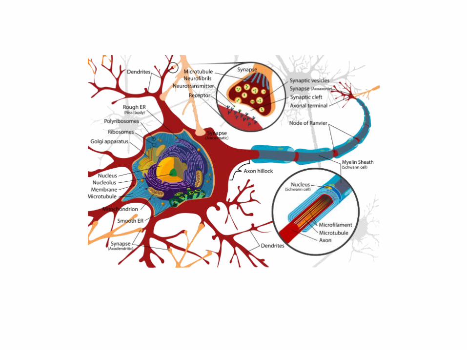
a neuron is made up of a cell body(soma), with its nucleus, and a number of processes called the
dendrites- short cluster of fiber hich recieve information from another neuron, and
axon which is a single long process that carries information destined for other neurons send impulses from the soma to other neurons.
axon hillock commences from the soma as conical projection and covered by a cellular sheath with an
insulating material called myelin. synaptic terminal- bulb at the end of an axon in which neurotransmitter molecules are stored and released.

Neural ImpulseImpulse controls the way we react or respond to a certain stimulus.Stages of a Neural Impulse:1. Resting Membrane Potential- in this stage the neuron is said to be in an inactive state. there are more negative ions inside the cell membrane and that there are more positive ions outside it. the charge is about 70 millivolts, and it is ready to fire once it is stimulated.
2. Action Potential- Once the stimulation reaches a certain threshold (strength of a stimulus that warrants a response), the neural membrane opens at one area and allows positively charged ions to rush in and the negative ions to rush out. At this stage, the internal state of the neuron becomes less negative until the charge inside the neuron rises to approximately +40 mv.
3. Absolute Refractory Period- After the action potential occurs, there is a brief period during which the neuron is unable to fire. Then, the charge inside the neuron drops to about -90 mv (refractory period) because at this stage, the cell membrane does not admit positive ions. A this stage, a neuron is said to be repolarized.
4. Relative Refractory Period- this is the stage where the neurons tries to quickly restore its charge by pumping out the positively charged ions and bringing back th negative ones. it is the period where a neuron may fire depending on the strength of the stimulus.

THE SYNAPSEA synapse is a connection between neurons. Neurons are meaningless if they exist alone. They must connect in order to communicate information. There are different types of synapse. There is so-called dendrodendritic connection which is a connection between dendrites of neighboring neurons. Another type is called axondendritic where the link is between the axon of one neuron and the dendrite of another neuron. And the other one is called axo-axonic where the connection is between the axons of neurons.

Neurotransmitters
Neurotransmitters is the process whereby neurotransmitters from a sending neuron are released to the presynaptic cleft and bind to the receptor sites of another neuron called receiving neuron or post-synaptic neuron.

The presynaptic neuron releases neurotransmitter, which activates receptors on the post-synaptic cell.
Types of Neurotransmitters
1. Acetylcholine (Ach)- found in parts of the peripheral nervous system, spinal cord, and areas of the brain. In the PNS, the acetylcholine acts as an axcitatory neurotransmitter; it activates muscles that help the body move.
2. Dopamine-neurotransmitter that is involved in movement, learning, and reinforcement. It has been associated with several clinical disorders.

Major division of the nervous system
Peripheral nervous system is made up of all of the nerves and the writing. This system sends the
messages from the brain to the rest of the body.PNS is subdivided to somatic and autonomic
nervous system. The former is responsible for the control of voluntary movements and the
communication of information to and from the different sense organ.
Autonomic nervous system is concerned with the parts of the body that function involuntarily like that of the heart muscle, which functions without our awareness.
ANS is split into two, namely: sympathetic that acts to prepare the body in stressful situation, on the other hand, parasympathetic nervous system, acts to calm down the body after an emergency situation, also called vegetative nervous system.

Central nervous sysytem
It includes two important structures: the brain-the cephalic or the superior portion and
the spinal cord- the caudal or the inferior portion.


The brain The function of the brain include
identification, organization, interpretation and response generation to sensory stimulation.
Five Major Division of the Brain and Its Structure
1.Myelencephalon-most inferior division of the brain, referred as medulla. It is composed of
tracts carrying signals between the rest of the brain and the body.

2.Metencepahlon- ascending and descending tracts and part of reticular formation. These structure collectively make up the pons which acts as a bridge to pass informtion regarding movement to the cerebellum.3. Mecencephalon- contains tectum(roof) and tegmentum. The tectum is composed of the inferior colliculli and the superior colliculli. The tegmentum contains 3 structures: the periaqueductal gray- rich in smallneuron and is a significant repository of the naturally occuring opium, morphine, endorphin.
4. Diencephalon- 2 important structure: the thalamus- a large, two lobed structure responsible for relaying message. The hypothalamus, on the other hand, is found underneath the thalamus, plays important role in the regulation of motivated behaviors since it controls the activities of the autonomic and the endocrine system.
5. Telencephalon- largest division of the brain and it mediates the most complex function as it holds the information- processing capacities of the brain.


Cerebral Cortex-thin layer of tissue covering the cerebral hemisphere. There are 2 cerebral hemispheres, the right and the left hemispheres., but they are connected by a large bundle of fibers called the corpus callosum. The presence of fissures further subdivides the hemispheres into four lobes, namely: the frontal, partial, temporal, and the occipital lobe.
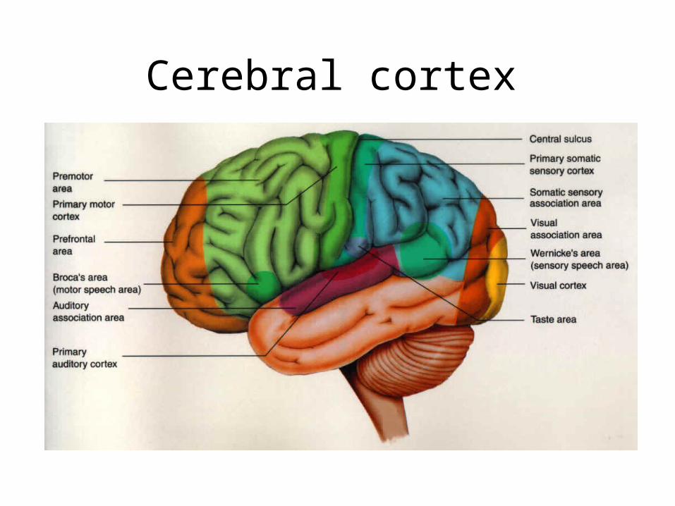
Cerebral cortex

Limbic System- involved in the regulation of motivated behaviors like aggresion, sexual behavior, and eating, among others.
Amygdala-almond-shaped structure responsible for memories with strong emotional content.
Hippocampus-keeps track of facts, it is one which decides whether memories are to be placed in short term or lng term storage.


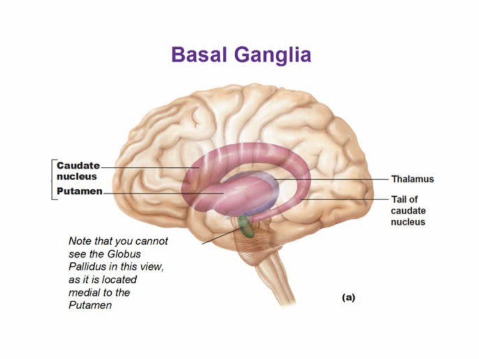
Basal ganglia-the major role played by this structure seems to be in the area of programming and executionof movements. Diseases of the basal ganglia are manifested by tremors and uncontrolled movements.

Spinal Cord the spinal cord is the major important part of
the nervous system. It is the most caudal part of the CNS. It begins at foramen magnumof the skull and terminates at the lumbar level. It is subdivided into levels corresponding to the vertebral regions surrounding it.

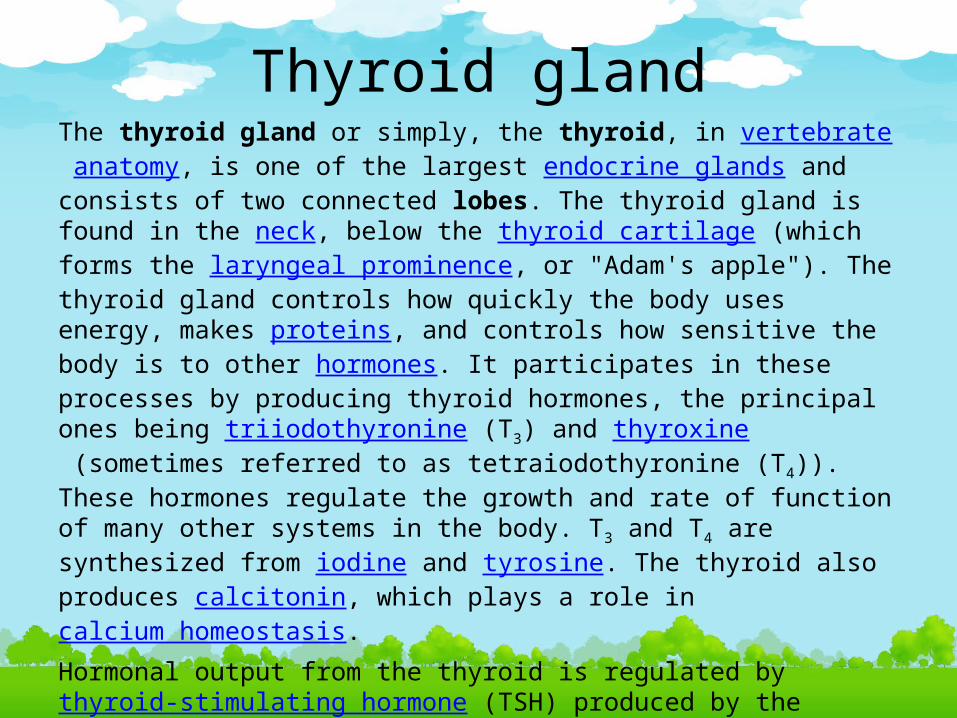
Thyroid glandThe thyroid gland or simply, the thyroid, in vertebrate anatomy, is one of the largest endocrine glands and consists of two connected lobes. The thyroid gland is found in the neck, below the thyroid cartilage (which forms the laryngeal prominence, or "Adam's apple"). The thyroid gland controls how quickly the body uses energy, makes proteins, and controls how sensitive the body is to other hormones. It participates in these processes by producing thyroid hormones, the principal ones being triiodothyronine (T3) and thyroxine (sometimes referred to as tetraiodothyronine (T4)). These hormones regulate the growth and rate of function of many other systems in the body. T3 and T4 are synthesized from iodine and tyrosine. The thyroid also produces calcitonin, which plays a role in calcium homeostasis.Hormonal output from the thyroid is regulated by thyroid-stimulating hormone (TSH) produced by the anterior pituitary, which itself is regulated by thyrotropin-releasing hormone (TRH) produced by the hypothalamus.

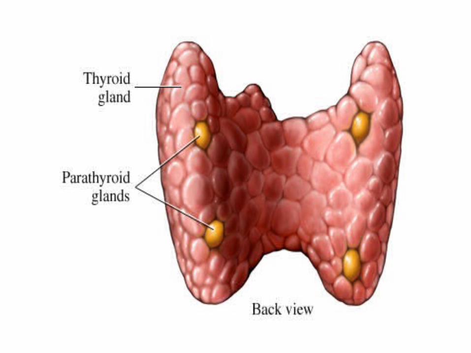
Parathyroid glandThe parathyroid glands are small endocrine glands in the neck of humans and other tetrapods that produce parathyroid hormone. Humans usually have four parathyroid glands, variably located on the back of the thyroid gland, although considerable variation exists. Parathyroid hormone and calcitonin (one of the hormones made by the thyroid gland) have key roles in regulating the amount of calcium in the blood and within the bones. The parathyroid glands are two pairs of glands usually positioned behind the left and right lobes of the thyroid. Each gland is a yellowish-brown flat ovoid that resembles a lentil seed, usually about 6 mm long and 3 to 4 mm wide, and 1 to 2 mm anteroposteriorly. There are typically four parathyroid glands. The two parathyroid glands on each side which are positioned higher are called the superior parathyroid glands, while the lower two are called the inferior parathyroid glands. Healthy parathyroid glands generally weight about 30mg in men and 35mg in women.
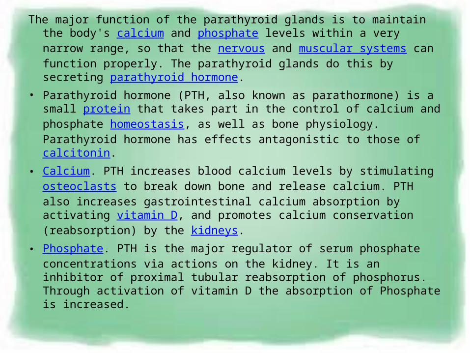
The major function of the parathyroid glands is to maintain the body's calcium and phosphate levels within a very narrow range, so that the nervous and muscular systems can function properly. The parathyroid glands do this by secreting parathyroid hormone.
• Parathyroid hormone (PTH, also known as parathormone) is a small protein that takes part in the control of calcium and phosphate homeostasis, as well as bone physiology. Parathyroid hormone has effects antagonistic to those of calcitonin.
• Calcium. PTH increases blood calcium levels by stimulating osteoclasts to break down bone and release calcium. PTH also increases gastrointestinal calcium absorption by activating vitamin D, and promotes calcium conservation (reabsorption) by the kidneys.
• Phosphate. PTH is the major regulator of serum phosphate concentrations via actions on the kidney. It is an inhibitor of proximal tubular reabsorption of phosphorus. Through activation of vitamin D the absorption of Phosphate is increased.
The gonads (sex gland)
The gonad is the organ that makes gametes. The gonads in males are the testes, and the gonads in females are the ovaries. spermatozoon and egg cells are gametes. The gonads are controlled gonadocilly by luteinizing hormone (LH) and follicle-stimulating hormone (FSH) secreted by the anterior pituitary gland. This secretion in turn is controlled by the hypothalamus' gonadotropin-releasing hormone.


Thymus glandThe thymus is a specialized organ of the immune system. Within the thymus, T-cells
mature.T cells are critical to the adaptive immune system, where they adapt specifically to
foreign invaders. Each T cell attacks a specific foreign substance which it identifies with its receptor. T cells have receptors which are generated by randomly shuffling gene segments. Each T cell attacks a different antigen. T cells that attack the body's own
proteins are eliminated in the thymus. Thymic epithelial cells express major proteins from elsewhere in the body. First, T cells undergo "Positive Selection" whereby the cell
comes in contact with self-MHC expressed by thymic epithelial cells; those with no interaction are destroyed. Second, the T cell undergoes "Negative Selection" by
interacting with thymic dendritic cell whereby T cells with high affinity interaction are eliminated throughapoptosis (to avoid autoimmunity), and those with intermediate
affinity survive.The thymus is composed of two identical lobes and is located anatomically in the
anterior superior mediastinum, in front of the heart and behind the sternum.

• Histologically, each lobe of the thymus can be divided into a central medulla and a peripheral cortex which is surrounded by an outer capsule. The cortex and medulla play different roles in the development of T-cells. Cells in the thymus can be divided into thymicstromal cells and cells of hematopoietic origin (derived from bone marrow resident hematopoietic stem cells). Developing T-cells are referred to as thymocytes and are of hematopoietic origin. Stromal cells include thymic cortical epithelial cells, thymic medullary epithelial cells, and dendritic cells.
• The thymus provides an inductive environment for development of T-lymphocytes from hematopoietic progenitor cells. In addition, thymic stromal cells allow for the selection of a functional and self-tolerant T-cell repertoire. Therefore, one of the most important roles of the thymus is the induction of central tolerance.
• The thymus is largest and most active during the neonatal and pre-adolescent periods. By the early teens, the thymus begins toatrophy and thymic stroma is mostly replaced by adipose (fat) tissue. Nevertheless, residual T lymphopoiesis continues throughout adult life.

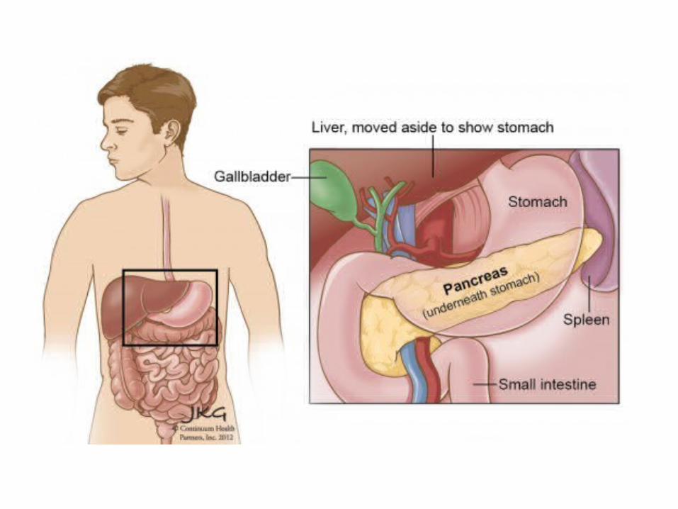
Pancreas endocrine organ that lies in the abdomen, specifically the upper, left abdomen. It is found behind the stomach, with the head of the pancreas surrounded by the duodenum. The pancreas is typically 5.75-9.5 cm long. The pancreas is divided into a head, which rests within the concavity of the duodenum, a body lying behind the base of the stomach, and a tail, which ends abutting the spleen. The neck of the pancreas lies between the body and head, and is in front of the superior mesenteric artery and vein. The head of the pancreas surrounds these two vessels, and a small uncinate processemerges from the lower part of the head, lying behind the superior mesenteric artery. The pancreas is a secretory structure with a internal hormonal role (endocrine) and an external digestive role (exocrine). It has two main ducts, the main pancreatic duct, and the accessory pancreatic duct. These drain enzymes through the ampulla of Vater into the duodenum.

The part of the pancreas with endocrine function is made up of approximately a million[7] cell clusters called islets of Langerhans. Four main cell types exist in the islets. They are relatively difficult to distinguish using standard staining techniques, but they can be classified by their secretion: α alpha cells secrete glucagon (increase glucose in blood), β beta cells secrete insulin (decrease glucose in blood), Δ delta cells secrete somatostatin (regulates/stops α and β cells) and PP cells, or γ (gamma) cells, secrete pancreatic polypeptide.the pancreas as an exocrine gland helps out the digestive system. It secretes pancreatic fluid that contains digestive enzymes that pass to the small intestine. These enzymes help to further break down the carbohydrates, proteins and lipids (fats) in the chyme.In humans, the secretory activity of the pancreas is regulated directly via the effect of hormones in the blood on the islets of Langerhans and indirectly through the effect of theautonomic nervous system on the blood flow.
The exocrine component of the pancreas, often called simply the exocrine pancreas, is the portion of the pancreas that performs exocrine functions. It has ducts that are arranged in clusters called acini (singular acinus). Pancreatic secretions are secreted into the lumen of the acinus, and then accumulate in intralobular ducts that drain to the mainpancreatic duct, which drains directly into the duodenum.Control of the exocrine function of the pancreas is via the hormones gastrin, cholecystokinin and secretin, which are hormones secreted by cells in the stomach and duodenum, in response to distension and/or food and which cause secretion of pancreatic juices.