neural communication note the similarities in the above brain regions, which are all engaged in...
TRANSCRIPT
Neural Communication
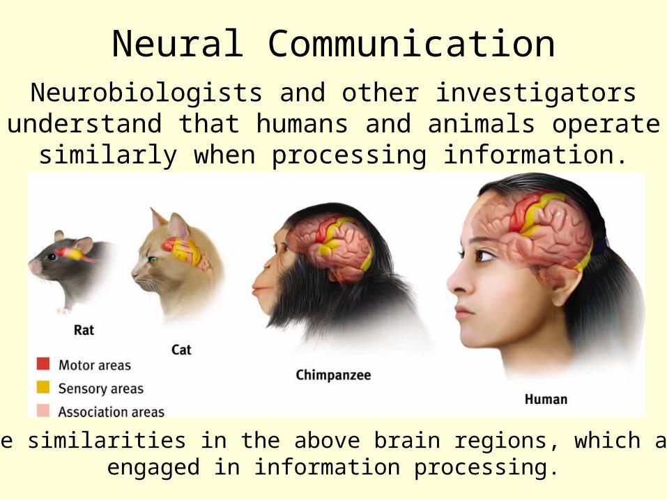
Note the similarities in the above brain regions, which are all engaged in information processing.
Neurobiologists and other investigators understand that humans and animals operate
similarly when processing information.
Neural Communication
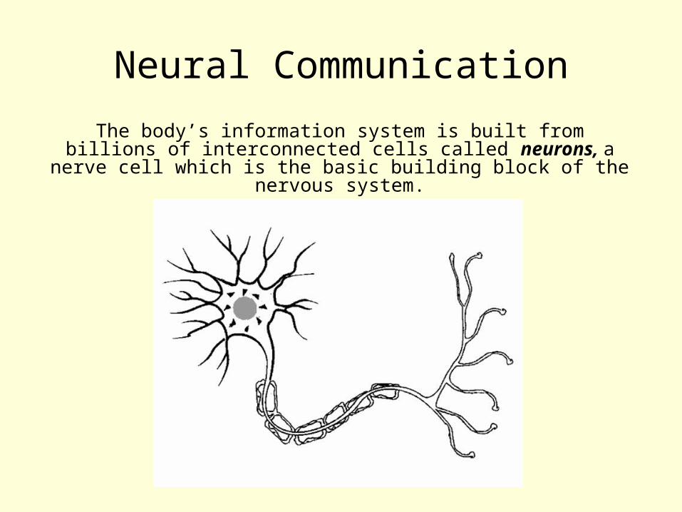
The body’s information system is built from billions of interconnected cells called neurons, a nerve cell which
is the basic building block of the nervous system.

Neuron A nerve cell, or a neuron, consists of many
different parts.

Parts of a Neuron
Cell Body: Life support center of the neuron.
Dendrites: Branching extensions at the cell body. Receive messages from other neurons.
Axon: Long single extension of a neuron, covered with myelin [MY-uh-lin] sheath to insulate and speed up messages through neurons.
Terminal Branches of axon: Branched endings of an axon that transmit messages to other neurons.

Fig. 3-2, p. 75

Action PotentialA neural impulse. A
brief electrical charge that travels down an
axon and is generated by the movement of positively charged atoms in and out of
channels in the axon’s membrane.
Potential = Voltage (Action potential refers to the impulse during action/Resting Potential refers to the impulse during rest)

Threshold Threshold: Each neuron receives excitatory (like
pushing the accelerator) and inhibitory (like
pressing the brake) signals from many neurons. When the excitatory signals
minus the inhibitory signals
exceed a minimum intensity
(threshold) the neuron fires an action potential.
Action Potential Properties
All-or-None Response: A strong stimulus can trigger more neurons to fire, and to fire more often, but it does not affect the
action potentials strength or speed.
Intensity of an action potential remains the same throughout the length of the
axon. It does not slow down as it travels.
What the what?
We have just described how an action potential moves through a single nerve
cell.
Now we will look at how that action potential gets from one nerve cell to
another!

Synapse Synapse: a junction between the axon tip of the sending neuron and the dendrite or cell
body of the receiving neuron. This tiny gap is called the synaptic gap or cleft.

NeurotransmittersNeurotransmitters
(chemicals) released from the sending
neuron travel across the synapse and bind to receptor sites on
the receiving neuron, thereby influencing it to generate an action
potential.
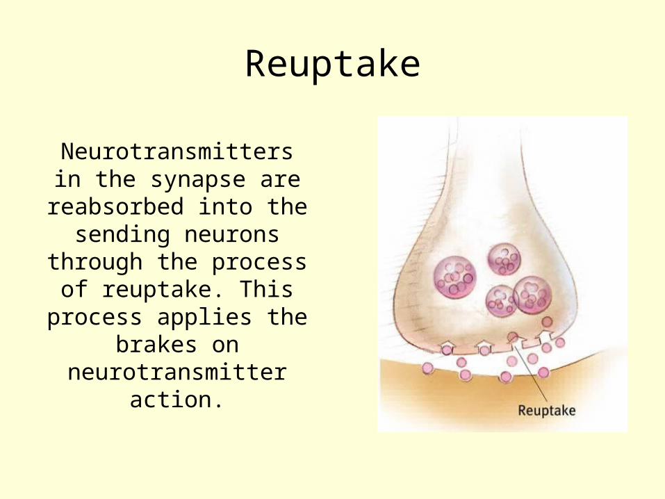
Reuptake
Neurotransmitters in the synapse are
reabsorbed into the sending neurons
through the process of reuptake. This
process applies the brakes on
neurotransmitter action.

Fig. 3-4, p. 78

Reuptake Inhibitors
• Serotonin: Regulation of mood, sleep, muscle contraction and some cognitive functions including memory and learning
• Selective Serotonin Reuptake Inhibitors (SSRI): treat depression and anxiety by preventing the reuptake of serotonin.
• Common SSRI’s: Paxil, Zoloft, Prozac

How Neurotransmitters Influence Us
Serotonin pathways are involved with mood regulation.
From Mapping the Mind, Rita Carter, © 1989 University of California Press
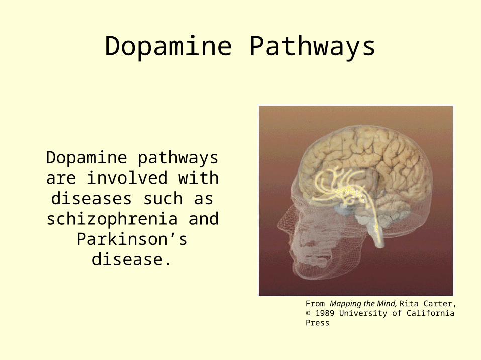
Dopamine Pathways
Dopamine pathways are involved with diseases such as
schizophrenia and Parkinson’s disease.
From Mapping the Mind, Rita Carter, © 1989 University of California Press

Parkinson’s Disease
• Different areas of the brain must communicate in order to produce smooth and coordinated muscle movements.
• Dopamine is a key neurotransmitter involved in this communication.
• Parkinson’s Disease is caused by a deterioration of brain neuron’s that produce dopamine (it is still unknown why this occurs).
• A lack of dopamine results in abnormal nerve functioning, causing a loss in the ability to control body movements.

Schizophrenia• Symptoms consist of
hallucinations, delusions and irrational behavior.
• Although not the sole cause of schizophrenia, dopamine unbalance is consistently seen found in patients with schizophrenia.
• Schizophrenia may result from biopsychosocial factors. Some evidence supports a genetic predisposition.
• Drugs that prevent dopamine from binding to receptors reduce the symptoms of schizophrenia.
A Schizophrenic’s paintings of his cat
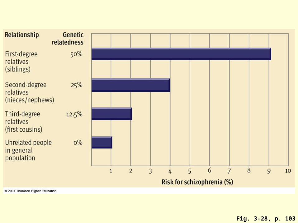
Fig. 3-28, p. 103
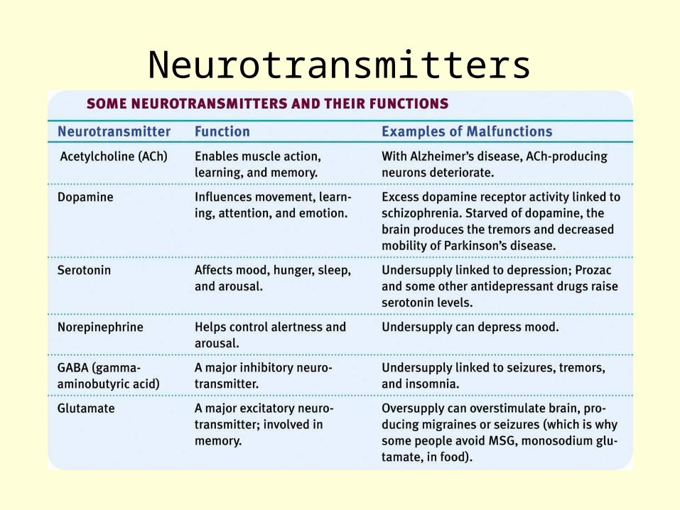
Neurotransmitters
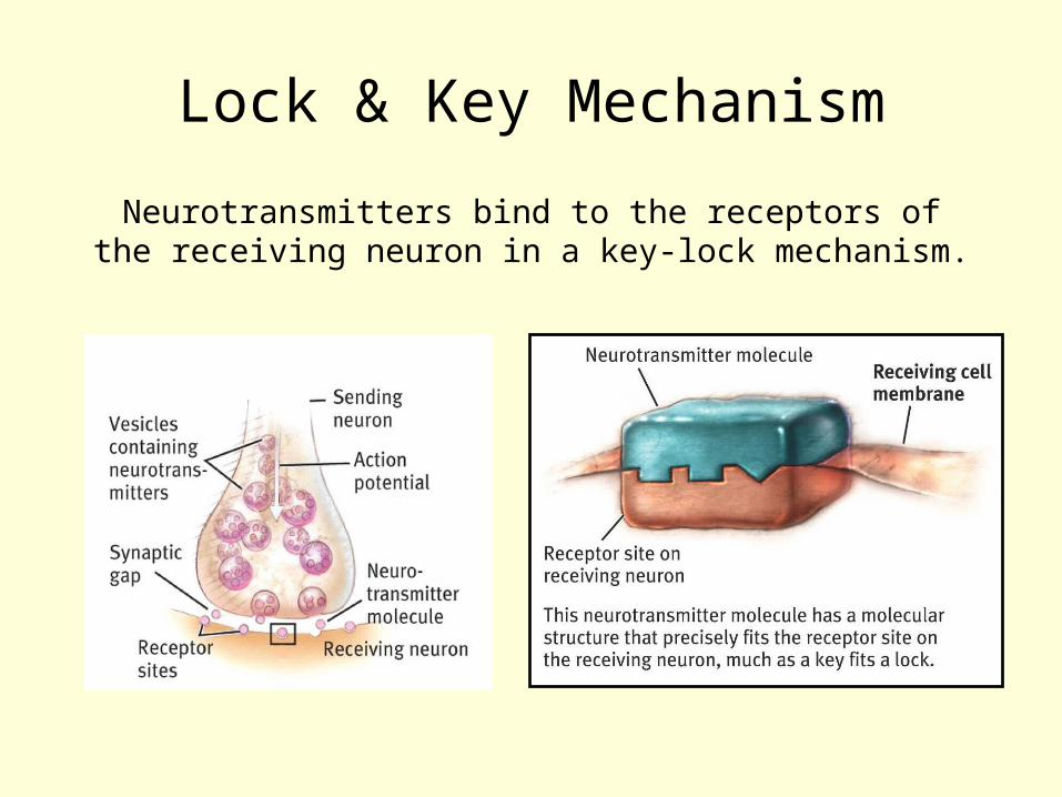
Lock & Key Mechanism
Neurotransmitters bind to the receptors of the receiving neuron in a key-lock mechanism.
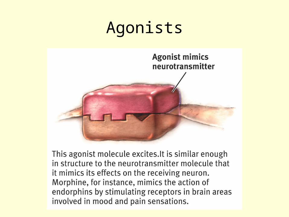
Agonists
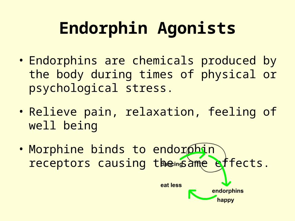
Endorphin Agonists
• Endorphins are chemicals produced by the body during times of physical or psychological stress.
• Relieve pain, relaxation, feeling of well being
• Morphine binds to endorphin receptors causing the same effects.

Antagonists
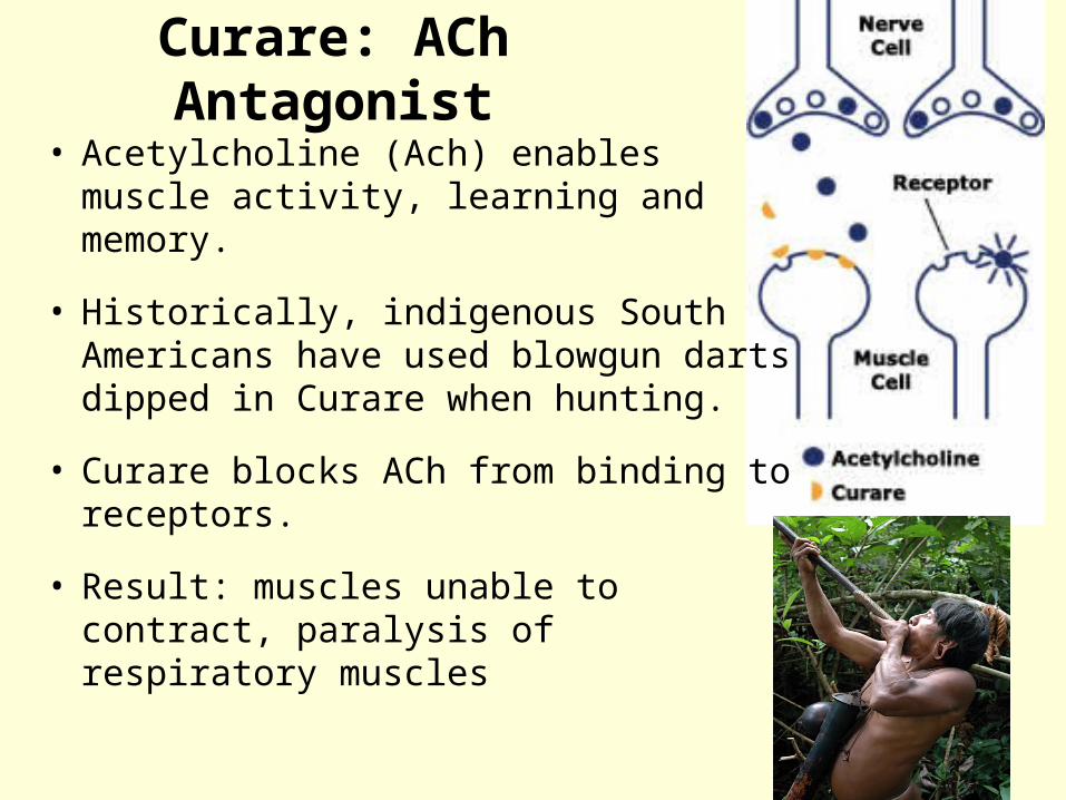
Curare: ACh Antagonist
• Acetylcholine (Ach) enables muscle activity, learning and memory.
• Historically, indigenous South Americans have used blowgun darts dipped in Curare when hunting.
• Curare blocks ACh from binding to receptors.
• Result: muscles unable to contract, paralysis of respiratory muscles

High blood Pressure
• Epinephrine (also called adrenaline because it is released by the adrenal gland) is release in times of stress (fight or flight, adrenaline rush).– Epinephrine “fits into” β-receptors
• Epinephrine in the blood reaches β-receptors (such as those in the heart) - prepares the body for an emergency.– Increases heart rate, elevates blood sugar, dilates
pupils, etc.– boosts the supply of oxygen and glucose to brain and
muscles– suppresses non-emergency bodily functions (ie:
digestion)
•In people with hypertension this response leads to complications such as blood vessel damage, stroke, heart attack, etc.

Beta-Blockers for High blood Pressure
• High blood pressure medications, called Beta-blockers, block beta receptors.
•In other words they act as epinephrine antagonists.

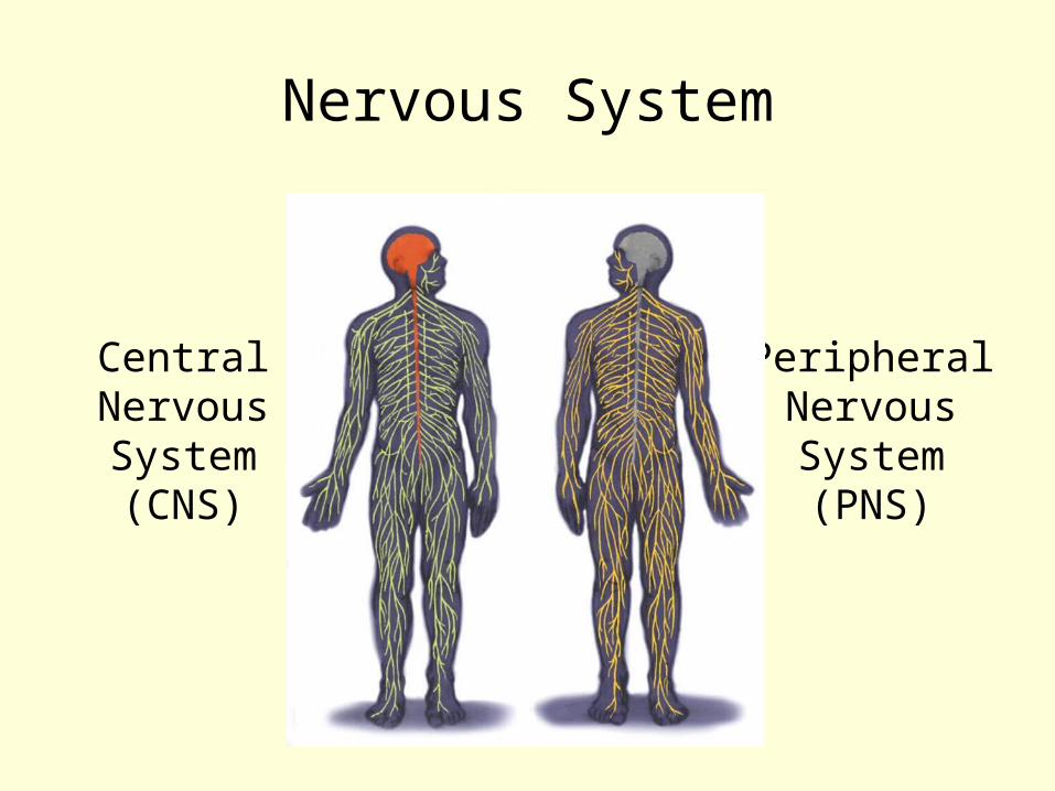
Nervous System
CentralNervousSystem(CNS)
PeripheralNervousSystem(PNS)
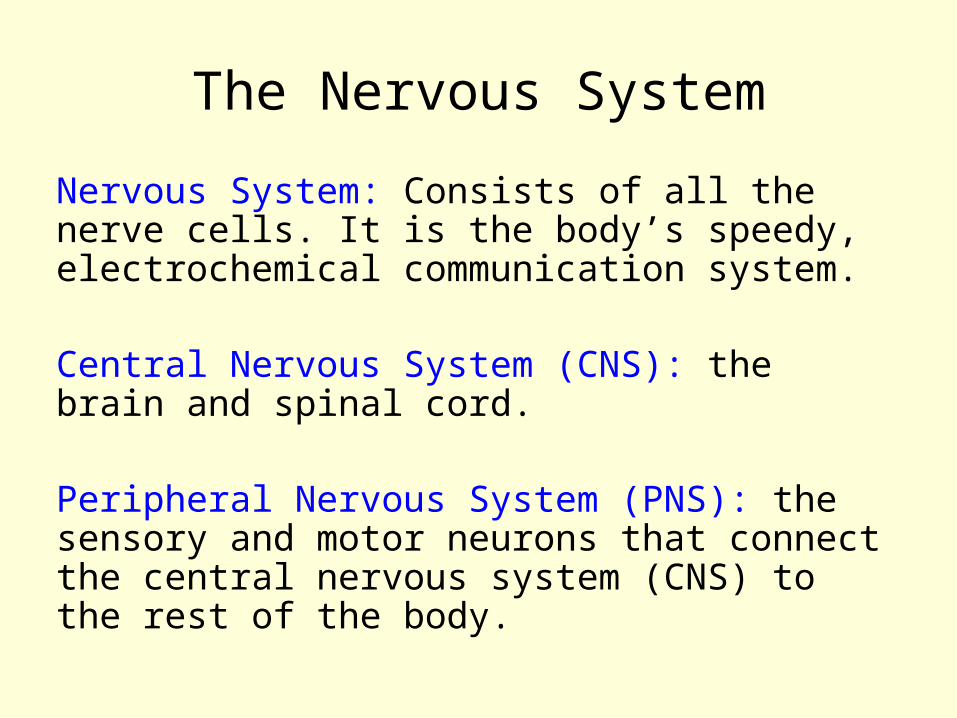
The Nervous System
Nervous System: Consists of all the nerve cells. It is the body’s speedy, electrochemical communication system.
Central Nervous System (CNS): the brain and spinal cord.
Peripheral Nervous System (PNS): the sensory and motor neurons that connect the central nervous system (CNS) to the rest of the body.
The Nervous System

The Brain and Neural Networks
Interconnected neurons form networks in the brain. Theses networks are complex and modify with growth and experience.
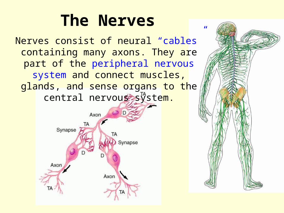
The NervesNerves consist of neural “cables”
containing many axons. They are part of the peripheral nervous system and connect muscles, glands, and sense
organs to the central nervous system.
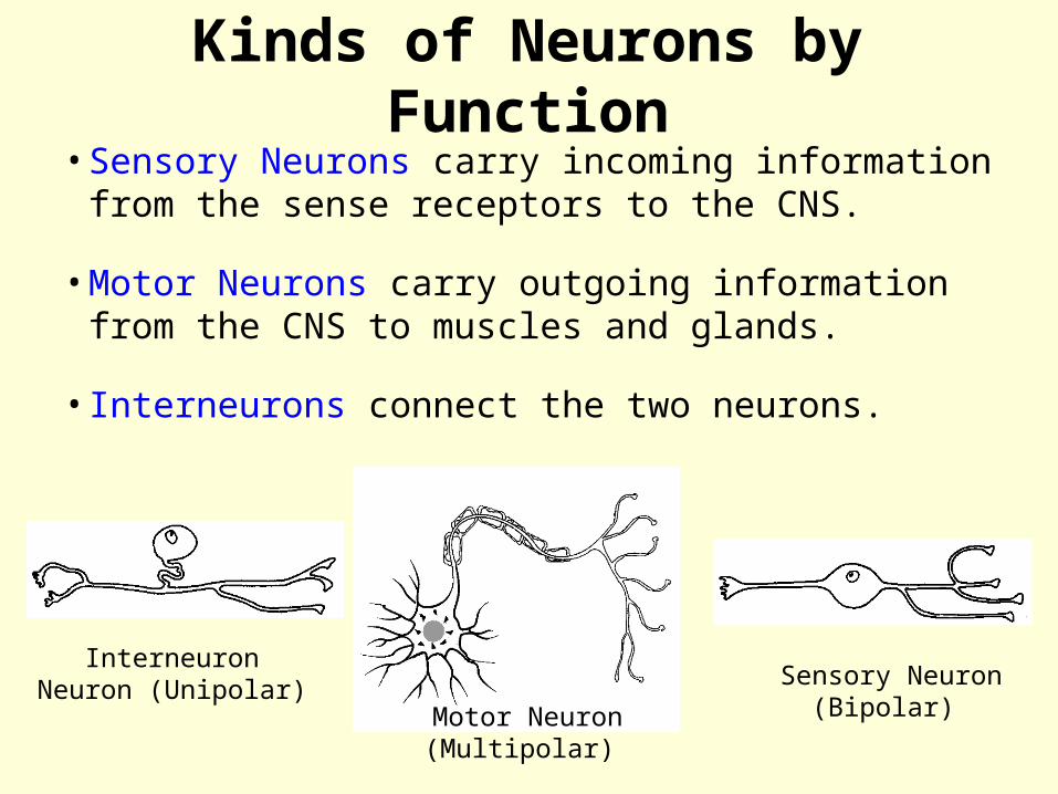
Kinds of Neurons by Function
•Sensory Neurons carry incoming information from the sense receptors to the CNS.
•Motor Neurons carry outgoing information from the CNS to muscles and glands.
•Interneurons connect the two neurons.
Sensory Neuron(Bipolar)
Interneuron Neuron (Unipolar)
Motor Neuron(Multipolar)
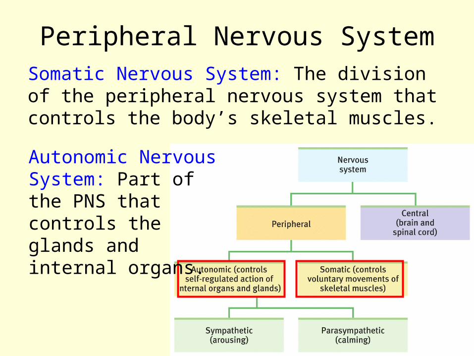
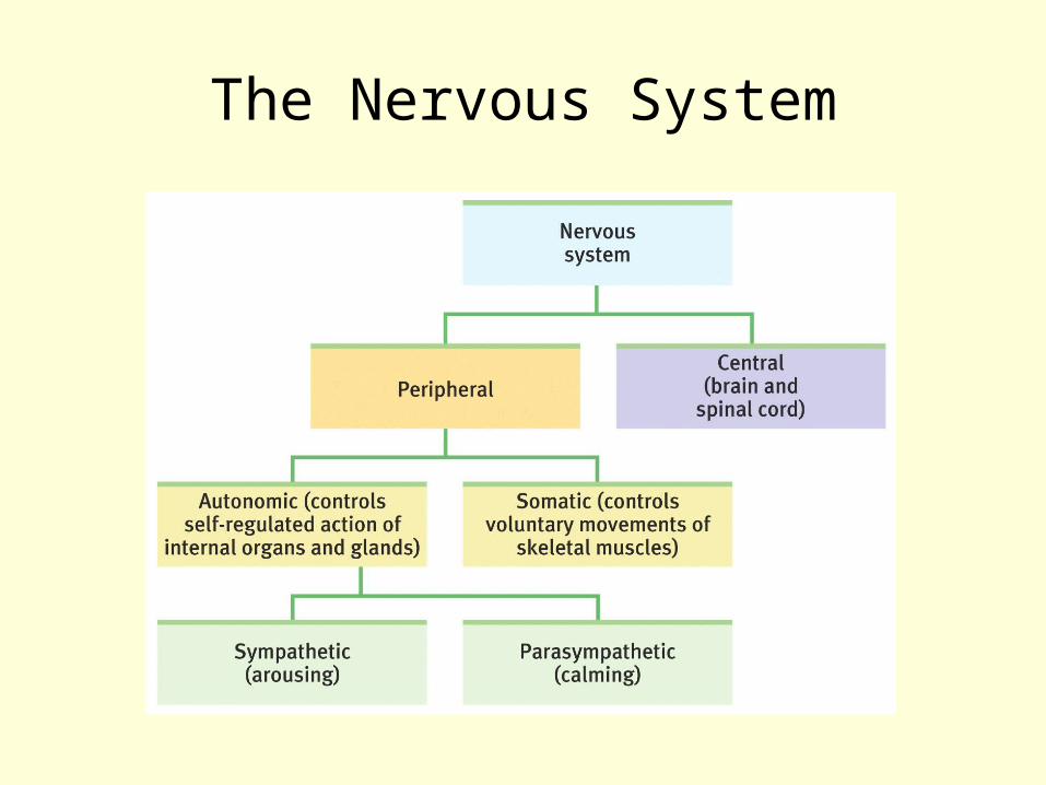
Peripheral Nervous SystemSomatic Nervous System: The division of the peripheral nervous system that controls the body’s skeletal muscles.
Autonomic Nervous System: Part of the PNS that controls the glands and internal organs.
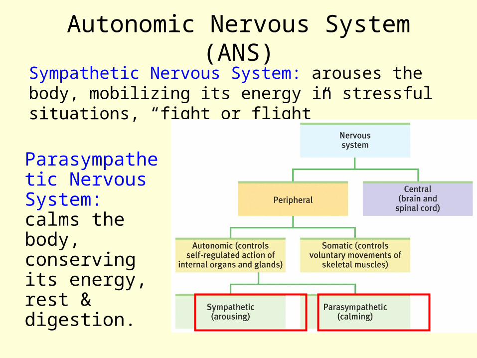
Autonomic Nervous System (ANS)Sympathetic Nervous System: arouses the body, mobilizing its energy in stressful situations, “fight or flight”
Parasympathetic Nervous System: calms the body, conserving its energy, rest & digestion.

Autonomic Nervous System (ANS)
Sympathetic NS “Arouses”
(fight-or-flight)
Parasympathetic NS “Calms”
(rest and digest)

Fig. 3-8, p. 83

Central Nervous SystemThe Spinal Cord and Reflexes
Simple Reflex

Nervous System vs. Endocrine System
Nervous System
• Chemicals of Nervous System = neurotransmitters
• Nervous system secretes neurotransmitters b/w nerve cells
• Signals sent in fractions of a second
Endocrine System
• Chemicals of Endocrine = hormones
• Endocrine system secretes hormones into blood stream
• Chemicals can take many seconds to begin working, effects can last longer (hours, days)
Both:Regulate conditions in body
Use chemicals for communication
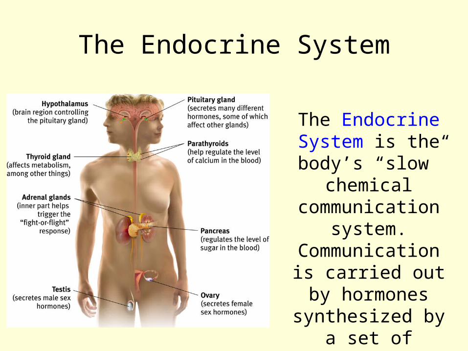
The Endocrine System
The Endocrine System is the body’s “slow”
chemical communication
system. Communication is
carried out by hormones
synthesized by a set of glands.

HormonesHormones:• chemicals synthesized by the endocrine glands• secreted in the bloodstream• affect the brain and many other tissues of the body.
Example: epinephrine (adrenaline) increases heart rate, blood pressure, blood sugar, and feelings of excitement during emergency situations.
Brain
Glands
Hormones

Hormones are Specific• Only certain cells in the body can respond to
hormones and, often, only at limited times. – In order for cells to respond they must have the
hormone receptor molecule Example:• Oxytocin is released by the posterior pituitary,
carried through the bloodstream to all the tissues in the body.
• It acts on only two tissues, the breasts and uterus in the female. • It acts only under certain conditions,
such as:•Oxytocin causes uterine contractions at the end of pregnancy.•Oxytocin causes breast tissue to eject milk only if the female has recently given birth and is nursing.

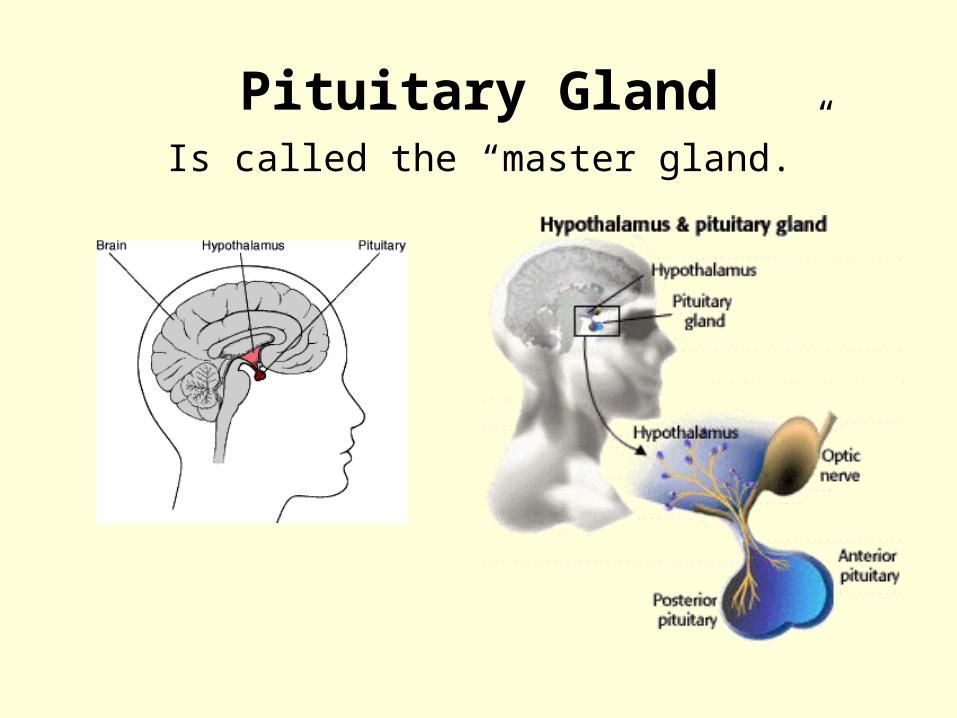
Pituitary Gland
Is called the “master gland.” The anterior pituitary lobe releases hormones that
regulate other glands. The posterior lobe regulates water and salt balance.
Pituitary GlandIs called the “master gland.”
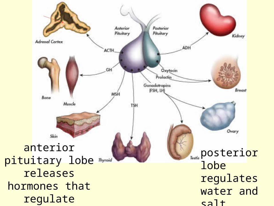
anterior pituitary lobe
releases hormones that
regulate organs and other glands.
posterior lobe regulates water and salt balance.

Thyroid & Parathyroid Glands
Regulate metabolic and calcium rate.
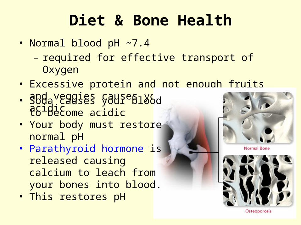
Diet & Bone Health• Normal blood pH ~7.4
– required for effective transport of Oxygen• Excessive protein and not enough fruits and
veggies causes your blood to become acidic
• Soda causes your blood to become acidic
• Your body must restore normal pH
• Parathyroid hormone is released causing calcium to leach from your bones into blood.
• This restores pH
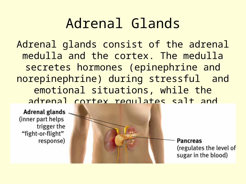
Adrenal GlandsAdrenal glands consist of the adrenal medulla and the cortex. The medulla secretes hormones (epinephrine and norepinephrine) during stressful and
emotional situations, while the adrenal cortex regulates salt and carbohydrate
metabolism.

Adrenal GlandsThe medulla secretes hormones (epinephrine and norepinephrine) during stressful and emotional situations.
The adrenal cortex regulates salt and carbohydrate metabolism.
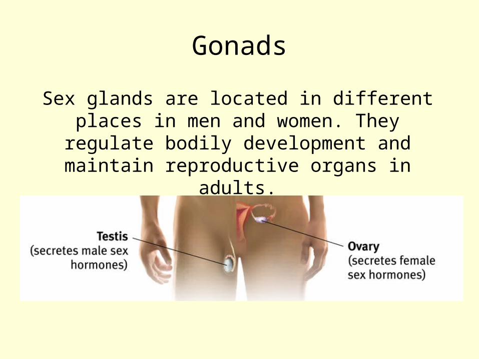
Gonads
Sex glands are located in different places in men and women. They regulate bodily
development and maintain reproductive organs in adults.

The BrainTechniques to Study the Brain
•A brain lesion experimentally destroys brain tissue to study animal behaviors after such destruction.
•Remember, the hippocampus was removed from rats at various times after learning a maze.
Hubel (1990)

Clinical ObservationClinical observations have shed light on a
number of brain disorders. Alterations in brain morphology due to neurological and
psychiatric diseases are now being catalogued.
Tom
Landers/ B
oston Globe
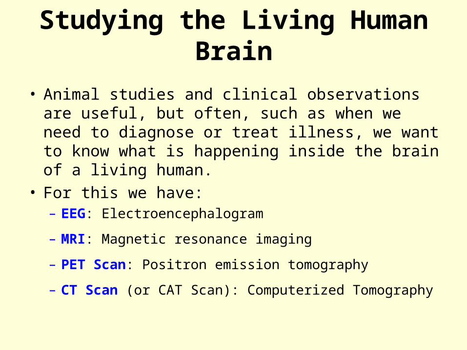
Studying the Living Human Brain
• Animal studies and clinical observations are useful, but often, such as when we need to diagnose or treat illness, we want to know what is happening inside the brain of a living human.
• For this we have:– EEG: Electroencephalogram
– MRI: Magnetic resonance imaging
– PET Scan: Positron emission tomography
– CT Scan (or CAT Scan): Computerized Tomography
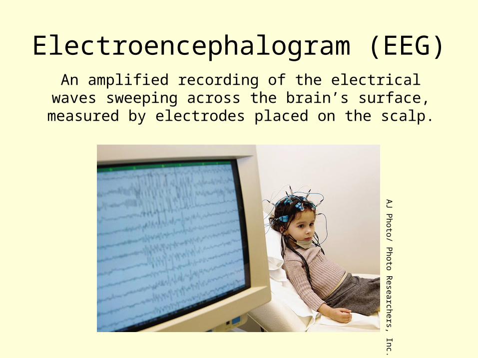
Electroencephalogram (EEG)An amplified recording of the electrical waves
sweeping across the brain’s surface, measured by electrodes placed on the scalp.
AJ P
hoto/ Photo R
esearchers, Inc.
MRI: Magnetic Resonance Imaging
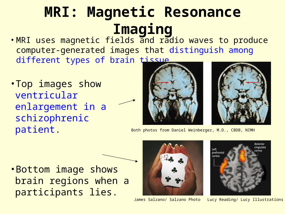
•MRI uses magnetic fields and radio waves to produce computer-generated images that distinguish among different types of brain tissue.
Both photos from Daniel Weinberger, M.D., CBDB, NIMH
James Salzano/ Salzano Photo Lucy Reading/ Lucy Illustrations
•Top images show ventricular enlargement in a schizophrenic patient.
•Bottom image shows brain regions when a participants lies.

PET Scan: Positron Emission Tomography
• PET Scan is a visual display of brain activity
• Detects gamma rays emitted by a radioactive form of glucose while the brain performs a given task.
Courtesy of N
ational Brookhaven N
ational Laboratories

CT Scan: Computerized Tomography
• X-ray rotates around head.
• May be done with or without contrasting dye.
• Images taken from different angles are assembled into 3-D image
• CT scan produces much more detailed image than x-ray of bone and soft tissue

The Brain The Brain

Brain stem CerebellumCerebrum

Cerebrum

Cerebellum
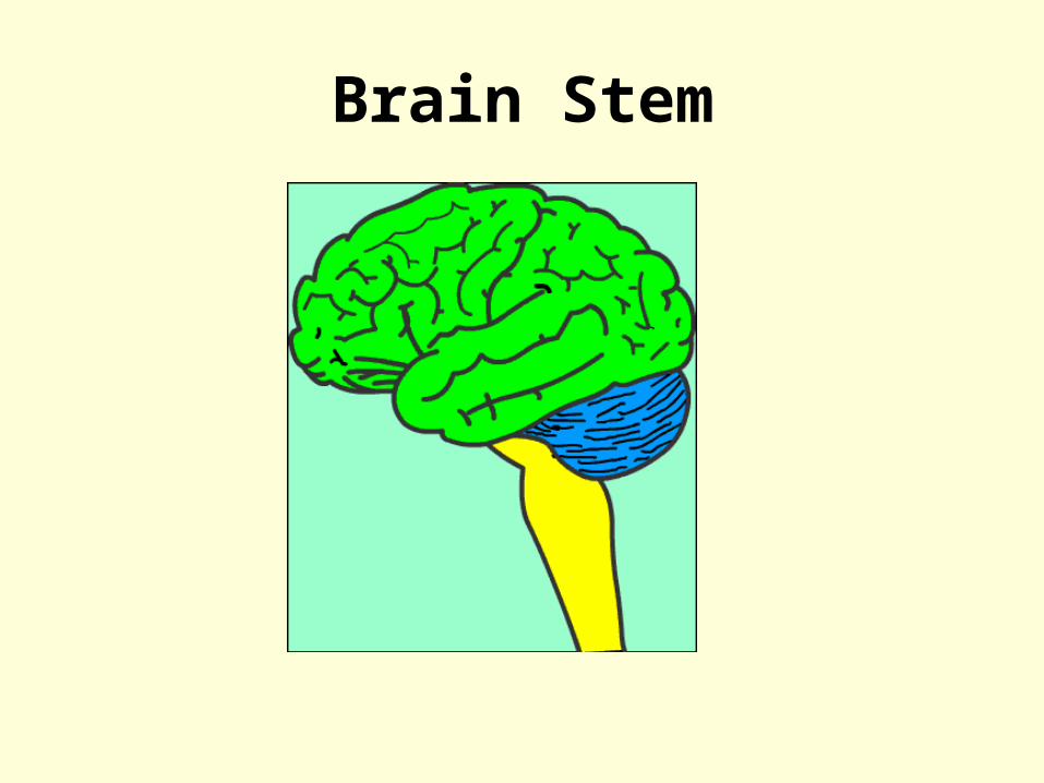
Brain Stem

The Brain Stem: Older Brain Structures
The Brainstem is the oldest part of the brain, beginning where the spinal cord swells and enters the skull. It is responsible for automatic survival
functions.


Brainstem

Thalamus
The Thalamus [THAL-uh-muss] is the brain’s sensory switchboard, located on top of the brainstem.
It directs messages to the sensory areas in the cortex and transmits replies to the cerebellum and medulla. To
medulla
To cerebellum
1.2.
3.
4.
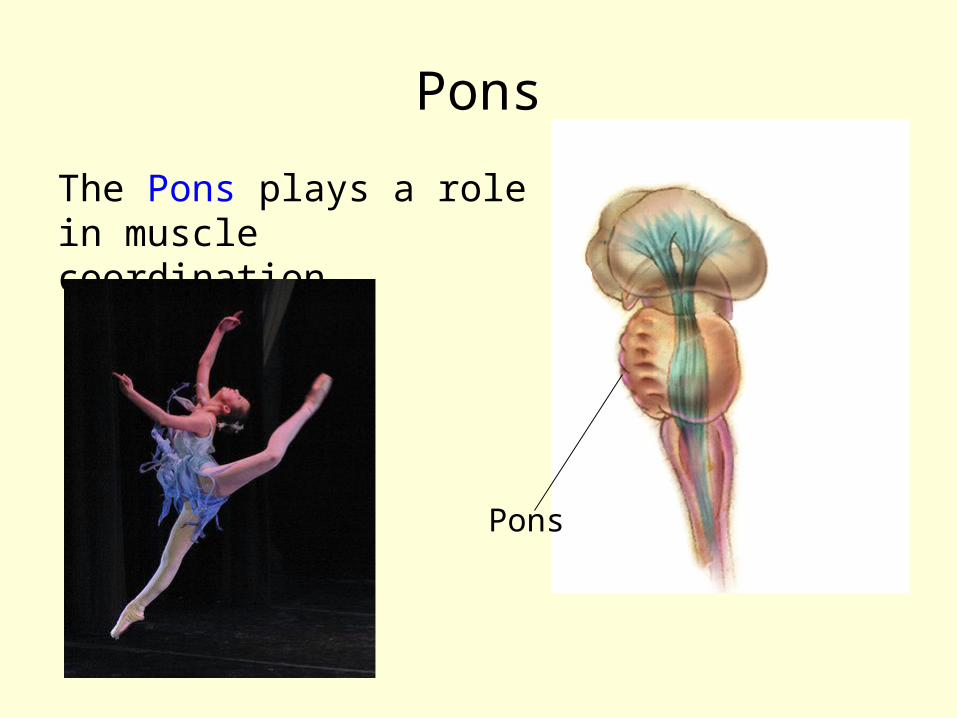
Pons
The Pons plays a role in muscle coordination.
Pons
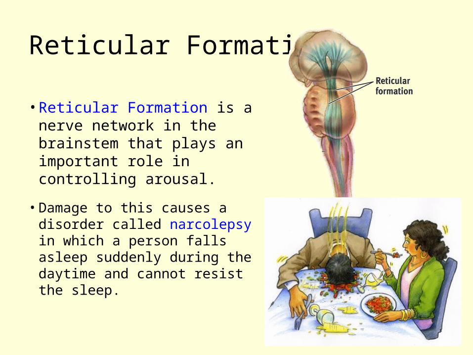
Reticular Formation
•Reticular Formation is a nerve network in the brainstem that plays an important role in controlling arousal.
•Damage to this causes a disorder called narcolepsy in which a person falls asleep suddenly during the daytime and cannot resist the sleep.

Medulla
The Medulla [muh-DUL-uh] is the base
of the brainstem that controls heartbeat
and breathing.
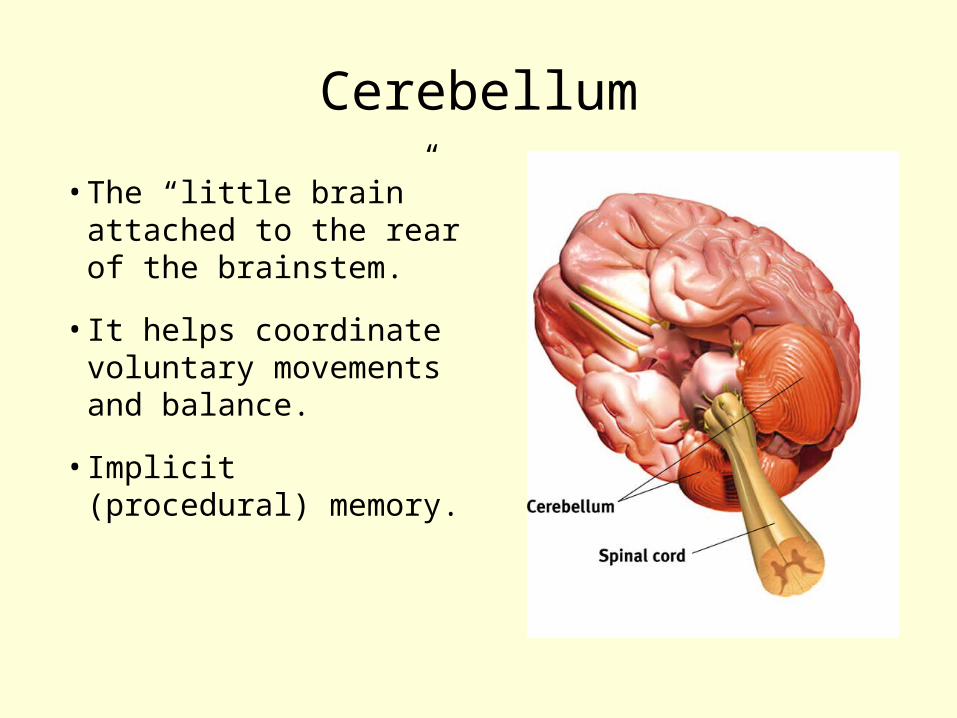
•The “little brain” attached to the rear of the brainstem.
•It helps coordinate voluntary movements and balance.
•Implicit (procedural) memory.
Cerebellum

•The Limbic System is a doughnut-shaped system of neural structures at the border of the brainstem and cerebrum
•Associated with emotions such as fear, aggression and drives for food and sex.
The Limbic System

Amygdala
The Amygdala [ah-MIG-dah-la] consists of two lima bean-sized neural clusters linked to the emotions of fear and
anger.

Hypothalamus•Hypothalamus lies below
(hypo) the thalamus. •Directs maintenance activities
like eating, drinking, body temperature, and control of emotions.
• It stimulates or inhibits pituitary gland other endocrine glands
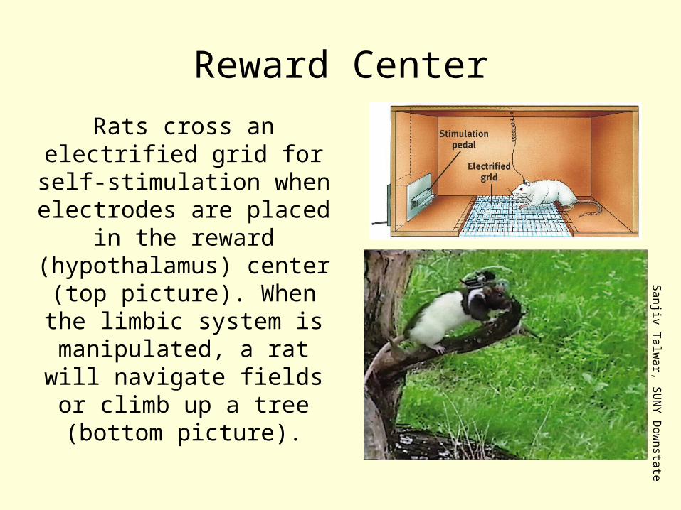
Rats cross an electrified grid for self-
stimulation when electrodes are placed
in the reward (hypothalamus) center (top picture). When the
limbic system is manipulated, a rat will navigate fields or climb
up a tree (bottom picture).
Reward CenterS
anjiv Talw
ar, SU
NY
Dow
nstate
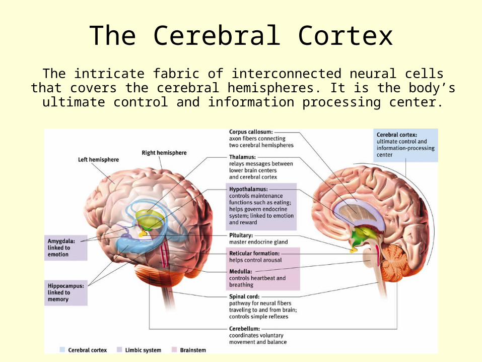
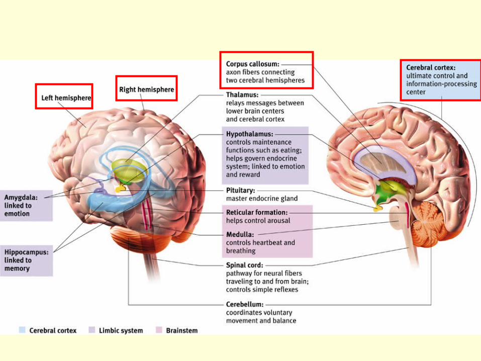
The Cerebral CortexThe intricate fabric of interconnected neural cells
that covers the cerebral hemispheres. It is the body’s ultimate control and information processing center.

Structure of the CortexEach brain hemisphere is divided into four lobes

Functions of the Cortex• The Motor Cortex is the area at the rear of the
frontal lobes that control voluntary movements. • The Sensory Cortex (parietal cortex) receives
information from skin surface and sense organs.


Visual Function
• Notice the visual cortex is located in the occipital lobe.
• The functional MRI scan shows the visual cortex is active as the subject looks at faces.
Courtesy of V
.P. Clark, K
. Keill, J. M
a. M
aisog, S. Courtney, L
.G.
Ungerleider, and J.V
. Haxby,
National Institute of M
ental Health
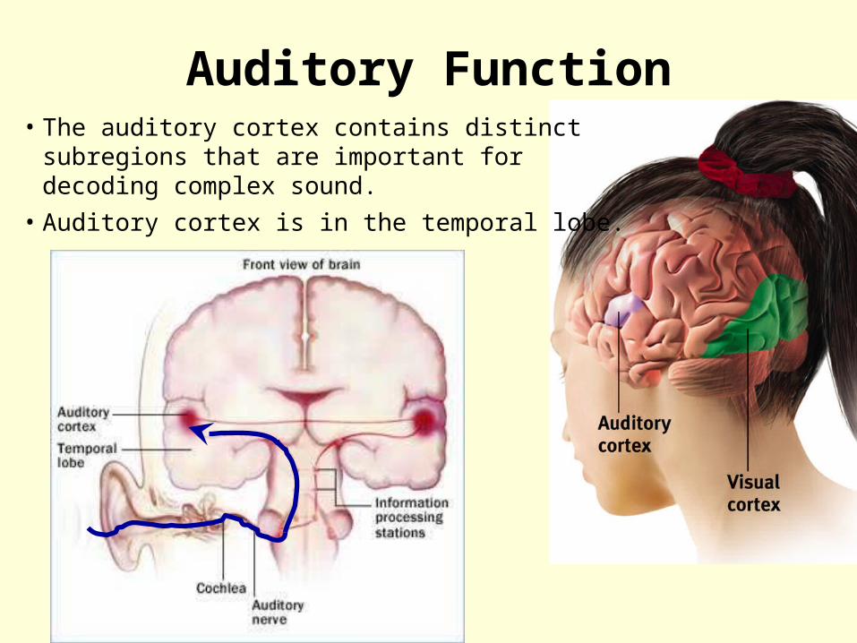
Auditory Function• The auditory cortex contains distinct
subregions that are important for decoding complex sound.
• Auditory cortex is in the temporal lobe.

2. information travels through the brainstem and midbrain to the auditory cortex.
3. information from the auditory cortex interacts with many other brain areas, especially the frontal lobe, for memory formation and interpretation.
4. The frontal lobe is involved in emotional evaluation.
5. The motor cortex is involved in sensory–motor feedback, in controlling movements needed to produce music using an instrument.
1. Sound waves enter ear, and are turned into neural impulses by the inner ear
1
2
3
4
5
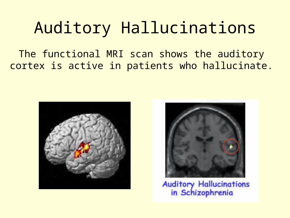
Auditory Hallucinations
The functional MRI scan shows the auditory cortex is active in patients who hallucinate.
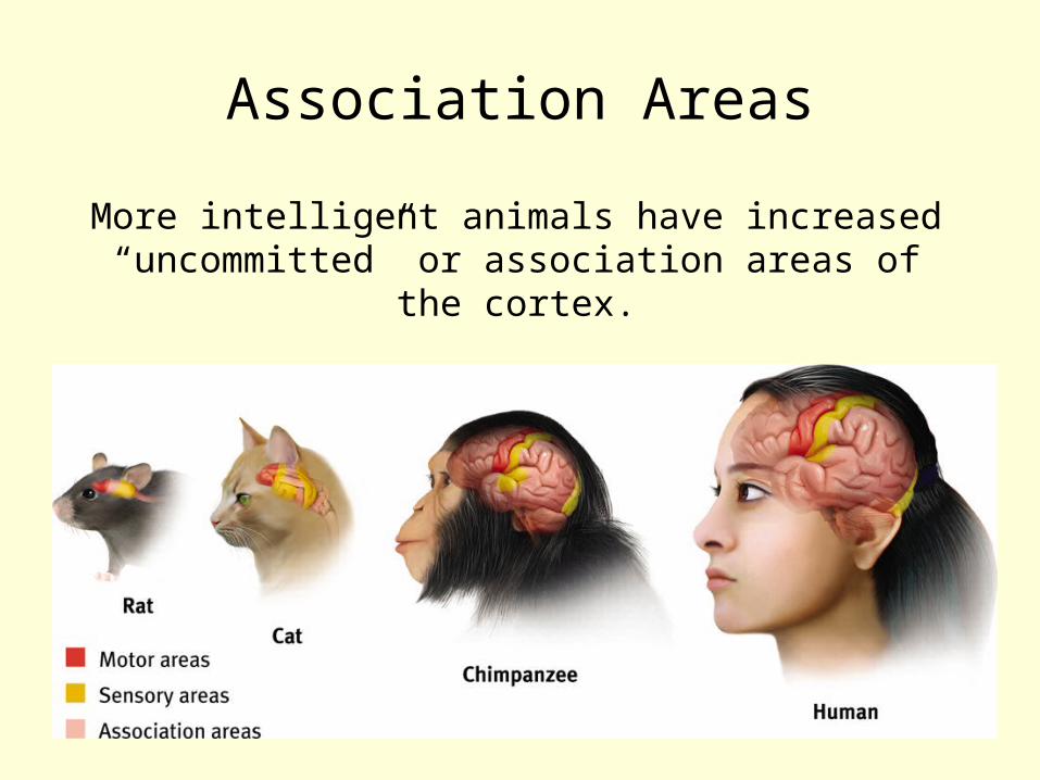
More intelligent animals have increased “uncommitted” or association areas of the
cortex.
Association Areas
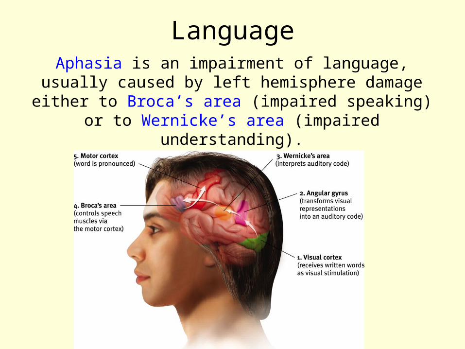
LanguageAphasia is an impairment of language, usually caused by left hemisphere damage either to
Broca’s area (impaired speaking) or to Wernicke’s area (impaired understanding).

Specialization & Integration
Brain activity when hearing, seeing, and speaking words
The brain is sculpted by our genes but also by our experiences.
Plasticity refers to the brain’s ability to modify itself after some types of injury or illness.
The Brain’s Plasticity
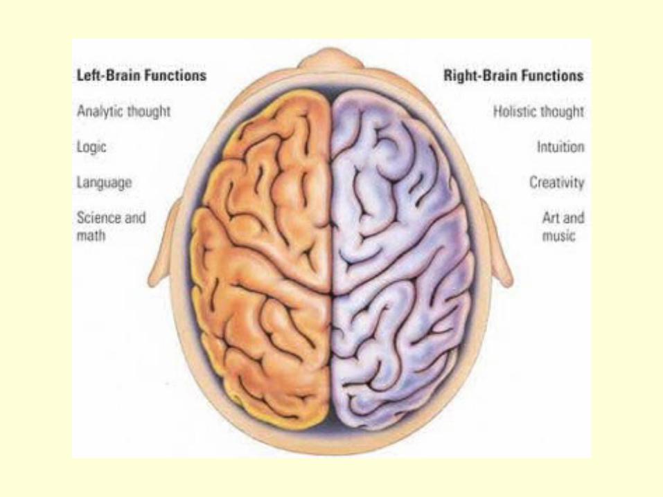
Our Divided Brain
Our brain is divided into two hemispheres. The left hemisphere processes reading,
writing, speaking, mathematics, and comprehension skills. In the 1960s, it was
termed as the dominant brain.

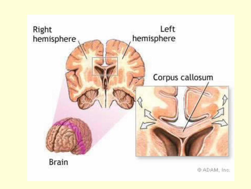
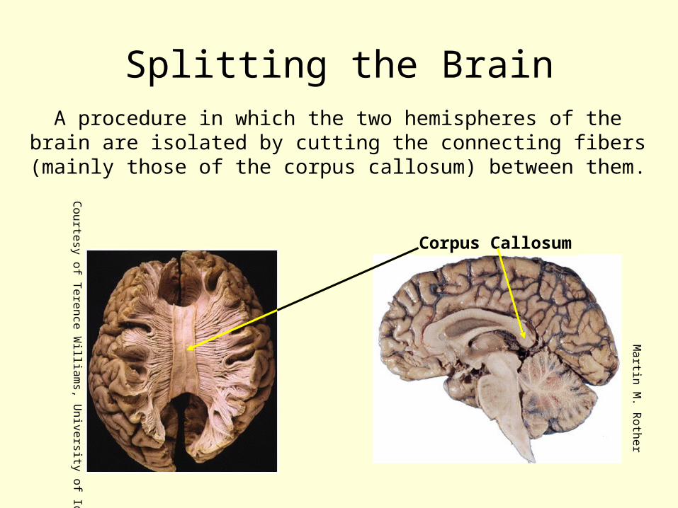
Splitting the BrainA procedure in which the two hemispheres of the brain are isolated by cutting the connecting fibers
(mainly those of the corpus callosum) between them.
Corpus Callosum
Ma
rtin M
. Ro
the
r
Courtesy of T
erence William
s, University of Iow
a
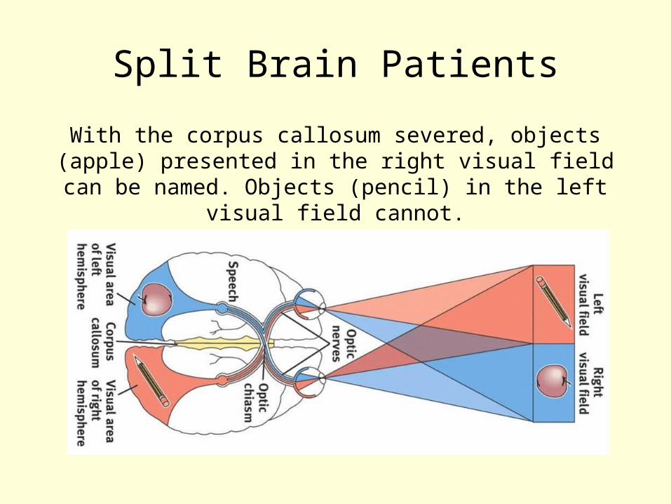
Split Brain Patients
With the corpus callosum severed, objects (apple) presented in the right visual field can be named.
Objects (pencil) in the left visual field cannot.

Try This!
Try drawing one shape with your left hand and one with your right hand, simultaneously.
BB
C
