nervous system physiology - kajalpetclinic.comkajalpetclinic.com/fileupload/editor/documents/nervous...
TRANSCRIPT
Nervous system
physiology
A. K.Goudarzi, D.V.M. Ph.D
Department of basic sciences
School of veterinary medicine
I.A. University
For PDF file visit:
www.kajalpetclinic.com

Introduction
The human nervous system is highly complex.
It is divided into the central nervous system,
consisting of the brain and spinal cord; the
peripheral nervous system, which includes
nerves innervating the muscles and nerves
sending sensory information from the skin,
muscle, and joints to the brain; and the
autonomic nervous system which controls the
involuntary processes of the body.

Functions of the Nervous System
communication system of the body.
Controls body functions and actions.
Maintains physiological homeostasis.

1. Sensory Functions: Sensory receptors detect both
internal and external stimuli.
Functional unit: Sensory or Afferent Neurons
2. Integrative Functions: CNS integrates sensory input and
makes decisions regarding appropriate responses
Functional Unit: Interneurons or Association Neurons
of the Brain and Spinal cord
3. Motor Functions: Response to integration decisions.
Functional Unit: Motor or Efferent Neurons
Functions of the Nervous System


Organization of a Nerve of the PNS
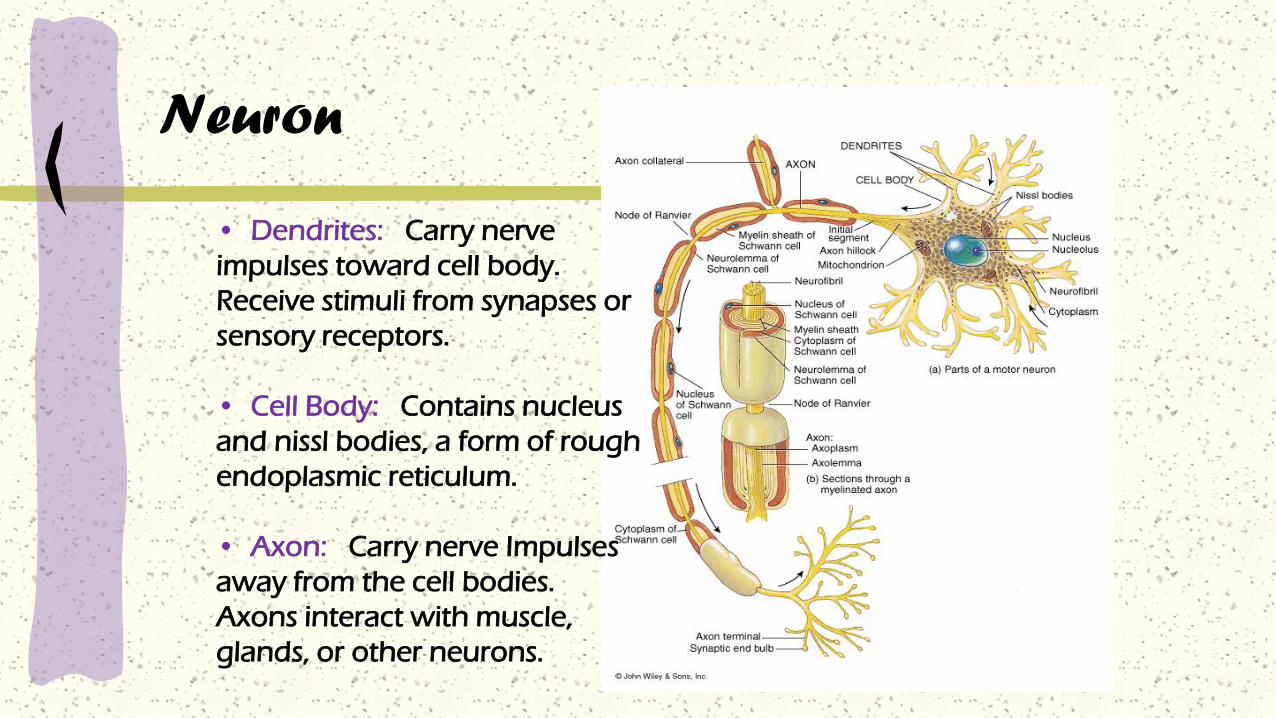
Neuron
• Dendrites: Carry nerve
impulses toward cell body.
Receive stimuli from synapses or
sensory receptors.
• Cell Body: Contains nucleus
and nissl bodies, a form of rough
endoplasmic reticulum.
• Axon: Carry nerve Impulses
away from the cell bodies.
Axons interact with muscle,
glands, or other neurons.

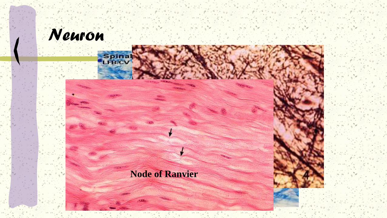
Neuron
Node of Ranvier
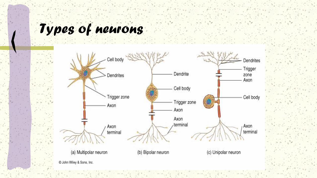
Types of neurons

Types of interneurons

Structural classificationPolarity
Different kinds of neurons:
1 Unipolar neuron
2 Bipolar neuron
3 Multipolar neuron
4 Pseudounipolar neuron
Types of neurons
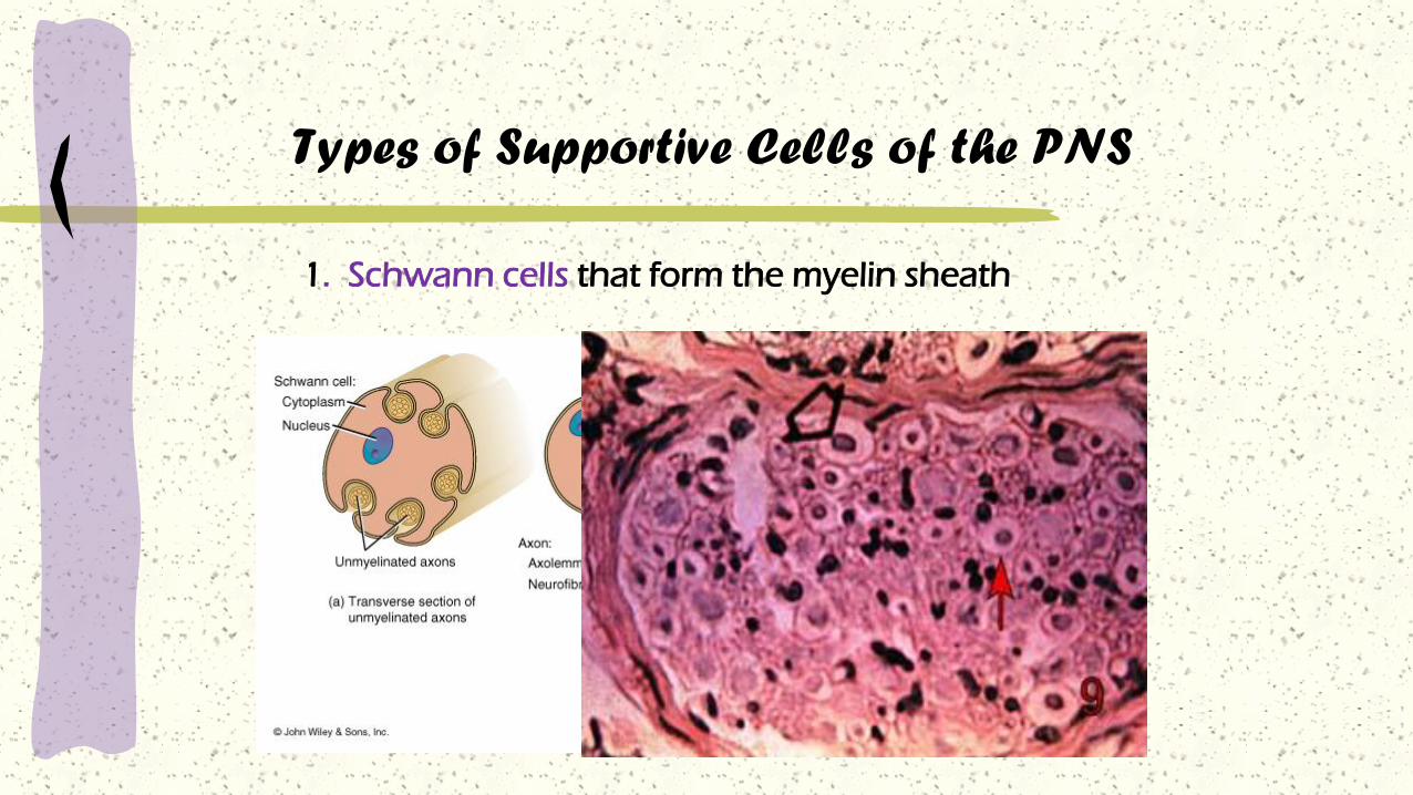
Types of Supportive Cells of the PNS
1. Schwann cells that form the myelin sheath

Types of Supportive Cells of the PNS
2. Satellite cells associated with sensory neuron cell bodies
• Precursors to skeletal muscle cells, able to give rise to
satellite cells or differentiated skeletal muscle cells. They
have the potential to provide additional myonuclei to
their parent muscle fiber, or return to a quiescent state.
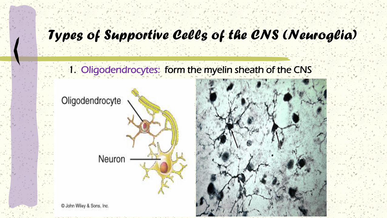
Types of Supportive Cells of the CNS (Neuroglia)
1. Oligodendrocytes: form the myelin sheath of the CNS

Types of Supportive Cells of the CNS (Neuroglia)
2. Astrocytes: Help form the blood-brain barrier, support
the appropriate chemical environment for neurons.
Types of Supportive Cells of the CNS (Neuroglia)
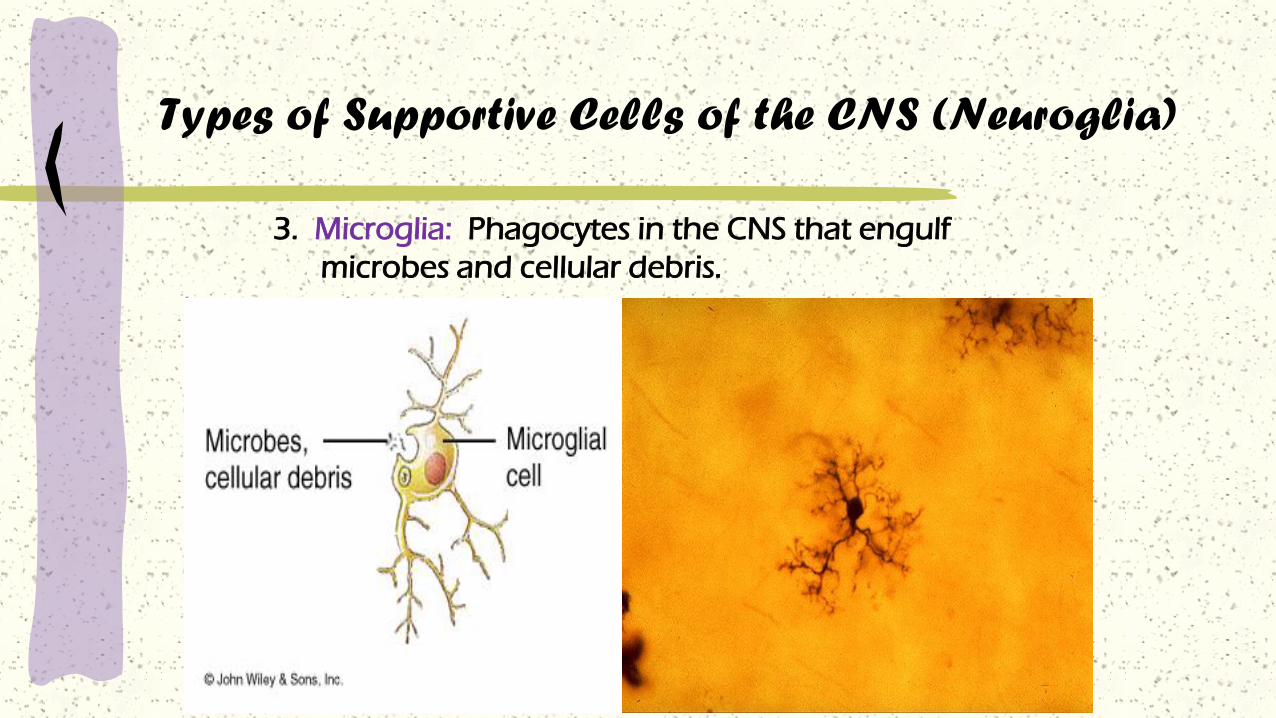
3. Microglia: Phagocytes in the CNS that engulf
microbes and cellular debris.

4. Ependymal Cells: Form blood-brain barrier in the brain
ventricles and central canal of spinal cord. Produce
cerebrospinal fluid and assist in its circulation.
Types of Supportive Cells of the CNS (Neuroglia)
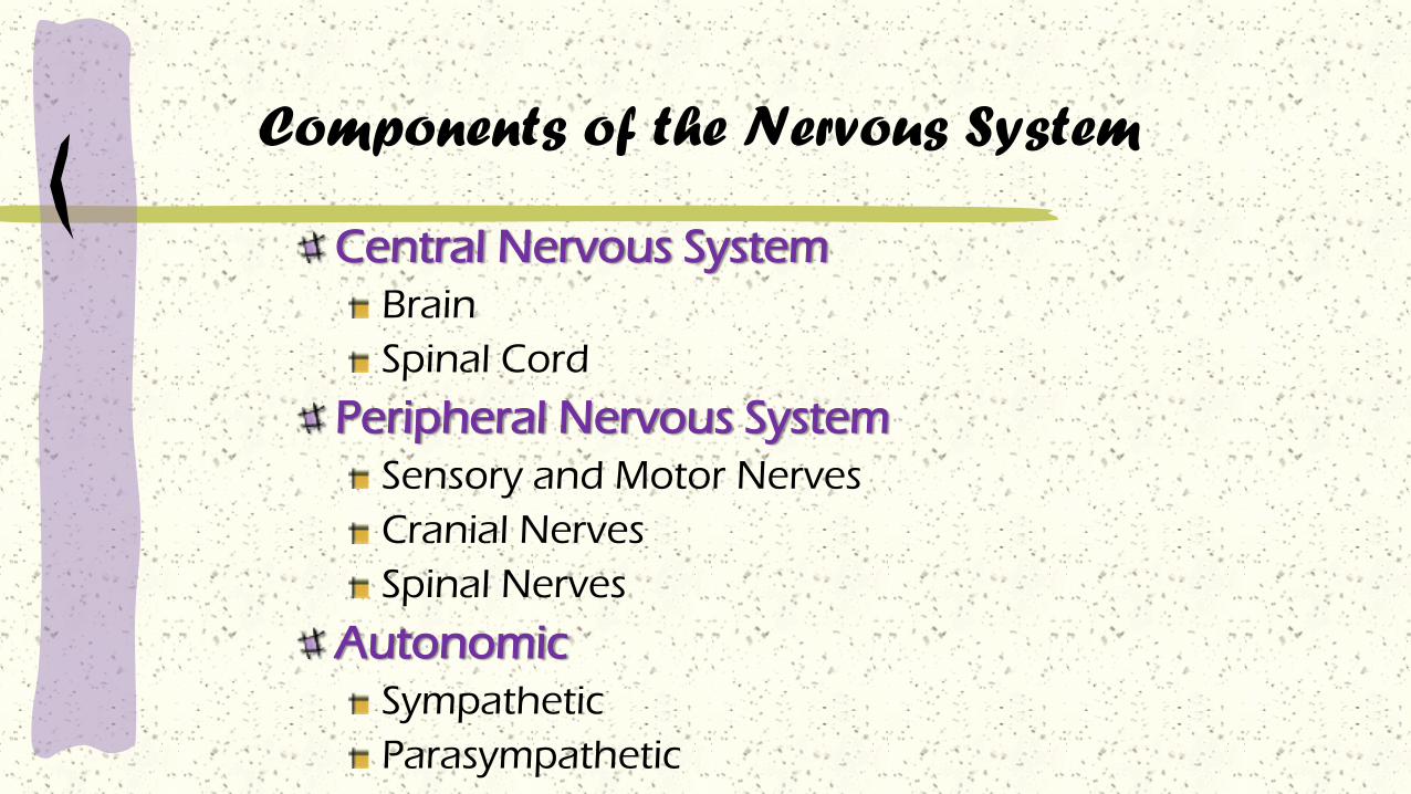
Components of the Nervous System
Central Nervous System
Brain
Spinal Cord
Peripheral Nervous System
Sensory and Motor Nerves
Cranial Nerves
Spinal Nerves
Autonomic
Sympathetic
Parasympathetic

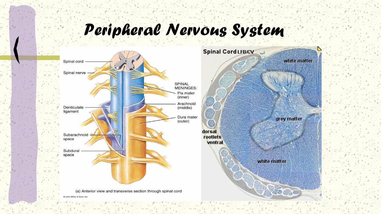
Central Nervous System (CNS)
Brain Spinal Cord
Peripheral Nervous System (PNS)
Sensory NeuronsMotor Neurons
Somatic Nervous System
• voluntary movements via skeletal muscles
Autonomic Nervous System
• organs, smooth muscles
Sympathetic
- “Fight-or-Flight” responsesParasympathetic
- maintenance
The Nervous System

Central Nervous System (CNS)
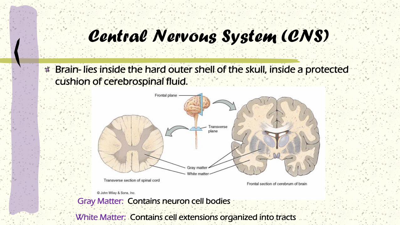
Brain- lies inside the hard outer shell of the skull, inside a protected
cushion of cerebrospinal fluid.
Gray Matter: Contains neuron cell bodies
White Matter: Contains cell extensions organized into tracts

Brain

Brain

Brain: Structure
Hindbrain carries out the most basic functions.
Midbrain coordinates signals.
Forebrain processes signals, stores memories, creates thought.

Hindbrain
Medulla: controls
autonomic fuctions.
Pons: controls sleep
stages.
Cerebellum:
coordinates
movement, stores
some motor memory.

Midbrain
Reticular formation:
the “traffic cops” of
the brain.
Filters sensory
input, which allows
us to concentrate.
Filtering can be
affected by higher
thoughts.

Try this:
Stop and think: What have you been paying
attention to for the last ten minutes?
Pay attention to the feel of your shirt on your
arms. Had you been noticing it during the last
ten minutes? That’s the reticular formation in
action.
What else have you not been paying attention
to?

Forebrain
Thalamus: relay
station channeling
sensory
information.
Limbic system:
basic emotions,
drives, and
behaviors.
Cortex: higher
thought

Limbic system
Hypothalamus: master
controller of the
endocrine system.
Amygdala: sensations of
pleasure or fear,
recognition of fear in
others.
Hippocampus: formation
of memories.

Cortex
Various areas control
sensory processing,
motor control, thought,
memory.
Wiring is plastic:
people blind from
birth, for example, use
parts of the visual
cortex to process
auditory signals.

Left brain, right brain?
While there is some specialization to each
hemisphere, the idea has been oversimplified.
The left brain controls the right half of the body;
the right brain controls the left half of the body.
However, “right brain” or “left brain” functions
such as math, language, etc. produce activity on
both sides of the brain, and processing of these
may be different in different people (males vs.
females, novices vs. experts, etc.).

Brain “maps”?
While hemispheric research shows some specialization between
hemispheres, most “brain maps” like this are nonsense.
Memory
How humans form memories is poorly understood.
“Working memory” appears to be distinct from long-term memory.
There may be short-term memory as well, things remembered for a
few days. Is this because the memory disappears, or because it
cannot be retrieved?

Models of Memory

Models of Memory
Craik & Lockhart, 1972
What is mind?
Many traditions, including psychology, separate
“brain” from “mind.”
What we perceive as “mind” (thought, will, self-
perception) does produce evidence of brain activity
in brain scans.
That “brain” influences “mind” is well-established;
but some evidence shows “mind” can influence
“brain”; as cognitive therapy for depression can
physically change the brain.
Neurology is a very young science, and there is
still much to learn about the brain-mind
connection.

Meninges are layers of non-nervous tissue
that surround and protect the brain and
spinal cord .
Dura Mater – a tough, fibrous membrane
that lies immediately internal to the skull and
encloses the brain and spinal cord.
Arachnoid- resembling a spider web, this is a
delicate layer and a thin, cellular membrane
with many silk-like tissue strands.
Central Nervous System (CNS)

Central Nervous System (CNS)
Pia Mater – loose tissue that covers the brain and
encases the blood vessels that supply the brain.
This is a thin, delicate and highly vascularized
membrane.
The cerebrospinal fluid lies in the space between
the arachnoid and pia mater layers. Its main
function is to act as a cushion, helping to
diminish the transmission of shocking forces.

Central Nervous System (CNS)
Central Nervous System (CNS)
Cerebrum – the largest part of the brain distinguished by the
folds or convolutions of much of its surface.
The cerebrum has four paired lobes – frontal, parietal, occipital, and
temporal.
Memory and conscious thought, speech, motor and sensory functions
are controlled by the cerebrum.

Central Nervous System (CNS)
Cerebellum – a mass that occupies the posterior part of the
cranium.
The cerebellum controls the automatic regulation of movement,
balance, and posture, as well as skilled movements.
Medulla Oblongata (Brain Stem) – connects the cerebrum and
cerebellum with the spinal cord.
The brain stem controls the heart rate, respiration, and body
temperature.
Central Nervous System (CNS)
Spinal Cord – A continuation of the brain
which provides pathways to and from the
brain, to and from the body.
The spinal cord is also surrounded, protected,
and nourished by cerebrospinal fluid.
The vertebrae also serve as a bony protection
to the spinal cord.
The spinal cord terminates with the cauda
equina.

Central Nervous System (CNS)

Senses
Sensory receptors
Receptors are found in the sense organs.
They receive stimuli from the environment
and transmit stimuli to neurons.
Primary humans senses: photoreception,
chemoreception, mechanoreception,
thermoreception.
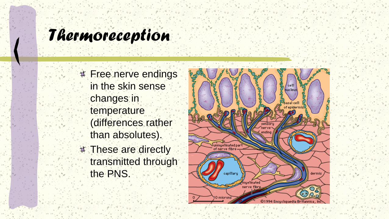
Thermoreception
Free nerve endings
in the skin sense
changes in
temperature
(differences rather
than absolutes).
These are directly
transmitted through
the PNS.

Mechanoreception
Hearing is a form of
mechanoreception.
Ears gather sound
waves from the
environment.
The inner ear bones
amplify sounds.
Sounds are transmitted
to the cochlea.
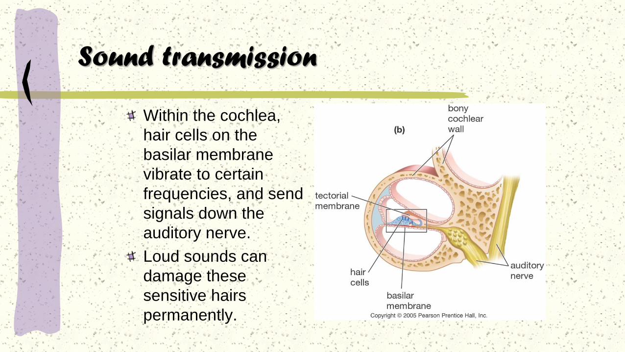
Sound transmission
Within the cochlea,
hair cells on the
basilar membrane
vibrate to certain
frequencies, and send
signals down the
auditory nerve.
Loud sounds can
damage these
sensitive hairs
permanently.

Photoreception
Sight is
photoreception.
Light enters the eye
through the cornea
and pupil.
Light is focused by the
lens.
Light strikes the retina,
and stimulates
receptors.
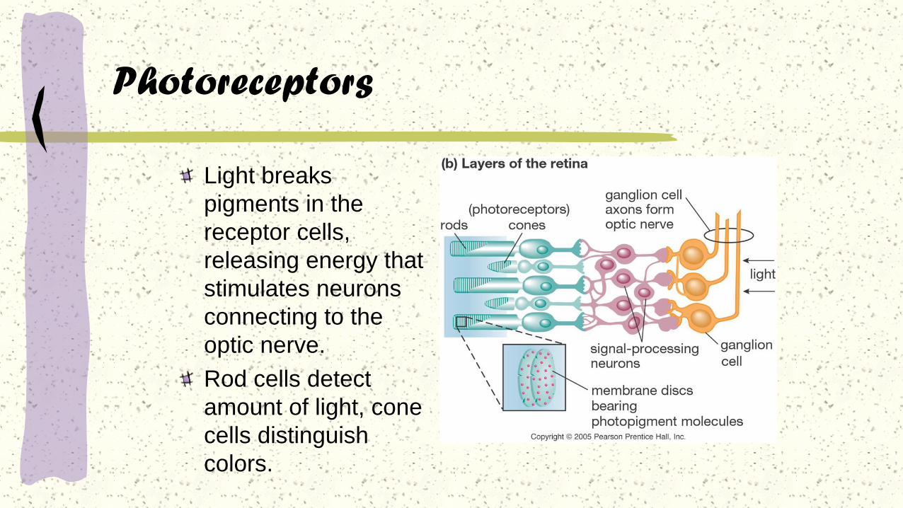
Photoreceptors
Light breaks
pigments in the
receptor cells,
releasing energy that
stimulates neurons
connecting to the
optic nerve.
Rod cells detect
amount of light, cone
cells distinguish
colors.
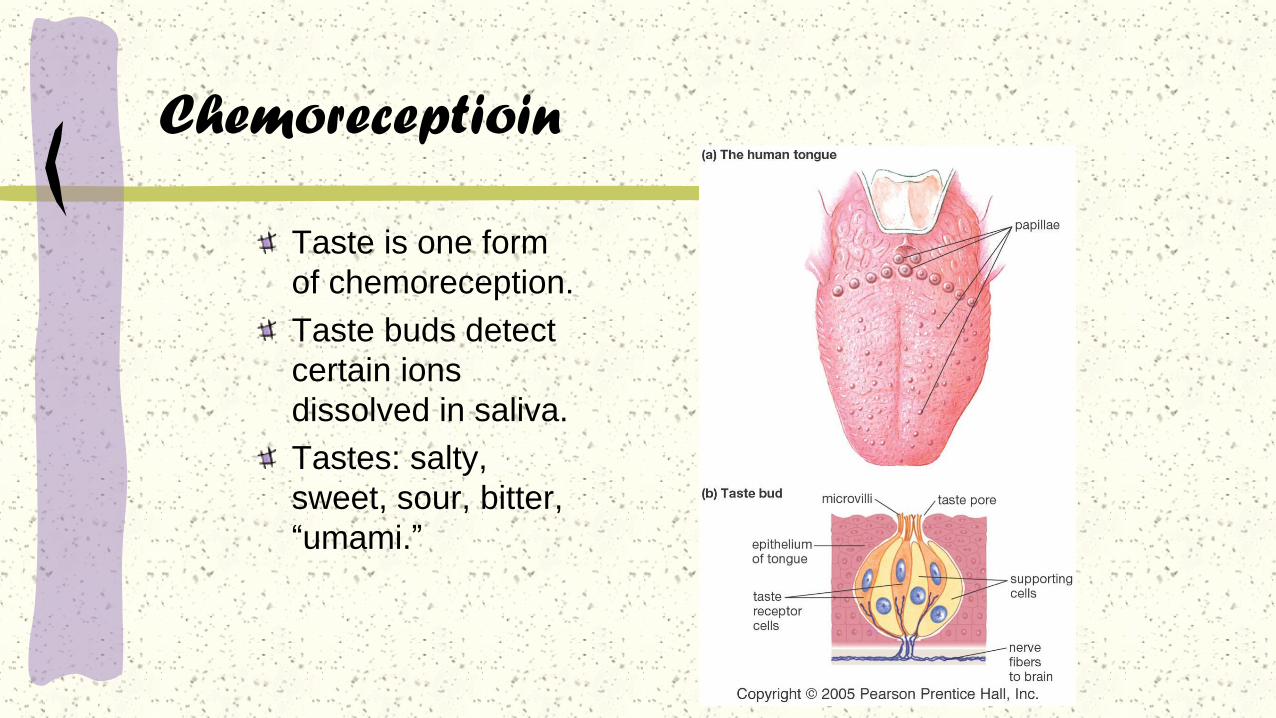
Chemoreceptioin
Taste is one form
of chemoreception.
Taste buds detect
certain ions
dissolved in saliva.
Tastes: salty,
sweet, sour, bitter,
“umami.”
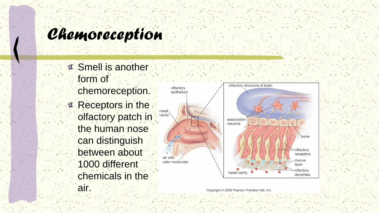
Chemoreception
Smell is another
form of
chemoreception.
Receptors in the
olfactory patch in
the human nose
can distinguish
between about
1000 different
chemicals in the
air.
“Flavor”
What we sense as the “flavor” of food is not taste alone. Smell and
taste together create the sensation of “flavor.”
This is why things don’t “taste” good when we have a cold; we lose
the sense of “flavor.”
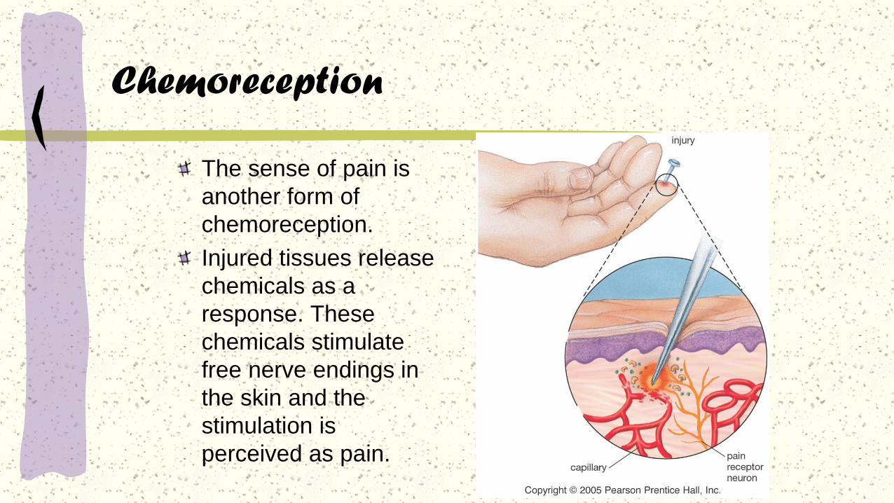
Chemoreception
The sense of pain is
another form of
chemoreception.
Injured tissues release
chemicals as a
response. These
chemicals stimulate
free nerve endings in
the skin and the
stimulation is
perceived as pain.

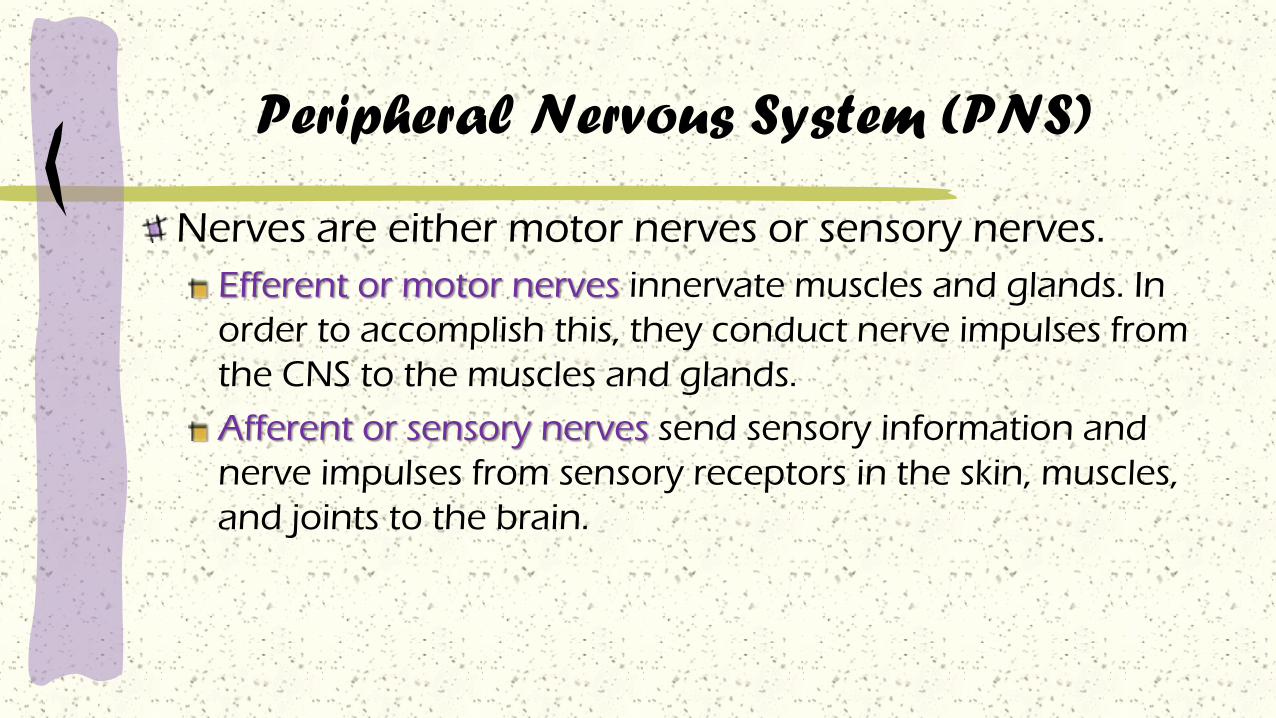
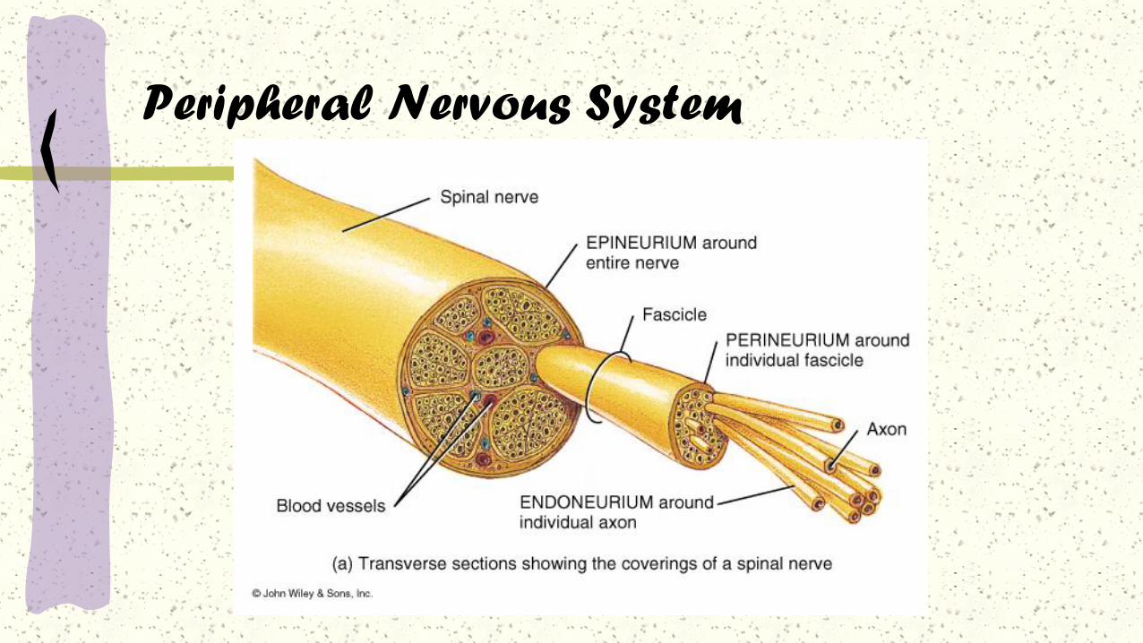
Peripheral Nervous System (PNS)
Nerves are either motor nerves or sensory nerves.
Efferent or motor nerves innervate muscles and glands. In
order to accomplish this, they conduct nerve impulses from
the CNS to the muscles and glands.
Afferent or sensory nerves send sensory information and
nerve impulses from sensory receptors in the skin, muscles,
and joints to the brain.
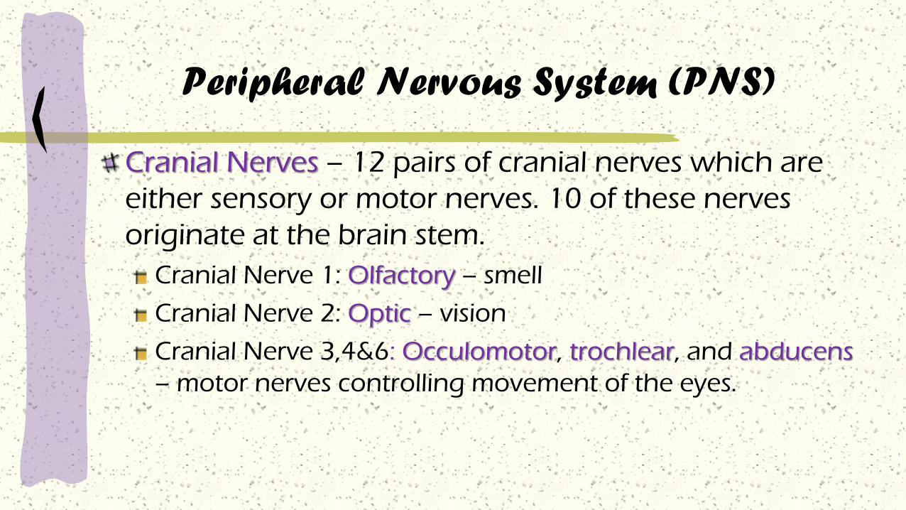
Peripheral Nervous System (PNS)
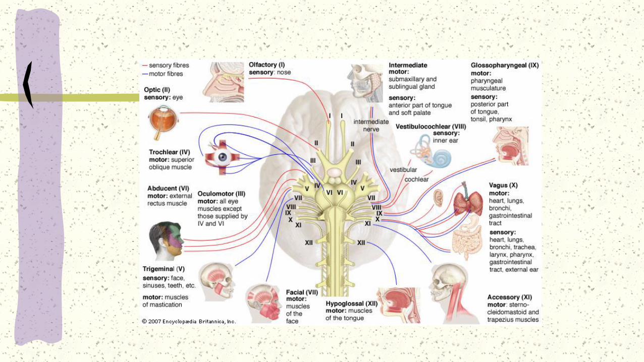
Cranial Nerves – 12 pairs of cranial nerves which are
either sensory or motor nerves. 10 of these nerves
originate at the brain stem.
Cranial Nerve 1: Olfactory – smell
Cranial Nerve 2: Optic – vision
Cranial Nerve 3,4&6: Occulomotor, trochlear, and abducens
– motor nerves controlling movement of the eyes.

Cranial Nerve 5: Trigeminal – sensation of the
head, face, and movements of the jaw
Cranial Nerve 7: Facial – taste, facial movements,
and secretions of tears and saliva
Cranial Nerve 8: Acoustic (vestibulocochlear) –
hearing and equilibrium
Cranial Nerve 9: Glossopharyngeal – taste,
sensation and movement in the pharynx, and
secretion of saliva
Peripheral Nervous System (PNS)

Cranial Nerve 10: Vagus – controls taste,
and movements in the pharynx and larynx
Cranial Nerve 11: Spinal accessory –
movements of the pharynx, larynx, head,
and shoulders
Cranial Nerve 12: Hypoglossal – movement
of the tongue
Peripheral Nervous System (PNS)

Peripheral Nervous System
Spinal Nerves – there are 31 pairs of spinal nerves
branching off the spinal cord.
8 cervical
12 thoracic
5 lumbar
5 sacral
1 coccygeal

Peripheral Nervous System
Peripheral Nervous System

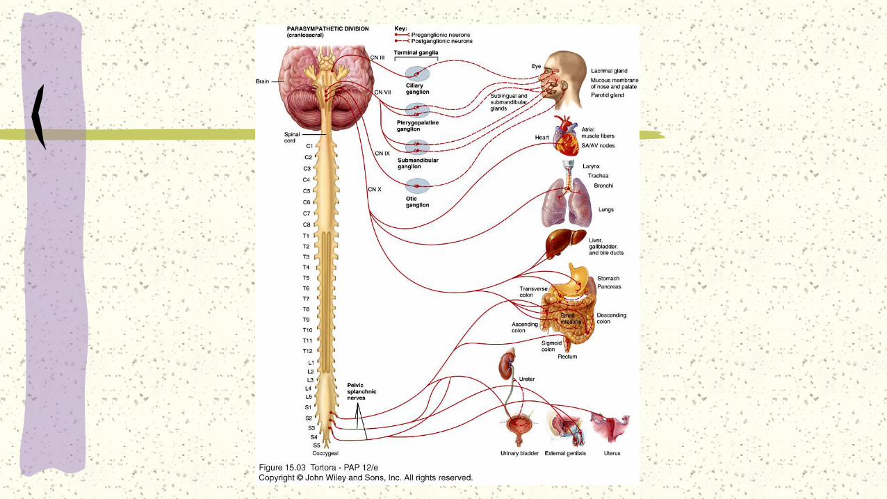
Autonomic Nervous System (ANS)
The autonomic or involuntary nervous
system is that portion of the nervous
system which regulates the activity of
cardiac muscle, smooth muscle, and the
glands.
The ANS has two parts:
Sympathetic
Parasympathetic

Autonomic Nervous System
Sympathetic – stimulates viscera
Prepares the body for emergency situations (“fight or flight”
response to stress)
Fear, emergency, physical exertion, and embarrassment are
responded to by this system
This system shifts energy and blood toward the skeletal
muscles, cardiac muscles, and respiration

Ganglion
In anatomy, a ganglion is a nerve cell cluster or a
group of nerve cell bodies located in the
autonomic nervous system and sensory system.
Ganglia house the cells bodies of afferent nerves
and efferent nerves.
A pseudoganglion looks like a ganglion but only
has nerve fibers and has no nerve cell bodies

Autonomic Nervous System
Parasympathetic – inhibits viscera
Energy conservation system
Restores body energy during rest
Responses toward digestion,
elimination of waste, and decreases
heart rate
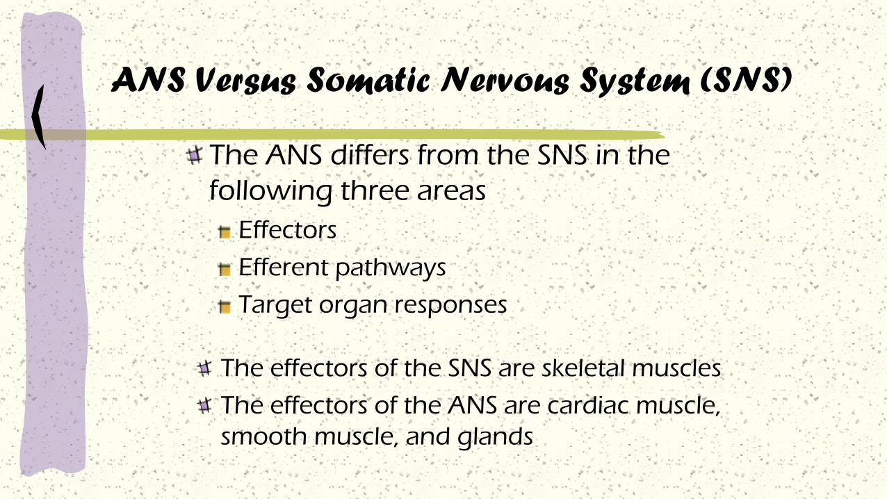
ANS Versus Somatic Nervous System (SNS)
The ANS differs from the SNS in the
following three areas
Effectors
Efferent pathways
Target organ responses
The effectors of the SNS are skeletal muscles
The effectors of the ANS are cardiac muscle,
smooth muscle, and glands

Efferent Pathways
Heavily myelinated axons of the somatic motor
neurons extend from the CNS to the effector (skeletal
muscles)
Axons of the ANS are a two-neuron chain
The preganglionic (first) neuron has a lightly myelinated
axon
The ganglionic (second) neuron extends to an effector
organ
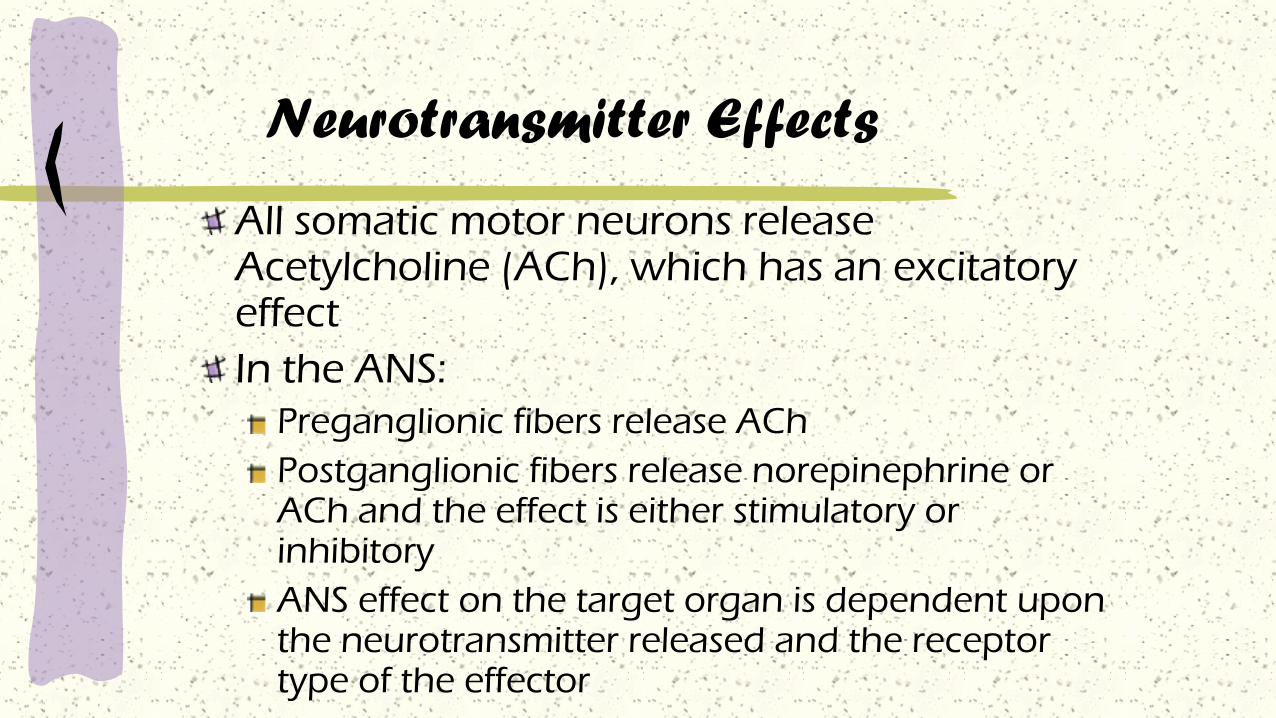
Neurotransmitter Effects
All somatic motor neurons release Acetylcholine (ACh), which has an excitatory effect
In the ANS:
Preganglionic fibers release ACh
Postganglionic fibers release norepinephrine or ACh and the effect is either stimulatory or inhibitory
ANS effect on the target organ is dependent upon the neurotransmitter released and the receptor type of the effector
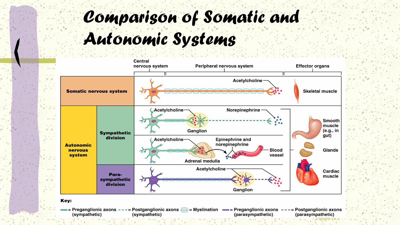
Comparison of Somatic and
Autonomic Systems
Figure 14.2
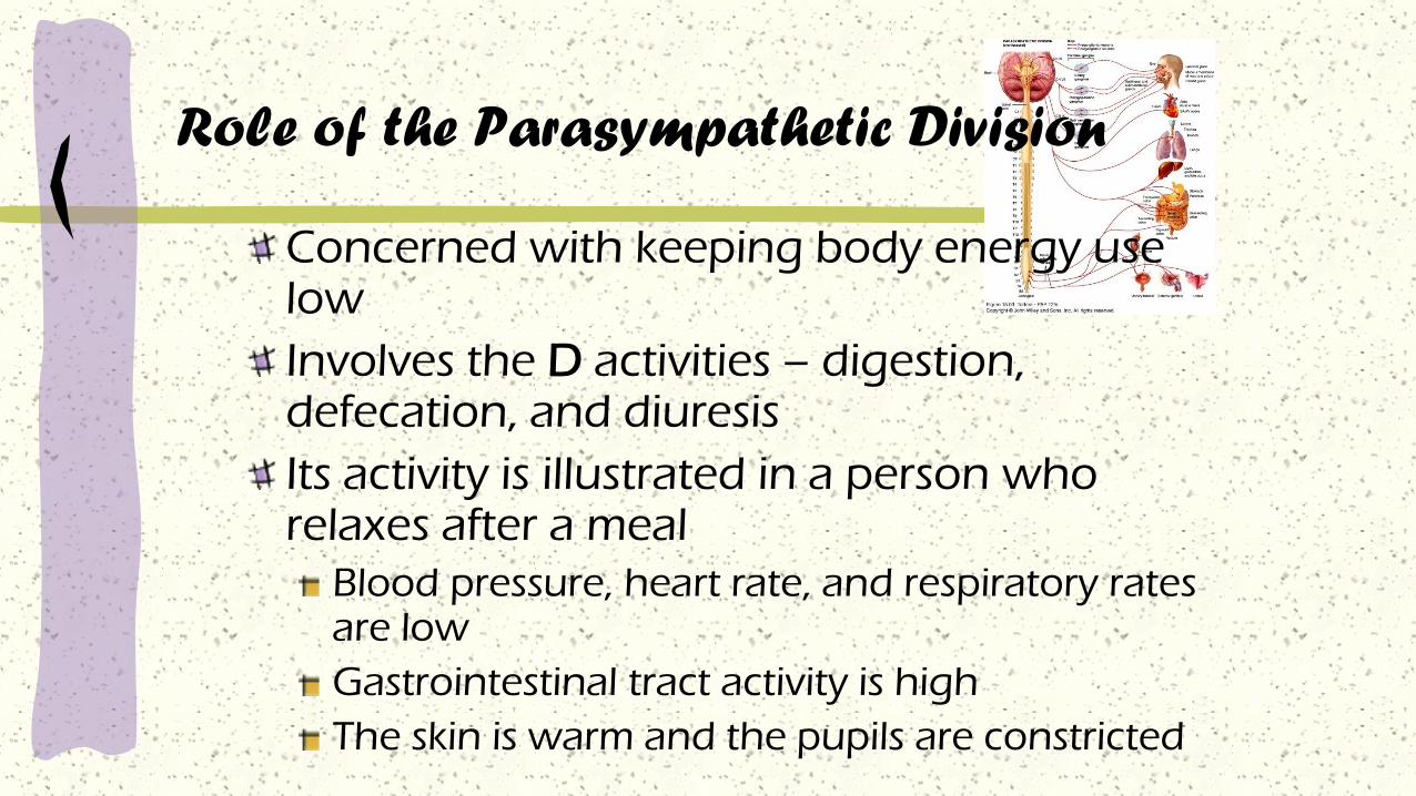
Role of the Parasympathetic Division
Concerned with keeping body energy use low
Involves the D activities – digestion, defecation, and diuresis
Its activity is illustrated in a person who relaxes after a meal
Blood pressure, heart rate, and respiratory rates are low
Gastrointestinal tract activity is high
The skin is warm and the pupils are constricted

Parasympathetic Responses
Rest-and-digest response.
Conserve and restore body energy.
↑ digestive and urinary function.
↓ body functions that support physical activity.
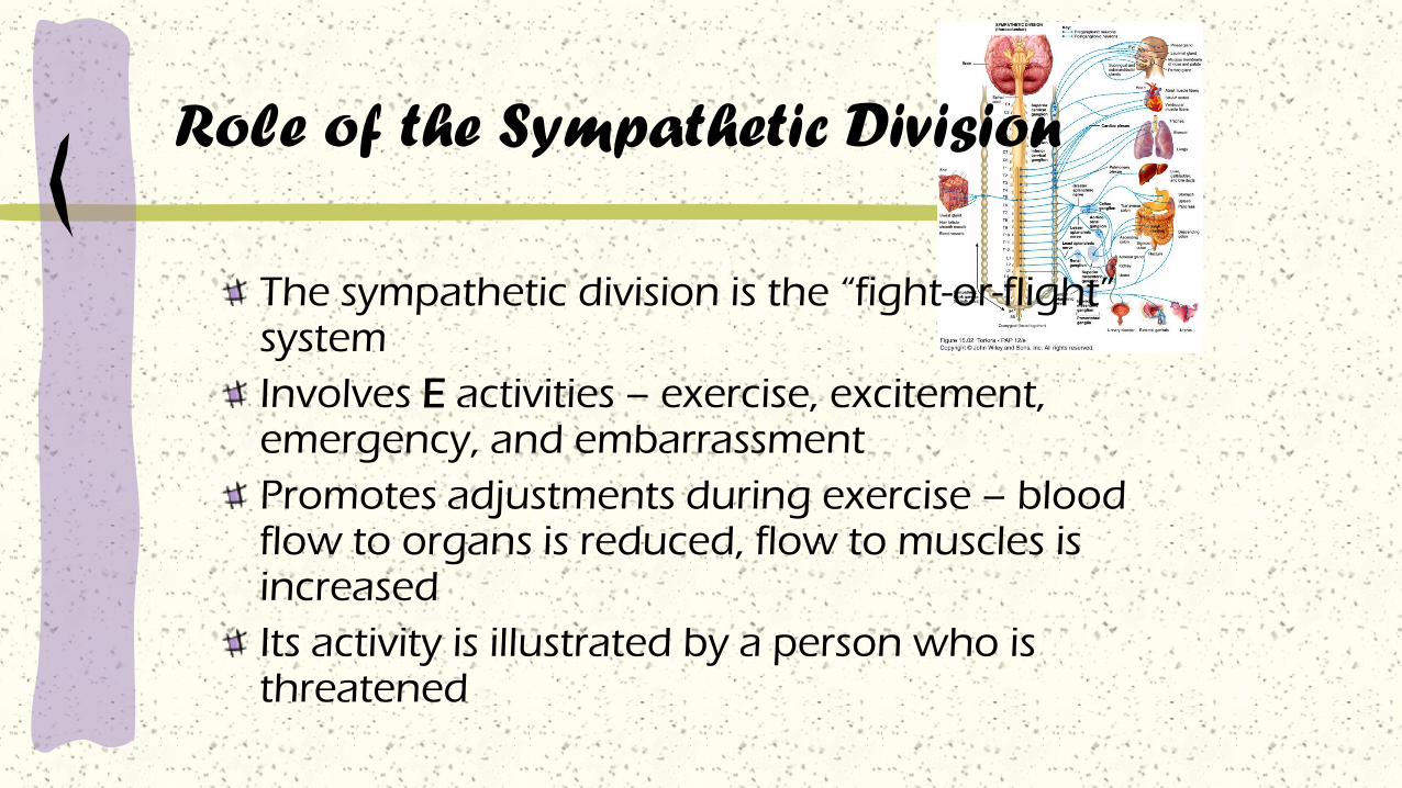
Role of the Sympathetic Division
The sympathetic division is the “fight-or-flight” system
Involves E activities – exercise, excitement, emergency, and embarrassment
Promotes adjustments during exercise – blood flow to organs is reduced, flow to muscles is increased
Its activity is illustrated by a person who is threatened

Sympathetic Responses
Stress ↑ sympathetic system ↑ fight-or-flight response.
↑ production of ATP.
Dilation of the pupils.
↑ heart rate and blood pressure.
Dilation of the airways.
Constriction of blood vessels that supply the kidneys
and gastrointestinal tract.

Sympathetic Responses
↑ blood supply to the skeletal muscles, cardiac muscle,
liver and adipose tissue
↑ glycogenolysis ↑ blood glucose.
↑ lipolysis.

Division Origin of Fibers Length of FibersLocation of
Ganglia
Sympathetic Thoracolumbar
region of the spinal
cord
Short preganglionic
and long
postganglionic
Close to the
spinal cord
Parasympathetic Brain and sacral
spinal cord
Long preganglionic
and short
postganglionic
In the visceral
effector organs
Anatomy of ANS

Cholinergic and Adrenergic Neurons in the
Autonomic Nervous System

Types of Neurotransmitters

Neurotransmitters and Receptors

Neurotransmitters overview
Neurotransmitters and Receptors
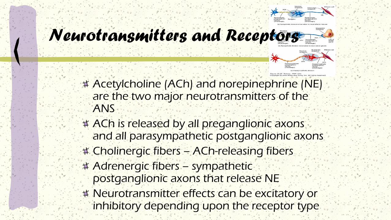
Acetylcholine (ACh) and norepinephrine (NE) are the two major neurotransmitters of the ANS
ACh is released by all preganglionic axons and all parasympathetic postganglionic axons
Cholinergic fibers – ACh-releasing fibers
Adrenergic fibers – sympathetic postganglionic axons that release NE
Neurotransmitter effects can be excitatory or inhibitory depending upon the receptor type
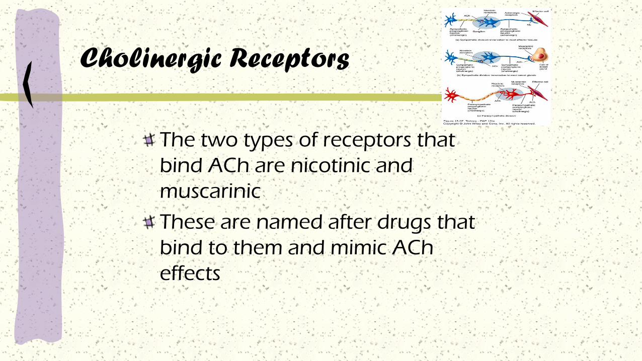
Cholinergic Receptors
The two types of receptors that
bind ACh are nicotinic and
muscarinic
These are named after drugs that
bind to them and mimic ACh
effects

Nicotinic Receptors
Nicotinic receptors are found on:
Motor end plates (somatic targets)
All ganglionic neurons of both sympathetic and
parasympathetic divisions
The hormone-producing cells of the adrenal medulla
The effect of ACh binding to nicotinic receptors is
always stimulatory

Muscarinic Receptors
Muscarinic receptors occur on all
effector cells stimulated by
postganglionic cholinergic fibers
The effect of ACh binding:
Can be either inhibitory or excitatory
Depends on the receptor type of the target
organ
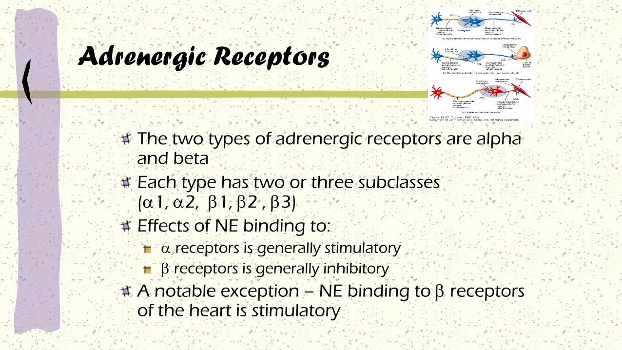
Adrenergic Receptors
The two types of adrenergic receptors are alpha and beta
Each type has two or three subclasses (1, 2, 1, 2 , 3)
Effects of NE binding to:
receptors is generally stimulatory
receptors is generally inhibitory
A notable exception – NE binding to receptors of the heart is stimulatory
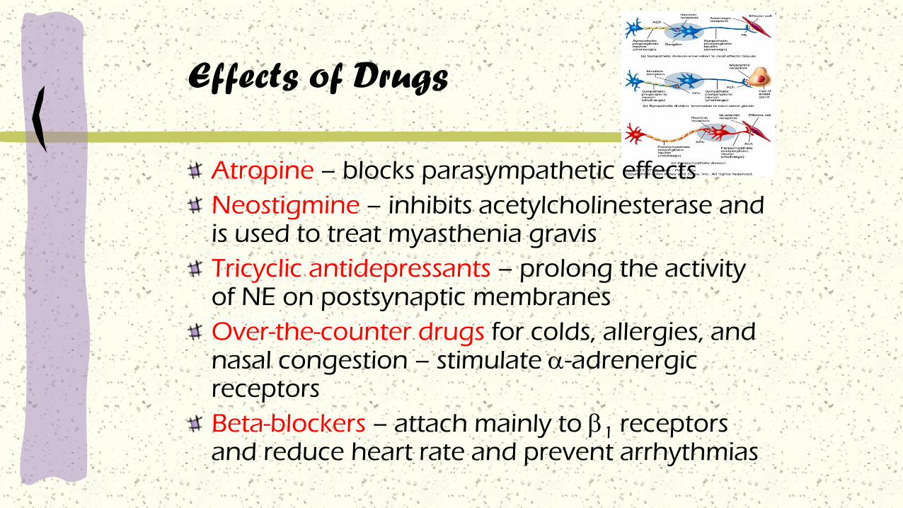
Effects of Drugs
Atropine – blocks parasympathetic effects
Neostigmine – inhibits acetylcholinesterase and is used to treat myasthenia gravis
Tricyclic antidepressants – prolong the activity of NE on postsynaptic membranes
Over-the-counter drugs for colds, allergies, and nasal congestion – stimulate -adrenergic receptors
Beta-blockers – attach mainly to 1 receptors and reduce heart rate and prevent arrhythmias
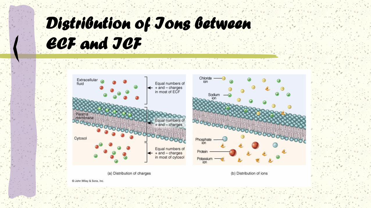
Distribution of Ions between
ECF and ICF

Membrane Potentials
All cell membranes are
electrically polarized
Unequal distribution of charges
Membrane potential (mV) =
difference in charge across the
membrane
Due to unequal ion
concentrations across cell
membrane (fixed anions)

Membrane Potentials

K+
[K+] higher inside cell than outside
Attracted to fixed anions inside cell
High membrane permeability
Flows slowly out of cell
Na+
[Na+] higher outside cell than inside
Attracted to fixed anions inside cell
Low membrane permeability
Flows slowly into cell
Membrane Potentials
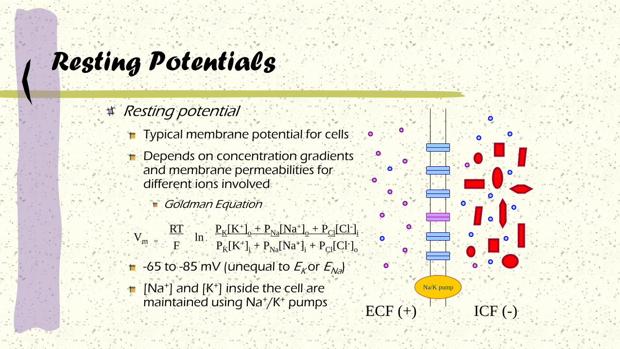
Resting Potentials
Resting potential
Typical membrane potential for cells
Depends on concentration gradients and membrane permeabilities for different ions involved
Goldman Equation
-65 to -85 mV (unequal to EK or ENa)
[Na+] and [K+] inside the cell are maintained using Na+/K+ pumps
Vm =
RT
Fln
PK[K+]o + PNa[Na+]o + PCl[Cl-]i
PK[K+]i + PNa[Na+]i + PCl[Cl-]o
ICF (-)ECF (+)
Na/K pump

Membrane Proteins Involved in Electrical
Signals
Non-gated ion channels (leak channels)
always open
specific for a particular ion
Gated Ion channelsopen only under particular conditions (stimulus)
voltage-gated, ligand-gated,
Ion pumpsactive (require ATP)
maintain ion gradients
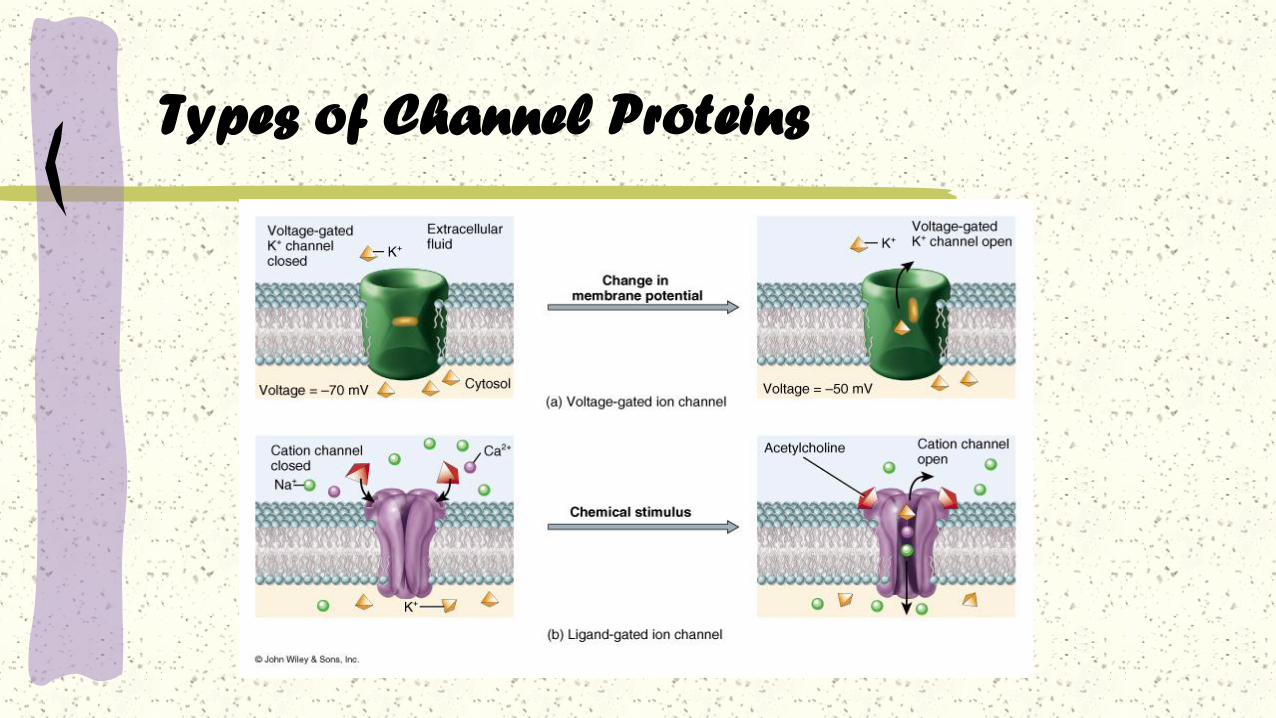
Types of Channel Proteins

Clinical correlations
History:
You examine an 8-year-old male boxer dog
whose owner complains that the dog
experiences seizures, weakness, and confusion
around the time he is fed.
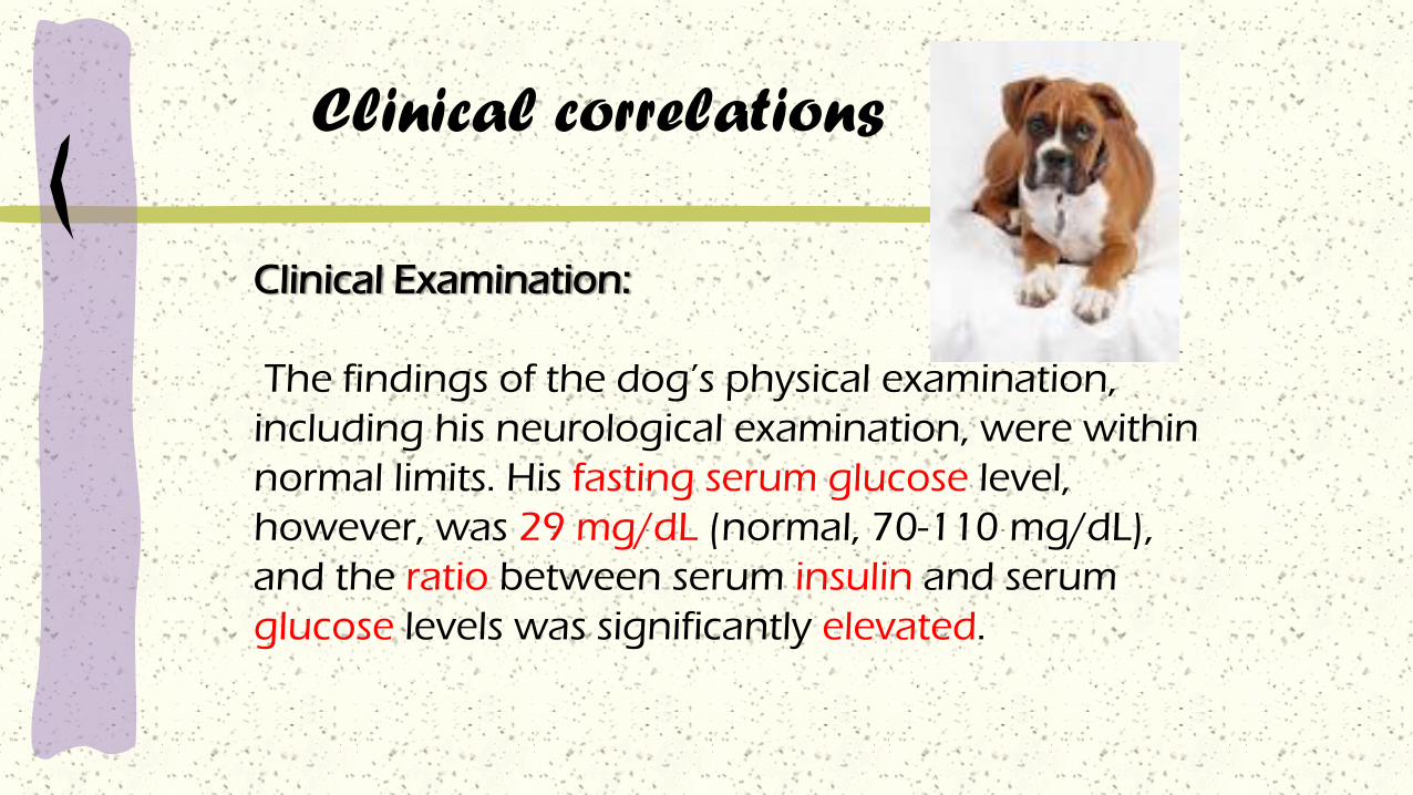
Clinical correlations
Clinical Examination:
The findings of the dog’s physical examination,
including his neurological examination, were within
normal limits. His fasting serum glucose level,
however, was 29 mg/dL (normal, 70-110 mg/dL),
and the ratio between serum insulin and serum
glucose levels was significantly elevated.

Clinical correlations
Comment.
Neurons depend primarily on oxygen and glucose as
metabolites for ATP energy production, and neurons cannot
store appreciable quantities of glucose. ATP is needed for
maintenance of the normal electrical membrane potential. When
deprived of glucose and subsequently ATP, the brain does not
function properly; associated clinical signs include seizures,
weakness, and confusion. In this animal, these signs were more
common at the time of feeding because as the dog anticipated
eating or actually did begin to eat, insulin was released, causing
hypoglycemia.

Clinical correlations
Diagnosis:
In this case the ratio of insulin to glucose is
elevated, probably because of an insulin-secreting
tumor of the pancreas. Because insulin facilitates
glucose transport through cell membranes, too
much insulin results in the transfer of too much
serum glucose to the cytoplasm of other cells of the
body, thus depriving the brain’s neurons of this
essential metabolite.
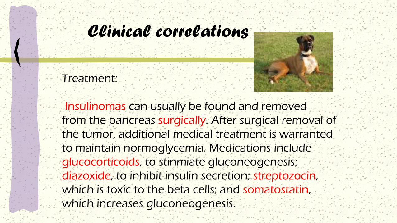
Treatment:
Insulinomas can usually be found and removed
from the pancreas surgically. After surgical removal of
the tumor, additional medical treatment is warranted
to maintain normoglycemia. Medications include
glucocorticoids, to stinmiate gluconeogenesis;
diazoxide, to inhibit insulin secretion; streptozocin,
which is toxic to the beta cells; and somatostatin,
which increases gluconeogenesis.
Clinical correlations

Action Potentials

Action Potentials

Action Potentials
begins at the axon hillock, travels down axon
brief, rapid reversal of membrane potential
Large change (~70-100 mV)
Opening of voltage-gated Na+ and K+ channels
self-propagating - strength of signal maintained
long distance transmission
mV
0
-70
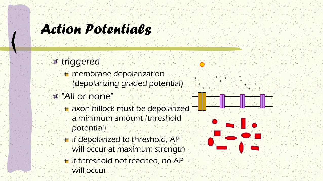
Action Potentials
triggered
membrane depolarization
(depolarizing graded potential)
"All or none"
axon hillock must be depolarized
a minimum amount (threshold
potential)
if depolarized to threshold, AP
will occur at maximum strength
if threshold not reached, no AP
will occur
+
++
++
++
++
++ +
++
+
++
++
+
++
++
++
+
++
++
+ +

Action Potential:Depolarization
Triggering event (graded potential) causes membrane to depolarize
slow increase until threshold is reached
mV
0
-70
+30
threshold
• voltage-gated Na+ channels open
– Na+ enters cell → further depolarization → more
channels open → further depolarization
• membrane reverses polarity (+30 mV)
Action Potential: Repolarization
• Na+ channels close
• Delayed opening of voltage-gated K+ channels
• K+ rushes out of the cell
– membrane potential restored
• K+ channels close
• [Na+] and [K+] restored by the Na+-K+ pump

The action potential
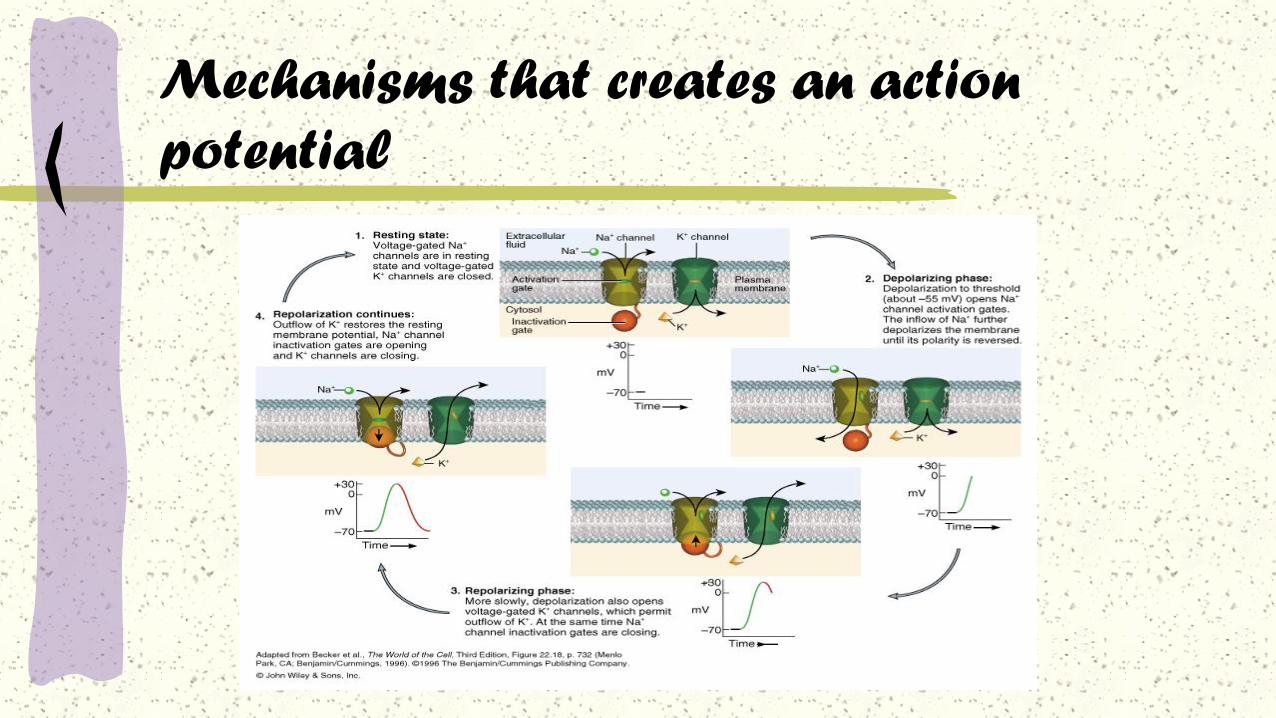
Mechanisms that creates an action
potential
Mechanisms that creates an action
potential
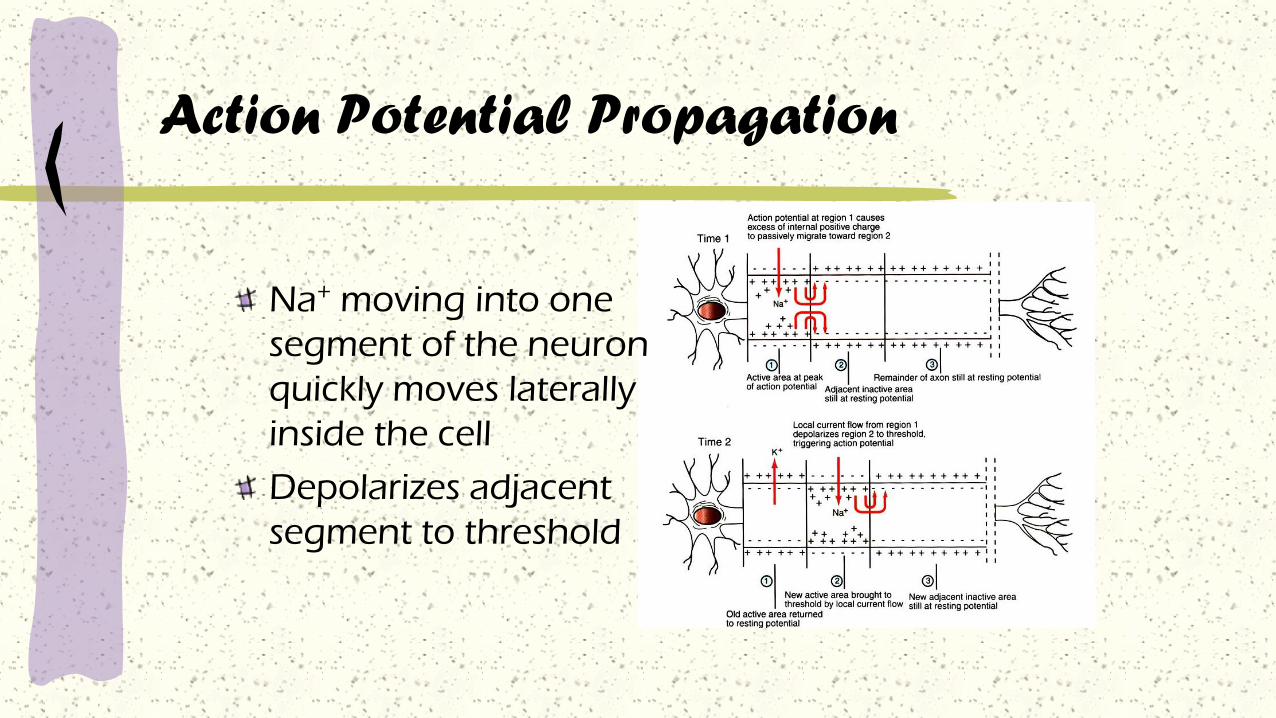
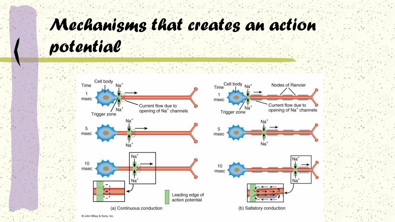
Action Potential Propagation
Na+ moving into one
segment of the neuron
quickly moves laterally
inside the cell
Depolarizes adjacent
segment to threshold

Conduction Velocity
Conduction velocity
speed at which the action
potential travels down the length
of an axon
dictates speed of response
Velocity directly related to axon
diameter
Increased diameter lowers internal
resistance to ion flow
V α √ D in unmyelinated axons
V α D in myelinated axons
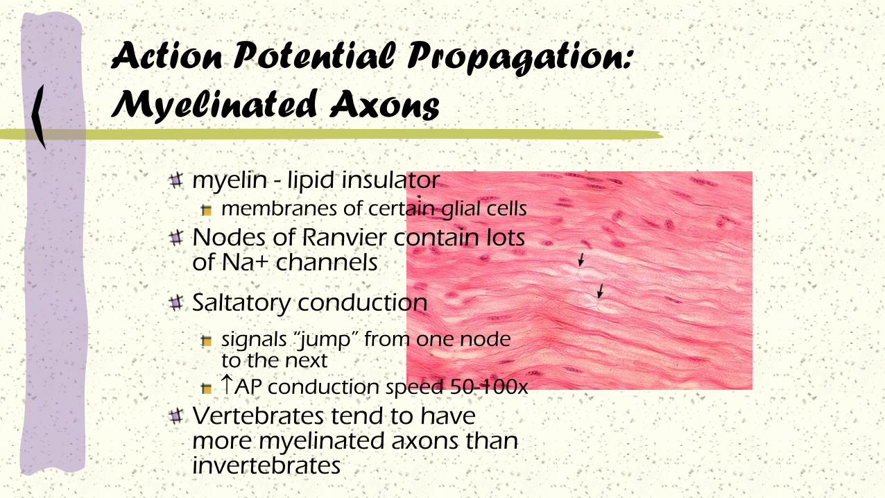
Action Potential Propagation:
Myelinated Axons
myelin - lipid insulator membranes of certain glial cells
Nodes of Ranvier contain lots of Na+ channels
Saltatory conduction
signals “jump” from one node to the next
AP conduction speed 50-100x
Vertebrates tend to have more myelinated axons than invertebrates

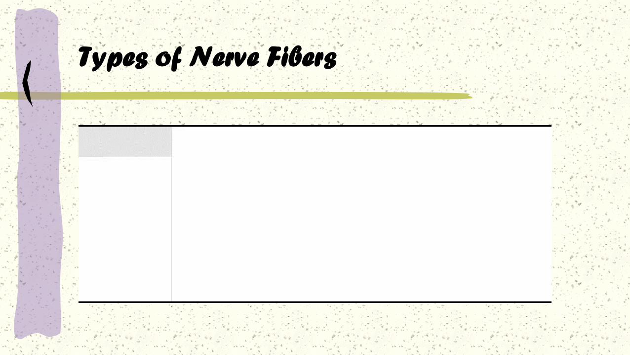
Types of Nerve Fibers(ERLANGER & GASSER system)
“A” fibers:
Largest diameter myelinated fibers with the fastest saltatory conduction (12-130 m/sec) and a brief absolute refractory period. Axons of motor neurons and axons of sensory neurons that conduct touch, pressure, and thermal sensations.
“B” fibers:
Intermediate diameter myelinated fibers With slower saltatory conduction than “A” fibers and longer absolute refractory periods. Dendrites of visceral sensory neurons and axons of presynaptic neurons of the ANS.

“C” fibers:
Smallest diameter unmyelinated fibers with slow
continuous conduction (.5 – 2 m/sec.) and the longest
absolute refractory periods. Axons of some somatic
sensory neuron that carry pain, touch, pressure and
thermal sensation, neuron that carry visceral pain
sensations, and postsynaptic neurons of the ANS
Types of Nerve Fibers
Types of Nerve Fibers
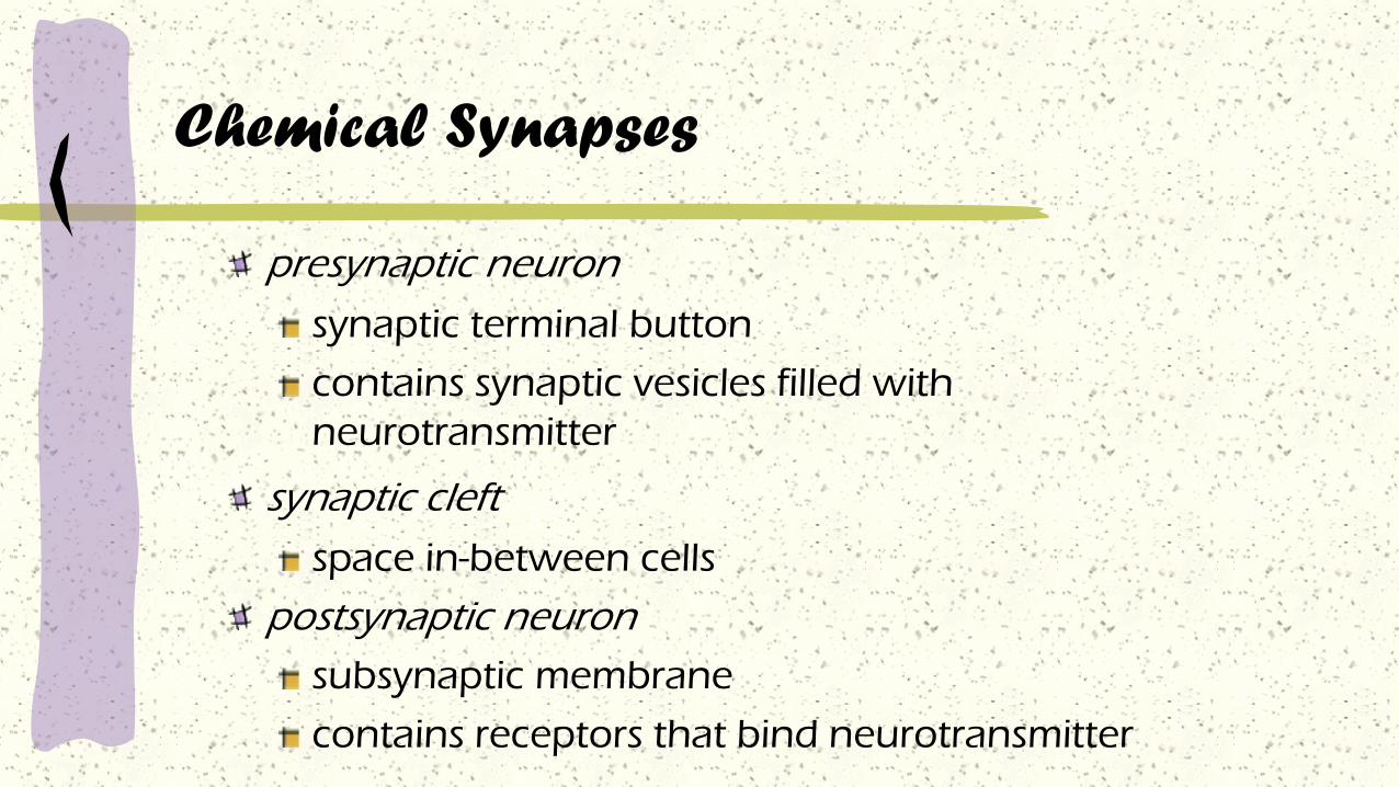
Chemical Synapses
presynaptic neuron
synaptic terminal button
contains synaptic vesicles filled with
neurotransmitter
synaptic cleft
space in-between cells
postsynaptic neuron
subsynaptic membrane
contains receptors that bind neurotransmitter

Chemical Synapses

The Synapse

Chemical Synapses
Many voltage-gated Ca2+
channels in the terminal button
AP in knob opens Ca2+
channels
Ca2+ rushes in.
Ca2+ induced exocytosis of synaptic vesicles
Transmitter diffuses across synaptic cleft and binds to receptors on subsynaptic membrane
Ca2+
Ca2+
Ca2+
Ca2+Ca2+
Ca2+
Ca2+
Ca2+
Calmodulin
Protein Kinase C
Synapsins
+
+
+
+
+
+
+
+ --
--
--
--

Chemical Synapses
Generate Postsynaptic Potentials
Specific ion channels in subsynaptic membrane open, altering membrane permeability
If depolarizing graded potential is strong enough to reach threshold - generates action potential in postsynaptic cell
Metabotropic actions
Long lasting effects (e.g., synaptic changes in learning and memory)

Clinical correlations
History:
You examine a 5-year-old female German
shepherd whose owner states that the dog
becomes progressively weaker with exercise. The
owner also states that recently, just after eating,
the dog has begun to vomit food in formed,
cylinder-shaped boluses.
Clinical Examination:
All abnormalities found on physical examination were
referable to the neuromuscular system. After resting, the
dog’s neurological examination findings were within normal
limits. With even moderate exercise, however, the dog
became progressively weaker, particularly in the front legs.
Intravenous injection of an acetylcholinesterase inhibitor,
edrophonium (Tensilon), eliminated all clinical signs of
weakness. Radiographs of the chest revealed an enlarged
esophagus and thymus.
Clinical correlations

Clinical correlations
Comment:
The history of an enlarged esophagus (megaesophagus) and
the response to an acetylcholinesterase inhibitor confirm the
diagnosis of myasthenia gravis (“grave muscle weakness”).
This is caused by a failure of transmission of acetyicholine at
the neuromuscular synapse. This transmission failure is caused
by antibodies produced by the body against its own
acetylcholine receptors.

Clinical correlations
Treatment :
Spontaneous remissions are common,
depending on the cause. Until then, oral
daily acetylcholinesterase inhibitors are
given. Surgical removal of mediastinal
masses may also be necessary.

Types of Postsynaptic Potentials
excitatory postsynaptic potentials (EPSPs)
Transmitter binding opens Na+ channels in the postsynaptic membrane
Small depolarization of postsynaptic neuron More positive inside the cell
closer to threshold
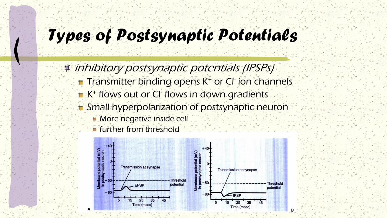
Types of Postsynaptic Potentials
inhibitory postsynaptic potentials (IPSPs) Transmitter binding opens K+ or Cl- ion channels
K+ flows out or Cl- flows in down gradients
Small hyperpolarization of postsynaptic neuron More negative inside cell
further from threshold

Summation
spatial summationnumerous presynaptic fibers may converge on a single postsynaptic neuron
additive effects of numerous neurons inducing EPSPs and IPSPs on the postsyn. neuron
temporal summation additive effects of EPSPs and IPSPs occurring in rapid succession
next synaptic event occurs before membrane recovers from previous event

Reflexes
Fairly fixed patterns of response or behavior similar
for any given stimulus. Fast, predictable, automatic
responses to changes in the environment that help
to protect the body.
Reflexes may be used as diagnostic tools to
determine nervous system disorders.
Reflex pathways consist of sensory fibers bringing
impulses into the spinal cord and motor fibers
capable of effecting a response, as well as all the
interconnections between the two.
Reflexes
Stretch reflex
Results in the contraction of a muscle when it is stretched
suddenly.
Example: patellar tendon reflex
Withdrawal reflex
Sudden contraction and removal of a body segment as the
result of a painful stimulus.
Example: hot stove reflex

Content of a reflex

Clinical correlations
History :
Worried owners call you about their 4-month- old Tennessee
Walking Horse colt. He appeared normal this morning when they
let him out to pasture with his mother, but later this afternoon,
the mare and the foal did not come in to be fed. The owners
went out to the pasture and found the mare with the foal, who
would not get up. He was lying on his side and seemed unable
to position himself sternal. When the owners tried to reposition
him, the foal thrashed, trying to get away. You tell the owners
not to move the foal and that you will be there soon.
Clinical correlations
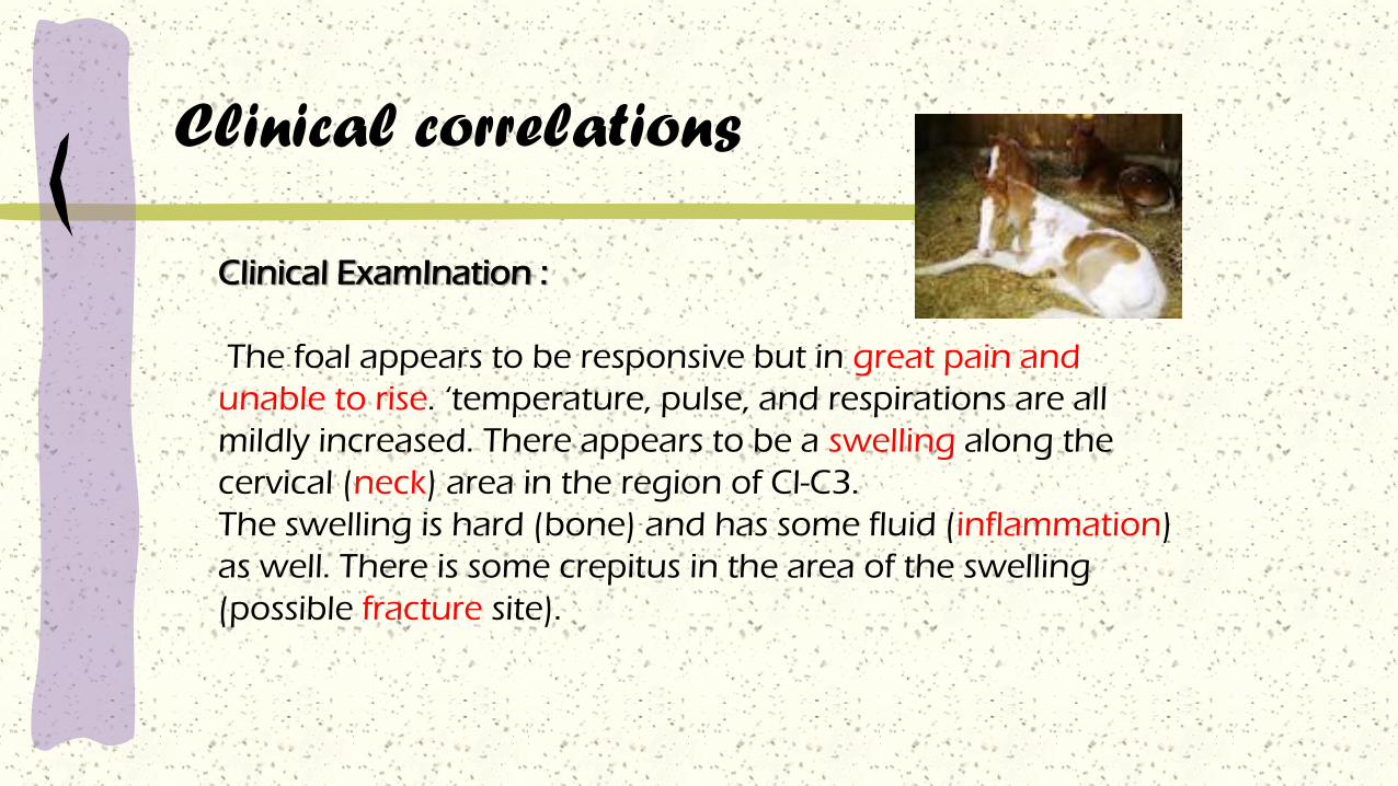
Clinical ExamInation :
The foal appears to be responsive but in great pain and
unable to rise. ‘temperature, pulse, and respirations are all
mildly increased. There appears to be a swelling along the
cervical (neck) area in the region of Cl-C3.
The swelling is hard (bone) and has some fluid (inflammation)
as well. There is some crepitus in the area of the swelling
(possible fracture site).

Clinical correlations
The foal displays no other areas of swelling or trauma.
Neurological examination reveals normal cranial nerves.
In the front limbs the biceps and triceps reflexes seem
increased on both sides. Deep pain is present, and
cutaneous sensation is increased bilaterally. In the hind
limbs the femoral, sciatic, and tibial responses are
increased.

Clinical correlations
Comment :
Although it is difficult to localize a fracture definitively, based
on history and physical examination a fracture seems likely.
The fracture appears to be in the region of Cl-C3.
Radiographs would be ideal to make a definitive diagnosis.
On neurological testing of the biceps, triceps, sciatic, femoral,
and cranial tibial responses, all assess segmental reflex arcs.
Because of a high cervical fracture, the descending motor
tracts that supply both the thoracic and the pelvic limbs are
affected.

Treatment.
The prognosis for this foal is poor Based on the physical
examination and clinical signs, a fracture is likely, and there
is little hope for recovery. The complications associated with
trying to manage a foal as the fracture heals are enormous.
The fracture may not heal, and the foal could have severe
residual neurological deficits. In most cases, these foals are
euthanized fairly quickly because of the poor prognosis.
Clinical correlations

Any Questions ?

PATHWAYS AND HIGHER-ORDER
FUNCTIONS
Introduction
There is a continuous flow of information between the brain, spinal cord, and peripheral nerves
- millions of sensory neurons deliver information to processing centers in the CNS, and millions of motor neurons are controlling or adjusting activities of peripheral effectors
- this process continues around the clock, with many brain stem centers active throughout our lives performing autonomic functions at the subconscious level
Many forms of interaction, feedback, and regulation link higher centers with the various components of the brain stem
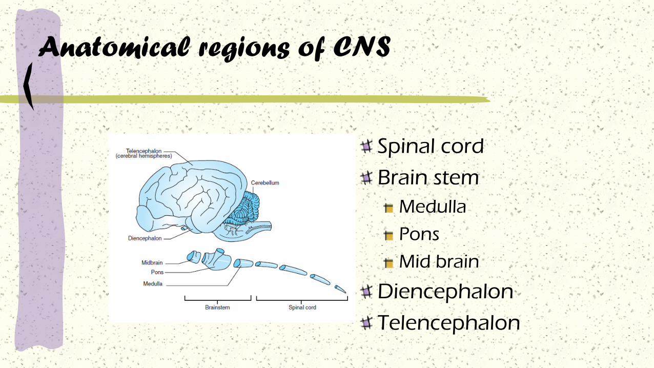
Anatomical regions of CNS
Spinal cord
Brain stem
Medulla
Pons
Mid brain
Diencephalon
Telencephalon

The spinal cord
The most caudal region
Sensory nerves enter from dorsal roots
Motor nerves exit from ventral roots
Contains
cell bodies and dendrites of motor neurons
Vertical tracts of sensory n. to the brain and motor n. from the brain
The isolated spinal cord can control simple reflexes,
such as muscle stretch reflexes and limb withdrawal from painful stimuli.

The spinal cord

Brain Stem
Medulla
Lies rostral to the spinal cord
The cell bodies of medullary neurons aggregates in sensory or motor
nuclei, called: cranial nerve nuclei.
play a critical role in life support functions:
Respiratory
Cardiovascular
Feeding (taste, tongue movement, swallowing, digestion)
Brain Stem
The pons
Rostral to the medulla
contains the cell bodies of large numbers of neurons in a two-neuron
chain that relays information from the cerebral cortex to the
cerebellum.
Receives sensory inf. from face, motor control of chewing.
The cerebellum is not a part of the brainstem
The cerebellum is important for smooth, accurate, coordinated
movement and for motor learning.

Brain stem
Mid brain (Mesencephalon)
Rostral to the pons
processing and relaying visual and auditory information
Directly controls eye movement
Reticula formation:
A netlike complex of many small clusters of cell bodies (nuclei),
modulating consciousness and arousal, pain perception, and spinal
reflexes, as well as in movement
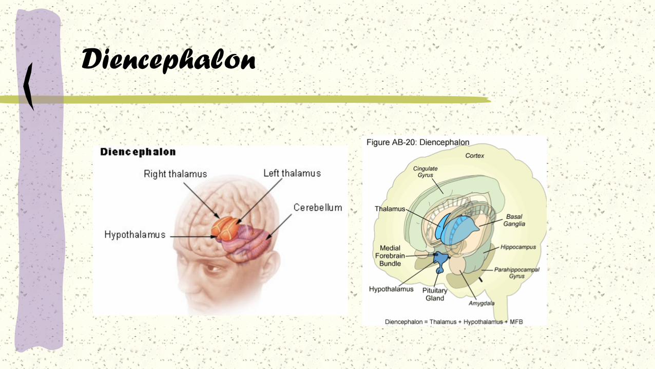
Diencephalon
contains the thalamus and the hypothalamus
The thalamus is a relay station of information
being passed to the cerebral cortex from sensory
systems and other brain regions
The hypothalamus regulates the autonomic
nervous system, controls hormone secretion of
the pituitary gland, and plays a major role in
physiological and behavioral aspects of
homeostasis (e.g., maintenance of temperature
and blood pressure; feeding).
Diencephalon

Telencephalon
made up of the cerebral cortex and a small
number of prominent subcortical structures, such as the basal ganglia
and hippocampus
The cerebral cortex mediates the most complex forms of sensory
integration and conscious sensory perception.
The basal ganglia are a collection of nuclei that modulate the motor
functions of cerebral cortex
the hippocampus plays an important role in memory and spatial
learning.

Telencephalon

Sensory and Motor Pathways
Nerve pathways, called tracts, relay
sensory and motor information between
the CNS and PNS
- consists of a chain of tracts and associated
nuclei
- number of synapses varies from 1 pathway to
another
- all involve both the brain and spinal cord
- tract name often indicates its origin and
destination

Ascending (sensory) pathways and descending
(motor) pathways:
1) these tracts are paired (bilaterally and symmetrically along
the spinal cord)
2) axons within each tract are grouped according to the body
region innervated
Sensory and Motor Pathways
Spinal Pathways - 1
Spinal Pathways - 2
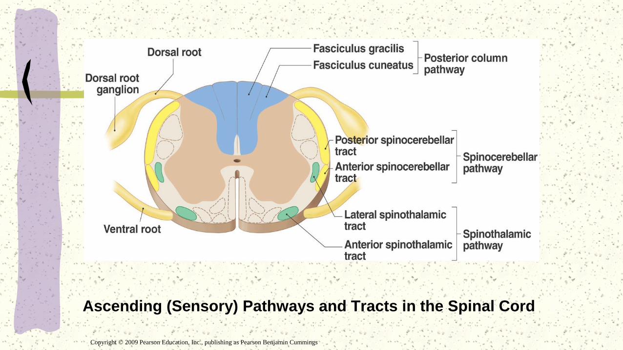
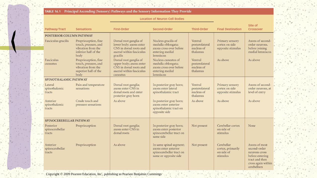
Ascending (Sensory) Pathways and Tracts in the Spinal Cord
Copyright © 2009 Pearson Education, Inc., publishing as Pearson Benjamin Cummings
Sensory PathwaysMonitor conditions both inside the body and in the external environment
Sensation - stimulated receptor passes information to the CNS
- form of action potentials in an afferent (sensory) fibers
- processing in the SC can produce a rapid motor response (stretch reflex)
- processing within the brain stem may result in complex motor activites (positional changes in the eye, head, trunk)
Most sensory information is processed in the SC, thalamus, or brain stem
- only ~1% reaches the cerebral cortex and our conscious awareness

Sensory Pathways
3 major somatic sensory pathways:
1. The posterior column pathway
2. The spinothalamic pathway
3. The spinocerebellar pathway

These pathways
involve a chain of
neurons:
First-order neuron – to
the CNS
Second-order neuron –
an interneuron located
in either the spinal cord
or the brain stem
Third-order neuron –
carries information
from the thalamus to
the cerebral cortex
Sensory Pathways
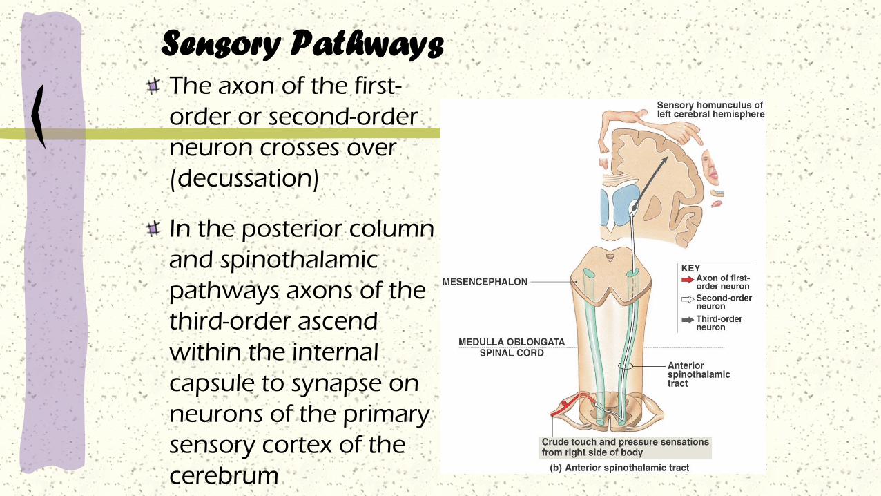
Sensory PathwaysThe axon of the first-
order or second-order
neuron crosses over
(decussation)
In the posterior column
and spinothalamic
pathways axons of the
third-order ascend
within the internal
capsule to synapse on
neurons of the primary
sensory cortex of the
cerebrum

Sensory Homunculus (‘little man’)
Functional map of the
primary sensory cortex
- proportions are distorted
because the area of sensory
cortex devoted to a particular
region is proportional to the
number of sensory receptors the
region contains
Example: tongue has 10s of
1000s of taste and touch
receptors while the back
touch receptors are few and
far between
Copyright © 2009 Pearson Education, Inc., publishing as Pearson Benjamin Cummings
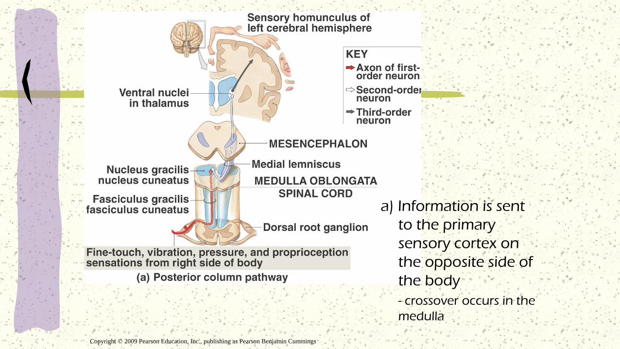
a) Information is sent
to the primary
sensory cortex on
the opposite side of
the body
- crossover occurs in the
medulla
Copyright © 2009 Pearson Education, Inc., publishing as Pearson Benjamin Cummings
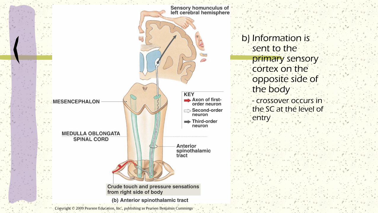
b) Information is sent to the primary sensory cortex on the opposite side of the body- crossover occurs in the SC at the level of entry
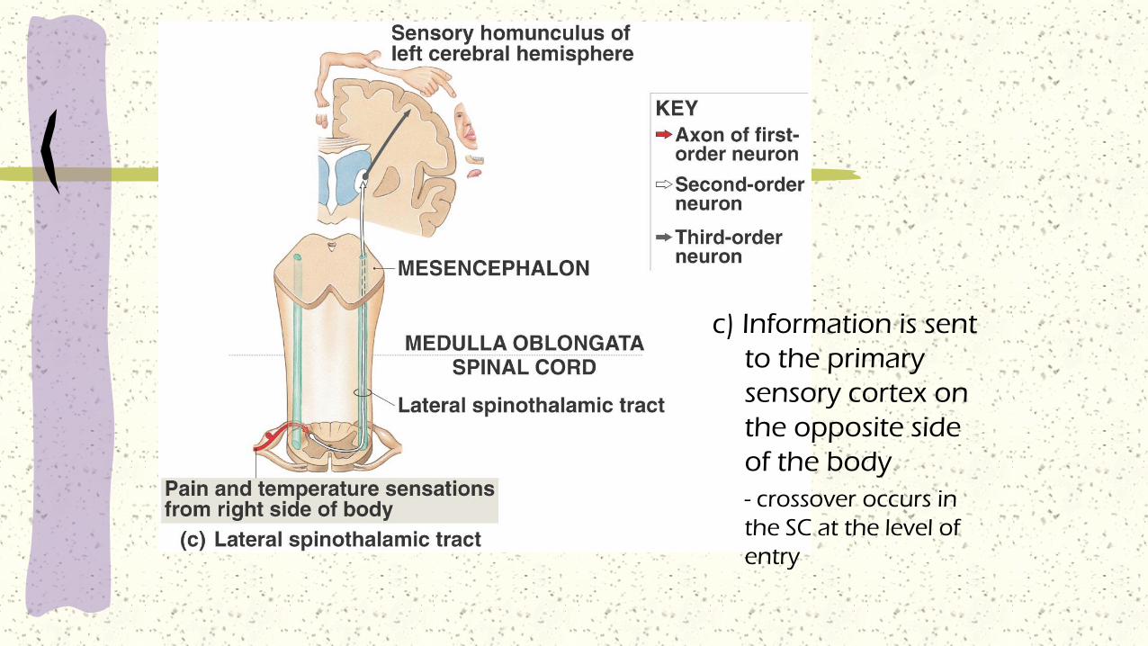
c) Information is sent
to the primary
sensory cortex on
the opposite side
of the body
- crossover occurs in
the SC at the level of
entry

Copyright © 2009 Pearson Education, Inc., publishing as Pearson Benjamin Cummings
d) Information sent to the cerebellum but only the anterior sends it to the opposite side of the body- cross before entering the tract and after within the cerebellum
Copyright © 2009 Pearson Education, Inc., publishing as Pearson Benjamin Cummings

Motor Pathways
CNS issues motor commands in response to information provided by sensory systems
- sent by the somatic nervous system (SNS) and the autonomic nervous system (ANS)
SNS – skeletal muscle contraction
ANS – innervates visceral effectors (smooth muscle, cardiac muscle, and glands)
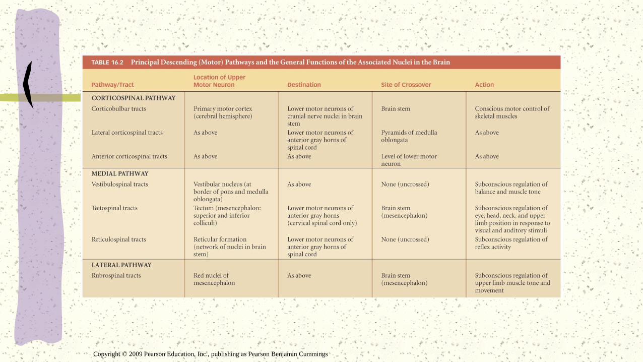
Conscious and subconscious motor commands control skeletal muscles by traveling over 3 integrated motor pathways

Motor Pathways
The corticospinal pathway – voluntary control- the corticobulbar tracts
- the corticospinal tracts
The medial and lateral pathways – modify or direct skeletal muscle contractions by stimulating, facilitating, or inhibiting lower motor neurons
Motor pathways usually contain 2 neurons
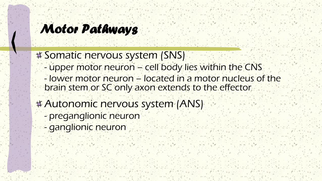
Somatic nervous system (SNS)- upper motor neuron – cell body lies within the CNS
- lower motor neuron – located in a motor nucleus of the brain stem or SC only axon extends to the effector
Autonomic nervous system (ANS)- preganglionic neuron
- ganglionic neuron
Motor Pathways

Copyright © 2009 Pearson Education, Inc., publishing as Pearson Benjamin Cummings
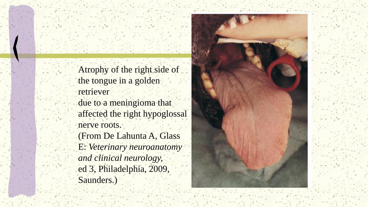
Atrophy of the right side of
the tongue in a golden
retriever
due to a meningioma that
affected the right hypoglossal
nerve roots.
(From De Lahunta A, Glass
E: Veterinary neuroanatomy
and clinical neurology,
ed 3, Philadelphia, 2009,
Saunders.)

Copyright © 2009 Pearson Education, Inc., publishing as Pearson Benjamin Cummings
Copyright © 2009 Pearson Education, Inc., publishing as Pearson Benjamin Cummings
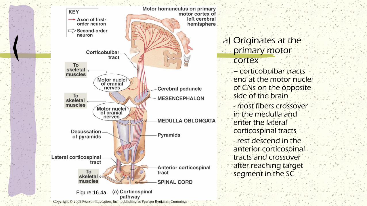
Figure 16.4a
a) Originates at the primary motor cortex – corticobulbar tracts end at the motor nuclei of CNs on the opposite side of the brain
- most fibers crossover in the medulla and enter the lateral corticospinal tracts
- rest descend in the anterior corticospinal tracts and crossover after reaching target segment in the SC
Copyright © 2009 Pearson Education, Inc., publishing as Pearson Benjamin Cummings
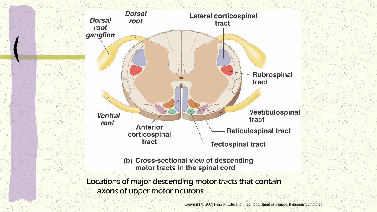
Locations of major descending motor tracts that contain
axons of upper motor neurons

Copyright © 2009 Pearson Education, Inc., publishing as Pearson Benjamin Cummings
Basal Ganglia – The Direct pathway
Basal Ganglia – The Indirect pathway
Copyright © 2009 Pearson Education, Inc., publishing as Pearson Benjamin Cummings


Higher-Order Functions
Characteristics:
- They are performed by the cerebral cortex
- They involve complex interconnections and
communication between areas within the
cerebral cortex and between the cerebral cortex
and other areas of the brain
- They involve both conscious and unconscious
information processing
- They are not part of the programmed ‘wiring’ of
the brains; therefore, the functions are subject to
modification and adjustment over time (learning)
Copyright © 2009 Pearson Education, Inc., publishing as Pearson Benjamin Cummings
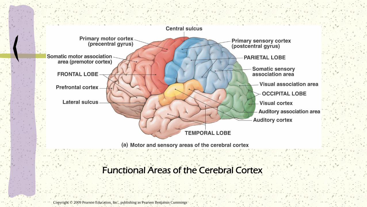
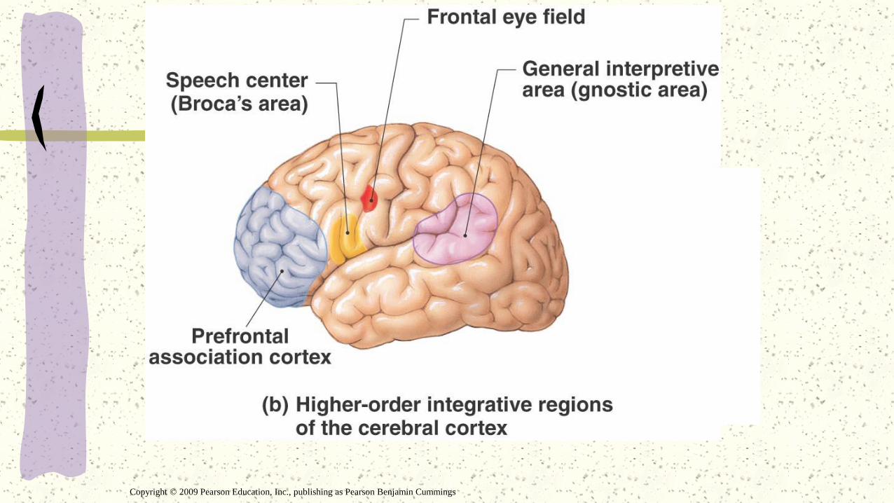
Functional Areas of the Cerebral Cortex

Integrative Regions of the Cerebral Cortex
Cortical areas that act as centers for complex
sensory stimuli and motor responses
- general interpretive area receives information from all
sensory association areas
- only present in one hemisphere, usually the left
Speech center – regulates patterns of breathing
and vocalization
Prefrontal cortex – coordinates information from
the secondary and special association areas of the
cortex
- performs abstract intellectual functions
Copyright © 2009 Pearson Education, Inc., publishing as Pearson Benjamin Cummings

Figure 16.8 Hemispheric Specialization
Copyright © 2009 Pearson Education, Inc., publishing as Pearson Benjamin Cummings

Memory
Process of accessing stored bits of information
acquired through experience
Short-term memories last seconds to hours
Long-term memories can last for years and are
stored in the cerebral cortex
Memory consolidation – conversion from a
short-term memory to a long-term memory
The amygdaloid body and the hippocampus
(limbic system) are essential to memory
consolidation
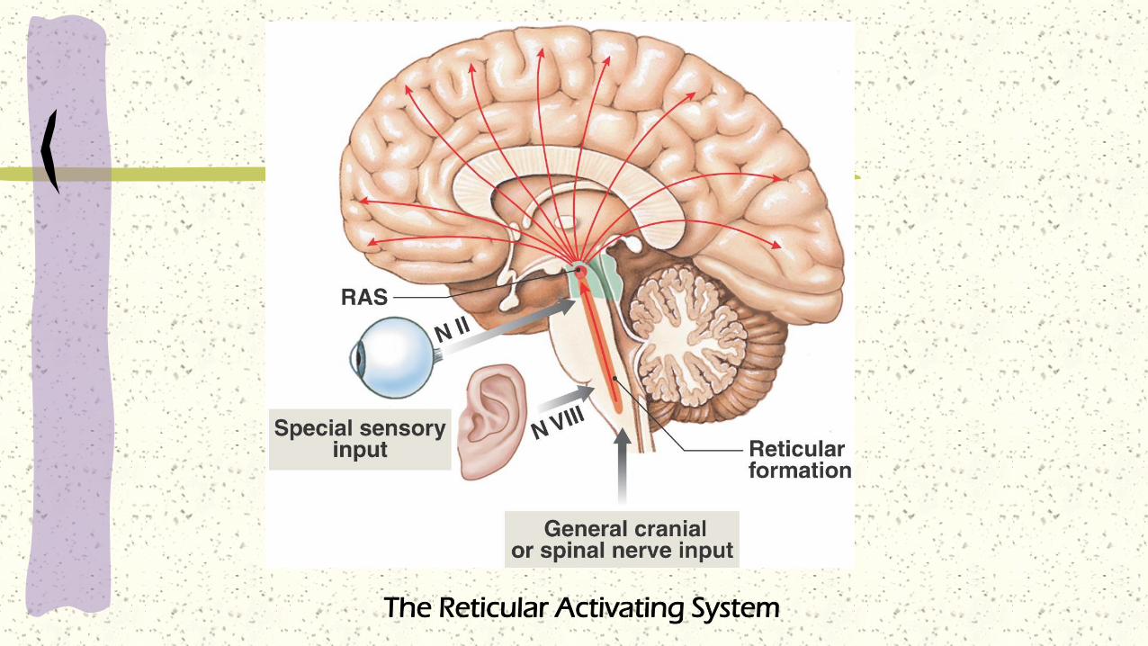
The Reticular Activating System


Aging and the Nervous System
Common, age-related anatomical changes in the
NS include the following:
- a reduction in brain size and weight
- a reduction in the number of neurons
- a decrease in blood flow to the brain
- changes in synaptic organization of the brain
- intracellular and extracellular changes in CNS
neurons
Any Questions ?

Injuries and Diseases of the Nervous
System
Cerebral Concussion
Paralysis
Cerebral Palsy
Cerebrovascular Accident (stroke)
Aneurysm
Parkinson’s Disease
Multiple Sclerosis (MS)
Cerebral Concussion
Despite its considerable protection, the brain is
subject to traumatic injury, often with serious
consequences.
A concussion literally means an agitation or
shaking of the brain by either a direct or
indirect blow.
A concussion is characterized by immediate
and transient impairment of neural functions
such as alteration of consciousness,
disturbance of vision, and equilibrium.

Cerebral Concussion
Concussions are classified by degree of
severity – 1st,2nd,3rd. These distinctions are
important for treatment and prognosis.
Variation in the classification of concussion
is common.
1st degree concussion experience no loss of
consciousness, possible memory loss, possible
dizziness and tinnitus (ringing in the ears), no
loss of coordination, and relatively rapid
recovery.
Cerebral Concussion
2nd degree concussions have momentary loss
(10 sec. to 5 min.) of consciousness, transient
confusion and mild retrograde amnesia
(amnesia for the events prior to the injury),
moderate dizziness and tinnitus, slight loss of
coordination and varied recovery time.
3rd degree concussion experiences a
prolonged loss consciousness, severe memory
loss , severe dizziness and tinnitus, marked loss
of coordination and a prolonged recovery time.

Cerebral Concussion
Coma and death can also result from a
serious concussion.
After a concussion, the athlete should not be
allowed to return to competition that day. In
fact, before resuming training, a head-injured
athlete must be free of headaches for 24
hours. Athletes who experience a loss of
consciousness for any period of time require
evaluation and monitoring by a physician.
Cerebral Concussion
The athlete who sustains repeated
concussions requires special evaluation
before returning to a sport with the potential
for further brain injury. Most team physicians
follow the “1-2-3” rule: one concussion= the
athlete is out of the game, two concussion=
out for the season, three concussions= the
athlete should no longer play.

Paralysis
The inability to voluntarily move a
muscle or limb.
Paralysis can be caused by damage to a
sensory nerve that results in a lack of
sensation in the area which that nerve
innervates.
Paralysis can also result from damage to
the spinal cord or motor nerve.

Paralysis
The higher the spinal cord is damaged, the greater the
extent of paralysis.
Paraplegia – paralysis of both lower extremities.
Quadriplegia – paralysis of both upper and lower
extremities.
Cerebral Palsy
A disorder of movement and posture caused by an irreparable lesion of the CNS.
Developmental defects of motor areas of the brain because of trauma at birth.
Individuals with cerebral palsy may have musculoskeletal problems, mental retardation, speech and hearing difficulties, eye problems, and seizures.
There is a great deal of variation among individuals with cerebral palsy – some are particularly bright; others have less musculoskeletal abnormalities.

Cerebrovascular Accident (Stoke)
This is the most common brain disorder.
Arteries that supply blood to the brain cause blood
clots to develop, obstructing blood flow to the brain,
precipitating a stroke.
Symptoms of a stroke include slurred speech,
loss/blurred vision, and paralysis of a limb or half the
body.

Aneurysm
Weak, swollen areas of a blood vessel supplying the
brain which alters the brain’s blood flow, resulting in a
partial or complete loss of consciousness.
Aneurysms develop slowly and are rarely associated with
symptoms.
If the weak area ruptures, massive hemorrhage occurs. This
can be fatal.

Parkinson’s Disease
A progressive disorder of the CNS (usually in
individuals over 60 years of age, but can occur in
younger patients).
Parkinson’s is thought to result from too little dopamine
being produced.
Symptoms include muscle tremors, muscle rigidity, and
slow, difficult movements. Walking and speech are often
affected.

Multiple Sclerosis (MS)
A progressive destruction of the myelin
sheaths of the nerves of the CNS. This
causes “short circuits” in nerve
transmissions.
There is no known cause or treatment.
MS most commonly strikes young women
in their 20’s, but can affect men as well.
Progressive loss of muscle function is the
main symptom.

The End
Any Questions???