nervous system organization - mt. san antonio …...organization of nervous system cns pns sensory...
TRANSCRIPT

Nervous System Organization
Nervous System Nervous System OrganizationOrganization
Dr. Carmen E. Dr. Carmen E. RexachRexachPhysiologyPhysiology
MtSACMtSAC Biology Dept. Biology Dept.

Organization of nervous system
CNS PNS
Sensory(afferent)
Motor(efferent)
autonomicsomatic
sympatheticparasympathetic
Single neuronpathway
Two neuronpathway

CNS
• Functions– Integrates information from PNS– Processes information– Cognition, learning, memory– Plans and executes voluntary
movements• Components
– Brain• Cerebrum• Diencephalon• Midbrain and Hindbrain
– Spinal cord• Ascending and descending tracts

Brain
• Cerebrum• Cerebral cortex
– frontal, parietal, temporal, occipital lobes• Cerebral lateralization
– Decussation of pyramids– split brain procedures of the corpus callosum in epilepsy

Major landmarks of CNS

Cerebrum• Only structure of the telencephalon.• Largest portion of brain (80% mass).• Responsible for higher mental
functions.• Corpus callosum:
– Major tract of axons that functionally interconnects right and left cerebral hemispheres.


Cerebral cortex• Convolutions
– Elevated folds: gyri– Depressed groves: sulci
• Frontal lobe– Anterior portion of each cerebral hemisphere.– Precentral gyri
• Contains upper motor neurons.• Involved in motor control.
• Body regions with the greatest amount of motor innervation are represented by largest areas of motor cortex.


Cerebral Cortex• Parietal lobe:
– Primary area responsible for perception of somatesthetic sensation.
– Body regions with highest densities of receptors are represented by largest areas of sensory cortex.
• Temporal lobe:– Contain auditory centers that receive sensory
fibers from cochlea.– Interpretation and association of auditory and
visual information.

Cerebral Cortex
• Occipital Lobe: – Primary area responsible for vision and
coordination of eye movements.• Insula:
– Implicated in memory encoding.– Integration of sensory information with
visceral responses.– Coordinated cardiovascular response to
stress.

Basal ganglia• Masses of gray matter
composed of neuronal cell bodies located deep within white matter.
• nuclei around thalamus that help plan voluntary movement– Corpus striatum = largest
part of basal ganglia• caudate nucleus• putamen• globus pallidus


Basal ganglia diseases
• Parkinson’s– Cause: lesions in
substantia nigra– Results: loss of
dopaminergicneurotransmitters
– Symptoms: tremor, rigidity, bradykinesia

Basal ganglia diseases
• Huntington’s chorea– Cause: genetic
disorder causing loss of striatopallidal and striatonigral neurons
– Result: loss of GABA (inhibitory)
– Symptoms: progressive dementia and bizarre involuntary movements

Cerebral Lateralization• Cerebral dominance:
– Specialization of one hemisphere.
• Left hemisphere: – More adept in language
and analytical abilities.– Damage:
• Severe speech problems.
• Right hemisphere: – Most adept at
visuospatial tasks.– Damage:
• Difficulty finding way around house.

Emotion and Motivation• Important in the neural basis of emotional
states are hypothalamus and limbic system.• Limbic system:
– Group of forebrain nuclei and fiber tracts that form a ring around the brain stem.• Center for basic emotional drives.
• Closed circuit (Papez circuit):– Fornix connects hippocampus to
hypothalamus, which projects to the thalamus which sends fibers back to limbic system.


Limbic system: functions• Controls emotional behavior, such as:
– aggression– fear– feeding– sex– goal directed behavior
• Papez circuit– Emotions and their expression governed by a
circuit of four structures interconnected by nerve fibers, not by a single structure
– Four structures: hypothalamus, anterior thalamic nucleus, cingulate gyrus, and hippocampus


Memory
• Several different systems of information storage
• declarative memory– ability to remember facts– short and long term memory– medial temporal lobe consolidates short term
into long term• protein synthesis consolidates memory• other structural changes in neurons and synapses
– formation of new synapses– growth of dendritic spines

Neuronal Stem Cells in Learning and Memory
• Neural stem cells:– Cells that both renew themselves through
mitosis and produce differentiated neurons and neuroglia.
• Hippocampus has been shown to contain stem cells (required for long-term memory).
• Neurogenesis = Production of new neurons• Indirect evidence that links neurogenesis
in hippocampus with learning and memory.

Diencephalon• Thalamus & epithalamus
(pineal gland)– relay center for sensory
information– alertness and arousal
from sleep• Hypothalamus &
pituitary gland– hunger, thirst centers– body temperature
regulation– visceral responses to
emotional state

Midbrain– Corpora quadrigemina:
• Superior colliculi:– Involved in visual reflexes.
• Inferior colliculi:– Relay centers for auditory information.
– Cerebral peduncles:• Composed of ascending and descending fiber
tracts.– Substantia nigra:
• Required for motor coordination.– Red nucleus:
• Maintains connections with cerebrum and cerebellum.
– Involved in motor coordination.
Hindbrain• Metencephalon
• Pons– apneustic and pneumotactic centers
• Cerebellum– coordination
• Myelencephalon• medulla oblongata

Myelencephalon:Medulla oblongata
• Pyramids• regulation of breathing
– respiratory center• regulation of cardiovascular responses
– vasomotor center -- enervation of blood vessels
– cardiac control center• RAS
– reticular activation system– nonspecific arousal of the cerebral cortex

Spinal cord tracts• Ascending
– sensory from proprioceptors, cutaneous, visual receptors
– decussation of pyramids• Descending
– Corticospinal– Extrapyramidal

Corticospinal (pyramidal) tract
• No synapse from cortex to spinal cord
• Most of the nuclei in the precentral gyrus
• Action: fine muscle control
• Lateral– decussates in the medulla– 80-90% of the tract
• Anterior– decussates in the spine

Extrapyramidal tract• Many synapses = more difficult to
diagnose location of stroke• Action: Gross motor control and
involuntary muscle excitation• Originates in the midbrain and brainstem• back circuits up to cortex and nuclei• Influence: trunk, neck, upper part of
limbs• Ex) Reticulospinal tract
– major descending pathway

Cranial and Spinal Nerves
• Cranial nerves:– 2 pairs arise from neuron cell bodies in
forebrain.– 10 pairs arise from the midbrain and hindbrain.– Most are mixed nerves containing both sensory
and motor fibers. • Spinal nerves:
– 31 pairs grouped into 8 cervical, 12 thoracic, 5 lumbar, 5 sacral, and l coccygeal.
– Mixed nerve that separates near the attachment of the nerve to spinal cord.
• Produces 2 roots to each nerve.– Dorsal root composed of sensory fibers.– Ventral root composed of motor fibers.
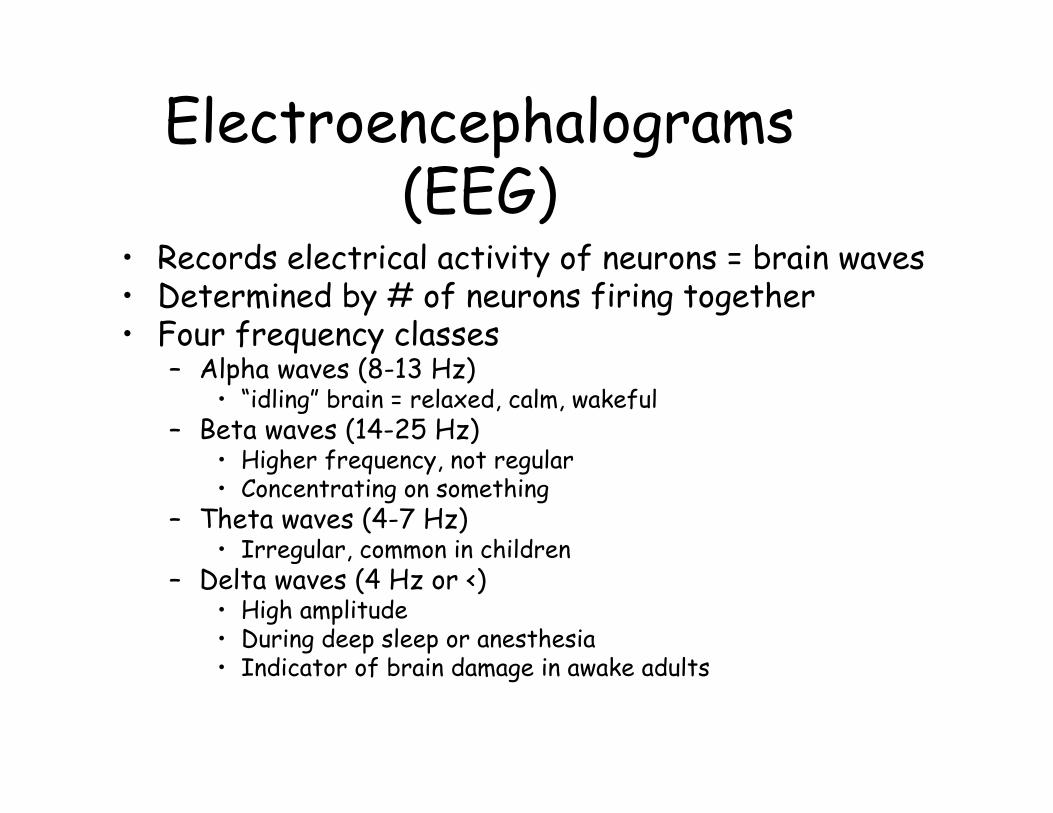
Electroencephalograms (EEG)
• Records electrical activity of neurons = brain waves• Determined by # of neurons firing together• Four frequency classes
– Alpha waves (8-13 Hz)• “idling” brain = relaxed, calm, wakeful
– Beta waves (14-25 Hz)• Higher frequency, not regular• Concentrating on something
– Theta waves (4-7 Hz)• Irregular, common in children
– Delta waves (4 Hz or <)• High amplitude• During deep sleep or anesthesia• Indicator of brain damage in awake adults

EEG
• Change with age, stimuli, brain disease
• Aids in diagnosis and localization of lesions, tumors, infarcts, epileptic lesions
• Absence of brain waves = brain death
Peripheral nervous system
• Sensory receptors• Somatic motor neurons• Autonomic motor neurons

Sensory (afferent) • Sense environmental stimuli and
transduce signal• Transmit information to CNS

Somatic (efferent)• Innervate skeletal
muscle fibers• One neuron
pathway• Nerve cell bodies in
CNS• Axons leave either
through ventral root or cranial nerve

Autonomic nervous system• Innervates organs not usually under
voluntary control• Two neuron pathways
– Pre-ganglionic neurons in CNS – Post-ganglionic neurons in PNS
• Sympathetic = fight or flight • Parasympathetic = rest or repose• Synapse on autonomic effectors
– Cardiac muscle– Smooth muscle– Glands

Characteristics of Autonomic Neurons
• Preganglionic autonomic fibers originate in midbrain, hindbrain, and upper thoracic to 4th sacral levels of the spinal cord.
• Autonomic ganglia are located in the head, neck, and abdomen.
• Presynaptic neuron is myelinated and postsynaptic neuron is unmyelinated.
• Autonomic nerves release NT that may be stimulatory or inhibitory.

Autonomic ganglia• Located in head, neck, abdomen• Sympathetic chain ganglia
CNS PNS
preganglionic postganglionic

Sympathetic division• Synapse close to the CNS & far away from
effector organ• Travel within spinal nerves• Mass activation due to convergence &
divergence• Sympathoadrenal system: converge on
adrenal medulla
preganglionicpostganglionic
AChNE

Sympathetic Division

Parasympathetic division
• Terminal ganglia -- close to effector organ• Fibers outside of spinal nerves (usually)• No stimulation to: cutaneous blood vessels,
blood vessels in skeletal muscle, sweat glands, arrector pili muscles
ACh
ACh
preganglionicpostganglionic

ParasympatheticDivision

Cranial nerves and Parasympathetic Division
• 4 of the 12 pairs of cranial nerves (III, VII, X, XI) contain preganglionic parasympathetic fibers.
• III, VII, XI synapse in ganglia located in the head.• X synapses in terminal ganglia located in widespread
regions of the body.• Vagus (X):
– Innervates heart, lungs esophagus, stomach, pancreas, liver, small intestine and upper half of the large intestine.
• Preganglionic fibers from the sacral level innervate the lower half of large intestine, the rectum, urinary and reproductive systems.

Functions of the ANS• Sympathetic = fight or flight
– Increased HR– Bronchiole dilation– Increase blood glucose, etc.
• Parasympathetic = rest and repose– Decrease HR– Incr digestion– Dilation visceral bv

Neurotransmitters• Adrenergic: release norepinephrine
(NE)– Adrenal medulla
• 85% epinephrine• 15% norepinephrine
• Cholinergic: release acetylcholine (ACh)
catecholamines

Adrenergic receptors• Beta adrenergic receptors:
– Produce their effects by stimulating production of cAMP.
– NE binds to receptor.– G-protein dissociates into α subunit or βγ−
complex.– Depending upon tissue, either α subunit or βγ−
complex produces the effects.• Alpha subunit activates adenylate cyclase, producing
cAMP.– cAMP activates protein kinase, opening ion channels.

Adrenergic receptors
• Alpha1 adrenergic receptors:– Produce their effects by the production of
Ca2+
– Epi binds to receptor.– Ca2+ binds to calmodulin– Calmodulin activates protein kinase, modifying
enzyme action• Alpha2 adrenergic receptors:
– Located on presynaptic terminal• Decreases release of NE
– Negative feedback control– Located on postsynaptic membrane
• When activated, produces vasoconstriction

Response to adrenergic stimulation
• Excitatory and inhibitory effects.• Responses due to different membrane
receptor proteins.1) α1 : constricts visceral smooth muscles.2) α2 : contraction of smooth muscle. 3) β1 : increases HR and force of contraction.4) β2 : relaxes bronchial smooth muscles.5) β3: adipose tissue, function unknown.

Cholinergic stimulation• All somatic motor neurons, all preganglionic
and most postganglionic parasympathetic neurons are cholinergic.– Release ACh as NT.– Somatic motor neurons and all preganglionic
autonomic neurons are excitatory.– Postganglionic axons, may be excitatory or
inhibitory.• Two types of receptors
– Muscarinic receptors– Nicotinic receptors