neonatal sepsis appropriate use of antibiotics. newborns are extremely susceptible to infection,...
TRANSCRIPT
Neonatal Sepsis
APPROPRIATE USEOF ANTIBIOTICS

Newborns are extremely susceptible to infection, and sepsis is a significant cause of morbidity and mortality in this population
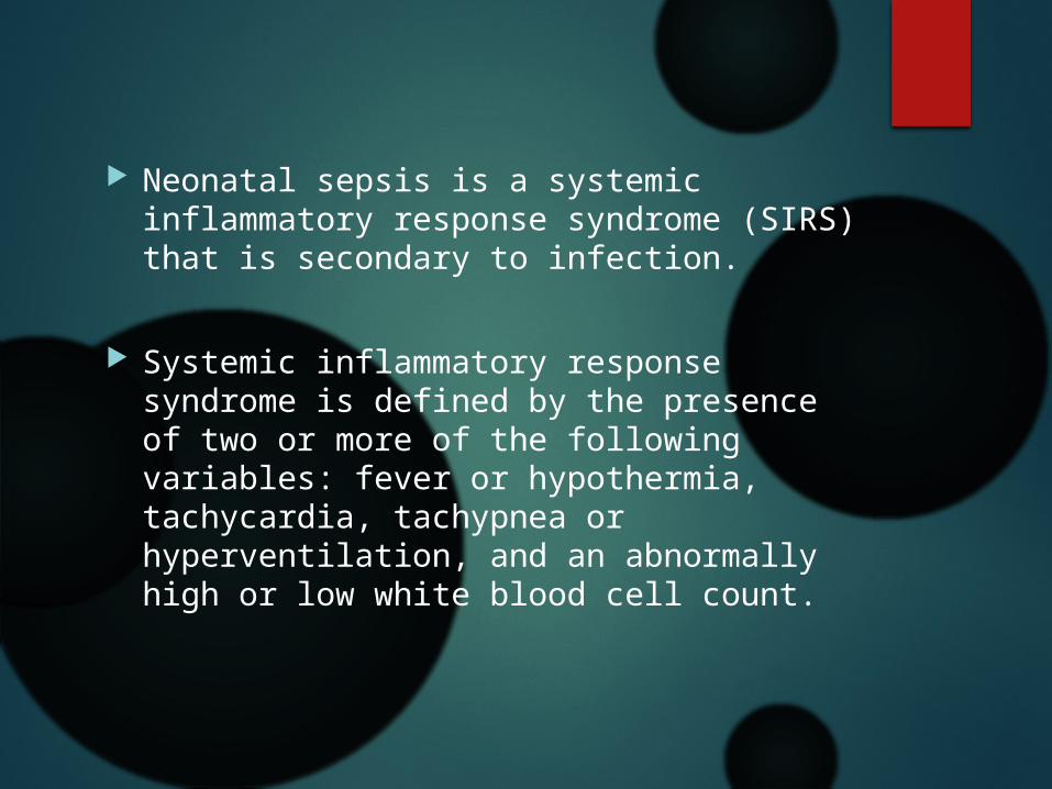
Neonatal sepsis is a systemic inflammatory response syndrome (SIRS) that is secondary to infection.
Systemic inflammatory response syndrome is defined by the presence of two or more of the following variables: fever or hypothermia, tachycardia, tachypnea or hyperventilation, and an abnormally high or low white blood cell count.

Neonatal sepsis is categorized according to the infant’s postnatal age at onset of disease.
early-onset sepsis occurring at or before 72 hours of life, and late onset occurring at greater than 72 hours to 7 days.

In early-onset sepsis,
vertical transmission through ascending amniotic fluid infection
acquisition of bacterial flora from the mother’s anogenital tract during vaginal delivery.
Some bacteria cross the placenta : Treponema pallidum and Listeria monocytogenes.
maternal anogenital colonization , most commonly responsible for early-onset sepsis: group B streptococci (GBS) and Gram-negative enteric bacilli.
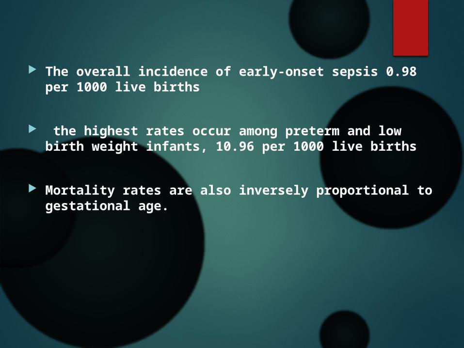
The overall incidence of early-onset sepsis 0.98 per 1000 live births
the highest rates occur among preterm and low birth weight infants, 10.96 per 1000 live births
Mortality rates are also inversely proportional to gestational age.
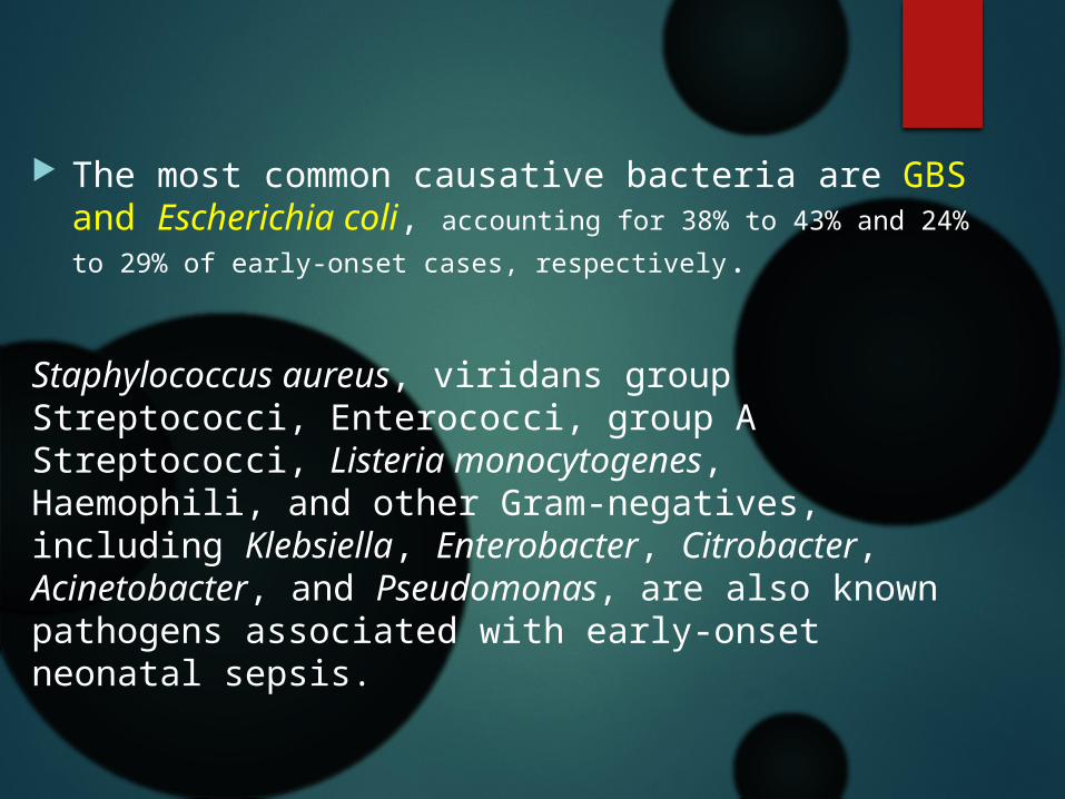
The most common causative bacteria are GBS and Escherichia coli, accounting for 38% to 43% and 24%
to 29% of early-onset cases, respectively.
Staphylococcus aureus, viridans group Streptococci, Enterococci, group A Streptococci, Listeria monocytogenes, Haemophili, and other Gram-negatives, including Klebsiella, Enterobacter, Citrobacter, Acinetobacter, and Pseudomonas, are also known pathogens associated with early-onset neonatal sepsis.

In late-onset sepsis, acquisition of infection is through the infant’s environment. including part of the flora of their caregivers.

Extreme prematurity is one of the greatest risk factors for late-onset
sepsis
long hospital stays and indwelling vascular catheters
improved surgical care and survival for neonates with congenital heart

Common causes of late-onset infection in very low birth weight and premature infants include coagulase-negative staphylococci and S. aureus, as well as invasive candidiasis, pseudomonas and other gram negative enterobacteriacea. Group B streptococcus and E. coli are also commonly implicated in late-onset sepsis.
Escherichia coli is frequently a cause of urosepsis in young infants.
Appropriate diagnosis is important for appropriate
antibiotics usage.
The clinical signs of sepsis in a neonate are often nonspecific. Detection requires that clinicians maintain a high index of suspicion.
Common presenting signs include respiratory distress, hemodynamic instability with poor perfusion or shock, and temperature instability.
Lethargy or poor feeding may be the only symptoms initially.
Metabolic changes may include hyperglycemia or hypoglycemia, acidosis, and jaundice.
Meningismus is uncommon in neonates
Full fontanelle, irritability, lethargy, and seizures may occur.

Because signs and symptoms are nonspecific, the clinical diagnosis of neonatal sepsis is extremely challenging.
A definitive diagnosis requires the isolation of a pathogen from a normally sterile body site, including blood, cerebrospinal fluid (CSF), and urine.

A blood culture (with a collection volume of 1 mL) should be drawn in any infant with suspected sepsis.
A lumbar puncture should also be considered in any infant with suspected sepsis.
There’s poor correlation between results of blood and CSF cultures
failure to perform a lumbar puncture may result in missed diagnoses.

Cerebrospinal fluid : based on the infant’s gestational age, postnatal age, and birth weight .
CSF WBC count of greater than 20 to 30 cells/μL is consistent with meningeal inflammation.
neonatal meningitis can occur with normal CSF parameters
some overlap in CSF values between neonates with and without meningitis

In suspected infants with NL CSF findings, repeat LP 24 to 48 hours later will show pleocytosis if true meningeal inflammation is present.
CSF Gram stain may be helpful,
a negative Gram stain does not exclude the diagnosis.
In traumatic LP, do not adjust the CSF WBC based on the red blood cell counts. treat presumptively for meningitis pending CSF culture results.

For early-onset sepsis, a urine culture is not required.
Suprapubic aspiration or sterile catheterization for urine cultures.

indirect markers of infection
none are definite
they can be used to help identify infected infants and guide decisions on duration of antimicrobial therapy.
WBC with differential
6 to 12 hours after birth are more likely to reflect an inflammatory response
both high and low WBC, low or high absolute neutrophil count, and high percentage of immature to total white blood cells.
flow cytometery for the future
C-reactive protein levels increase within 6 to 8 hours after infection and peak after 24 hours.
The sensitivity of CRP for neonatal sepsis is lowest early in infection, and sensitivity generally increases with serial values obtained 24 to 48 hours after onset of symptoms.
Serial determinations may be useful for R/O sepsis
or in monitoring response to treatment.

Procalcitonin peak 12 hours following infection. Increased levels also with noninfectious causes, such as respiratory
distress syndrome.
Procalcitonin appears to have better sensitivity but less specificity than cRP
Specific cytokines

the most important information guiding clinical
decisions continues to be the patient’s overall clinical status and culture data.

MANAGEMENT
APPROPRIATE USE OF ANTIBIOTICS

Ampicillin and an aminoglycoside are recommended as empiric therapy for early-onset sepsis.
coverage against GBS and E. coli, the most common causative pathogens, as well as Listeria monocytogenes.
Empiric use of third-generation cephalosporins is not recommended.
(because of concerns for development of resistance and the increased risk for invasive candidiasis)
if Gram negative meningitis is suspected, then it is recommended to add cefotaxime to the empiric regimen, given its excellent CNS penetration.
Cefotaxime is preferred over ceftriaxone
(because risk for kernicterus and its association with biliary sludging.)

Empiric therapy for late-onset sepsis usually consists of vancomycin and an aminoglycoside.
providing coverage for coagulase-negative Staphylococci, S. aureus, and Gram-negative organisms.
In a stable neonate & where MRSA is not prevalent start with cloxacillin and aminoglycoside ?!
consider empiric antifungal coverage for invasive
candidiasis. (GA<28 wk, CV line, ET, PLT<100000, carbapenems)

if Gram-negative meningitis is suspected, consider
adding a third-generation cephalosporin.
A fourth generation cephalosporin (cefepime) or
Carbapenems may be considered, depending on
local resistance patterns or if the patient had
previously received therapy with a third-generation
cephalosporin.
Recommended meningitic doses of meropenem may
be toxic at lower gestational ages and may produce
seizures.
Consider Acyclovir if signs of encephalitis

Once a pathogen is identified, therapy should be tailored to the species and antimicrobial susceptibilities.
Duration determined by : the site of infection and the patient’s clinical response.

Bacteremia without a focus of infection is usually treated for 7 to 10 days.
Early preterm infants (<32 weeks’ gestational age) may require slightly longer treatment courses of 10 to 14 days.
Gram-negative bacteremia tends to be treated with longer courses of 10 to 14 days.

uncomplicated GBS meningitis is treated for 14 to 21 days.
Longer courses are needed for other focal complications of GBS infection.
Aminoglycoside continues until CSF sterilization. (not
more than 2 weeks)
Gram-negative bacterial meningitis, treatment is for 21 days, or 2 weeks beyond
the first negative CSF culture, whichever is longer.
A lumbar puncture before discontinuation of
antibiotics to confirm response to treatment.
Neonatal pneumonia is also categorized into two patterns of disease according to timing and route of acquisition.
Empiric antibiotic treatment for early-onset pneumonia includes ampicillin and an aminoglycoside.
for late onset pneumonia it depend on local bacterial resistanc patterns in both the hospital and the community.
Vancomycin and an aminoglycoside are commonly used as empiric therapy to provide coverage against coagulase negative Staphylococci and
methicillin-resistant S.aureus (MRSA).
If Pseudomonas is suspected, an aminoglycoside
plus an anti-pseudomonal beta-lactam, such as
ceftazidime or piperacillin-tazobactam, should be
given.
Duration of therapy for uncomplicated pneumonia is 7
to10 days.

Urinary tract infection
Sterile urine culture
Any growth of a urinary pathogen from bladder aspirate is considered significant. Catheterization cultures with greater than 1000 colony-forming units per milliliter are considered meaningful.
Urinalysis lacks specificity and sensitivity for diagnosis of UTI in neonates.
Blood cultures one third will have bacteremia with
the same organism.
very low threshold for performing a lumbar
puncture the risk for concomitant meningitis is 1%
tact infectiono 2%.
Empiric antibiotic includes ampicillin and an aminoglycoside.
In hospitalized infants with late-onset infections vancomycin and an aminoglycoside to provide coverage for hospital associated infections with coagulase-negative Staphylococci and MRSA.
a urine culture may need to be repeated 2 to 3 days after start of treatment to confirm sterilization of the urine. If cultures remain positive further evaluated for a potential reservoir of infection.
The duration of therapy is generally 10 to 14 days for uncomplicated bacterial UTI. Neonates are usually given intravenous antibiotics for the entire course,
Although in older infants oral antibiotics after clinical improvement.

OSTEOMYELITIS AND SEPTIC ARTHRITIS
In neonates, the most common route for infection to the bone or joint is by hematogenous spread.
The most common causative organisms are S. aureus,E. coli, and GBS.
Less common pathogens include group A Streptococcus, N. gonorrhoeae, and other enteric Gram-negative bacilli.

Fluid collections in soft tissue or bone should be aspirated and sent for Gram stain and culture. Patients with joint involvement should have synovial fluid sent for Gram stain and culture.
Blood cultures should be obtained in all patients positive in 50% of cases.
Lumbar puncture should be performed on ill-appearing infants or those with positive blood cultures.

Recommended empiric antibiotic treatment includes vancomycin and an aminoglycoside
Therapy can be narrowed once the causative organism is isolated.
Joint infections and bony abscesses require surgical drainage.
The total duration of therapy is 4 to 6 weeks for uncomplicated cases.
Parenteral therapy at least until clinical improvement and normalization of the inflammatory markers .

when cultures are negative, It is often difficult to determine an appropriate duration of antibiotic therapy for suspected sepsis.
In well-appearing infants without clinical or hematologic evidence for infection, standard
practice is to discontinue antibiotics if cultures
have been negative after 48 hours.
More Challenging Management decisions are much more challenging for those
infants in whom sepsis is highly suspected but cultures are negative, which is often the case for preterm infants.
Infants whose mothers received antibiotics during labor may have false-negative blood cultures because of antibiotic suppression.
Cerebrospinal fluid culture data may be lacking in infants who are not clinically stable enough to tolerate a lumbar puncture.
Noninfectious conditions mimicking sepsis can also complicate the clinical picture.
in such cases, consider each patient’s clinical course.
Retrospective cohort studies : potential harm associated with longer duration of empiric antibiotics when cultures are negative, including increased risk for necrotizing enterocolitis and death among premature infants.

Thank you


Bacterial Infections
by Organ System

MENINGITIS
Bacterial meningitis is more common in the first month of life than at any other age.
Incidence of neonatal bacterial meningitis is estimated to be 0.25 per 1000 live births.
As with neonatal sepsis, neonatal meningitis can be categorized into two patterns of disease: early onset and late onset.
The causative pathogens for neonatal meningitis are similar to those for neonatal sepsis.
Group B streptococcus is the most common cause of neonatal meningitis.
Gram-negative enteric bacilli cause 30% to 40% of cases of neonatal meningitis, and E. coli accounts for approximately 50% of the Gram-negative isolates
important Gram-negative organisms include Klebsiella, Enterobacter, Citrobacter, and Serratia species.
A known complication of Citrobacter meningitis is brain abscesses, obtain brain imaging
Listeria monocytogenes ,relatively uncommon, association with thromboencephalitis.
Nosocomial pathogens include coagulase-negative Staphylococci, Candida, and resistant Gram-negative organisms, particularly Pseudomonas.

Cerebrospinal fluid : based on the infant’s gestational age, postnatal age, and birth weight .
CSF WBC count of greater than 20 to 30 cells/μL is consistent with meningeal inflammation.
neonatal meningitis can occur with normal CSF parameters
some overlap in CSF values between neonates with and without meningitis
In suspected infants with NL CSF findings, a repeat lumbar puncture obtained 24 to 48 hours later will show pleocytosis if true meningeal inflammation is present.
CSF Gram stain may be helpful,
a negative Gram stain does not exclude the diagnosis.
In traumatic LP, do not adjust the CSF WBC based on the red blood cell counts. treat presumptively for meningitis pending CSF culture results.

Empiric therapy for early-onset meningitis includes ampicillin and an aminoglycoside. If infection with a Gram-negative organism is suspected, the regimen should be expanded to include cefotaxime in addition to ampicillin and an aminoglycoside.
Aminoglycosides are not used as monotherapy because of their poor CNS penetration.
Once a pathogen is identified, therapy should be tailored according to the causative organism.

Gram-negative meningitis is treated with a thirdgeneration cephalosporin (cefotaxime) for at least 21 days, or for 14 days after the first negative CSF culture.
An aminoglycoside is added until CSF sterilization.
A fourthgeneration cephalosporin (cefepime) or a carbapenem
(meropenem) in combination with an aminoglycoside should be
considered for infection with members of the Enterobacteriaceae
family with inducible beta-lactamase resistance (e.g.,
Citrobacter, Enterobacter, Serratia) and for Pseudomonas.
recommended meningitic doses of meropenem may be toxic at
lower gestational ages and may produce seizures.
A lumbar puncture should be considered before discontinuation of
antibiotics to confirm response to treatment.
Listeria monocytogenes is not susceptible to cephalosporins and should be treated with ampicillin and an aminoglycoside until CSF sterilization, followed by ampicillin monotherapy for 14 days after first negative culture.
The same therapy is recommended for infection with Enterococcus.
Meningitis with coagulase-negative Staphylococci is seen in
preterm infants and is often associated with the presence of a
foreign body in the CNS. Most of these organisms are resistant to
penicillin, and treatment with vancomycin is often required.
Duration is generally 14 to 21 days after CSF sterilization, with
removal of any foreign body if possible.
There is no evidence that corticosteroids improve the outcome for
neonatal bacterial meningitis other than tuberculosis meningitis.
PNEUMONIA
Neonatal pneumonia is also categorized into two patterns of disease according to timing and route of acquisition.
Early-onset pneumonia is usually acquired within the first 3 days of life via vertical transmission, including aspiration of infected amniotic fluid and transplacental transmission.

Late-onset pneumonia occurs after the first week of life,
and infection arises from pathogenic organisms in the infant’s environment. The risk for late-onset pneumonia is highest among infants who require mechanical ventilation. Other risk factors include extreme prematurity, prolonged hospitalization, and previous bloodstream infection.

The most common causes of early-onset pneumonia are GBS, S. pneumoniae, nontypable H. influenzae, S. aureus, E. coli, Klebsiella, and atypical organisms. Ureaplasma urealyticum (development of chronic lung disease ).
Chlamydia trachomatis pneumonia can occur in the first week of life, typically between 2 and 4 weeks of age.
Syphilis, Listeria monocytogenes, and Mycobacterium tuberculosis (TB) are much less common

In late-onset pneumonia ,
Streptococcus pneumoniae a predominant causative pathogen
Other important pathogens include S. aureus, S. pyogenes, non-typable H. influenza, Gram-negative enteric organisms.
Staphylococcus aureus, Streptococci, Klebsiella pneumoniae,
Citrobacter, Enterobacter, Serratia, and Pseudomonas have
the potential to cause extensive lung injury, abscess formation,
empyema, pneumatoceles.
Bordetella pertussis infection in young infants can lead to respiratory failure and death.

Empiric antibiotic treatment for early-onset pneumonia
includes ampicillin and an aminoglycoside.
Empiric antibiotic therapy for late onset pneumonia depend on local bacterial resistanc patterns in both the hospital and the community.
Vancomycin and an aminoglycoside are commonly used
as empiric therapy to provide coverage against
coagulase negative Staphylococci and methicillin-resistant S.
aureus (MRSA).
If Pseudomonas is suspected, an aminoglycoside
plus an anti-pseudomonal beta-lactam, such as ceftazidime
or piperacillin-tazobactam, should be given.
Recommended duration of therapy for uncomplicated
pneumonia is 7 to 10 days.

URINARY TRACT INFECTIONS
The incidence of bacteriuria in term newborns is estimated to be 0.1% to 1%.1,9 The incidence is higher in preterm infants and is thought to be around 2%.
Infection of the urinary tract in neonates is thought to be acquired either through hematogenous spread or by ascending infection, which is often associated with anatomic abnormalities
Urinary tract abnormalities are seen
in approximately 20% to 50% of infants with UTI
The most common causative pathogen for neonatal UTI is E. coli, which has been isolated in approximately 80% of cases in some large series.
Other common causative organisms : Enterobacteriaceae, Klebsiella, Enterobacter, Proteus, Citrobacter, Salmonella, and Serratia.
Gram-positive organisms are isolated much less frequently than Gram-negative organisms, although Enterococci, S. aureus, and coagulase negative Staphylococci are also known to cause UTI in newborns.

Definitive diagnosis must be made by urine culture with specimens obtained by catheterization or suprapubic bladder aspiration.
Any growth of a urinary pathogen from bladder aspirate is considered significant. Catheterization cultures with greater than 1000 colony-forming units per milliliter are considered
meaningful.
Urinalysis lacks specificity and sensitivity for diagnosis of UTI in
neonates.
Blood cultures should be obtaine for all infants with suspected
UTI, as one third with UTI will have bacteremia with the same
organism.
Clinicians should have a very low threshold for performing a
lumbar puncture in neonates with UTI because the risk
for concomitant meningitis is 1% to 2%.

Empiric antibiotic treatment for neonatal UTI includes ampicillin and an aminoglycoside. Hospitalized infants with late-onset infections should be given vancomycin and an aminoglycoside to provide coverage for hospital associated infections with coagulase-negative Staphylococci and MRSA. If the infection is caused by a highly resistant pathogen, or if there is a known anatomical abnormality,
a urine culture may need to be repeated 2 to 3 days after start of treatment to confirm sterilization of the urine.
If cultures remain positive despite adequate therapy, the infant should be further evaluated for a potential reservoir of infection.
The duration of therapy is generally 10 to 14 days for uncomplicated bacterial UTI. Neonates are usually given intravenous antibiotics for the entire course, although older infants are often switched to oral antibiotics after demonstration of clinical improvement.

All neonates with UTI should undergo radiographic evaluation because of the high prevalence of urinary tract abnormalities. If prenatal ultrasound data are not available, a renal ultrasound should be performed after the infant has clinically stabilized. Ultrasound can detect structural abnormalities, although it cannot detect vesicoureteral reflux or renal scarring. Voiding cystourethrogram is performed to detect vesicoureteral reflux, and this is usually done 3 to 6 weeks after completion of antibiotic treatment.
If renal damage is suggested by ultrasound,
renal cortical scintigraphy can be performed .

OSTEOMYELITIS AND SEPTIC ARTHRITIS
In neonates, the most common route for infection to the bone or joint is by hematogenous spread.
The most common causative organisms are S. aureus,E. coli, and GBS.
Less common pathogens include group A Streptococcus, N. gonorrhoeae, and other enteric Gram-negative bacilli.

Imaging studies should be obtained for any patient with suspected osteomyelitis or septic arthritis. The radiologic investigation usually starts with plain radiographs.
Soft tissue swelling is the earliest finding 48 hours after onset of infection.
Bony changes, 7 to 10 days after onset of infection. further imaging is often required for patients in whom there is a high index of suspicion.
Ultrasound can detect subperiosteal collections and joint effusions, so it is very useful in cases of suspected septic arthritis.
Ultrasound can also detect findings of acute osteomyelitis, although this is highly dependent on the duration of infection as well as operator experience.
Bone scintigraphy is more sensitive than plain films for detecting osteomyelitis early on, and it is useful for detecting multiple foci of infection.

Magnetic resonance imaging (MRI) is extremely useful in the evaluation for osteomyelitis. Bony changes can be detected within 24 to 48 hours after onset of symptoms.
it provides excellent anatomic detail. It also provides better information on growth plate involvement than does bone scintigraphy.

Fluid collections in soft tissue or bone should be aspirated and sent for Gram stain and culture. Patients with joint involvement should have synovial fluid sent for Gram stain and culture. Blood cultures should be obtained in all patients positive in 50% of cases.
Lumbar puncture should be performed on ill-appearing infants or those with positive blood cultures.

Recommended empiric antibiotic treatment includes vancomycin for broad Gram-positive coverage and an aminoglycoside or third-generation cephalosporin for Gram-negative coverage.
Therapy can be narrowed once the causative organism is isolated.

Joint infections and bony abscesses require surgical drainage.
The total duration of therapy is 4 to 6 weeks for uncomplicated cases.
Parenteral therapy should be continued at least until the patient demonstrates clinical improvement and the inflammatory markers have normalized.
OPHTHALMIA NEONATORUM
Ophthalmia neonatorum is conjunctivitis that occurs in the first month of life.
Bacterial causes include S. aureus, nontypable H. influenzae, S. pneumoniae, enteric Gramnegative bacilli, GBS, N gonorrhoeae, and Chlamydia trachomatis.
The differential also includes viral causes, particularly herpes simplex virus, and non-infectious causes such as chemical conjunctivitis.
Conjunctivitis in neonates is managed aggressively because of the high risk for associated systemic illness and complications.
Prophylactic administration of ophthalmic antibiotic agents shortly after birth greatly reduces the risk of gonococcal conjunctivitis, but not chlamydial disease.
Regimens with equal efficacy include 0.5% erythromycin ointment, 1% tetracycline ointment, and silver nitrate solution.
If gonococcal infection is suspected, CSF and blood cultures
should be obtained
Purulent ocular discharge should be sent for Gram stain and culture. Culture on selective media (e.g., Thayer-Martin) is needed to evaluate for gonococcal infection.
Additionally, conjunctival specimens obtained from an everted eyelid should be sent for C. trachomatis testing. Specimens must contain conjunctival epithelial cells; ocular exudates are not adequate specimens for testing.
Culture of the organism is the gold standard for diagnosis of C. trachomatis infection in infants. Nucleic acid amplification tests may also be considered because they have been found to have high specificity and sensitivity
In infants with signs of pneumonia, testing for C. trachomatis can
be sent from nasopharyngeal specimens.
Any infant with signs of systemic illness should have blood and CSF cultures obtained.

Systemic antibiotics are required to treat conjunctivitis
resulting from N. gonorrhoeae and C. trachomatis. For
other causes of bacterial conjunctivitis, topical antibiotic
ointment or solution given for 7 to 10 days provides
adequate therapy. Infants with gonococcal eye disease
should be hospitalized and monitored for response
to treatment as well as for signs of disseminated
disease. Recommended treatment for conjunctivitis is a
single dose of intramuscular or intravenous ceftriaxone
(25–50 mg/kg). The eyes should also be irrigated frequently
with sterile saline until the discharge has resolved.
Recommended treatment for either chlamydial conjunctivitis
or pneumonia is oral erythromycin (50 mg/kg per
day in four divided doses) for 14 days.

OMPHALITIS Bacteria can reach the bloodstream through patent vessels
of the newly cut cord and lead to systemic infection and severe complications.
Common pathogens include S. aureus, group A Streptococci, GBS, and Gramnegative bacilli, including E. coli, Klebsiella, and Pseudomonas.
Polymicrobial infections may occur.
Anaerobic bacteria can contribute to infection, especially in infants born to mothers with chorioamnionitis.

The clinical presentation is characterized by purulent
drainage from the umbilical stump, with surrounding erythema, induration, and tenderness.
Foul smelling drainage is suggestive of infection with anaerobic bacteria.
Infants with more severe infection will display systemic signs of illness.
Involvement of the abdominal wall or extensive edema should prompt consideration for necrotizing fasciitis as a complication. Other complications include peritonitis, intra-abdominal abscess, suppurative thrombophlebitis of portal or umbilical veins.

Purulent discharge for Gram stain and culture.
Blood and CSF cultures should be obtained from infants with signs of systemic illness.
Treatment an anti-staphylococcal penicillin and an aminoglycoside.
If the prevalence of methicillin-resistant S. aureus is high, vancomycin should be used.
The addition of clindamycin or metronidazole for anaerobic involvement, especially for foul smelling discharge or history of maternal chorioamnionitis.
For uncomplicated cases, duration of treatment is typically 10 days. A switch to oral therapy for completion of the course of treatment may be considered.