neonatal jaundice: etiologies and how and when to …/media/files/medical professionals... ·...
TRANSCRIPT

© Children’s Specialty Group. All rights reserved.
Neonatal Jaundice: Etiologies and
how and when to evaluate
© Children’s Specialty Group. All rights reserved.
10:30 – 11:15 a.m.
Bernadette Vitola, MD

© Children’s Specialty Group. All rights reserved.
Disclosures
I have no relevant financial
relationships to disclose.

© Children’s Specialty Group. All rights reserved.
Objectives
• Describe neonatal jaundice
• Identify clinical findings
• Review the etiologies of unconjugated and
conjugated hyperbilirubinemia
• Understand important tests needed to confirm
diagnosis to determine treatment

© Children’s Specialty Group. All rights reserved.
Types of Jaundice
• Neonatal jaundice can be caused by
either unconjugated or conjugated
hyperbilirubinemia
• Most causes of unconjugated
hyperbilirubinemia are relatively benign
but can frequently require phototherapy
• Conjugated hyperbilirubinemia is NEVER
normal and should always be
investigated

© Children’s Specialty Group. All rights reserved.
Physical Findings
• There are no physical findings that can differentiate between unconjugated and conjugated hyperbilirubinemia
• Jaundice starts in the face/eyes and progresses caudally, but visual assessment of degree of jaundice is often inaccurate
• Jaundice may be difficult to identify in darker-skinned infants. Scleral icterus is helpful.
• Signs/symptoms of dehydration and/or sepsis

© Children’s Specialty Group. All rights reserved.
When To Evaluate
• Jaundice in 1st 24 hours, T bili >5 mg/dl
• Jaundice that seems excessive for
infant’s age
• Jaundice beyond 2 weeks in formula-
fed infant and 3 weeks in breast-fed
infant

© Children’s Specialty Group. All rights reserved.
Unconjugated Hyperbilirubinemia

© Children’s Specialty Group. All rights reserved.
Risk Factors for Severe
Unconjugated Hyperbilirubinemia
• Prematurity
• Breastfeeding
• Poor feeding
• Hemolysis
– ABO incompatibility, cephalohematoma, spherocytosis, G6PD
• Sepsis
• Asphyxia
• Gilbert syndrome
• Sibling required phototherapy

© Children’s Specialty Group. All rights reserved.
Treatment
• Use nomograms to determine when to
treat with phototherapy or exchange
transfusion
• If total bilirubin >25 mg/dl at any age,
medical emergency requiring
admission for phototx +/- exchange
transfusion
• Consider IVIG if isoimmune hemolysis
© Children’s Specialty Group. All rights reserved.
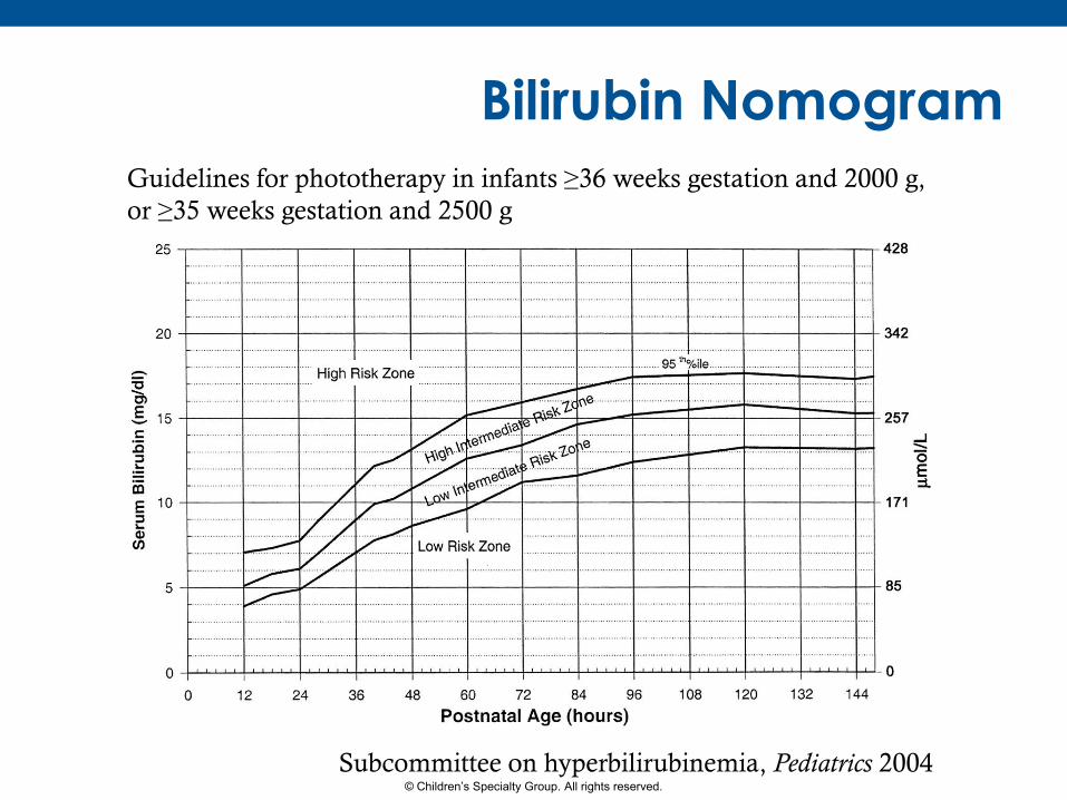
Bilirubin Nomogram
Guidelines for phototherapy in infants ≥36 weeks gestation and 2000 g,
or ≥35 weeks gestation and 2500 g
Subcommittee on hyperbilirubinemia, Pediatrics 2004

© Children’s Specialty Group. All rights reserved.
Bilirubin Nomogram Guidelines for exchange transfusion in ≥35 weeks gestation
Subcommittee on hyperbilirubinemia, Pediatrics 2004

© Children’s Specialty Group. All rights reserved.
Case Study
• A 4 week old full-term healthy breastfeeding male presents with persistent jaundice since shortly after birth.
• He is eating and growing well and the parents report no issues. His stool is yellow per parents report.
• A fractionated bilirubin shows a total bilirubin of 8.2 mg/dL, unconjugated bilirubin 3.5 mg/dL, conjugated bilirubin 4.1 mg/dL.
• What next?

© Children’s Specialty Group. All rights reserved.
Conjugated Hyperbilirubinemia

© Children’s Specialty Group. All rights reserved.
Conjugated
Hyperbilirubinemia
• Cholestasis is defined as reduced bile flow and
abnormal accumulation of conjugated, or direct,
bilirubin, indicating impaired hepatobiliary function
• Conjugated hyperbilirubinemia is defined as >20% of
total bilirubin
• Occurs in 1:2500 live births
• NEVER a normal finding and needs evaluation
• Is NOT caused by breast milk jaundice
• Early referral to Pediatric Gastroenterology
recommended since certain diseases require timely
intervention
© Children’s Specialty Group. All rights reserved.
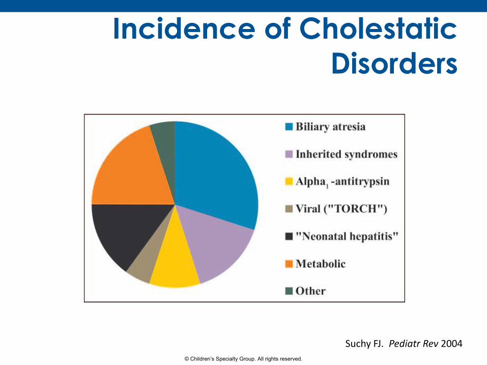
Incidence of Cholestatic
Disorders
Suchy FJ. Pediatr Rev 2004

© Children’s Specialty Group. All rights reserved.
Etiologies
• Infectious – Sepsis, UTI, CMV, E. Coli sepsis assoc w/galactosemia
• Anatomic – Biliary atresia, choledochal cyst, inspissated bile/sludge
• Metabolic – Galactosemia, tyrosinemia, fatty acid oxidation
disorder, congenital disorder of glycosylation, mitochondrial disorders
• Endocrine – Hypothyroidism, panhypopituitarism

© Children’s Specialty Group. All rights reserved.
Etiologies
• Genetic – Alagille syndrome, cystic fibrosis, alpha-1 antitrypsin
deficiency, progressive familial intrahepatic cholestasis, congenital hepatic fibrosis
• Toxins – Medications, TPN
• Vascular – Heart failure
– Shock
– Budd Chiari (occlusion of hepatic veins)

© Children’s Specialty Group. All rights reserved.
Clinical Presentation
• Aside from jaundice, clinical presentation of the cholestatic infant can vary widely depending on the etiology.
• Infants with cholestasis due to infection or a metabolic disease, such as galactosemia or tyrosinemia, are often ill-appearing.
• Infants with biliary atresia are often healthy-appearing with no apparent symptoms until later in the disease course. However, can be associated with situs inversus, malrotation, polysplenia, asplenia.

© Children’s Specialty Group. All rights reserved.
Clinical Presentation
• Congenital TORCH infection often associated
with LBW, microcephaly, purpura, chorioretinitis,
intracranial calcifications
Baselga et al. J Am Acad Dermatol, 1997

© Children’s Specialty Group. All rights reserved.
Clinical Presentation
• Cholestasis as a result
of a genetic mutation
may have associated
physical findings,
such as a heart
murmur, vertebral
anomalies, typical
facial features and
eye findings in
Alagille syndrome.
http://greatnonprofits.org/organizations/gallery/alagille-syndrome-alliance

© Children’s Specialty Group. All rights reserved.
Acholic Stool
• The triad of jaundice,
acholic stools and
dark urine should
alert the clinician to
the possibility of
cholestasis, especially
biliary atresia.

© Children’s Specialty Group. All rights reserved.
Case Study
• A 4 week old full-term healthy breastfeeding male presents with persistent jaundice since shortly after birth.
• He is eating and growing well and the parents report no issues. His stool is yellow per parents report.
• A fractionated bilirubin shows a total bilirubin of 8.2 mg/dL, unconjugated bilirubin 3.5 mg/dL, conjugated bilirubin 4.1 mg/dL.
• What next?

© Children’s Specialty Group. All rights reserved.
Stool Color Card

© Children’s Specialty Group. All rights reserved.
Initial Evaluation
• Look for signs of sepsis/acute illness
• Assess liver synthetic function
– PT/INR
– Albumin
– Glucose
• Look for other evidence of cholestasis
– Alkaline phosphatase
– γ- glutamyl transferase (GGT)

© Children’s Specialty Group. All rights reserved.
Diagnosis
• Anatomic – Abd US to evaluate for choledochal cyst,
sludge, inspissated bile
• Endocrine – TSH, free T4, cortisol
• Infectious – UA, urine culture, septic workup if appears
ill or febrile, urine CMV, consider adenovirus, enterovirus, TORCH workup

© Children’s Specialty Group. All rights reserved.
Diagnosis
• Metabolic – Review newborn screen
– Serum amino acids (organic and amino acidemias), urine
organic acids (tyrosinemia), acylcarnitine profile and free
carnitine (fatty acid oxidation disorders), ammonia (urea
cycle defects), lactate/pyruvate (mitochondrial)
• Genetic – Sweat test
– Alpha-1 antitrypsin phenotyping
– Genetic mutations (JAG1, FIC1, BSEP, MDR3)
– Ophtho exam, thoracic films, echo

© Children’s Specialty Group. All rights reserved.
Background of Biliary Atresia
• Incidence 1/4,000-1/20,000 live births
• An obliterative cholangiopathy of unknown etiology
• Universally fatal by 1-2 years of life if untreated
• Primary treatment is excision of atretic biliary tree and Kasai portoenterostomy
• Supportive care with optimizing nutrition, vitamin supplementation, portal hypertension management, and other sequelae of chronic liver disease

© Children’s Specialty Group. All rights reserved.
Types of Biliary Atresia

© Children’s Specialty Group. All rights reserved.
Kasai Portoenterostomy

© Children’s Specialty Group. All rights reserved.
Effects of Age at Kasai
Chardot C, et al. J Hepatol, 2013.

© Children’s Specialty Group. All rights reserved.
Effects of Age at Kasai
Chardot C, et al. J Hepatol, 2013.

© Children’s Specialty Group. All rights reserved.
Effect of Race on Diagnosis Kasai ≤ 60 DOL Kasai > 60 DOL
Mean age at Kasai (d) 46.9
(95% CI 43.5-50.4)
82.6
(95% CI 76.6-88.7)
Gender
Male
Female
N (%)
170 (55.6)
136 (44.4)
N (%)
158 (39.5)
242 (60.5)
Race
Caucasian
African American
Hispanic
Asian/Pacific Islander
Other
Missing information
179 (57.8)
16 (5.2)
33 (10.8)
56 (18.3)
16 (5.8)
6 (2.1)
182 (45.6)
82 (20.5)*
53 (13.1)
38 (9.6)
31 (7.6)
14 (3.6)
Payor
Medicaid
Private Insurance
Other
143 (46.7)
147 (48.2)
16 (5.1)
165 (48.8)
159 (46.8)
15 (4.4)
*P<0.03 Vitola, et al. NASPGHAN,2015

© Children’s Specialty Group. All rights reserved.
Measuring Fractionated Bilirubin
Harpavat S, et al. Pediatrics, 2011.
© Children’s Specialty Group. All rights reserved.
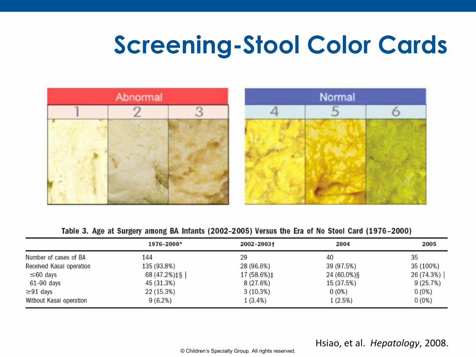
Screening-Stool Color Cards
Hsiao, et al. Hepatology, 2008.

© Children’s Specialty Group. All rights reserved.
Stool Color Card and Survival
Lien TH, et al. Hepatology, 2011.

© Children’s Specialty Group. All rights reserved.
Take Home Points
• Conjugated or direct bilirubin should be obtained in
ANY infant more than 3 wks of age with jaundice.
Don’t forget to check the sclera and the stool!
• Conjugated hyperbilirubinemia is NEVER normal
even if less than 20% of the total.
• PT/INR and glucose should be sent on any infant
with conjugated hyperbilirubinemia to assess liver
function.
• Early referral to a Pediatric Gastroenterologist is
imperative to ensure timely assessment and to
establish a diagnosis, but…
• YOUR early assessment is crucial for the best
outcomes for these babies.

© Children’s Specialty Group. All rights reserved.
Questions?

© Children’s Specialty Group. All rights reserved.
References
• American Academy of Pediatrics Subcommittee on
Hyperbilirubinemia. Management of Hyperbilirubinemia in the
Newborn Infant 35 or More Weeks of Gestation. Pediatrics 2004;114;294-316.
• Suchy FJ. Neonatal cholestasis. Pediatr Rev 2004;25;388-395.
• Moyer V, Freese DK, Whitington PF, et al. Guideline for the
evaluation of cholestatic jaundice in infants:
recommendations of the North American Society for Pediatric
Gastroenterology, Hepatology and Nutrition. J Pediatr
Gastroenterol Nutr 2004;39(2):115-28.
• Suchy FJ. Approach to the infant with cholestasis. In: Suchy FJ,
Sokol RJ, Balistreri WF, editors. Liver disease in children, 3rd ed. New York, NY: Cambridge University Press; 2007. p. 179-89.
• Benchimol EI, Walsh CM, Ling SC. Early diagnosis of neonatal
cholestatic jaundice. Can Fam Physician 2009;55:1184-92.

© Children’s Specialty Group. All rights reserved.
References
• Chardot C, Buet C, Serinet MO, Golmard JL, Lachaux A, Roquelaure B, Gottrand F, Broue P, Dabadie A, Gauthier F, Jacquemin E. Improving outcomes of biliary atresia: French national series 1986-2009. J Hepatol. 2013;58:1209-17.
• Harpavat S, Finegold MJ, Karpen SJ. Patients with biliary atresia have elevated direct/conjugated bilirubin levels shortly after birth. Pediatrics. 2001;128(6):1428-33.
• Hsiao CH, Cheng MH, Chen HL, Lee HC, et al. Universal screening for biliary atresia using an infant stool color card in Taiwan. Hepatology. 2008;47:1233-40.
• Lien TH, Chang MH, Wu JF, Chen HL, et al. Effects of the infant stool color card screening program on 5-year outcome of biliary atresia in Taiwan. Hepatology. 2011;53:202-08.
• Matta S, Sood M, Lerret S, Yan K, Vitola B. The role of race and payor on age at Kasai portoenterostomies for biliary atresia in the United States: A study from a national inpatient database 2007-2001. NASPGHAN 2015.

© Children’s Specialty Group. All rights reserved.
Contact Information
Bernadette E. Vitola, MD, MPH (414) 266-4829
Physician Consultation and Referral: (800) 266-0366