nema a. mohamed1, awatef m. ali1, shnoudy a. bakhoum2 heba
TRANSCRIPT
Egyptian Journal of Aquatic Biology & Fisheries
Zoology Department, Faculty of Science,
Ain Shams University, Cairo, Egypt.
ISSN 1110 – 6131
Vol. 23(2): 165 – 182 (2019)
www.ejabf.journals.ekb.eg
Monitoring of Oxidative Stress Biomarkers and Toxicity of Lead and Mercury
in Catfish of Lake Mariout, Egypt: The Role of Meso -2, 3-Dimercaptosuccinic
Acid (DMSA)
Nema A. Mohamed1, Awatef M. Ali
1, Shnoudy A. Bakhoum
2,
Heba H. Abdel-Kader2*
and Mohamed A. Ahmed
2
1- Department of Zoology, Faculty of Science, Alexandria University, Egypt.
2- Fish Physiology Lab., National Institute of Oceanography and Fisheries, Egypt. *Corresponding author: [email protected]
ARTICLE INFO ABSTRACT Article History:
Received: Feb. 19, 2019
Accepted: march 30, 2019
Online: April 2019
_______________
Keywords:
Monitoring
oxidative stress
Lead
Mercury
toxicity
Catfish
Lake Mariout
The present investigation was designed to evaluate the dangerous
effects of the Pb and Hg on Clarias gariepinus inhabiting Lake Mariout and
assay the possible protective effect of meso-2, 3 dimercaptosuccinic acid
(DMSA). The test fish were divided into four groups kept in plastic tanks (20
fish each) containing 30L of Lake Mariout water. Group (1): Standard group
(zero time); Group (2): Unexposed DMSA group, Group (3): Low dose
DMSA group; Group (4): High dose DMSA group. Fish exposed to DMSA
showed significant decrease in lead (P≤0.05) and mercury concentrations
(P≤0.05) in kidney, liver, gills, muscle and blood than unexposed group. The
present results clearly indicate a significant decrease in RBCs, Hb, Hct, and
platelet counts while a significant increase in MCH, MCHC, and WBCs in
the Clarias gariepinus collected from the main basin of Lake Mariout. A
marked significant decrease in AST, ALT, urea, creatinine (P≤0.05) was
observed in DMSA groups. A significant increase in CAT, GPX and SOD
was observed after exposure to DMSA. In addition, DMSA exposure
improved the histopathological alterations in fish liver and kidney.
INTRODUCTION
Egypt and the Middle East region are facing water pollution, which is one of the
environmental and public health problems. Pollution is generally associated with
industries waste that is one of the river Nile ecological problems (El-Sheikh et al.,
2010).
Heavy metals are the most common pollutants in the coastal area (Krishna et
al., 2013). Lead (Pb) and mercury (Hg) are toxic at even low concentrations
(Pugazhvendan et al., 2012). Excessive exposure to lead may cause chronic
nephropathy, hypertension, pathological changes and also reproductive impairments
(Falayi and Amatosero, 2014). Mercury Hg is a wide spread metal pollutant of high
toxicity to aquatic animals including fishes. Oxidative stress, inflammation,
thrombosis and mitochondrial dysfunction are the most side effects of mercury
(Begam and Sengupta, 2014).
Lake Mariout is the major source of pollution in the Mediterranean Sea through
El-Max Bay in South Alexandria. The Lake Mariout receives polluted water from
Nema A. Mohamed et al. 166
many sources, including industrial and domestic effluents. The total discharge of
primary treated sewage is about 916,000 m3/day (El-Bestawy et al., 2014).
African catfish (Clarias gariepinus) has a great commercial importance
because it is considered as one of the healthiest and cheapest fish as a source of
protein and sufficient omega fatty acids (Ebrahimi and Taherianfard, 2011).
Generally, fish is not only used for human consumption, but also used as a good
source of animal meal (Emam and Badia, 2014). Consumption of fish from Lake
Mariout is considered a health problem. Amr et al. (2005) stated that the
concentrations of heavy metals in fish samples from the Main Basin were higher than
those in water samples. It can therefore be a good model to study responses to
various environmental pollutants (Damian et al., 2014).
Metals are well known inducers of oxidative damage in fishes reflecting metal
contamination of the aquatic ecosystem (Kumar and Nandan, 2014). Antioxidant is
an agent that can neutralize the oxidant effect of free radicals, including some natural
and other substances (Mahmoud et al., 2013). Meso-2, 3-dimercaptosuccinic acid
(DMSA) is a dithiol compound, has two-SH groups (Flora et al., 2011). It was 40
years ago, DMSA was known as an effective antidote to heavy metal poisoning in
human.
To avoid the aquatic pollution there is need to use new strategies and
technologies for detoxifying the river water to improve the health of its fish, and, in
turn, that of the humans who consume them. Thus, the objective of the present study
was to evaluate the possible protective effects of DMSA against the lead and
mercury toxicity on blood, liver and kidney and the oxidant status of Clarias
gariepinus which accompanied by physiological and histopathological studies.
MATERIALS AND METHODS
Chemical
The antidote dimercaptosuccinic acid (DMSA, Succimer), a white powder with
molecular weight 182.21. DMSA, was obtained from the Acros Organics Company,
USA.
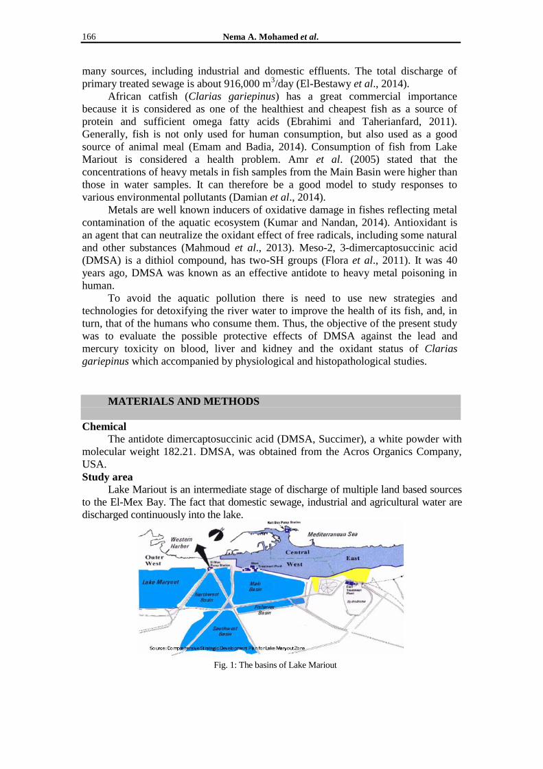
Study area
Lake Mariout is an intermediate stage of discharge of multiple land based sources
to the El-Mex Bay. The fact that domestic sewage, industrial and agricultural water are
discharged continuously into the lake.
Fig. 1: The basins of Lake Mariout

Monitoring of oxidative stress biomarkers and toxicity of lead and mercury in catfish 167
Experimental fish
Clarias gariepinus (Catfish) (115±5.0 g, 20 to 23 cm) were caught from the main
basin of Lake Mariout. In the laboratory, fish were placed in 30L aquaria (Twenty
fish/aquarium) of Lake Mariout water, which was renewed weekly. Aquaria were
maintained within an appropriate range of temperature (22°C–25°C) and pH (8.0–
8.8). Four recirculating aquarium systems the bio-filters and aeration were used. Fish
were acclimated to the laboratory conditions for 15 days. For the entire duration of
the experiment (60 days), the fish were fed with commercial fish feed, a rate of 3%
body weight daily. All animal procedures and the experimental protocols were carried
out according to the guidelines of the National Institutes of Health (NIH).
Experimental design:
In the laboratory, fish were divided into 4 groups (20 each) as follows: Group I: Standard group (Zero time): fish of this group were sacrificed on the same day of
collection. Group II: Unexposed DMSA group: fish of this group stayed without treatment.
Group III: Low dose DMSA group: fish of this group treated with 5 mg/kg BW/L of
DMSA.
Group VI: High dose DMSA group: fish of this group treated with 18 mg/kg BW/L of
DMSA.
The experiment continued for 60 days. The low and high doses were selected
according to Palaniappan and Vijayasundaram (2009).
Water analysis
Lead levels in water samples were estimated by using Air/Acetylene flame
atomic absorption spectrophotometer (UNICAM 696 AA Spectrometer). Mercury
levels in water samples were estimated by using Flameless atomic absorption
spectrophotometer equipped with (MHS) mercury hybrid system "Cold Vapor
Technique".
Bioaccumulation analysis
Lead and mercury concentrations in tissue were determined by using the
methods of Al-Balawi et al. (2013) and Diaz-Ravina et al. (1994), respectively. Lead
(Selander and Cramer, 1968) and mercury (Kanno et al., 1985) concentrations in
blood were also determined.
Determination of transfer factor (TF):
Transfer factor is the ratio between the accumulated concentration of a given
pollutant in any organ and its dissolved concentration in water. It is calculated by
dividing the concentrations of a heavy metal in fish organ by the heavy metal found
in water (Olabanji and Oluyemi, 2014).
Transfer ratio (TF) =
Preparation of blood and serum
The blood samples were collected by caudal vein into clean, dry, sterile
container containing EDTA and free anti-coagulated container. The anti-coagulated
blood sample was used for the determination of the hematological parameters. Free
anti-coagulated blood samples were used for the serum preparation. They were
centrifuged at 3000 rpm for 15 minutes and the serum was collected in capped sterile
tubes and used for biochemical measurements.
Hematological studies
Hematological parameters include erythrocyte count (RBC), hemoglobin
concentration (Hb), hematocrit value (Hct) platelet counts and the blood indices
Metal concentration in fish organ
Metal concentration in water

Nema A. Mohamed et al. 168
(MCV, MCH, MCHC) were calculated. Furthermore, the total number of leukocytes
and a differential white blood cell count were counted (Dacie and Lewis, 1975).
Biochemical analysis
The levels of aspartate aminotransferase (AST) (E.C.2.6.1.2.) and alanine
aminotransferase (ALT) (Reitmena and Frankel., 1957) were determined. Urea and
creatinine were estimated according to the methods of Henry (1974) and Tietz
(1995), respectively. Catalase (CAT) (EC 1.11.1.6), superoxide dismutase (SOD)
(EC 1.15.1.1) and glutathione peroxidase (GPX) (EC 1.11.1.9) were estimated in
liver according to the methods of Abei (1984), Nishikimi et al. (1972) and Paglia and
Valentine (1967), respectively.
Light and electron microscope examination
Tissue specimens of the liver and kidney were removed, fixed in 10% formalin
solution and routinely processed for paraffin embedding. Sections were cut at 3 mm
and stained with Haematoxylin and Eosin (H&E) according to Bancroft and Gamble
(2002). Liver and kidney samples were fixed by immersion in 2.5% glutaraldehyde in
phosphate buffer solution (pH 7.2) at 4oC. The samples were post-fixed in 2%
osmium tetroxide for two hours at 4oC. They were rinsed again in the buffer,
dehydrated in a graded ethanol series, and embedded in Epon-Araldite mixture.
Ultrathin sections were cut and stained with lead citrate and uranyl acetate according
to Reynolds (1963). The specimens were viewed in Joel 100 CX TEM.
Statistical analysis Data were tested using one-way ANOVA followed by Tukey honest-significant
difference test (TSD) for the comparison between different treatments, when F-values
were significant. All collected data in the present study were subjected to statistical
analysis by using SPSS version 22 software.
RESULTS
The effect of DMSA exposure on the water lead and mercury concentrations
The heavy metal residues in Lake Mariout water exposed to DMSA showed a
significant (p≤0.05) decrease in lead and mercury concentrations as compared to the
unexposed group and standard group (Table 1). Tukey honest-significant difference
test (TSD) revealed that the difference in the concentration of lead was significantly
different in the two doses of DMSA. While mercury concentration was statistically
insignificant (p≤0.05) between the low and high dose DMSA groups (Table 1).
Table 1: Lead and mercury concentrations (mg/l) in water of Lake Maryout before and after DMSA
exposure.
Standard group
(Zero time)
Unexposed
DMSA group
(60 days)
Low dose
DMSA group
(60 days)
High dose
DMSA group
(60 days)
Water
(mg/l)
Lead Mercury Lead Mercury Lead Mercury Lead Mercury
5.25
± 0.92a
1.60
± 0.43a
4.60
± 0.14 b
1.90
± 0.28 b
2.25 ±
0.21 c
0.59 ±
0.18 c
1.75 ±
0.07cd 0.53 ±
0.09 c
Values are expressed as mean±SD, n=10. Means in a row with no common superscripts (a, b, c, d) are
significantly different (p≤0.05).
The effect of DMSA exposure on the tissue lead and mercury concentrations
Fish exposed to DMSA showed significant (p≤0.05) decrease in lead and
mercury concentrations in kidney, liver, gills, muscle and blood in comparison with
unexposed groups (Table 2). TSD revealed that the difference in the concentrations of
tissue lead and mercury was statistically significant between the low and high dose DMSA
groups (Table 2).
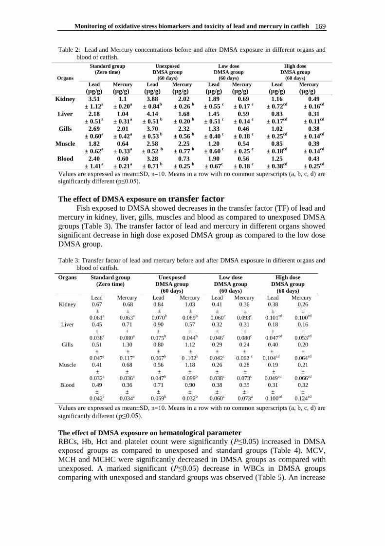
Monitoring of oxidative stress biomarkers and toxicity of lead and mercury in catfish 169
Table 2: Lead and Mercury concentrations before and after DMSA exposure in different organs and
blood of catfish.
Values are expressed as mean±SD, n=10. Means in a row with no common superscripts (a, b, c, d) are
significantly different (p≤0.05).
The effect of DMSA exposure on transfer factor Fish exposed to DMSA showed decreases in the transfer factor (TF) of lead and
mercury in kidney, liver, gills, muscles and blood as compared to unexposed DMSA
groups (Table 3). The transfer factor of lead and mercury in different organs showed
significant decrease in high dose exposed DMSA group as compared to the low dose
DMSA group. Table 3: Transfer factor of lead and mercury before and after DMSA exposure in different organs and
blood of catfish.
Values are expressed as mean±SD, n=10. Means in a row with no common superscripts (a, b, c, d) are
significantly different (p≤0.05).
The effect of DMSA exposure on hematological parameter
RBCs, Hb, Hct and platelet count were significantly (P≤0.05) increased in DMSA
exposed groups as compared to unexposed and standard groups (Table 4). MCV,
MCH and MCHC were significantly decreased in DMSA groups as compared with
unexposed. A marked significant (P≤0.05) decrease in WBCs in DMSA groups
comparing with unexposed and standard groups was observed (Table 5). An increase
Organs
Standard group
(Zero time)
Unexposed
DMSA group
(60 days)
Low dose
DMSA group
(60 days)
High dose
DMSA group
(60 days)
Lead
(µg/g)
Mercury
(µg/g)
Lead
(µg/g)
Mercury
(µg/g)
Lead
(µg/g)
Mercury
(µg/g)
Lead
(µg/g)
Mercury
(µg/g)
Kidney
3.51
± 1.12a
1.1
± 0.20a
3.88
± 0.84b
2.02
± 0.26 b
1.89
± 0.55 c
0.69
± 0.17 c
1.16
± 0.72cd
0.49
± 0.16cd
Liver
2.18
± 0.51a
1.04
± 0.31a
4.14
± 0.51 b
1.68
± 0.20 b
1.45
± 0.51 c
0.59
± 0.14 c
0.83
± 0.17cd
0.31
± 0.11cd
Gills
2.69
± 0.60a
2.01
± 0.42a
3.70
± 0.53 b
2.32
± 0.56 b
1.33
± 0.40 c
0.46
± 0.18 c
1.02
± 0.25cd
0.38
± 0.14cd
Muscle
1.82
± 0.62a
0.64
± 0.33a
2.58
± 0.52 b
2.25
± 0.77 b
1.20
± 0.60 c
0.54
± 0.25 c
0.85
± 0.18cd
0.39
± 0.14cd
Blood
2.40
± 1.41a
0.60
± 0.21a
3.28
± 0.71 b
0.73
± 0.25 b
1.90
± 0.67c
0.56
± 0.18 c
1.25
± 0.38cd
0.43
± 0.25cd
Organs Standard group
(Zero time)
Unexposed
DMSA group
(60 days)
Low dose
DMSA group
(60 days)
High dose
DMSA group
(60 days)
Lead Mercury Lead Mercury Lead Mercury Lead Mercury
Kidney
0.67
±
0.061a
0.68
±
0.063a
0.84
±
0.070b
1.03
±
0.089b
0.41
±
0.060c
0.36
±
0.093c
0.38
±
0.101cd
0.26
±
0.100cd
Liver
0.45
±
0.038a
0.71
±
0.080a
0.90
±
0.075b
0.57
±
0.044b
0.32
±
0.046c
0.31
±
0.080c
0.18
±
0.047cd
0.16
±
0.053cd
Gills
0.51
±
0.047a
1.30
±
0.117a
0.80
±
0.067b
1.12
±
0 .102b
0.29
±
0.042c
0.24
±
0.062 c
0.40
±
0.104cd
0.20
±
0.064cd
Muscle
0.41
±
0.032a
0.68
±
0.036a
0.56
±
0.047b
1.18
±
0.099b
0.26
±
0.038c
0.28
±
0.073c
0.19
±
0.049cd
0.21
±
0.066cd
Blood
0.49
±
0.042a
0.36
±
0.034a
0.71
±
0.059b
0.90
±
0.032b
0.38
±
0.060c
0.35
±
0.073a
0.31
±
0.100cd
0.32
±
0.124cd

Nema A. Mohamed et al. 170
in lymphocyte and a decrease in monocyte and neutrophil counts were observed in
DMSA exposed groups in comparison with unexposed and standard groups.
Table 4: Hematological changes in catfish before and after DMSA exposure:
High dose DMSA
group (60 days)
Low dose DMSA
group (60 days)
Unexposed DMSA
group (60 days)
Standard group
(Zero time)
Parameter
4.30
±
0.52cd
4.15
±
0.40c
3.00
±
0.60a
3.39
±
0.30a
RBCs (x106/ mm3)
12.18
±
1.29cd
11.18
±
1.29c
7.22
±
1.61b
9.50
±
0.81a
Hb (g/dl)
39.40
±
7.60 cd
34.40
±
5.60 c
25.00
±
5.24 a
27.88
±
4.92a
Hct (%)
81.11
±
08.75d
82.60
±
7.05c
92.70
±
22.63 a
90.36
±
11.59a
MCV (fl)
24.38
±
1.64d
25.96
±
2.05c
34.47
±
8.65a
34.28
±
2.30 a
MCH (pg)
27.34
±
1.64d
28.71
±
1.84c
38.94
±
3.28a
39.00
±
3.31a
MCHC (g/dL)
247.60
±
52.54cd
237.60
±
51.81c
188.60
±
25.11a
186.60
±
40.32a
PLt (x103/µl)
Values are expressed as mean±SD, n=10. Means in a row with no common superscripts (a, b, c, d) are
significantly different (p≤0.05).
Table 5: WBCs changes in catfish before and after DMSA exposure:
High dose DMSA
group (60 days)
Low dose
DMSA
group (60
days)
Unexposed DMSA
group (60 days)
Standard group
(Zero time)
Parameter
6640 ± 2.9290 c 7840 ± 2.3362 c 10660 ± 1.5143 b 9470 ± 4.4292a WBCs (x103/mm3)
45.00 ± 5.61 c 45.50 ± 4.87c 57.00 ± 5.20a 57.90 ± 6.2a Neutrophils (%)
1.40 ± 0.71c 1.30 ± 0.45 c 3.80 ± 0.55a 3.10 ± 0.5a Eosinophils (%)
46.20 ± 5.08 c 46.40 ± 5.23c 30.00 ± 5.00a 30.00 ± 6a Lymphocytes (%)
6.80 ± 1.30 c 6.80 ± 1.30 c 9.20 ± 2.39a 9.00 ± 2.6a Monocytes (%)
Values are expressed as mean±SD, n=10. Means in a row with no common superscripts (a, b, c, d) are
significantly different (p≤0.05).
The effect of DMSA exposure on liver function A significant (p≤0.05) decrease in AST and ALT activities after DMSA
exposure as compared to unexposed and standard groups (Table 6). Also, TSD test
revealed that there was a significant difference in AST and ALT activities between
low and high dose DMSA exposed groups.
Table 6: Changes in liver function of catfish before and after DMSA exposure:
Parameters
Standard
group
(Zero time)
Unexposed
DMSA group
(60 days)
Low dose
MSA Group
(60 days)
High dose
DMSA group
(60 days)
AST (U/L) 107.7 ± 23.7 a 206.20 ± 67.32
b 96.00 ± 29.59
c 87.00 ± 17.39
cd
ALT (U/L) 113.4 ± 35.00a 217.40 ± 29.20
b 69.40 ± 12.58
c 57.00 ± 8.78
cd
Values are expressed as mean±SD, n=10. Means in a row with no common superscripts (a, b, c, d) are
significantly different (p≤0.05).

Monitoring of oxidative stress biomarkers and toxicity of lead and mercury in catfish 171
The effect of DMSA exposure on kidney function
A significant (P≤0.05) decrease in urea and creatinine values after DMSA
exposure as compared to unexposed and standard groups (Table 7). TSD test revealed
that there was significant (P≤0.05) difference in urea and creatinine values between
low and high dose DMSA exposed groups.
Table 7: Changes in kidney function of catfish before and after DMSA exposure:
Parameters Standard group
(Zero time)
Unexposed
DMSA group
(60 days)
Low dose
DMSA group
(60 days)
High dose
DMSA group
(60 days)
Urea
(mg/dl)
32.60 ± 5.68 a
34.6 ±5.2b
24.00 ± 7.97c
20.80 ± 5.54 cd
Creatinine
(mg/dl)
0.63 ± 0.15 a
0.87± 0 .5b
0.57 ± 0.47c
0.43 ± 0.17cd
Values are expressed as mean±SD, n=10. Means in a row with no common superscripts (a, b, c, d) are
significantly different (p≤0.05).
Oxidative marker and antioxidant enzymes A marked significant (p≤0.05) increase in catalase (CAT) glutathione
peroxidase (GPX) and superoxide dismutase (SOD) was observed after exposure to
DMSA than that of unexposed and standard groups (Table 8). TSD test revealed that
there was no significant difference in CAT, GPX and SOD activities between low
and high dose DMSA exposed groups.
Table 8: Changes in serum antioxidant enzyme activities of catfish before and after DMSA exposure:
Parameter
Standard
group
(Zero time)
Unexposed
DMSA group
(60 days)
Low dose
DMSA group
(60 days)
High dose
DMSA group
(60 days)
CAT (U/ml) 32.60 ± 9.6a 29.80 ± 6.54
b 51.20 ± 9.42
c 52.80 ± 6.57
c
GPx (mU/ml) 35.00 ± 16.9a
28.60 ± 7.37b 60.40 ± 18.26
c 61.00 ± 9.35
c
SOD (U/ml) 34.70 ± 12.2a 23.60 ± 5.77
b 62.40 ± 12.90
c 63.00 ± 13.7
c
Values are expressed as mean±SD, n=10. Means in a row with no common superscripts (a, b, c, d) are
significantly different (p≤0.05).
Histopathological findings
Light microscope
The standard liver section of Clarias gariepinus (G1) revealed cytoplasmic
vacuolation presenting a foamy appearance with Kupffer cells and fibrosis increment
(Fig. A). The unexposed DMSA liver section (G2) displayed severe disorganization
of the hepatic cords, damaged cell membrane, excessive necrosis of hepatocytes,
associated with hemolysis, melanomacrophages aggregation and inflammatory
leukocytic infiltration (Fig. B). The liver section of G3 exhibited less organized
architecture with vacuolated area. Most of the hepatocyte with large spherical nuclei
containing central prominent nucleolus and large Kupffer cell rest on the luminal
surface of the sinusoids (Fig. C). The liver section of G4 showed clear signs of
improvement included the arrangement of polygonal hepatocytes with centrally
located nuclei separated by narrow blood sinusoids lined by endothelial cells (Fig. D).
The standard kidney section of G1 exhibited necrosis interstitial tissues, severe
vacuolation of tubular epithelium with large intracytoplasmic vacuoles. Also, wide
Bowman's space and fibrosis were noticed (Fig. A). The unexposed DMSA kidney
section (G2) demonstrated atrophied glomeruli capillaries with wide Bowman's
space, complete necrosis of fused renal tubules and hyperplasia of interstitial tissues
with complete loss of cellular histological architecture (Fig. B). The kidney section of

Nema A. Mohamed et al. 172
G3 showed partial improved state where, the tubular segment is lined with epithelial
cells with central basal nuclei and presence of glomeruli separated by nearly normal
interstitial tissues (Fig. C). The kidney section of G4 showed healthy renal tubules
with high cuboidal cells and weak eosinophilic cytoplasm (Fig. D).
Light micrographs (100X H&E stain): (A) G1 liver sections showing disappearance of normal architecture with
fibrosis (circles) in the cytoplasm and around bile ductule (Bd) with foamy area and Kupffer cells. (B) G2 liver
sections showing aggregation of leukocytes (circle), melanomacrophage (white asterisk) and hemolysis (dashed
circle). (C) G3 liver sections showing: vacuolated area (circle), pale nucleus (N) with large nucleolus and
hypertrophied Kupffer cell (Kc). (D) G4 liver sections showing improved polyhedral hepatocytes with central
nucleus (N), sinusoids lined with endothelial cell (EC) and contained Kupffer cell (Kc).
Light micrographs (40X. H&E stain): (A) G1 kidney sections showing atrophied glomerulus with lytic Bowman's
capsule (dashed arrow), vacuolated renal tubules (circle), fibrosis (dashed circle) and necrosis (asterisk). (B) G2
kidney sections showing atrophy with destruction of glomerular capillaries (arrow), wide Bowman's space
(asterisk), tubules fusion (circle), and vacuolated tubule (dashed-circle). (C) G3 kidney sections showing less
improved renal tubules (arrows) with basal nuclei in interstitial tissues (asterisk) and Bowman's capsule (square).
(D) G4 kidney sections showing improved glomerulus (G) surrounded by the Bowman's capsule (arrow),
Bowman's spaces (Bs) round renal tubules (dashed arrow).
Electron microscope
The standard liver hepatocytes (G1) showed disorganized cytoplasm with
absence of most organelles and occupied by lipid vesicles (Fig. A). Also, abnormal

Monitoring of oxidative stress biomarkers and toxicity of lead and mercury in catfish 173
nuclei with prominent nucleolus, folded nuclear envelope, proliferation of bizarre-
shaped mitochondria, loss of structural integrity of rER, increase in the number of
primary and secondary lysosomes with variable sizes (Fig. B). The unexposed DMSA
liver section (G2) showed nuclei with severe irregularities in shape and irregular
masses of heterochromatin covered the whole nucleus.
Electron micrographs: (A, B) G1 liver section showing lipid augmentation (arrows), mitochondria (M), lysosome
(Ly), irregular nucleus (N), disrupted nucleolus (Nu), destruction of nuclear envelope (Ne), depletion of rough
endoplasmic reticulum (rER), degenerated mitochondria (star), primary (Ly1) and secondary (Ly2) lysosomes. (C)
G2 liver section showing irregular nuclei (N-square), proliferative mitochondria (M), fat droplets (L) in affected
cytoplasm. (D, E) G3 liver sections showing round nuclei (N) with peripheral masses of heterochromatin, rough
endoplasmic reticulum with parallel cristae (rER), proliferation of primary lysosome (arrow), secondary lysosome
(Ly2) and peroxisome (P), lipid droplets (L), aggregation of small mitochondria (M) or depletion (M1), glycogen
(g). (F) G4 liver sections showing improved binucleated (N) hepatocyte with prominent nucleolus (Nu),
D
B
E F
A
C
C
Nema A. Mohamed et al. 174
mitochondria (M), few lipid droplet (L), organized rough endoplasmic reticulum (rER), and Kupffer cells (Kc-
square).
There was an increase in the lipid droplets and severe aggregation of small
mitochondria Fig. C. Liver section G3 showed partial improvement in nuclei with
random distribution of heterochromatin, rough endoplasmic reticulum stacks with
non-fenestrated cisternae arranged in parallel array with round and mega-
mitochondria (Fig. D). Proliferation of lysosomal elements including primary,
secondary with few lipid droplets and large deposits of glycogen (Fig. E). The liver
section G4 represented high improvement where, the hexagonal shape of typical
hepatocytes, central round nucleus with distinct single nucleolus with high electron
density and heterochromatin dispersed along the nucleoplasm. The rough
endoplasmic reticulum was evident and arranged in parallel stacks of cisternae
possessing high densities of ribosome adjacent to the nucleus Fig. F.
DISCUSSION
To accelerate the restoration of Lake Mariout to better conditions for saving
them as grounds for fisheries, the quality of the sewage and industrial wastes
dumping directly into them or indirectly via agricultural drains must be much
improved by suitable means, at least by using DMSA for limiting the amounts of
heavy metals demanding industrial wastes, especially lead and mercury . It was
established that the polluted water weakens the fish host defenses against parasites,
allowing increased opportunities for epizootic diseases to affect fish populations
(Zeitoun and Mehana, 2014). From the achieved results, it was confirmed that water
quality is an integral part of any aquaculture system and plays a major role in fish
health. So, DMSA groups showed improve in water quality and increase the fish host
defenses against infections and parasites.
In the current study, the lead (Pb) accumulation among the different organs was
arranged as follows: kidney˃gills˃blood˃liver˃muscle. The mercury (Hg)
accumulation pattern was liver˃kidney˃muscle˃gills˃blood as observed in this work.
The presence of these elements in both the gills and other tissues of fish in that
study area agree with study of El-Shehawi et al. (2014) who stated that Lake Mariout
is highly contaminated with heavy metals. Metals transport through the blood, thus,
they are brought into contact with different organs and tissues of fish and
consequently accumulated (Kaoud and El-Dahshan, 2010). The lead accumulation in
the tissues of C. gariepinus was dependent on the exposure period and lead
concentration (Al-Balawi et al., 2013). Kusemiju et al. (2012) found that the C.
gariepinus gills contained the highest concentration of all the detected heavy metals,
while the muscle tissue was the lowest. The excessive intake of metals may lead to
high accumulation in the gills causing the precipitation of mucous on the gill surface
membrane (Siraj et al., 2014) and structural damage (Abumourad et al., 2013).
Pugazhvendan et al. (2012) suggested that the liver is one of the important
organs to accumulate Pb and disturb its regular function leads to death of fish. The
high accumulation of these metals in the liver could be related to the fact that the liver
played an important role in the accumulation and detoxification (Al-Balawi et al.,
2013). Meanwhile, the kidney is the gateway for heavy metal detoxification in the
body (Vinodhini and Narayanan, 2008). The enhancement of these metals may be due
to presence of industrial, sewage and agricultural discharge (Kaoud and El-Dahshan,
2010).
Monitoring of oxidative stress biomarkers and toxicity of lead and mercury in catfish 175
The determination of the transfer factor (TF) and the positive correlations
(P≤0.05) between concentrations of metals in fish muscle and water indicated the
direct accumulation of metals from the water to the fish. The transfer factor provides
a straightforward, constructive method for assessing heavy metal accumulation
for the purposes of health risk assessment of humans consuming the fish. In the
present work, transfer factor for organs of DMSA groups was lower than that of
unexposed, this important to save human health who consuming the fish from this
Lake as reported by Canpolat et al. (2014).
In this study, DMSA exposure showed significant decrease in lead and mercury
concentrations in kidney, liver, gill, muscle and blood. Volans et al. (2010) recorded
a decrease in blood lead or mercury concentrations in the poisoned child using
DMSA over the short- and long-term. The positive effect of DMSA may be attributed
to its properties as a chelating agent (Flora et al., 2008). Lead binds to adjoining
sulfur and oxygen atoms whereas mercury binds to sulfur atoms in succimer. DMSA
has a wide therapeutic index and has advantages over dimercaprol and CaNa2EDTA
(Lowry, 2010). DMSA has been recommended for the treatment of lead and mercury
in human (Masters et al., 2008). DMSA preferentially binds to lead, but can also
increase the excretion of several other toxic metals, including mercury, to a lesser
extent (Adams, 2009). DMSA may bond with lead or mercuric ions through a
mechanism of thiol competition and conjugates mercury or lead (Bridges et al.,
2009).
Anemia in lead poisoning results from impairment of hemoglobin production
and changes in the RBC membrane. Lead's interference in heme biosynthesis is
characterized by several unique enzyme blockades causing increased urinary delta-
aminolevulinic acid (ALA), urinary coproporphyrin and erythrocyte zinc
protoporphyrin (Lowry, 2010). Also, lead and mercury showed a high affinity for
erythrocytes (Alves et al., 2006). Furthermore, the impairment in iron synthesis and
absorption, causing microcytic hypochromic anemia (Kumar and Nandan, 2014).
There is also good evidence linking mercury with anemia, including hemolytic
anemia and aplastic anemia as mercury is thought to compete with iron for binding to
hemoglobin which can result in the impaired hemoglobin formation (Pyszel et al.,
2005). The present finding came accordance with the results of Adeyemo (2007) and
Mahmoud et al. (2013) who observed a decrease in RBC, Hb, and Hct values in
Clarias gariepinus exposed to lead.
In this study, DMSA exposure completely improved the hematological
parameters (RBC, Hb, Hct, MCV, MCH and MCHC). The present findings were in
agreement with Gurer et al. (1998) who reported that treatment with a thiol
antioxidant chelating agent (Succimer) has a reverse effect on the lead induced
oxidative stress against accumulation and antioxidation of aminolevulinic acid
(ALAD). Adams et al. (2009) recorded greatly improved abnormal platelet counts,
suggesting a significant decrease in inflammation after oral DMSA therapy for
children with autism spectrum disorders ages 3-8 years.
In the current work, the pronounced alterations in white blood cells were
leucocytosis and lymphocytopenia in the unexposed DMSA group. This increase in
WBCs may be attributed to immune response in lead exposed fish (Altindag, 2005).
In addition, it has been known for many years that mercury impairs immune system
function most likely via its deleterious effects on the polymorphonuclear leukocytes
(PMNs). Mercury through suppression of adrenocorticosteroids production prevents
normal stimulation of PMNs production and also affects the PMN function by
inhibiting their ability to destroy foreign substances (Wada et al., 2009). These results

Nema A. Mohamed et al. 176
came accordance with Olugh and Omerebele (2010) who recorded that there was a
significant leucocytosis in the fish Clarias gariepinus exposed to lead poisoning. Zaki
et al. (2014) reported that long term exposure of Clarias lazera to Pb and Hg caused
a gradual increase in WBCs count. In this study, DMSA exposure completely
improved the enhancement in WBC count. Significant increase in lymphocyte in
exposed DMSA groups comparing with unexposed and standard groups.
Monitoring of liver enzyme leakage into the blood has proved to be a useful
tool in liver toxicological studies (Abedi et al., 2013; Chavan and Muley, 2014). The
increased activity of these enzymes in blood is correlated either with leakage of
enzymes from the damaged hepatic cells into the blood stream or increased synthesis
of these enzymes as a result of heavy metal toxicities (Osman et al., 2010). The
activity of AST and ALT enzymes in blood may also be used as a stress indicator as
mentioned by Awad Elkareem et al. (2014). Okonkw and Ejike (2012) and Olojo et
al. (2012) stated that elevations in ALT and AST concentrations in serum of the
catfish may be attributed to disruption of hepatic cells as a result of necrosis or
altered membrane permeability after exposure to lead. The present findings were in
agreement with Çoğun and Şahin (2013) who reported that Pb exposure increased the
activities of ALT and AST in serum of Clarias gariepinus.
DMSA treatment showed a protective effect against hepatotoxicity as indicated
by a significant decrease in AST and ALT activities. Adams et al. (2009) recorded
that DMSA had no significant effects on liver transaminases (AST and ALT),
perhaps because they treated for only 3 days at a time therapy for children with
autism spectrum disorders ages 3-8 years. The present data from the liver function
tests are corroborated by the histological changes observed in the liver sections of the
test fish, relative to the control fish.
The level of Pb and its accumulation over time may cause serious damage to
the renal system, over time, by continuous consumption of the fish and the water
(ATSDR, 2007). The increase in creatinine level might be induced by glomerular
insufficiency, increased muscle tissue catabolism or the impairment of carbohydrates
metabolism (Hadi et al., 2009). Urea and creatinine are nitrogenous end products of
protein metabolism, taken together the BUN and creatinine levels provide a very
accurate estimation of how well the kidneys are working (Ajeniyi and Solomon,
2014).
Abdel El -Moneium et al. (2008) showed an insignificant difference in blood
urea and significant increase in creatinine after exposure Clarias lazera to different
doses of industrial effluent. An increase in the urea and creatinine in Clarias
gariepinus after exposure to mercury and Pb was observed (Mahmoud et al., 2013).
Zaki et al. (2014) reported that long term exposure of Clarias lazera to mercuric
chloride and/or to Pb, Hg, Ca and/or lead acetate caused gradual elevation of serum
urea and creatinine.
The present study revealed that DMSA had a protective effect against renal
dysfunction as indicated by a decrease in the urea and creatinine levels. Chelating
agents are capable of binding to toxic metal ions to form complex structures which
are easily excreted from the body. Hydrophilic chelators like meso-2,3-
dimercaptosuccinic acid effectively promote renal metal excretion (Flora et al.,
2011). These results in the same line with Adams et al. (2009) who recorded high
urinary excretion of toxic metals (Pb and mercury) suggesting an improvement in
kidney functions after oral dimercaptosuccinic acid (DMSA) therapy for children
with autism spectrum disorders ages 3-8 years. Bradberry and Vale (2009) stated that
Monitoring of oxidative stress biomarkers and toxicity of lead and mercury in catfish 177
DMSA at a dose 30 mg/kg/day was more effective than 10 or 20 mg/kg/day in
enhancing urinary lead excretion.
Fish inhabiting the highly polluted sites developed an increased state of
oxidative stress characterized by increased levels of lipid peroxidation (Rajeshkumar
et al., 2013). Lead alters the activity of antioxidant enzymes like SOD, CAT and GPx
(Flora et al., 2008). Lead and mercury have electron-sharing affinities causing the
formation of covalent attachments mainly between heavy metal and sulfhydryl groups
of several enzymes in antioxidant defense system, causing inactivation of these
enzymes. Overall, these inhibitory effects of lead on various enzymes would probably
result in impaired antioxidant defenses by cells and render cells more vulnerable to
oxidative attacks.
Saliu and Bawa-Allah (2012) showed reduced levels of SOD and CAT occurred
in Clarias gariepinus exposed to lead. They studied the effects of mercury on the
activities of antioxidant defenses in intestinal macrophages of fresh water teleost
Channa punctatus suggesting the use of these antioxidants as potential biomarkers of
toxicity associated with exposure of freshwater fish to contaminants. Mercury has a
high affinity for sulfydryl (-SH) groups, inactivating sulfur-containing antioxidants
with subsequent decreased oxidant defense and increased oxidative stress (Begam
and Sengupta, 2014).
DMSA exposure has an antioxidant effect as documented by elevations in the
antioxidant enzymes activities. Flora et al. (2008) recorded a reduction in oxidative
stress in the blood of male Wistar rats were exposed to 0.1% lead acetate in drinking
water for 3 months after DMSA therapy. Sompamit et al. (2013) recorded a
reduction in oxidative stress in blood mice after DMSA at a dose of 25 mg/kg or 50
mg/kg for 5 consecutive days after 8 weeks of Cd exposure.
Most DMSA in plasma is protein-bound through a disulfide bond with cysteine;
only a very small amount is present as free drug, which is filtered at the glomerulus
then extensively reabsorbed into proximal tubule cells. DMSA therefore accumulates
in the kidney where it is extensively metabolized to mixed disulfides of cysteine
which are active chelating agents. A chelating agent has at least two negatively
charged groups that allow it to form complexes with metal ions with multiple positive
charges, such as lead and mercury. The chelate that is thus formed is nontoxic and
can be excreted in the urine (Bradberry and Vale, 2009).
In the present study, the histopathological alterations in the liver of the studied
fish collected from the Lake Mariout (Standard group) and unexposed DMSA group
might be due to the oxygen deficiency as a result of gill degeneration which is the
most common cause of cellular degeneration of the liver. Also, hepatocytes wall
degeneration, high number of Küffer cells, infiltration of leukocytes indicate increased
inflammation and fibrosis in the hepatic tissues as reported by Koca (2008). The
manifestation of cytopathologic changes suggests a severe hepatic dysfunction and the
impairment of the physiometabolic process in the liver (Dezfuli et al., 2006). Al-
Balawi et al. (2013) showed that necrosis of hepatocytes was apparent in the fishes
(Clarias gariepinus) exposed to lead.
In this study, ultra–examination of standard liver hepatocytes showed the
necrotic appearance with lytic nuclei or pyknotic nucleus with multivesicular
appearance and an extremely folded nuclear envelope. In addition, blood sinusoid
lined with small flat endothelial cell has irregular nucleus, hypertrophied Kupffer cell
and strongly developed cytoplasmic myelinated bodies. These can be explained as,
disturbance of living processes at the molecular and subcellular levels of biological
organization by xenobiotics which can lead to cell injury, resulting in degenerative
Nema A. Mohamed et al. 178
and neoplastic diseases in target organs (Pacheco & Santos, 2002). The increased
number of lysosomes, a result of the attempt to digest heavy metals or toxic
substances, is considered a general manifestation of injury. These results were
consistent with the results of Abdel-Moneim and Abdel-Mohsen (2010) and Sayed et
al. (2012).
In the present study, the standard fish kidneys and unexposed DMSA group
showed histopathological changes. The changes in the size, structure of the epithelial
cells and the narrow lumen of the renal tubule inhibits kidney functioning (Gupta and
Srivastava, 2006) and these were well documented in this study by biochemical and
physiological results. Mohamed (2009) observed necrosis of tubular epithelium,
hypertrophied epithelial cells of renal tubules, narrowing of the tubular lumen,
expansion of space inside the Bowman's capsules and contraction of the glomerulus in
C. carpio exposed to sewage. Abd-Elghaffar et al. (2015) recorded that the kidneys
treated with lead acetate showed severe tubular necrosis, periglomerular lymphoid cell
reaction, dilatation of renal tubule, hyaline tubular cast associated with hemorrhage.
It has been indicated that chelating agent, like DMSA, reduces the toxic effect
of heavy metals on the histological alterations of soft tissues (Raafat et al., 2011).
Abd-Elghaffar et al. (2015) proved that rats treated with lead acetate combined with
DMSA (50 mg/kg /week) only have improved effect.
CONCLUSION
The protective effect of DMSA may be attributed to its activity as a chelating
agent which is act as a scavenger molecule with at least two negatively charged groups
that allow it to form complexes with metal ions such as lead and mercury.
Conflict of interest:
The authors declare no conflict of interest.
REFERENCES
Abd-Elghaffar, S.K.H.; El-Sayed, M.F.; Adly, M.A. and Abdel-Samei, W.M. (2015).
The protective effects of DMSA and some vitamins against toxicity induced by
lead in male Albino rats. J. Pharm. Appl. Chem., 1(1): 1-8.
Abdel-Moneim, A.M. and Abdel-Mohsen, H.A. (2010). Ultrastructure changes in
hepatocytes of catfish (Clarias gariepinus) from Lake Maryout. Egypt. J.
Enviro. Biol., 31(5): 715-720.
Abdel-Moneium, A.M.; Abou Shabana, N.M.; Khadre, S.E.M. and Abdel-Kader,
H.H. (2008). Physiological and histological effects on (Clarias lazera) exposed
to dyestuff and chemical wastewater. Int. J. Zool. Res., 4(4): 189-202.
Abedi, Z.; Hasantabar, F.; Khalesi, K. and Babaei, S. (2013). Effect of sublethal
concentrations of cadmium, lead and chromium on some enzymatic activities of
common carp, Cyprinus carpio. World J. Zoo., 8(1): 98-105.
Abei, H. (1984). Determination of Malondialdehyde. Method Enzymol., 105: 121-
126.
Abumourad IMK, Mohammad MN and Abbas WAT. 2013. Heavy Metal Pollution
and Metallothionein Expression: A Survey on Egyptian Tilapia Farms. J.
Appl.Sci. Res., 9 (1): 612-619.
Adams, J.B.; Baral, M. and Geis, E. (2009). Safety and efficacy of oral DMSA
therapy for children with autism spectrum disorders: Part A-Medical results.
BMC Clini. Pharm. 9:1186-1472.

Monitoring of oxidative stress biomarkers and toxicity of lead and mercury in catfish 179
Adeyemo, O.K. (2007). Haematological profile of (Clarias gariepinus) exposed to
lead. Turkish J. Fish. Aqua. Sci., 7: 163-169.
Agency for Toxic Substances and Disease Registry (ATSDR). (2007). Toxicological
profile for lead. Public Health Service, U.S. Department of Health and Human
Services, Atlanta, GA: U.S. Department of Public Health and Human Services,
Public Health Service.
Ajeniyi, S.A. and Solomon, R.J. (2014). Urea and creatinine of (Clarias gariepinus)
in three different commercial ponds. Nature and Science, 12(10).
Al-Balawi, H.F.A.; Al-Akel, A.S.; Al-Misned, F.; Suliman, E.M.; Al-Ghanim, KA.;
Mahboob, S. and Ahmad Z. (2013). Effects of sub-lethal exposure of lead
acetate on histopathology of gills, liver, kidney and muscle and its
accumulation in these organs of (Clarias gariepinus). Brazil. Arch. Biol. Tech.
56(2): 293-302.
Altindag, A. and Yigit, S. (2005). Assesment of heavy metal concentration in the
food web of Lake Beysehir, Turkey. Chemosphere. 60: 552-556.
Alves, L.C. and Wood, C.M. (2006). The chronic effects of dietary lead in freshwater
juvenile rainbow trout (Oncorhynchus mykiss) fed elevated calcium diets.
Aquat. Toxicol. 78: 217.
Amr, H.M.; El-Tawila, M.M. and Ramadan, M.H.M. (2005). Assessment of pollution
levels in fish and water of main basin, Lake Mariout. The Journal of the
Egyptian Public Health Association (JEPHAss.), (80): 1, 2.
Awad Elkareem, M.M.A.; Karrar, A.M.H. and Ali, A.K.S. (2014). Relationship of
biometric size-weight, nutritive value, and metal concentrations in (Clarias
lazera) (Cuvier and Valenciennes) reared in treated waste water. Jordan Journal
of Biological Sciences. , 7(3): 217 – 225.
Bancroft, J.D. and Gamble, M. (2002). Theory and practice of histological
techniques. Neuro. J & Exp. Neuro., 67(6):633.
Begam, M. and Sengupta, M. (2014). Effects of mercury on the activities of
antioxidant defenses in intestinal macrophages of fresh water teleost (Channa
punctatus). Int. J. of Fish. and Aqu. Stud., 2(1): 172-179.
Bradberry, S. and Vale, A. (2009). Dimercaptosuccinic acid (succimer; DMSA) in
inorganic lead poisoning. Clin Toxicol (Phila)., 47(7):617-31.
Bridges, C.C.; Joshee, L. and Zalups, R.K. (2009). Effect of DMPS and DMSA on
the placental and fetal disposition of methylmercury. Placenta, 30:800-805.
Canpolat, Ö.; Mücahit, E.; Mehmet, Z.Ç. and Mustafa, D. (2014). Transfer factors
and bioaccumulation of some heavy metals in muscle of a freshwater fish
species: a human health concern. Fresenius Environmental Bulletin, 23(2):418-
425.
Chavan, V.R. and Muley, D.V. (2014). Effect of heavy metals on liver and gill of fish
(Cirrhinus mrigala). Int. J. Curr. Microbiol. App. Sci., 3(5): 277-288.
Cogun, H.Y. and Şahin, M. (2012). The effect of zeolite on reduction of lead toxicity
in Nile Tilapia (Oreochromis niloticus Linnaeus, 1758). Kafkas Univ Vet Fak
Derg. 18 (1): 135-140.
Dacie, J.V. and Lewis, S.M. (1975). Practical haematology. The English Language
Book Society and Churchill Livingston. 32-34.
Damian, E.C.; Afulenu, N.L.; Obinna, O.M. and Ndidi, O.C. (2014).
Bioaccumulation of heavy metals in fish sourced from environmentally stressed
axis of River Niger: Threat to ecosystem and public health. In. J. Enviro. Prot.
Pol., 2(4): 126-131.

Nema A. Mohamed et al. 180
Dezfuli, B.S.; Simoni, E.; Giari, L. and Manera, M. (2006). Effects of experimental
terbuthylazine exposure on the cells of (Dicentrarchus labrax). Chemosphere,
64: 1684–1694.
Diaz-Ravina, M.; Baath, E. and Frostegard, A. (1994). Multiple heavy metal
tolerance of soil bacterialcommunities and its measurement by a thymidine
incorporation technique. Appl. Environ. Microbio., 60: 2238–2247.
Ebrahimi, M. and Taherianfard, M. (2011). The effects of heavy metals exposure on
reproductive systems of cyprinid fish from Kor River. Iranian J. Fish. Sci., 10
(1): 13.
El-Bestawy, A.H.; Attia, A.M. and Zahran, H. (2014). Biodegradation of persistent
chlorinated hydrocarbons using selectedfreshwater bacteria. J. Bio. & Biod. J
Bioremed Biodeg., 5:4.
El-Shehawi, A.M.; Ali, F.K. and Seehy, M.A. (2014). Estimation of water pollution
by genetic biomarkers in tilapia and catfish species shows species-site
interaction. African Journal of Biotechnology, 6 (7): 840-846.
El-Sheikh, M.A.; Saleh, H.I.; El-Quosy, D.E. and Mahmoud, A.A. (2010). Improving
water quality inpolluted drains with free water surface constructed Wetlands.
Ecol. Engineer., 36: 1478-1484.
Emam, A.N.M. and Badia, A. (2014). Seasonal histological changes in gonads of the
catfish (Clarias lazera). Fish. Aquac J., 5: 087.
Falayi, B.A. and Amatosero, R.B. (2014). The effects of lead (Pb) on (Clarias
gariepinus) (B.) Juveniles in captivity research. J. Agricul. Environ. Manag.,
3(8): 353-360.
Flora, S.J.S.; Mittal, M. and Mehta, A. (2008). Heavy metal induced oxidative stress
& its possible reversal by chelation therapy. Indian. J. Med. Res.,128: 501-523.
Flora, S.J.; Pachauri, V. and Saxena, G. (2011). Arsenic, cadmium and lead. In:
Reproductive and Developmental Toxicology. Acad. Press., 415–438.
Gupta, A.K. and Kumar, A. (2006). Tilaknagar Udaipur. Histopathological lesions in
the selected tissues of (Cirrhinus mrigala) (Ham.) fingerlings exposed to sub
lethal concentration of mercury. J. Environ. Biol., 27(2):235-239.
Gurer, H.; Ozgunes, H.; Neal, R.; Spitz, D.R. and Ercal, N. (1998). Antioxidant
effects of N-acetyl cysteine and succimer in red blood cells from lead exposed
rats. Toxicol., 128: 181-9.
Hadi, A.; Shokr, A. and Alwan, S.V. (2009). Effects of aluminum on the biochemical
parameters of fresh waterfish (Tilapia zillii). J. Sci. Applica., 3: 33
Henry, R.J.; Cannon, D.C. and Winkelman, W. (1974). Clinical chemistry principles
and techniques. 11th
ed., Harper and Row Publishers, New York. 1629.
Kanno, J.; Akagi, H. and Takabatake, E. (1985). A method for determination of
methylmercury in environmental samples, particularly in sediment, Japanese. J.
Toxicol. Enviro. Heal. (Eisei kagaku),. 31: 260-268.
Kaoud, H.A. and El-Dahshan, A.R. (2010). Bioaccumulation and histopathological
alterations of the heavy metals in (Oreochromis niloticus) fish. Nat. Sci., 8(4).
Koca, S.; Koca, Y.B.; Yildiz, S. and Gürcü, B. (2008). Genotoxic and
histopathological effects of water pollution on two fish species, (Barbus capito
pectoralis and Chondrostoma nasus) in the Büyük Menderes River, Turkey. J.
Biol. Trace Elem. Res., 122: 276–291.
Krishna, K.; Rama, M.K. and Murthy, N.N. (2013). Assessment of heavy metal
contamination in soils around chromite mining areas, Nuggihalli, Karnataka,
India. Environmental Earth Sciences, 70(2): 699.

Monitoring of oxidative stress biomarkers and toxicity of lead and mercury in catfish 181
Kumar, G.B. and Nandan, B.S. (2014). Copper Toxicity: haematological and
histopathological changes and prophylactic role of vitamin C in the fish,
Anabastestudineus Bloch, 179. J. Zool. Stud., 1(3): 04-13.
Kusemiju, V.; Patience, A.; Aderinola, J. and Oluwatoyin. (2012). Accumulation of
lead in the tissues of freshwater catfish (Clarias gariepinus) exposed to static
nominal concentrations of lead nitrate. Agric. Biol. J. N. Am., 3(12): 510-515.
Lowry, J.A. (2010). Oral chelation therapy for patients with lead poisoning. Division
of Clinical Pharmacology and Medical Toxicology, 4(1):173-.
Mahmoud, U.M.; Ebied, A.B. and Mohamed, S.M. (2013). Effect of lead on some
haematological and biochemical characteristics of Clarias gariepinus dietary
supplemented with lycopene and vitamin E. Egypt. Acad. J. Biolog. Sci., 5: (1):
67 – 89.
Masters, S.B.; Trevor, A.J. and Katzung, B.G. (2008). Katzung & Trevor's
Pharmacology: Examination & Board Review (8th Ed.). McG. H. Med. 481–3.
Mohamed, F.A.S. (2009). Histopathological studies on (Tilapia zillii and Solea
vulgaris) from Lake Qarun, Egypt. W. J. Fish. Mar. Sci., 1 (1): 29-39.
Nishikimi, M.; Roa, N.A. and Yogi, K. (1972). Biochem. Bioph.Res.Common., 46:
849-854.
Okonkwo, F.O. and Ejike, C.E. (2011). Simulation of heavy metal contamination of
fresh water bodies: toxic effects in thecatfish and its amelioration with co-
contamination with glyphosate. J. Appl. Sci. Environ. Manage., 15(2): 341 –
345.
Olabanji, I.O. and Oluyemi, E.A. (2014). Preliminary assessment of heavy metal
pollution of Opa Reservoir. Ile- Ife, Southwest Nigeria using Mormyrus Rumea
(Tilapia zillii). Ife. J. Sci. 16: 1.
Olojo, E.A.A.; Abass, A.A.; Olurin, K.B. and Mbaka, G. (2012). The potential use of
certain protein metabolism parameters as biomarkers of heavy metal (lead)
stress in the African cat fish (Clarias gariepinus). Agri. J., 7: (5):316-322.
Oluah, N.S. and Omerebele, U.A.M. (2010). Changes in haematological parameters
of (Clarias gariepinus) exposed to lead poisoning. J. Fish. Int., 5(4):72-76.
Osman, A.G.M.; Al-Awadhi, R.M.; Harabawy, A.S.A. and Mahmoud, U.M. (2010).
Evaluation of the use of protein electrophoresis of the African catfish (Clarias
gariepinus) for biomonitoring aquatic pollution. Environ. Res. J., 4(3): 235-
243.
Pacheco, M. and Santos, M.A. (2002). Biotransformation, genotoxic, and
histopathological effects of environmental contaminants in European ell
(Anguilla anguilla L.). Ecotoxicol. Environ. Saf., 53: 331-347.
Paglia, D.E. and Valentine, W.N. (1976). Studies on the quantitative and qualitative
characterization of erythrocyte of glutathione peroxidase. J. Lab. Clin. Med.,
7:158-169.
Palaniappan, R.M. and Vijayasundaram, V. (2009). The effect of arsenic exposure
and the efficacy of DMSA on the proteins and lipids of the gill tissues of
(Labeo rohita). Food. Chem. Toxicol., 47:1752–1759.
Pugazhvendan, S.R.; Mariappan, M.; Leon, P.S. and Balakrishnan, J.K. (2012).
Bioaccumulation of lead in fresh water fish (Cyprinus Carpio). Int. J. C. Res.,
4 (7):146-148.
Pyszel, A.; Wrobel, T.; Szuba, A. and Andrzejak, R. (2005). Effect of metals,
benzene, pesticides and ethylene oxide on the haematopoietic system. Med Pr.,
56(3):249–255.

Nema A. Mohamed et al. 182
Raafat, M.; El-Barbary, A.; Touson, E. and Aziz, S. (2011). Dimercapto succinic acid
(DMSA) and vitamin C chelating potency in lead intoxication, regarding
oxidative stress and apoptotic proteins in rabbit. J. Gen. Engin. Biotech., 9:121-
131.
Rajeshkumar, S.; Jayaprakash, M. and Munuswamy, N. (2013). Effects of heavy
metals on antioxidants and expression of HSP70 in different tissues of milk fish
(Chanos chanos) of Kaattuppalli Island, Chennai, India. Ecot. Environ. Saf., 98
(1): 8–18.
Reitman, S. and Frankel, S.A. (1957). Colorimetric method for the determination of
serum glutamic oxalacetic and glutamic pyruvic transaminases. Am. J. Clin.
Pathol., 28: 56-63.
Reynolds, E.S. (1963). The use of lead citrate at high pH as an electron-opaque stain
in electron microscopy. J. Cell Biol. 17: 208.
Saliu, J.K.; Saliu, K.A and Bawa-Allah. (2012). Toxicological effects of lead and
zinc on the antioxidant enzyme activities of post juvenile Clarias gariepinus.
Resources and Environment., 2(1): 21-26.
Sayed, A.H.; Mekkawy, I.A and Mahmoud, U.M. (2012). Histopathological
alterations in some body organs of adult (Clarias gariepinus) exposed to 4-
Nonylphenol, Zoology, Dr. María- Dolores García (Ed.). Zoology. 8, 163-184.
Selander, S. and Cramer, K. (I968). Determination of lead in blood by atomic
absorption spectrophotometry. Brit. J. Industr. Med. 25: 209.
Siraj, M.; Shaheen, M.; Sthanadar, A.A.; Khan, A.; Douglas, P.; Chivers, A.M. and
Yousafzai. (2014). A comparative study of bioaccumulation of heavy metals in
two fresh water species (Aorichthys seenghala and Ompok bimaculatous) at
River Kabul, Khyber Pakhtunkhwa. Pakistan J. Bio. Env. Sci., 4(3):40-54.
Sompamit, K.; Kukongviriyapan, U.; Donpunha, W.; Nakmareong S. and
Kukongviriyapan V. (2010). Reversal of cadmium-induced vascular
dysfunction and oxidative stress by meso-2,3-dimercaptosuccinic acid in mice.
Toxicol Lett.,198(1):77-82.
Tietz NW. (1995). Clinical Guide to Laboratory Tests. 3rd ed., Philadelphia, PA: WA
Saunders Co. 622-626.
Vinodhini, R. and Narayanan, M. (2009). The impact of toxic heavy metals on the
hematological parameters in common carp (Cyprinus carpio L.) Iran. J.
Environ. Health. Sci. Eng. 6(1):23-2823.
Volans, G.N.; Karalliedde, L and Wiseman HM. (2010). Review of succimer for
treatment of lead poisoning. Medical Toxicology Information Services, Mary
Sheridan House, Guy's Hospital, London SE1 9RT.
Wada H.; Cristol DA.; McNabb, FM. and Hopkins WA. (2009). Suppressed
adrenocortical responses and thyroid hormone levels in birds near a mercury-
contaminated river. Environ Sci Technol., 43(15):6031–6038.
Zaki, M. and Osman, A. (2005). Histological changes in gill tissues of (Tilapia
nilotica) exposed to lead chloride. Bull. Natio. Res. Cent. Cairo., 28(1):87-100.
Zeitoun, M.M. and Mehana, E.E. (2014). Impact of water pollution with heavy
metals on fish health: Overview and Updates Global Veterinaria. 12(2): 219-
231.