needle emg guidance for improved outcomes · most importantly, emg provides ongoing information...
TRANSCRIPT
NEEDLE EMG GUIDANCE FOR IMPROVED OUTCOMES
INTRONIX TECHNOLOGIES CORPORATION
VALUE - INNOVATION - PERFORMANCE
© Dr. Evan Friedman, June 26, 2011
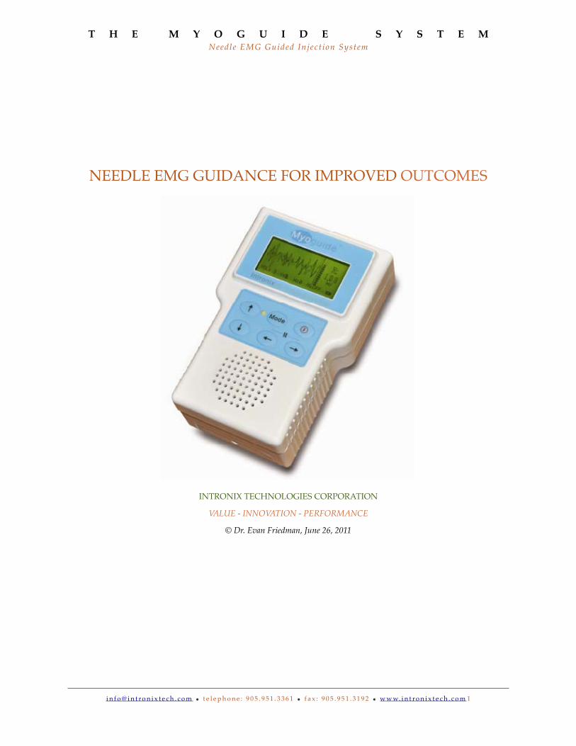
T H E M Y O G U I D E S Y S T E MNeedle EMG Guided Inject ion System
i n f o @ i n t r o n i x t e c h . c o m • t e l e p h o n e : 9 0 5 . 9 5 1 . 3 3 6 1 • f a x : 9 0 5 . 9 5 1 . 3 1 9 2 • w w w. i n t r o n i x t e c h . c o m 1
Introduction
Myoguide products are designed to aid in diagnosis and management of musculoskeletal pain, spasticity, and fibromyalgia. There are applications in Physical and Rehabilitation Medicine, Sports Medicine, Oto-laryngology, Urology, Anesthesiology delivered pain management, and for cosmetic injections using Botulinum Toxin (or other medication), as well as dry needling.
Myoguide is a handheld guidance system that enables clinicians to see and hear, as well as challenge, the injection sites, before any treatment is delivered or drug is injected. Myoguide can be instru-mental in improving treatment outcomes, and reducing side ef-fects, in a number of very impor-tant clinical areas.
High fidelity EMG audio, a well featured, powerful stimulator, and EMG signal display are all inte-grated into a battery operated, handheld package. Myoguide em-bodies innovation, value and per-formance in the palm of your hand.
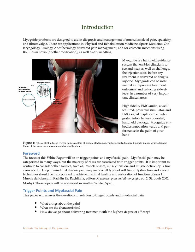
!"#$%&'()''!"#$%#&'()*$&+,-.$/0$'(+11#($2/+&'.$%/&')+&$)3&/(4)*$#*#%'(/45/1()2"+%$)%'+6+'57$*/%)*+8#,$4-.%*#$.2).47$9"+*#$),:)%#&'$;+3#(.$/0$'"#$.)4#$4-.%*#$(#4)+&#,$#*#%'(+%)**5$.+*#&'<
!"#$%"#&The focus of this White Paper will be on trigger points and myofascial pain. Myofascial pain may be categorized in many ways, but the majority of cases are associated with trigger points. It is important to continue to consider other sources, such as, muscle spasm, muscle tension, and muscle deficiency. Clini-cians need to keep in mind that chronic pain may involve all types of soft tissue dysfunction and varied techniques should be incorporated to achieve maximal healing and restoration of function (Kraus H: Muscle deficiency. In Rachlin ES, Rachlin IS, editors Myofascial pain and fibromyalgia, ed. 2, St. Louis 2002, Mosby). These topics will be addressed in another White Paper. . '
'#())$#*+"(,-.*/,&*01"2/.3(/4*+/(,This paper will answer the questions, in relation to trigger points and myofascial pain:
• What brings about the pain? • What are the characteristics? • How do we go about delivering treatment with the highest degree of efficacy?
I n t r o n i x Te c h n o l o g i e s C o r p o r a t i o n! W h i t e P a p e r
1
The majority of clinical pain is myofascial. It has been reported that 85% of back pain and 54.6% of chronic headache and neck pain is myofascial pain (Fishbain DA, Goldberg M, Steele R, et al: DSM-III diagnosis of patients with myofascial pain syndrome(fibrositis), Arch Phys Med Rehabil 70:433-438, 1989).
Myofascial pain includes various types of soft tissue dysfunction. An analysis of such soft tissue pain involves at least the following types of pathology: tissue inflammation, contracture, microcirculatory de-ficiency, tissue degeneration, tissue adhesion, scarring, and biomechanical imbalance of the musculo-skeletal system.
Early work on tenderness and motor points described the beginning of an important new medical subject “neuropathic pain”. Electromyographic (EMG) evidence of neuropathy in the nerves, tender muscles, increased insertion activity, polyphasic action potentials, prolonged motor action potentials, etc., demon-strated this. Pain can be of muscular origin as well. This can be due to damaged muscle fibers leading to spasm. ( Dr. Chan Gunn: "Tenderness and Motor Points" (Journal of Bones and Joint Surgery,Vol. 58A, No.6 Sept. 1976)).
Muscular injury can occur when soft tissues are exposed to single or recurrent episodes of biomechanical overloading. Muscular pain is often attributed to a myofascial pain disorder, a condition originally de-scribed by Drs Janet Travell ,David Simons & Lois Simons(1999). Myofascial Pain and Dysfunction: The Trigger Point Manual (2 vol. set, 2nd Ed.). USA: Lippincott Williams & Williams.
Muscles with activity or injury-related pain are usually abnormally shortened with increased tone and tension. In addition, myofascial pain disorders can be characterized by the presence of tender, firm nod-ules called trigger points (figure 1).
Research has demonstrated that muscular contraction knots are the histopathologic characteristics of trig-ger points. Chan Gunn’s work supported this notion, and suggested a similar histologic structure of the shortened muscles with palpable sensitive or painful bands, or points.
Details about the physical and molecular basis of the muscle can be found in many textbooks today, and is not the focus of this White Paper. Knowledge of these processes is key to understand the pathology of pain and trigger points. We will touch lightly on this.
Travell’s etiological definition of a myofascial trigger point is a cluster of electrically active loci, each of which is associated with a contraction knot and a dysfunctional motor endplate in skeletal muscle.
The clinical definition describes a hyper-irritable spot in skeletal muscle that is associated with a hyper-sensitive palpable nodule in a taut band. The spot is painful on compression and gives rise to characteris-tic referred pain, referred tenderness, motor dysfunction and autonomic phenomena.
Palpation of this spot within the trigger point provokes radiating, aching-type pain into localized refer-ence zones. Research suggests that myofascial pain and dysfunction with characteristic trigger points and taut-bands are a spinal reflex disorder caused by a reverberating circuit of sustained neural activity in a specific spinal cord segment. The treatment of myofascial pain disorders requires that symptomatic trig-ger points and muscles are identified as primary or ancillary pain generators.
I n t r o n i x Te c h n o l o g i e s C o r p o r a t i o n! W h i t e P a p e r
2
If a muscle fiber is damaged, the sarcoplasmic reticulum may be unable to take back the cytoplasmic cal-cium into storage. The contractile protein molecules cannot decouple because the high concentration cy-toplasmic calcium. The results in persistent contraction for the muscle fiber, even without any further impulse from the motor nerve. If this energy consuming process continues, sensitive muscle bands are formed. '
Mechanical, thermal and chemical treatments, which neurophysiologically, or physically, denervate the neural loop of the trigger point, can result in reduced pain and temporary resolution of muscular over-contraction. Most experts believe that appropriate treatment should be directed at the trigger point to restore normal muscle length and proper biomechanical orientation of myofascial elements, followed by treatment that includes strengthening and stretching of the affected muscle.
54$3-#"61")#/7819):(&/,3$*;$#.:.*<4(,&*,$$&4$*(,.$#=",.
There are many compelling reasons to use EMG guidance. The first is that EMG ensures that the needle is located in a muscle and in a muscle that is actively contracting in association with the disorder. Speel-man and Brans (Cervical dystonia and botulinum treatment: is electromyographic guidance necessary? (Letter). Mov Disor 1995;10(6):802) showed that even the most experienced “blind” injectors were fre-quently inaccurate in identifying needle placement in muscles of the neck. The error rate ranged from 15% in an easily palpated superficial cervical muscle, such as sternocleidomastoid, to greater than 50% in deeper muscles, such as levator scapulae and semispinalis capitis (] Dressler D. Botulinum toxin therapy. New York: Theime Stuttgart; 2000).
Comella and colleagues (Botulinum toxin injection for spasmodic torticollis: increased magnitude of benefit with electromyographic assistance. Neurology 1992;42:878–82), in this published study comparing experienced investigators using EMG versus palpation, showed that EMG was superior in terms of re-ducing side effects and obtaining clinical benefit.
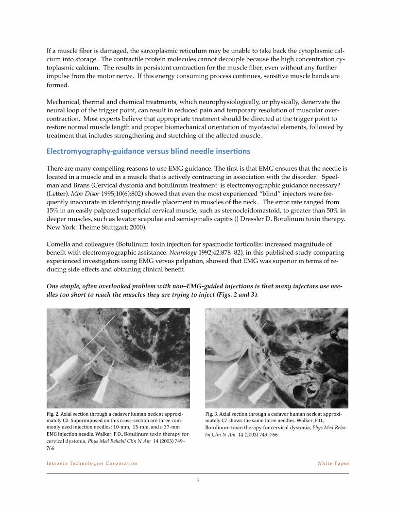
One simple, often overlooked problem with non–EMG-guided injections is that many injectors use nee-dles too short to reach the muscles they are trying to inject (Figs. 2 and 3).
=+1<$><$?@+)*$.#%'+/&$'"(/-1"$)$%),)6#($"-4)&$&#%A$)'$)22(/@+B4)'#*5$C><$D-2#(+42/.#,$/&$'"+.$%(/..B.#%'+/&$)(#$'"(##$%/4B4/&*5$-.#,$+&:#%'+/&$&##,*#.E$FGB447$$FHB447$)&,$)$IJB44$KLM$+&:#%'+/&$&##,*#<$N)*A#(7$=<O<7$Botulinum toxin therapy for cervical dystonia, Phys Med Rehabil Clin N Am 14 (2003) 749–766
=+1<$I<$?@+)*$.#%'+/&$'"(/-1"$)$%),)6#($"-4)&$&#%A$)'$)22(/@+B4)'#*5$CJ$."/9.$'"#$.)4#$'"(##$&##,*#.<$N)*A#(7$=<O<7$Botulinum toxin therapy for cervical dystonia, Phys Med Reha-bil Clin N Am 14 (2003) 749–766.
I n t r o n i x Te c h n o l o g i e s C o r p o r a t i o n! W h i t e P a p e r
3
Most importantly, EMG provides ongoing information regarding anatomy and activation patterns of muscle to the injector not available from any other technique. Pre-injection evaluation provides the injec-tor with unique information regarding:
•Recognition of patterns of activation •Location of muscles •Aspects of anatomy and kinesiology
This is particularly valuable for newer injectors, who rarely have access to large numbers of patients with which to gather experience, familiarity and use of EMG seems to be warranted (Cervical dystonia and botulinum treatment: is electromyographic guidance necessary? (Letter). Mov Disor 1995;10(6):802).
Patients with cervical dystonia may have muscle hypertrophy, so the cadaver measure, in the cited study, may underestimate the depth needed to find target muscles. This is also true for obese patients.
There are are other valuable benefits from needle EMG guidance, which include:
•Confirmation of muscle is particularly useful in cases where the site may be surrounded by essential nerves and blood vessels
•Pre-injection evaluation can lead to use of reduced drug dose and volume, thereby contributing to limit-ing drug diffusion into adjacent areas. This is especially pertinent in cases where drugs will be re-injected on follow up appointments, as the basal EMG activity level can be assessed before a dose decision is final-ized.
>*?"#&*><":-*@#1*A$$&4(,)Acupuncture needling creates a tiny lesion, with bleeding, in the tissue, the contracting muscle immedi-ately relaxes and blood circulation improves in the area. The needling breaks the cycle maintaining the contracture. The needle created lesion disturbs the surrounding tissue and generates an electrical current, or lesion current. Small local bleeding caused by the lesion stimulates secretion of numerous growth fac-tors such as platelet derived growth factor and neurotrophic factors. This promotes healing and regen-eration of damaged tissues, and initiates repairs within a few days. All this happens without the injection of medication into the tissue. (Dr. Chan Gunn, Gunn Approach to the Treatment of Chronic Pain: Intra-muscular Stimulation for Myofascial Pain of Radiculopathic, Churchill Livingstone: 1996)( Yun-Tao Ma, Biomedical Acupuncture for Sports and Trauma Rehabilitation Dry Needling Techniques, Elsevier, 2010)EMG guidance provides value to these procedures by confirming proper needle positions. Myo-guide provides the ability to see and hear the EMG signal, on a real-time basis.
50B*C(),/4*+(3D9:7There are three electrodes used for an EMG guidance procedure: a surface reference electrode, a surface ground electrode, and a hypodermic needle electrode. The hypodermic needle electrode is designed for simultaneous Botulinum Toxin (or other medication) injection and EMG recording, or electrical stimula-tion. The beauty of the process is that once an optimal site is located, the medication can be injected im-mediately through the electrode itself.
Trigger points are located with the help of EMG audio (sound), Signal display (sight), processed EMG indicators (quantitative measures), and possibly stimulation location (used to distinguish specific digits)
Dry needling would simply replace the hypodermic needle with a thin needle electrode for monitoring the site. All the benefits of the procedure would be in place to support optimal electrode placement.
I n t r o n i x Te c h n o l o g i e s C o r p o r a t i o n! W h i t e P a p e r!
4
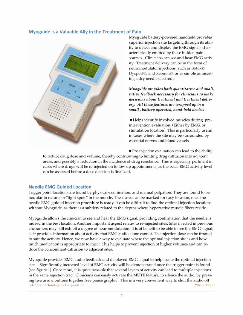
01"):(&$*(.*/*E/4:/<4$*>441*(,*-8$*'#$/-6$,-*"2*+/(,Myoguide battery-powered handheld provides superior injection site targeting through its abil-ity to detect and display the EMG signals char-acteristically emitted by these hidden pain sources. Clinicians can see and hear EMG activ-ity. Treatment delivery can be in the form of neuromodulator injections, such as Botox©, Dysport©, and Xeomin©, or as simple as insert-ing a dry needle electrode.
Myoguide provides both quantitative and quali-tative feedback necessary for clinicians to make decisions about treatment and treatment deliv-ery. All these features are wrapped up in a small , battery operated, hand-held device: •Helps identify involved muscles during pre-intervention evaluation. (Either by EMG, or stimulation location). This is particularly useful in cases where the site may be surrounded by essential nerves and blood vessels
•Pre-injection evaluation can lead to the ability to reduce drug dose and volume, thereby contributing to limiting drug diffusion into adjacent areas, and possibly a reduction in the incidence of drug resistance. This is especially pertinent in cases where drugs will be re-injected on follow up appointments, as the basal EMG activity level can be assessed before a dose decision is finalized.
A$$&4$*50B*B:(&$&*F"3/=",Trigger point locations are found by physical examination, and manual palpation. They are found to be nodular in nature, or "tight spots" in the muscle. These areas an be marked for easy location, once the needle EMG guided injection procedure is ready. It can be difficult to find the optimal injection locations without Myoguide, as there is a subtlety related to the depths where hyperactive muscle fibers reside.
Myoguide allows the clinician to see and hear the EMG signal, providing confirmation that the needle is indeed in the best location. Another important aspect relates to re-injected sites. Sites injected in previous encounters may still exhibit a degree of neuromodulation. It is of benefit to be able to see the EMG signal, as it provides information about activity that EMG audio alone cannot. The injection dose can be titrated to suit the activity. Hence, we now have a way to evaluate where the optimal injection site is and how much medication is appropriate to inject. This helps to prevent injection of higher volumes and can re-duce the concomitant diffusion to adjacent sites.
Myoguide provides EMG audio feedback and displayed EMG signal to help locate the optimal injection site. Significantly increased level of EMG activity will be demonstrated once the trigger point is found (see figure 1). Once more, it is quite possible that several layers of activity can lead to multiple injections in the same injection tract. Clinicians can easily activate the MUTE feature, to silence the audio, by press-ing two arrow buttons together (see pause graphic). This is a very convenient way to shut the audio off I n t r o n i x Te c h n o l o g i e s C o r p o r a t i o n! W h i t e P a p e r!
5
while waiting for the next injection. Any button press will switch the EMG audio back to the previously set volume.
01"):(&$*C=6:4/=",*F"3/=",*G59C=6HMulti-focal sites, such as the hand, can require the use of stimulation to identify the correct injection sites. EMG itself cannot separate the different fingers, or toes, as they all emit EMG signals. Stimulation will invoke a twitch response and identify the digit.
Myoguide’s powerful built in stimulator can challenge the injection site, and invoke a twitch response. Stimulation location is essential in these areas. The site is located by palpation during digit movement. The hypodermic needle electrode is inserted and the patient is asked to move the digit in question (if pos-sible). The site is reinforced as EMG is evoked when the movement is initiated. Stimulation through the same needle will confirm the injection site is related to that digit by evoking a muscle contraction.
Stimulation parameters can be changed easily with several pulse widths, frequency of pulse presentation, and constant current level. Stimulation evokes movement of the digit, as a twitch response, and is achieved at a very comfortable stimulation current. Stimulation can be presented continuously in order to simplify the clinician’s work. A bright yellow panel light illuminates when actively stimulating.
Clinicians can easily activate the stimulation PAUSE feature, to halt stimulation, by pressing two arrow buttons together (see pause graphic). The yellow light will extinguish when the system is paused and the status on the display will indicate “Stimulation Pause”. This is a very convenient way to halt stimula-tion, yet retain all the stimulation parameters. This is useful while repositioning the needle to an im-proved location. Any button press will switch the stimulator back to the previously parameters, and will continue stimulation.
I",34:.(",Trigger point location is an important aspect of myofascial pain management. Myoguide’s comprehensive design provides valuable tools for clinicians treating musculoskeletal problems, associated with pain, spasticity, and movement disorders. Myoguide is a handheld injection guidance system that enables cli-nicians to see and hear, as well as challenge, the injection sites, before any drug is injected. Myoguide quickly becomes an essential tool because it makes finding these sites straightforward. Myoguide pro-vides both quantitative and qualitative feedback necessary for clinicians to make decisions about treat-ment and treatment delivery.
I n t r o n i x Te c h n o l o g i e s C o r p o r a t i o n! W h i t e P a p e r!
6