neck injuries in sports thomas m. howard, md sport medicine
TRANSCRIPT

Neck Injuries in Sports
Thomas M. Howard, MD
Sport Medicine

Anatomy 3-joint complex 50% Flex-Ext Atlanto-
occipital 50% rotation C1-C2 Center of motion
– Flex C 5-6– Ext C 6-7
C2 and C7 most prominent spinous processes

Anatomy 8 cervical roots Normal lordodic
curve helps absorb energy of blows to head and neck
This lordosis is lost @ 30 deg forward flexion

Exam- Motor C5-Deltoid, biceps C6- Biceps, wrist
ext C7-elbow ext, wrist
flex, finger ext C8- finger flexors T1-hand intrinsics

Exam-sensory C5-lateral Deltoid area C6-dorsal thenar web
space C7-MF & RF C8-ulnar side of hand T1-axilla

Diagnoses Cervical Strain Stingers CCN
– Transient Quadraparesis
– Burning Hands Syndrome
Cervical Instability Fractures/subluxation

Epidemiology 10,000 C-spine
injuries/yr in US 5-10% related to
sports Football risk
1.9/100,000 player-yrs Football, wrestling,
gymnastics, diving, surfing, skiing, hockey, rugby
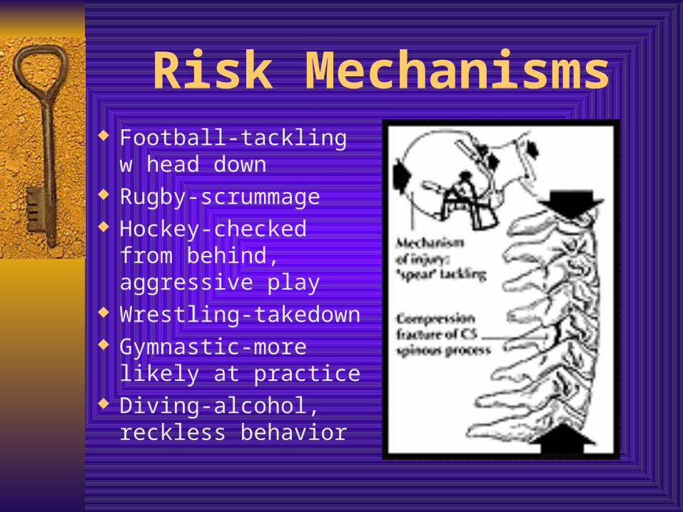
Risk Mechanisms Football-tackling w
head down Rugby-scrummage Hockey-checked from
behind, aggressive play Wrestling-takedown Gymnastic-more likely
at practice Diving-alcohol, reckless
behavior

Cervical Strain AKA Whiplash injury Up to 40% w sx @ 15 yrs Disability highly
associated with job dissatisfaction, female gender, low back pain and prior neck pain
Single best estimate of handicap was return of normal ROM

Stingers Transient UE
neuropraxia of root or brachial plexus– Traction-plexus– Compression-root
Burning in arm Weakness in C5 and C6
distribution– Deltoid, biceps, RC,
wrist extensors, pronator teres
Positive Spurling’s

Stinger RTP Full cervical ROM
w/o pain Neg Spurling’s Full strength

Complicated Stingers Recurrent, prolonged
disability Consider EMG and
MRI of C-spine and plexus
Consider equipment changes upon return
Cervical strengthening

Cervical Cord Neuropraxia Cervical cord “pinch”
– Reduced AP diameter and in-folding of ligamentum flavum
Axial load with hyperextension or flexion Sx last 10 min-48 hrs Pressure on cord causes local increase in
intracellular calcium Mixed neuro findings in 2 limbs or all four

Cervical Spinal Stenosis Acquired stenosis Normal AP diameter 15
mm– 13 considered to be narrow
Torg ratio < 0.8 predictive of future risk of catastrophic injury– Torg ratio < 0.5 with one
episode of neuropraxia have 75% risk of repeat episodes
MRI-functional stenosis– Spinal cord contour
deformation and loss of surrounding CSF

On-field Management Assess LOC and simple
neuro exam by question without moving athlete
Stabilize C-spine and log-roll if necessary to move athlete to back
“Leave helmet on”– Helmet and shoulder pads
Manage airway by removing face mask

Cervical Instability Often following
whiplash-type insult Persistent pain after
appropriate time to recover
>3.5 mm translatory displacement or 11 deg angulation w adjacent vertebrae

Immediate Transport Unconscious athlete Neuro symptoms in 2
limbs Spinous process
tenderness with concerning MOI
Beware of distracting injuries

Clearing C-spine on Field Awake and alert Nl neuro exam No spinous process
pain Full voluntary range
of motion– FF 60 deg– Ext 70 deg– Lat Flexion 45 deg– Rotation 80 deg

Imaging Not Required if… No midline tenderness No focal neuro sx Normal LOC No drugs/meds No distracting injuries

Fractures C1 C2 Flexion injuries Extension injuries

C1 Jefferson fx
– Vertical compression
– Stable
Atlantoaxial rotatory displacement– Rotatory locking of
facets

C2 Odontoid fx Hangman’s Fx
– Hyperextension injury
– Bilat neural arch fx

Flexion injuries Anterior wedge Anterior subluxation
– Post lig complex dispruption
Unilateral locked facets
Bilat locked facets– Jumped and locked
facets– High incidence of cord
damage

Flexion Injuries Clay Shoveler’s Fx
– Avulsion of C6 or 7 spinous process
Teardrop burst fx– Simple or complex
– Most severe with posterior displacement into canal

Extension injuries Pre-vertebral STS Posterior body
displacement Anterior widening of
IVDS Anterior-inferior
avulsion fx Nerve root
compression and cord injury

RTP Full, pain-free Rom Normal neuro
examination Appropriate imaging
studies and specialty consultation
Informed consent of athlete

No Contraindication to Participation*Resolved burnerSpina bifida occultaType 2 Klippel-Feil congenital one-level fusionDevelopmental stenosis of spinal canal (canal/vertebral body ratio <0.8)Mild ligamentous sprain with no laxityHealed, stable compression fracture of vertebral bodyHealed, stable end-plate fractureHealed "clay shoveler's" fractureHealed intervertebral disk bulgeStable, one-level anterior or posterior surgical fusion

Relative Contraindications to Participation*Recurrent acute and chronic burnersDevelopmental canal stenosis with: - episode of cervical cord neurapraxia - intervertebral disk disease - MRI evidence of cord compressionLigamentous sprain with mild laxity (<3.5 mm anteroposterior displacement and 11° rotation)Healed, nondisplaced Jefferson fractureHealed, stable, mildly displaced vertebral body fracture without a sagittal component or neural ring involvementHealed, stable neural ring fracturesHealed intervertebral disk herniationStable, two-level anterior or posterior surgical fusion

Absolute Contraindications to Participation #1
Odontoid agenesis, hypoplasia, or os odontoidiumAtlanto-occipital fusionType 1 Klippel-Feil mass fusionDevelopmental canal stenosis with: - ligamentous instability - cervical cord neurapraxia with signs or symptoms lasting more than 36 hours - multiple episodes of cervical cord neurapraxiaSpear tackler's spineAtlantoaxial instabilityAtlantoaxial rotatory fixation

Absolute Contraindications to Participation #2
Acute cervical fractureLigamentous laxity (>3.5 mm anteroposterior displacement or 11° rotation)Vertebral body fracture with a sagittal componentVertebral body fracture with associated posterior arch fractures and/or ligamentous laxityVertebral body fracture with displacement into the spinal canalHealed fractures with associated neurologic findings or symptoms, pain, or limitation of cervical range of motionIntervertebral disk herniation with neurologic signs or symptoms, pain, or limitation of cervical range of motionAnterior or posterior fusion of three or more levels