nbrc annual report 2014-15 | 1
TRANSCRIPT

NBRC Annual Report 2014-15 | 1 |

| 2 | NBRC Annual Report 2014-15
Compiled and edited by Dr. V Rema and Kedar Singh Bajetha
Front cover: Untitled 32B, A confocal photomicrograph from a P0 parvalbin/tdTomato mouse: V Rema
Back cover: 'Exploring the World' A wall painting: Sheikh Touseef Ahmad

Annual Report 2014-15
National Brain Research Centre Manesar, Haryana (India)


NBRC Annual Report 2014-15 | III |
Contents
Mandate ...................................................................................................................................................... V
From the Director’s Desk ..................................................................................................................... VIII
Molecular & Cellular Neuroscience Division ...................................................................................... 1
• Dr. Sourav Banerjee .......................................................................................................................... 3• Dr. Anirban Basu .............................................................................................................................. 7• Dr. Ranjit Kumar Giri ..................................................................................................................... 14• Prof. Nihar Ranjan Jana ................................................................................................................. 20• Dr. Anindya Ghosh Roy ................................................................................................................. 24• Dr. Ellora Sen ................................................................................................................................... 26• Prof. Pankaj Seth ............................................................................................................................. 30• Dr. Shiv Kumar Sharma ................................................................................................................. 35• Prof. Subrata Sinha ......................................................................................................................... 39
Systems & Cognitive Neuroscience Division..................................................................................... 45
• Dr. Yoganarasimha Doreswamy .................................................................................................. 47• Dr. Soumya Iyengar ........................................................................................................................ 50• Prof. Neeraj Jain .............................................................................................................................. 55
Computational Neuroscience & Neuroimaging Division ............................................................... 59
• Dr. Arpan Banerjee ........................................................................................................................ 61• Prof. Pravat Kumar Mandal .......................................................................................................... 66• Prof. Prasun Kumar Roy ................................................................................................................ 70• Dr.NandiniChatterjeeSingh ........................................................................................................ 78

| IV | NBRC Annual Report 2014-15
Publications, Patents & Presentations .............................................................................................. 87
• Publications ..................................................................................................................................... 89• Patents .............................................................................................................................................. 91• Presentations ................................................................................................................................... 92
Externally Funded Research Projects ................................................................................................... 97
Distinctions, Honours & Awards ........................................................................................................ 103
Academic Programmes .......................................................................................................................... 107
• Ph.D. in Neuroscience .................................................................................................................. 109• Integrated Ph.D. in Neuroscience ............................................................................................... 109• Summer Training & Short Term Programmes ......................................................................... 110
Core Facilities ......................................................................................................................................... 111
• Distributed Information Centre (DIC) ....................................................................................... 113• Animal Facility .............................................................................................................................. 115• Library ............................................................................................................................................ 117
DBT’s Electronic Library Consortium (DeLCON) ........................................................................... 121
National Neuroimaging Facility ......................................................................................................... 127
Translational Research: Clinical Unit ................................................................................................ 131
Centre of Excellence .............................................................................................................................. 135
Lectures, Meetings & Workshops ....................................................................................................... 139
General & Academic Administration ................................................................................................. 147
Institutional Governance Structure & People at NBRC ................................................................. 151
Annual Financial Statements ............................................................................................................... 163

NBRC Annual Report 2014-15 | V |
Mandate
MANDATE• Pursue basic research to understand brain
function in health and disease.• Generate trained human resources with the
capability to carry out inter-disciplinary research in neuroscience.
• Promote neuroscience in India through networking among institutions across the country
OBJECTIVES• To undertake, aid, promote, guide and
coordinate research of high caliber in basic and clinical neuroscience related to diseases and disorders of the nervous system.
• To develop NBRC as the national apex centre for neuroscience research and promoteneuroscienceresearchatdifferentcentres in the country and to provide consulting services to other institutions, agencies and industries.
• To promote, encourage and augment effectivelinkages,alliancesandaffiliationsbetween the Centre and National and International Scientific and ResearchInstitutions, bodies, agencies/laboratories and other organizations working in the fieldofbrainandneurosciencesresearch.
• To establish one or more satellite centers toservedifferentregionsofthecountryfor
efficient achievement of the objectives ofthe Center.
• To collect, assimilate, publish and disseminate data and information on relevant aspects of neuroscience to the scientificcommunity.
• To establish, operate and maintain state-of-the-art facilities as well as databases for carrying research and development activities and make such facilities and databases available to scientists and researchers from all over the country and abroad.
• To provide for instructions and training in such other branches of learning as the Centremaydeemfit.
• To provide facilities for the advancement of research and development to facilitate learning and dissemination of knowledge.
• To undertake extramural studies, extension programmesandfieldoutreachactivitiestocontribute to the development of society.
• To promote, develop, collaborate or otherwise assist in providing services of research, training, consulting or guidance related to neurosciences activities comprising biological, psychological, sociological and clinical aspects; and
• To do all such other acts and things as may be necessary or desirable to further the objectives of the Centre.

| VI | NBRC Annual Report 2014-15

NBRC Annual Report 2014-15 | VII |
The National Brain Research Centre is now in its teens, and in this phase of growth, the results of the converging philosophies that gave rise to the centre are visible, though in a nascent form.Brainresearchhasoftenmeantdifferentthings to different people. Arewe talking offundamental neuroscience – molecules, cells and circuits? Are we talking of disease as it manifests in the patient? From the NBRC perspective, it is both. The thought gap between basic and applied science is sometimes as wide as that between science and humanities as was envisaged by C.P. Snow. However, small as we are in numbers, this does not circumscribe our thoughts and we are always trying to bridge the gap. The generation of new knowledge and perspectives and ‘science for public good’ are not mutually exclusive and contradictory, but form part of a seamless spectrum, a philosophy which we also hope to inculcate in ourstudents.Wecansaywithsomeconfidencethat the breadth of exposure of our students to the gamut of disciplines forming modern neuroscience is amongst the best anywhere.
Research highlights from NBRC include those from Dr. Nihar Ranjan Jana’s laboratory which hasdemonstratedthepotentialbeneficialeffectof enriched environment in the reversal of
Angelman syndrome phenotype in a mouse model of the disease. Dr Ranjit Giri’s laboratory, which also works on neurodegenerative disorders has developed a robust and novel cellular model of prion and Alzheimer’s disease utilizing a CNS stem/progenitor cell culture system. The significance of these cellularmodels of prion and Alzheimer’s disease is that they mimic the cellular pathologies seen in animal models, making it suitable to study molecular mechanisms associated with cellular pathologies in a controlled manner.
The Neuro AIDS laboratory led by Prof Pankaj Seth, is the only laboratory in India using primary cultures of human brain cells to understand cellular and molecular basis of HIV-1 neuropathogenesis. Having carved a niche in area of Neuro AIDS worldwide with its research contributions to discover the clade specificityofHIVneurotoxicity,ithasrecentlyuncovered the role of purinergic receptors in glia-mediated neuronal damage in HIV-1 neuropathogenesis. Currently, the laboratory is involved in deriving human inducible pluripotent cells (Human iPSCs) from blood cells that would offer an iPSC platform forvarious laboratories within NBRC and the neuroscience community of India.
From theDirector’s Desk

| VIII | NBRC Annual Report 2014-15
A major health problem in India is encephalitis, caused by the Japanese Encephalitis Virus (JEV). In endemic areas, JEV causes yearly epidemics of encephalitis which affects both childrenand adults. Dr Anirban Basu’s laboratory at NBRC, whose major focus has been to study JEV, has recently evaluated the involvement of micro RNA-155 in modulating JEV-induced neuroinflammation. TheyhaveobservedthatmiR-155 expression was up-regulated during JEV infection in both mouse and human brain (data obtained from post mortem JE samples). They have also showed that the modulation of miR-155 could be a novel strategy to regulate JEV-induced neuroinflammation. In the pastyear, they have also concluded a clinical trial at King George Medical University at Lucknow on the use of the antibiotic Minocycline for treating JEV infections. This trial demonstrated that Minocyclinehadabeneficialeffect inpatientswith Acute Encephalitis Syndrome (AES), especially in those patients who survived the initial days in hospital. These findings couldform the basis for planning a larger study and possibly including minocycline in the management of AES.
Dr Shiv Kumar Sharma’s group which works on processes involved in memory formation has been interested in the fact that several protein modifications play critical roles in synapticplasticity and memory. However, it is unclear whether thesedifferentmodifications interactin the service of memory. Their recent results suggestthatthemodificationsoftwoproteinsinteract in memory formation.
Research at NBRC also focuses on studying mechanisms underlying various cancers which affect the brain and possible curative agents.Recent studies in Dr Ellora Sen’s laboratory have indicated the involvement of oxidative stress in metabolic programming of glioma cells. Dr Ranjit Giri’s laboratory has shown that curcumin, the active principle of turmeric can induce a newer group of tumour suppressor Bex genes and regulate cell death in cultures of neuroblastoma cells. Further, they have discovered the anti-cancer properties of a novel
DNA intercalating agent [b)quinoline-4(3H)-one] on neuroblastoma cells.
Dr. Sourav Banerjee’s laboratory is focused on investigating fundamental mechanisms related to the development and functions of neuronal circuitry using various biochemical, cell biological, and microscopy based tools as well as whole cell patch-clamp recording. This study will enhance our understanding of how developmental as well as functional impairment of precise neural circuitry leads to emergence of various neurodevelopmental disorders including autism.
Some of the research groups at NBRC are involved in studying various aspects of spinal cord injuries, with a view to understanding changes in the brain and various neural circuits following such injury. Prof Neeraj Jain’s laboratory which has been focusing on spinal cord injury has recently shown that subcortical mechanisms are the major mediators of brain plasticity, with perhaps only a smaller contribution from the cortex. In Dr Anindya Ghosh’s laboratory which works on neural circuit function and repair using the nematode Caenorhabditis elegans as a model system, multiple femto-second lasers were used simultaneously to ablate neuronal processes across the whole length of the worm’s body. C. elegans provides a particularly interesting model to study various questions regarding systems neuroscience, since the connections of every one of its 302 neurons have been mapped and each has been characterized genetically as well.
From the Cognitive and Computational Neuroscience division, Prof Pravat Mandal’s research group has discovered a diagnostic biomarker for Alzheimer disease which has 100% sensitivity and 92% specificity,using state-of-the-art non-invasive imaging techniques. This biomarker was tested on a large cohort of elderly subjects which included normal healthy subjects, subjects with mild cognitive impairment and Alzheimer patients. Incontinuingeffortsto improvethe
From theDirector’s Desk

NBRC Annual Report 2014-15 | IX |
communication skills of autistic children, Dr Nandini C. Singh’s laboratory has recently shown that 'sung word processing' is preserved inthesechildrenandiseffectiveinimprovingboth eye contact and social gestures in such kids, using neuroimaging and behaviour.
On the academic front, in its role as a Deemed University, NBRC awarded the degree of Doctor of Philosophy (PhD) in Neuroscience to 9 students. With these students successfully defending their doctoral research, the total number of PhD degrees that have been granted by NBRC from its inception is 50. One of the major changes in the academics at NBRC is the introduction of the Master of Sciences (M.Sc) programme in Neuroscience which will begin in August, 2015. The M.Sc programme has been envisaged as a two-year programme wherein thefirstyearwillprovideathoroughgroundingin all the subjects which comprise Neuroscience as well as related areas. Besides course-work, a laboratory equipped with various instruments and computer workstations has been dedicated for hands-on practical training for M.Sc students. NBRC continues to provide hands-on training to summer trainees (under the aegis of the Indian Academy of Science, Bangalore, Indian National Science Academy, New Delhi and National Academy of Sciences, Allahabad). Eachtraineeisallottedtoalabandisprovidedhands-on training in Neuroscience for a period of two months to introduce them to the subject. Besides academics, NBRC also encourages students to participate in extra-curricular activities and sports. These are showcased in its annual student festival Tantrika, which combines scientific events, sports, arts andcrafts and is organized by students at NBRC. This year’s Tantrika celebrations included a talk by Dr Maithreyi Narasimha (Dept. of Biological Sciences, TIFR, Mumbai) entitled “How tissues are built and how they heal themselves: insights from epithelial morphogenesis in Drosophila” and culminated in a colourful programme of music and dance performances.
Since its inception, NBRC has used its Foundation Day as an opportunity for
community outreach. The centre’s 11th Foundation Day was celebrated on 16th December,2014byinvitingstudentsfromfiveschools in the Gurgaon/Manesar region were invitedtoattendlectures,posterpresentations,demonstrations and a science quiz on the NBRC campus almost entirely conducted by NBRC students. The highlight of the day was the public lecture entitled “How the Brain Tells theNewfromtheOld(andWhythisMatters)”by Prof Mani Ramaswami, (Trinity College, Dublin, Ireland) who is interested in linking molecular and circuit mechanisms with simple learned behaviours in fruit flies (Drosophilamelanogaster). Another community outreach programme at NBRC is the Brain Awareness Programme, for spreading awareness of the normal physiology of the brain and brain disorders in colleges and schools. NBRC actively supports these events and also provides resources for holding the Brain Awareness Week every year in March. This year, the Brain Awareness Week was held in collaboration with the Presidency University, Kolkata which included talks by Dr Sourav Banerjee and Dr Arpan Banerjee (NBRC). An awareness camp was also organized jointly in Indore by the Seek a Miracle Ataxia Group and NBRC, to increase awareness about ataxia amongst patients, whichwasverywellattended.
It is our sincere hope that the unique blend of research and teaching that is a hallmark of NBRC will form a unique model that will flourishandexpand.Drawingstrengthsfromcontinuity of purpose, as well as adaptability in a rapidly changing world, we shall continue to strive for a greater understanding of the organ that is the least understood frontier of modern biology – the human brain.
Professor Subrata Sinha

| X | NBRC Annual Report 2014-15
From theDirector’s Desk

NBRC Annual Report 2014-15 | 1 |
Molecular and Cellular Neuroscience

| 2 | NBRC Annual Report 2014-15

NBRC Annual Report 2014-15 | 3 |
Tools to connect: novel players in development and functions of neuronal connections
Principal InvestigatorSourav Banerjee
Research Fellows Sarbani Samaddar Pushpa Kumari Balakumar Srinivasan Abir Mondal
Technical Assistant D. Narendar Musadiq Hussain
Brain functions ranging from simple reflexesto complex behaviors are regulated through a vast network of interconnected neurons. Precise neuronal connections or synapses are established with spatial and temporal precision during development, as well as in the adult brain.Synapsesaredynamicandmodificationof synaptic connections or synaptic plasticity occurs in response to neuronal activity. Thus, identifying the processes that regulate synaptic development and function will be essential to understand how development and function of synapses are regulated and how these programs are impaired during various neurodevelopmewntal disorders including autism spectrum disorder.
Regulatory mechanisms of synapse formation by Ubiquitin Proteasome System:
Although we have identified critical factors,such as cell adhesion molecules, ligand-receptor complexes and signaling molecules, we know very little about how these factorsare modulated during synapse formation in vivo. More recently, much interest has been focused on post-transcriptional control of synapse formation, in particular by Ubiquitin Proteasome System (UPS) as they can reversibly fine-tune gene expressionwith spatio-temporal precision. Importantly, emerging reports have turned the spotlight on the non-canonical functions of the UPS in
modulation of nervous system; apart from its conventional role in protein degradation. Of particular interest, we focused on E3 ubiquitin ligases and deubiquitinases (DUBs), critical component of UPS, that can reversibly regulate ubiquitination of target protein to modulate various cellular process. Although, degradative control of synaptogenic program through ubiqutination has been demonstrated previously, novel mechanisms of synapse formation involving non-canonical functions of ubiquitination remains to be elucidated. These non-canonical mechanisms potentially include assembly of protein complexes, protein sorting, protein transport and modulation of activity of signaling molecules.
Towards this goal, we have focused on some of the intriguing questions. These include: (i) are these E3 ligases or deubiquitanases differentiallyexpressedinresponsetoneuronalactivity during synapse formation? (ii) are these E3 ligases or deubiquitinases can specificallymodulate excitatory or inhibitory synapse formation? (iii) do they regulate balance between excitation and inhibition in developing nervous system? (iv) are these factors employ novel mechanisms, other than tagging protein for degradation, to modulate functional synapse formation? (v) What are their targets and how they modulate synaptogenic program with spatio-temporal precision?

| 4 | NBRC Annual Report 2014-15
Figure 1: E3 ligase expression in specific brain area and neuronal type.
Using cultured hippocampal neurons as a model system, our research programme aims to assess role of E3 ubiquitin ligase and deubiquitinases in spatio-temporal modulation of synapse formation. Of particular interest, we are investigating non-canonical role of ubiquitination that can potentially modulate synaptogenic programme. Towards this aspect, wehaveidentifiedspecificcandidateE3ligasesandDUBsthosethataredifferentiallyexpressedduring temporal window of activity regulated phase of functional synapse development.
evaluated by calcium imaging using two-photon microscopy.
Taken together, this study will not only address novel mechansisms of synapse formation through non-canonical functions of ubiquitination but also will elucidate how impairment of these synaptogenic programme leads to onset of neurodevelopment disorders, such as Angelman Syndrome that occurs due to impairment of ubiquitin proteasome system.
Molecular mechanisms of synaptic plasticity involving miRNA-mediated control of protein synthesis at the synapse:
Spatio-temporal regulation of dendritic protein synthesis has emerged as a key modulator of synaptic plasticity. Neuronal activity can induce new protein synthesis at discrete locations along the dendrite that results in persistent structural, physiological, and biochemical changes in dendritic spines. The reversibility of miRNA-mediated regulation of their targets makes them perfect candidates for activity-dependent translational control of neuronal plasticity. miRNAs guide a multi-protein complex, known as the RNA-induced silencing complex (RISC), to specific sites onmRNAs targeted for translational silencing or transcript degradation. Although emerging studies have demonstrated mechanisms involved in RISC –mediated control of dendritic protein synthesis, some intriguing questions are yet to be addressed. These include: (i) how miRNA activity itself is regulated at the synapse? (ii) can modulation of miRNA activity fine-tune structural and functionalchanges at the synapse? (iii) how localized modification of synapse contribute to specificcognitive function including formation of long-term memory?
A recent study has demonstrated that miRNAs can be rapidly degraded in retina upon light induced neuronal activity. This additional layer of regulatory control on miRNA activity has been proposed to be responsible for rapid fine-tuning of miRNA expression. However,detailed mechanisms of activity regulated miRNAturnover,itsimportanceinfine-tuning
Molecular and Cellular Neuroscience Division
Following validation of our screen, we have selected candidate E3 ligases those that are enriched in specific brain area, such ashippocampus, to evaluate their role in synapse maturation. We have employed range of biochemical and cell biological techniques to assess the role of these E3 ligases in development of excitatory and inhibitory synapses after RNA interferance (RNAi) –mediated loss of function. Furthermore, using whole cell patch clamp recording, we are evaluating contribution of these E3 ligases in synapse maturation and maintaing balance between excitation and inhibition of hippocampal circuitry.
To analyze the role of these E3 ligases in synapse formation in vivo, we have performed loss of function before the onset of synapse formation by introducing RNAi through in utero electroporation of embryonic brain. Implications of E3 ligases in regulating functions of hippocampal circuitry are being

NBRC Annual Report 2014-15 | 5 |
Figure 2: in utero electroporation of control lentivirus expressing GFP. Lentivirus co-expressing control short hairpin RNA and GFP was injected into lateral ventricle of Embryonic day 15 (E15) rat embryo and GFP was visualized 28 days (P28) after birth.
of synaptic function de novo, implication of these local changes in modulation of neural circuitry and associated behaviour are poorly understood. Prompted by these observations, we aim to assess how activity regulated miRNA turnover modulate dendritic protein synthesistofine-tunelong-termmodificationsofsynapsesandhowlocalizedmodificationofthese synapses regulate functions of neuronal circuitry involved in cognitive function, such as formation of long-term memory.
To investigate miRNA-mediated control of synaptic plasticity mechanisms in response to neuronal activity, it is critical to identify specificmiRNAsthosethatareenrichedatthesynaptic compartment. Towards this purpose, we have identified several synapticallyenriched miRNAs by deep sequencing based analysis of small RNA library prepared from hippocampal synaptic fraction. After validating synaptic enrichment of candidate miRNAs by quantitative PCR (qPCR) and in situ hybridization, we have selected a subset of miRNAs and measured activity regulated (temporal dynamics) of their turnover using qPCR as well as sensor assay using photoconvertible protein fused to miRNA sensor.
WeobservedthatspecificmiRNAsaredegradedat the synapse upon synaptic activation and their rapid degradation occurs within minute time scale. The time scale of rapid turnover of selective miRNAs suggests that degradative control of miRNA activity could play a role in modulating protein synathesis dependent form of long lasting synaptic plasticity. Furthermore, to investigate mechanistic details of miRNA turnover –mediated control of synaptic plasticity, we have identified factors that can
potentially modulate rapid miRNA turnover and subsequently release translational suppression of specific subset of miRNAregulated transcripts localized at the synapse. WehaveidentificiedtragetofaspecificmiRNAthat is rapidly degradred at the synapse. Experiments are in progress to address how miRNA turnover –mediated control of protein synthesis at the synapse can make long lasting modification of the activated synapse andhow these localized modification can finetune neural circuitry to regulate long-term memory formation. Taken together our study will enumerate novel mechanisms of miRNA-mediated protein synthesis dependent form of long lasting plasticity and its implication in long-term memory formation.
Figure 3: miRNA at the synapse. Synaptic miRNA was visualized by LNA probe based in situ hybridization coupled with immuno staining with synaptic marker, Synapsin I.
| 6 | NBRC Annual Report 2014-15
Molecular and Cellular Neuroscience Division
Presentations
1. Sarbani Samaddar and Sourav Banerjee. “Constructive destruction: Regulatory control of dendritic protein synthesis by activity dependent microRNA turnover”. Cold Spring Harbor Laboratory meeting on “Axon guidance, synapse formation and regeneration.” September 2014.
2. Balakumar Srinivasan and Sourav Banerjee. “Regulation of feeding behaviour by high fat diet induced adult neurogenesis.” National Center for Biological Sciences meeting on “Neuro Modulation of Behaviour.” October 2014.
3. Sourav Banerjee. Dynamic connections: Regulatory control of synaptic plasticity by miRNAs. IBRO School on “Basic & Research Concepts of Depression & Cognitive Dysfunction.” Punjab University, Chandigarh. October 2014.
4. Sourav Banerjee. “Think locally modulate globally: Implications of local control of gene expression in synaptic plasticity”. SERB School on “Brain Circuits” at IISER Pune. December 2014.
5. Sourav Banerjee and Sarbani Samaddar. “Making connections: Role of long non-coding RNA in activity regulated synaptogenesis”. Keystone Symposia, USA. March 2015.
Funding
Ramalingaswami Fellowship, DBT
RNAi grant, DBT
NBRC core fund
Collaborator
Dr. Dasaradhi Palakodeti, inSTEM, Bangalore
Dr. Sharba Bandyopadhyay, IIT, Kharagpur
Dr. Nihar Ranjan Jana, NBRC, Manesar.

NBRC Annual Report 2014-15 | 7 |
Molecular approaches to understand the pathophysiology and pharmacology of infection and inflammatory disorders of Central Nervous System
Principal InvestigatorAnirban Basu
Research Fellow Sourish Ghosh, Shalini Swaroop, Abhishek Verma
DST Inspire Fellow Sriparna Mukherjee
Research Associate Dr Bibhabasu Hazra
Project Assistant Noopur Singh
Technical Assistant Kanhaiya Lal Kumawat
Lab attendant Manish Dogra
While the blood–brain barrier (BBB) protects the central nervous system (CNS) from peripheral immuneandinflammatoryactivation,theCNSis also able to induce the protective innate immune system in response to injury, including trauma, infection, stroke and neurotoxins. This acuteinflammatoryresponseisshort-livedandgenerally beneficial in neutralizing potentialthreats to the CNS by minimizing cellular damage. On the other hand, a sustained chronic neuroinflammatory response can bedetrimental and initiate neuronal damage, neuronal circuits impairments, astrocytic and microglia involvement and neurodegeneration via long-lasting formation and accumulation ofneurotoxicproinflammatorymediators.Therecognition of microglia as the brain’s intrinsic immune system, and the understanding that chronic activation of this system leads to pathologic sequelae, has led to the modern conceptofneuroinflammation.
Our research question evolves around the understanding the molecular basis of host-pathogen interaction in viral infection of the brain and the signaling events associated with neuro-inflammation. In last few yearsour research have been primarily focused on neuropathology of host pathogen interaction in Japanese encephalitis Virus (JEV), and Chandipura Virus (CHPV). Besides viral
encephalitis we are also interested to know the molecular mechanism of IL-1β mediatedmicroglial activation.
Different microRNAs (miRNAs) have beenshown to regulate microglia activation under various pathological conditions including neuroviral infections. Till date, detailed studies pertaining to the involvement of miRNAs in JEV infection have not been reported. Hence, we sought to evaluate possible regulation by miRNAs in mediating JEV induced microglial activation. Our initial screening depicted significant up-regulation of miR-29b in JEVinfected mouse microglia cell line and primary microglia cells. Previous studies have reported tumor necrosis factor alpha-induced protein 3 (TNFAIP3), a negative regulator of NF-ҡBactivation as a potential target of miR-29b. We observed that in vitro knockdown of miR-29b resulted in significant up-regulation ofTNFAIP3 and subsequently decreased nuclear translocationofpNF-ҡB.Further,JEVinducedelevation in inducible nitric oxide synthase (iNOS), cyclooxygenase-2 (COX-2) and pro-inflammatory cytokines were diminishedin BV-2 cells after miR-29b knockdown. Collectively, our study demonstrates miR-29b’s involvement in regulating JEV induced microglialactivation,andpresentsfirstdetailedaccount on the involvement of miRNAs in JEV infection of microglia.

| 8 | NBRC Annual Report 2014-15
Very recently, we have also showed the involvement of miR-155 in modulating Japanese Encephalitis Virus (JEV) induced neuro-inflammation. We observed that miR-155 expression was up regulated during JEV infection of mouse primary microglia, BV-2 microglia cell line, and in both mouse and human brain. In vitro and in vivo knockdown of miR-155 minimized JEV-induced inflammatory responses. We have showedthat the targeting of SHIP1 3’UTR by miR-
155 in the context of JEV infection. Inhibition of SHIP1 by miR-155 resulted in higher interferonbeta (IFN-β) andpro-inflammatorycytokine production through activation of TANK-binding kinase 1 (TBK-1). Based on these observations we conclude that miR-155 modulates neuroinflammatory responseduring JEV infection via negative regulation of SHIP1 expression. Thus, modulation of miR-155 could be a novel strategy to regulate JEV inducedneuroinflammation.(Figure1)
Figure 1: miR-155 expression is up regulated during Japanese encephalitis virus infection (A) Expression of miR-155 in JEV-infected BALB/c mice brain. BALB/c mice were infected with JEV (MOI 5) or mock infected with PBS and brain samples were collected after 0, 3, 5 or 7 days for analysis of miR-155 expression using qPCR. For in situ hybridization brain samples were collected 7 days post infection (magnification = 40X; scale bar = 20µm). * p<0.05 compared to day 0 (un-infected) mouse brain. CON represents brain samples collected from PBS treated mice. (B) miR-155 expression in JEV-infected human brain samples. Formalin-fixed paraffin-embedded sections of control and JEV infected human brain sections were used to isolate miR-155. miR-155 was localized by in situ hybridization (magnification = 20X; scale bar = 20µm). * p<0.05 compared to non-JEV infected human brain. CON represents paraffin-embedded sections prepared from biopsy of non-JEV infected human brain sample. (C) Time-dependent expression of miR-155 in BV-2 cells exposed to JEV or UV irradiated JEV. BV-2 cells were exposed to JEV or UV-irradiated JEV for 0, 3, 6, 12 or 24 hr and expression of miR-155 was evaluated using qPCR. * p<0.05 compared to un-infected BV-2 cells. (D) Expression of miR-155 in JEV- infected primary microglia cells. Primary microglia cells were isolated from P0-P2 (post natal day 2) old BALB/c mouse pups, cultured for 12 days, and exposed to JEV for 12 or 24 hr. * p<0.05 compared to un-infected primary microglia cells. CON represents primary microglia cells treated with PBS for 12 or 24 hr. Expression of miR-155 was evaluated using qPCR. Data represent mean ± SEM from 5 mice/3 human samples per group or 3 independent experiments for in vitro study.
Molecular and Cellular Neuroscience Division

NBRC Annual Report 2014-15 | 9 |
The impact of emerging and reemerging viral induced encephalitis is being felt globally. In India, although many encephalitis outbreaks have been associated with JEV several outbreaks have remained undiagnosed. The association of Chandipura virus (CHPV) with the recent outbreak of acute encephalitis in Andhra Pradesh implicates that this virus should be considered as an emerging pathogen of substantial public health importance. Chandipura Virus (CHPV) belongs to the Rhabdoviridae family in the order Mononegavirales of genus Vesiculovirus. Interestingly, its continuing mutating trend has enhanced its lethality to cause human infections unlike its genetic cousin, the Vesiculo Stomatitis Virus (VSV). Moreover, its complete genome sequencing has proved the uniqueness that the virus has acquired to be recognized as a new threat to humanity. Presently it has been reported that CHPV is more closely related to its Asian kin the Isfahan Virus (ISFV) and also have similarities in sequence with those of the new world viruses such as Indiana, New Jersey, Alagoas and Piry Virus which happens tobedifferentserotypesofVSV.
CHPV has been reported to be an emerging human pathogen in Indian subcontinent with a fatality rate of around 55-77%. Children below 15 years of age are more vulnerable than adults who show symptoms similar to any other encephalitis such as high grade fever, vomiting, altered sensorium, generalized convulsions, decerebrate posture and grade IV coma. Sand fly of Phlebotomus spp. and Sergentomyia spp. has been reported to be the major carriers of CHPV during the outbreaks in recent years. CHPV is (-) stranded enveloped RNA virus with an approximate genome length of 11kb comprising of 5 genes which translates a glycoprotein (G), a matrix protein (M), a nucleoprotein (N), a phosphoprotein (P) and a large/ polymerase (L) protein that helps the virus to successfully enter the host and replicate. There have been few studies that have identified general pathological mechanisms
associated with the infection. As common with other neurotropic viruses CHPV has also been reported to breach the blood brain barrier (BBB) and enter the CNS to cause encephalitis and neuronal death. But the precise mechanism by which the virus is causing neuronal death has not been deciphered so far.
Chandipura Virus has been previously reported to induce neuronal death through Fas-mediated extrinsic apoptosis pathway from our lab, but it was unclear that what propelled this apoptosis. Lipid metabolism has been evidenced to play a vital role in viral replication. Also oxysterol binding proteins along with oxysterols have been reported to be key players in neurodegeneration. In our present study we identified upregulationof genes like Apolipoprotein E (ApoE), Cholesterol 24-hydroxylase (Cyp46a1), Sterol regulatory element-binding transcription factor 1 (Srebf-1) and Sterol-4-alpha-carboxylate 3-dehydrogenase, decarboxylating (Nsdhl) from CHPV infected brain sample. CHPV replication demands influx of lipidswhich is supplied by ApoE which transports cholesterol through LDL receptor which has been illustrated in our present study with over-expression of LDL receptors in CHPV infected neurons. Hence an upsurge of cholesterol concentration has been evidenced within neurons which triggers the expression of Cyp46a1 enzyme culminating into conversion of cholesterol to 24 (S) Hydoroxycholesterol (24S- OHC). The increase of oxysterols within cells stimulates the expression of oxysterol binding protein (OSBP) and its related proteins (ORPs). Increased 24(S)- OHC concentration is also toxic to neurons, which propels neuronal apoptosis through the Fas-mediated extrinsic apoptosis pathway. This is for the first timeperturbation of cholesterol homeostasis in brain is reported to be utilized by the viruses for both maturation and the release of its virions outside the cells for continuing the process of neuropathogenesis. (Figure 2)

| 10 | NBRC Annual Report 2014-15
Molecular and Cellular Neuroscience Division
Figure 2: LDL receptor knockdown hindered internalization of externally supplied cholesterol in Neuro2a cells. Cholesterol internalization in neurons happens through ApoE-LDLr association in vivo. In order to verify the role of LDLr in vitro system we supplied 200µM cholesterol and 5 µM ApoE to the cell culture medium post knocking down the expression of LDLr, hence infected the cells with CHPV. The experiment had 4 treatment groups as: mock infected, CHPV infected, CHPV infected + esiLDLr treated, CHPV infected + esiEGFP treated (scrambled). Double immunocytochemistry staining of LDLr/ Nile Red was performed to analyze the internalization of cholesterol post CHPV infection. The representative image panels (A) show internalization of externally supplied cholesterol was significantly reduced in the LDLr knockdown treatment group compared to CHPV and scrambled groups (image scale= 20 µm; inset scale= 10µm) (n=3). Immunoblot analyses (B) show expression of LDLr and CHPV N protein from the similar analysis groups as in (A). β-Actin was used as a loading control (n= 3). The immunoblot result shows successful knockdown of LDLr which resulted in reduced internalization of cholesterol as shown in (A). Hence this resulted in reduced CHPV replication as evident from the immunoblot analysis of CHPV N protein.
Alongside with viral encephalitis, our laboratory is also deeply engaged in basic research to understand the transcriptional regulation of microglial activation. Microglia are the resident macrophages of the Central Nervous System (CNS), which secrete several pro- and anti-inflammatory cyto-chemokines in response to pathogenic stimuli. One key player that is believed to drive this neuroinflammatory process is interleukin(IL)-1β, a pro-inflammatory cytokine that isup-regulated in Alzheimer’s disease (AD), Parkinson’s disease, multiple sclerosis, and other neurodegenerative disorders. Once secreted, IL-1β binds to IL-1 receptor presenton microglia and initiates the production of inflammatorycytokinesinmicroglia.Microgliaare the resident macrophages of the CNS, which secrete several pro- and anti-inflammatory
cyto-chemokines including interleukin-1β (IL-1β), in response to pathogenic stimuli. Oncesecreted, IL-1β binds to IL-1 receptor presenton microglia and initiates the production of inflammatory cytokines in microglia.However, the detailed information regarding themolecularmechanisms of IL-1β triggeredinflammatorypathwaysinmicrogliaislacking.Recently we have performed the proteomic profiling of the N9 microglia cells with andwithout IL-1β treatment and identified thedifferentially expressed protein spots withthe help of MALDI-TOF MS/MS. We observed that the proteins being affected by IL-1βadministration in microglia are involved in unfolded protein response (ER stress), oxidative stress, apoptosis and cytoskeleton proteins and are involved in the activation of various cellular stress pathways (Figure 3).

NBRC Annual Report 2014-15 | 11 |
Figure 3: IL-1β up-regulates the expression of HSP60 in microglia. Primary microglia and N9 murine microglia cells were treated with IL-1β at a concentration of 5 ng/mL for 3 hours. (A.) Immunostaining of primary microglia for HSP60 [Alexa flour 488; green], Iba-1[Alexa flour 594; Red], 4,6-diamidino-2-phenylindole (DAPI) (blue) and merged images (HSP60+ Iba-1+ DAPI) (B.) Immunostaining of N9 murine microglia for Expression of HSP60 [Alexa flour 488; green], 4,6-diamidino-2-phenylindole (DAPI) (blue) and merged images (HSP60+ DAPI). HSP60 increases in IL-1β treated primary microglia and N9 murine microglia as compared to untreated control cells as shown by the arrows. Images were captured using Zeiss apotome fluorescence microscope (Scale bar-20 µm; magnification- 40X).
Publications
1. N Sharma, R Verma, K L Kumawat, A Basu and S K Singh (2015) miR-146a suppresses cellular immune response during Japanese encephalitis virus JaOArS982 strain infection in human microglialcells.JournalofNeuroinflammation18;12(1):30
2. S Mukherjee, S Ghosh, A Nazmi, and A Basu (2015) RIG-I Knockdown Impedes Neurogenesis in a Murine Model of Japanese Encephalitis. Cell Biol Int 39(2):224-9.
3. P P Manna, S K Hira, A Basu, and S Bandyopadhyay (2014) Cellular therapy by allogeneic macrophagesagainstvisceralleishmaniasis:roleofTNF-α.CellularImmunology290(1):152-163.
4. ANazmi,SMukherjee,KKundu,KDutta,AMahadevan,SKShankar,andABasu(2014)TLR7isa key regulator of innate immunity against Japanese Encephalitis Virus infection. Neurobiology of Disease 69: 235-247.
5. D K Kaushik, M C Thounajam, A Mitra, and A Basu (2014) Vespa tropica venom suppresses lipopolysaccharide mediated secretion of pro-inflammatory cyto-chemokines by abrogatingnuclearfactor-κBactivationinmicroglia.InflammationResearch63(8):657-65.
6. M C Thounajam, K Kundu, D K Kaushik, S Swaroop, A Mahadevan, S K Shankar, and A Basu (2014)MicroRNA-155RegulatesJapaneseEncephalitisVirusInducedInflammatoryResponsebyTargeting src Homology 2-Containing Inositol Phosphatase-1. Journal of Virology 88(9): 4798–4810
7. M C Thounajam, D K Kaushik, K Kundu, and A Basu (2014) microRNA-29b Modulates Japanese Encephalitis Virus Induced Microglia Activation by Targeting Tumor Necrosis Factor Alpha-induced Protein 3 (TNFAIP3) Journ of Neurochemistry 129(1):143-54
8. G D Manocha, R Mishra, N Sharma, K L Kumawat, A Basu and S K Singh (2014) Regulatory role of TRIM21 in type-I interferon pathway in Japanese encephalitis virus infected human microglial cells. Journal of Neuroinflammation 11:24
As the information on the mechanistic details of IL-1β mediated microglial activation islacking, our study will enable us to understand
the overall molecular mechanistic study of neuro-inflammation and will fill the gaps inour understanding of the same.

| 12 | NBRC Annual Report 2014-15
Molecular and Cellular Neuroscience Division
Review/Editorial/Commentary 9. ANazmi,BHazra,KDutta,andABasu(2014)RoleofPatternRecognitionReceptorsinFlavivirus
Infections. Virus Research 185:32-40.
Presentation
1. A Basu (2015) Japanese Encephalitis-Basic to translational research Pub Kamrup College, Baihata Chairali, Kamrup, Assam; 26th March, 2015. [Invited speaker as a part of DBT CTEP sponsored series of popular lecture programme]
2. A Basu (2015)Neuroinflammation: a potential therapeutic target. 21stAnnual Symposium onNeurodegeneration-Ranbaxy Science Foundation; 9th March 2015, ICGEB, New Delhi.
3. A Basu (2015) Heating things up-how inflammation drives brain disorders: From basic totranslational research. Presidency University, Kolkata; 3rd March 2015.
4. ABasu(2015)IL-1βmediatedinflammatoryresponseinmicroglia.Neurocon-2015,Haldia,WestBengal; 7-10th Januray, 2015.
5. A Basu (2014) Host pathogen interaction in Japanese encephalitis virus infection: from bench to bedside. 17th International Symposium of Molecular Medicine, 9-11th October, 2014, Athens-Greece [Speaker and session chair]
6. A Basu (2014) Host pathogen interaction in Japanese encephalitis virus infection: basic and translational studies; 10th September, 2014; CCMB, Hyderabad.
7. A Basu (2014) The transcriptional regulation of microglial function: the tightrope walk of CNS innateimmuneresponse.12thmeetingoftheAsian-PacificSocietyforNeurochemistry,23-26thAugust, 2014, Kaohsiung, Taiwan[Symposium organizer, speaker, and session chair]
8. A Basu (2014) Drug Discovery in Academic Set up. Salwan Public School, Gurgaon; 26th July, 20149. ABasu(2014)NeglectedTropicalDiseasesinconflictZones.InternationalWorkshoponPerspectives
on Science & Technology Diplomacy for Sustainable Development in NAM and other Developing Countries, 27th-30th May, 2014; Heritage Village Resort, Manesar. [Speaker and session chair]
Funding
• Identificationandcharacterizationofbraincellularmembranecomponentsactingasreceptorsfor Japanese Encephalitis virus.[CSIR, 27(0307)/14/EMR-II]
• To study the molecular mechanism of microglial activation and identify the therapeutics targets critical for IL-1β signaling inbrain following inflammation. [Departmentof Scienceand Technology, No SB/SO/HS-070/2013]
• Implementing proteomic approach to understand the etiology of Neuropathogenesis induces Chandipura Virus infection [Department of Biotechnology (BT/PR7907/MED/29/702/2013)]
Collaborators
• Arpan Banerjee and Pankaj Seth, NBRC• Rashmi Kumar, Dept of Pediatrics,CSM Medical University, Lucknow

NBRC Annual Report 2014-15 | 13 |
• SK Shankar, and Anita Mahadevan, NIMHANS, Bangalore• James Gomes, School of Biological Sciences, IITD• BiswadevBishayi,DepartmentofPhysiology,UniversityofCalcutta,Kolkata• Sunit Singh, BHU, Varanasi.• Dhrubajyoti Chattopadyay, BC Guha Center for Genetic Engineering and Biotechnology,
UniversityofCalcutta,Kolkata• SomaChattopadhyayandAmolSuryawanshi,InstituteofLifeSciences,Bhubneswar.• Sudhanshu Vrati, Arup Banerjee and Guruprasad Medhigeshi, Vaccine and Infection Disease
Research Center, THSTI, Faridabad
Award
1. Tata Innovation Fellowship (Department of Biotechnology)-2014-152. Rajib Goyal Prize in Life Sciences-2012-13 (has been awarded in the year 2015)3. J B Srivastav Oration Award of ICMR-2011 (has been awarded in the year 2015)
Degree Awarded (Ph.D.)Arshed Nazmi

| 14 | NBRC Annual Report 2014-15
Development of neurosphere and mouse models to study the effect of prion replication on the mechanisms behind cellular and brain pathologies of prion disease
Principal InvestigatorRanjit Kumar Giri
Research Fellow Himakshi Sidhar
Technical Assistant Rajiv Kumar Mishra
Lab Attendant Lalit Bidla
Although rare, prion disease itself includes varieties of irreversible neurodegenerative disorders such as Kuru, Creutzfeldt-Jakobdisease (CJD), variant CJD (vCJD), Gerstmann-Sträussler-Scheinker syndrome (GSS) and fatal familial insomnia (FFI) in humans; scrapie in sheep and goats, and bovine spongiform encephalopathy (BSE) in cattle. Unlike otherneurodegenerative diseases, all prion diseases generate a defective protein which itself is infectious. Infectious nature of prion disease poses a serious health risk to cattle, deer,goat, sheep, as well as to human from food derived from these animals, blood transfusion and organ transplantation. In prion disease, the normal cellular prion protein (PrPC) is converted post-translationally to pathological, infectious and alternatively folded isoforms (PrPSc). Replication of PrPSc and subsequent multimerization and accumulation in brain parenchyma is the key step in the onset and pathoprogression of prion disease.
In a prion diseased brain, all types of adult brain cells come in contact with prion aggregates but neurons are mainly killed as disease progress. Whether PrPSc accumulation is sufficient totrigger neurodegeneration is questionable. Both human diseases and animal models of these diseases have not been able to differentiatethe effect of prion protein on various brain
cell types. To avoid such complexities, cell culture models of prion disease have been reported. However, such models also fail to address various mature brain cell types and replicate defective protein consistently. CNS stem cells containing neurosphere cultures can be isolated from embryo and adult brains, can be grown over long period of time and most importantly,differentiatetowardsmajoradultbrain cell types such as, neurons, astrocytes and oligodendrocytes. Therefore, we proposed the feasibility of testing the effect of prionreplication on various brain cell types and their cytopathological changes employing neurosphere cultures replicating prion proteins de novo.
In order to study the cytopathological changes in matured brain cells to prion replication, we developed neurosphere lines from fetal mouse brain (E15) of C57BL/6J mice. Fetal mouse brain cells when grown in neurobasal media supplemented with Glutamax, N2 supplement, EGF, FGF-b and LIF grew into ball of cells within 15-20 days (Figure 1A) and express nestin (commonly used marker for CNS stem/progenitor cells) in majorities of cells (data not shown here). These neurosphere cultures were tested for the expression of cellular prion protein (PrPC), the substrate for replication of prion protein. Western blot analysis show

NBRC Annual Report 2014-15 | 15 |
C57BL/6J neurosphere cultures express PrPC and these proteins are mostly present in diglycosylated form as reported in mouse brain (Figure 1B). To validate our Western blot result we also performed immunohistochemistry on neurosphere sections. The results show, PrPC protein is expressed in most of the cells in neurospheres making it suitable in vitro system to replicate mouse prion protein (RML scrapie protein serially passaged in CD1 mice) (Figure 1C).
Figure 1: Development of neurosphere cultures from C57BL/6J mice embryos and characterization of PrPC ex-pression in these neurospheres by Western blot and Immuno-histochemistry analysis.
Furthermore, to validate the alteration of cellular processes seen in neurosphere cultures, we have also developed a robust mouse model of prion disease similar to other prion labs across the world. Both CD1 and C57BL6 mice were injected intracerebrally with RML scrapie mouse brain homogenate or left untreated. Infected mice exhibit plastic tail and have serious gait problem during the late stage of disease. At the end stage of the disease (when animals have difficulty ininitiating walking), the brain was isolated, cut
sagitally into two parts and frozen rapidly on ethanol dry ice bath. Left hemispheres were utilized for cryo-sectioning followed by either histoblot or immunohistochemical analysis. Right hemispheres were used for biochemical analysis. Results from histo-blot clearly demonstrate the accumulation of prion protein in RML prion infected mouse brain and are resistant PK-mediated proteolysis (Figure 2A). Accumulation of prion protein is more prominent in cortex, thalamus, pons and
cerebellum of the brain. Dot blot analysis also suggests the presence of PK-resistant prion protein only in RML-infected mice brains but not at all in normal mouse brain (Figure 2B). Both accumulation and PK-resistant prion protein is also seen in western blot analysis (Figure 2C). Taken together, mice infected with prion inoculum synthesized new prion protein, accumulate in brain and progressively develop the prion disease both in CD1 and C57BL/6J mice.

| 16 | NBRC Annual Report 2014-15
Molecular and Cellular Neuroscience Division
Figure 2: De novo synthesis and accumulation of mouse prion protein in terminally sick CD1 mice infected initially with RML mouse scrapie prions as demonstrated by histo-blot, Dot blot and western blot analysis.
Reactive astrogliosis is another key pathology of prion disease and in animal model of prion disease. Brain sections from uninfected and infectedmicebrainswereobtained,fixedin4%paraformaldehyde and immunostained with anti-GFAP antibody. Results show a dramatic increase in GFAP positive cells in RML infected mouse brain and at various brain regions like cortex, thalamus, cerebellum and olfactory lobe suggesting increased astrogliosis in prion diseased mice brains is similar to earlier reports. Furthermore, the intensity of GFAP
stain in GFAP positive cells are visibly higher in RML prion infected brain than control brain indicating reactive astrogliosis as seen in prion diseases of human and animals (Figure 3).
Collectively, increased prion accumulation and reactive astrogliosis in RML scrapie prion infected mice established mouse models of prion disease for our future studies on the mechanisms behind brain pathologies of prion disease.
Figure 3: Increased reactive astrogliosis is observed in RML scrapie prion infected mice brains than uninfected mice by immunohistochemistry for GFAP.
A B
C

NBRC Annual Report 2014-15 | 17 |
Presentation
1. Giri RK, Cellular models of prion disease. Indo-US symposium on viral infections on nervous system, NBRC, Gurgaon, India, February 2014.
Funding
This work is funded by NBRC Core and partially by Ramalingaswami fellowship (102/IFD/SAN/758/2007-08) and a grant on prion disease (BT/PR10721/Med/30/105/2008) from DBT, New Delhi.

| 18 | NBRC Annual Report 2014-15
Curcumin-mediated neuro 2a neuroblastoma cell death involves intrinsic apoptotic pathway and induction of proapoptotic Bex genes
Principal InvestigatorRanjit Kumar Giri
Research Fellow Himakshi Sidhar
Reseach Assistant Upasana Sahu
Lab Attendant Lalit Bidla
Neuroblastoma is the most common childhood tumor of extra cranial nervous system. These tumorsdevelopfromuncommittedneuralcrestcells and are present along the sympathetic chain, mostly in abdomen or chest. According to International Neuroblastoma Staging System, these tumors are categorized into four differentstages(I-IV).Neuroblastomapatientswith stage III or IV tumors are at higher risk of death. Stage IV tumors are metastatic and spread to distant sites like lung, liver and/or bone marrow, and their prognosis is very poor. These neuroblastomas are not sensitive to commonly used chemotherapeutic agents.
Genome of tumor cells is perturbed in almost all cancerous cells. Proapoptotic genes are either silenced or inactivated in tumor cells. The Bex (Brain–Expressed X- linked) genes belong to a small family of genes including Bex1, Bex2, Bex3, Bex4 and Bex6 in mouse while Bex5 instead of Bex6 in humans. All these genes are located on X-chromosome except Bex6, which is located on chromosome 16 in the mouse genome. These genes are shown to be expressed majorly in brain and show high sequence similarities. Bex1 andBex2havebeen identifiedas tumorsuppressor genes and silenced in malignant glioma. Viral mediated re-expression of Bex1 or Bex2 gene enhanced chemosensitization and apoptosis in glioma cells. Bex3 has also been
reported as a pro-apoptotic protein by inducing apoptotic signaling through its interaction with p75NTR. Interestingly, role of Bex1, 2 and 3 have not been studied in any neuroblastoma cells and there is no report on the association of Bex4 and Bex6 genes with any type of cancer. Currently, it is not possible to re-express these genes employing gene therapy in wide range of cancers and at various tissues simultaneously. Therefore, manipulating tumor cells genome by safe natural compounds to induce Bex genes can be of importance to control cancer cells growth.
There is no report on utilization of a small molecule or phytochemical to induce Bex gene family endogenously. Literature survey strongly suggests that curcumin, an active ingredient of turmeric, has a wide range of molecular targets and it activates many pro-apoptotic genes to induce apoptosis in various cancer cell types. Curcumin also activates transcription factor p53 that has been known to play an important role in apoptosis. Therefore, we hypothesized that curcumin-mediated neuroblastoma cell death might engage Bex genes products by inducing endogenous Bex genes expression.
In this study, we found that curcumin rightly engages cellular machineries to induce both

NBRC Annual Report 2014-15 | 19 |
Bex genes as well as apoptosis in a murine neuroblastoma neuro 2a cell line. The cellular signaling behind curcumin-mediated induction of Bex genes suggests the involvement of p53 activation. In addition, Pifithrin-α, a knownp53 inhibitor inhibited curcumin-mediated Bex genes induction and apoptotic neuro2a cells death suggesting the direct involvement of Bex genes in apoptotic cell deaths. Other cellular targetofpifithrin-α, suchas,Egr-1 andHSF1
are also activated prior to Bex genes induction suggesting the role of p53, Egr-1 and HSF1 in curcumin-mediated induction of Bex genes. Thus, for the first time these studies suggestthat induction of Bex genes are associated with cellapoptosisandcanbe inducedspecificallyby curcumin, the active principle of turmeric, used as a curry spice since ages.
Figure 1: Curcumin-mediated cell death involve intrinsic apoptotic pathways and induce Bex genes. A) LIVE DEAD assay indicates curcumin kills neuro 2a cells in a dose-dependent manner. Green=Calcein=Live and Red=Ethidium homodimer=Dead. B) Curcumin treated neuro 2a cells show increased nucleosomal DNA fragmentation in a dose dependent manner indicating the involvement of apoptotic pathways. C) Curcumin activates intrinsic apoptotic pathway in neuro 2a cells. D) Curcumin induces the proapoptotic Bex1, 2, 3, 4 and 6 genes in neuro 2a cells which are sustained upto 24 hours. GAPDH is used as loading control.
Presentation
1. Sidhar H and Giri RK. Curcumin-Mediated Neuro 2a Neuroblastoma Cell Apoptosis is Associated with Induction of Bex Gene Family. 32nd IAN annual meeting, NIMHANS, Bengaluru, Karnataka, India, November, 2014 (Poster Presentation).
2. Giri RK. Understanding the Mechanisms of Neuroblastoma Cell Death by a Novel Anticancer Agent, 8-Methoxypyrimido[4',5':4,5]thieno(2,3-b) Quinoline-4(3H)-One. Vellore Institute of Technology University, Vellore, Tamilnadu, India, November 2014 (Invited Lecture).
Funding
This work is supported by NBRC Core and partially by Ramalingaswami fellowship (102/IFD/SAN/758/2007-08) and a grant on Alzheimer’s disease from BIRAC (BIRAC/CRS/CRS-0004/CRS-01/2012), New Delhi, India.
Collaborator
Sathees C. Raghavan, Dept. of Biochemistry, Indian Institute of Science, Bangalore 560012
| 20 | NBRC Annual Report 2014-15
Molecular and Cellular Neuroscience Division
Understanding the physiological function of Ube3a and pathogenesis of Angelman syndrome
Principal InvestigatorNihar Ranjan Jana
Research Associate Vivek Tripathi
Research fellows Imran Jamal Brijesh Kumar Singh Naman Vatsa Shashi Sekhar Vipendra Kumar Shruti Nagaral
Technical assistants Ankit Sharma Mahendra Singh
Ube3a was first identified as a cellularprotein connected with ubiquitin-dependent proteolysis of tumour suppressor p53 in assistance with E6 oncoprotein of the human papilloma virus (HPV). Subsequently, it is characterized as an E3 ubiquitin ligase involved intargetingspecificproteinforubiquitinationand degradation through proteasome. Ube3a also acts as a transcriptional co-activator of steroid hormone receptors. Ube3a shows neuronspecificimprintinganddysfunctionofmaternally inherited Ube3a causes Angelman syndrome (AS), a neurodevelopmental disorder characterized by severe mental retardation, speech impairment, susceptibility to seizures, ataxia and unique behavioural features such as improper laughter and autistic features. Duplication of the UBE3A gene is also reported in autism. Ube3amaternal deficientmice (AS mice) recapitulates many essential features of AS, including cognitive and motor dysfunctions. These AS mice also display defects in hippocampal LTP, altered function of hippocampal CAMK-IIα and abnormaldendritic spine morphology. Studies in these mice also provided further evidence that Ube3a is perhaps necessary for development of synapse and experience-dependent synaptic plasticity.
Past several years we are engaged in exploring the physiological function of Ube3a and how its altered level is linked with either AS or autism. Our primary aim is to identify and characterise novel substrates of Ube3a. We are also trying to understand the molecular mechanism of behavioural deficits inAS mouse model andhow that can be reversed.
Recently, we have shown that altered glucocorticoid receptor signalling in the hippocampus could lead to chronic stress and increased anxiety-like behaviour in a mouse model of AS (Godavarthi et al. Hum. Mol. Genet., 2012). We have also observed that chronic stress in these mice could lead to down-regulation of parvalbumin (PV)-positive GABAergic interneuron in the hippocampus and basolateral amygdale (BLA) from early postnatal days, which can be partially reversed bychronictreatmentoffluoxetine(Godavarthiet al. J. Neurochem., 2014). Since AS mice exhibit deficits in experience-dependentsynaptic plasticity and are under chronic stress, we further tested the influence of enrichedenvironment in the recovery of behavioural abnormalities of these mice. Rearing AS mice under enriched environment just after weaning for5-9monthssignificantlyimprovedcognitive

NBRC Annual Report 2014-15 | 21 |
andmotor deficits as seen from novel objectrecognition and rotarod tests respectively. Enriched environment also significantlyalleviates anxiety-like behaviour and partially restored blood corticosterone level in these mice. Immunoblot analysis revealed rescue of altered levels of BDNF, GR and pThr286CaMKII in the hippocampus of AS mice that are reared in enriched environment. Interestingly, enriched environment also significantly increased thenumber of PV-positive GABAergic neurons in the hippocampus and BLA of AS mice. These results indicate potential beneficialeffectofenrichedenvironment in thereversalof AS phenotype. In another project, we are exploring novel inducers of Ube3a and we have preliminary data indicating that HDAC inhibitor induces the expression of Ube3a. Currently, we are studying the role of HDAC inhibitor in the reversal of disease progression in AS mouse model.
Role of Ube3a in the progression of neurodegenerative disorders using mice models
The accumulation of intracellular protein deposits as inclusion bodies is the common pathological hallmark of most age-related neurodegenerative disorders including Huntington’s disease (HD) and Alzheimer’s disease (AD). Appearance of aggregates of the misfolded mutant disease proteins suggest that thecellsareunabletoefficientlydegradethem,and failure of clearance leads to the severe disturbances of the cellular protein quality control system. Earlier we have demonstrated that Ube3a function as a cellular protein quality control ubiquitin ligase and involved in the clearance of misfolded mutant huntingtin that causes HD (Mishra et al. J. Biol. Chem., 2008, 2009). We have also recently demonstrated that the partial loss of function of Ube3a might be associated with synaptic dysfunction in HD transgenic mice brain (Maheshwari et al. J. Biol. Chem. 2012).
In order obtain further insight of the role of Ube3a in HD pathogenesis, we have generated brain Ube3a deficient HD transgenic mice.Ube3a gene is paternally imprinted in the brain and we have taken the advantages of preferential expression of maternal Ube3a to generate Ube3a-maternal deficient HD micethat will not express Ube3a in the brain. We observed that removal of Ube3a selectively from HD mice brain caused accelerated disease phenotype and shorter lifespan compared with HD mice (Maheshwari et al. Hum. Mol. Genet. 2014). The deficiency of Ube3a inHDmice brain also caused significant increase inglobal aggregates load and these aggregates were less ubiquitinated when compared with age-matched HD mice. These results indicates the crucial role of Ube3a in the progression of HD and its enormous potential as therapeutic target. The UBE3A gene is paternally imprinted inthebrainwithpreferentialmaternal-specificexpression particularly in neurons. Therefore, reactivation of paternal Ube3a expression could beanattractivestrategytoslowdowntheHDprogression. Interestingly, a recent report has demonstrated that toposimerase inhibitors can activate the dormant expression of Ube3a in neurons. Some of the topoisomerase inhibitors are FDA approved drugs for treating particular types of cancer. We started our investigation with a specific topoisomerase inhibitors(topotecan) that can cross blood brain barrier. Initially, we spend several months to optimize the tolerable dose of topotecan in mice and found that topotecan at a dose of 2mg/kg body weight (intra peritoneal injection) is well tolerable. After optimizing the dose, we injected topotecan to HD mice of 8 weeks old. Each HD mice received 4 doses at two days interval. It is important to note that most of the behavioural abnormalities can be clearly visible from 8 weeks onwards that progressively increased over time and mice begin to die by 11-12 weeks of age. We have observed that injection of topotecan significantlyimprovedthemotorabnormalities

| 22 | NBRC Annual Report 2014-15
Molecular and Cellular Neuroscience Division
in HD mice as evident from the clasping experiment and rotarod performance task. Most interestingly, the lifespan of topotecan treated HDmicewassignificantlyimprovedcomparedto HD controls. Currently, we are checking the possibleeffectoftopotecanonglobalaggregateload, Ube3a expression, neuronal degeneration and other possible effect.We are also testingalternate route of drug administrations like tail vein and intrathecal injection.
We have also found that the expression of Ube3a is significantly decreased in AD mice brain
compared to age-matched wild type control and this decrease was age-dependent. Since Ube3a level decreased in AD mice brain and itsdeficiencyisassociatedwithaggressiveHDphenotype (described above), we further aimed to study the role of Ube3a in AD pathogenesis using its mouse model. We have generated the Ube3a deficientADmice by crossing Ube3a-maternaldeficientfemaleswithADmales.Weare now testing various behavioural parameters atdifferenttimepoints.
Publications
1. J. Chakraborty, U. Rajamma, N. R. Jana and K.P. Mohanakumar. Quercetin improves the activityofubiquitinproteasomalsystemin150QmHttexpressingcells,butexertsdetrimentaleffectsonneuronalsurvivability.JournalofNeuroscienceResearch.2015(InPress).
2. M. Chakraborti, B. Paul, T. Nayak, A. Das, N.R. Jana and S. Bhutani . The E3 ligase Ube3a is required for learning in Drasophila melanogaster. Biochemical and Biophysical Research Communications. 462, 71-77, 2015.
3. M. Maheshwari, S. Shekhar, B. K. Singh, I. Jamal, N. Vatsa, V. Kumar, A. Sharma and N. R. Jana.DeficiencyofUbe3ainHuntington'sdiseasemicebrainincreasesaggregateloadandaccelerates disease pathology. Human Molecular Genetics, 23, 6235-6245, 2014
4. S. Palmal,N.R. Jana andN.R. Jana. Inhibition of amyloidfibril growthbynanoparticlecoated with histidine-based polymer. Journal of Physical Chemistry C, 118, 21630-21638, 2014.
5. S. Godavarthi, A. Sharma and N. R. Jana. Reversal of reduced parvalbumin neurons in hippocampus and amygdala of Angelman syndrome model mice by chronic treatment of fluoxetine.JournalofNeurochemistry,130,444-454,2014.
Presentations
1. N. R. Jana. Toxic protein aggregation in neurodegenerative diseases. IBRO School, Chandigarh, November, 2014.
2. N.R.Jana.Ube3adeficiencyacceleratesdiseasepathologyinamousemodelofHuntington’sdisease. Annual BSBE winter workshop, IIT Kanpur, December, 2014.
3. N. R. Jana. Neurodegenerative disorders involving protein aggregation. Shaheed Rajguru College, Delhi, December, 2014.

NBRC Annual Report 2014-15 | 23 |
Funding
• Deregulation of micro RNA in cell and animal models of Huntington’s disease: role of alteredmicroRNAinneuronaldifferentiationandcellcycleregulation.AjointprojectwithBiomedical Genomics Centre and SNIP, Kolkata. Department of Biotechnology. Govt. of India. Grant No: BT/PR7185/MED/30/910/2012.
• Ube3a as a therapeutic target of Huntington’s disease. TATA Innovation project, Department of Biotechnology, Govt. of India. Grant No: BT/HRD/35/01/03/2013
Collaborators
1. Dr. Nobuyuki Nukina, Juntendo University, Japan. 2. Dr. Nikhil Jana, Indian Association for the Cultivation of Science, Kolkata.3. Dr.NitaiBhattacharya,BiomedicalGenomicsCentre,Kolkata.4. Drs. Supriya Bhutani and Ranjit Giri, NBRC.

| 24 | NBRC Annual Report 2014-15
Development and repair of neural circuit in C. elegans
Principal InvestigatorAnindya Ghosh Roy
Research Fellows Neeraj Singh Atrayee Basu
Post-doctoral Fellow Nilanjana Das Saha
Project Assistant Prerna Srivastava Giriraj Kishore Sharma
Technical Assistant Yunis Khan
Background:
Our lab is interested in understanding how nervous system develops and after injury how it repairs. Towards this goal we use a combination of genetics, biochemistry and imaging in Caenorhabditis elegans. To address these questions we focused our attention tothe regulation of microtubule cytoskeleton in neuron.
1) Cell biological mechanisms regulating neuronal polarity and maintainenace:
Microtubule (MT) cytoskeleton is the basis of the polarized structure of neuron. We found that loss of the kinesin-13 family depolymerizing factor KLP-7 stabilizes microtubules and causes multi-polar neuron formation (Figure1). To find out novel regulators of microtubulecytoskeleton in neuron, we have screened and identified mutants those suppress theneuronal phenotype of klp-7 mutant. None of the known microtubule stabilizing factors involving plus or minus end binding proteins, and centrosomal proteins suppressed klp-7(lf). However, the drug Colchicine that destabilizes MTs suppressed the same. This indicated that our genetic screening might identify novel regulators neuronal cytoskeleton.
To understand how microtubules are maintained after axon development, we are studying posttranslational modification oftubulin involving tyrosination. We found that simultaneous loss of two tubulin carboxypeptidases suppresses the axon overgrowth phenotype caused due to lack of E3 family ligase rpm-1. This indicated that there is a link between neuronal homeostatic signallingandpost-translationmodificationoftubulin. We are investigating the mechanistic link between these two pathways.
2) Behavior and perturbed neural circuit:
Since the entire nervous system is anatomically mapped in C. elegans, many of the behaviors are being studied at the level of neural circuit. We are interested in understanding how a given neural circuit is regulated after neuronal injury. We are particularly interested in mechanosensory and locomotion behavior. In order to pursue this question, we have established the femto-second laser injury assay in our 2-photon lab. In future, we will combine calcium imaging, molecular genetics and behavioral assays towards this goal. Overall, identifying genetic determinants of neuronal response to injury should help inform therapeutic strategy for the treatment of nervous system abnormalities and injuries in humans.

NBRC Annual Report 2014-15 | 25 |
Presentation
Anindya Ghosh Roy: Regulation of neuronal microtubule cytoskeleton in Caenorhabditis elegans. Wellcome Trust-DBT Fellow meeting. October, 2014
Funding
Wellcome Trust-DBT
NBRC Core
Collaborators
Dulal Panda, IIT-Bombay, India
Shalini Gupta, IIT-Delhi, India
Sourav Banerjee, NBRC, India
Award
Wellcome Trust-DBT Intermediate fellowship-2013-2018

| 26 | NBRC Annual Report 2014-15
Regulation of immune escape mechanisms and resistance to apoptosis in Glioblastoma: Involvement of aberrant metabolism
Principal InvestigatorEllora Sen
Research Fellows Deobrat Dixit Sadashib Ghosh Ruchi Ghildiyal Piyushi Gupta Fahim Ahmad S. Touseef Ahmad
Post Doctoral Fellows Dr. Arpita Chatterjee, Dr. Pinaki Mondal
Technical Assistant Shanker Dutt Joshi
Lab Attendant Rajesh Kumar Kumawat
Background and significance
It is well documented that the metabolic status of cancer cell is critical to their survival. Glioblastoma multiforme (GBM) - the most malignant of brain cancers is characterized by aberrant metabolic profile. In addition todysregulated metabolism, the inflammatorytumor microenvironment is also an integral component of tumor evolution. Recent evidences have suggested that metabolic signals play a central role in inflammation,with considerable overlap being observed between the signaling mediators in metabolic adaptation and inflammation. Regulation ofchromatin dynamics is pivotal to proper gene regulation, with cross talk among differentchromatin modifications being crucial in theregulation of transcription. Moreover, aberrant chromatin modification has been clearlyimplicated in the pathogenesis of cancer. As metabolic reprogramming and inflammationare both essential to deregulate a number of cellular functions, in the focus of our group is to understand how inflammation affectstranscription factors/ chromatin remodelers to regulate genes associated resistance to apoptosis, metabolism and immune evasive responses in the glioma tumor microenvironment.
Understanding mechanisms associated with immune evasive responses
(i) Besides playing a pivotal role in the induction and maintenance of adaptive immune responses, MHC I also serve as an important component in cancer immuno-surveillance. As mobility of MHC I-peptide complexes regulates the sensitivity of antigen recognition, understanding mechanisms regulating its clustering is crucial for understanding immune escape mechanisms in tumors. Previous studies from our group have demonstrated that inflammation induced hypoxia induciblefactor (HIF-1α is a pivotal transcriptionfactor that links inflammation and gliomaprogression.Usingfluorescencerecoveryafterphotobleaching (FRAP), we observed that TNFα mediated increased MHC-I expressionis concurrent with the formation of stable clusters and increased tethering with actin cytoskeleton in a HIF-1α dependent manner.Interestingly, siRNA mediated knockdown of HIF-1α delocalized mitochondriallybound hexokinase II (HKII); and this altered subcellular HKII localization affected TNFαinduced actin dynamics and MHC cluster stability. In addition, colocalization of MHC I membraneclusterswithperipheralactinfibres

NBRC Annual Report 2014-15 | 27 |
exhibited both actin and HKII dependency as evidenced by FRAP studies. This study indicates how inflammation can affect thefunctionofmetabolicproteinHKIItoinfluencean immune related outcome.
(ii) Tumor-associated macrophages (TAMs) constitute major infiltrates of solid tumors.One of the hallmarks of malignancy is the polarization of TAMs from a pro-immune (M1-like) phenotype to an immune-suppressive (M2-like) phenotype. Gliomas are hypoxic and TAMs accumulate in hypoxic tumor regions. TAMs located at these sites of solid tumor respond to the hypoxia with altered gene expression, leading to the acquisition of a skewed macrophage population with distinct pro-tumor phenotype. Using a 3D glioma-macrophage co-culture model, we are investigating how metabolic profileof the tumor microenvironment effect theexpression of immune-regulatory genes (MHC I,TLR4etc)oninfiltratingmacrophages,anditsconsequences on macrophage polarization and tumor progression.
Understand mechanisms that confer resistance of GBM to apoptosis Warburg effect- a metabolic adaptation of cancercells characterised by enhanced glycolysis and suppressed oxidative phosphorylation (OXPHOS) is associated with drug resistance in cancer cells. As a consequence, agents targeting glycolysis have shown promising efficacyin reversing drug resistance and are being considered as potential anti-cancer targets.
(i) As our previous finding suggested theinvolvement of casein kinase (CK2) in conferring resistance to apoptosis in glioma cells, we investigated the association of CK2 with deregulated metabolism in cancer cells. Inhibition of CK2 increased expression of metabolic modeler AMP-activated protein kinase (AMPK) and Pyruvate dehydrogenase Kinase 4 (PDK4) in glioma cells. While CK2
inhibition decreased ATP generation and glucose uptake in a PDK4 dependent manner, AMPK regulated CK2 inhibitor (CK2-I) induced glioma cell apoptosis. CK2 inhibitor TBB significantly retarded the growth ofglioma xenografts in athymic nude mouse model. Coherent with the in vitro findings,TBB treated tumor exhibited elevated pAMPK and PDK4 levels. This study suggests that CK2 induced PDK4 -AMPK axis regulates survival as well the metabolic requirement of glioma cells.
(ii) The telomeric protein TIN2 is known to regulate mitochondrial oxidative phosphorylation. Excessive ROS causes oxidative mitochondrial damage and cell death. The ability of diverse chemotherapeutic agents to induce glioma cell apoptosis through increased intracellular ROS generation, prompted us to investigate whether inhibition of telomerase activity could affect gliomacell viability through perturbation of redox homeostasis. Given the link between telomeric proteins and metabolic control with oxidative stress, the effect of TERT inhibition (eitherthrough siRNA mediated knockdown or through small molecule inhibitor) on the metabolic status of glioma cells is currently being under investigated.
(iii) Following up on our previous study that cell death-inducing DNA fragmentation factor-α-likeeffector-A(CIDEA)iselevatedinglioma cells undergoing apoptosis in reponse to chemotherapeutics; we are currently investigating the role of CIDEA in conferring resistance to apoptosis. Over-expression of CIDEA was found to induce apoptosis in glioma cells and in a JNK dependent manner. As expression of CIDEA levels in glioma cells is low, we are investigating into mechanisms that contributes to decreased expression of CIDEA in glioma cells. Given the well known ability of the immune-stimulatory cytokine IFNγ toexhibit anti-tumorigenic effects, its ability toeffect glioma cell survival was investigated.Though IFNγ had no effect on glioma cellviability, it induced cell cycle arrest, increased

| 28 | NBRC Annual Report 2014-15
Molecular and Cellular Neuroscience Division
Publications
1. AhmadF,GhoshS,SinhaS,JoshiSD,MehtaVS,SenE(2015).TGF-β-inducedhCG-βregulatesredox homeostasis in glioma cells. Mol Cell Biochem.;399(1-2):105-12
2. Dixit D, Ghildiyal R, Anto NP, Sen E. (2014). Chaetocin-induced ROS-mediated apoptosis involves ATM-YAP1 axis and JNK-dependent inhibition of glucose metabolism. Cell Death Dis.;5:e1212. doi: 10.1038/cddis.2014.179.
Presentations
1. Ellora Sen. Targeting aberrant cellular bioenergetics in Glioblastoma: Implications in therapy. Carcinogenesis Foundation (USA) and ACTREC Tata Memorial Centre, Navi Mumbai, “Molecular Pathways toTherapeutics: Paradigms and Challenges inOncology” from the 11th-13th February, 2015.
2. ElloraSen.Inflammatoryshiftsinmetabolism:Amodelofexaptation???AIIMS,March20153. Ellora Sen. Signaling networks in cancer cells: Target for therapy Institutional level
Biotechnology Hub, Pub Kamrup College, Guwahati, 26th March, 2015 4. ElloraSen.“Motivatingstudentstowardsscientificresearch”.SalwanPublicSchool.Gurgaon.
July 26th 20145. Ellora Sen. “Why Science?” Pub Kamrup College, Guwahati, 26th March, 2015 6. Ellora Sen. Targeting oxidative stress in glioblastoma: Implications in therapy. 19th World
Congress on Advances in Oncology, Athens, Greece, October 11th 2014. (Invited Speaker and session chair).
7. Ellora Sen. "Cancer metabolism: Bridging the gap". Department of Medical Biology. UiT. The Arctic University of Norway, Tromso. 27th January (Invited lecture).
8. Sadashib Ghosh. Hexokinase 2 activity modulates cofilin mediated actin dynamics toaffectMHCClassIclusterstabilityinTNFatreatedgliomacells.SocietyforNeuroscience,Washington DC, USA, November 2014
Funding
Role of chromatin remodelers in regulating genes associated with resistance to apoptosis under inflammatoryandhypoxicconditionsingliomacells.DBT(BT/PR5818/MED/30/839/2012)
expression of retinoic acid inducible gene (RIG-I) and histone methyltransferase (HMT) G9a. An increased mitochondrial localization ofRIG-IuponIFNγtreatmentwasconcomitantwith the ability of RIG-I to regulate ROS generation. Interestingly, IFNγ inducedPGC-1α positively regulated RIG-I, and G9a
inhibition negatively regulated IFNγ inducedchangesinRIG-IandPGC-1α.Inaddition,G9ainhibition induced apoptosis in IFNγ treatedgliomacells.ThesefindingsdemonstratehowHMT G9a regulated metabolic adaptation is crucial for glioma cells survival.

NBRC Annual Report 2014-15 | 29 |
Collaborator
Dr. VS Mehta, Paras Hospital, Gurgoan.
Awards
Elected Fellow of The National Academy Of Sciences, India (NASI) 2014
National Bioscience Award, 2013

| 30 | NBRC Annual Report 2014-15
Cellular and molecular mechanisms of HIV-1 neuropathogenesis
Principal InvestigatorPankaj Seth
Research Fellows Shaily Malik, Manju Tewari, Mahar Fatima Chitra Singal, Hriday Pandey and Reshma Bhagat
Project Assistants Anindita Mandal, Banshi Nath, Rina Kumari and Monika Sharma
Technical Assistant Durgalal Meena and Naushad Alam
Human immunodeficiency virus (HIV-1)infections of central nervous system cause neurocognitive and motor deficits in HIV/AIDSpatientsaffectingtheirskillsofexecutivefunctioning including reasoning and memory recall. Collectively, such neurological deficitsare referred to as HIV-1 associated neurological disorders (HAND). The current understanding ofHANDsuggeststhatneurocognitivedeficitsin HIV/AIDS patients is due to irreversible neuronal damage in patients, particularly in the basal ganglion, subcortical and frontal cortex regions of the brain. Interestingly, HIV-1 does not infect neurons however prominent neuronal damage is evident in brain autopsy studies of HIV/AIDS patients. Release of neurotoxic viral proteins from HIV-infected microglia and astrocytes are key mediators thatindirectlyandadverselyaffectneurons.Itis believed that most of the neuronal damage occurs via glial cells, particularly astrocytes. HIV-1 Trans Activator of Transcription (Tat) protein is a known neurotoxin and is detected in the central nervous system of HIV infected individuals. Tat is a viral regulatory protein and significantly increases expression ofmonocytechemoattractantprotein(MCP-1) inhuman brains as well as in fetal brain derived human astrocyte cultures. The elevated levels of MCP-1 positively correlate with the degree of HIV-dementia in patients and have been
suggested as a biomarker for HIV-1 associated dementia. While the role of astrocytes in HIV-1 neuropathogenesis is established, precise cellular and molecular mechanisms are still lacking. Detailed investigations into the subject of astrocyte mediated neuronal damage in HIV-1 neuropathogenesis are warranted. Our laboratory at the Centre has taken up the task to delineate the molecular and cellular pathways that may be important for astrocyte mediated HIV-1 neuropathogenesis.
Among the glial cells, the star shapped cells or the astrocytes are most abundant cell type in human brain. Astrocytes support neuronal functions by providing structural, trophic and metabolic support and assist neurons to perform optimally through a dynamic crosstalk between the two most important cells of brain, astrocytes and neurons. Any puturbation in the astrocyte-neuron crosstalk is bound to have profound affect on neuronal health andfunctionality. Adenosine triphosphate (ATP), an important source of “ready” energy in a cell, isreleasedasaneurotransmitterduringnormalbrain activity. ATP is also an extracellular signaling molecule used by astrocytes to communicate with neighbouring astrocytes or neurons. During pathological conditions ATP levelsareelevatedincerebrospinalfluid(CSF)as injured neuron releaseATP as a ‘findme’

NBRC Annual Report 2014-15 | 31 |
signal for microglia chemotaxis whereas UDP another ligand for purinergic receptor signals microglia to engulf the damaged neurons. Once released in the extracellular space ATP binds to various purinergic receptors. Purinergic receptors are divided into adenosine receptor (P1) and P2 receptors subtypes. P2 receptors are further characterized into ligand gated ion channel (P2X) and G-protein coupled metabotropic (P2Y) receptors. The P2X7 receptor is a non-selective and ATP-sensitive ligand-gated cation channel, expressed in glial cells of central and peripheral nervous system like astrocytes, microglia, oligodendrocytes, ependymal cells, Schwann cells, radial glia and satellite cells. P2X7R has low sensitivity for ATP and hence requires high amounts of ATP for its activation that are possible only under pathological conditions. Growing evidences suggest important role of P2X7 receptor in various neurological disorders.
Our laboratory studied the role of purinergic receptor in astrocyte mediated neuronal death in HIV-1 neuropathogenesis. Using molecular and cell biology approaches, we investigated if HIV-1 Tat induced neurotoxicity was mediated via the P2X7 receptors. We also studied whether Tat mediated elevations in MCP-1 levels was mediated via P2X7R. During the course of experimentation in this area we gained several novel insights. An increased expression of P2X7R was noticed when human astrocytes were exposed to HIV-1 Tat protein. We observed that HIV-1 Tat mediated neuronal damage was via the P2X7 receptor on astrocytes. Furthermore, addition of recombinant HIV-1 Tat protein to primary cultures of human astrocytes resulted in a concomitant increase in MCP-1 levels which appeared to be calcium and ERK1/2 pathway dependent. Calcium chelators and MAP-kinase pathway inhibitors blocked the Tat mediated MCP-1 increase and neuronal damage. We also noticed a substantial increase in ATP release from astrocytes that were treated with HIV-1 Tat, these elevated levels were sufficient to activate the P2X7Rand cause neurtoxicity. Exposure of neurons to
supernatants from astrocytes treated with HIV-1 Tat resulted in increased neuronal damage. Small interfering RNA (siRNA) and antagonists of P2X7R like oxidized-ATP attenuated theMCP-1 release as well as astrocyte mediated neuronal damage induced by HIV-1 Tat. These observations strongly correlate to astrocyte mediated neuronal damage seen in HIV/AIDS patients. Our findings highlight the fact thatastrocytes would require special emphasis in the attempt of the field towards eradicationof HIV-1 from brain. Novel strategies need to be evolved for tackling HIV-1 infected astrocytes to achieve virus eradication and to prevent astrocyte mediated neuronal damage. In this regard, downregulating P2X7 by pharmacological inhibitors and molecular toolsofferanattractivestrategyformodulatingastrocyte mediated neuronal injury in HIV-1 associated neurocognitive disorders.
Furthermore, we are also pursuing yet another interesting project on understanding how HIV-1 and its protein alter the human neural stem cell properties of self proliferation. We had previously reported that HIV-1 Tat protein stalls the proliferation of human neural stem cells (hNSCs). Taking this work forward we are investigating the mechanisms by which Tat significantly slowsdown theproliferationof hNSCs. More precisely, we are focusing onto a novel factor that may be important for HIV-1 Tat induced neurodegeneration. We are investigating role of a neural stem cell determinant, Tripartite containing motif 32 (TRIM32) in HIV-1 Tat induced quiescence of human neural stem cells (hNSCs). TRIM32 was originally discovered as a Tat interacting protein and hence we are investigating whether TRIM32 plays any role in HIV-1 Tat induced attenuation of proliferation ofhNSCs. Our findings so far indicate HIV-1Tat up-regulated levels of TRIM32 in hNSCs and helps in nuclear localization of TRIM32. Cellular localization and levels of TRIM32 are critical regulators of stemness of NSCs, hence studying nuclear translocation of TRIM32 may help in gaining novel insights into HIV-

| 32 | NBRC Annual Report 2014-15
Molecular and Cellular Neuroscience Division
1 Tat induced neurodegeneration. We strongly believethatourfindingsmayhelpusdiscovera novel molecular cascade involving TRIM32 leading to HIV-1 Tat induced attenuatedproliferation of hNSCs. Detailed studies are currently in progress. We also plan to validate these findings in autopsy brain sections from
AIDS patients and are coordinating with the Brain bank at NIMHANS, Bangalore, India.
Our findings provide novel insights intoHIV-1 neuropathogenesis and have immense potential to be utilized to design strategies for better therapeutic management of HIV-1associated neurocognitive disorder patients.
Legend of the figure: Proposed mechanism for role of purinergic receptor, P2X7R, in HIV-1 Tat induced neuroinflammation and neuronal apoptosis: Tat protein mediates increase in P2X7R expression on astrocytes, which on activation evokes sustained increase in intracellular calcium, which further leads to phosphorylation of ERK1/2 pathway and release of CCL2 (major inflammatory biomarker in HIV) from astrocytes thus contributing to neuroinflammation. P2X7R also regulates the Tat mediated direct and indirect (mediated via astrocytes) neuronal death as it can be abrogated by inhibiting P2X7R using a wide variety of P2X7R antagonist (oxATP, BBG and A437089) (Tewari et al 2015; J. Neurochemistry 132: 464-476).
Publications
1. *S. Malik, R. Saha and P. Seth (2014). Involvement of Extracellular signal-regulated kinase (ERK1/2)-p53-p21 axis in mediating neural stem/progenitor cell cycle arrest in co-morbid HIV-Drug abuse exposure. J NeuroImmune Pharmacology, 9:340-353.
2. M. Tewari, Monika, R. Verghese, M. Menon and P. Seth (2014). Astrocytes mediate HIV-1 Tat-induced neuronal damage via ligand-gated ion channel, P2X7R. Journal of Neurochemistry, 132: 464-476. (Featured on Cover page of the Journal).
3. M. Duan, H. Yao, Y. Cai, K. Liao, P. Seth and S. Buch (2014). HIV-1 Tat disrupts CX3CL1-CX3CR1AxisinMicrogliaviatheNF-κBYY1Pathway.CurrentHIVResearch,12:189-200.
Book Chapter
4. N. Roy and P. Seth (2015). Stem Cell and Their Application. In: Biotechnology – Progress and Prospects. Publishers – Studium Press LLC, USA, Chapter 15, Pages 383-398.
*Was listed in “In Press” in last Annual Report.

NBRC Annual Report 2014-15 | 33 |
Presentations
1. P. Seth (Invited Speaker), HIV Dementia, 21st Annual Symposium – Neurodegeneration, Organized by Ranbaxy Science Foundation, ICGEB, New Delhi, India on March 9, 2015.
2. P. Seth (Guest Faculty), Neural Stem Cells as a Tool to Understand NeuroAIDS, Organized by Collaborative Undergraduate Biology Education (CUBE), HBCSE, Tata Institute of Fundamental Research, Mumbai, India on January 17, 2015.
3. P. Seth (Resource Person for Lecture Workshop) Sponsored by Lecture Workshop sponsored by Science Academies’ Education Panel on “Building of an Organism” at The Department of Life Sciences at Sophia College, Mumbai, India. November 21-22, 2014.
4. P. Seth (Invited Speaker), Molecular Basis of HIV-1 Induced Neuronal Damage, National Conference of Molecular Virology, Jamia Milia Islamia, New Delhi, India. November 17-18, 2014.
5. P. Seth (Invited Faculty for IBRO/APRC Neuroscience School), Neural Stem cells as model for neurodegeneration organized at Panjab University, Chandigarh, India. November 3-8, 2014.
6. P. Seth (Invited Speaker), Friends turn foe - Role of astrocytes in HIV-1 neurodegeneration at 31st Annual Meeting of Indian Academy of Neurosciences at NIMHANS Bangalore, India, November 1-3, 2014.
7. P. Seth (Invited Speaker), “Role of Purinergic Receptor P2X7 receptor in Astrocyte Mediated NeuronalInjuryinNeuroAIDS”.ThreeDecadesofResearchinPMLandDisordersAffectingthe CNS. National Institute of Neurological Disorders and Stroke, National Institutes of Health, Bethesda USA, June 20, 2014.
Funding
This work is supported by NBRC Core and DBT and NIH-RO1 funds.
CollaboratorS. Sharma, NBRC, India.
S. Sharma, B. Sindhu and A. Singh, Civil Hospital, Gurgaon, India.
U. Ranga, JNCASR, Bangalore, India.
C. Pardo, Johns Hopkins University, Baltimore, USA.
A. Nath, National Institute of Health, Bethesda, USA.
S. Buch, University of Nebraska Medical College, Nebraska, USA.
J. Schwamborn, University of Luxembourg, Luxembourg (EU).

| 34 | NBRC Annual Report 2014-15
Molecular and Cellular Neuroscience Division
Award
Manju Tewari -
• Awarded DST - ITS travel grant for Poster presentation at Gordon Research Seminar and Gordon Research Conference on Glia Biology at Ventura, California, USA, Febraury 28 - March 6, 2015.
• Awarded Lalita and Ravindra Nath International Travel Fellowship from Indian Academy of Neuroscience for poster presentation at Gordon Research Seminar and Gordon Research Conference on Glia Biology at Ventura, California, USA, February 28 - March 6, 2015.
Degrees Awarded (Ph.D.)
Shaily Malik (Integrated M.Sc-Ph.D) – December 2014

NBRC Annual Report 2014-15 | 35 |
Understanding the molecular and cellular mechanisms of memory formation has been one of the major endeavors in Neuroscience research. Using various approaches, research overtheyearshasmadesignificantimpactonour understanding of how memory is formed. We use molecular, cellular and behavioral approaches to gain insights into the processes involved in memory formation. It has been established that several different kinds onactivity-dependent protein modificationscontribute to the development of synaptic plasticity and memory formation. Whether and how different kinds of posttranslationalmodificationsinteractwitheachotherinmemoryformation, is not clearly understood. We have previously described our detailed study on activity-dependent protein modifications. Wehavenowstartedexaminingwhetherdifferentkindsofmodificationsinteractwitheachotherin memory formation. For these studies, we use a memory task, which relies on the ability of animals to explore an object. In this task, the animals are exposed to two copies of an objects in an arena. The animals try to learn about the object, and make memory of. At a later time, when the animals are exposed to the old object
used during training, and a new object, the animals tend to explore the new object more than the old object. More exploration of the new object compared to the old object used during training is considered an indication of memory for the old object.
Using pharmacological approach, we tried to enhance one kind of protein modificationand examine its effect of another proteinmodification.Ourresultsshowthatincreasingone protein modification led to increase inanother protein modification. This is indeedaninterestingfindingthatshowsthatthelevelof one protein modification may affect thelevel of anotherproteinmodification.Havingestablished that there is a cross-talk between the twomodifications,wenextaskedwhetherthisprocess contributes to memory formation. Our results show that indeed there is a cross-talk betweenthetwomodificationsunderstudy,inmemory formation. It would be interesting to work out more details of the cross-talk between different modifications in synaptic plasticityand memory formation.
Activity-dependent protein modifications relevant for synaptic plasticity and memory
Principal InvestigatorShiv K Sharma
Research Fellows Kaushik Sharma Kiran Pandey Kautuk Kamboj Biswaranjan Sahoo
Lab Attendant Narayanan

| 36 | NBRC Annual Report 2014-15
Presentation1. SK Sharma, KP Sharma, K Pandey (2014). Effects of histone deacetylase inhibitor onmassed
pattern-inducedsynapticplasticityandmemory.PosterpresentationatSocietyforNeurosciencemeeting, Nov. 15-19, 2014, Washington, DC, USA.
FundingNBRC core.
Degree awardedKiran Pandey
Molecular and Cellular Neuroscience Division

NBRC Annual Report 2014-15 | 37 |
Principal InvestigatorShiv K Sharma
Dementia is a devastating condition. Alzheimer’s disease is the most common cause of dementia amongst the elderly. The number ofpeopleaffectedby thisdisease isprojectedto increase in the times to come. Thus, the social and economic burden will be enormous due to more number of people living with the disease. Amyloid beta, a small peptide that is produced after the proteolytic processing of a larger protein, amyloid precursor protein, is considered a primary causative agent in this disease. Amyloidplaquesandneurofibrillarytangles are prominent pathological features of this disease. Amyloid plaques are present outside the cells, and the tangles are found inside the cells.
It has now become clear that amyloid beta exists in different forms including the oligomericform. Studies have shown that the oligomeric form of amyloid beta may be more relevant to the development of AD, at least in the initial stages. It is diffusible and causes neuronalcell death, synaptic failure and, impairment in synaptic plasticity and memory. In our studies, we use oligomeric form of amyloid beta which is prepared using the amyloid beta peptide. We characterize the preparation by gel electrophoresis.
Effects of an Alkaloid on Amyloid Beta-Induced Neurotoxicity
The brains of AD patients show neuronal cell death which is a contributing factor for development this disease. Considering the importance of neuronal cell death in AD, significant efforts are directed towardsunderstanding the mechanisms of cell death, as well as identifying compounds which can confer neuroprotection. Preventing cell death is considered to be helpful in at least delaying the progression of this devastating disease. One mechanisms by which the toxic peptide, amyloid beta, causes cell death is by directly affectingtheneurons.Inadditiontothisdirectneuronal toxicity, amyloid beta affects glialcells which release toxic factor that impair neuronal viability. This indirect mode of neuronal cell death also plays important role in the development of AD. One of our aims has been to identify compounds that can protect neurons from amyloid beta-induced toxicity. In the current study, we are asking whether an alkaloid can reduce the production of toxic factors from glial cells. Using cell lines, we found that the alkaloid reduces the production of reactive oxygen species from the astrocytic cells. In addition, this alkaloid protects hippocampal neuronal cells from indirect toxicity.
Alzheimer’s disease: neuroprotection against amyloid beta-induced toxicity
Research Fellows Apurv Agarwal Tushar Arora Richa Awasthi
Lab Attendant Narayanan

| 38 | NBRC Annual Report 2014-15
Presentation1. Protection against amyloid beta-induced neurotoxicity. Invited presentation in Neurcon-2015,
Haldia, West Bengal, January 7-10, 2015.
Funding Department of Biotechnology and NBRC core.
CollaboratorDr. Pankaj Seth, NBRC. Prof. Prashant Mishra, IIT-Delhi.
Degree awardedShilpa Mishra Shukla
Molecular and Cellular Neuroscience Division

NBRC Annual Report 2014-15 | 39 |
Therapy of glioma: role of hypoxia and aberrant gene expression
Principal InvestigatorSubrata Sinha
A major focus of my laboratory has been glial tumour biology. This work is in collaboration with Prof Parthaprasad Chattopadhyayand Dr Kunzang Chosdol, department of Biochemistry, AIIMS. Hypoxia is a stress that all tumours face, and depending on its extent, hypoxia determines their response to additional stresses like chemotherapy. Hypoxia contributes to genomic instability, invasiveness, cell migration and other phenotypes linked to adverse outcomes. One of the important areas of work is related to the relationship of hypoxia induced genes and the Notch family of genes with reference to Grade IV glioma behavior. Our findings show that the Notch-axis maximally associated with hypoxia in resected GBM, which might be prognostically relevant. Its upregulation in hypoxia-exposed gliomaspheressignifythemasabetterin-vitromodel for studying hypoxia-Notch interactions than monolayer cultures.
We have also been studying the role of the gene FAT1, which is homologous to the similarly named Drosophila tumour suppressor gene. Our initial work on identifying differences
between tumour and corresponding normal DNA by profiling differences in their DNAfingerprintingpatternshadidentifiedaregionin the chromosomal region 4q34 – 35, as being altered in a significant proportion of glialtumours.AnalysisofLOHprofilesindicatedthatit harboured a tumour suppressor gene (TSG), the most likely being the human homologue of the Drosophila TSG FAT 1 (Chosdol et al 2009). While FAT1 was earlier thought to be a tumour suppressor gene (TSG), we have shown that depending on the context, it can be either an oncogene or a TSG. A major action of FAT1 is in upregulatingpro-inflammatorycytokinesandthus generating a micro-environment of that is ‘enabling’ for the pro-neoplastic phenotype, (Dikshit et al 2013). Our current work focused on how FAT1 may be modulating the hypoxic response in glioma. The relationship of FAT1 with some of the hypoxia responsive signaling pathways is modulated by the oxygenation status of these tumours and in several cases the hypoxia response in the reverse of the one seen in normoxia. The processes leading to this modulation are being elucidated.

| 40 | NBRC Annual Report 2014-15
Publication
1. Irshad K, Mohapatra S.K., Srivastava C, Garg H, Mishra S, Dikshit B, Sarkar C, Gupta D, Sarat PC,ChattopadhyayP,SinhaS*;ChosdolK.(2015)"Acombinedgenesignatureofhypoxiaand Notch pathway in human glioblastoma and its prognostic relevance". PLOS One 2015 March 3 DOI:10.1371/journal.pone 0118201 Joint corresponding author
Collaborators
DrKunzangChosdol(Biochemistry),DrParthaprasadChattopadhyay(Biochemistry),DrChitraSarkar (Pathology), Dr. P S Chandra (Neurosurgery) and Dr Deepak Gupta (Neurosurgery), AIIMS
Dr Tapasya Srivastava, University of Delhi South Campus
Dr Ellora Sen, NBRC
Molecular and Cellular Neuroscience Division

NBRC Annual Report 2014-15 | 41 |
Targeting strategies: Combined recombinant and promoter based strategies for the onco-developmental agent, the Placental Isozyme of Alkaline Phosphatase
Principal InvestigatorSubrata Sinha
Of the four isozymes of alkaline phosphatase (AP) the placental isozyme (PAP) and the Placental Like Isozyme (PLAP) are oncodevelopmental in nature, and in addition to the normal placenta also are expressed ectopically in tumours. , PAP and PLAP/GCAP are very highly homologous and are immunologically indistinguishable. While both PAP and PLAP/GCAP are highly expressed in germ cell tumours, these are also expressed in a variety of cancers, including cervix, breast etc. Germ cell tumours comprise of 2 to 10% of childhood brain tumours. In addition, PAP is expressed on tumours metastatic to be brain, depending on tumour type.
We have been able to successfully combine targeting using recombinant antibodies as well astissuespecificpromotersinordertoachievean effective dual specificity. This has beenshown useful in targeting PLAP expressing tumour cells. The cell killing modality has been either Transcriptional Gene Silencing for the suppression of the c-myc gene gene dependent enzyme prodrug therapy (GDEPT).
A similar combined strategy is being tried for liver cancer. Liver specific delivery byunmodified Sendai virosomes ismediated by
the Asialoglycoprotein receptor on the liver cells. The alpha-fetoprotein promoter has beenutilizedasamodelofapromoterspecificfor a particular tissue type. The cell killing modality has been either Transcriptional Gene Silencing for the suppression of the c-myc gene dependent enzyme prodrug therapy (GDEPT). The combination of both targeting modalities is successfully able to induce cell death only in transformed (but not untransformed) liver cells.
Targeting of Infectious diseases: Generation of neutralizing antibodies to HIV1 clade C and to Hepatitis B
The recombinant library from the HIV infected EBV transformed cells reported by us earlier, is now being utilized to isolate neutralizing clones and their ability to be effective acrossdifferent primary isolates is being tested.Weare also attempting to design epitopes thatcan generate broadly neutralizing antibody responses. For Hepatitis, we are generating neutralizing antibodies to the pre S1 region of the Hepatitis B surface antigen to target the epitopesresponsibleforattachmentofthevirusto Hepatocytes.
Research Associats Dr Mukesh Kumar
Lab Technician P. Manish

| 42 | NBRC Annual Report 2014-15
Publications
1. KumarM,ManiP,PratheeshP,ChandraS,JeyakkodiM,ChattopadhyayP,SarkarDPandSinha S. Membrane fusion mediated targeted cytosolic drug delivery through scFv engineered Sendai viral envelopes Current Molecular Medicine, 2015 15 386 – 400
2. ZakariaM,KhanI,ManiP,ChattopadhyayP,SarkarDP,SinhaS.CombinationofhepatocytespecificdeliveryandtransformationdependentexpressionofshRNAinducingtranscriptionalgene silencing of c-Myc promoter in hepatocellular carcinoma cells. BMC Cancer, 2014, 14:582
Collaborators
Dr. Parthaprasad Chattopadhyay, Dr. Kunzang Chosdol, Dr. Kalpana Luthra, (Biochemistry),AIIMS
Prof. Debi P Sarkar, Dept. of Biochemistry, University of Delhi, South Campus
Dr Ashutosh Tiwari THSTI
Molecular and Cellular Neuroscience Division

NBRC Annual Report 2014-15 | 43 |
Genetic analysis of dyslexia
Principal InvestigatorSubrata Sinha
Research Fellow Bharat Prajapati
DST Cognitive Science Fellow D Shubhashree
Research Fellow Teesta Naskar
Technical Assistant P. Manish
Dyslexia, or specific learning disability affectsabout 5 – 10% of the population, and has a strong familial basis. It is a complex phenotype, which is influenced by gene environmentinteractions. Multiple genetic factors come together to affect the final phenotype. While,by definition, children with dyslexia do nothave intellectual disability, they often have co-morbidities like ADHD. Genetic studies using both candidate gene approaches, as well as high throughput sequencing methods like exome sequencing have been tried and and a number of genetic associations identifiedearlier. Dr Nandini Singh has carried out screening and diagnosis of a number of cases of dyslexia, including a number of familial cases. Hergrouphas identified three largeextendedmulti-generational families from differentendogamous groups, as well as nuclear families with one or two affected siblings. We arestudyingtheinheritancepatternsinfamiliesofdyslexia. While the candidate gene approach is beingusedforaffectednuclearfamilies,exomesequencing followed by validation used for the large extended families.
The results show different patterns ofinheritance, with one family showing a dominant pattern and another having arecessive pattern of inheritance. The diseaseassociatedlocidifferfromfamilytofamily.Theresults are still being validated. However, the results indicate that there are multiple pathways to a similar dyslexic phenotype, which however may have subtle variations that are not always possible to distinguish by routine testing. Several candidate SNPs and other variations with significant linkage to the disease havebeenidentified.Thesearebeingfollowedupforvalidation and functional characterization.
The long term aim is to understand how a complexpatternofco-inheritancecandeterminethe dyslexia phenotype, how the genes interact with each other, and how differentendophenotypes are established. A possibility of identifying physiological functions as well as pathophysiology arises from doing molecular biology and in vitro-electrophysiological studies using either fetal derived neural progenitor cells or iPS derived lines.
Collaborators
Dr. Nandini Singh and Dr Pankaj Seth NBRC
Dr. Mitali Mukherjee and Dr M Faruq, Institute of Genomics and Integrative Biology (IGIB)

| 44 | NBRC Annual Report 2014-15

NBRC Annual Report 2014-15 | 45 |
Systems and CognitiveNeuroscience

| 46 | NBRC Annual Report 2014-15

NBRC Annual Report 2014-15 | 47 |
Neural mechanisms of spatial navigation
Principal InvestigatorYoganarasimha Doreswamy
Research Fellows Chetan Yadav Apoorv Sharma Guncha Bhasin Indrajit Nair
Project Assistant Kuldeep Shrivasthava
The research focus of our laboratory is to understand how the brain constructs an internal representation of the outside world and how those representations are stored and recalled as conscious memories, thus forming a spatial relation between an organism and its environment. The hippocampus and related medial temporal lobe areas play a major role in learning and memory. The spatial component of the memory is encoded in these brain areas as a cognitive map of the external environment, resulting in efficient spatial navigation,orientation and successful interpretation of external sensory cues. Place cells in the hippocampal formation, Grid cells in the medial entorhinal cortex and head direction system play a very critical role in spatial memory and navigation, and acts as model system for deciphering the neural network mechanisms by which the brain constructs these cognitive representations from multimodal inputs. Spatially active place cells selectively fires atspecificlocationinanenvironment,indicatingthat the hippocampus may form the locus of a cognitive map of the surrounding environment. Head direction cells present in various cortical andsubcorticalareas,fireselectivelywhentherat’s head is pointed in a particular direction in allocentric space regardless of its location and serve as internal compass for the animal. Strong coupling between head direction cells and place
cells have been reported, and the head direction system is suggested to govern the orientation of the hippocampal spatial representation relative to the external environment. However, there are no direct anatomical connections from brain regions containing head direction cells to the hippocampus, but subicular complex region, an interface brain region between the hippocampus and entorhinal cortex, has reciprocal connection with the anterodorsal thalamic region containing head direction cells. Thus, the subicular complex region (consisting of subiculum proper, presubiculum, postsubiculum and parasubiculum) may act as an integrator of directional information onto the spatial framework. In order to understand the role of heading direction during spatial navigation and in orienting the spatial representation, we carried out behavioural analysis of heading dynamics and in vivo neurophysiological recordings from subicular complex neurons.
Head Direction: A Critical System For Spatial Navigation.
Spatial navigation consists of extensively displayed elements like homing, grooming, walking etc. and rare events like looping and lateral head scanning. The dynamics of these rare events is less understood as compared

| 48 | NBRC Annual Report 2014-15
Systems and CognitiveNeuroscience Division
to comparatively easily noticeable dominant features like home base establishment. Animals may spontaneously optimize spatial informationflowbymakinglateralheadscansat familiar locations (home base) to update their local and global position in spatial coordinates. This conceptual mechanics is in line with “self-organized criticality” phenomenon wherein a systemdealingwithinformationflowposeitselfat a critical point where loss of information is minimized and processing is facilitated and has been suggested to be a scale free phenomenon. To address this question, we analysed exploratory behaviour in rats on an elevated openfield (1mx 1m)placed in the centreofthe room surrounded by a circular curtain with four salient landmarks. The environment was novel for the animals and each rat was allowed toexploretheopenfieldfor90minutes.Thesebehavioural sessions were video recorded and analysedofflineby extracting the still imagesfrom the video data. The animal's contour was estimated in 2 dimensional space and the centroid was computed for obtaining the body position. Head position was calculated based on the first derivative of contour. Two endpoints of contour (nose tip and the tail) were detected as derivative peaks and nearby values were compared. After obtaining body, head and tail positions, we computed the heading angle with respect to horizontal plane and detected the lateral head scans. A bivariate histogram of animal's body position was calculated and the location with highest cumulative time of animal's stay was characterized as the home base. The spatial distribution of these head scaneventswereidentifiedbysuperimposingthem on the occupancy histogram. The time series of head scans were used for computation of major variables for characterization of the dynamics, such as the duration of lateral head scans, inter-event intervals and magnitude of the lateral head scans. Probability distribution functions of life time (duration), waiting time (inter-event intervals) and magnitude were computed from histograms transformed for probability mass function.
We observed that most of the lateral head scans occurred at the home base location and occasional scans happen at distinct locations in order to have error proof consolidation of spatial information. These head scans were non-periodic and continuously happening spontaneous process. Probability distribution function analysis of life Time (head scan time duration for information gathering), waiting Time (time interval between successive head scans) and event magnitudes followed power laws with highly correlated significant slopevalues. Our findings suggests existence ofa critical point in dynamic attractor thatfacilitates and optimizes spatial information flow, i.e., continuous updation of locationaland orientational information for successful navigationinspacewithoutgettinglost.
Functional Properties of Subicular Complex Neurons During Spatial Navigation.
During this period we continued in vivo neurophysiology studies to understand the functional properties of subicular complex neurons during spatial navigation. We used multi-channel electrophysiology system to record ongoing neural activity from subicular complex region in awake freely moving animals to understand spatial information processing in rodent brain in vivo. The custom made multitetrode recording device (microdrive) with independently movable tetrodes was stereotaxically implanted on the right hemisphere of the rat’s brain under surgical anesthesia, targeting subicular complex region. Upon postsurgical recovery, the tetrodes were lowered into the subicular complex area and the rats were subjected to behavioural training. The rats were trained to run clockwise for food reward on an elevated textured circular track placed in the centre of the behavioural room, surrounded by a black circular curtain having salient distal landmarks. Once the tetrodes were lowered to the target brain areas, the experimental recording sessions were carried out while the rat navigated clockwise on the circular track, followed by foraging on a circular platform.

NBRC Annual Report 2014-15 | 49 |
Various cell types within the subicular complex area and their representational properties were analysed through neurophysiological recordings while the rats moved clockwise on the circular track and exploration on a circular platform. The data was analysed offline toisolate the firings from individual neuronsto study the locational and directional firingproperties. Based on this, the cell types within the subicular complex region were categorised as either a place cell, head direction cell, grid cell or a place x direction cell. Further, the spatial specificity and stability of neuronalrepresentation was assessed by conducting three recording sessions in the familiar environment keeping constant the relationship between the global landmarks and proximal cues on the circular track. All types of cells within subicular complex region showed spatial and directional specificity upon exposure tothe familiar environment, suggesting a highly stable neural representation.
The network dynamics of neural representation in subicular complex region was assessed in
various experimental conditions by creating either a cue-conflict condition by rotating theglobal landmarks and the textured circular track in opposite direction, or making the landmarks unstable, or changing the rats directional headings. Analysis of our data has revealed an attractor-like network activity insubicular complex region, wherein differenttypes of cells encode the environmental novelty as an ensemble showing strong coherence between place cells, head direction cells and place x direction cells in various experimental conditions. Further, we observed integration of directional information onto the spatial framework at the subicular complex region, as evidenced by switching of directional bearings to stable landmarks, thus impacting the orientation of the spatial representations. Our results have also suggested dominance of landmarks in reorienting the spatial representations in familiar environments due to a shift in reference frame anchoring. The results so far have revealed a distinct way of information processing in subicular complex region.
Presentations
1. D. Yoganarasimha: The Map In The Brain: Neural Basis of Spatial Navigation. Guest Lecture Series at The South Asian University, New Delhi. 07 Nov 2014.
2. D. Yoganarasimha: The Map in the Brain: The 2014 Nobel Prize in Physiology or Medicine for discovering Brain's Navigational System. Invited lecture at the Jawaharlal Nehru National Science, Mathematics and Environment Exhibition (JNNSMEE) of children held at Chandigarh, organised by the NCERT. 13 Nov 2014.
3. D.Yoganarasimha:ReflectionsonNobelPrizes2014. Invited lectureat Indian InstituteofTechnology, New Delhi. 18 Feb 2015.
4. D. Yoganarasimha: Neural mechanisms underlying spatial navigation. Invited lecture at Indian Institute of Science Education and Research, Pune. 23 March 2015.
Funding:
NBRC Core funds.
Department of Biotechnology, Govt. of India.

| 50 | NBRC Annual Report 2014-15
Development of the human auditory cortex
Principal InvestigatorSoumya Iyengar
Research Fellow Utkarsha A. Singh
Technical Assistants OP Sharma Arvind Singh Pundir
Project Assistants Nikhil Ahuja Sonal Makhija
Lab Assistant Ram Mehar
Earlier research (Moore and Guan, 2001) had demonstrated that with the exception of Layer I, all other layers of the human auditory cortex were almost devoid of axons until approximately 3 postnatal years. Contrary to thesefindings,wehadshownthattheauditorycortex was populated with axons positive for neurofilament proteins as early as 25GW(gestation weeks). To study whether these axons were cortico-cortical or thalamocortical in origin, we had studied that expression of the vesicular glutamate transporters (VGLUT) 1 and 2, which are present in cortico-cortical and thalamocortical synapses, respectively. Using qRT-PCR (quantative RT-PCR), we found that both VGLUT-1 and VGLUT-2 mRNA are expressed in the human auditory cortex before birth, in the second trimester. Whereas levels of VGLUT-2 mRNA are higher than those of VGLUT-1 during the prenatal period in the presumptive auditory cortex, this trend is reversed during the postnatal period. Interestingly, levels of VGLUT-1 mRNA were low before birth and increased during postnatal development to peak during childhood and then began to decrease in adolescence. Both
VGLUT-1 and VGLUT-2 proteins were present in the human auditory cortex as early as 15GW. Further, immunohistochemistry revealed that the supra- and infragranular layers were more immunoreactive for VGLUT-1 compared to that in Layer IV at 34GWand this patternwas maintained until adulthood. We have recently quantified VGLUT-1 and VGLUT-2protein expression by analysing puncta density in sections stained by immunofluorescence(Dumitriu et al., 2012). Stacks of optical sections were captured using a confocal microscope and deconvolved, after which they were subjected to a procedure called vamping using specialized software. Using this method, we found that VGLUT-1-positive synapse density peaked in childhood, after which it decreased in adolescence. Surprisingly, the density of VGLUT-2 labelled synapses peaked during adolescence followed by a decrease in adulthood, suggesting that thalamocortical axons continue to be remodelled well into adolescence in the human auditory cortex. Our results suggest that excitatory synapses in the humanauditorycortexundergorefinementandpruning comparatively late in development.

NBRC Annual Report 2014-15 | 51 |
Funding
This study is supported by NBRC core funds.
Collaborators
Prof. PC Dikshit, MAMC, Delhi
Prof. SK Shankar, NIMHANS, Bangalore
Dr. Anita Mahadevan, NIMHANS, Bangalore
Prof. B Radotra, PGIMER, Chandigarh
Brig. P. Kumar, Army Base Hospital, New Delhi
Prof. TS Roy, All India Institute of Medical Sciences, New Delhi
| 52 | NBRC Annual Report 2014-15
Cognition in corvids
Principal InvestigatorSoumya Iyengar
Research Fellow Shankhamala Sen Pooja Parishar
Technicians Arvind Singh Pundir Krishan Sharma
Lab Assistant Ram Mehar
Crows provide an interesting model system to study advanced cognitive abilities such as attention, learning andmemory, tooluse andtheory of mind in an avian species. They belong to the genus Corvidacea which include crows, rooks, jays and starlings and some of these birds are known to perform tasks requiring cognition at par with non-human primates. Although earlier studies have demonstrated that the caudal nidopallium (NC) is the avian homologue of the prefrontal cortex, which underlies the cognitive abilities of mammals, few studies exist on brain structure or function in general or of the NC in particular of corvids. We have been studying the dopaminergic system of indigenous species of corvids (house crows, Corvus splendens, as a first step tounderstanding the crow brain, since it is known to play an important role in cognition.
A combination of western blots and RT-PCR was used to confirm the presence of proteinand mRNA for tyrosine hydroxylase (TH, an enzyme involved in the biosynthesis of dopamine) and DARPP-32 (a protein involved in signalling cascades downstream of dopamine receptors). Further, immunohistochemistry was used to detect the presence of dopaminergic neurons and their terminals in differentpartsof thecorvidbrain. We foundthat TH-positive neurons were present in the
VTA-SNc and septum. All parts of the striatum contained a dense plexus of fibers highlyimmunoreactive for TH, such that the striatum could be clearly distinguished from the pallium (cortex). The NC contained a high density of TH-positive fibers although it could not beclearly demarcated from other pallial regions. However, the NCL region was characterized by neuronal somata which were surrounded by a TH-positive ‘basket’ of terminals, which can be used to delineate this region (Figure 1a). We also found that the highest levels of DARPP-32 were present in the avian striatum, followed by pallial areas. Interestingly, neuropil in the NCL was more immunoreactive for DARPP-32 than other parts of the pallium, suggesting that it can be used for delineating NCL in crows (Figure 1b). We are currently quantifying the number and intensity of DARPP-32 staining in different parts of the NC to confirmour descriptive findings. Our preliminarydata demonstrates levels of DARPP-32 immunoreactivity are the highest in the neuropil of dorsal NC. Further, immunoreactivity for DARPP-32 is intermediate in intermediate NC and the lightest in ventral NC compared to other divisions of NC. Our results suggest that the NCL region can be delineated in the house crow brain based on staining for TH and DARPP-32, which is important for further studies on structure-function relationships in the cognitive abilities of corvids.

NBRC Annual Report 2014-15 | 53 |
Figure 1: (a) The NCL region in crows is characterized by an increased density of dopaminergic fibers which form TH+ baskets around neurons. (b) Neuropil is more immunoreactive for DARPP-32 in the NCL compared to the surrounding NC region. High power images demonstrate that DARPP-32 labels neurons with elaborate dendritic arbors in the intermediate NC region compared to other parts of the NC.
Presentations
1. S Iyengar, AS Pundir, UA Singh, N Ahuja, B Radotra, P Kumar, PC Dikshit, SK Shankar, A Mahadevan: Development of cortico-cortical and thalamocortical excitatory neural circuits in the human auditory cortex. International Conference on Auditory Cortex, Madgeburg, Germany, Sept 13th - 17th, 2014. Abstracted in "Proceedings of the 5th International Conference on Auditory Cortex - Towards a synthesis of Human and Auditory Research" Edited by Eike Budinger, pp. 74
2. Soumya Iyengar: Development of Neural Circuits in the Human Auditory Cortex. Invited lecture, International Symposium on Translational Neuroscience and XXII Annual Conference of Indian Academy of Neuroscience, NIMHANS, Bangalore, November 2, 2014.
3. AS Pundir, UA Singh, N Ahuja, B Radotra, P Kumar, PC Dikshit, SK Shankar, A Mahadevan, S Iyengar: Establishment of Cortico-Cortical and Thalamocortical Circuits in the Human Auditory Cortex. Poster presented at the XXII Annual Conference of IAN, Nov 2014, Bangalore.

| 54 | NBRC Annual Report 2014-15
Systems and CognitiveNeuroscience Division
4. S Sen, S Iyengar: The Dopaminergic System in Corvids. Poster presented at the XXII Annual Conference of IAN, Nov 2014, Bangalore.
5. Soumya Iyengar: Auditory System. Eigth DST-SERB School in Neuroscience, Centre for Neural and Cognitive Sciences, IISER Pune, December 13, 2014.
6. Soumya Iyengar: Principles of Immunohistochemistry. Invited lecture, Department of Anatomy, Maulana Azad Medical College, New Delhi, March 13, 2015.
Funding
This study is supported by a grant from DST (SR/CSI/03/2010) “Neurobiology and Understanding the Circadian System Linkage of Cognitive Performance in an Avian Model System” awarded in 2010 and NBRC Core funds.
Collaborator
Prof. Vinod Kumar, Delhi University, Delhi.
NBRC Annual Report 2014-15 | 55 |
Organization of somatosensory and Motor Systems and the effects of Spinal Cord injuries
Principal InvestigatorNeeraj Jain
Post Doctoral Fellow Prem Chand
Research Fellows Hisham Mohammed, Priyabrata Halder John Thomas, Atanu Datta
Project Assistants Kalpana Gupta, Shanah Rachel John
Technical Assistant Mithlesh Kumar Singh
Laboratory Assistant Hari Shankar
Our lab is interested in determining how spinalcordinjuriesaffectthebrain.Spinalcordinjuries lead to permanent motor and sensory disabilities and result in brain plasticity. Both spontaneous as well as physiotherapeutic recoveries have been proposed to be mediated by brain plasticity. However, mechanisms of brain plasticity are not known. Efforts inmy laboratory are focused on understanding the extent and mechanisms of brain plasticity following unilateral lesions of dorsal columns of the spinal cord.
Our model of partial spinal cord injuries consists of unilateral surgical lesions of dorsal columns of the spinal cord at cervical levels. These injuries deafferent sensory inputs fromparts of the body below level of the lesion. Deafferentation results in topographicalreorganization of somatosensory area 3b (the primary somatosensory cortex), and areas S2 and PV (somatosensory areas in the upper bank of the lateral sulcus; Tandon et al., Journal of Neuroscience, 2009), ventroposterior nucleus of the thalamus and brain stem nuclei. In all these areas, intact face inputs expand to reactivate neurons in the deafferented handrepresentation. Interestingly, areas S2 and PV undergo reorganization, although they continue to receive normal intact hand inputs via spinothalamic pathways after dorsal
column lesions. We have also shown that loss of sensoryinputsresultsinsubtlebutsignificantchanges in movement representation in the primary motor cortex (Kambi et al., Journal of Neuroscience, 2011).
Major focus of research during the past years has been on determining mechanisms of large-scale brain reorganization. During the previous yearswe had reported our findingsthat the observed brain-wide plasticity of the somatosensory system is due to a key change in the cuneate nucleus of the brain stem. Cortical reorganization is upstream reflectionof plasticity of cuneate nucleus (Kambi et al., Nature Communications 2014). This year we performed additional experiments to determine if there is any neuronal sprouting at the cortical level that might contribute to shaping to the receptivefields inarea3bandhigher somatosensory areas.
Brain reorganization following spinal cord injury, which as described above, occurs at multiple sites in the brain - area 3b, areas S2/PV, ventroposterior nucleus of the thalamus and cuneate nucleus of the brain stem, shifts topographic boundaries to such an extent that it cannot be mediated by changes in synaptic strengths of any preexisting connections. Therefore, reorganization is likely mediated

| 56 | NBRC Annual Report 2014-15
Systems and CognitiveNeuroscience Division
by axonal sprouting (Jain et al., PNAS 2000). It has been proposed that sprouting can take place at one or more sites in the somatosensory system. For example, the growth could be (i) corticocortical, (ii) subcortical, or (iii) taking place at multiple sites independently. Our results presented last year conclusively show that expansion of chin inputs into the cuneate nucleus is essential for expression of plasticity in the cortex.However, deafferentation couldinduce sprouting in other regions of the brain, which although may not provide novel driving inputs, might contribute to shaping of the receptive fields. Knowledge of extentof sprouting is also essential to understand full consequences of spinal cord injuries. We, therefore, determined if there is any corticocortical sprouting between the chin and thedeafferentedhand representations in area3b in monkeys with spinal cord injuries.
We injected neuroanatomical tracers in the hand and the face representations, close to the hand-face border, of monkeys with dorsal column injuries. These monkeys had expansion of chin representation into the deafferentedhand region of area 3b (Fig. 1). Location of the injection sites with respect to the hand-faceborderwasconfirmedinsectionofcortex
stained for myelin (Fig. 2). Results showed that as in case of normal monkeys, very few interconnections are present across the hand-
Fig 1. Left. Somatotopic map in area 3b of a normal monkey showing representation of the hand (blue), chin (orange) and rest of the face (red). Blue arrowhead marks location of the hand-face border. Nature of the neuronal responses evoked at each recording site is marked with a dots (see legend). Right. Somatotopic map of a monkey with chronic lesion of the dorsal columns of the spinal cord. Note the medially expanded chin representation into the deafferented hand region. At few sites neurons responded to tactile stimulation of the hand (blue) and arm (green) because there was sparing of some of the dorsal column fibers. Blue arrowhead marks the hand-face border. Large dots represent vigorous neuronal responses and small dots weak responses. Small triangles mark sites with very weak responses. Sites at which no responses were evoked are marked with crosses. D, dorsal; M, medial.
Fig 2. Photomicrograph of a myelin-stained section of the flattened cortex showing area 3b of a macaque monkey with chronic lesion of the dorsal columns. Blue arrowhead points to the myelin-light hand-face septum marking the hand-face border, which separates medial hand and lateral face representations. Other medial myelin light septa are interdigital septa. M, medial; R, rostral.
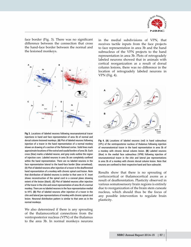
NBRC Annual Report 2014-15 | 57 |
face border (Fig. 3). Therewas no significantdifference between the connection that crossthe hand-face border between the normal and the lesioned monkeys.
Fig 3. Locations of labeled neurons following neuroanatomical tracer injections in hand and face representation of area 3b of normal and dorsal column lesioned monkeys. (A) Plot of labeled neurons following injection of a tracer in the hand representation of a normal monkey shown on drawing of a section of the flattened cortex. Solid lines mark approximate locations of the rostral and caudal borders of area 3b. Each cross (blue) marks a labeled neuron, and grey ovals outline the region of injection core. Labeled neurons in area 3b are completely confined within the hand representation. There are no labeled neurons in the face representation lateral to the hand-face border (blue arrowhead). (B) Plot of labeled neurons after injection of a tracer in the deafferented hand representation of a monkey with chronic spinal cord lesion. Note that distribution of labeled neurons is similar to that seen in ‘A’. Inset shows reconstruction of the spinal cord in a coronal plane showing extent of the lesion (black). (C) Plot of labeled neurons after injection of the tracer in the chin and snout representation of area 3b of a normal monkey. There are no labeled neurons in the face representation medial to HFS. (D) Plot of labeled neurons after injection of a tracer in the chin and lateral jaw representations of monkey with chronic spinal cord lesion. Neuronal distribution pattern is similar to that seen as in the normal monkeys.
We also determined if there is any sprouting of the thalamocortical connections from the ventroposterior nucleus (VPN) of the thalamus to the area 3b. In normal monkeys neurons
Fig 4. (A) Locations of labeled neurons (red) in hand subnucleus (VPL) of the ventroposterior nucleus of thalamus following injection of neuroanatomical tracer in the hand representation in area 3b of a monkey with chronic dorsal column lesion. (B) Labeled neurons (blue) in the medial face subnucleus (VPM) following injection of neuroanatomical tracer in the chin and lateral jaw representations in area 3b of a monkey with chronic dorsal column lesion. Note that neurons are confined to their respective hand and face subnuclei.
Results show that there is no sprouting of corticocortical or thalamocortical axons as a resultofdeafferentation.Plasticityobservedinvarious somatosensory brain regions is entirely due to reorganization of the brain stem cuneate nucleus, which should thus be the focus of any possible intervention to regulate brain plasticity.
in the medial subdivisions of VPN, that receives tactile inputs from the face projects to face representation in area 3b and the hand subnucleus of the VPN projects to the hand representation in area 3b. Plots of retrogradely labeled neurons showed that in animals with cortical reorganization as a result of dorsal columnlesions,therewasnodifferenceinthelocation of retrogradely labeled neurons in VPN (Fig. 4).

| 58 | NBRC Annual Report 2014-15
Systems and CognitiveNeuroscience Division
Patent
‘A capillary-electrode device for simultaneous injection and neuronal recordings from the brain’ Indian Patent Application No. 0189/DEL/2015 (Filed).
Presentations:
1. Neeraj Jain: ‘Search for a second motor area in the rat cortex’. National Conference on Recent AdvancesinZoologyatJiwajiUniversity,Gwalior;May2015.
2. Neeraj Jain: ‘Spinal Cord Injuries: Mechanisms of Brain Reorganization and Development of Brain-Computer Interface’, Indian Institute of Technology Hyderabad; March 2015.
3. NeerajJain:‘Tactileinformationprocessinginthebrainandtheeffectsofspinalcordinjuries’.Plenary talk at 2nd International Conference on Perception and Machine Intelligence (PerMin15) organized by Centre for Development of Advance Computing (CDAC), Kolkata, India; Februar 2015.
4. Neeraj Jain: ‘Mechanisms of Brain Plasticity’, Plenary talk at IFCAM Workshop on ‘Statistical and Mathematical Biology’, Indo-French Centre for Advanced Mathematics, Indian Institute of Science, Bangalore; July 2014.
5. Neeraj Jain: ‘Vision’ and 'Brain Reorganization Following Spinal Cord Injuries', Faculty Development Programme on ‘Computer Vision, Video and Image Processing’ at Dept of Biomedical Engineering, PSG College of Technology, Coimbatore; July 2014.
Funding
This work is supported by Department of Biotechnology and NBRC Core funds.
Collaborators
Prof Mrignaka Sur, MIT, USA
Prof P Raghunathan, NBRC
Degrees Awarded (Ph.D.)
Niranjan Kambi (Ph.D.)

NBRC Annual Report 2014-15 | 59 |
Computational Neuroscience and Neuroimaging

| 60 | NBRC Annual Report 2014-15

NBRC Annual Report 2014-15 | 61 |
Investigating neuro-cognitive network mechanisms using multimodal neuroimaging
Principal InvestigatorArpan Banerjee
Research Associate Dipanjan Ray
Junior Research Fellow Amit Kumar Jaiswal G. Vinodh Kumar
Research & Development Engineer Abhishek Mukherjee
Project Assistant Tamesh Halder
The concept of Neuro-cognitive networks: large-scale brain networks of distributed and interconnected neuronal populations required for supporting cognitive functions has been increasingly gaining ground in recent years. In these formative years several questions needs to be answered, how large/ small is large-scale, how can one define interconnectednessin space and time? Are there any general principles which govern the behavior of neuro-cognitive networks that are invariant across modalities used for studying their operations? Often the responses to these questions results in intense debates with opinions shaped from methodological constraints such as the modality of neuro-imaging tool in use, electro- & magneto-encephalography (EEG & MEG) or functional magnetic resonance imaging (fMRI). Our lab uses multimodal techniques EEG, MEG & fMRI to understand operational principles of neuro-cognitive networks underlying speech perception and complex auditory processing. We are also engaged in developing new tools to characterize the network mechanisms from neurophysiological recordings. We have also set up a network analysis pipeline in collaboration with lab of Dr Anirban Basu to study protein pathwaysunderlyingneuroinflammation.
Brain networks underlying speech perception
Speech perception emerges from harmonious interaction of multiple neural systems. “McGurk illusion”, a classic example of how visual feedback shapes speech perception, provides an entry point to study the underlying brain networks. Earlier research has shown that modulating the degree of audio-visual (AV) integration by psychophysical parameters weakened the effect. Here, we performedan fMRI study on human volunteers when McGurk-stimuli (incongruent audio-video signal) were presented with varying AV lags. We observed across a large group of 16 volunteers there was a regime where the illusory perception was maximum, [-150, 300] ms in concordance with earlier studies. When the block at which maximum illusory response occurred was pooled for a group statistical parametric mapping (SPM) analysis with respect to rest we observed significantactivations in inferior frontal gyrus (IFG), posterior superior temporal sulcus (pSTS), V5 and superior temporal gyrus (STG) (Fig 1). In a conjunction analysis with blocks where minimum illusory perception was reported by volunteers,thehighestdifferencewasfoundinpSTS. In a regime where auditory preceded the

| 62 | NBRC Annual Report 2014-15
visual stimulus, pSTS was not even activated. Functional connectivity among network nodes involving IFG, pSTS, auditory cortex/ STG and V5 using partial correlations were not altered by illusory perception but changed from a negative to positive AV lag. Overall, our results indicate neural activity in pSTS is most likely reflective ofmultisensory perception, but notmultisensory stimulus processing.
ComputationalNeuroscience andNeuroimaging Division
Figure 1: (a) shows the number of responses of each of the three types: “/pa”, “/ta” and “other” normalized and grouped over all 42 subjects (including non-perceivers) for each AV Lag. The perception of the illusion was the highest when the stimulus was synchronous and this perception reduced with the introduction of asynchrony. T-tests were done for: (b) Task > Rest, and (c) Rest > Task over the group of the 34 McGurk-effect perceivers for each AV Lag at p<0.01 (uncorrected). We find that the significant activity of the pSTS correlates with the group behavior in (b). We used the mask of SC (superior colliculi) in (c) and found significant activity only in the synchronous condition (26 voxels) and AV Lag of 450 ms (1 voxel).
EEG/MEG Source Localization
A critical problem in EEG-MEG is that the neural activity we measure outside the head is generated by unknown number of neural sources and also corrupted by volume conduction artifacts. To circumvent this issue researchers have developed source imaging techniques, also known as inverse methods. In mathematical sense this is an ill-posed

NBRC Annual Report 2014-15 | 63 |
problem, since there is an unknown number of sourcesandfixednumberofsensors.Wehaverecently developed a preprocessing step which can enhance the performance of a very popular source analysis technique, standardized low resolution electromagnetic tomography (sLORETA). Oscillatory brain electromagnetic activity is an established tool to study neurophysiological mechanisms of human behavior. State of the art source localization methods such as standardized low resolution electromagnetic tomography (sLORETA) that incorporates minimum norm estimates gives a distributed map of brain activity underlying sustained and transient responses during neuroimaging studies of behavior. The volume conductioneffectsandnoiseoftheenvironmentplay a considerable role in adding uncertainty to source localization. Additionally, specificspectral events exist within a milieu of non-specific spectral components that ultimatelycontribute to adding uncertainty in source localization. Here, we introduce Compressive
Sensing (CS) as a pre-processing technique to estimate sources of spectral event generators in the cortex. We use simulated data to validate that accuracy and sensitivity of source localization is enhanced dramatically while using a combined CS-sLORETA algorithm than a stand-alone sLORETA. CS-sLORETA yielded focal sources when applied to empirical EEG data on human volunteers featuring stimulus evoked 40 Hz response to binaural auditory stimulus. Furthermore, we got focal localization for transient N100/ P100 component of the auditory evoked potential response, thus establishing the validity of CS-sLORETA for both sustained and transient responses. Our results fall in line with observations from previous studies such as auditory cortical activations for N100 and steady-state 40Hz rhythms and right hemisphere dominance of brain activations. Hence, we propose an analysis pipeline featuring CS-sLORETA significantlyexpands thepossibilitiesofEEG/MEG source localization.
Figure 2: (a) Source localization results on simulated EEG data plotted on Left, Top, and Right view of MNI brain surface. Red patches show the nearest surface locations from an estimated 3D brain voxel using CS-sLORETA, green represents sLORETA results and blue diamond depicts the original source locations. CS-sLORETA voxels were a subset of sLORETA voxels. (b) Source localization for empirical EEG data. Left, top and right view of cortical activation averaged over all participants for 40 Hz activity. Sum of distances of all sLORETA and CS-sLORETA clusters from the respective centroids were 60 and 53 mm respectively for steady state 40Hz response.

| 64 | NBRC Annual Report 2014-15
ComputationalNeuroscience andNeuroimaging Division
Graph-theoretic analysis of protein networks
Complex protein networks underlie any cellular function. Certain proteins play a pivotal role in many network configurations, disruptionof whose expression proves fatal to the cell. An efficient method to tease out such keyproteins in a network is still unavailable. Here, we used graph-theoretic measures on protein-protein interaction data (interactome) to extract biophysically relevant information about individual protein regulation and network properties such as formation of function specific modules (sub-networks) of proteins.We took 5 major proteins that are involved in neuronal apoptosis post Chandipura Virus
(CHPV) infection as seed proteins in a database to create a meta-network of immediately interacting proteins (1st order network). Graph theoretic measures were employed to rank the proteins in terms of their connectivity and the degree up to which they can be organized into smaller modules (hubs). We repeated the analysis on 2nd order interactome that includes proteins connected directly with proteins of 1st order. FADD and Casp-3 were connected maximally to other proteins in both analyses, thus indicating their importance in neuronal apoptosis. Thus, our analysis provides a blueprint for the detection and validation of protein networks disrupted by viral infections.
Figure 3: (a) Interactions between monitored proteins Fas, FADD, Casp-8, Casp-3 and XIAP estimated using STRING 9.1 database. (b) Proteins interacting directly with Fas, FADD, Casp-8, Casp-3 and XIAP were estimated using STRING 9.1 database. The nodes represent the proteins while the lines indicate interactions in this 1st order interactome. Only those proteins reported at a confidence level of 95% are considered. (c) The proteins interacting directly with the nodes of 1st order interactome were extracted analogously to capture the 2nd order interactome.

NBRC Annual Report 2014-15 | 65 |
Publication
1. Ghosh, S., Kumar, V. G. , Basu, A. & Banerjee, A. (2015) : Graph theoretic network analysis reveals protein pathways underlying cell death following neurotropic viral infection (submitted)
Presentation/ Invited talks
1. Large-scale neural models to interpret functional brain connectivity, IFCAM Workshop in Statistics and Mathematical Biology, IISc Bangalore, July 2014
2. How does math help us understand the brain? Kendriya Vidyalaya Manesar, Dec 20143. Neurocognitive networks: Linking brain, mind, behavior, Brain Awareness Week, Presidency
University, Kolkata, March 2015
Funding
NBRC Core
Ramalingaswami Fellowship, Department of Biotechnology
Collaborations
Abhijit Sen, Institute for Plasma Research (IPR), Gandhinagar
Anirban Basu, NBRC
Dipanjan Roy, International Institute of Information Technology (IIIT), Hyderabad
Manjari Tripathi, All India Institute of Medical Sciences (AIIMS), New Delhi
Partha Ragunathan, NBRC
Award
Innovative Young Biotechnologist Award (IYBA), Department of Biotechnology

| 66 | NBRC Annual Report 2014-15
Early diagnostic biomarker of Alzheimer’s disease: A longitudinal multi modal brain imaging study
Principal InvestigatorPravat Kumar Mandal
Researchers Dr. Sumiti Saharan Sarah A Khan Monika Grewal Shammi More Sourav Bhaduri Marium Siddique Shipra Jain Vijaya Rathore
Objective: The overall objective of the brain longitudinal study is
• Comprehensive analysis and evaluation of potential biomarkers of AD using noninvasive in vivo MRS
• Neuropsychological assessment and its correlation with level of biomarker
Alzheimer’s disease (AD) is the most common causeofdementiaintheworldaffectingnearly44 million people as of 2015. It is characterized byneuriticplaqueandneurofibrillarytangles.Scientists expect that the number of people living with dementia will rise up to 115 million by 2050. This alarming projection of increase in patients has catapulted the research in the direction of detecting and diagnosing AD at its incipient stages. Glutathione (GSH), a predominant brain antioxidant responsible for neutralization of reactive oxygen species (ROS), has been correlated with cognitive impairment and is emerging as a potential biomarker. GSH levels in the brain can be evaluated by proton magnetic resonance spectroscopy (1H
MRS) which is a noninvasive in vivo method. However, the detection of GSH by MRS is obscured by other neurometabolites and results are ambiguous. To overcome this predicament and to achieve a reliable result with improved GSH signal, we have employed MEscher-GArwood-PRESS (MEGA PRESS) spectral editing technique. This has paved the way for betterquantitationofGSHlevelinbrain.
We conducted our study on 64 subjects and another 66 subjects for MRS. The study included scanning for key regions of brain affected in AD chiefly the hippocampi (HP)and frontal cortices (FC). Out of these 64 HP subjects recruited, 21 were AD patients, 22 were MCI (mild cognitive impairment) and 21 were healthy controls. Among 66 FC subjects, 19 were AD, 19 MCI and 28 HC. The exclusion criteria for the subjects were age less than 50 years, magnetic resonance incompatibility and presence of any current or previous disorders whichmightaffectthebrain.Thedemographicand clinical characteristics of the subjects are stated in the following Table1.

NBRC Annual Report 2014-15 | 67 |
The MRS data was acquired through 3T MRI Scanner (Achieva, Philips, Netherlands) and data was analyzed by an in-house MATLAB based novel MRS data processing toolbox called “KALPANA”. Neuropsychological assessment was done to establish any correlation between GSH levels and cognitive impairment by the use of Mini-Mental State Examination (MMSE),
Clinical Dementia Rating (CDR) and Trail Making Test (TMT). Evaluation of GSH level in various regions of the brain was done by Statistical Package for Social Sciences (SPSS, version 18.0, Chicago, IL, USA). Sensitivity-specificitytestsweredoneandtheresultswereplottedasgiveninFigure1.

| 68 | NBRC Annual Report 2014-15
Figure1. GSH levels are reduced in MCI and AD. (A,B) Box plots of GSH concentrations in (A) the HP regions, that is, LH and RH and (B) the FC regions, that is, LFC and RFC in HC (green), MCI (blue), and AD (red) subjects. There is significant deterioration in GSH levels in all brain regions of AD when compared with HC. GSH levels are significantly reduced in the LH and RH of MCI with respect to HC subjects. Furthermore, GSH levels are significantly reduced in the LFC and RFC of AD compared with MCI. Median values are marked by a bar. Circles represent medium outliers and asterisks represent extreme outliers. #p , .06, *p , .05, **p , .01, ***p , .001. (C–E) Receiver operator characteristic curve analyses of GSH for discriminating (C) AD patients from HC controls, (D) MCI patients from HC subjects, and (E) AD patients from MCI patients. Combined GSH estimation in both HP regions yielded high AUCs for differentiating AD from HC. GSH estimation in LH was found to yield the highest AUC for discriminating MCI from HC, whereas GSH estimation in FC yielded highest AUC for discriminating AD from MCI. AD, Alzheimer’s disease; AUC, area under the curve; FC, bilateral frontal cortices; GSH, brain antioxidant glutathione; HC, healthy old controls; HP, bilateral hippocampi; LFC, left frontal cortex; LH, left hippocampus; MCI, mild cognitive impairment; RFC, right frontal cortex; RH, right hippocampus.
Sensitivity of 87.5% and specificity of 100%were obtained for distinguishing MCI from HC on the basis of GSH estimation in LH (Left hippocampus). Sensitivity of 91.7% and specificity of 100% were obtained fordistinguishing MCI from AD subjects on the basis of GSH level in FC. Hence, it’s predictive thatGSHcanbeanefficientbiomarkerofAD.Ourfindings substantiate thatGSH levelsareselectively reduced in the HP of MCI compared with HC subjects and in the FC regions of AD compared with MCI patients. Also, our neuropsychological outcomes further validate that GSH levels positively correlate with MMSE scores.
The purpose of our project was to discern
and characterize a novel biomarker for AD detection by in vivo imaging techniques. We have successfully concatenate the results from MRS scanning and neuropsychological testing, estimated GSH levels in HP and FC among AD, MCI and healthy controls and elucidated that GSHlevelssignificantlyreducesintheHPandFC regions of brain in AD patients.
Hence, the above results foster the pivotal role played by GSH as a biomarker for detection of AD. We are currently investigating the GSH levels with additional subjects and longitudinally performing a follow up to further validate our results. In the long run, we aim to provide insights in the etiology and progression of AD and signify the role of GSH for future therapeutic avenues.
ComputationalNeuroscience andNeuroimaging Division

NBRC Annual Report 2014-15 | 69 |
Publications
1. Pravat K Mandal, Sumiti Saharan, Manjari Tripathi and Geetanjali Murari "Brain Glutathione Levels – A Novel Biomarker for Mild Cognitive Impairment and Alzheimer’s Disease" Biological Psychiatry doi:10.1016/j.biopsych.2015.04.005.
2. Pravat K Mandal, Sumiti Saharan, Sarah Khan, Mithun James "Apps for Dementia Screening: ACost-effectiveandPortableSolution", JournalofAlzheimer'sDisease,doi:10.3233/JAD-150255.
Funding
Department of Biotechnology, Govt. of India
Collaborator
Dr. Manjari Tripathi, MD, DM, Department of Neurology, All India Institute of Medical Sciences, New Delhi.

| 70 | NBRC Annual Report 2014-15
Spatiotemporal processing and Information transmission in brain
Principal InvestigatorPrasun Roy
Research Fellow Rajiv Ramaswamy Vikas Pareek
Research engineer VPS Rallabandi
An important aspect of both basic and applied neuroscience is understanding how flow ortransport processes occur, whether that of energy, information, current, drugs, cells or tissue displacement. Conventional linear quantitative models of flow processes, havedifficulty in accounting for experimentally-observedpreferentialflowinaxialdirectioninlayered tissues as brain or muscle. Hence, one needs a quantitative computational approach that can clarify and predict the various transport processes and their modulation across the brain, vis-à-vis tissue orientation or angulation. The recent advance of multimodal neuroimaging, beyond simply scalar imaging, implies a broad general perspective towards pathophysiological processes, wherein one can study the various transport processes or rotational parameters in the brain parenchyma, cellular permeation, tissue transposition, electrical conduction or information transmission. The overall objective of this program is to obtain basic insights and translational implications, regarding the physiological or pathological change of the various transport or flowprocesses in the brain parenchyma, mirroring the plasticity and dynamical processes. We pursue this aspect in extreme ranges of age: older age (neurodegenerative/involutional stage) and early age (neuroregenerative/neurodevelopmental stage).
Trajectory of Brain Connectomics & Compensatory Neural Re-modelling across Normal Ageing along the Life-Span
We delineate the spectrum of ageing process (neurodegenerative change). We study a critical but neglected issue of brain’s plasticity and coping with old ageing, via tensor MRI and its myelination biomarker, in about 400 individuals, 20-90 years age [fig. 1(a)].By devising our novel tailor-novel Brain Connectomics analysis (metric spanning indices), we show that as ageing-occurs the brain’sinformationflowenshrinedinthewhitematter dynamics (diffusion tensor imaging),undergoes full-scale re-modelling [fig. 1(b)].We found that the information processing by centralized white-matter tracts channelsincreases, to compensate for the decrease of information processing by peripheral white-matter channels. The peripheral transmissionby the outlying channels decreases, since with ageing, diffuse plaques deposit around thecortexandthenpermeatetoperipheralfibres,lessening therein the information transmission.
The maximal level of compensatory re-modelling occurs at 6th to 7th decade of life (mean 56 years for men, 62 years for women) [fig. 1(c)]. Ours is the first report unexpectedold-age neuroplasticity, neural diffusivity

NBRC Annual Report 2014-15 | 71 |
flow and neural re-modelling. Our imagingformulation are corroborated by histological findings which shows that in older age theoligodendroglial cellular myelination activity maximizes around the age of 50-60 years. Such restitutive remodelling as a coping mechanism is observed in electrically excitable tissue, as brain, heart or muscle. For instance, in the electrically active myoneural tissue of the heart, if there is decrease in haemodynamic flowin some neurovascular channels due to age-induced dystonia or ischaemia, then there is myocardial re-modelling, and compensatorily haemodynamic flow increases via collateralother channels.
Fast Epilepsy Screening by MRI using Neurodevelopmental Analysis
We now come to the imaging and analysis of the neuroregenerative/developmental stage, pertinent to mobility processes of brain tissue. An important issue is the mobility and rotation of the basal brain, rhinenceplanon (as amygdala and parahippocampal formation), during brain
development and growth. In the commonest seizure condition, temporal lobe epilepsy (TLE) which is a neurodevelopmental aberration, there is alteration of this mobility and rotation of the rhinencephalon. The neurological disorder that has the maximal disease burden in India is Epilepsy, having treatment gap of 96% with the unattendedpatientsinIndiabeing22millionasper lifetime prevalence.
Waiting for expert epileptologists and neuroradiologists for diagnostic screening causes delay. As important issue is to develop a rapid MRI-based screening of temporal lobe epilepsy. We have developed a rapid single-
a
c
b
Figure 1. (a): Brain Connectomics and Information flux motif from Diffusion Tensor Image of brain. (b): Topological interlinkage indices mapped using the connectomics platform. (c): Maximization of the Distributed Information Processing Efficiency at late age: Inverted U-shaped ageing-curves of males and females in brown and blue respectively. The two vertical red lines show the maxima of the male and female curve envelopes; the left red line is the maxima of males (mean 56 years), the right red line is that for females (mean 62 years).
slice MRI screening program (15 minutes) which uses 1-D axial rotational measurements of brain regions for screening the epilepsy cases [fig. 2(a)]. Our technique circumventsthe customary time-consuming methods as multi-slice 3-D hippocampal volumetry that can take 40-70minutes.Wefind that theParahippocampal Angle of rotation (namely, its bilateral asymmetry index of 0.1 threshold)

| 72 | NBRC Annual Report 2014-15
ComputationalNeuroscience andNeuroimaging Division
satisfactorily demarcates epileptics from normal[fig.2(b)].WealsofindthattheReceiver-
Operating Characteristic of the method is superior,theAUCindexbeing0.984[fig.2(c)].
Figure 2. (a): Fast single-slice estimation of parahippocampal rotational angulation. (b): Demarcation between normal and epileptic (TLE) subjects based on bilateral asymmetry of parahippocampal rotation, thresholded at 0.1 as shown by dashed line (c): Sensitivity-Specificity curve of screening the epileptic patients based on parahippocampal angulation.
Publications
1. Datta, S., Chakraborty, S, Mulpuru, S., Tiwary,B., Chakrabarti, N., and Roy, P. K.. MRIcharacteristics of temporal lobe epilepsy using rapidly-measurable spatial indices with hemispheric asymmetry and gender features. Neuroradiology, (Accepted). 2015. DOI: 10.1007/s00234-015-1540-6.
2. OtteyW,DiesenE,RajivR,SubhadipPal,SubramanyamV,Roy,P.K.AgeingAlterationsinWhole-Brain Neworks during Adulthood Mapped with Minimum Spanning Tree Indices: The Interplay of Density, Connectivity Cost and Life Time Trajectory, NeuroImage: Journal of Brain Function, 109, 171-189, 2015.
Patent
3. Roy P K, Subramanyam V P, Medical Image Enhancement Device based on Image Transform Resonance. Assignee: Dept. of Biotechnology (DBT) and National Brain Research Centre, PatentNo.8774480B2,U.S.PatentOffice,Washington,2014[PatentgrantedonJuly2014].
Presentation1. SaiKrishna,VPSubramanyam,PKRoy,Asia-PacificProgramsonBrainGrids:Multicentric
Analysis for Dementia/MCI Screening and Monitoring, Asia-Pacific Conference onAlzheimer’s Disease, New Delhi, Nov. 2014.
2. RishuRathee,VPSRallabandi,PKRoy,Age-relateddifferences inwhitematter integrityinhealthyhumanbrain:Theevidence fromstructuralMRIanddiffusion tensor imaging,International Conference on Emerging Trends in Biotechnology, JNU, New Delhi, Aug. 2014.
3. Suhela Kapoor, Systems biology approach to neurovascular disorders, Max Planck Institute of Cell Biology, Dresden, Germany, Nov. 2014.
NBRC Annual Report 2014-15 | 73 |
4. Prasun Roy, Tissue Mechanics as a key to Healthy Lifespan for a Greying World. Symposium on Translational Medicine and the Future of Human Health, Chinese National Academy of Engineering and Chinese National Academy of Medical Science, Beijing, China, June 2014.
5. Prasun Roy, Towards Health Grid Initiatives in India: Prospects and Insights from the Brain Grid,WorkshoponAffordableTechnologiesforHealthCare,GenopoleCampus-Universityof Evry, Paris, France, Oct 2014
6. Prasun Roy, A Systems Approach to Multi-Organ Amyloid Models: Towards a Human Simulator Platform for Clinical Trials. Strategy Formulation Meeting on Amyloid Disorders: I.T. in Health Care, Dept. of Electronics & Information Technology, Govt. of India, Jan 2015.
Funding
Dept. of Biotechnology, Govt. of India.Utrecht University Research Foundation, The Netherlands.
Collaborators
• Dr Anirban Basu, National Brain Research Centre.• Dr Luijten, Dutch Centre for Translational Medicine & Utrecht University.• Dr MV Padma & Dr TS Toy, All-India Institute of Medical Sciences, Delhi.• Dr Santanu Choudhury & Dr Raj Khanna, Indian Institute of Technology, Delhi.• Dr Shinjini Bhatnagar, Translational Health Science & Technology Institute, Gurgoan.
Awards
S. Pal. IBRO Travel Fellowship Award for participating at Society of Neuroscience Meeting, Washington DC, International Brain Research Organization (IBRO), June 2014.
S. Kapoor. Best Research Proposal Writing Award, Route 28 Workshop in Neurobiology, Munich, Aug. 2014.
Degree Awarded
Monica Arya. Studies on MRI and MRS of Malignant Brain Tumours. (Dissertation for B. Tech degree in Biomedical Engineering, under supervision of Prasun Roy at NBRC). DCR University of Science & Technology-Haryana, Sonepat. 2014.

| 74 | NBRC Annual Report 2014-15
Application of stochastic activation and stability analysis for brain imaging and therapy
Principal InvestigatorPrasun Roy
Research Fellow Subhadip Paul Suhela Kapoor
Research Associate Aftab Alam
As per the current scenario in neurobiology, it transpires that Perturbation-induced activation, anemergingresearchfield,offersapromisingprospecttoenhancetheefficiencyofdiagnosticor therapeutic applications in neuroscience. This effect, namely Stochastic activation,noise-aided resonance or fluctuation-inducedtransition, is a general principle of nonlinear biological systems, and occurs basically due to the statistical kinetic nature of the components that exhibits probabilistic fluctuations ofparameters. However the practical application of stochastic activation effect as a noveltechnique in diagnostic or therapeutic radiology has not been systematically pursued, and the applicability is the aim of our project.
Neuroimagiong-based Planning of Enhanced Tumour Control and Normal Tissue Protection by augmenting secondary Radiosensitivity parameter.
In the coming days, a critical application of neuroimaging is to be in the field oftreatment planning and optimization. Utilizing MRI image analysis, we have utilized the perturbative activation approach to enhance the efficiency of tumour cell elimination, aswell as decrease elimination of normal tissue, under therapeutic radiology, with particular application to neurooncology. Antitumour
treatment, such as using as radiotherapy or chemotherapy agent, has a lethal disadvantage, namely that the therapy agent damages both malignant tissue, together with normal tissue, leading to serious therapy-induced toxicity. Wehave investigated theeffectof impressingperturbation in the radiotherapy photon beam (stochastic radiotherapy) so as alter of the DNA double-strand interference (linear-quadratic formulation of two-hit event). This perturbation is impressed by a Gaussian function generator in the pulse circuit of the linac beam. An critical issue of oncology is to enhance Normal Tissue Protection, while at the same time augment the Tumour Cell kill. In earlier years, we have shown that stochastic radiotherapy enables the latter. This year, we develop a novelmethodology to enable the former.
Fig 1(a) shows the contrast-enhanced template image of the tumour that is used for developing the template for therapy modelling and therapy planning.Fromourformulationwefindthatthesecondary radiosensitivity index β (denotingdouble-strand interference by the photon) has an unexpected manifestation as follows. As one increases the secondary radiosensitivity β during stochastic radiotherapy, the tumourtissuedamageincreases[fig.1(b)],relativebuttherelativenormaltissuedamagedecreases[fig.1(c)], meaning that the normal tissue protection

NBRC Annual Report 2014-15 | 75 |
enhances. Fig 1(d) shows that the tumour tissue damageincreasesby10%asβrisesby10%;thisβriseisalsoassociatedsurprisinglybydecreaseof normal tissue damage by about 30%. [Fig 1(e)]Thusaugmentationofβradiosensitivity,enables the therapeutic differential to riseby 40% under stochastic radiotherapy. Some therapeutic radiosentitizers can elevate the β
radiosensitivity index, such as gadolinium, heavy metal colloids etc. Chemotherapy can also be integrated as multimodal therapy, this acts synergistically effectively enablingcomplete tumour elimination, and this can be explored for neurooncologal tumours as oligodendroglioma or melanoblastoma [fig1(f)].
Figure1: (a). MRI image: Gadolinium-contrast enhanced image of the tumour enables the construction of the MRI template to formulate the biological effect of stochastic radiotherapy on the tumour. (b). Tumour tissue under effect of Perturbative Radiotherapy: Increase of Tumour cell damage [(%)] as the secondary radiosensitivity index (double-strand interference index) increases (arrow); the radiation dose is 3 Grays (Gy or centirads) per day, for 30 day therapy protocol. (c). Normal tissue under Perturbative Radiotherapy: Decrease of Normal Cell damage [(%)] as the secondary radiosensitivity index (double-strand interference index) increases (arrow). (d). Alteration of Tumour tissue damage as radiobiological parameters β,μ,δ increases by 10% each; the bar M denotes that the Tumour tissue damage increases by about 10%. When β rises by 10% (e). Alteration of Normal tissue damage as radiobiological parameters increases by 10% each; the bar N denotes that the Normal tissue damage decreases by about 30%. as β is increased by 10% Combining data of (d) and (e), the stochastic radiotherapy decreases relative Normal tissue damage by 40%, when radiobiological index β in increased by 10% using secondary radiosensitizer agents. (f): Complete tumour elimination by multimodal therapy; the tumour cell population is eliminated at 41 days of therapy.

| 76 | NBRC Annual Report 2014-15
ComputationalNeuroscience andNeuroimaging Division
Publications
1. Subhadip Pal, Roy, P. K. Strategy for Stochastic Dose-rate induced Enhanced Elimination of Malignant Tumour without Needing Dose-escalation, Mathematical Medicine & Biology, 2015 (Accepted). DOI: 10.1093/imammb/dqv012.
2. Chandrashekhar, R, Radhakant, P, Kapoor, S, Subramanyam V, Roy, P. K. Multimodal Therapy for Complete Regression of Malignant Melanoblastoma using Constrained Nonlinear Optimal Dynamic Inversion, Biomedical Signal Processing and Control. 13, 198-211 (2014) [shown as accepted last year].
Patent
3. RoyPK,PalS.Atechniquetoenhancetheclinicalefficiencyofradiotherapyusingperturbativebeaming. Assignee: Dept. of Biotechnology (DBT) and National Brain Research Centre, Patent No.2012/01067003A1,U.S.PatentOffice,Washington,2014[PatentgrantedonNov.2014].
Presentation
1. Subhadip Paul, Alteration of small-world anatomical networks in the patients with brain lesions: Glioma as case-study, Society for Neuroscience, Washington DC, Oct. 2014.
2. Aftab Alam, Harnessing the biphasic role of immunomodulation using a quantitative systems pathology approach, Indian Academy of Neuroscience, NIMHANS, Bangalore, Nov.. 2014.
3. Suhela Kapoor, Systems Biology Approach to Stem Cell Regeneration, International Workshop on Adult Neurogenesis: Concepts and Applications, Karolinska Institute, Stockholm, May 2014.
4. Vikas Pareek, Neuroprotection in Brain Injury: A Computed Neurobiology Platform, Foundation Day Conference, Translational Health Science & Technology Institute, New Delhi, July 2014.
5. Prasun Roy, Harnessing Neuroprotection Kinetics with Multicentric Approach via Brain Grids, Indian Institute of Science, Bangalore, July, 2014.
6. Prasun Roy, Optimizing Therapeutic Neurogenesis and Synaptogenesis, University of Oxford, Oxford, U.K., Nov. 2014.
Collaborators
• Dr Alan Evans, Montreal Neurological Institute, McGill University• Dr. Manjari Tripathi & Dr P Sarat Chandra, All-India Institute of Medical Sciences, Delhi.• Dr Ralph Martins, Edith Cowan University & CRC Mental Health Instt., University of
Melbourne.• Dr R. K. Padhi, Indian Institute of Science, Bangalore.• Dr T R Seshadri, University of Delhi.

NBRC Annual Report 2014-15 | 77 |
Funding
Ministry of Information Technology, Govt. of India.OfficeofPrincipalScienceAdviser,Govt.ofIndia.
Awards
A. Alam, Travel Fellowship for research presentation at 25th Biennial meeting of APSN, Australian Society of Neuroscience, Sydney, March 2015.
S. Kapoor. First Prize in Foundation Day Research Presentation, Translational Health Science Technology Institute, New Delhi, July 2014.
Degree Awarded
Subhadip Paul, Higher Order Diffusion Tensor Imaging and Stochastic Perturbation in BrainTumour. PhD Degree. National Brain Research Centre, 2015.

| 78 | NBRC Annual Report 2014-15
The influence of print to speech mapping on reading networks in simultaneous biliterate children
Principal InvestigatorNandini Chatterjee Singh
Research Fellow Sarika Cherodath
Background – About two-thirds of the world’s children grow up in bilingual environment (Bhatia & Ritchie, 2012; Crystal, 2003). In many such environments, children are instructed simultaneously in two languages (Bialystok, Luk, & Kwan, 2005) and are often biliterate in that they learn to read languages that belong to distinct writing systems. The writing system thata languageusesaffects children’sacquisition of literacy because writing systems vary in their consistency in mapping of sounds ontounitsofprint(Ziegler&Goswami,2005)also known as orthographic depth (Katz &Frost, 1992). Based on this feature, transparent languages like Spanish with near univalent spelling-to-sound mapping occupy one end of a continuum and opaque orthographies such as English fall on the other end (Seymour, Aro, & Erskine, 2003).
Simultaneous reading instruction in native and second language is becoming an increasingly common feature of language learning environments across the globe, wherein reading acquisition in non-native language parallels language learning itself (Ranguelov, De Coster, Norani, & Paolini, 2012; Saiegh-Haddad & Geva, 2010; Silver, Hu, & Iino, 2002; Wang, 2007). This scenario opens up an exciting avenue to investigate neuroplasticity of the developing brain. The urban education environment in
India is a natural setting admirably suited toexplore this owing to its multilingual home environments and its education policy which requires children to learn more than one language at school (Annamalai, 2001).
The participants in our study were provided simultaneously reading instruction in Hindi and English, languages with distinct writing systems. Hindi, written in Devanagari scripthas a near transparent orthography with almost univalent mapping of sounds onto the basic written units, also calledAksharas.Devanagari is an alphasyllabary with distinct consonants and vowels akin to alphabetic scripts, but each grapheme corresponds to a syllable similar to syllabic scripts (Vaid & Gupta, 2002). On the other hand, English is alphabetic and has an opaque orthography which necessitates recruitment of whole-word processes for reading familiar stimuli, like words and assembly of phonological units for novel stimuli (Jobard et al., 2003). Conversely, the transparent nature of Devanagari allows the usage of phonological assembly for reading both novel and familiar stimuli (Das et al., 2011; Rao, Vaid, Srinivasan, & Chen, 2011). However, both English and Devanagari (Nag, Caravolas, & Snowling, 2011) represent phoneme level information, making it central to reading both languages as per the psycholinguistic grain size

NBRC Annual Report 2014-15 | 79 |
theory (Ziegler &Goswami, 2005) of readingacquisition. The primary focus of this study was to investigate whether this cross-linguistic variation in orthographic depth impacts reading pathways in biliterate children.
We used functional neuroimaging to measure brain activity during word and nonword reading tasks to investigate neural reading circuits in Hindi-English biliterate children. Based on literature on biliteracy and reports from simultaneous Hindi-English adult readers, we hypothesized that Hindi-English biliterate children would exhibit (1) overlap in reading pathways for reading Hindi and English (2) word and non-word processing strategies in the two languages which reflectdisparity in orthographic depth.
Methods:
A detailed procedure was adopted to select participants for the study with similar language background. Forty four typically developing, right handed, Hindi-English biliterate children (23 males) (mean age - 9.21 years, SD = 0.69) from a private school in National Capital Region, Delhi in northern India participated in the study which was approved by the HumanEthicsCommitteeoftheInstitute.Therecruitment of participants from a single school ensured matched instruction and reduced variability in the literacy environment. The native language for all participants was Hindi,
while English was acquired primarily through schooling. At school, children simultaneously learnt to read both Hindi and English (from 5 years onwards), with the latter being themedium of instruction and within-classroom communication. All participants belonged to middle income families and as per the parental questionnaire, the education of parents was of graduate level and home environment typically involved usage of Hindi as well as English.Allparticipantsmetourcut-offscoreon age appropriate reading tests developed in both languages and had no history of reading difficultyasperschoolandparentalreports.
The final study sample consisted of 34participants (16 males) None of the participants had any history of sensory or neurological deficits, had normal or corrected to normalvision and normal intelligence (mean IQ = 104.81, SD = 7.94).
The fMRI paradigms employed reading tasks in Hindi and English in a simple and identical block design. The task comprised of alternating word and nonword reading blocks separated by rest blocks with a visual baseline. Participants were instructed to read aloud words and nonwords that appeared during the taskblocks,andtofixateonthesymbolstringsdisplayed during rest blocks without any oral response.
Analysis of neuroimaging data showed is shown in Fig. 1.

| 80 | NBRC Annual Report 2014-15
ComputationalNeuroscience and Neuroimaging Division
Anatomical sections of activation maps for the four conditions tested – English words (EW), English nonwords (ENW), Hindi words (HW), Hindi nonwords (HNW). Imaging results revealed a robust shared network for reading across two languages. This shared cortical reading network comprised of bilateral hippocampi, precentral and postcentral gyri (BA 3/4), middle (BA 19) and inferior occipital gyri (BA 18) and cerebellum.
Toexplorecross-linguisticeffectsinactivation
patterns, we performed subtraction analysesacross pairs of language conditions (English-Hindi, EW-HW,
ENW-HNW) in both directions, which did not showanysignificantdifferences.Toisolatethecontributionofeachlanguagetothemaineffectof stimulus type, subtractions between word and nonword reading conditions in English and Hindi was performed. The subtractions revealedsignificantdifferencesinEnglishFig.2(a) , but not in Hindi (Fig 2(b)).
Fig. 2(a)
Fig 2(b)

NBRC Annual Report 2014-15 | 81 |
Conclusions
Our results provide evidence that while children receiving simultaneous reading instruction in distinct writing systems engage a largely shared network for reading, they alsoexhibiteffectsoforthographicdepth.Ourresults indicate that simultaneous biliterate instruction provides an environment wherein cortical reading strategies are sensitive to cross-linguistic differences in orthographic depth.Further studies comparing subpopulations with varying levels of second language exposure are required to elucidate effects ofdifferential exposure on reading circuits inbiliterate children.
Long term goals –
These findings have significant ramificationsfor bilingual environments across the globe where children receive simultaneous reading instruction in non-native and native languages and consequently learn to read the non-native language in parallel with language learning itself. Given the current global scenario of growing English language learning populations, these results provide an interesting perspective for understanding reading development in multicultural environments wherein children are bilingual as well as biliterate.

| 82 | NBRC Annual Report 2014-15
Emotional responses to Hindustani Raga music: The role of musical structure
Principal InvestigatorNandini Chatterjee Singh
Research Fellow Avantika Mathur
Background – An extensive body of ancient Indian scripts belonging to the early centuries A.D. have documented the emotions associated with ragas (Bhatkhande, 1934; Natyashastra by Bharata translated by Vatsyayan, 1996), but there has been little scientific investigationof this idea. In the research reported here, we used self-reports by participants as a measure to study the subjectively experienced feeling of emotion while listening to ragas of North Indian Classical music. North Indian Classical music (NICM), born out of a cultural synthesis of the Vedic chant tradition and traditional Persian music has been known to induce emotions (Kaufmann, 1965). The central notion in this system of music is that of a raga. The word ‘raga’ originates in Sanskrit and isdefinedas'the act of colouring or dyeing' (the mind and mood/emotions in this context) and therefore refers metaphorically to 'any feeling or passion especially love, affection, sympathy, desire,interest, motivation, joy, or delight’. Using notes in particular combinations characteristic to the raga, the performer creates a mood (rasa) or atmosphere that is unique to the raga. We used the following emotion labels used for this study, namely, happy, romantic, devotional, calm/soothed, angry, longing/yearning, tensed/restless and sad.
A raga composition is typically presented as a specific sequence of events, namely the alaapfollowed by the gat. Alaap is the note by note delineation of a raga bound by a slow tempo, but not bound by any rhythmic cycle. Gat is the composition rendered at a faster tempo with accompaniment of a percussion instrument that provides a rhythmic cycle. The rhythmic cycle is measured in terms of time units or beats. These rhythmic structures can vary in the degree of pulse clarity. Pulse clarity is the estimate, on a large time scale, of how clearly the underlying pulsation in music is perceivable and is regarded as a measure for the underlying periodicity of the music (Lartillot et al., 2008). Thus, pulse clarity provides a measure of rhythmic regularity. Besides features such as pulse clarity, tempo is an important factor contributing to the perception of rhythm, which can be estimated as the number of notes presented per second. For the purpose of this study, rhythmic regularity was determined by estimating pulse clarity while tempo was determined in terms of note density of raga excerpts.
The specific objectives of this study were to(1) discriminate the emotion experienced by alaap and gat for various ragas across a large population of listeners’ and (2) investigate the
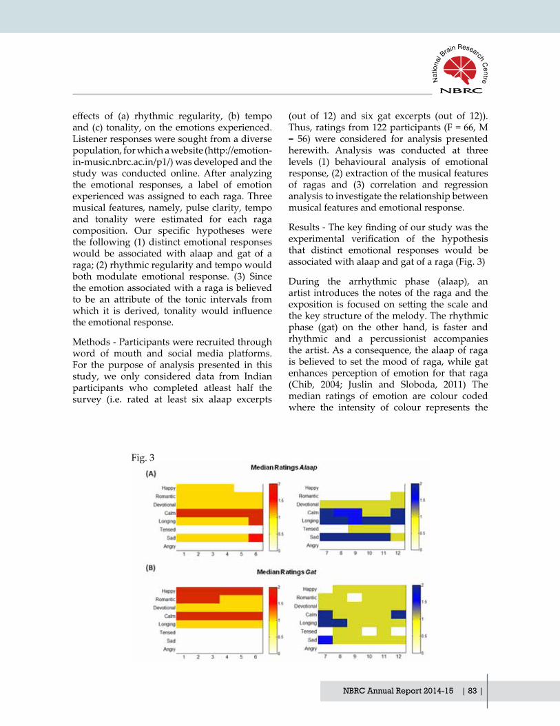
NBRC Annual Report 2014-15 | 83 |
effects of (a) rhythmic regularity, (b) tempoand (c) tonality, on the emotions experienced. Listener responses were sought from a diverse population,forwhichawebsite(http://emotion-in-music.nbrc.ac.in/p1/) was developed and the study was conducted online. After analyzing the emotional responses, a label of emotion experienced was assigned to each raga. Three musical features, namely, pulse clarity, tempo and tonality were estimated for each raga composition. Our specific hypotheses werethe following (1) distinct emotional responses would be associated with alaap and gat of a raga; (2) rhythmic regularity and tempo would both modulate emotional response. (3) Since the emotion associated with a raga is believed to be an attribute of the tonic intervals fromwhich it is derived, tonality would influencethe emotional response.
Methods - Participants were recruited through word of mouth and social media platforms. For the purpose of analysis presented in this study, we only considered data from Indian participants who completed atleast half the survey (i.e. rated at least six alaap excerpts
(out of 12) and six gat excerpts (out of 12)). Thus, ratings from 122 participants (F = 66, M = 56) were considered for analysis presented herewith. Analysis was conducted at three levels (1) behavioural analysis of emotional response, (2) extraction of the musical features of ragas and (3) correlation and regression analysis to investigate the relationship between musical features and emotional response.
Results-Thekeyfindingofourstudywastheexperimental verification of the hypothesisthat distinct emotional responses would be associated with alaap and gat of a raga (Fig. 3)
During the arrhythmic phase (alaap), an artist introduces the notes of the raga and the exposition is focusedon setting the scale andthe key structure of the melody. The rhythmic phase (gat) on the other hand, is faster and rhythmic and a percussionist accompanies the artist. As a consequence, the alaap of raga is believed to set the mood of raga, while gat enhances perception of emotion for that raga (Chib, 2004; Juslin and Sloboda, 2011) The median ratings of emotion are colour coded where the intensity of colour represents the
Fig. 3
| 84 | NBRC Annual Report 2014-15
ComputationalNeuroscience andNeuroimaging Division
strength of the emotional response. The highest median rating for ragas rated as ‘calm/soothing’ during ‘alaap’ shifted to ‘happy’ when played in gat. On the other hand, the highest median rating for ragas rated as ‘sad’ shifted to ‘longing/yearning’ or ‘tensed/restless’ during gat. ‘Angry’ remained the lowest rated emotion for both categories of ragas.
The results revealed that pulse clarity (estimate of rhythmic regularity) and note density (estimate of tempo) differ among ragaswith different experienced emotions, wherehigh arousal emotions (happy/tensed) are associated with a faster rhythm. In addition, tonality significantly influenced the emotionexperienced as the increase in mean frequency of occurrence of minor intervals was associated with ‘tensed’ emotion whereas increase in mean
frequency of occurrence of major intervals was associated with ‘happy’ emotion Thus, our results indicated that the tonal distribution patternsdeterminetheunderlyingmood(rasa)of a raga and the presence of rhythm changes the level of arousal of emotions experienced.
Finally, one of the most interesting findingsof our study was the association of the minor second with ‘tensed’ emotion. This is distinct from past work in Western classical music that has shown an association for the minor third with sadness in Western music (Curtis and Bharucha, 2010). Current work will attemptto extend these findings to larger populationin order to delineate influences of culture,familiarity and musical training on emotion experienced.
Publications
1. Megha Sharda, Budhachandra Khudrapakam, Alan Evans and Nandini C Singh. Structural Covariance Networks in Autism are modulated by verbal ability, Brain, Structure and Function, Brain, Structure and Function, DOI 10.1007/s00429-014-0953-z, 2014.
2. Megha Sharda, Rashi Midha, Supriya Malik, Shaneel Mukerji, and Nandini C. Singh. Fronto-Temporal Connectivity is Preserved During Sung but Not Spoken Word Listening, Across the Autism Spectrum, Autism Research, 8, 174-176, 2015.
3. SarikaCherodath andNandiniC Singh. The influence of orthographic depth on readingnetworks in simultaneous biliterate children, Brain and Language, 143, 42-51, 2015.
4. Chaitra Rao and Nandini C Singh. Visuo-spatial complexity modulates reading in the brain, Brain and Language, 141, 50-61 2015.
5. Avantika Mathur, Suhas H. Vijayakumar, Bhismadev Chakrabarti and Nandini C. Singh. Emotional responses to Hindustani raga music: the role of musical structure, Frontiers in Psychology (Emotion Science), doi: 10.3389/fpsyg.2015.00513, 2015.
Presentations
1. Nandini C Singh, reading pathways in children reading two distinct writing systems, Invited talk, Haskins Laboratories, Yale University, USA, April 2014.
2. Nandini C Singh - ‘Reading Assessments in a bilingual-biscriptal setting’, MaharshtraDyslexia Association, Mumbai, October 2014.
3. Nandini C Singh, Visuo-spatial complexity modulates reading in the brain, Invited talk, University of Taiwal, Taipei, November 2014.

NBRC Annual Report 2014-15 | 85 |
Grants
• Research grant on ‘Development and validation of screening tool to identify learning disability (teacher administered screening tool’, Department of Science and Technology, 2013-14.
• Research grant on ‘Speech and Music Processing in Autism’, from Department of Science and Technology, 2012 -2015.
• Research grant on ‘A functional imaging study of dyslexia in biscriptal Indian Children’ from Department of Biotechnology (2011-2014)
Collaborators
1. Dr. V. S. Mehta, Director of Neuorsciences, Paras Hospital, Gurgaon, India.
2. Dr. Amit Sen, Children First, New Delhi.

| 86 | NBRC Annual Report 2014-15
ComputationalNeuroscience &Neuroimaging Division

NBRC Annual Report 2014-15 | 87 |
Publications, Patents and Presentations

| 88 | NBRC Annual Report 2014-15

NBRC Annual Report 2014-15 | 89 |
Publications
• Irshad K, Mohapatra S.K., Srivastava C, Garg H, Mishra S, Dikshit B, Sarkar C, Gupta D, Sarat PC, Chattopadhyay P,Sinha S*; Chosdol K. (2015) "A combined gene signature of hypoxia and Notch pathway in human glioblastoma and its prognostic relevance". PLOS One 2015 March 3 DOI:10.1371/journal.pone 0118201 * Joint corresponding author
• Kumar M, Mani P, Pratheesh P, Chandra S,JeyakkodiM,ChattopadhyayP,SarkarDP and Sinha S. Membrane fusion mediated targeted cytosolic drug delivery through scFv engineered Sendai viral envelopes Current Molecular Medicine, 2015 15 386 – 400
• ZakariaM,KhanI,ManiP,ChattopadhyayP, Sarkar DP, Sinha S. Combination of hepatocyte specific delivery andtransformation dependent expression of shRNA inducing transcriptional gene silencing of c-Myc promoter in hepatocellular carcinoma cells. BMC Cancer, 2014, 14:582
• J. Chakraborty, U. Rajamma, N. R. Jana and K.P. Mohanakumar. Quercetin improves the activity of ubiquitin proteasomal system in 150Q mHttexpressing cells, but exerts detrimental effectsonneuronalsurvivability.Journal of Neuroscience Research. 2015 (In Press).
• M. Chakraborti, B. Paul, T. Nayak, A. Das, N.R. Jana and S. Bhutani . The E3 ligase Ube3a is required for learning in Drasophila melanogaster. Biochemical and Biophysical Research Communications. 462, 71-77, 2015.
• M. Maheshwari, S. Shekhar, B. K. Singh, I. Jamal, N. Vatsa, V. Kumar, A. Sharma and N. R. Jana. Deficiency of Ube3a inHuntington's disease mice brain increases aggregate load and accelerates disease pathology. Human Molecular Genetics, 23, 6235-6245, 2014
• S. Palmal, N. R. Jana and N. R. Jana. Inhibition of amyloid fibril growth bynanoparticle coated with histidine-based polymer. Journal of Physical Chemistry C, 118, 21630-21638, 2014.
• S. Godavarthi, A. Sharma and N. R. Jana. Reversal of reduced parvalbumin neurons in hippocampus and amygdala of Angelman syndrome model mice by chronictreatmentoffluoxetine.Journal of Neurochemistry, 130, 444-454, 2014.
• *S. Malik, R. Saha and P. Seth (2014). Involvement of Extracellular signal-regulated kinase (ERK1/2)-p53-p21 axis in mediating neural stem/progenitor cell cycle arrest in co-morbid HIV-Drug abuse exposure. J NeuroImmune Pharmacology, 9:340-353.
• M. Tewari, Monika, R. Verghese, M. Menon and P. Seth (2014). Astrocytes mediate HIV-1 Tat-induced neuronal damage via ligand-gated ion channel, P2X7R. Journal of Neurochemistry, 132: 464-476.
• M. Duan, H. Yao, Y. Cai, K. Liao, P. Seth and S. Buch (2014). HIV-1 Tat disrupts CX3CL1-CX3CR1 Axis in Microglia via the NF-κBYY1 Pathway. Current HIV Research, 12: 189-200.
• N Sharma, R Verma, K L Kumawat, A Basu and S K Singh (2015) miR-146a suppresses cellular immune response during
Publications, Patents and Presentations

| 90 | NBRC Annual Report 2014-15
Publications, Patents and Presentations
Japanese encephalitis virus JaOArS982 strain infection in human microglial cells. Journal of Neuroinflammation 18; 12(1):30
• S Mukherjee, S Ghosh, A Nazmi, and A Basu (2015) RIG-I Knockdown Impedes Neurogenesis in a Murine Model of Japanese Encephalitis. Cell Biol Int 39(2):224-9.
• P P Manna, S K Hira, A Basu, and S Bandyopadhyay (2014) Cellular therapy by allogeneic macrophages against visceral leishmaniasis: role of TNF-α.Cellular Immunology 290(1):152-163.
• A Nazmi, S Mukherjee, K Kundu, K Dutta,AMahadevan, SK Shankar, andA Basu (2014) TLR7 is a key regulator of innate immunity against Japanese Encephalitis Virus infection. Neurobiology of Disease 69: 235-247.
• D K Kaushik, M C Thounajam, A Mitra, and A Basu (2014) Vespa tropica venom suppresses lipopolysaccharide mediated secretion of pro-inflammatory cyto-chemokines by abrogating nuclear factor-κ B activation in microglia. Inflammation Research 63(8):657-65.
• M C Thounajam, K Kundu, D K Kaushik, S Swaroop, A Mahadevan, S K Shankar, and A Basu (2014) MicroRNA-155 Regulates Japanese Encephalitis Virus Induced Inflammatory Response byTargeting src Homology 2-Containing Inositol Phosphatase-1. Journal of Virology 88(9): 4798–4810
• M C Thounajam, D K Kaushik, K Kundu, and A Basu (2014) microRNA-29b Modulates Japanese Encephalitis Virus Induced Microglia Activation by Targeting Tumor Necrosis Factor Alpha-induced Protein 3 (TNFAIP3) Journ of Neurochemistry 129(1):143-54
• G D Manocha, R Mishra, N Sharma, K L Kumawat, A Basu and S K Singh (2014) Regulatory role of TRIM21 in type-I interferon pathway in Japanese encephalitis virus infected
human microglial cells. Journal of Neuroinflammation 11:24
• A Nazmi, B Hazra, K Dutta, and ABasu (2014) Role of Pattern RecognitionReceptors in Flavivirus Infections. Virus Research 185:32-40.
• Ahmad F, Ghosh S, Sinha S, Joshi SD, Mehta VS, Sen E (2015). TGF-β-induced hCG-β regulates redoxhomeostasis in glioma cells. Mol Cell Biochem.;399(1-2):105-12
• Dixit D, Ghildiyal R, Anto NP, Sen E. (2014). Chaetocin-induced ROS-mediated apoptosis involves ATM-YAP1 axis and JNK-dependent inhibition of glucose metabolism. Cell Death Dis.;5:e1212. doi: 10.1038/cddis.2014.179.
• Megha Sharda, Budhachandra Khudrapakam, Alan Evans and Nandini C Singh. Structural Covariance Networks in Autism are modulated by verbal ability, Brain, Structure and Function, Brain, Structure and Function, DOI 10.1007/s00429-014-0953-z, 2014.
• Megha Sharda, Rashi Midha, Supriya Malik, Shaneel Mukerji, and Nandini C. Singh. Fronto-Temporal Connectivity is Preserved During Sung but Not Spoken Word Listening, Across the Autism Spectrum, Autism Research, 8, 174-176, 2015.
• Sarika Cherodath and Nandini C Singh. The influence of orthographic depthon reading networks in simultaneous biliterate children, Brain and Language, 143, 42-51, 2015.
• Chaitra Rao and Nandini C Singh. Visuo-spatial complexity modulates reading in the brain, Brain and Language, 141, 50-61 2015.
• Avantika Mathur, Suhas H. Vijayakumar, Bhismadev Chakrabarti and Nandini C. Singh. Emotional responses to Hindustani raga music: the role of musical structure, Frontiers in Psychology (Emotion Science), doi: 10.3389/fpsyg.2015.00513, 2015.

NBRC Annual Report 2014-15 | 91 |
• Datta,S.,Chakraborty, S,Mulpuru,S., Tiwary,B., Chakrabarti, N., and Roy, P. K.. MRI characteristics of temporal lobe epilepsy using rapidly-measurable spatial indices with hemispheric asymmetry and gender features. Neuroradiology, (Accepted). 2015. DOI: 10.1007/s00234-015-1540-6.
• Ottey W, Diesen E, Rajiv R,Subhadip Pal, Subramanyam V, Roy, P. K. Ageing Alterations in Whole-Brain Neworks during Adulthood Mapped with Minimum Spanning Tree Indices: The Interplay of Density, Connectivity Cost and Life Time Trajectory, NeuroImage: Journal of Brain Function, 109, 171-189, 2015.
• Subhadip Pal, Roy, P. K. Strategy for Stochastic Dose-rate induced Enhanced Elimination of Malignant Tumour without Needing Dose-escalation, Mathematical Medicine & Biology, 2015 (Accepted). DOI: 10.1093/imammb/dqv012.
• Chandrashekhar, R, Radhakant, P, Kapoor, S, Subramanyam V, Roy,
P. K. Multimodal Therapy for Complete Regression of Malignant Melanoblastoma using Constrained Nonlinear Optimal Dynamic Inversion, Biomedical Signal Processing and Control. 13, 198-211 (2014) [shown as accepted last year].
• Pravat K Mandal, Sumiti Saharan, Manjari Tripathi and Geetanjali Murari "Brain Glutathione Levels – A Novel Biomarker for Mild Cognitive Impairment and Alzheimer’s Disease" Biological Psychiatry (In Press) April 2015, DOI:10.1016/j.biopsych.2015.04.005
• Pravat K Mandal, Sumiti Saharan, Sarah Khan, Mithun James "Apps for Dementia Screening:ACost-effectiveandPortableSolution", Journal of Alzheimer's Disease, Vol 47, Number 4 (In Press) May 2015, DOI: 10.3233/JAD-150255
• Ghosh, S., Kumar, V. G., Basu, A. & Banerjee, A. (2015) : Graph theoretic network analysis reveals protein pathways underlying cell death following neurotropicviralinfection(submitted)
* Was listed in “as Press” in last Annual Report.
Patent
• Neeraj Jain: ‘A capillary-electrode device for simultaneous injection and neuronal recordings from the brain’ Indian Patent Application No. 0189/DEL/2015 (Filed).
• Roy P K, Subramanyam V P, Medical Image Enhancement Device based on Image Transform Resonance. Assignee: Dept. of Biotechnology (DBT) and National Brain Research Centre, Patent No. 8774480B2, U.S. Patent Office, Washington, 2014 [Patent granted on July 2014].
• Roy PK, Pal S. A technique to enhance the clinical efficiency of radiotherapy using perturbative beaming. Assignee: Dept. of Biotechnology (DBT) and National
Brain Research Centre, Patent No. 2012/01067003A1, U.S. Patent Office, Washington, 2014 [Patent granted on Nov. 2014].
Book Chapter
• N. Roy and P. Seth (2015). Stem Cell and Their Application. In: Biotechnology – Progress and Prospects. Publishers – Studium Press LLC, USA, Chapter 15, Pages 383-398.

| 92 | NBRC Annual Report 2014-15
Presentations
• N. R. Jana. Toxic protein aggregation in neurodegenerative diseases. IBRO School, Chandigarh, November, 2014.
• N. R. Jana. Ube3a deficiency accelerates disease pathology in a mouse model of Huntington’s disease. Annual BSBE winter workshop, IIT Kanpur, December, 2014.
• N. R. Jana. Neurodegenerative disorders involving protein aggregation. Shaheed Rajguru College, Delhi, December, 2014.
• P. Seth (Invited Speaker), HIV Dementia, 21st Annual Symposium – Neurodegeneration, Organized by Ranbaxy Science Foundation, ICGEB, New Delhi, India on March 9, 2015.
• P. Seth (Guest Faculty), Neural Stem Cells as a Tool to Understand NeuroAIDS, Organized by Collaborative Undergraduate Biology Education (CUBE), HBCSE, Tata Institute of Fundamental Research, Mumbai, India on January 17, 2015.
• P. Seth (Resource Person for Lecture Workshop) Sponsored by Lecture Workshop sponsored by Science Academies’ Education Panel on “Building of an Organism” at The Department of Life Sciences at Sophia College, Mumbai, India. November 21-22, 2014.
• P. Seth (Invited Speaker), Molecular Basis of HIV-1 Induced Neuronal Damage, National Conference of Molecular Virology, Jamia Milia Islamia, New Delhi, India. November 17-18, 2014.
• P. Seth (Invited Faculty for IBRO/APRC Neuroscience School), Neural Stem cells as model for neurodegeneration organized at Panjab University, Chandigarh, India. November 3-8, 2014.
• P. Seth (Invited Speaker), Friends turn foe - Role of astrocytes in HIV-1 neurodegeneration at 31st Annual Meeting of Indian Academy of Neurosciences at NIMHANS Bangalore, India, November 1-3, 2014.
Publications, Patents and Presentations
• P. Seth (Invited Speaker), “Role of Purinergic Receptor P2X7 receptor in Astrocyte Mediated Neuronal Injury in NeuroAIDS”. Three Decades of Research in PML and Disorders Affecting theCNS. National Institute of Neurological Disorders and Stroke, National Institutes of Health, Bethesda USA, June 20, 2014.
• A Basu (2015) Japanese Encephalitis-Basic to translational research Pub Kamrup College, Baihata Chairali, Kamrup, Assam; 26th March, 2015. [Invited speaker as a part of DBT CTEP sponsored series of popular lecture programme]
• A Basu (2015) Neuroinflammation: a potential therapeutic target. 21st Annual Symposium on Neurodegeneration-Ranbaxy Science Foundation; 9th March 2015, ICGEB, New Delhi.
• A Basu (2015) Heating things up-how inflammation drives brain disorders: From basic to translational research. Presidency University, Kolkata; 3rd March 2015.
• ABasu(2015)IL-1mediated inflammatory response in microglia. Neurocon-2015, Haldia, West Bengal; 7-10th Januray, 2015.
• A Basu (2014) Host pathogen interaction in Japanese encephalitis virus infection: from bench to bedside. 17th International Symposium of Molecular Medicine, 9-11th October, 2014, Athens-Greece [Speaker and session chair]
• A Basu (2014) Host pathogen interaction in Japanese encephalitis virus infection: basic and translational studies; 10th September, 2014; CCMB, Hyderabad.
• A Basu (2014) The transcriptional regulation of microglial function: the tightrope walk of CNS innate immune response. 12th meeting of the Asian-Pacific Societyfor Neurochemistry, 23-26th August, 2014, Kaohsiung, Taiwan[Symposium organizer, speaker, and session chair]

NBRC Annual Report 2014-15 | 93 |
• A Basu (2014) Drug Discovery in Academic Set up. Salwan Public School, Gurgaon; 26th July, 2014
• A Basu (2014) Neglected Tropical Diseases in conflict Zones. International Workshop on Perspectives on Science & Technology Diplomacy for Sustainable Development in NAM and other Developing Countries, 27th-30th May, 2014; Heritage Village Resort, Manesar. [Speaker and session chair]
• Ellora Sen. Targeting aberrant cellular bioenergetics in Glioblastoma: Implications in therapy. Carcinogenesis Foundation (USA) and ACTREC Tata Memorial Centre, Navi Mumbai, “Molecular Pathways toTherapeutics: Paradigms and Challenges inOncology” from the 11th-13th February, 2015.
• Ellora Sen. Inflammatory shifts in metabolism: A model of exaptation??? AIIMS, March 2015
• Ellora Sen. Signaling networks in cancer cells: Target for therapy Institutional level Biotechnology Hub, Pub Kamrup College, Guwahati, 26th March, 2015
• Ellora Sen. “Motivating students towards scientific research”. Salwan Public School. Gurgaon. July 26th 2014
• Ellora Sen. “Why Science?” Pub Kamrup College, Guwahati, 26th March, 2015
• Ellora Sen. Targeting oxidative stress in glioblastoma: Implications in therapy. 19th World Congress on Advances in Oncology, Athens, Greece, October 11th 2014. (Invited Speaker and session chair).
• Ellora Sen. "Cancer metabolism: Bridging the gap". Department of Medical Biology. UiT. The Arctic University of Norway, Tromso. 27th January (Invited lecture).
• Sadashib Ghosh. Hexokinase 2 activity modulates cofilin mediated actin dynamics to affect MHC Class I cluster stability in TNFa treated glioma cells. Society for Neuroscience, Washington DC, USA, November 2014
• SK Sharma, KP Sharma, K Pandey (2014). Effects of histone deacetylase inhibitor on massed pattern-induced synaptic plasticity and memory. Poster presentation at Society for Neuroscience meeting, Nov. 15-19, 2014, Washington, DC, USA.
• SK Sharma Protection against amyloid beta-induced neurotoxicity. Invited presentation in Neurcon-2015, Haldia, West Bengal, January 7-10, 2015.
• Giri RK, Cellular models of prion disease. Indo-US symposium on viral infections on nervous system, NBRC, Gurgaon, India, February 2014.
• Sidhar H and Giri RK. Curcumin-Mediated Neuro 2a Neuroblastoma Cell Apoptosis is Associated with Induction of Bex Gene Family. 32nd IAN annual meeting, NIMHANS, Bengaluru, Karnataka, India, November, 2014 (Poster Presentation).
• Giri RK. Understanding the Mechanisms of Neuroblastoma Cell Death by a Novel Anticancer Agent, 8-Methoxypyrimido[4',5':4,5]thieno(2,3-b Quinoline-4(3H)-One. Vellore Institute of Technology University, Vellore, Tamilnadu, India, November 2014 (Invited Lecture).
• Sarbani Samaddar and Sourav Banerjee. “Constructive destruction: Regulatory control of dendritic protein synthesis by activity dependent microRNA turnover”. Cold Spring Harbor Laboratory meeting on “Axon guidance, synapse formation and regeneration.” September 2014.
• Balakumar Srinivasan and Sourav Banerjee. "Regulation of feeding behaviour by high fat diet induced adult neurogenesis." National Center for Biological Sciences meeting on “Neuro Modulation of Behaviour.” October 2014.
• Sourav Banerjee. Dynamic connections: Regulatory control of synaptic plasticity by miRNAs. IBRO School on “Basic & Research

| 94 | NBRC Annual Report 2014-15
Concepts of Depression & Cognitive Dysfunction.” Punjab University, Chandigarh. October 2014.
• Sourav Banerjee. “Think locally modulate globally: Implications of local control of gene expression in synaptic plasticity”. SERB School on “Brain Circuits” at IISER Pune. December 2014.
• Sourav Banerjee and Sarbani Samaddar. “Making connections: Role of long non-coding RNA in activity regulated synaptogenesis”. Keystone Symposia, USA. March 2015.
• Anindya Ghosh Roy: Regulation of neuronal microtubule cytoskeleton in Caenorhabditis elegans. Wellcome Trust-DBT Fellow meeting. October, 2014
• Neeraj Jain: ‘Search for a second motor area in the rat cortex’. National Conference on Recent Advances in Zoology at JiwajiUniversity, Gwalior; May 2015.
• Neeraj Jain: ‘Spinal Cord Injuries: Mechanisms of Brain Reorganization and Development of Brain-Computer Interface’, Indian Institute of Technology Hyderabad; March 2015.
• Neeraj Jain: ‘Tactile information processing in the brain and the effects of spinal cord injuries’. Plenary talk at 2nd International Conference on Perception and Machine Intelligence (PerMin15) organized by Centre for Development of Advance Computing (CDAC), Kolkata, India; Februar 2015.
• Neeraj Jain: ‘Mechanisms of Brain Plasticity’, Plenary talk at IFCAM Workshop on ‘Statistical and Mathematical Biology’, Indo-French Centre for Advanced Mathematics, Indian Institute of Science, Bangalore; July 2014.
• Neeraj Jain: ‘Vision’ and 'Brain Reorganization Following Spinal Cord Injuries', Faculty Development Programme on ‘Computer Vision, Video and Image Processing’ at Dept of
Biomedical Engineering, PSG College of Technology, Coimbatore; July 2014.
• S Iyengar, AS Pundir, UA Singh, N Ahuja, B Radotra, P Kumar, PC Dikshit, SK Shankar, A Mahadevan: Development of cortico-cortical and thalamocortical excitatory neural circuits in the human auditory cortex. International Conference on Auditory Cortex, Madgeburg, Germany, Sept 13th - 17th, 2014. Abstracted in "Proceedings of the 5th International Conference on Auditory Cortex - Towards a synthesis of Human and Auditory Research" Edited by Eike Budinger, pp. 74
• Soumya Iyengar: Development of Neural Circuits in the Human Auditory Cortex. Invited lecture, International Symposium on Translational Neuroscience and XXII Annual Conference of Indian Academy of Neuroscience, NIMHANS, Bangalore, November 2, 2014.
• AS Pundir, UA Singh, N Ahuja, B Radotra, P Kumar, PC Dikshit, SK Shankar, A Mahadevan, S Iyengar: Establishment of Cortico-Cortical and Thalamocortical Circuits in the Human Auditory Cortex. Poster presented at the XXII Annual Conference of IAN, Nov 2014, Bangalore.
• S Sen, S Iyengar: The Dopaminergic System in Corvids. Poster presented at the XXII Annual Conference of IAN, Nov 2014, Bangalore.
• Soumya Iyengar: Auditory System. Eigth DST-SERB School in Neuroscience, Centre for Neural and Cognitive Sciences, IISER Pune, December 13, 2014.
• Soumya Iyengar: Principles of Immunohistochemistry. Invited lecture, Department of Anatomy, Maulana Azad Medical College, New Delhi, March 13, 2015.
• D. Yoganarasimha: The Map In The Brain: Neural Basis of Spatial Navigation.
Publications, Patents and Presentations
NBRC Annual Report 2014-15 | 95 |
Guest Lecture Series at The South Asian University, New Delhi. 07 Nov 2014.
• D. Yoganarasimha: The Map in the Brain: The 2014 Nobel Prize in Physiology or Medicine for discovering Brain's Navigational System. Invited lecture at the Jawaharlal Nehru National Science, Mathematics and Environment Exhibition (JNNSMEE) of children held at Chandigarh, organised by the NCERT. 13 Nov 2014.
• D. Yoganarasimha: Reflections on Nobel Prizes 2014. Invited lecture at Indian Institute of Technology, New Delhi. 18 Feb 2015.
• D. Yoganarasimha: Neural mechanisms underlying spatial navigation. Invited lecture at Indian Institute of Science Education and Research, Pune. 23 March 2015.
• Nandini C Singh, reading pathways in children reading two distinct writing systems, Invited talk, Haskins Laboratories, Yale University, USA, April 2014.
• Nandini C Singh - ‘Reading Assessments in a bilingual-biscriptal setting’,Maharshtra Dyslexia Association, Mumbai, October 2014.
• Nandini C Singh, Visuo-spatial complexity modulates reading in the brain, Invited talk, University of Taiwal, Taipei, November 2014.
• Sai Krishna, VP Subramanyam, PK Roy, Asia-Pacific Programs on Brain Grids:Multicentric Analysis for Dementia/MCI Screening and Monitoring, Asia-PacificConference on Alzheimer’s Disease, New Delhi, Nov. 2014.
• Rishu Rathee, VPS Rallabandi, P K Roy, Age-related differences in white matter integrity in healthy human brain: The evidence from structural MRI and diffusion tensor imaging, International Conference on Emerging Trends in Biotechnology, JNU, New Delhi, Aug. 2014.
• Suhela Kapoor, Systems biology approach to neurovascular disorders, Max Planck Institute of Cell Biology, Dresden, Germany, Nov. 2014.
• Prasun Roy, Tissue Mechanics as a key to Healthy Lifespan for a Greying World. Symposium on Translational Medicine and the Future of Human Health, Chinese National Academy of Engineering and Chinese National Academy of Medical Science, Beijing, China, June 2014.
• Prasun Roy, Towards Health Grid Initiatives in India: Prospects and Insights from the Brain Grid, Workshop on AffordableTechnologies for Health Care, Genopole Campus-University of Evry, Paris, France, Oct 2014
• Prasun Roy, A Systems Approach to Multi-Organ Amyloid Models: Towards a Human Simulator Platform for Clinical Trials. Strategy Formulation Meeting on Amyloid Disorders: I.T. in Health Care, Dept. of Electronics & Information Technology, Govt. of India, Jan 2015.
• Subhadip Paul, Alteration of small-world anatomical networks in the patients with brain lesions: Glioma as case-study, Society for Neuroscience, Washington DC, Oct. 2014.
• Aftab Alam, Harnessing the biphasic role of immunomodulation using a quantitative systems pathology approach, Indian Academy of Neuroscience, NIMHANS, Bangalore, Nov.. 2014.
• Suhela Kapoor, Systems Biology Approach to Stem Cell Regeneration, International Workshop on Adult Neurogenesis: Concepts and Applications, Karolinska Institute, Stockholm, May 2014.
• Vikas Pareek, Harnessing Neuroprotection in Brain Injury: A Computed Neurobiology Platform, Foundation Day Conference, Translational Health Science & Technology Institute, New Delhi, July 2014.

| 96 | NBRC Annual Report 2014-15
• Prasun Roy, Harnessing Neuroprotection Kinetics with Multicentric Approach via Brain Grids, Indian Institute of Science, Bangalore, July, 2014.
• Prasun Roy, Optimizing Therapeutic Neurogenesis and Synaptogenesis, University of Oxford, Oxford, U.K., Nov. 2014.
• Arpan Banerjee Large-scale neural models to interpret functional brain connectivity, IFCAM Workshop in Statistics and Mathematical Biology, IISc Bangalore, July 2014
• Arpan Banerjee How does math help us understand the brain? Kendriya Vidyalaya Manesar, Dec 2014
• Arpan Banerjee Neurocognitive networks: Linking brain, mind, behavior, Brain Awareness Week, Presidency University, Kolkata, March 2015
Publications, Patents and Presentations

NBRC Annual Report 2014-15 | 97 |
Externally Funded Research Projects

| 98 | NBRC Annual Report 2014-15

NBRC Annual Report 2014-15 | 99 |
Externally Funded Research Projects
Sr. No.
Name of P.I. Project S.No.
Name of Project Name of the Implem enting Agency
Date of Sanction of Project
Original Sanct ioned Cost (Rs. In Lakh)
Date of Completion
Sanction No.
1 Dr Neeraj Jain 1 Two Photon microscope facility for advance research in basic neuroscience and unraveling the mechanism of brain disease
D.B.T. 17.09.2010 934.00 16.09.2015 BT/BF/IM/ NBRC/2010
2 Mechanisms of Adult Brain Reorganization
D.B.T. 28.05.2014 35.74 27.05.2018 BT/PR7180/ MED/30/ 907/2012
2 Dr. Anirban Basu
5 Implementing Proteomic approach to understand the Etiology of Neuropathogenesis induced by Cha ndipura Virus infectiona
DBT 21.08.2013 37.4 20.08.2016 BT/PR7907/ MED/29/ 702/2013
6 To Study the molecular mechanism of microbial activation and identify the therapeutic targets critical for IL-IB signaling in brain followinginflammation
S.E.R.B. 20.10.2014 41.95 19.10.2017 SB/SO/ HS-070/2013
7 IdentificationandCharacterization of brain cellular membrane components acting as receptors as receptors for japanese encephalitis virus
C.S.I.R. 21.11.2014 15.00 20.11.2017 27(0307)/ 14/EMR-II
8 MicroRNAs as a potential therapeutic target in Neuro tropic viral infection (Tata Innovation fellowship)
D.B.T. 01.05.2015 27.00 31.04.2018 BT/HRD/ 35/01/02/ 2014

| 100 | NBRC Annual Report 2014-15
Externally Funded Research Projects
3 Dr. Ellora Sen 9 Understanding inflammationdriven regulation of macrophages function: Implications in glioblastoma progression (National Bioscience Award)
D.B.T. 25.11.2014 15.00 24.11.2017 BT/HRd/ NBA/ 34/01/ 2012-13(V)
10 Role of Chromatin Remodelers in regulating associated with resistance to apoptosis under inflammatoryandhypoxic conditions in glioma cells
D.B.T. 25.07.2013 36.75 24.07.2016 BT/PR5818/ MED/ 30/839/2012
4 Dr. Pankaj Seth 15 Understanding Neuron-Glia Crosstalk in HIV Research in Health and Disease
D.B.T. 22.03.2012 35.14 21.03.2016 BT/PR5350/ MED/30/ 811/2012
5 Prof. Prasun kumar Roy
17 India Integration with Global Imaging System via McGill Linkage (NKN)
NICS 06.07.2011 89.89 05.07.2017 110416/ GEN/ND
18 Collaboration for translation & Clinical Research Between Translational Health science and technology institute (glue Grant)
D.B.T. 30.08.2011 234.22 29.08.2016
19 Using Stereo X-Ray image to develop a ready automated method for screening of Alzheimer type mild cognitive impairment from normal ageing in resource constrained setting(TataInnovationFellowship Award)
D.B.T. 04.05.2012 22.02 03.05.2015 BT/HRD/ 25/01/04/ 2011
20 Spatiotemporal Dynamics of the Neurai System (DEIT)
DEIT 01.01.2014 66.28 31.12.2017 DeitY/R&D/ TDC/13(6)/ 2013
6 Dr. Nandini C. Singh
21 Neuro plasticity and language Anatomical Correlates of Vedic Recitation (ITPAR)
D.S.T 13.03.2013 31.65 12.03.2016 INT/ITALY/ ITPAR-III/ Cog-P(4)/ 2013(G)
22 Speech and music processing in Autism Spectrum Disorder A functional neuroimaging study
D.S.T. 20.03.2012 27.12 19.03.2015 SR/CSI/30/ 2011(G)
24 Development and validation of screening tool to Identity learning disability (Teacher administered Screening Tool)
D.S.T. 12.02.2013 75.00 11.02.2014 SR/CSI/349/ 2012(G)
25 A longitudinal study to responsiveness to song based stimuli in children with autism behavioranddiffusiontensor Imaging (National Women Bioscientists Awards)
D.B.T. 12.11.2013 25.00 11.11.2018 BT/HRD/ NWBA/ 36/01/ 2011-12(ii)

NBRC Annual Report 2014-15 | 101 |
7 Dr. Soumya Iyengar
26 Neurobiology and understanding circadian system linkage of cognitive performance in an avian model system (CSI)
D.S.T. 20.07.2011 25.80 15.02.2015 SR/CSI/ 03/2010
8 Dr. Pravat kumar Mandal
27 Pulse Sequence (invivo) and processing scheme development for Anesthetics and amyloid beta petide interactions using 19F MRS
D.B.T. 29.03.2012 13.73 27.09.2014 BT/PR5386/MED/ 30/816/2012
28 Non-invasive Imaging based detection and of brain oxidative (U.S. Airforce)
AOARD (Tokyo)
01.04.2013 $30,000 31.03.2015
29 Characterizing biomarkers of Alzheimer's disease: A longitudinal multi modal brain imaging study (Brain imagining)
D.B.T. 25.09.2013 120.68 24.09.2018 BT/PR7361/ MED/30/ 953/2013
30 National Programme On Perception Engg.Phase II
D.E.I.T. 20.12.2013 86.40 19.12.2017 DeitY/R&D/ TDC/13(5)/ 2013
31 Non-invasive imaging Technology Development to aid DifferentialDiagnosisof Alzheimer, Dementia with Lewy body and parkinson Disease from Brain Glutathione Quantiation and ph Mapping (Tata Innovation Fellowship)
D.B.T. 01.04.2015 27.00 31.03.2018 BT/HRD/35/ 01/05/2014
9 Dr.Nihar Ranjan Jana
32 Deregulation of micro RNA in cell and animal models of Huntington's disease: role of altered micro RNA in neuronal differentiationandcellcycle regulation
D.B.T. 02.08.2013 15.56 01.08.2016 BT/PR7185/ MED/30/ 910/2012
33 Tata Innovation Fellow D.B.T. 22.01.2014 18.00 21.01.2017 BT/HRD/35/ 01/03/2013
10 Dr. Sourav Banarjee
34 Ramalingaswamy fellowship 2011-12
D.S.T. 07.06.2012 74.50 06.06.2017 BT/RLF/ Re-entry/ 32/2011
11 Dr. Shiv Kumar Sharma
37 National Initiative on glial cell research in health and disease
D.B.T. 27.03.2012 31.64 26.03.2015 BT/PR3998/ MED/30/ 668/2011

| 102 | NBRC Annual Report 2014-15
12 Dr. Subrata Sinha
38 Epilepsy Project (M.E.G.)
D.B.T. 11.02.2011 2776.76 10.02.2016 BT/01/ COE/09/08
39 Neuroscience education research fellowships in clinical neuroscience and Neuro-informatics & Computational neuroscience
D.B.T. 27.09.2012 620.00 26.09.2017 BT/Med/ Neuro -Initiative/2012
40 Dist information Centre (DIC)
D.B.T. 22.12.1999 72.4 BT/BI/03/012/ 2002
41 Dementia Programme D.B.T. 14.09.2007 37.50 BT/PR-NBRC/ 2008
42 Delcon (E- Library) Project
D.B.T. 01.09.1999 2180.3 BT/BI/12/053/ 2012
13 Dr. Yoganarasimha Doreswamy
43 Natural Network Mechanism
D.B.T. 21.01.2011 97.42 23.01.2016 BT/PR14057/ Med/30/352/ 2010
14 Dr. Supriya Bhavnani
45 INSPIRE Project D.S.T. 27.08.2012 19.00 26.08.2017 DST/INSPIRE faculty/award/2012
46 Innovative Young Biotechnologist Award-2013 (IYBA 2013)
D.B.T. 30.06.2014 36.92 29.06.2017 BT/07/IYBA/2013-18
15 Dr. D. Subhashri 47 Identificationofassociations between genetic varients in the neural migration pathway and behavioral correlates of dyslexia linking
D.S.T. 07.03.2012 12.48 06.03.2014 SR/CSI/PDF-58/2011
16 Dr. Arpan Banerjee
48 Neuro -Cognitive networks underlying goal Directed Behavior
D.B.T. 28.11.2013 82.00 27.11.2018 BT/RLF/Re-entry/ 31/2011
49 How do vision guide speech perception (IYBA-2013)
D.B.T. 21.05.2014 38.81 20.05.2017 BT/07/IYBA/2013-2
17 Dr. Anindya Ghosh Roy
50 Wellcome Trust/DBT Indian Alliance
D.B.T. 01.12.2013 321.93 30.11.2018
18 Dr. Chetan Yadav
52 Influenceofsocialcuesomapatial cognition
D.S.T. 16.04.2013 14.88 15.04.2015 SR/CSI/ PDF-87/2012
19 Dr. Yogita K Adlakha
53 Innovation in science pursuit for inspired research (INSPIRE)
D.S.T. 01.07.2014 35.00 31.06.2019
20 Dr. Prem Chand
54 C.S.I.R. Research Associate
C.S.I.R. 1.08.2012 2.84
21 Dr. Sayali Chintamani Ranade
55 Prenatal programming of the hippocampus under perinatal choronic energy deficiency(CED)'Women scientist scheme (Wos-A)
D.S.T. 09.05.2014 24.10 08.05.2017 SR/WOS-A/ LS-383/2013(G)
22 Dr. Aparna Dixit
56 Deciphering the role of the multifaceted kinase CDK5 in intractable epilepsy
D.S.T. 21.10.2014 27.21 20.10.2017 BT/Bio-CARe/ 07/9816/2013-14
Externally Funded Research Projects

NBRC Annual Report 2014-15 | 103 |
Distinctions, Honours and Awards

| 104 | NBRC Annual Report 2014-15

NBRC Annual Report 2014-15 | 105 |
Details of students who have obtained Ph.D. degree along with the Title of Thesis during the period from 01-04-2014 to 31-03-2015
Distinctions, Honours and Awards
S.No. Name of candidate Date of Award of Degree
Enrolment No. Title of the thesis
1. Dr. Niranjan A. Kambi
16.04.2014 0028 Extent and Mechanisms of Plasticity in the Somatomotor Cortex following partial spinal cord injuries in monkeys (Guide : Neeraj Jain)
2. Dr. Arshed Nazmi 05.05.2014 0064 RoleofPatternRecognitionReceptorsinthepathogenesis of Japanese Encephalitis and Novel Therapeutic Strategies (Guide : Anirban Basu)
3. Dr. Megha Sharda 02.06.2014 00015 Auditory Processing in Autism Spectrum Disorders (Guide : Nandni Singh)
4. Dr. Varsha Jain 22.07.2014 00004 Brn3-Expressing Retinal Ganglion Cells and Their Role in Vision (Guide : Narender Dhingra)
5. Dr. K.M. Sharika 21.08.2014 0048 Contextual control in the planning of sequential saccades (Guide : Aditya Murti)
6. Dr. Deobrat Dixit 10.11.2014 0060 Study of Signaling and Transcriptional networks underlying resistance to apoptosis in Glioblastoma (Guide : Ellora Sen)
7. Dr. Shaily Malik 31.12.2014 00006 Cellular and Molecular Events during HIV-1 and Drug Abuse Co-morbidity (Guide : Pankaj Seth)
8. Dr. Kiran 31.12.2014 0061 ProteinModifications:RelevancetoSynapticPlasticity and Memory (Guide : Shiv K. Sharma)
9. Dr. Shilpa Mishra Shukla 08.01.2015 0043 Neuroprotection against Amyloid Beta-Induced ToxicityandeffectsofAmyloidBetaonGrowthFactor Signaling (Guide : Shiv K. Sharma)
Honours and Awards for the period from 01-04-2014 to 31-03-2015
Mr. Arkoprovo Paul,JuniorResearchFellow(Project)wasawardedforattendingtheInternationalMeeting for Autism Research (IMFAR) conference, Atlanta, Georgia, U.S.A.
Course-Work
Mr. Neeraj Singh
Ph.D.student,hasbeenawardedfirstrankuponcompletionofCourse-Workduringtheyear2013-14andacertificatewasgiventohimonthe11thFoundationDay,the16thDecember2014.
Mr. S Balakumar
Ph.D. student, has been awarded second rank upon completion of Course-Work during the year 2013-14andacertificatewasgiventohimonthe11thFoundationDay,the16thDecember2014.
He has also been awarded for the Comprehensive Viva-Voce of Course-Work during the same year.

| 106 | NBRC Annual Report 2014-15

NBRC Annual Report 2014-15 | 107 |
Academic Programmes

| 108 | NBRC Annual Report 2014-15

NBRC Annual Report 2014-15 | 109 |
Deemed University Status
NBRC was awarded Deemed University status (de-novo category) in 2002 under Section 3 ofUGCAct,1956(3of1956)videnotificationNo.F.9-52/2001-U.3 dated 20th May, 2002 issued by Ministry of Human Resources Development, Government of India. NBRC is the first Institute among the Institutes of theDepartment of Biotechnology to attain thisstatus.
On completion of 5 years period from the time NBRC has been given de-novo deemed Universitystatus,aCommittee(dulyconstitutedby UGC) visited NBRC for reviewing the ‘Deemed to be University’ status and recommended further extension. The deemed university status has also been reviewed by an independent Committee constituted byMinistry of HRD. The Committee gave anexcellent report and placed this University / Institute under “A” category.
UGC desired to re-assess and review the deemed university status and again a duly constituted Committee visited NBRC againandgaveaverygoodreport.Thenotificationfrom Ministry of HRD is awaited.
Courses Offered:
Ph.D. in Neuroscience
NBRC has a Ph.D. Programme in Neuroscience to develop trained manpower having a broad overviewofdifferentaspectsofNeuroscience.
NBRC inducts students for its Ph.D. programme from diverse backgrounds including Masters
degree in any branch related to Neurosciences, Psychology or M.B.B.S., B.E., or B.Tech. NBRC recognizes that understanding brain functions requires a fusion of knowledge from multiple disciplines.
Fellowship for Junior Research Fellows is ` 25,000/- per month and for Senior Research Fellows it is ` 28,000/-
Integrated-Ph.D. in Neuroscience
NBRC has an Integrated Ph.D. Programme in Neuroscience to develop trained manpower havingabroadoverviewofdifferentaspectsofNeuroscience.
NBRC inducts students for its Integrated Ph.D. programme from diverse backgrounds including Bachelor’s degree in any branch related to Neurosciences, M.B.B.S., B.E., B.Tech. or Psychology. NBRC recognizes that understanding brain functions requires a fusion of knowledge from multiple disciplines.
Integrated Ph.D. Students are provided a fellowship of ` 5000/- permonth for thefirsttwo years. From third year onwards they are paid fellowship on par with Ph.D. students. After completion of the Integrated Ph.D. programme, the students will be given dual degree (M.Sc. and Ph.D.). NBRC is one of the first Institutes in the country to developan integrated multidisciplinary teaching programme in Life Sciences.
NBRCofferscertainbenefitstoitsstudentsinthe form of fellowships, hostel accommodation, transportation facility, medical reimbursement to its students.
Academic Programmes

| 110 | NBRC Annual Report 2014-15
Summer Training and Short-term Programmes
NBRC conducted Summer Training Programme for the Students through three National Science Academies viz: (1) Indian Academy of Science, Bangalore (2) Indian National Science Academy, New Delhi (3) National Academy of Sciences, Allahabad. The summer training was for a period of 8 weeks. The Trainees were provided with shared accommodation in the Hostel of NBRC during their training period. Summer trainees were encouraged to attendseminars and journal clubs organized at the Institute.
The summer training projects give students an exposure to Neuroscience and encourage them to consider it as a future career option.
Academic Programmes

NBRC Annual Report 2014-15 | 111 |
Core Facilities

| 112 | NBRC Annual Report 2014-15

NBRC Annual Report 2014-15 | 113 |
Distributed Information Centre (DIC)The Computing Facility of the Institute which is typically referred as Distributed Information Centre (DIC) provides and manages the computing, information technology and communication systems of the organization. It includes:
A) Campus Converged Network : The campus converged network consists of campus wide Local Area Network runningon10Gbpsfiberopticbackbonewith redundant paths over manageable switching fabric. The redundancy and robustness is built in the network architecture itself. The core LAN network is further integrated with wireless access points installed across the campus and managed through central wireless controller. The network is further supplemented with secure firewall andunified threat management appliancesfor network safety, intrusion detection system, gateway level antivirus, VPN facility, managing IT policy and detailed auditing / logging etc. The campus network is a fully IPv6 compliant and IPv6 services are functional in dual stack.
The campus converged network of the institute is further integrated with National Knowledge Network (NKN), the last mile link to NKN-Delhi POP is on1GbpsopticalfibrelinkprovidedbyBSNL. The NKN linkage is instrumental in the running of several scientificprojects for multi-site high volume data applications like –
a) Multi site neuro-imaging data repository project (Model NKN project PI : Prof. P K Roy)
b) NBRC-AIIMS data pipeline for MEG as part of collaborative Centre of Excellence in Epilepsy project funded by DBT.
B) IP-PBX facility: The computing centre has upgraded to IP-PBX and the campus networkisusedtocarrythevoicetrafficas well, the user endpoints are IP-Phones connected to LAN. The facility is running on active-passive automatic failover mode on virtualized servers from institute’s datacenter. The external incoming and outgoing voice traffic isrouted on E1-PRI of BSNL. The users are also provided with various facilities like multi-point conferencing, voicemail, directory, call forwarding etc. over the provided end-points.
C) Institute Core and Application Servers: The computing facility manages and maintains the server infrastructure of the institute; they are housed and maintained in the mini-datacenter facility. In essence the institute currently has four numbers of fully utilized 42U server racks in the datacenter facility. The various service runningontheseservercanbeclassifiedas under :
a. Web-servers for the institute website (http://www.nbrc.ac.in) and otherwebsitelikehttp://neuroscienceacademy.org.in&http://snci.nbrc.ac.in.Inadditionvarious web-servers related to ongoing computational projects and applications ofvariousscientificgroupsisalsohostedand managed in the central facility
Core Facilities

| 114 | NBRC Annual Report 2014-15
b. E-mail servers for institute mailing along with list servers.
c. DNS servers for the official and hosteddomains.
d. Virtualization servers for providing virtualized hardware to run various applications and service in a more managed manner and to consolidate and utilize the existing physical server infrastructure.
e. Radius and authentication servers for access, accounting and authorization of computing resources
f. License management servers for managing institutional site/network/concurrent licenses.
g. Antivirus and security servers for providing protection to user end-points across the campus.
h. Central Storage servers along with backup servers handling storage requirements of the users and laboratories for online central storage and data processing.
i. Application servers running on windows and Linux platforms for common computing requirements of the users and also other specialized computing servers forspecificdataprocessingrequirementsof various laboratories.
D) Other Facilities & Services: a. The computing facility also provides
support and maintenance activities for the entire computing infrastructure of
the institute which also includes user endpoints like computers, peripherals, software’s etc. An online support ticketing system with automated workflowmanagement is functional forsupport activities.
b. The computing facility also undertakes software development activities in line with the institute requirements, several scientificande-Governanceapplicationshave been developed in-house.
c. The computing facility also undertakes planning and implementation of new computational infrastructure facilities and services, software/hardware/network upgradations of Institute computers/peripherals etc.
E) Planned Future expansions :a. A Disaster Recover storage solution has
been proposed in collaboration with another DBT institution in near future subject to necessary administrative and financial approval from the competentauthority.
b. Educational Roaming (EDUROAM) is proposed to be implemented in FY-2015-16 to provide wireless access to NBRCusersvisitingotherscientificandeducational institutions across the globe.
c. Digital Classroom with facility for webcasting etc. has also been proposed while upgrading the existing Video-Conferencing Facility in near future.
Core Facilities

NBRC Annual Report 2014-15 | 115 |
Animal FacilityNBRC has a state-of-the-art animal facility to meet the requirements of the scientists for advanced neuroscience research to understand normal brain function and neurobiology of brain disorders. The Institute recognizes that the use of laboratory animals in research is an important privilege accompanied with a great ethical responsibility to ensure the humane care and use of these valuable subjects. To ensure appropriate care and use, detailed programs of excellent veterinary and husbandry care, and programs for the peer-reviewed evaluation of all activities prior to the use of any animal in research, are in place.NBRC is committedto the highest standards of research and recognizes that laboratory animals must receive the best possible care, not only to obtain valid research data, but also to ensure the health and safety of animals, researchers, and animal caretakers. The Animal Facility is registeredwiththeCommitteeforthePurposeof Control and Supervision of Experiments on Animals (CPCSEA), Ministry of Environment and Forests, Government of India, New Delhi. (Registration number: 464/GO/bc/2001/CPCSEA, dated 24/08/2001. All activities of the Laboratory Animal Facility are carried out as per standard operating procedures (SOPs), which meet all the rules, regulations and guidelines of Government of India and CPCSEA. The Animal Facility maintains the records of day-to-day activities as well as breeding, maintenance and experimental records of the animals as per the statutory requirements of CPCSEA. Animal Facility procures and breed a wide variety of species of laboratory animals and supplies quality animals to in-house researchers. The animal facility staff ensureshumane and appropriate animal care. Highest degree of hygienic conditions is maintained in the animal house by regular cleaning and sterilizationofthecages,waterbottles,beddingand feed. The animal rooms are also regularly disinfected. Heavy-duty steam autoclaves have been installed for these purposes. A hot vapour jet machine is used for cleaning the
large rabbitandmonkeycages.Thestaffsarerequired to take shower, and change to work-overalls before entering the animal rooms; and again in the evening after finishing theworkand changing to personal clothes. All users are required to wear lab coats, facemasks and gloves before handling animals.
All the animal species are housed in species appropriate cages, which are designed to meet or exceed CPCSEA guidelines.. The outdoor play area for non-human primates has six large interconnected enclosures that provide a flexible layout foroptimising enrichment andsocial interactions. The transgenic, knock-out and mutant mice are housed under germ-free conditions in filter top cages or individuallyventilated cages (IVC). Such animals are handled in laminar hoods, and transferred to fresh cages in cage-changing station under hepa-filteredair.
The animals are maintained under controlled environmental conditions as specified inCPCSEA guidelines, with temperature maintained between 22(±20)OC, relative humidity between 45-55%, 12:12 hr light-dark cycle, and 12-15 air changes per hour. The air-handling system uses 100% fresh air for each change. Qualified and trained veterinariansoversee all the animal health concerns, and provide all necessary veterinary care to ensure that healthy animals are available for research. All animals are procured as per CPCSEA guidelines. A health surveillance program for screening incoming animals is carried out to assess animal quality. Animals procured from other places are kept in quarantine to minimize risk of introduction of infection.
The animal facility has a state-of-art surgical suite equipped with intensity controlled surgical lights, advanced surgical microscopes, gas anesthesia machines, equipment for monitoring the physiological state of the animals, including heart rate monitor, pulse oximeter and rectal thermometers. For cleaning and sterilization of the surgical instruments there is an ultrasonic instrument cleaner, glass

| 116 | NBRC Annual Report 2014-15
Core Facilities
bead sterilizer and ethylene oxide gas sterilizer. The animal facility has a necropsy room, perfusion room with a specially designed perfusion hood, deep freezer for carcass storage, and incinerator for disposal of the animal carcass. The animal facility has been equipped with a card reader security system. The access is restricted to the animal house staff; maintenance staff and the investigatorswho are listed in the IAEC approved protocols. All the personnel who handle animals are required to have a current tetanus vaccination, and those who handle non-human primates (NHP) are screened for tuberculosis. Everyone handling NHP's is trained in the procedures for thefirst-aidincaseofaninjuryfromananimalbite or scratch.
Close circuit monitoring cameras have been installed at various locations in the facility to help in effectivemonitoring of the animalfacility.
TheVeterinarystaffofAnimalFacilityconductsa short-term training in laboratory animal science for students and other scientific staff.The training covers ethical guidelines on the regulationofscientificexperimentonanimals,the three R’s, general biology and reproduction of the laboratory animals, animal identification techniques, blood collection, injections, anesthesia and monitoring, handling and restraint, husbandry and care, sex differentiation, humane euthanasia, etc
The animal facility currently maintains the following species and strains of laboratory animals. Mice:
• SWISS • BALB/c • C57BL/6J • CD1
Transgenic Mice:
• B6C3-Tg(APP695)85DboTg(PSEN1) 85Dbo (Alzheimer disease model)
• UBC-GFP(Greenfluorescentprotein)
• B6CBA-Tg (Hdexon1) 62Gpb/3J (Huntington disease model)
• B6.Cg–Mapttm1(EGFPKltTg(MAPT)8cPdav/J (Alzheimer disease model)
• B6.129P2-Gsk3btm1Dgen/J (Alzheimer disease model)
• B6;129P2Pvalb<tm1(cre)Arbr>/J• B6.CgGt(ROSA)26Sor<tm9
(CAGtdTomato)• B6.CgTg(Scnn1acre)3Aibs/J• STOCKGad2<tm2(cre)Zjh>/J
Knock Out Mice:
• UBE3A null mice (Angelman syndrome model)
Mutant Mice
• CBA/J mice (Retinal degeneration model)
Rats:
• Long Evans • Sprague Dawley • Wistar
Non-human primates
• RhesusMonkeys(Macacamulatta)Bonnet Monkeys (Macaca radiata)
Birds
• Zebrafinches(Taeniopygiaguttata)• House crows (Corvus splendens) • Jungle crows (Corvus levaillantii)
All the mice strains are maintained by inbreeding and the rat strains by out breeding. Zebra finch colonies are maintained by outbreeding. The transgenic and knockout mice are maintained under a specialized breeding program guided by molecular genotyping to determine based on presence or absence of the genes of interest by the concerned investigators.

NBRC Annual Report 2014-15 | 117 |
Library The NBRC Library plays a vital role in the collection, development and dissemination of scientificandtechnicalinformationtomeetthepresent and future needs of the centre and also provides facilities and support to the scientists, researchers, students, staff and NBRC’snetworked centers.
The NBRC library has a large collection of journals, books and other relevant research materials on Neuroscience and allied subjects such as Biochemistry, Genetics, Molecular Biology, Immunology & Microbiology, Pharmacology and Toxicology, Psychology, Physics, Mathematics, Computer Science and General Subjects. The NBRC Library currently subscribes to 963 online journals through the DBT e-Library Consortium (DeLCON) and 14 journals for the Centre. It also maintains digital archives and news-clips about the Centre and subscribes to Newspapers and News Letters.The collection of the NBRC Library is growing day-by-day along with new developments in research and knowledge in the field ofNeuroscience and related areas.
To provide optimum service to all users, the NBRC library is currently digitizing its list of collections using the LSEASE software, to which all users will have full access. A barcode technology has also been installed for accurate and speedy circulation and the management of all library documents. The new software will also help in efficient library operations viz.administration, acquisition, circulation, serial control, cataloguing and information retrieval.
The Library has set up 22 IBM PC-Pentium-IV Computers to provide services for use of researchers and students and has been providing electronic access to the subscribed journals within the campus portal.
The NBRC Library also provides “Inter Library Loan” Services to NBRC’s 48 network centres alloverIndia.Researchersatdifferentcenterssend their requirement for research material or
journal articles through email to NBRC Library ([email protected]), or to the Librarian Sh. D. D. Lal ([email protected]), and the downloaded articles are sent free of cost. The library entertains an average of approximately 448 requests for articles and this number.
The NBRC Library regularly evaluates its information services to ensure that the Institution’s requirements are met. It promotes resource sharing and cooperation activities among librariesbyprovidinganefficientandreliable means of resource sharing, that is, the inter library loan for the maximum use of resources.
Main Activities of NBRC Library are
1. Book Acquisition 2. Periodicals Acquisition3. Selective Dissemination Information
(SDI), 4. Current Awareness Services (CAS) 5. Inter Library Loan6. Resource Sharing 7. Circulation services 8. Reference Services, Bibliographic services 9. Indexing and Special Services10. Collects maintains, store and retrieves
information and data keeping in the view of evolving needs of its researchers
11. Provide service to Network Centres.A new two-storey library, which is currently being furnished will have reading room, reference room, video conferencing, online journal access facility, book section, internet access and reprographic facilities etc. The main aim of the NBRC Library staff is to provideexcellent services to users in NBRC and all centers associated with the Institute.
Networking
A major goal of NBRC is to network the existing neuroscience groups/ institutions in the country and promote multidisciplinary

| 118 | NBRC Annual Report 2014-15
Core Facilities
research in neuroscience. This is aimed to prevent unnecessary duplication of the work and facilities already existing. It also facilitates sharing of expertise and available infrastructure formutualbenefit.Networkingalso helps to bring together researchers from varying backgrounds to pursue common objectives that may be beyond the capacity of an individual investigator, group or institution. This is important because major achievements in neuroscience are being
made through a multidisciplinary approach by bringing together scientists working in different disciplines into the main streamof neuroscience and brain research activity. The networking is possible by information sharing through electronic network and identifying “Collaborating” centres for mutual interaction. Currently 48 centres throughout India are networked to NBRC. The following institutions/universities are member of our network activities
List of Network Centres
1. All India Institute of Medical Sciences (AIIMS), New Delhi.2. Banaras Hindu University (BHU), Varanasi.3. Bangur Institute of Neurology, Kolkata.4. Centre for Behavioural and Cognitive Sciences (CBCS), University of Allahabad, Allahabad.5. Centre for Cellular & Molecular Biology (CCMB), Hyderabad.6. Central Drug Research Institute (CDRI), Lucknow.7. Centre for DNA Fingerprinting and Diagnostic, Hyderabad.8. Central Food and Technological Research Institute (CFTRI), Mysore.9. Cochin University of Science and Technology, Cochin.10. Department of Biotechnology. New Delhi.11. Delhi University, South Campus, Delhi.12. Dr. A. L. Neurosurgical Centre, Chennai.13. Indo American Hospital Brain and Spine Center, Kerala.14. Institute of Cybernetics, Systems and Information Technology, Kolkata. 15. International Centre for Genetic Engineering and Biotechnology (ICGEB), New Delhi.16. Institute of Genomics and Integrative Biology (IGIB), Delhi.17. Institute of Human Behaviour & Allied Sciences (IHBAS), Delhi.18. Indian Institute of Information Technology (IIIT), Allahabad.19. Indian Institute of Technology (IIT), Mumbai.20. Indian Institute of Technology (IIT), Delhi.21. Indian Institute of Technology (IIT), Kanpur.22. Indian Institute of Science (IIS), Bangalore.23. Indian Institute of Chemical Biology (IICB), Kolkata. 24. Institute of Nuclear Medicine and Allied Sciences (INMAS), New Delhi.25. Industrial Toxicology Research Centre (ITRC), Lucknow.

NBRC Annual Report 2014-15 | 119 |
26. Indian Statistical Institute, Kolkata.27. International School of Photomics, Cochin.28. Jagadguru Sri Shivarathreeshwara Medical College, Mysore 29. Jawaharlal Nehru University (JNU), New Delhi.30. JawaharlalNehruCentreforAdvanceScientificResearch(JNCASR),Bangalore.31. Jiwaji University, Gwalior.32. M.S. University of Baroda (Dept. of Microbiology and Biotechnology Centre), Baroda.33. National Centre for Biological Sciences (NCBS), Bangalore.34. National Informatics Centre (Medical Informatics and Telemedicine Division), (NIC)
New Delhi.35. National Institute of Mental Health & Neuroscience (NIMHANS), Bangalore.36. Nizam’s Institute for Medical Sciences (NIMS), Hyderabad.37. Rajiv Gandhi Centre for Biotechnology, Trivandrum.38. National Neuroscience Centre (NNC), Kolkata.39. Sanjay Gandhi Post-Graduate Institute of Medical Sciences (SGPGIMS), Lucknow.40. School of Information Technology, West Bengal University.41. Shree Chitra Tirunal Institute for Medical Sciences and Technology, Thiruvanthapuram.42. Sri Venkateswara Institute of Medical Sciences, Tirupati.43. Tata Institute of Fundamental Research (TIFR), Mumbai.44. University College of Medical Sciences (UCMS), Delhi.45. University of Hyderabad, Hyderabad.46. UniversityofCalcutta,Kolkata.47. Vidyasagar Institute of Mental Health and Neuroscience (VIMHANS), New Delhi.48. Vision Research Foundation, Chennai

| 120 | NBRC Annual Report 2014-15

NBRC Annual Report 2014-15 | 121 |
DBT's Electronic Library Consortium
(DeLCON

| 122 | NBRC Annual Report 2014-15

NBRC Annual Report 2014-15 | 123 |
DeLCON Consortium: An Unique National Library Consortium in the Area of Life Sciences & Biotechnology of Department of Biotechnology, hosted and administered by NBRC
The ‘DBT’s Electronic Library Consortium (DeLCON)’ is major project of the ‘Department of Biotechnology (DBT)’ to bring qualitative change in their research Institutions. It was launched in January, 2009 with the 10 DBT member Institutions (including DBT H.Q. & ICGEB) with a large number of high impact online journals. It is a national initiative for providing access to scholarly electronic resources including full-text and bibliographic databases in all the life science subject disciplines to DBT Institutional community. It facilitates access to high quality e-resources to DBT research Institutions in the country to improve teaching, learning and research.
The facility was extended to 17 more DBT Institutions in 2nd phase of extension in the year 2010, and 7 additional members were added in the 3rd phase in 2011. The ‘DeLCON Consortium’ provides current as well as archival access to more than 963 core and peer-reviewed journals and SCOPUS bibliographic database indifferentdisciplinessourcedfrom21 publishers and aggregators.
DeLCON is fully funded by DBT, and provides network connectivity among DBT institutions. Individual Institution have unique static IP address through which access is given by the publishers to the subscribed journals. However whole programme is administered, monitored and maintained by ‘DeLCON Nodal Centre’ at NBRC and ‘DeLCON National Steering Committee’.
DeLCON currently compromises of the following 34 Member Institution.
DBT Institutions 1. Department of Biotechnology (DBT),
New Delhi2. National Brain Research Centre (NBRC),
Manesar 3. National Institute of Plant Genome
Research (NIPGR) - New Delhi 4. National Institute of Immunology (NII) -
New Delhi 5. National Centre for Cell Science (NCCS)
- Pune 6. Institute of Life Sciences (ILS) –
Bhubaneshwar7. Institute of Bioresources and Sustainable
Development (ISBD) - Imphal 8. Centre for DNA Fingerprinting and
Diagnostics (CDFD) - Hyderabad 9. Rajiv Gandhi Centre for Biotechnology
(RGCB) – Thiruvananthapuram10. International Centre for Genetics and
Engineering Biotechnology (ICGEB), New Delhi
11. National Agri-Food Biotechnology Institute (NABI), Mohali, Punjab
12. National Institute of Biomedical Genomics (NIBMG), Kalyani, Kolkata
13. National Institute of Animal Biotechnology (NIAB), Hyderabad
14. Regional Centre for Biotechnology (RBC), Faridabad
15. Transnational Health Science & Technology Institute (THSTI), Faridabad
DBT's Electronic Library Consortium (DeLCON)

| 124 | NBRC Annual Report 2014-15
16. Biotechnology Industry Research Assistance Council (BIRAC), New Delhi
North Eastern Region (NER) Institutes
17. Dibrugarh University, Assam 18. Assam University, Silchar 19. North Eastern Regional Institute of
Science & Technology, Arunachal Pradesh
20. North East Institute of Science & Technology, Assam
21. Mizoram University, Mizoram 22. D. M. College of Science (DMC), Mizoram
University, Manipur 23. Sikkim University, Gangtok 24. College of Veterinary Science, Assam
Agricultural University, Guwahati 25. Gauhati University, Assam 26. Manipur University, Imphal 27. College of Veterinary Science & Animal
Husbandry Central Agricultural University, Mizoram
28. Rajiv Gandhi University, Arunachal Pradesh
29. Nagaland University, Nagaland 30. North-Eastern Hill University (NEHU),
Shillong 31. St. Anthony’s College (SAC), NEHU,
Meghalaya32. Indian Institute of Technology Guwahati,
Guwahati, Assam33. Tezpur University, Tezpur, Sonitpur,
Assam34. Sikkim State Council of Science and
Technology, Gangtok, SikkimIn terms of number of users, the DBT’s Electronic Library Consortium (DeLCON) is the largest Consortium in India in the area of Biotechnology and Life Sciences with a vision and plan to reach out to all DBT Institutes, Departments, Research Institutes, Universities,
DBT's Electronic Library Consortium (DeLCON)
and their colleges affiliated to DBT, over aperiod of time.
Subject Areas of DeLCON Consortium
The DeLCON Consortium cover-up all the disciplines and subjects coming under Life Sciences i.e. Biotechnology, Bioinformatics, Biochemistry, Biology, Chemical Biology, Sciences, Immunology, Neuroscience, Plant Genome, Plant Biology, Microbiology, Physiology, Psychology, Physiotherapy, Psychotherapy, Genome, Gene, Genetics, Mathematics, Physics, Chemistry, Radiology, Medicines, Computational Biology, Cell Biology, Cell Sciences, Molecular biology, Molecular and Cellular Biology, Computational Neuroscience, System Neuroscience etc.
All e-resources were evaluated for their i) qualitative and quantitative contents; ii) coverage; iii) availability on differentplatforms and their comparative advantages/disadvantages; and iv) rates applicable for these resources to individual institutions as well as the other consortia. The electronic resources for consortia- based subscription are selected based on the following major criteria:
• Resources from scholarly societies, university presses and non-for-profitprojects are preferred over commercial publishers.
• Well-established multi disciplinary resources with broad cover coverage are preferred over highly specialized sources targeted for specialists.
• Electronic resources already on subscription in the DBT research institutions are preferred over those which are not being used in any of them.
• Resources that are ‘electronic-only’ are preferred over those that are print based.
• Resources those are very important even though cost-intensive are preferred over those which are less important or less used but low cost.

NBRC Annual Report 2014-15 | 125 |
• Resources where electronic versions are made available free on subscription to their print versions are avoided as far as possible.
• Selections are made on usage/suitability of e-resources to DBT Institutions.
Sr. No. Name of Publisher No. of Journals1 American Association for Advancement of Science (AAAS) 32 American Association for Cancer Research (AACR) 83 American Chemical Society (ACS) 414 Annual Reviews 235 American Society for Biochemistry and Molecular Biology 26 American Society For Microbiology 127 Cold Spring Harbor Laboratory Press Journals 48 Informa Healthcare / Taylor and Francis 409 LippincottWilliamandWilkins(LWW)/WolterandKluwer/OVID 1110 Marry ANN Liebert 711 Nature Publications 4012 Oxford University Press (OUP) 2213 Springer India 23714 Society for General Microbiology 315 Society for Haematology 116 Wiley-Blackwell 8617 Elsevier Science (Science Direct) 42518 American Society of Plant Biologist 219 American Association of Immunologist 120 Scopus Database 1 (Database)21 Proceedings of the National Academy of Sciences (PNAS) 122 New England Journal of Medicine 1
List of full-text publishers and aggregators providing resources (e-journals) and bibliographic databases to DeLCON:
Benefits of DeLCON Consortium
The consortia-based subscription to e-resources is a viable solution for increasing the access to electronic resources across DBT institutions at a lowerrateofsubscription.MajorbenefitsofDeLCON Consortium are as follows:
• DeLCON acts as a single-window service for a large number of DBT Institutions with their diverse research and academic interest.
• DeLCON with its collective strength of participating institutions, attractshighlydiscounted rates of subscription with most favourable terms of agreement for a wider range of e-resources. Most of the e-publishers have responded positively to the call of the Consortium. The rates offered to the consortium are lower by66% to 99% depending upon the category of DBT institutions.

| 126 | NBRC Annual Report 2014-15
DBT's Electronic Library Consortium (DeLCON)
• DeLCON expected to trigger remarkable increase in sharing of electronic resources amongst participating DeLCON members
• The research productivity of DBT institutions is expected to improve with increased access to international full-text resources (Journals and database).
• Users have immediate access to material previously not subscribed to, at no incrementalcostforaccessingbackfiles.
• It improves the existing library services and reduced the subscription cost.
• DeLCON is open so that other DBT institution can also join the DeLCON Consortium.
• DeLCON offered better terms ofagreement for use, archival access and preservation of subscribed electronic
resources, which would not have been possible for any single institutions.
• Members of the DeLCON Consortium have the benefit of cap on the annualincrease in the rates of subscription. While the usual increase in price of e- resources is vary from 15% to 20%, but the DeLCON members enjoy a cap on increase in price ranging from 5% to 7%.
• Since the subscribed resources is accessible online in electronic format, the DBT institutions have less pressure on space requirement for storing and managing print-based library resources.

NBRC Annual Report 2014-15 | 127 |
National Neuroimaging
Facility

| 128 | NBRC Annual Report 2014-15

NBRC Annual Report 2014-15 | 129 |
National Neuroimaging Facility
National Neuroimaging Facility
National Neuroimaging facility, sponsored by the Department of Biotechnology, Govt. of India, came into existence in the year of 2006. The main purpose this National Facility to facilitate/support cutting edge brain imagingresearch. The facility is equipped with three state-of-the-art equipments such as,
• T Magnetic Resonance Imaging (MRI)• Electroencephalogram (EEG)• Transcranial Magnetic Stimulation (TMS)
Magnetic Resonance Imaging (MRI)
MRI provides much greater contrast between thedifferentsofttissuesofthebodycomparedto computed tomography (CT), making it especially useful in neurological (brain), musculoskeletal, cardiovascular. Various imaging modalities also play important role providing crucial information which can aid to various diagnostic process. There are various imaging modalities, which are:
• MR Spectroscopy (MRS) which provides non-invasive neurochemical level estimations and enables clinical correlation.
• Functional MRI (fMRI) which, as the name suggests correlates functional (haemodynamics) activity with images of brain activation
The 3 Tesla Phillips whole body MRI scanner at our Facility is equipped with state-of-the-art hardware, software and data processing software required for each imaging modality. The facility is being used for performing structural, metabolic (multinuclear, e.g. proton
and phosphorous) and functional MRI. In addition to understanding brain function and clinical research, the center also is closely interacting with leading imaging centers within the country and across the globe.
Electroencephalography (EEG) is a method that measures and records the electrical activity of the brain. Special sensors are attached tothe head and hooked by wires to a computer. The computer records brain's electrical activity on the screen or on paper as wavy lines. Certain conditions, such as epilepsy, dementia, consciousness and narcolepsy (sleeping disorder) can be studied by EEG. Evoked Response Potential Recording (ERP) is an electrical potential recorded using EEG equipment from the nervous system of a human or other animal following presentation of a stimulus. Evoked potential amplitudes tend to be low, ranging from less than a microvolt to several microvolts. Clinical studies on patients with Alzheimer’s Parkinson’s and Autism as well as monitoring of aging in normal healthy brain is being performed extensively in this National Neuro imaging facility. NBRC is equipped with a Neuroscan 64 channel EEG system.
Transcranial Magnetic Stimulation (TMS) is a non-invasive method to induce electric field changes in targeted brain areas byapplying a rapidly changing magnetic fieldpulse. It has a two-fold use, 1) studying basic neuronal mechanisms by searching for loci of specific cognitive or sensorimotor functionsby stimulation of relevant brain areas & 2) stimulationofspecificbrainareasasatherapyfor neurological and psychiatric disorders such as depression, schizophrenia, migraine, stoke,

| 130 | NBRC Annual Report 2014-15
National Neuroimaging Facility
dystonia, Parkinson’s disease, etc. NBRC is equipped with state-of-the-art MagPro X100 Stimulator from MagVenture.
Eye Tracking Currently, we have a standalone eye-tracker from Eye Tribe Inc, to record gaze positions and eye movements at 60 Hz resolution. This is compatible with our Neuroscan EEG
system recordings. Additionally we have an eye-movement recording camera for MR environment from MRC systems. This system can record videos at 60 Hz simultaneously with fMRI data collection and interfaces with our Nordic Neurolab stimulus presentation system.

NBRC Annual Report 2014-15 | 131 |
Translational Research: Clinical Unit

| 132 | NBRC Annual Report 2014-15

NBRC Annual Report 2014-15 | 133 |
The unit is located at the Government General Hospital, Civil Lines, Gurgaon 122 001.
Investigation facilities:
The following facilities are available to the patients of the unit through the hospital/clinics at concessional rates:
MRI system: Siemens Magnetom 1.5 Tesla scanner with various study protocols
CT system
Ultrasonography
Neurophysiology: EEG, Evoked response, EMG.
X-ray and Contrast imaging.
Laboratory facilities:
Biochemistry, Microbiology, Haematology, Pathology & Immunology.
The expertise of the following faculty are available at the NBRC Unit:
Consultant Clinical Professor: Dr. V. S. Mehta
Consultant Clinical Assistant Professor: Dr Kapil Agarwal
Consultant Clinical Assistant Professor: Dr Rajnish Kumar
Clinical Neuropsychologist: Sumati Chauhan
Clinic Assistant: Hanuman Singh and Pawan Kumar
Translational research aims to connect basic research to patient care bidirectionally for mutual benefit: “From the Bench lab to the
Bedside patient and back to the Bench”. The Clinical Research Unit of NBRC covers the full spectrum of clinical neuroscience: Neurology, Neurosurgery, Neuropsychology, Neuropsychiatry, Behavioral therapy, Psychology, and Psychometry. The unit has a morning outpatient facility, at the Government GeneralHospitalfivedaysaweek,eachoftheconsultant clinical faculty is available on one of the designated days. The NBRC Unit has integrated well with the Civil hospital medical team and there is an increasing number of referrals from other in-house departments and local hospitals. If a patient of the unit requires indoor treatment or observation, then, with courtesy of Neuropsychiatrists and Specialist Clinicians of internal medicine of the General Hospital, the patient is taken care of.
The out-patients facility is busy, and on some days attendance can exceed50 to 60patients.The follow up by the patients is about 80%. Male to female ratio is almost equal. Paediatric group patientattendanceismainlyformanagementofEpileptic Seizure and disorders of the Mentally Challenged. There are also Elderly or Geriatric patients attending, and Movement Disordersareanimportantcauseofattendance.
Patients attending the OPD at Civil Hospitalcome from old Gurgaon township and the villages and towns in the surrounding districts of Haryana, while some come from neighbouringstatesasRajasthan,Uttarkhand,Delhi,Uttaranchal,PunjabandUttarPradesh.
Patients requiring advanced specialist neurology in-patient care are referred to All-India Institute of Medical Sciences (AIIMS), Institute of Postgraduate Medical Education & Research – Rohtak, Institute of Human
Translational Research: Clinical Unit

| 134 | NBRC Annual Report 2014-15
Behaviour & Allied Sciences (IHBAS), or Vardhaman Medical College (Safdarjung Hospital), New Delhi or to other tertiary hospital as per the choice of the patient, if he so desires. The Clinical Neuroscience OPD rooms have been refurbished and space has been allottedforNBRCintheoutpatientarea,whichcan accommodate other members of our team.
Outpatient case records in neurology are maintained from the outset, aside from relevant entry into the patient OP case sheets, which the patients keep in their possession. A comprehensive Neurology case sheet has been formulated and formatted byDistributed Information Centre of NBRC. We are undertaking to prospectively enter all the medical data of new patients, to create computer database with relevant patient data along with any planned imaging/molecular/neurophysiological studies at the NBRC labs, thus creating a well documented “clinical window” for our research institute. In this effort to narrow the gap between BasicNeuroscience and Applied Neuroscience, an ethics committee has been formulated jointlywith the Government General Hospital/Government of Haryana.
The association of NBRC with Alzheimer’s & Related Disorders Society of India (ARDSI) which has been going on for a decade, has been further fostered. This interaction promises to growtothemutualbenefitofbothinstitutionsconcerned primarily with the common goal of the care of the elderly in its varied aspects. Besides medical and neurological health conditions, one is exposed to the psychosocial and public health problems of the ageing populace in their home environment.
Electrophysiological facility incorporates support like Electromyography (EMG) and Neurophysiological studies as Nerve Conduction velocity system and Neurometry.
For proper functioning and further clinical support, the NBRC Unit at the Hospital receives the fullest cooperation of the Ministry of Health - Government of Haryana, and the Deputy Commissioner - Gurgaon, as well as fromtheChiefMedicalOfficer&CivilSurgeonaswellasthePrincipalMedicalOfficeroftheHospital.
Translational Research: Clinical Unit

NBRC Annual Report 2014-15 | 135 |
Centre of Excellence

| 136 | NBRC Annual Report 2014-15

NBRC Annual Report 2014-15 | 137 |
Centre of Excellence
Centre of Excellence (CoE) in Epilepsy & Magnetoencephalography (MEG)
Project Principal Investigator : Prof. Sarat Chandra, AIIMS
Co-Principal Investigator : Prof. Manjari Tripathi, AIIMS
Co- Investigators : Dr. Arpan Banerjee Prof. Pravat Mandal, Prof. Prasun Kumar Roy, NBRC
CoE in Epilepsy and MEG became operational in the year of 2014. The main purpose of the CoE is developing cutting edge non-invasiveimaging protocols to aid neurosurgeons in identificationofbrainnetworksthatcontributesto initiation of epileptic discharges and to understand neuronal mechanisms of human cognitive function. The facility is equipped with three state-of-the-art equipments such as,
1. Magnetoencephalography (MEG) 2. High density electroencephalogram (EEG) 3. Neuro-navigation aided transcranial
Magnetic Stimulation (TMS)Magnetoencephalography (MEG) is a non-invasive method by which minute changes in the magnetic fields of brain areas aredirectly measured by a highly sensitive sensor array made from Superconducting Quantum Interference Devices (SQUIDs). Temporal resolution of MEG is similar to EEG, in the order of milliseconds but the spatial resolution is considerably advanced because of minimization of volume conduction artifacts. MEGalsosensestheprimarycurrents,flowingacross an axon albeit with high degree of summation. Due to better spatial localizationcapabilities than EEG, this method is ideally suited to characterize the generators of spike discharges, neurophysiological events thought to be pre-cursor to an epileptic discharge and

| 138 | NBRC Annual Report 2014-15
Centre of Excellence
hence their importance in clinical neurology. One can also perform neurophysiological studies of various cognitive functions using this equipment. NBRC has the Triux 306 sensor MEG system from Elekta Neuromag that also allows simultaneous MEG-EEG recordings. Until now 400 patients from all over India and from other countries have been imaged in the MEG system.
High density Electroencephalography (EEG) is a method that enhances the capacity of traditional EEG (usually 10-20, 32 or 64 sensors) by very finespatialsamplinginsensorspace.Thishasshowntoprovidebettersourcelocalizationofcortical activation such as one can detect not only the mere presence of an epileptic spike, but actually detect its underlying neuronal source. CoE is equipped with the gel-free
Electrical Geodesic 128-channel EEG system which saves time in participant preparation and thus increasing the throughput of research.
Transcranial Magnetic Stimulation (TMS) is a non-invasive method to induce electric fieldchanges in targeted brain areas by applying a rapidly changingmagnetic field pulse. Thecurrent generation NEXTSTIM system in CoE allows MRI guided targeted stimulation of brain networks. Accurate visualization of targeted cortical regions on application of magnetic field outside is achieved with thissystem. This is ideal for cortical mapping of brain function for subsequent planning of neurosurgery and testing cortical foci of speech production, as well as visual and auditory and speech perception from clinical perspective.

NBRC Annual Report 2014-15 | 139 |
Lectures, Meetings and Workshops

| 140 | NBRC Annual Report 2014-15

NBRC Annual Report 2014-15 | 141 |
Lectures, Meetings and Workshops
Sr. No. Name of the Speaker Title of the Lecture Date1. Dr. Jyoti Mishra
University of California, San Francisco
Developing closed loops to enhance attention
April 9, 2014
2. Dr. Krishanu Ray Associate Professor, Department of Biological Sciences, Tata Institute of Fundamental Research, Mumbai,
Axonal transport of soluble and vesicular cargoes by Kinesin-2
April 21, 2014
3. Dr. Nicole Le Douarin, Secrétarire Perpetual Honoraire, French Academy of Sciences, Institute France, Paris, France
The neural crest, an embryonic structure of the vertebrate embry critical for forebrain and midbrain development
April 22, 2014
4. Dr. Mriganka Sur Paul E. and Lilah Newton Professor of Neuroscience, Director, Simons Center for the Social Brain, MassachusettsInstituteofTechnology(MIT)
The functional logic of cortical circuits
May 9, 2014
5. Dr. Sanjay Pandey Fellowship in Parkinson disease and movement disorder, NINDS, NIH, Bethesda, USA Associate Professor Neurology Chief, Parkinson disease and Movement disorder GB Pant Hospital, Delhi
Trans-cranial magnetic stimulation in movement disorder patients
May 12, 2014
6. Dr. Santosh Mishra StaffScientist National Institute of Dental and Craniofacial Research, National Institutes of Health (NIH),Bethesda, MD 20892, USA
Dissecting the molecular pathways involved in itch and pain
May 27, 2014
7. Dr. Tapan Kumar Gandhi MIT, USA
Face perception: nature or nurture? June 10, 2014
8. Dr.SomaChattopadhyay Scientist Institute of Life Sciences , Bhubaneswar
Faster and enhanced replication of recent outbreak strains of chikungunya virus: implication in understanding emergence of the epidemic
June 4, 2014
9. Dr. Sowmya Venkataramani, Research Associate, Oregon Health and Science University,USA
Retinal ganglion cell circuits in rabbit retina
June 23, 2014
Invited Speakers at NBRC

| 142 | NBRC Annual Report 2014-15
10. Dr. Saguna Verma Associate Professor Dept of Tropical Medicine, Medical Microbiology and Pharmacology John A. Burns School of Medicine University of Hawaii, Honolulu, HI, 96813
West nile virus pathogenesis: an interplay between virus,blood-brain barrier and innate immune pathways
August 1, 2014
11. Dr. Preeti Singh, Ph.D Neuropsychologist Paras hospital
Psychological testing and NeuroprofilinginBrainDisorders
August 1, 2014
12. Dr. Venkatakrishnan Ramaswamy (Hebrew University, Jerusalem)
Theoretical connectomics September 18, 2014
13. Dr. Anand Swaroop Senior Investigator National Eye Institute National Institutes of Health, USA
Genetic and epigenetic control ofneuronaldifferentiation:implications for designing novel treatments of retinal neurodegenerative diseases
September 30, 2014
14. Dr. Hans Herzog Institute of Neuroscience & Medicine, Juelich, Germany
Overview of neuroimaging research October 8, 2014
15. Dr. M. Balasubramanyam Assistant Director & Senior Scientist Madras Diabetes Research Foundation (MDRF) 4, Conran Smith Road Gopalapuram, Chennai - 600086
Molecular pathogenesis of type 2 diabetes: the brain connection
November 25, 2014
16. Dr. Nitin Williams MRC, Cambridge
On pipelines, networks and solutions: analysis methods to understand imaging data from the human brain.
December 10, 2014
17. Dr. Nicholas R Forsyth, Ph.D from the Department of Bioengineering and Therapeutics The Guy Hilton Research Laboratories Keele University Medical School, UK
Hypoxic culture of mesenchymal stem cells and implications for the era of cell therapy
December 10, 2014
18. Dr. Deepashri Agarwal Hannover medical School, Germany
Sounds of emotion and music December 11, 2014
19. Dr. Sonia Guha Project Scientist, Department of Ophthalmology, Jules Stein Eye Institute (JSEI), University of California at Los Angeles (UCLA)
The eye-brain axis in vision disorders:when“trafficsignals”gowrong!
January 2, 2015
20. Dr. Parthiv Haldipur Postdoctoral Researcher MillenLabSeattleChildren'sResearchInstitute Center for Integrative Brain Research University of Washington
Foxc1 dependent mesenchymal signalling controls neurogenesis and migration in the embryonic cerebellum
January 14, 2015
21. Dr.BrigitteRoder Biological Psychology and Neuropsychology University Hamburg D-20146 Hamburg (Germany)
Multisensory processing and age dependent plasticity - Recovery in blind
January 19, 2015
Lectures, Meetings and Workshops

NBRC Annual Report 2014-15 | 143 |
22. Dr. Lijun Sun Associate Professor Harvard Medical School Director, Center for Drug Discovery and Translational Research Department of Surgery Beth Israel Deaconess Medical Center, Boston, USA
Targeting cancer stem cells” February 16, 2015
23. Dr. Suryadeep Dash Robarts Research Institute, Department of Physiology and Pharmacology,Western University,Ontario, Canada
Continuous updating of visuospatial memory in superior colliculus during slow eye movements
March 4, 2015
24. Dr. Rudi Balling Director, Luxembourg Centre for Systems Biomedicine, University of Luxembourg
Systems approach to parkinson’s disease
March 10, 2015
25. Dr. V. R. Rama Raju Nizam’s Institute of Medical Sciences, Hyderabad
Effectivenessofleadpositionwith microelectrode recording in determining subthalamic nuclei-based deep brain stimulation
March 17, 2015

| 144 | NBRC Annual Report 2014-15
Lectures, Meetings & Workshops
The National Brain Research Centre, organized its annual event of B. Ramamurthi Memorial lecture, on April 10th, 2015, in the memory of late Prof B Ramamurthi, a stalwart of Indian neurosurgery. The Xth Dr. B. Ramamurthi Memorial Lecture was delivered by Dr. M.K. Bhan, National Science Professor, IIT-Delhi, Former Secretary to GOI Department of Biotechnology, Ministry of Science & Technology. Initiated in 2006, the annual lecture was conceived to honour Prof. Ramamurthi for his significant and for his critical role in theestablishment of the National Brain Research Centre. On this occasion Prof. P.N. Tandon, President NBRC society, enlightened us about
Prof. Ramamurthi significant contribution toIndian neuroscience and to establishing of the NBRC. Prof. Subrata Sinha, Director, NBRC presented highlights of recent developments and achievements at the Centre.
Prof. Bhan delivered a mesmerizing lecture on ‘Human Nutrition, Development and Consequences and The Indian Enigma’. During his lecture, Prof Bhan provided scientificevidence of correlation of gut microflora tohealth. His talk highlighted the importance of nutrition on human health and brain development gave us insights of consequences ofnutritionaldeficiencyonhumanhealth.
Professor B. Ramamurthi Memorial Lecture

NBRC Annual Report 2014-15 | 145 |
The Foundation Day of National Brain Research Centre was celebrated on 16 December 2014. The Foundation Day marks the anniversary of the dedication of NBRC to our nation by the then President of India, His Excellency, Dr. A.P.J.AbdulKalam.Studentsfromdifferentschools in Haryana were invited to tour the laboratories and were enlightened about various research projects being done at NBRC related to the functioning of the brain in health and disease. Posters made by NBRC students were displayed to explain the ongoing research
activities at the centre and encourage school students to think of neuroscience research as a future career option. The students took part in a quiz competition and awards were given to the winning teams and all the participants. On this occasion a public lecture was delivered by Prof. Mani Ramaswami, Trinity College, Dublin, Ireland, on ‘How the Brain Tells the NewfromtheOld(andWhythisMatters)’
Foundation Day

| 146 | NBRC Annual Report 2014-15
Lectures, Meetings & Workshops
The Indo-German seminar on Medical Imaging and Neuroscience was held on 07 December2014.Thisseminarwasajointeffortof Indian and German leading institutes in Brain Research involving Medical Imaging in diagnosis, clinical neuroscience and Analysis and Technology development in this area. The focal points comprise the development of novel methods in the area of ultra-high field MRI,
MEG/ EEG, and the development of hybrid imaging combining MRI with PET. Such combinations present unique opportunities for the simultaneous acquisition of structural changes, physiological and biochemical data and provide excellent perspectives for clinical application to neurological diseases, e.g., for the diagnosis and therapy of brain tumours.
Indo-German Seminar on Medical Imaging and Neurosciences: Diagnosis, Analysis and Treatment

NBRC Annual Report 2014-15 | 147 |
General and Academic Administration

| 148 | NBRC Annual Report 2014-15
NBRC Annual Report 2014-15 | 149 |
General and Academic Administration
General & Academic Administration – A ProfileThe General Administration of the Institute consists of the following major wings:
1. General Administration, headed by the Chief Administrative Officer, whois responsible for overall Management of Establishment, Personnel & Administration Wing, Stores & Purchase Wing, Import & Project Cell, Finance & Accounts Wing, Estate Management & Engineering Maintenance Wing – Civil, Electrical & Mechanical. The officer isalso responsible for the administration of DIC.
2. Academic Administration is headed by the Registrar, who is responsible for the students’ administration, project co-ordination, new students’ admissions, course co-ordination etc. The officer isalso responsible for administration of all the projects.
During the year under review, the Administration of NBRC observed all the important days as directed by the Government of India such as Anti-terrorism day, Martyr’s Day, Sadbhavana Diwas, Independence Day, Hindi Week, Vigilance Awareness week etc. The Administration achieved excellence in execution of the following activities at NBRC:
• The annual cultural festival of NBRC, ‘TANTRIKA 2014’ was organized within the campus which included a variety of cultural and sports events during 11th to 13th September, 2014. Students, officers, and staff ofNBRC participated
in the event. On 11th September, 2014, a special guest lecture by Dr. Maithreyi Narasimha, Department of Biological Sciences, TIFR, Mumbai was organized.
• Provided necessary logistics in conducting international and national conferences/seminars organized in the campus as well outside the campus.
• Made major imports from differentcountries in terms of equipment and other consumables with meticulous planning and adhered to a precise schedule.
• The 11th Foundation Day of NBRC was held on 16th day of December, 2014. On this occasion, several programmes were organized within and outside the campus. The daylong celebrations included the poster presentations on ongoing research activities of NBRC. Students from various schools were invited to interact with NBRC scientists and they visited the laboratories. A quiz programme for students from local schools was also organized on this occasion. On this august occasion, Prof. Mani Ramaswami, Trinity College, Dublin, Ireland delivered the Foundation Day lecture on “How the Brain Tells the New from the Old (and Why this Matters)” to the students and scientificcommunity at India International Centre, New Delhi.
• The Sadbhavana Diwas was observed in NBRC on August 20, 2014. The faculty, students and staff were administereda solemn pledge to work in harmony and emotional oneness of all people regardless of region, caste, religion or language.

| 150 | NBRC Annual Report 2014-15
• The Rashtriya Ekta Diwas was organized at NBRC on 31st October, 2014. Besides elocution/speech competiton on “Sardar Patel’s contribution to India’s Unity, Safety and Security” a Run for the Unity by faculty, staff and students wereorganized. The winners of the speech competition were felicitated with cash prizes.
Implementation of Official Language
NBRC Administration has given due importance for the implementation of Hindi as the Official Language at this centre andhasmade full efforts to implement theuseofOfficialLanguageinalltheadministrativejobssuch as internal officialmeetings, interviews,debates, general applications etc. During this year on the occasion of celebration of HINDI PAKHWADA, a lecture competition and poem recitation competition on Hindi was organized on 15.09.2014. Mr. Bhopal Singh, Assistant Director (Official Language), DBTwas the external guest. Students and staffparticipated in the competition. The winners were distributed the prize money.
RTI Act
The provisions of RTI Act are being followed at NBRCinletterandinspirit.AllRTIapplicationsreceived during 2014-15 seeking information on various matters concerning NBRC wereprovided the requisite information within the prescribed time limit. The quarterly reports containing number of requests received with date, details of compliance, amount of charges etc., were sent to CIC and updated in NBRC website.
General and Academic Administration
Women Empowerment
NBRC has a distinct feature of giving equal opportunity to women. The Committees,constituted to do various work of Administration, Academics and scientificactivities, have women members in them which ensure fair participation and protection ofwomen.Thereisacommitteeforredressalof complaints relating to any sexual harassment of women at NBRC and grievances, if any, from aggrieved girl students/ women employees of NBRC. Any lady/ woman of NBRC, among the Students/ Employees who is subjected to sexual harassmentcanapproachanyofthecommitteemembers.
Reservations and concessions in Employment & Admissions of Students
NBRC follows reservations & concessions as per rules of Government of India in employment, andinthematterofstudents’admissions,theprovisionofexemptionasprovidedinGazetteNotificationNo. 5 dated 4th January, 2007 isimplemented.
Vigilance
The Institute has a Chief Vigilance Officer.As per the guidelines of DBT, one of the scientists of NBRC has been nominated as Chief Vigilance Officer of the Centre. NBRCobserved the Vigilance Awareness Week, 2014 during 27th October to 1st November, 2014 and a pledge taking ceremony was organized on 27th October, 2014.

NBRC Annual Report 2014-15 | 151 |
Institutional Governance Structure and People
at NBRC

| 152 | NBRC Annual Report 2014-15

NBRC Annual Report 2014-15 | 153 |
Institutional Governance Structure and People at NBRC
1. Prof. P.N. Tandon (President) No. 1, Jagriti Enclave, Vikas Marg, New Delhi – 110 092 Email: [email protected]
2. Prof. K. VijayRaghavan Secretary, Department of Biotechnology, C.G.O Complex, New Delhi – 110 003 Email: [email protected]
3. Prof. Ashutosh Sharma Secretary, Department of Science & Technology, Technology Bhawan, New Mehrauli Road, New Delhi – 110 016, E mail: [email protected]
4. Director-General Indian Council of Medical Research, New Delhi – 110 029 Email: [email protected]
5. Dr. Sandip K. Basu JC Bose Chair Professor, National Institute of Science Commination & Information Resources (NISCAIR) 14,Satsang Vihar Marg, New Delhi – 110 067 Email: [email protected]
6. Shri J.B. Mohapatra, IRS Joint Secretary & Financial Advisor, Department of Biotechnology, New Delhi – 110 003, E mail: [email protected]
7. Dr. M.O. Garg Director General CSIR Institute of Genomics & Integrative Biology, Mall Road, Near Jubilee Hall, Delhi – 110 007 Email: [email protected]
Members of NBRC Society8. Dr. Suman Govil
Adviser, Department of Biotechnology, New Delhi Email: [email protected]
9. Dr. Gourie Devi Director (Retd.), Flat –9, Doctors Apartments, Vasundhara Enclave, Delhi – 110 096 Email: [email protected]
10. Dr. L. M. Patnaik CSA Department Indian Institute of Science Bangalore - 560012 Email: [email protected]
11. Dr. Kalluri Subba Rao (INSA Hon. Scientist & Professor) School of Medical Sciences University of Hyderabad Hyderabad – 500 046 Email: [email protected], [email protected], [email protected]
12. Prof. Gomathy Gopinath Flat # 001, Kanchanjunga Apartments, 122/2, nagavarapalaya, Varthur Road, Bangalore – 560 093 Email: [email protected], [email protected]
13. Prof. Subrata Sinha Director National Brain Research Centre Nainwal Mode, Manesar – 122 051, Gurgaon, Haryana Email: [email protected]

| 154 | NBRC Annual Report 2014-15
Institutional Governance Structure and People at NBRC
1. Prof. K. Vijay Raghavan (Chairman) (Ex-officio)Secretary Department of Biotechnology Lodhi Road, CGO Complex, New Delhi – 110 003 Email: [email protected]
2. Prof. P.N. Tandon (Ex-officio)No.1,JagritiEnclave Vikas Marg, Delhi – 110 092 Email: [email protected]
3. Prof. Upinder S. Bhalla Scientist, National Centre for Biological Sciences, NCBS, Bellary Road Bangalore- 560 065 Email : [email protected]
4. Prof. Dinakar M. Salunke Executive Director Regional Centre for Biotechnology 180, Udhyog Vihar Phase I, Gurgaon – 122 016 Email: [email protected]
5. Dr. A.K. Agarwal (Ex-Officio) Dean, Director, Professor & HOD Maulana Azad Medical College New Delhi – 110 002 Email : [email protected]
6. Professor G. Mehta FNA, FRS Bhartia Chair School of Chemistry University of Hyderabad Hyderabad 500046, INDIA Email: [email protected] [email protected], [email protected]
7. Dr. Chitra Sarkar Department of Pathology All India Institute of Medical Sciences Ansari Nagar, New Delhi – 110 029 Email: [email protected]
Members of NBRC Governing Council8. Prof. Seyed E. Hasnain
Professor, Kusuma School of Biological Sciences, Indian Institute of Technology (IIT Delhi), Hauz Khas, Delhi – 110016 Email: [email protected]
9. Shri J.B. Mohapatra IRS, Joint Secretary & Financial Advisor Department of Biotechnology New Delhi – 110 003 Email: [email protected]
10. Director General (Ex-officio) Indian Council for Medical Research, V. Ramalingaswamy Bhawan, Ansari Nagar, New Delhi – 110 029 Email: [email protected]
11. Prof. Ashutosh Sharma Secretary,(Ex-officio) Department of Science & Technology Technology Bhawan, New Mehrauli Road, New Delhi – 110 016 Email: [email protected]
12. Dr. Suman Govil Adviser Department of Biotechnology Lodhi Road, CGO Complex New Delhi – 110 003 Email : [email protected]
13. Dr. Sanjeev Jain, Professor & HOD Department of Psychiatry NIMHANS, Bangalore E-mail: [email protected] [email protected]
14. Prof. Subrata Sinha (Ex-officio) Director National Brain Research Centre Nainwal More, Manesar – 122 050 Haryana Email: [email protected]

NBRC Annual Report 2014-15 | 155 |
1. Prof. K. Vijay Raghavan Chairman(Ex-officio) Secretary Department of Biotechnology, Lodhi Road, CGO Complex, New Delhi – 110 003 Email : [email protected]
2. Shri J.B. Mohapatra, IRS,Member(Ex-officio) Joint Secretary & Financial Advisor Department of Biotechnology New Delhi – 110 003 Email: [email protected]
3. Joint Secretary Member(Ex-officio) University Grants Commission (UGC) Bahadur Shah Jafar Marg, New Delhi – 110 002
4. Dr. Suman Govil, (Member) Adviser, Department of Biotechnology, Lodhi Road, CGO Complex, New Delhi – 110 003
Members of NBRC Finance Committee5. Prof. Dinakar M. Salunke
(Member) Executive Director, Regional Centre for Biotechnology, 180, Udhyog Vihar Phase I, Gurgaon – 122 016 Email:[email protected]
6. Prof. Seyed E. Hasnain (Member) Professor, Kusuma School of Biological Sciences, Indian Institute of Technology (IIT Delhi), Hauz Khas, Delhi – 110016 Email: [email protected]
7. Prof. Subrata Sinha, (Member) Director, National Brain Research Centre, Nainwal Mode, Manesar-122050, Gurgaon, Haryana, Email: [email protected]
8. Shri Rajesh K. Vyas, (Non-Member-Secretary) F&AO, National Brain Research Centre, Nainwal Mode, Manesar-122050, Gurgaon, Haryana Email: [email protected]
1. Prof. P.N. Tandon, (Chairperson) President, NBRC Society
2. Prof. Upinder S. Bhalla, (Co-Chairperson) Scientist, National Centre for Biological Sciences, NCBS, Bellary Road, Bangalore- 560 065
Members:3. Prof. K. Vijay Raghavan
Secretary, Department of Biotechnology CGO Complex, Lodi Road New Delhi – 110 003
4. Prof. Vijayalakshmi Ravindranath Chairperson, Centre for Neuroscience Indian Institute of Sciences (IISc) Bangalore – 560 012
5. Prof.AmitabhaChattopadhyay Scientist, Centre for Cellular and Molecular Biology (CCMB), Uppal Road, Hyderabad
6. Dr. Ayub Qadri Scientist, National Institute of Immunology (NII), Aruna Asaf Ali Marg, New Delhi – 110 067
Members of Scientific Advisory Committee7. Prof. Dinakar M. Salunke
Executive Director Regional Centre for Biotechnology, 180, Udhyog Vihar Phase I, Gurgaon – 122 016
8. Prof. Siddhartha Roy Director Indian Institute of Chemical Biology (IICB) 4 Raja S.C. Mullick Road, Kolkata-700 032
9. Prof. Jyotsna Dhawan Scientist, Institute for Stem Cell Biology and Regenerative Medicine (inStem), NCBS, TIFR, GKVK, Bellary Road, Bangalore-560065
10. Prof. Rohit Manchanda Biomedical Engineering Group School of Biosciences and Bioengineering IITBombay, Mumbai-400076
11. Prof. B.N. Mallick Professor,Deptt.OfLifeSciences Jawaharlal Nehru University, New Delhi
12. Dr. V. Rajshekhar Department of Neurological Sciences, Christian Medical College Hospital, CMC, Vellore, Pin : 632 004 (India)

| 156 | NBRC Annual Report 2014-15
Member of Academic Council
Institutional Governance Structure and People at NBRC
1. Prof. Subrata Sinha (Chairman) Director National Brain Research Centre Manesar, Haryana
2. Prof. Basabi Bhaumik Depatment of Electrical Engineering, Indian Institute of Technology New Delhi
3. Dr. V. S Mehta Paras Hospitals, Gurgaon
4. Prof. K Muralidhar Head,Dept.ofZoology,UniversityofDelhi,Delhi
5. Dr. Anirban Basu National Brain Research Centre Manesar, Haryana
6. Dr. Arpan Banerjee National Brain Research Centre Manesar, Haryana
7. Dr. Sourav Banerjee National Brain Research Centre Manesar, Haryana
8. Dr. Narender K. Dhingra National Brain Research Centre Manesar, Haryana
9. Dr. Yoganarasimha Doreswamy National Brain Research Centre Manesar, Haryana
10. Dr. Ranjit K. Giri National Brain Research Centre Manesar, Haryana
11. Dr Soumya Iyengar National Brain Research Centre Manesar, Haryana
12. Prof. Neeraj Jain National Brain Research Centre Manesar, Haryana
13. Prof. Nihar Rajan Jana National Brain Research Centre Manesar, Haryana
14. Dr. Amitabha Majumdar National Brain Research Centre Manesar, Haryana (until 03. 03. 2015)
15. Prof. Pravat K. Mandal National Brain Research Centre Manesar, Haryana
16. Dr. Anindya Ghosh Roy National Brain Research Centre Manesar, Haryana
17. Prof. Prasun K. Roy National Brain Research Centre Manesar, Haryana
18. Dr. Ellora Sen National Brain Research Centre Manesar, Haryana
19. Prof. Pankaj Seth National Brain Research Centre Manesar, Haryana
20. Dr. Shivkumar Sharma National Brain Research Centre Manesar, Haryana
21. Dr. Nandini C. Singh National Brain Research Centre Manesar, Haryana
22. Mr. K. V. S. Kameswara Rao National Brain Research Centre Manesar, Haryana
1. Dr. Suman Govil Adviser, DBT (Chairperson)
2. Prof. Subrata Sinha (Director) National Brain Research Centre, Manesar Haryana
3. Dr. S.K. Gupta (Deputy Director, NII)
4. Mr. M.K. Gupta Engineer-In-Charge (Civil), Inter University Accelerator Centre
Members of Building Committee
13. Dr. Sanjeev Jain Head of the Department, Department of Psychiatry, NIMHANS, Bangalore
14. Prof. Sudipta Maiti Deptt.ofChemicalSciences,TIFR,HomiBhabha Road, Colaba, Mumbai - 400 005
15. Prof. N.R. Jagannathan Head of the Department of NMR and MRI Facility, All India Institute of Medical Sciences (AIIMS), Ansari Nagar New Delhi – 110 029
16. Dr. Chitra Sarkar Department of Pathology All India Institute of Medical Sciences Ansari Nagar, New Delhi– 110 029
17. Prof. Ajoy Ray Vice-Chancellor Bengal Engineering & Science University Shibpore, Howrah-711103, West Bengal
18. Prof.ARIELRUIZiALTABA Professor, Faculty of Medicine, University of Geneva, Department of Medicinal Genetics Switzerland.
19. Prof.BaronessSusanGreenfield Professor, Department of Pharmacology Lincoln College, Oxford University, UK
20. Prof. Thomas D. Albright Professor, The Salk Institute for Biological Studies California, USA
21. Prof. Michael W. Weiner MD Director of the Center for Imaging of Neurodegenerative Diseases, SFVAMC, Professor of Radiology, Medicine, Psychiatry and Neurology, UCSF
22. Dr. Suman Govil, Adviser, Department of Biotechnology, CGO Complex, Lodi Road, New Delhi – 110 003

NBRC Annual Report 2014-15 | 157 |
Member of Board of Studies 1. Prof. Subrata Sinha
Director National Brain Research Centre Manesar, Haryana
2. Prof. D. N. Rao Indian Institute of Science, Bangalore, Karnatka
3. Prof. Rohit Manchanda, Indian Institute of Technology, Mumbai, Maharashtra
4. Dr. Anirban Basu National Brain Research Centre Manesar, Haryana
5. Dr. Arpan Banerjee National Brain Research Centre Manesar, Haryana
6. Dr. Sourav Banerjee National Brain Research Centre Manesar, Haryana
7. Dr. Narender K. Dhingra National Brain Research Centre Manesar, Haryana
8. Dr. Yoganarasimha Doreswamy National Brain Research Centre Manesar, Haryana
9. Dr. Ranjit K. Giri National Brain Research Centre Manesar, Haryana
10. Dr Soumya Iyengar National Brain Research Centre Manesar, Haryana
11. Prof. Neeraj Jain National Brain Research Centre Manesar, Haryana
12. Prof. Nihar Rajan Jana National Brain Research Centre Manesar, Haryana
13. Dr. Amitabha Majumdar National Brain Research Centre Manesar, Haryana (until 03. 03. 2015)
14. Prof. Pravat K. Mandal National Brain Research Centre Manesar, Haryana
15. Dr. Anindya Ghosh Roy National Brain Research Centre Manesar, Haryana
16. Prof. Prasun K. Roy National Brain Research Centre Manesar, Haryana
17. Dr. Ellora Sen National Brain Research Centre Manesar, Haryana
18. Prof. Pankaj Seth National Brain Research Centre Manesar, Haryana
19. Dr. Shivkumar Sharma National Brain Research Centre Manesar, Haryana
20. Dr. Nandini C. Singh National Brain Research Centre Manesar, Haryana
21. Mr. K. V. S. Kameswara Rao National Brain Research Centre Manesar, Haryana
Ph.D. Students1. Mr. Niranjan A Kambi
(Till 17-04-2014 A.N.)2. Ms. Radhika Rajan
(Till 16-04-2014 A.N.)3. Dr. Shilpa Mishra Shukla
(Till 12-01-2015)4. Mr. Pankaj Sadashiv Ghate
(Till 28-11-2014 A.N.)5. Ms. Kiran (Till 06-11-2014 A.N.)
Scientists1. Prof. Subrata Sinha (Director)2. Prof. Prasun Kumar Roy3. Prof. Neeraj Jain4. Dr. Nihar Ranjan Jana5. Dr. Pravat Kumar Mandal6. Dr. Pankaj Seth7. Dr. Narender K. Dhingra8. Dr. Shiv Kumar Sharma9. Dr. Ranjit Kumar Giri10. Dr. Nandini C. Singh11. Dr. Soumya Iyengar12. Dr. Anirban Basu13. Dr. Yoganarasimha Doreswamy14. Dr. Ellora Sen15. Dr. Sourav Banerjee16. Dr. Arpan Banerjee 17. Dr. Anindya Ghosh Roy 18. Dr. Amitabha Majumdar
(Till 03.03.2015 A/N)
Consultants1. Dr. Rema Velayudhan 2. Prof. Partha Raghunathan3. Mr. Suman Kumar
DST-INSPIRE Faculty1. Dr. Supriya Bhavnani2. Dr. Yogita Kapil Adlakha
Scientific Staff
NBRC Students

| 158 | NBRC Annual Report 2014-15
Institutional Governance Structure and People at NBRC
6. Mr. Arshed Nazmi (Till 04-09-2014 A.N.)
7. Mr. Vasav J Arora (Till 26-01-2015)
8. Mr. Hemant Kumar Srivastava (Till 05-06-2014 A.N.)
9. Mohammed Hisham P.M10. Dr. Subhadip Paul11. Mr. Kaushik Pramod Sharma12. Mr. Rahul Chaudhary13. Mr. Deobrat Dixit14. Mr. Apoorv Sharma15. Ms. Manju Tewari16. Mr. Sandeep Kumar17. Mr. Sourish Ghosh18. Mr. Bharat Prajapati19. Ms. Mahar Fatima20. Mr. Brijesh Kumar Singh21. Mr. John Thomas22. Mr. Kautuk Kamboj23. Mr. Biswaranjan Sahoo24. Mr. Indrajith R. Nair25. Ms. Pushpa Kumari26. Ms. Shalini Swaroop27. Mr. Shashi Shekhar Kumar28. Mr. Touseef Ahmad Sheikh29. Mr. Tushar Arora30. Mr. Neeraj Singh31. Mr. S Balakumar32. Mr. G Vinodh Kumar33. Ms. Arti Kumari34. Mr. Debajit Bagchi35. Mr. Dharmendra Puri36. Ms. Mukta Kumari37. Mr. Raghav Shankar38 Md. Tipu Khan
Integrated Ph.D. Students1. Mr. Ajit Ray2. Ms. Shaily Malik
(Till 10-11-14 A.N.)
3. Mr. Sadashib Ghosh4. Mr. Deepak Poria5. Ms. Manvi Goel6. Mr. Atul Gopal P.A7. Dr. Megha Sharda
(Till 03-06-14 A.N.)8. Ms. Suhela Kapoor9. Ms. Guncha Bhasin10. Ms. Sarika Cherodath11. Ms. Himakshi12. Ms. Ruchi Ghildiyal13. Ms. Piyushi Gupta14. Ms. Avantika Mathur15. Ms. Shankhamala Sen16. Mr. Priyabrata Halder17. Mr. Imran Jamal18. Mr. Fahim Ahmad19. Ms. Manika Arora20. Ms. Uzma Din21. Ms. Chitra Mohinder Singh Singal22. Ms. Utkarsha A Singh23. Ms. Pooja Parishar24. Mr. Apurva Agrawal25. Mr.AtanuDatta26. Mr. Naman Vatsa27. Mr. Abhishek Kumar Verma28. Mr. Hriday Shanker Pandey29. Ms. Reshma30. Mr. Vikas Pareek31. Mr. Vipendra Kumar32. Ms. Atrayee Basu33. Ms. Jyoti Vashisht
(Till 26-08-14 A.N.)34. Ms. Priyanka35. Ms. Shruti F Nagaral36. Mr. Gourav Sharma37. Ms. Harjot Kaur38 Mr. Pruthvi S.G39. Ms. Shelly Pal40. Mr. Shubham Krishna

NBRC Annual Report 2014-15 | 159 |
Project Assistant1. Ms. Jyothirmayi Vadlamudi
(Till 06-08-14 A.N.)2. Mr. Raghu Ram Katreddi
(Till 21-07-14 A.N.)3. Ms. Keerthi Ramanujan
(Till 13-06-14 A.N.)4. Ms. Sudha Sharma
(Till 06-06-14 A.N.)5. Ms. Rabia Khatoon
(Till 30-06-14 A.N.)6. Mr. Prajwal Pradeep Thakre
(Till 03-06-14 A.N.)7. Mr. Ipsit Srivastava
(Till 18-09-14 A.N.)8. Mr. Venkatesh Jalubula
(Till 29-12-14 A.N.)9. Ms. Suchismita Muduli
(Till 08-08-14 A.N.)10. Ms. Sreetama Basu
(Till 01-08-14 A.N.)11. Ms. Tanmoyita Nayak
(Till 25-04-14 F.N.)12. Ms.SuchetanaBiasDutta
(Till 01-08-14 A.N.)13. Mr.SnehasishBhattacharjee
(Till 11-08-14 A.N.)14. Ms. Shriya Palchaudhuri
(Till 18-11-14 A.N.)15. Mr. Blesswin Victor
(Till 27-02-15 A.N.)16. Mr. Arpit Agarwal
(Till 12-11-14 A.N.)17. Ms. Richa Awasthi
(Till 20-06-14 A.N.)18. Mr. Abijeet Singh Mehta
(Till 05-08-14 A.N.)19. Mr. Amit Kumar Jaiswal
(Till 23-07-14 A.N.)20. Ms.SomdattaSaha
(Till 14-07-14 A.N.)21. Mr. Soham Karmakar
(Till 27-02-15 A.N.)22. Ms. Subarna Choudhury
(Till 26-06-14 A.N.)
NBRC Project Staff
23. Ms. Pramita Garai (Till 22-07-2014 A.N.)
24. Mr. Rishov Goswami (Till 16-12-2014 A.N.)
25. Ms. Vijay Laxmi Rathore (Till 09-03-2015 A.N.)
26. Mr. Joag Hiranmay Girish (Till 03-03-2015 A.N.)
27. Mr. Pradeep Kumar Banerjee (Till 28-11-2014 A.N.)
28. Ms. Teesta Naskar29. Ms. Sarbani Samaddar30. Ms. Anindita Mandal31. Mr. Blesson K Paul32. Ms. Prerna Srivastava33. Ms. Rina Kumari34. Ms. Kalpana Gupta35. Ms. Sonal Makhija36. Mr. Tamesh Halder37. Ms. Shanah Rachel John38 Mr. Bathini Praveen39. Mr. Kuldeep Shrivastava40. Mr. Alok Nath Mohapatra41. Mr. Abir Mondal42. Mr. Giri Raj Kishore Sharma43. Ms. Noopur Singh44. Ms. Srujana Raili45. Ms. Rishu46. Ms. Priyanka Singh Kshatriya47. Ms. Tanya Singh
Project Employees1. Dr. Prem Chand
Research Associate-32. Ms. D. Suvarnalata Xanthate
R&D Engineer (Project)3. Mr. V.P. Subramanyam Rallabandi
SeniorResearchOfficer (Computer Engineering)
4. Mr. Arkoprovo Paul Junior Research Fellow (Project)
5. Ms. Shammi More Senior R&D Engineer (Project)
| 160 | NBRC Annual Report 2014-15
Institutional Governance Structure and People at NBRC
6. Dr.ArpitaChatterjee Research Associate-3
7. Dr. Chetan Kumar Yadav Research Associate-3
8. Mr. Rajiv Ramaswamy Senior Research Fellow (Project)
9. Dr. Pinaki Mondal Research Associate-2
10. Ms. Archana Vadiraj Malagi Junior Research Fellow (Project)
11. Dr. Bibhabasu Hazra Research Associate-2
12. Mr. Abhishek Mukherjee R&D Engineer (Project)
13. Dr. Md. Aftab Alam Research Associate-1
14. Dr. Sumiti Saharan Research Scientist (Project)
15. Ms. Monika Senior R&D Engineer (Project)
16. Ms. T. Ammaponnu@Sumathi Senior Research Fellow (Project)
17. Dr. Neelanjana Roy Research Associate-1
18. Ms. Rashi Midha ProjectOfficer
19. Mr. Bharath H.N Senior R&D Engineer (Project)
20. Ms. Monika Junior Research Fellow (Project)
21. Dr. Nabonita Sengupta ICMR-Research Associate-2
22. Dr. Deepak Ranjan Sahoo Research Associate-1
23. Mr. Mulpuru Sai Krishna TechnicalOfficer(Project)
24. Ms. Vidya S Moorthy Research Assistant (Project)
25. Ms. Shikha Ahuja Psychologist (Project)
26. Ms. Rajnesh Kumari Yadav Junior R&D Engineer (Project)
27. Mr. Joshi Jitesh Narendra Senior R&D Engineer (Project)
28. Mr. Thakur Saurabh Vijay Senior Engineer (Project)
29. Dr. Mukesh Kumar ICMR-Research Associate-2
30. Ms. Geetanjali Clinical Coordinator (Project)
31. Mr. Sourav Bhaduri Senior R&D Engineer (Project)
32. Dr. Manna Shyamshree Shyamlendu Shekar Clinical Research Manager (Project)
33. Mr. Kamal Bharti Technologist (MEG Project)
34. Mr. Vibhin V. Technologist (MEG Project)
35. Ms.Km.RuchikaMittal Data Entry Coordinator (Project)
36. Ms. Monikonkona Neog R&D Engineer (Project)
37. Dr. Nilanjana Das Saha Research Associate-2
38 Dr. Neloy Kumar Chakroborty Research Associate-1
39. Dr. Vivek Kumar Tripathi Research Associate-2
40. Dr. Dipanjan Ray Research Associate-2
41. Ms. Richa Awasthi Junior Research Fellow (Project)
42. Mr. Sanjeev Bhardwaj Manager (MEG Project)
43. Ms. Khan Sarah Aziz Neuropsychologist (Project)
44. Mr. Manjit LabAttendant(MEGProject)
45. Mr. Rakesh Yadav Nursing Orderly (MEG Project)
46. Dr. Aparna Dixit Assistant Professor (MEG Project)
47. Dr. Sayali Chintamani Ranade Women Scientist Scheme - A (WOS-A)
48. Dr. Jyotirmoy Banerjee Assistant Professor (MEG Project)
49. Dr. Chaitra Rao DST-PDF
50. Dr.NarottamSharma CasualtyMedicalOfficer(MEGProject)
51. Dr.Md.KhalidZakaria Research Associate-1
52. Mr. Ashok Kumar, Nurse (MEG Project)

NBRC Annual Report 2014-15 | 161 |
5. Mr. Jitender Ahlawat6. Mr. Arvind Singh Pundir7. Dr. Inderjeet Yadav8. Mr. Kanhaiya Lal Kumawat9. Mr. Kedar Singh Bajetha10. Mr.ShankarDuttJoshi11. Mr. Sumit Kumar Sinha Mahapatra12. Mr. D. Narender13. Mr. Sanjay Kumar14. Mr. Mithlesh Kumar Singh15. Mr. Ankit Sharma16. Mr. Sanjeev Bhardwaj17. Mr. Yunis Khan18. Ms. Seepika19. Mr. Amit Kumar Gaurav20. Mr. Sachin Kumar 21. Ms. Tarnnum Mansoori22. Mr. Durgalal Meena23. Mr. Irshad Alam24. Mr. P. Manish25. Mr. Dil Bahadur Karki26. Mr. Rammehar27. Mr. Manish Kumar28. Mr. Hari Shankar29. Mr. Mahendra Singh30. Mr. Sanjay Kumar Singh
Administrative Staff1. Mr. K.V.S. Kameswara Rao2. Mr.TanmoyBhattacharyya3. Mr. Rajesh Kumar Vyas4. Mr. Santosh Kumar Choudhary5. Mr.DebashishBhattacharjee6. Mr. Ravinder Pal7. Mr. Sunil Kumar Dwivedi 8. Ms. Pooja Gosain9. Mr. Sanjay Kumar Gupta10. Mr. Suraj Bhan 11. Mr. Ajay Kumar Dehariaya12. Mr. Himanshu Mal13. Mr. Rakesh Kumar Yadav14. Mr. Parmander Singh Rawat
Other Staff
53. Mr. Gaurav Singh Technician (MEG Project)
54. Mr. Vivek Singh Technician (MEG Project)
55. Mr. Amit Kumar Jaiswal Junior Research Fellow (Project)
56. Mr. Om Prakash Jakhar Nurse (MEG Project)
57. Mr. Tony C. Paikada Nurse (MEG Project)
58. Ms. Mini Mohan Nurse (MEG Project)
59. Mr. Banshi Nath Junior Research Fellow (Project)
60. Dr. Kiran Research Associate-1
61. Dr. Shaily Malik Research Associate-1
62. Mr. Sounak Mohanta Junor Research Fellow (Project)
63. Dr. Shabir Ahmad Ganai Research Associate-1
64. Mr. Ankush Edmond Jacob Junior Research Fellow (Project)
65. Ms. Monika TechnicalOfficer(Imaging)(Project)
66. Ms. Mariam Siddiqui R&D Scientist (Project)
67. Mr. Sushil Kumar Gupta Technical Assistant (Libray)
68. Ms. Shipra Jain Clinical Coordinator (Project)
69. Ms. Devina Sharma Junior Research Fellow (Project)
70. Ms. Vijay Laxmi Rathore Junior R&D Engineer (Project)
71. Dr. Megha Sharda Research Associate-1
Technical Staff1. Mr. Rajbir Singh2. Mr. Sanjeev K. Choudhary3. Mr. Dev Das Lal 4. Mr. Mahender Kumar Singh

| 162 | NBRC Annual Report 2014-15
Institutional Governance Structure and People at NBRC
15. Mr. Surender Kumar16. Mr. Bhupender Pal Sharma17. Mr. Satish Kumar
DIC Project Staff1. Ms. Reema Saxena2. Mr. Amit Kumar 3. Ms. Sunita 4. Mr. R. Ganesh Gurumoorthy
DeLCON Project Staff 1. Mr. Sushil Kumar Gupta
Contractual Staff 1. Dr. Mithun James2. Dr. Karan Singh3. Ms. Nisha Devi4. Mr. Mukesh Chauhan5. Ms. Shweta Mishra6. Mr. Hanish Kumar Sauda

NBRC Annual Report 2014-15 | 163 |
Annual Financial Statements

| 164 | NBRC Annual Report 2014-15
Annual Financial Statements

NBRC Annual Report 2014-15 | 165 |
Annual Financial Statements
N. C. Mittal & Co.Chartered Accountants
AUDITORS’S REPORT
Re. The Members of General Body of M/s National Brain Research Centre, Gurgaon, Haryana.
1. We have audited the attached Balance Sheet of M/s National Brain Research Centre, Nainwal More, Near NSG Campus, Manesar, Gurgaon as at March 31, 2015 and therelatedstatementsfortheperiodendedonthatdate,annexedthereto.Thesefinancialstatements are the responsibility of the management. Our responsibility is to express an opiniononthesefinancialstatementsbasedonouraudit.
2. We have conducted our audit in accordance with the auditing standards generally accepted in India. These standards require that we plan and perform the audit to obtain reasonable assurance aboutwhether thefinancial statements are free ofmaterialmisstatements. Anaudit includes, examining on test basis, evidence supporting the amounts and disclosures infinancialstatements.Anauditalsoincludesassessingtheaccountingprinciplesusedandsignificantestimatesmadeby themanagement,aswellasevaluating theoverallfinancialstatement presentation. We believe that our audit provides a reasonable basis for our opinion.
3. Further we report that :a) Subject to the observations as referred to in sub-para (d) hereinafter, we have obtained
all the information and explanations, which to the best of our knowledge and belief were necessary for the purpose of our audit.
b) In our opinion, proper books of accounts have been kept by the Centre, so far as appears from our examination of such books.
c) The Balance Sheet, Income & Expenditure Account & Receipt and Payment Account dealt with by this report are in agreement with the books of account.
d) Subject to the observation as per Clauses(4), (9), (11) , (12) & (14) in Notes on Accounts (Schedule-17) forming part of annual set of accounts for current year, the impact whereof on the results of operation for theyear& the state of affairs as atMarch 31, 2015, is notdeterminable due to pending status, in our opinion and to the best of our information and according to the explanations given to us , the said accounts and give a true and fair view:

| 166 | NBRC Annual Report 2014-15
Annual Financial Statements
i. InthecaseoftheBalanceSheet,ofthestateofaffairsoftheOrganizationasatMarch31,2015;ii. In the case of Income and Expenditure Account, of excess of expenditure over income of
the Organization for the year ended on that date;iii. In the case of Receipt and Payment Account, of receipts & payment of the Organization
during the year ended on that date.
For N.C.Mittal & Co. Chartered Accountants (FRN-000237N)
Place: New DelhiDate: August 13, 2015
(CA. Kapil Mittal) B.com(H), F.C.A, D.I.S.A(ICAI), A.I.I.S.L.A Sr. Partner M.No.: 503378

NBRC Annual Report 2014-15 | 167 |
Rajesh Kumar Vyas Prof. Subrata Sinha Finance&AccountsOfficer Director
As per our separate report of even date attached
For N.C. Mittal & Co Chartered Accountants (Frn000237n)
Kapil Mittal Partner Membership No.503378
National Brain Research CentreNh-8, Nainwal More, Manesar, Gurgaon
Balance Sheet As At 31st March 2015
Schedule Current Year Previous Year
CORPUS / CAPITAL FUND AND LIABILITIES
CORPUS/ CAPITAL FUND 1 1,028,502,000.00 988,502,000.00
RESERVE AND SURPLUS 2 -215,604,830.59 -124,568,030.41
EARMARKED/ ENDOWMENT FUNDS 3 670,261,801.89 743,382,367.25
CURRENT LIABILITIES AND PROVISIONS 4 34,156,907.63 22,434,137.48
TOTAL 1,517,315,878.93 1,629,750,474.32
ASSETS
FIXED ASSETS 5 1,346,975,988.92 1,248,680,665.00
INVESTMENTS - CPF FUND 6 19,346,141.12 17,507,551.82
CURRENT ASSETS, LOANS, ADVANCES ETC. 7 150,993,748.89 363,562,257.50
TOTAL 1,517,315,878.93 1,629,750,474.32
NOTES ON ACCOUNTS 17

| 168 | NBRC Annual Report 2014-15
National Brain Research CentreNh-8, Nainwal More, Manesar, Gurgaon
Income And Expenditure Account For The Year Ended 31.03.2015
Schedule Current Year Previous Year
INCOME
Grants/ Subsidies (Revenue ) from DBT 169,300,000.00 4,000,000.00
Fees/Subscriptions 8 3,926,330.39 3,009,315.23
Interest Earned 9 3,943,056.23 33,409,628.87
Other Income 10 4,725,795.00 6,934,546.18
TOTAL (A) 181,895,181.62 47,353,490.28
EXPENDITURE
Establishment Expenses 11 76,228,094.00 68,363,124.00
Other Administrative/Lab Expenses etc. 12 11,433,219.97 17,617,532.70
Repair & Maintenance 13 85,405,136.22 65,548,634.75
Training and Networking Expenses 14 14,854,075.78 14,472,931.72
Laboratory and Animal house consumables 15 35,047,432.69 39,655,752.54
Prior Period Items 16 12,246,556.14 0.00
Depreciation 5 37,717,467.00 39,808,735.00
TOTAL (B) 272,931,981.80 245,466,710.71
BALANCE BEING SURPLUS/(DEFECIT) CARRIED TO RESERVE & SURPLUS (A-B)
-91,036,800.18 -198,113,220.43
Annual Financial Statements
Rajesh Kumar Vyas Prof. Subrata Sinha Finance&AccountsOfficer Director
As per our separate report of even date attached
For N.C. Mittal & Co Chartered Accountants (Frn000237n)
Kapil Mittal Partner Membership No.503378
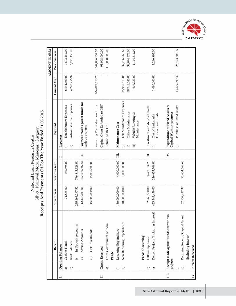
NBRC Annual Report 2014-15 | 169 |
Nat
iona
l Bra
in R
esea
rch
Cen
tre
Nh-
8, N
ainw
al M
ore,
Man
esar
, Gur
gaon
Rec
eipt
s And
Pay
men
ts O
f For
The
Yea
r End
ed 3
1.03
.201
5
AM
OU
NT
IN (R
S.)
Rec
eipt
Cur
rent
Yea
rPr
evio
us Y
ear
Paym
ent
Cur
rent
Yea
rPr
evio
us Y
ear
I.O
peni
ng B
alan
ces
I.Ex
penc
es
a)C
ash
in H
and
71,
885.
00
150
,698
.00
i)Es
tabl
ishm
ent E
xpen
ses
8,6
44,4
09.0
0 9
,603
,152
.00
b)Ba
nk B
alan
ces
ii)A
dmin
istr
ativ
e Ex
pens
es
4,2
20,7
56.9
7 6
,721
,155
.70
i)In
Dep
osit
Acc
ount
s 2
30,1
63,2
97.5
2 7
96,8
43,3
28.0
0
ii)Sa
ving
Acc
ount
s 1
12,1
36,0
11.0
1 3
07,6
39,3
07.5
4 II
.Pa
ymen
t mad
e ag
ains
t fun
ds fo
r va
riou
s pr
ojec
ts
iii)
CPF
Inve
stm
ents
15,
000,
000.
00
15,
036,
600.
00
Recu
rrin
g /C
apita
l exp
endi
ture
6
34,0
71,6
10.2
0 6
44,0
86,9
57.5
2
II.
Gra
nts
Rec
eive
dC
apita
l Gra
nt R
efun
ded
to D
BT -
91,
098,
000.
00
a)Fr
om G
over
nmen
t of I
ndia
Refu
nd to
RC
GB
- 1
10,0
00,0
00.0
0
PLA
N
i)Re
curr
ing
Expe
nditu
re 1
50,0
00,0
00.0
0 4
,000
,000
.00
III.
Mai
nten
ance
Cos
t
ii)N
on-R
ecur
ring
Exp
endi
ture
40,
000,
000.
00
1,0
00,0
00.0
0 i)
Lab
Mai
nten
ance
Exp
ense
s 3
5,95
5,51
3.05
3
7,76
6,06
0.68
ii)OfficeMaintenance
50,
741,
546.
00
38,
074,
373.
00
iii)
Vehi
cle
Runn
ing
&
Mai
nten
ance
619
,733
.00
1,1
44,5
14.0
0
PLA
N (R
ecur
ring
)
b)Fe
llow
ship
Gra
nt 2
,968
,550
.00
3,6
77,3
14.2
3 II
I.In
vest
men
t and
dep
osit
mad
e
c)D
elco
n Pr
ojec
ts (I
nclu
ding
Inte
rest
) 4
23,3
43,4
39.0
0 2
46,6
53,7
09.0
0 i)
Out
of E
arkm
ark/
Endo
wm
ent f
unds
1,0
80,0
00.0
0 1
,246
,802
.00
III.
Rec
eipt
mad
e ag
ains
t fun
ds fo
r var
ious
pr
ojec
tsIV
.Ex
pend
iture
of F
ixed
Ass
ets
&
Cap
ital W
ork-
in-p
rogr
ess
i)i)
PurchaseofFixedAsstts
13,
529,
090.
32
28,
473,
602.
39
ii)Re
curr
ing
Rece
ipt/
Cap
ital G
rant
67,
957,
417.
37
91,
634,
664.
60
(Incl
udin
g In
tere
st)
IV.
Inte
rest
Rec
eive
d

| 170 | NBRC Annual Report 2014-15
i)O
n Ba
nk d
epos
its -
32,
961,
672.
52
ii)Sa
ving
s Acc
ount
1,2
72,8
89.0
0 4
,783
,520
.00
iii)
On
CPF
Fun
d 1
,490
,728
.00
79,
346.
00
V.
Trai
ning
Exp
ense
s 1
,963
,564
.28
2,7
23,2
21.1
2
iv)
Oth
er In
tere
st 2
40,7
56.0
0 1
05,5
55.0
0
VI.
Oth
er P
aym
ents
(Spe
cify
)
VA
ny o
ther
rece
ipt
i)A
dvan
ce to
Sup
plie
r 4
,356
,329
.23
16,
108,
752.
24
Indi
rect
Inco
me
ii)
Advancetostaff
5,6
08,9
26.2
8 7
,200
,812
.00
i)A
dvan
ce to
Sup
plie
r rec
eive
d 7
5,14
5.00
6
11,7
84.0
0 iii
)Le
ave
Enca
shm
ent/
LTC
/ Bo
nus
311
,990
.00
705
,190
.00
ii)AdvancetoStaffreceived
1,3
98,7
53.2
4 9
39,3
48.0
0 iv
)Se
curi
ety
depo
sit
rece
ived
(pai
d) 7
80,6
72.6
7 1
15,4
61.0
0
iii)
Sale
of t
ende
r doc
umen
ts 1
4,50
0.00
4
8,50
0.00
v)
EMD
Ref
unde
d 3
,750
,339
.72
2,2
37,6
00.0
0
iv)
Mis
c.Re
ceip
ts.
470
,280
.87
878
,529
.00
vii)
TDS
Paid
6,5
70,4
72.0
0 5
,680
,647
.00
v)
Earn
est D
epos
it M
oney
rece
ived
1,4
82,4
26.0
0 2
,892
,400
.00
ix)
Impe
rest
256
,887
.00
199
,060
.00
vi)
Sale
of S
crap
12,
000.
00
167
,100
.00
xii)
Paym
ent O
f Cur
rent
Li
abili
ties.
151
,385
,403
.00
152
,472
,340
.71
vii)
Gue
st H
ouse
Cha
rges
212
,550
.00
198
,729
.00
viii)
Hos
tel D
epos
it 1
56,0
00.0
0 2
20,0
00.0
0 V
II.
Clo
sing
bal
ance
s
ix)
CPF
Fun
d re
ceiv
ed 1
,875
,804
.00
1,6
48,9
13.0
0 a)
Cas
h in
Han
d 1
50,8
68.0
0 7
1,88
5.00
x)Li
brar
y D
epos
it 4
4,00
0.00
5
8,00
0.00
b)
Bank
Bal
ance
xi)
Cur
rent
Lia
bilit
ies
rec.
1,6
46,6
79.0
0 7
99,8
77.0
0 i)
In D
epos
it A
ccou
nts
- 2
30,1
63,2
97.5
2
ii)Sa
ving
Acc
ount
s 1
11,6
35,0
00.2
9 1
12,1
36,0
11.0
1
iii)
CPF
Inve
stm
ents
16,
400,
000.
00
15,
000,
000.
00
TOTA
L 1
,052
,033
,111
.01
1,5
13,0
28,8
94.8
9 TO
TAL
1,0
52,0
33,1
11.0
1 1
,513
,028
,894
.89
Annual Financial Statements
For N
.C. M
ittal
& C
o C
hart
ered
Acc
ount
ants
(
Frn0
0023
7n)
Kap
il M
ittal
Part
ner
Mem
bers
hip
No.
5033
78
Raj
esh
Kum
ar V
yas
Finance&AccountsOfficer
Prof
. Sub
rata
Sin
ha
Dir
ecto
r

NBRC Annual Report 2014-15 | 171 |
Rajesh Kumar Vyas Prof. Subrata Sinha Finance&AccountsOfficer Director
For N.C. Mittal & Co Chartered Accountants (Frn000237n)
Kapil Mittal Partner Membership No.503378
National Brain Research CentreNh-8, Nainwal More, Manesar, Gurgaon
Schedule Forming Part Of Balance Sheet As At 31.03.15
AMOUNT IN (RS)
SCHEDULE 1-CORPUS/CAPITAL FUND: Current Year Previous Year
1. Grant-in-Aid - Balance as at the beginning of the year 988,502,000.00 1,131,074,000.00
Add: Contribution towards Corpus/Capital Fund 40,000,000.00 -142,572,000.00
40,000,000.00 -142,572,000.00
Balance as at the Year - End 1,028,502,000.00 988,502,000.00

| 172 | NBRC Annual Report 2014-15
National Brain Research CentreNh-8, Nainwal More, Manesar, Gurgaon
Schedule Forming Part Of Balance Sheet As At 31.03.15
SCHEDULE 2 - RESERVES AND SURPLUS:AMOUNT IN (RS)
Current Year Previous Year
1. GENERAL RESERVE
As per last Account -124,568,030.41 73,545,190.02
Addition during the Year -91,036,800.18 -198,113,220.43
Less : Deductions during the year (deficit) -215,604,830.59 -124,568,030.41
Total -215,604,830.59 -124,568,030.41
Annual Financial Statements
Rajesh Kumar Vyas Prof. Subrata Sinha Finance&AccountsOfficer Director
For N.C. Mittal & Co Chartered Accountants (Frn000237n)
Kapil Mittal Partner Membership No.503378

NBRC Annual Report 2014-15 | 173 |
SCHEDULE 3 - EARMARKED/ENDOWMENT FUNDS
AMOUNT IN (RS.)
Current Year Previous Year
(Refer Annexures I - LXXII)
A. Opening Balance of Project Fund 267,919,296.53 468,094,766.81
Add : Grants Received during the year 60,172,722.37 60,488,617.60
Less : Grants Refunded During the year 0.00 0.00
328,092,018.90 528,583,384.41
Add:Interest Earned 5,683,682.25 33,582,656.65
Any other addition during the year 0.00 333,775,701.15 0.00 562,166,041.06
Less: Utilization/Expenditure towards objectives of funds
a) Capital Expenditure
Fixed Assets 177,929,748.18 261,628,355.73
Others 0.00 0.00
b) Revenue Expenditure
Salaries and wages and 20,656,324.73 12,617,890.00
Other Administrative Expenses 32,687,316.64 20,000,498.80
231,273,389.55 294,246,744.53
Total (a) 102,502,311.60 267,919,296.53
Opening Balance of Fixed Asset Fund (Project)
344,558,377.56 146,014,697.14
B. Add: Addition During the year 177,929,748.18 258,934,925.73
Less: Depreciation for the period 2014-15 77,730,151.58 60,391,245.32
Total (b) 444,757,974.16 344,558,377.56
Opening balance of Donation received 2,631,788.00 2,631,788.00
C. Add: Additions during the year 2,631,788.00 0.00 2,631,788.00
Endowment fund created for Buildings Opening Balance
85,473,182.00 81,284,807.00
D. Add: Additions / (Paymnet) during the year
85,473,182.00 4,188,375.00 85,473,182.00
Contributory Provident Fund 9,902,908.00 9,902,908.00
Add: Additions / (Payment) during the year
795,804.00 10,698,712.00 0.00 9,902,908.00
E.
DeLcon E-library Consortium
F. Opening balance of Consortium 32,896,815.16 162,314,298.17
Add : Grants Received during the year 421,449,577.00 245,582,438.00
Add interest Earned 1893862.00 2964007.00
National Brain Research CentreNh-8, Nainwal More, Manesar, Gurgaon
Schedule Forming Part Of Balance Sheet As At 31.03.15

| 174 | NBRC Annual Report 2014-15
Less: Utilization/Expenditure towards objectives of funds
432,042,420.03 377,963,928.01
24,197,834.13 32,896,815.16
Escrow Account-DBT
G. Opening balance 0.00 57,526,000.00
Less: Grant Received During the Year 0.00 0.00 57,526,000.00 0.00
Grand total (A+B+C+D+E+F+G) 670,261,801.89 743,382,367.25
Annual Financial Statements
Rajesh Kumar Vyas Prof. Subrata Sinha Finance&AccountsOfficer Director
For N.C. Mittal & Co Chartered Accountants (Frn000237n)
Kapil Mittal Partner Membership No.503378

NBRC Annual Report 2014-15 | 175 |
SCHEDULE-4 CURRENT LIABILITIES AND PROVISIONS
AMOUNT IN (RS.)
Current Year Previous Year
A. CURRENT LIABILITIES
1 Sundry Creditors 796,753.25 783,179.25
2 Advances Received (Security deposit) 5,013,848.62 3,260,345.29
3 Other Liabilities-TDS Payable 822,205.50 943,149.50
4 Earnest Money Deposit 3,005,008.00 5,228,671.72
5 Hostel Deposit 624,000.00 692,000.00
6 Library Deposit 160,000.00 170,000.00
7 Expenses Payable 12,607,531.46 301,941.92
8 CPF Payable 54,437.00 42,727.00
9 GIS Payable 2,764.00 2,774.00
10 Salary Payable 5,314.00 14,008.00
11 NPS(Employees Subscription) 24,090.00 80,766.00
12 Labour Cess Payable 338,537.00 167,334.00
23,454,488.83 11,686,896.68
TOTAL (A) 23,454,488.83 11,686,896.68
B. PROVISIONS
1 Gratuity 6743850.00 6,737,555.00
2 Accumulated Leave Encashment 3958568.80 4,009,685.80
10,702,418.80 10,747,240.80
TOTAL (B) 10,702,418.80 10,747,240.80
TOTAL (A+B) 34,156,907.63 22,434,137.48
National Brain Research CentreNh-8, Nainwal More, Manesar, Gurgaon
Schedule Forming Part Of Balance Sheet As At 31.03.15
Rajesh Kumar Vyas Prof. Subrata Sinha Finance&AccountsOfficer Director
For N.C. Mittal & Co Chartered Accountants (Frn000237n)
Kapil Mittal Partner Membership No.503378

| 176 | NBRC Annual Report 2014-15
SCH
EDU
LE 5
- FI
XED
A
SSET
S/ D
EPR
ECIA
TON
GR
OSS
BLO
CK
DEP
RIC
IATI
ON
NET
BLO
CK
Cos
t /va
luat
ion
As
at
begi
nnin
g o
f th
e Ye
ar
Add
ition
s
duri
ng
the
Year
D.D
.YC
ost /
valu
atio
n A
s at
end
of t
he
Year
As
at th
e be
ginn
ing
of
the
Year
Dep
reci
atio
n fo
r cur
rent
ye
ar
On
D
.D.Y
Tota
l Dep
n.
Upt
o 31
.03.
13A
s at
Cur
rent
ye
ar-e
ndA
s at
Pre
viou
s ye
ar-e
nd
A.
Fixe
d A
sset
s
1Bu
ildin
g64
,653
,850
.00
4,91
4,16
4.00
0.00
69,5
68,0
14.0
012
,647
,821
.86
5692
019.
000.
0018
,339
,840
.86
51,2
28,1
73.1
452
,006
,028
.14
2Pl
ant &
Mac
hine
ry A
nd
Equi
pmen
t25
5,52
1,41
0.12
7,56
9,13
8.64
0.00
263,
090,
548.
7696
,253
,779
.54
24,8
71,2
59.0
00.
0012
1,12
5,03
8.54
141,
965,
510.
2215
9,26
7,63
0.58
3Ve
hicl
es2,
086,
342.
000.
000.
002,
086,
342.
001,
148,
982.
4814
0,60
4.00
0.00
1,28
9,58
6.48
796,
755.
5293
7,35
9.52
4Fu
rnitu
re &
Fix
ture
s32
,698
,661
.00
1,91
8,20
6.00
0.00
34,6
16,8
67.0
013
,773
,730
.32
2,05
6,34
7.00
0.00
15,8
30,0
77.3
218
,786
,789
.68
18,9
24,9
30.6
8
5C
ompu
ter &
Sof
twar
e3,
750,
822.
8181
2,68
6.00
0.00
4,56
3,50
8.81
2,54
0,05
1.00
1,16
1,04
9.00
0.00
3,70
1,10
0.00
862,
408.
811,
210,
771.
81
6OfficeEquipment
28,0
52,1
54.1
13,
088,
692.
680.
0031
,140
,846
.79
10,7
48,8
83.1
23,
044,
058.
000.
0013
,792
,941
.12
17,3
47,9
05.6
717
,303
,270
.99
TOTA
L O
F TH
E C
UR
REN
T YE
AR
386,
763,
240.
0418
,302
,887
.32
0.00
405,
066,
127.
3613
7,11
3,24
8.32
36,9
65,3
36.0
00.
0017
4,07
8,58
4.32
230,
987,
543.
0424
9,64
9,99
1.72
B.Fi
xed
Ass
ets
(Pro
ject
s)
1Pr
ojec
t Equ
ipm
ents
496,
268,
331.
9517
7,92
9,74
8.18
0.00
674,
198,
080.
1315
1,70
9,95
4.40
77,7
30,1
51.5
80.
0022
9,44
0,10
5.98
444,
757,
974.
1534
4,55
8,37
7.55
TOTA
L O
F TH
E C
UR
REN
T YE
AR
(A+B
)88
3,03
1,57
1.99
196,
232,
635.
500.
001,
079,
264,
207.
4928
8,82
3,20
2.72
114,
695,
487.
580.
0040
3,51
8,69
0.30
675,
745,
517.
1959
4,20
8,36
9.27
Prev
ious
Yea
r56
8,99
4,37
4.51
C.
Cap
ital W
ork
In
Prog
ress
1Bu
ildin
gs
Cap
ital w
ork-
in-
prog
ress
incl
udin
g ad
vanc
es,
652,
314,
275.
7316
,659
,809
.00
0.00
668,
974,
084.
730.
000.
000.
000.
0066
8,97
4,08
4.73
652,
314,
275.
73
cons
truc
tion
mat
eria
ls
and
build
ing
unde
r
cons
truc
tion
(net
of
reco
very
)
DIn
tang
ible
Ass
ets
1Pa
tent
And
Cop
y Ri
ght
(App
licat
ion
Fee)
3,50
7,36
6.00
850,
498.
000.
004,
357,
864.
001,
349,
346.
0075
2,13
1.00
0.00
2,10
1,47
7.00
2,25
6,38
7.00
2,15
8,02
0.00
TOTA
L (A
+ B
+C)
1,53
8,85
3,21
3.72
213,
742,
942.
500.
001,
752,
596,
156.
2229
0,17
2,54
8.72
115,
447,
618.
580.
0040
5,62
0,16
7.30
1,34
6,97
5,98
8.92
1,24
8,68
0,66
5.00
Nat
iona
l Bra
in R
esea
rch
Cen
tre
Nh-
8, N
ainw
al M
ore,
Man
esar
, Gur
gaon
Sche
dule
For
min
g Pa
rt O
f Bal
ance
She
et A
s At 3
1.03
.15
For N
.C. M
ittal
& C
o C
hart
ered
Acc
ount
ants
(
Frn0
0023
7n)
Kap
il M
ittal
Part
ner
Mem
bers
hip
No.
5033
78
Raj
esh
Kum
ar V
yas
Finance&AccountsOfficer
Prof
. Sub
rata
Sin
ha
Dir
ecto
r
Annual Financial Statements

NBRC Annual Report 2014-15 | 177 |
National Brain Research CentreNh-8, Nainwal More, Manesar, Gurgaon
Schedule Forming Part Of Balance Sheet As At 31.03.15
SCHEDULE 6- INVESTMENTS - CPF FUNDAMOUNT IN (RS.)
Current Year Previous Year
1.FDR In scheduled Banks 16,400,000.00 15,000,000.00
2.Balance with Savings Bank Account 2,946,141.12 2,507,551.82
19,346,141.12 17,507,551.82
Rajesh Kumar Vyas Prof. Subrata Sinha Finance&AccountsOfficer Director
For N.C. Mittal & Co Chartered Accountants (Frn000237n)
Kapil Mittal Partner Membership No.503378

| 178 | NBRC Annual Report 2014-15
National Brain Research CentreNh-8, Nainwal More, Manesar, Gurgaon
Schedule Forming Part Of Balance Sheet As At 31.03.15
SCHEDULE 7 - CURRENT ASSETS, LOANS, ADVANCES ETC.
AMOUNT IN (RS.)
Current Year Previous Year
A CURRENT ASSETS
1 Cash Balances in hand (Including Cheques/Drafts) 150,868.00 71,885.00
2 Bank Balances:
a) With Schduled Banks:
-In Deposit Account 0.00 150,163,297.52
-In Saving Accounts (Core & Projects) 108,688,859.18 109,628,459.20
-In Deposit Against various Project Assets 0.00 108,688,859.18 80,000,000.00 339,791,756.72
3 Interest Accrued on FD(CPF) 1,475,662.76 0.00
4 Grant Receivable (DBT) 19,300,000.00 0.00
TOTAL (A) 129,615,389.94 339,863,641.72
B LOANS, ADVANCES AND OTHER ASETS
1 Advances and other amounts receivable in cash
or in kind or for value to be received
a)Staff 8,819,350.59 8,578,072.49
b) Imprest 86,462.00 62,180.00
c) Advance to Parties 5,173,043.57 7,561,032.50
d) Others(Security & other Deposits) 2,016,902.20 2,198,318.20
e) TDS Receivable 4,342,380.59 4,341,980.59
f) Prepaid Insurance 940,220.00 957,032.00
21,378,358.95 23,698,615.78
TOTAL (B) 21,378,358.95 23,698,615.78
TOTAL (A + B) 150,993,748.89 363,562,257.50
Annual Financial Statements
Rajesh Kumar Vyas Prof. Subrata Sinha Finance&AccountsOfficer Director
For N.C. Mittal & Co Chartered Accountants (Frn000237n)
Kapil Mittal Partner Membership No.503378

NBRC Annual Report 2014-15 | 179 |
National Brain Research CentreNh-8, Nainwal More, Manesar, Gurgaon
Schedule Forming Part Of Income And Expenditure For The Year Ended 31.03.2015
SCHEDULE 8 - FEES/ SUBSCRIPTIONSAMOUNT IN (RS.)
Current Year Previous Year
1.Application Fees (Net) 922,996.39 347,391.00
2.Annual Fees/ Subscription to Journals 201,652.00 298,556.00
3.Others (Specify)-Fellowship Grants 2,801,682.00 2,363,368.23
TOTAL 3,926,330.39 3,009,315.23
Rajesh Kumar Vyas Prof. Subrata Sinha Finance&AccountsOfficer Director
For N.C. Mittal & Co Chartered Accountants (Frn000237n)
Kapil Mittal Partner Membership No.503378

| 180 | NBRC Annual Report 2014-15
National Brain Research CentreNh-8, Nainwal More, Manesar, Gurgaon
Schedule Forming Part Of Income And Expenditure For The Year Ended 31.03.2015
SCHEDULE 9 - INTEREST EARNEDAMOUNT IN (RS.)
Current Year Previous Year
1) On Term Deposits:
a) With Scheduled Banks 2,499,857.23 28,488,351.87
2) On Savings Accounts:
a) With Scheduled Banks 1,316,304.00 4,783,520.00
3) On Advances: 126,895.00 137,757.00
TOTAL 3,943,056.23 33,409,628.87
Annual Financial Statements
Rajesh Kumar Vyas Prof. Subrata Sinha Finance&AccountsOfficer Director
For N.C. Mittal & Co Chartered Accountants (Frn000237n)
Kapil Mittal Partner Membership No.503378

NBRC Annual Report 2014-15 | 181 |
National Brain Research CentreNh-8, Nainwal More, Manesar, Gurgaon
Schedule Forming Part Of Income And Expenditure For The Year Ended 31.03.2015
SCHEDULE 10-OTHER INCOMEAMOUNT IN (RS.)
Current Year Previous Year
1. Projects Receipts 4080538.00 5877535.18
2. Tender Form 14500.00 48500.00
3. Miscellaneous (Scrap & Others) 65843.00 174199.00
4. Insurance Claim Received 0.00 35200.00
5. Medical Contribution Recovery 187900.00 194480.00
6. Licence Fee Recovery 105764.00 134563.00
7. Establishment Charges 0.00 127790.00
8. Guest House Charges 271250.00 342279.00
TOTAL 4,725,795.00 6,934,546.18
Rajesh Kumar Vyas Prof. Subrata Sinha Finance&AccountsOfficer Director
For N.C. Mittal & Co Chartered Accountants (Frn000237n)
Kapil Mittal Partner Membership No.503378
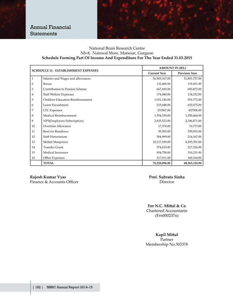
| 182 | NBRC Annual Report 2014-15
National Brain Research CentreNh-8, Nainwal More, Manesar, Gurgaon
Schedule Forming Part Of Income And Expenditure For The Year Ended 31.03.2015
SCHEDULE 11 - ESTABLISHMENT EXPENSESAMOUNT IN (RS.)
Current Year Previous Year
1 Salaries and Wages and allowances 56,905,167.00 51,891,757.00
2 Bonus 132,400.00 119,451.00
3 Contribution to Pension Scheme 667,692.00 693,872.00
4 StaffWelfareExpenses 174,480.00 118,252.00
5 Children Education Reimbursement 1,031,140.00 976,772.00
6 Leave Encashment 215,048.00 652,075.00
7 LTC Expenses 531867.00 457506.00
8 Medical Reimbursement 1,554,330.00 1,350,464.00
9 NPS(Employers Subscription) 2,835,523.00 2,346,871.00
10 Overtime Allowance 17,374.00 19,773.00
11 Rent for Residence 93,583.00 539,052.00
12 StaffHonorarium 304,999.00 214,167.00
13 Skilled Manpower 10,117,189.00 8,295,391.00
14 Transfer Grant 374,633.00 217,326.00
15 Medical Insurance 954,758.00 310,231.00
16 OfficeExpenses 317,911.00 160,164.00
TOTAL 76,228,094.00 68,363,124.00
Annual Financial Statements
Rajesh Kumar Vyas Prof. Subrata Sinha Finance&AccountsOfficer Director
For N.C. Mittal & Co Chartered Accountants (Frn000237n)
Kapil Mittal Partner Membership No.503378

NBRC Annual Report 2014-15 | 183 |
National Brain Research CentreNh-8, Nainwal More, Manesar, Gurgaon
Schedule Forming Part Of Income And Expenditure For The Year Ended 31.03.2015
SCHEDULE 12 - OTHER ADMINISTRATIVE EXPENSESAMOUNT IN (RS.)
Current Year Previous Year
1. Postage, Telephone and Communication Charges 926,210.70 1,072,898.00
2. Printing and Stationary 539,370.00 2,312,270.00
3. Travelling Expenses 5,531,622.81 8,043,294.23
4. Auditors Remuneration 18,090.00 30,899.00
5. Hospitality/Local Meeting Expenses 489,991.00 829,645.00
6. Legal & Professional Charges 804,785.00 628,003.00
7. Lease Rent 1,000,000.00 1,000,000.00
8. Bank Charges 925.46 4,263.62
9. Advertisement and Publicity 760,566.00 1,558,763.00
10. Misc. Expenses 408,053.00 1,130,251.85
11. Books & Periodicals 78,836.00 97,002.00
12. Transportation charges 381,668.00 295,346.00
13. Conveyance Reimbursement 97,769.00 73,756.00
14. Honorarium (Others) 395,333.00 541,141.00
TOTAL 11,433,219.97 17,617,532.70
Rajesh Kumar Vyas Prof. Subrata Sinha Finance&AccountsOfficer Director
For N.C. Mittal & Co Chartered Accountants (Frn000237n)
Kapil Mittal Partner Membership No.503378

| 184 | NBRC Annual Report 2014-15
National Brain Research CentreNh-8, Nainwal More, Manesar, Gurgaon
Schedule Forming Part Of Income And Expenditure For The Year Ended 31.03.2015
SCHEDULE 13 - REPAIR & MAINTENANCEAMOUNT IN (RS.)
Current Year Previous Year
1. Electricity and Water Charges 46,189,319.00 33,650,076.00
2. Insurance Others 1,447,596.00 1,306,026.00
3.Repairs&maintenance(Office) 14,760,109.00 13,225,276.00
4. Manpower (House Keeping) 1,456,007.00 1,862,471.00
5. Vehicle Running and Maintenance 174,687.00 179,638.00
6. Manpower (Security) 5,985,738.00 4,701,718.00
7. Horticulture 2,149,562.00 2,199,144.00
8. Repairs & Maintenance (Buildings) 3,502,223.00 1,796,066.00
9. Repairs & Maintenance (Lab Equipment) 5,953,932.00 4,495,994.75
10.Repairs&Maintenance(OfficeEquipment) 166,833.00 315,877.00
11. Insurance Charges vehicle 36,671.00 51,670.00
12.Repairs&maintenanceofficeequip.(AMC) 2,907,788.22 287,749.00
13. Petrol Diesel CNG etc. 674,671.00 1,476,929.00
TOTAL 85,405,136.22 65,548,634.75
Annual Financial Statements
Rajesh Kumar Vyas Prof. Subrata Sinha Finance&AccountsOfficer Director
For N.C. Mittal & Co Chartered Accountants (Frn000237n)
Kapil Mittal Partner Membership No.503378

NBRC Annual Report 2014-15 | 185 |
National Brain Research CentreNh-8, Nainwal More, Manesar, Gurgaon
Schedule Forming Part Of Income And Expenditure For The Year Ended 31.03.2015
SCHEDULE 14 - TRAINING AND NETWORKING EXPENSESAMOUNT IN (RS.)
Current Year Previous Year
1. Subscription to Journals 854,258.78 2,152,808.72
2. Training Expenses 13,005,220.00 9,332,116.00
3. Contingencies (CSIR/UGC/DBT/ICMR Students) 94,902.00 110,000.00
4. Conference & workshop Expenses 753,586.00 2,780,526.00
5. Student Medical Exp. 146,109.00 97,481.00
TOTAL 14,854,075.78 14,472,931.72
Rajesh Kumar Vyas Prof. Subrata Sinha Finance&AccountsOfficer Director
For N.C. Mittal & Co Chartered Accountants (Frn000237n)
Kapil Mittal Partner Membership No.503378

| 186 | NBRC Annual Report 2014-15
National Brain Research CentreNh-8, Nainwal More, Manesar, Gurgaon
Schedule Forming Part Of Income And Expenditure For The Year Ended 31.03.2015
SCHEDULE 15 - LABORATORY AND ANIMAL HOUSE CONSUMABLSAMOUNT IN (RS.)
Current Year Previous Year
1.Lab consumables and chemicals 33,229,916.19 38,232,404.54
2.Medicines and Consumables Animal 1,817,516.50 1,423,348.00
TOTAL 35,047,432.69 39,655,752.54
Annual Financial Statements
Rajesh Kumar Vyas Prof. Subrata Sinha Finance&AccountsOfficer Director
For N.C. Mittal & Co Chartered Accountants (Frn000237n)
Kapil Mittal Partner Membership No.503378
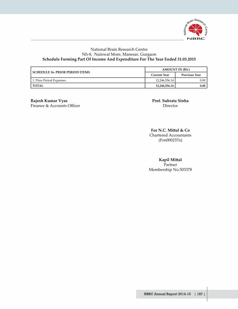
NBRC Annual Report 2014-15 | 187 |
National Brain Research CentreNh-8, Nainwal More, Manesar, Gurgaon
Schedule Forming Part Of Income And Expenditure For The Year Ended 31.03.2015
SCHEDULE 16- PRIOR PERIOD ITEMSAMOUNT IN (RS.)
Current Year Previous Year
1. Prior Period Expenses 12,246,556.14 0.00
TOTAL 12,246,556.14 0.00
Rajesh Kumar Vyas Prof. Subrata Sinha Finance&AccountsOfficer Director
For N.C. Mittal & Co Chartered Accountants (Frn000237n)
Kapil Mittal Partner Membership No.503378
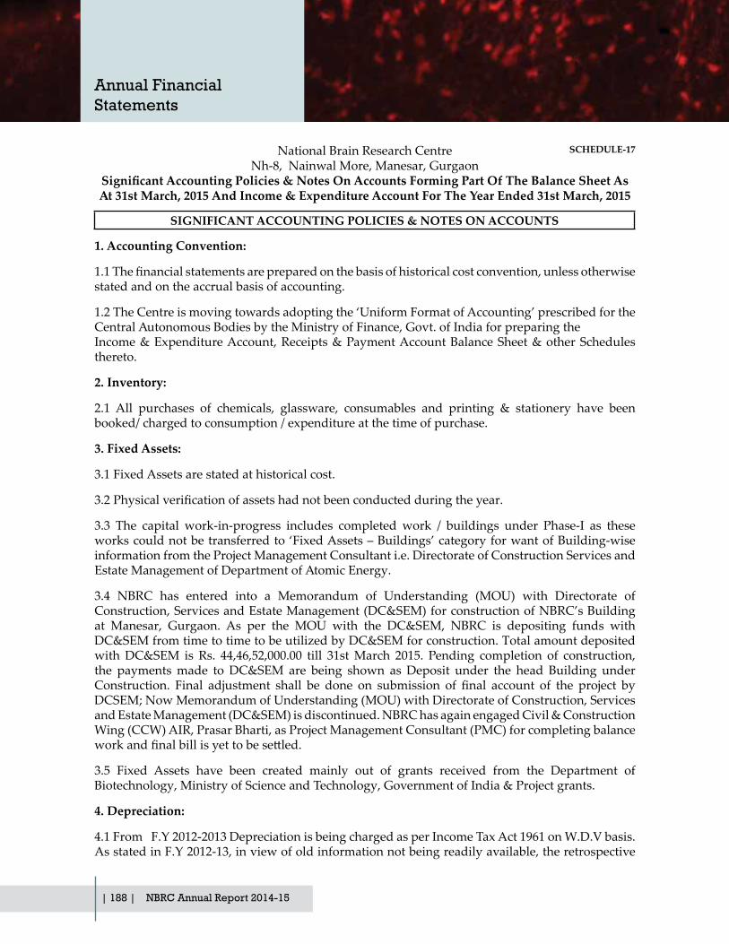
| 188 | NBRC Annual Report 2014-15
1. Accounting Convention:
1.1Thefinancialstatementsarepreparedonthebasisofhistoricalcostconvention,unlessotherwisestated and on the accrual basis of accounting.
1.2 The Centre is moving towards adopting the ‘Uniform Format of Accounting’ prescribed for the Central Autonomous Bodies by the Ministry of Finance, Govt. of India for preparing the Income & Expenditure Account, Receipts & Payment Account Balance Sheet & other Schedules thereto.
2. Inventory:
2.1 All purchases of chemicals, glassware, consumables and printing & stationery have been booked/ charged to consumption / expenditure at the time of purchase.
3. Fixed Assets:
3.1 Fixed Assets are stated at historical cost.
3.2Physicalverificationofassetshadnotbeenconductedduringtheyear.
3.3 The capital work-in-progress includes completed work / buildings under Phase-I as these works could not be transferred to ‘Fixed Assets – Buildings’ category for want of Building-wise information from the Project Management Consultant i.e. Directorate of Construction Services and Estate Management of Department of Atomic Energy.
3.4 NBRC has entered into a Memorandum of Understanding (MOU) with Directorate of Construction, Services and Estate Management (DC&SEM) for construction of NBRC’s Building at Manesar, Gurgaon. As per the MOU with the DC&SEM, NBRC is depositing funds with DC&SEM from time to time to be utilized by DC&SEM for construction. Total amount deposited with DC&SEM is Rs. 44,46,52,000.00 till 31st March 2015. Pending completion of construction, the payments made to DC&SEM are being shown as Deposit under the head Building under Construction. Final adjustment shall be done on submission of final account of the project byDCSEM; Now Memorandum of Understanding (MOU) with Directorate of Construction, Services and Estate Management (DC&SEM) is discontinued. NBRC has again engaged Civil & Construction Wing (CCW) AIR, Prasar Bharti, as Project Management Consultant (PMC) for completing balance workandfinalbillisyettobesettled.
3.5 Fixed Assets have been created mainly out of grants received from the Department of Biotechnology, Ministry of Science and Technology, Government of India & Project grants.
4. Depreciation:
4.1 From F.Y 2012-2013 Depreciation is being charged as per Income Tax Act 1961 on W.D.V basis. As stated in F.Y 2012-13, in view of old information not being readily available, the retrospective
National Brain Research CentreNh-8, Nainwal More, Manesar, Gurgaon
Significant Accounting Policies & Notes On Accounts Forming Part Of The Balance Sheet As At 31st March, 2015 And Income & Expenditure Account For The Year Ended 31st March, 2015
SIGNIFICANT ACCOUNTING POLICIES & NOTES ON ACCOUNTS
SCHEDULE-17
Annual Financial Statements

NBRC Annual Report 2014-15 | 189 |
calculation of depreciation as per Income Tax Act 1961 for adjustment of excess/short depreciation is vis-à-vis the old rates, as required under the Accounting Standard-6 issued by Institute of Chartered Accounts of India (ICAI), could not be made till date. The same shall be made in due course of the determination of the same.
4.2 Depreciation provided for current year on the fixed assets of Project for Rs. 7,77,30,151.58andwhichhasbeendirectlydebitedtothefixedassetsfundsaccount.Theseassetswerecreatedthrough the Non-Recurring and project based grant from the funding agencies. Depreciation for otherthanprojectassetsamountingtoRs.3,77,17,467.00forcurrentfinancialyearhadbeendebitedto Income & Expenditure Account.
5. Investments:
5.1 Investments in term deposits with banks are basically for Current Investments and are therefore valued on cost.
5.2 Interest received on term deposits are accounted for on accrual basis, which results in increase inprofitability.
6. Government Grants / Subsidies:
6.1Governmentgrantsofthenatureofcontributiontowardscapitalcostofsettingupprojectsaretreated as Capital Reserve/Fund.
6.2 Government grants / subsidy are accounted for in accordance with the sanctioned terms.
6.3 Interest on Government Grant has been considered under the respective projects due to which loss has increased by
Rs. 75,77,544.25.
7. Foreign Currency Transactions/ Grants:
7.1 Transactions denominated in foreign currency are accounted at the exchange rate prevailing at the date of the transaction.
7.2 The Centre had one FCRA Bank Accounts PNB Manesar related to the Grants. The submission of the returns of these accounts has been made up to Financial Year 31.03.2009 under the FCR Act.
8. Lease:
The Centre is located on the leasehold land at Manesar taken from Indian Vaccine Corporation Ltd. for Rs. 10,00,000/- per annum lease rent with certain semi-built structure at a cost Rs. 45,17,000/- towards such structures. The lease is for the period of 33 years, after which the land along with premisesthereonaretobehandedovertothelessor.Noamortization/writeoffhavebeendoneinrespect of the assets acquired on lease.
9. Retirement Benefits:
9.1 The Centre is registered with the Provident Fund authorities and it maintains a separate CPF Trust, which is yet to be recognized and the CPF fund required the separate accounting.
9.2TheCentrehasnotmadeanyprovisionforgratuityandleaveencashmentduringfinancialyear

| 190 | NBRC Annual Report 2014-15
2014-2015 as against the requirement of AS-15 issued by ICAI. However the amount of gratuity and leave encashment to the extent of Rs.67,43,850/- and Rs.39,58,568/- already exists on 31.03.2015, against provision made earlier.
10. Taxation:
In view of the tax exemption status of the Center, no provision for income tax has been considered necessary.
11. Loans & Advances
Advances appearing under the head Current assets, Loans & Advances under Schedule-7 are subjecttoconfirmationfromparties.
12. Bank Balance
Bank balance in Axis Bank Limited (A/c No.056010100453998) as on 31st March, 2015 of Rs. 10,53,63,459.58 , is subject to reconciliation.
13. Prior Period Items
Accounting Standard-5 Issued by Institute of Chartered Accountants of India (ICAI), Prior Period items are income or expenses, which arises, in current period as a result of error or omission in thepreparationoffinancialstatementofoneormorepriorperiods.Inthecurrentyear,thePriorPerioditemsrecognized,relatedtoexpenditureforthefinancialyear2013-14thatwasomittedinthat year.
14. Others
14.1 The Balance in the name of various parties under the head Advance to Suppliers & Receivable fromcustomers andpayable to SundryCreditors are subject to confirmation/ reconciliationbyrespective parties. The total amount payable to Creditors is Rs. 7,96,753.25.
14.2 Schedules 1 to 16 along with Annexure 1 to 93 are annexed to and form an integral part of the Balance Sheet as at 31st March, 2015 and the Income and Expenditure Account for the year ended on that date.
14.3Correspondingfigures for theprevious yearhavebeen regrouped / rearranged,wherevernecessary.
Accounting polices not referred to otherwise be consistent with Generally Accepted Accounting Principles (GAAP).
Rajesh Kumar Vyas Prof. Subrata Sinha Finance&AccountsOfficer Director
Kapil Mittal As per our separate report Partner of even date attached MembershipNo.503378 ForN.C.Mittal&Co Chartered Accountants Place: Delhi (Frn000237n) Dated: 13.08.2015
Annual Financial Statements
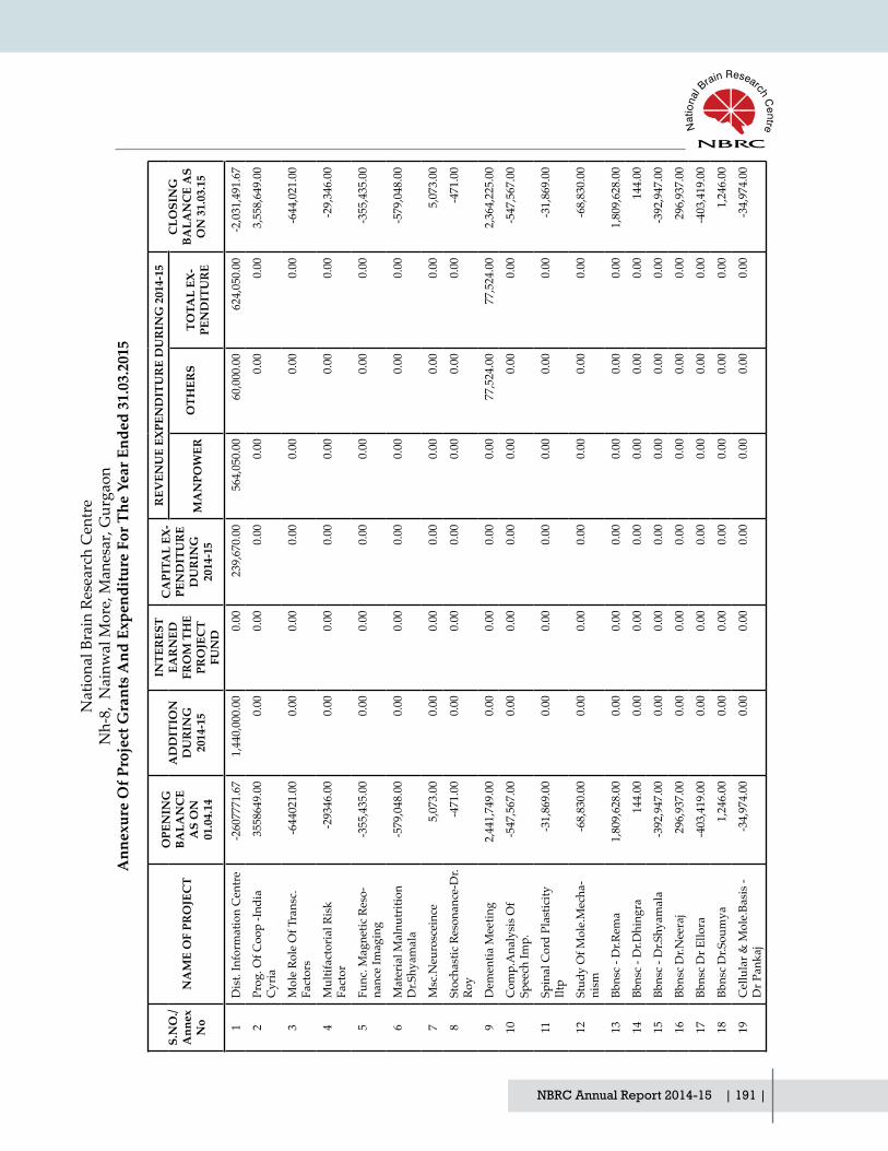
NBRC Annual Report 2014-15 | 191 |
S.N
O./
Ann
ex
No
NA
ME
OF
PRO
JEC
T
OPE
NIN
G
BALA
NC
E A
S O
N
01.0
4.14
AD
DIT
ION
D
UR
ING
20
14-1
5
INTE
RES
T EA
RN
ED
FRO
M T
HE
PRO
JEC
T FU
ND
CA
PITA
L EX
-PE
ND
ITU
RE
DU
RIN
G
2014
-15
REV
ENU
E EX
PEN
DIT
UR
E D
UR
ING
201
4-15
CLO
SIN
G
BALA
NC
E A
S O
N 3
1.03
.15
MA
NPO
WER
OTH
ERS
TOTA
L EX
-PE
ND
ITU
RE
1D
ist.
Info
rmat
ion
Cen
tre
-260
7771
.67
1,44
0,00
0.00
0.00
239,
670.
0056
4,05
0.00
60,0
00.0
062
4,05
0.00
-2,0
31,4
91.6
7
2Pr
og. O
f Coo
p -In
dia
Cyr
ia35
5864
9.00
0.00
0.00
0.00
0.00
0.00
0.00
3,55
8,64
9.00
3M
ole
Role
Of T
rans
c.
Fact
ors
-644
021.
000.
000.
000.
000.
000.
000.
00-6
44,0
21.0
0
4M
ultif
acto
rial R
isk
Fact
or-2
9346
.00
0.00
0.00
0.00
0.00
0.00
0.00
-29,
346.
00
5Fu
nc. M
agne
tic R
eso-
nanc
e Im
agin
g
-355
,435
.00
0.00
0.00
0.00
0.00
0.00
0.00
-355
,435
.00
6M
ater
ial M
alnu
triti
on
Dr.S
hyam
ala
-579
,048
.00
0.00
0.00
0.00
0.00
0.00
0.00
-579
,048
.00
7M
sc.N
euro
scei
nce
5,07
3.00
0.00
0.00
0.00
0.00
0.00
0.00
5,07
3.00
8St
ocha
stic
Res
onan
ce-D
r.Ro
y-4
71.0
00.
000.
000.
000.
000.
000.
00-4
71.0
0
9D
emen
tia M
eetin
g2,
441,
749.
000.
000.
000.
000.
0077
,524
.00
77,5
24.0
02,
364,
225.
00
10C
omp.
Ana
lysi
s Of
Spee
ch Im
p.-5
47,5
67.0
00.
000.
000.
000.
000.
000.
00-5
47,5
67.0
0
11Sp
inal
Cor
d Pl
astic
ity
Iltp
-31,
869.
000.
000.
000.
000.
000.
000.
00-3
1,86
9.00
12St
udy
Of M
ole.
Mec
ha-
nism
-68,
830.
000.
000.
000.
000.
000.
000.
00-6
8,83
0.00
13Bb
nsc
- Dr.R
ema
1,80
9,62
8.00
0.00
0.00
0.00
0.00
0.00
0.00
1,80
9,62
8.00
14Bb
nsc
- Dr.D
hing
ra14
4.00
0.00
0.00
0.00
0.00
0.00
0.00
144.
00
15Bb
nsc
- Dr.S
hyam
ala
-392
,947
.00
0.00
0.00
0.00
0.00
0.00
0.00
-392
,947
.00
16Bb
nsc
Dr.N
eera
j29
6,93
7.00
0.00
0.00
0.00
0.00
0.00
0.00
296,
937.
00
17Bb
nsc
Dr E
llora
-403
,419
.00
0.00
0.00
0.00
0.00
0.00
0.00
-403
,419
.00
18Bb
nsc
Dr.S
oum
ya1,
246.
000.
000.
000.
000.
000.
000.
001,
246.
00
19C
ellu
lar &
Mol
e.Ba
sis -
D
r Pan
kaj
-34,
974.
000.
000.
000.
000.
000.
000.
00-3
4,97
4.00
Nat
iona
l Bra
in R
esea
rch
Cen
tre
Nh-
8, N
ainw
al M
ore,
Man
esar
, Gur
gaon
Ann
exur
e O
f Pro
ject
Gra
nts A
nd E
xpen
ditu
re F
or T
he Y
ear E
nded
31.
03.2
015

| 192 | NBRC Annual Report 2014-15
20Es
t.Of T
rans
latio
nal R
es.
Uni
t4,
307,
442.
000.
000.
000.
000.
000.
000.
004,
307,
442.
00
21Ja
pane
se E
ncep
h.Vi
rus-
Dr.B
asu
451.
000.
000.
000.
000.
000.
000.
0045
1.00
22Fu
nctio
nal R
ole
Of E
6-A
p-D
r.Jan
a16
8.00
0.00
0.00
0.00
0.00
0.00
0.00
168.
00
23C
hara
c.O
f Mol
ecul
ar
Inte
rac.
-Pra
vat
2,10
6.00
0.00
0.00
0.00
0.00
0.00
0.00
2,10
6.00
24C
ogni
tive
Neu
ro S
cien
ce
W/S
-Adi
tiya
Mur
thy
-437
,464
.00
0.00
0.00
0.00
0.00
0.00
0.00
-437
,464
.00
25Eb
m In
clud
ing
Alz
-he
imer
Dis
ease
-Dr.
Vija
ylax
mi
-230
,717
.00
0.00
0.00
0.00
0.00
0.00
0.00
-230
,717
.00
26Ra
mal
inga
Sw
amy-
Dr.
Ranj
it K
r. G
iri-6
8,44
0.70
0.00
0.00
0.00
0.00
0.00
0.00
-68,
440.
70
27M
ultil
ingu
alis
m-D
r. N
andi
ni82
3.00
0.00
0.00
0.00
0.00
0.00
0.00
823.
00
28D
bt G
rant
-Dr.
Kal
lol
Dut
ta7,
920.
000.
000.
000.
000.
000.
000.
007,
920.
00
29In
do-U
s & N
ih R
o1-D
r. Pa
nkaj
616,
981.
580.
000.
000.
000.
0047
4,89
4.00
474,
894.
0014
2,08
7.58
30C
sir-
Dr.
Nih
ar R
anja
n Ja
na73
,089
.50
0.00
0.00
0.00
0.00
0.00
0.00
73,0
89.5
0
31Pe
rcep
tion
Engi
neer
ing
Proj
ect O
f Dit-
Dr.
Nee
raj
Jain
-1.3
10.
000.
000.
000.
000.
000.
00-1
.31
32Fu
nctio
nal I
mag
ing
Stud
y O
f Dys
lexi
a-D
r. N
.C S
ingh
339,
302.
77-1
,803
.00
0.00
0.00
288,
000.
0049
,500
.00
337,
500.
00-0
.23
33Ep
ileps
y Pr
ojec
t Of N
brc
110,
609,
523.
597,
542,
000.
004,
737,
051.
0073
,774
,120
.32
4,59
2,15
3.00
2,42
3,36
3.00
7,01
5,51
6.00
42,0
98,9
38.2
7
34M
otiv
ated
Beh
avio
ur In
M
ale
Zebr
a Fi
nche
s-D
r. So
umya
200,
481.
65-2
,525
.00
0.00
0.00
0.00
124,
762.
0012
4,76
2.00
73,1
94.6
5
35M
ulti
Dis
plin
ary
Syst
em
Of P
arki
nson
Dis
ease
-D
r. N
c Si
ngh
1,18
9,00
0.00
0.00
0.00
0.00
0.00
0.00
0.00
1,18
9,00
0.00
Annual Financial Statements

NBRC Annual Report 2014-15 | 193 |
37U
nder
stan
ding
The
Si
gnal
ing
Circ
uitie
s-D
r. El
lora
Sen
-575
,915
.39
0.00
0.00
0.00
0.00
0.00
0.00
-575
,915
.39
38Tw
o Ph
oton
Mic
rosc
ope
Faci
lity
For A
dvan
ce
Rese
arch
112,
448,
703.
810.
0045
1,71
2.25
89,6
17,6
88.9
70.
0010
,060
.00
10,0
60.0
023
,272
,667
.09
39U
nder
stan
ding
The
Ps
ycho
logi
cal F
unct
ion
Of M
alin
350.
110.
000.
000.
000.
000.
000.
0035
0.11
40N
eura
l Net
wor
k M
echa
nism
-Dr.
Yoga
nar-
ashi
mha
-933
,372
.66
178,
000.
000.
000.
0012
3,67
7.00
66,7
69.0
019
0,44
6.00
-945
,818
.66
41Ib
ro S
choo
l Wor
ksho
p-
Prof
. P. K
. Roy
-672
,839
.00
0.00
0.00
0.00
0.00
0.00
0.00
-672
,839
.00
42D
st S
erc
Scho
ol
Wor
ksho
p-D
r. So
umya
Iy
enga
r
36,3
76.0
00.
000.
000.
000.
000.
000.
0036
,376
.00
43C
ircad
ian
Syst
em L
ink-
age
(Dst
) Dr.
Soum
ya
Iyen
gar
-216
,963
.61
800,
000.
000.
0063
,816
.00
189,
000.
0032
6,04
8.00
515,
048.
004,
172.
39
44C
olla
bora
tion
For T
rans
. &
Clin
. Res
. (G
lue)
Pro
f. P
K R
oy
1,24
5,48
5.00
0.00
0.00
0.00
777,
754.
0051
8,43
5.00
1,29
6,18
9.00
-50,
704.
00
45C
sir -
Ii St
udy
The
Role
O
f Neu
ral I
mm
une
Resp
nce
Dr.
Bas
u
-168
,365
.93
0.00
0.00
0.00
0.00
0.00
0.00
-168
,365
.93
46D
st A
ustis
m S
pect
rum
D
isor
der D
r. N
andi
ni C
. Si
ngh
57,6
44.0
070
0,00
0.00
0.00
0.00
391,
097.
0020
1,23
8.00
592,
335.
0016
5,30
9.00
47D
st C
ogni
tive
Scie
nce
Rese
arch
Initi
ativ
e (C
si)
Dr.
Cha
itra
Rao
-948
,000
.00
624,
000.
000.
000.
000.
000.
000.
00-3
24,0
00.0
0
48D
it M
cgill
Lin
kage
(N
kn) P
rof.
Pras
um
Kum
ar R
oy
821,
174.
160.
000.
000.
0068
7,46
8.00
16,3
38.0
070
3,80
6.00
117,
368.
16
49Ic
mr H
iv A
ssoc
iate
d N
euro
cogn
itive
Dis
orde
r (H
and)
Dr.
Pank
aj
30,8
60.0
00.
000.
000.
000.
0030
,860
.00
30,8
60.0
00.
00

| 194 | NBRC Annual Report 2014-15
50Ro
le O
f Hum
an U
m-
bilic
al C
ord
Bloo
d St
em
(Aiim
s) D
r. Pa
nkaj
-299
,944
.56
300,
000.
000.
000.
000.
000.
000.
0055
.44
51D
bt N
atio
nal B
iosc
ienc
e A
war
d 20
10 (D
r. A
nir-
ban
Basu
)
240,
553.
900.
000.
0023
9,96
8.25
0.00
0.00
0.00
585.
65
52D
bt 5
Th M
eetin
g O
f Eg
n-C
db (D
r. Sh
iv K
u-m
ar S
harm
a)
67,8
75.0
00.
000.
000.
000.
000.
000.
0067
,875
.00
53D
st It
par W
orks
hop
On
Cog
nitiv
e N
euro
scie
nce
(Dr.
Nan
dini
C. )
84,7
93.0
0-8
4,79
3.00
0.00
0.00
0.00
0.00
0.00
0.00
54D
bt C
si D
evel
opm
ent
And
Val
idat
ion
Of
Scre
enin
g To
ols P
roje
ct-
Dr.
Nan
dini
541,
129.
000.
000.
0099
4,59
9.00
175,
000.
0018
1,82
5.00
356,
825.
00-8
10,2
95.0
0
55D
bt E
duca
tiona
l Neu
-ro
scie
nce
Mee
ting-
Dr.
Nan
dini
C S
ingh
476.
000.
000.
000.
000.
000.
000.
0047
6.00
56D
bt It
par G
rant
-Dr.
Nan
dini
C.S
ingh
2,15
8,63
8.08
0.00
0.00
576,
410.
3418
2,47
7.00
434,
991.
0061
7,46
8.00
964,
759.
74
57N
atio
nal I
nitia
tive
On
Glia
Cel
l Res
earc
h Pr
o-je
ct -D
r. Pa
nkaj
Set
h
222,
807.
860.
000.
000.
0011
6,90
3.00
998,
162.
151,
115,
065.
15-8
92,2
57.2
9
58D
bt P
ulse
Seq
uenc
e And
Pr
oces
sing
Pro
ject
-Dr.
Prav
at K
umar
Man
dal
-571
,350
.27
636,
600.
000.
000.
0061
,249
.73
4,00
0.00
65,2
49.7
30.
00
59D
bt B
irac
Und
er C
rs
Sche
me
Proj
ectg
rant
-Dr.
Ranj
it G
iri
676,
269.
2079
0,00
0.00
0.00
10,1
00.0
00.
0062
4,62
8.70
624,
628.
7083
1,54
0.50
60D
bt R
amal
inga
swam
y Fe
llow
ship
Dr.S
aura
v Ba
nerje
e
-1,1
92,3
72.0
03,
280,
000.
000.
000.
001,
182,
500.
001,
255,
145.
192,
437,
645.
19-3
50,0
17.1
9
61 D
st P
df P
roje
ct U
nder
C
si-D
r.D S
ubha
shre
e-4
54,7
48.0
062
4,00
0.00
0.00
0.00
216,
774.
0027
6,47
8.00
493,
252.
00-3
24,0
00.0
0
62D
st In
spire
Fac
ulty
A
war
d -D
r. Su
priy
a Bh
avan
i
1,70
8,26
8.56
0.00
0.00
0.00
1,00
0,04
0.00
955,
778.
211,
955,
818.
21-2
47,5
49.6
5
Annual Financial Statements

NBRC Annual Report 2014-15 | 195 |
63D
bt T
ata
Inno
vatio
n Fe
l-lo
wsh
ip -D
r P.
K.R
oy-7
0,18
8.40
690,
000.
000.
000.
0010
0,00
0.00
553,
445.
0065
3,44
5.00
-33,
633.
40
64D
bt In
cre
Gra
nt(N
brc)
(Pro
f Sub
rata
Sin
ha)
9,43
4,33
7.00
0.00
0.00
0.00
3,15
0,00
0.00
3,50
2,50
0.00
6,65
2,50
0.00
2,78
1,83
7.00
65Th
e W
elco
me
Trus
t/D
bt
Indi
a A
llian
ce P
roje
ct D
r. Sh
arba
Ban
dhop
adhy
ay
Dhy
ag
800,
278.
00-8
00,2
78.0
00.
000.
000.
000.
000.
000.
00
66N
atio
nal I
nstit
ute
Glia
l C
ell R
esea
rch-
Shiv
Ku-
mar
Sha
rma
150,
281.
130.
000.
000.
0012
7,10
0.00
490,
440.
0061
7,54
0.00
-467
,258
.87
67W
ellc
om T
rust
/Dbt
In
dian
Alli
ance
-Dr.
Am
-ita
bha
Maj
umda
r
3,80
7,20
2.00
9,05
9,84
6.00
0.00
5,07
1,31
0.33
437,
329.
004,
586,
502.
335,
023,
831.
332,
771,
906.
34
68W
ellc
om T
rust
/Dbt
In
dian
Alli
ance
-Dr.
Ani
ndya
Gho
sh R
oy
10,6
18,8
41.0
013
,353
,081
.00
401,
722.
005,
014,
017.
020.
003,
897,
243.
683,
897,
243.
6815
,462
,383
.30
69Im
plem
entin
g Pr
oteo
mic
A
ppro
ach
To U
nder
-st
and
The
Etio
logy
Of
Neu
ropa
thog
enes
is
Indu
ced
By C
hand
ipur
a Vi
rus I
nfec
tiona
:dr.
Ani
r-ba
n Ba
su
1,23
3,68
2.00
1,03
1,00
0.00
55,7
10.0
00.
000.
001,
705,
116.
921,
705,
116.
9261
5,27
5.08
70N
euro
-Cog
nitiv
e N
et-
wor
ks U
nder
lyin
g G
oal
Dire
cted
Beh
avio
r :D
r. A
rpan
Ban
erje
e
1,29
8,06
5.00
1,87
3,12
9.00
0.00
0.00
0.00
2,76
7,39
7.00
2,76
7,39
7.00
403,
797.
00
71Ro
le O
f Chr
omat
in
Rem
odel
ers I
n Re
gula
t-in
g A
ssoc
iate
d W
ith
Resi
stan
ce T
o A
popt
osis
U
nder
Iflam
mat
ory
And
H
ypox
ic C
nditi
ons I
n G
liom
a C
ells
: Dr.
Ello
ra
Sen
1,70
6,80
2.00
0.00
0.00
1,00
0,00
0.00
0.00
1,36
1,27
0.58
1,36
1,27
0.58
-654
,468
.58
72Fi
rst A
nnua
l Con
fere
nce
Of T
he A
ssoc
iatio
n Fo
r C
ognt
ive
Scie
nce:
Dr.
Nan
dini
C. S
ingh
17,5
33.0
00.
000.
000.
000.
003,
973.
003,
973.
0013
,560
.00

| 196 | NBRC Annual Report 2014-15
73A
Lon
gitu
dian
al S
tudy
To
Res
pons
iven
ess T
o So
ng B
ased
Stim
uli I
n C
hild
ren
With
Aut
ism
Be
havi
or A
nd D
iffus
ion
Tens
or Im
agin
g: D
r. N
andi
ni C
. Sin
gh
478,
871.
000.
000.
000.
0040
5,59
6.00
164,
553.
0057
0,14
9.00
-91,
278.
00
74Sc
reen
ing
Com
mitt
ee
Mee
ting
Und
er C
ogni
-tiv
e Sc
ienc
e Re
sear
ch
Initi
ativ
e (C
si):
Dr.
Nan
dini
C. S
ingh
34,5
92.0
00.
000.
000.
000.
0019
,614
.00
19,6
14.0
014
,978
.00
75D
ereg
ulat
ion
Of M
icro
Rn
a In
Cel
l And
Ani
mal
M
odel
s Of H
untin
gton
's D
isea
se: R
ole
Role
Of
Alte
red
Mic
ro R
na In
N
euro
nal D
iffer
en-
tiatio
n A
nd C
ell C
ycle
Re
gula
tion:
dr.N
ihar
Ra
njan
Jana
516,
185.
000.
000.
000.
0018
5,69
1.00
450,
333.
5963
6,02
4.59
-119
,839
.59
76D
bt W
orks
hop
On
Sci-
entifi
c G
rant
Writ
ing:
Dr
Pank
aj S
eth
-21,
476.
000.
000.
000.
000.
000.
000.
00-2
1,47
6.00
77In
do-U
s Sym
po-
sium
On
Vira
l Inf
ac-
tion
Of T
he N
ervo
us
Syst
em(Iu
sstf)
:Dr.
Pank
aj S
eth
280,
847.
280.
000.
000.
000.
0010
9,58
5.00
109,
585.
0017
1,26
2.28
78N
on-In
vasi
ve Im
agin
g Ba
sed
Det
ectio
n A
nd
Of B
rain
Oxi
dativ
e (U
.S. A
irfor
e):D
r. Pr
avat
K
umar
Man
dal
1,35
2,06
2.85
0.00
0.00
0.00
300,
000.
0012
4,80
6.00
424,
806.
0092
7,25
6.85
79C
hara
cter
izin
g Bi
o-m
arke
rs O
f Alz
heim
er's
Dis
ease
:A L
ongi
tudi
a-na
l M
ultii
Mod
al B
rain
Im
agin
g St
udy
(Bra
in
Imag
inin
g ) D
r. Pr
avat
K
umar
Man
dal
1,48
7,00
8.00
0.00
0.00
302,
200.
0086
1,60
6.00
386,
080.
001,
247,
686.
00-6
2,87
8.00
Annual Financial Statements

NBRC Annual Report 2014-15 | 197 |
80N
atio
nal P
rogr
amm
e O
n Pr
ecep
tion
Engi
neer
-in
g - P
hase
Ii D
r. Pr
avat
K
umar
Man
dal
688,
589.
001,
250,
000.
000.
0016
9,26
6.00
1,95
6,82
2.00
304,
053.
902,
260,
875.
90-4
91,5
52.9
0
81In
fluen
ce O
f Soc
ial C
ues
Om
apat
ial C
ogni
tion:
Dr
Che
tan
Yada
v
269,
859.
0060
0,00
0.00
0.00
0.00
420,
000.
0033
1,74
3.00
751,
743.
0011
8,11
6.00
82N
atio
nal P
rogr
amm
e O
n Pe
rcep
tion
Engi
neer
ing
Prof
Pk
Roy
500,
000.
001,
218,
000.
000.
0014
0,44
5.00
419,
806.
0041
6,59
6.00
836,
402.
0074
1,15
3.00
83C
ean
Retu
rn H
ome
Rew
ard
- Dr A
mita
bh
Maj
umda
r
0.00
477,
865.
370.
0026
9,06
2.46
0.00
0.00
0.00
208,
802.
91
84C
sir J
apan
ese
Encc
epha
-lit
ies D
r Ani
rban
Bas
u0.
0012
5,00
0.00
0.00
0.00
0.00
14,1
20.0
014
,120
.00
110,
880.
00
85M
olec
ular
Mec
hani
sm
Of M
icro
bial
Act
ivat
ion
Dr A
nirb
an B
asu
0.00
1,80
0,00
0.00
0.00
0.00
0.00
527,
180.
0052
7,18
0.00
1,27
2,82
0.00
86M
ultif
acet
ed K
inas
e C
dk5
Dr A
parn
a D
ixit
0.00
694,
600.
000.
000.
0021
,774
.00
38,7
50.0
060
,524
.00
634,
076.
00
87Vi
sion
Gui
de S
peec
h Pe
rcen
tion-
Dr A
rpan
Ba
nerje
e (D
bt)
0.00
1,95
0,00
0.00
0.00
0.00
208,
000.
0010
0,00
0.00
308,
000.
001,
642,
000.
00
88N
atio
nal B
iosc
ienc
e A
war
d- D
r Ello
ra S
en0.
0050
0,00
0.00
0.00
0.00
0.00
32,7
73.8
032
,773
.80
467,
226.
20
89Ta
ta In
nova
tion
Fello
w-
ship
- Dr N
ihar
Ran
jan
Jana
0.00
900,
000.
000.
000.
0027
5,00
0.00
151,
728.
5942
6,72
8.59
473,
271.
41
90W
omen
Sci
entis
t Sch
eme
-Dr S
ayal
i Ran
ade(
Dst
)0.
0082
0,00
0.00
0.00
0.00
280,
000.
0031
2,09
8.00
592,
098.
0022
7,90
2.00
91In
nova
tive
Youn
g Bi
o-te
chno
lgis
t Aw
ard
2013
-D
r Sup
riya
Bhav
anan
i
0.00
1,88
7,00
0.00
37,4
87.0
041
8,93
5.49
108,
000.
0069
2,60
2.00
800,
602.
0070
4,94
9.51
92In
nova
tion
In S
cien
ce
Purs
uit F
or In
spire
d Re
sear
ch(In
spire
)- D
r Yo
gita
0.00
1,90
0,00
0.00
0.00
0.00
789,
677.
0038
3,35
3.00
1,17
3,03
0.00
726,
970.
00

| 198 | NBRC Annual Report 2014-15
93M
echa
nism
s Of A
dult
Brai
n Re
orga
nisa
tion-
Dr
Nee
raj J
ain
0.00
4,01
8,00
0.00
0.00
28,1
39.0
061
,781
.00
148,
659.
0021
0,44
0.00
3,77
9,42
1.00
Tota
l (A
)26
7,91
9,29
6.53
60,1
72,7
22.3
75,
683,
682.
2517
7,92
9,74
8.18
20,6
56,3
24.7
332
,687
,316
.64
53,3
43,6
41.3
710
2,50
2,31
1.60
36D
elco
n E-
Libr
ary
Con
-so
rtiu
m (B
)*32
,896
,815
.16
4214
4957
7.00
1893
862.
000.
0017
9721
4.00
4302
4520
6.03
432,
042,
420.
0324
,197
,834
.13
Gra
nd T
otal
(A+B
)30
0,81
6,11
1.69
481,
622,
299.
377,
577,
544.
2517
7,92
9,74
8.18
22,4
53,5
38.7
346
2,93
2,52
2.67
485,
386,
061.
4012
6,70
0,14
5.73
For N
.C. M
ittal
& C
o C
hart
ered
Acc
ount
ants
(
Frn0
0023
7n)
Kap
il M
ittal
Part
ner
Mem
bers
hip
No.
5033
78
Raj
esh
Kum
ar V
yas
Finance&AccountsOfficer
Prof
. Sub
rata
Sin
ha
Dir
ecto
r
Annual Financial Statements

NBRC Annual Report 2014-15 | 199 |

| 200 | NBRC Annual Report 2014-15