natural history of impetigo
TRANSCRIPT

Natural History of Impetigo
I. SITE SEQUENCE OF ACQUISITION AND FAMILIAL
PATTERNS OF SPREAD OF CUTANEOUS STREPTOCOCCI
PATRICIA FERmRY, ADNAN S. DAJANI, LEWIS W. WANNAMAKER, andS. STEPHEN CHAPMAN
From the Departments of Pediatrics and Microbiology, University of MinnesotaMedical School, Minneapolis, Minnesota 55455
A B S T R A C T The appearance on and spread of GroupA streptococci among different body sites in relationshipto the development of impetigo were studied prospectivelyin 31 children in five families. During July and August1969 intensive clinical, bacteriological, and serologicalobservations were made, including cultures taken at leastevery other day.
In individual children, site sequence of spread ofGroup A streptococci was from normal skin to lesionsand finally to respiratory tract. Streptococci were recov-ered from normal skin before development of lesions(mean interval of 10 days) in 74% of episodes. Re-covery of streptococci from nose and throat followed (bymeans of 14 and 20 days, respectively) skin acquisitionof streptococci (97% of episodes) and lesions (74% ofepisodes).
Distribution of positive normal skin sites amongwrist, ankle, and back was similar (28-37%) although62% of lesions were on the legs. Recovery of a serotypefrom normal skin was associated with a high risk (76%)of subsequent development of lesions due to that type.New streptococcal serotypes usually entered a family
during the peak or decline of a preceding serotoype witha tendency of one to predominate. Among family mem-bers the mean interval from index to secondary skin ac-quisition of streptococci was 4.8 days, but 21 days elapsedfrom first appearance to last acquisition of skin disease.
This work was presented in part at the annual meetingof the Midwest Society for Pediatric Research, Chicago,Ill., November 1970.This work was done during Dr. Ferrieri's tenure as a
Career Investigator Fellow of the American Heart Asso-ciation. Dr. Dajani was supported by a Research CareerDevelopment Award from the National Institute of Allergyand Infectious Diseases. Dr. Wannamaker is a Career In-vestigator of the American Heart Association.Received for publication 11 April 1972 and in revised
form 6 July 1972.
In the population as a whole, streptococci were re-covered in high frequency from normal skin before theincrease in prevalence of lesions and also later in the fallwhen cutaneous infections were absent.
INTRODUCTIONStudies from a number of centers in recent years haverevealed that Group A streptococci, usually of certainserological types, are commonly associated with impetigoand that skin infections with some of these types maylead to acute glomerulonephritis (1-5).However, there are still many unanswered questions
relating to the epidemiology and pathogenesis of strep-tococcal skin infections (6). Of paramount interest arethe source(s) of the streptococci which cause skin in-fections, factors which contribute to and facilitate thedevelopment of cutaneous lesions, the modes by whichstreptococci spread within families and from site to sitein an individual, and the effect of penicillin on acquisi-tion of streptococci on the skin.
Streptococcal pyoderma has been an endemic problemat the Red Lake Indian Reservation in northern Min-nesota for at least 20 years. Two well-documented out-breaks of acute glomerulonephritis associated with strep-tococcal M-type 49 skin infections occurred there in1953 (7) and 1966 (5) and sporadic cases of pyodermanephritis continue to occur (8). Group A streptococcalserotypes which were associated with skin lesions dur-ing the summer months were often found in the respira-tory tract in the fall, a finding of Anthony, Kaplan, Chap-man, Quie, and Wannamaker (5) which was confirmedin studies in Tennessee (9) and Alabama (H. C. Dillon,personal communication). Later studies in 1968 at RedLake suggested that Group A streptococci were com-monly isolated from the normal skin of children, and that
The Journal of Clinical Investigation Volume 51 November 1972 2851

skin lesions frequently developed later in such children(10).Therefore, during the summer and early fall of 1969
a prospective study was conducted at the Red Lake In-dian Reservation to document more precisely the appear-ance and sequential spread of Group A streptococciamong different body sites within the same individual andthe relationship of these events to the development ofimpetigo. In addition, it was possible to elucidate pat-terns of introduction into and spread among family mem-bers of various streptococcal serotypes and to make ob-servations on the reisolation of streptococci on normalskin and recurrence of skin lesions after penicillin treat-ment. Studies of the bacterial agents commonly associ-ated with impetigo and their interaction in serially cul-tured lesions are presented in a separate communication(11).
METHODS
Beginning July 1 and continuing through August 1969, 37children between the ages of 1 and 15 yr from six familiesliving at the Red Lake Indian Reservation were examineddaily except Saturday and Sunday. All the children in thisage group in any single family were included. Routine cul-tures of the nose, throat, and normal skin sites (wrist, ankle,back) were obtained three times weekly. When skin lesionswere present, repeated cultures of one or more lesions weretaken, at least three times a week and often daily. Urine wastested once weekly for hematuria and proteinuria by the"Hemacombistix" method (Ames Co., Inc., Elkhart, Ind.).Blood specimens were obtained at the initiation of the studyand at 4 and 8 wk. After these 2 months of intensive ob-servations, the children were reexamined and cultures weretaken on September 3, 10, 17, and October 1. A majority ofthe children were also examined and cultured on November5. Written parental consent was obtained for each child inthe study.
Cultures were obtained on home visits and swabbed imme-diately on 6% sheep-blood trypticase-soy agar plates con-taining 1%o Todd-Hewitt broth and incubated overnight at37° C. The plates were examined at 18 hr, left at roomtemperature and reexamined on the 2nd and 3rd day. Drycotton swabs were used to obtain cultures from the throat(posterior pharynx and tonsillar areas) and nose (anterior
TABLE IPrevalence of Group A Streptococci at Different Culture Sites
No. posi-No. tive for
cultures group A PerSite taken streptococci cent
Nose 656 72 11Throat 656 64 10Normal skin
(wrist, ankle, back) 1989 624 31Skin lesions 720 617 86
4021 1377 34
nares). Cotton-tip applicators moistened with Todd-Hewittbroth were used to swab 4 X 4 cm areas of the volar aspectof the wrist, the ankle above the medial malleolus, and themid-back. Skin lesions were cultured by cleansing with 70%alcohol, lifting the crust with a sterile 20 gauge needle, andtouching the base or lesion fluid with a cotton-tip applicator.Three colonies of beta hemolytic streptococci from each
positive culture were transferred separately to Todd-Hewittblood broth, grown overnight at 370C, and frozen for group-ing and typing. All Group A isolates were further identifiedby T-protein agglutination patterns and M-protein precipita-tion reactions.1 Sera for grouping, M-typing, and T-aggluti-nation were obtained from the Center for Disease Control,Atlanta, Ga. Other antisera were provided by Dr. RebeccaLancefield of The Rockefeller University, New York, andby the Central Public Health Laboratory at Colindale, Eng-land, through the courtesy of Dr. W. R. Maxted and Dr.M. T. Parker.Four streptococcal antibody determinations were performed
on the sera. Antistreptolysin 0 (ASO) ,' antideoxyribonu-clease B (anti-DNase B), and anti-nicotinamide adeninedinucleotidase (anti-NADase) titers were measured, as de-scribed in previous publications (12-15). Streptococcal GroupA carbohydrate antibodies were determined by the radio-immune precipitation technique of Halpern and Goldstein(16) as modified by Dudding and Ayoub (17).
RESULTS
Prevalence and scrotypes of streptococci. FromJuly 1 to October 1, 1969, there were 4021 cultures takenfrom 31 children distributed among the five families in-cluded in this analysis.3 34% of these cultures were posi-tive for Group A streptococci. Table I summarizes thenumber of cultures taken from each site and the per-centage positive at each site for Group A streptococci.Cultures of the normal skin, regardless of site, wereabout three times more likely to be positive for Group Astreptococci than those of the nose or throat. Group Astreptococci were isolated from a high percentage oflesions, usually in large numbers (50 or greater col-onies per plate).' Quantitation of Group A streptococcirecovered from normnal body sites (normal skin, nose,and throat) revealed similarities and differences among
1Specific M-antisera available were: 1-6, 8, 9, 11-15, 17-19, 22-33, 36-44, 46-49, and 51-57.2Abbreviations used in this paper: anti-DNase B, anti-
deoxyribonuclease B; anti-NADase, anti-nicotinamide ade-nine dinucleotidase; ASO, antistreptolysin; CFU, colony-forming units.3One family of six children had so few streptococcal iso-
lates that analysis of spread among different body sites oramong different family members was not feasible. This fam-ily was therefore omitted from the analysis reported herebut is included in the subsequent report (11) dealing withstaphylococci as well as streptococci. Furthermore, cultureresults for all the families from the November 5 visit wereomitted because there were few streptococcal isolates recov-ered and no lesions were present.
' Other groups of streptococci were not commonly isolatedfrom lesions: Group G in four instances, Groups B and Cin none.
2852 P. Ferrieri, A. S. Dajani, L. W. Wannamaker, and S. S. Chapman

TABLE IIQuantitation of Group A Streptococci Recovered
from Normal Body Sites
Number of coloniesper plate (per cent
Positive cultures distribution)
50 orSite Number 1-9 10-49 greater
Nose 72 40 40 20Throat 64 52 27 21Normal skin 624 57 33 10
these sites (Table II). The percentage of positive nosecultures with 10 or more colony-forming units (CFU)per primary culture plate was higher (60%) than thatfor normal skin cultures (43%) or for throat cultures(48%).Table III lists the six streptococcal serotypes isolated
from the five families during the study period, their fre-quency of recovery among all cultures taken and theirper cent distribution among all Group A streptococcirecovered. M-57, the major type, accounting for 50% oftotal Group A streptococci recovered and for 17% of allcultures taken, was associated with acute nephritis intwo children in the study (8). Skin lesions due to M-type57 occurred in 15 other children in the study, but with-out associated nephritis. The other five streptococcalserotypes accounted for smaller percentages of positivecultures. Three of the five families had two streptococcaltypes, one family had four types, and one other familyhad only one type, M-57, recovered. 73% of all GroupA streptococci isolated in this study were M-typable,which compares favorably with typability of pyodermastrains reported previously from studies at the CassLake and Red Lake Indian Reservation (18) as well asfrom other areas (2, 3, 9, 19, 20).
In Fig. 1 the percentages of children with positivecultures of Group A streptococci from the nose, throat,normal skin sites, and skin lesions are plotted for eachweek of the study. For the first 3 wk the frequencies ofpositive skin sites and lesions showed some fluctuationbut were at comparable levels. The distinct upswing inprevalence of lesions lagged behind that for positivenormal skin sites during late July and early August, andthroughout the remainder of the study there was main-tenance of a higher level of positive normal skin sitesthan lesions. The peak frequency of positive normal skinsites and skin lesions occurred during August 4-10, asdid the frequency of positive nose cultures. The sharpincrease in prevalence of positive nose cultures followedthat for normal skin sites and lesions. The prevalence ofpositive throat cultures showed little fluctuation. Therewas a higher frequency of children with positive normal
TABLE IIIStreptococcal Serotypes Recovered
Per centSerological classification of all Per cent
cultures of group AT-pattern M-type taken streptococci
8/25/1-19 57 17 5028 56 7 1912 NT* 4 1211 NT 2 8
6/15/17/23/47 NT 2 714/49 49 1 4
* Not M-typable with available antisera.
skin sites (one or more of three sites cultured) and ofchildren with skin lesions than of children with positiverespiratory tract cultures from the beginning of thestudy until the end of August. A rather sharp declinein skin lesions occurred at the beginning of September,but a high percentage of individuals with pbsitive nor-mal skin site cultures continued (range 47-68%)throughout September. On the final November 5 visit20 children were cultured, four of whom (20%) hadpositive normal skin site cultures. No lesions were pres-ent at that time.
Distribution of lesions compared with positive normalskin sites. In Fig. 2 is contrasted the per cent distri-bution of positive normal skin cultures and skin lesionsamong different body sites. Although the percentagedistribution of normal skin sites positive for Group Astreptococci was generally similar among upper and
- * Nose~D-Throol GROUPANormo0/~STREP.Skin
A-A Lesions0 80 _Z
0? 60
40240A
20
7- 14- 21- 28- 4- I- 18- 25- 3 10 17JUY 13 20 27 AUa.3 0 17 24 sEPT2 OCT
FIGURE 1 Per cent of children with positive cultures forGroup A streptococci from the nose, throat, normal skinsites, and skin lesions for each week of the study. (Normalsites were considered positive if positive on any of threeculture days during the week. Normal skin sites were con-sidered positive if one or more of three sites cultured waspositive. Lesions were cultured if present on any day andconsidered positive if Group A streptococci were recoveredat any time during the week.)
Site Sequence of Acquisition and Spread of Skin Streptococci 285'"3
UpperEx/remites
Trunk a|
LowerExtremities
i11. IV1717,.1111"IIIIIIIIIIIIIIIIIIIIA
_.1 .;vm.'t,!n
E_ sag..
e}*sor
C 4Per cent
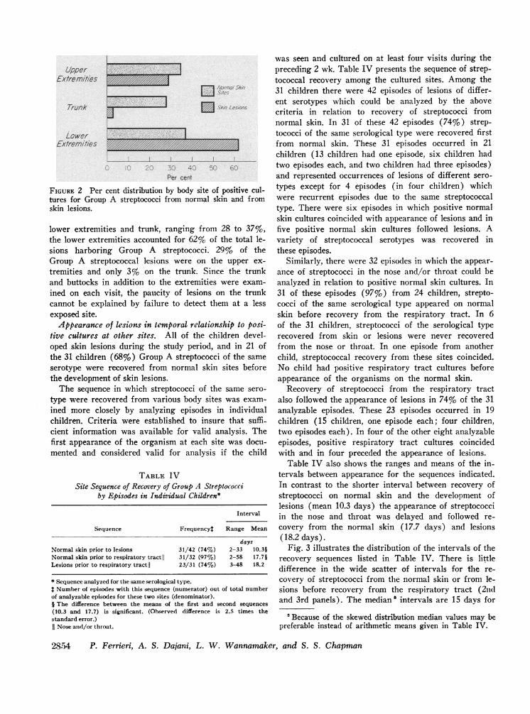
FIGURE 2 Per cent distribution by body site of positive cul-tures for Group A streptococci from normal skin and fromskin lesions.
lower extremities and trunk, ranging from 28 to 37%,the lower extremities accounted for 62% of the total le-sions harboring Group A streptococci. 29% of theGroup A streptococcal lesions were on the upper ex-tremities and only 3% on the trunk. Since the trunkand buttocks in addition to the extremities were exam-ined on each visit, the paucity of lesions on the trunkcannot be explained by failure to detect them at a lessexposed site.Appearance of lesions in temporal relationship to posi-
tive cultures at other sites. All of the children devel-oped skin lesions during the study period, and in 21 ofthe 31 children (68%) Group A streptococci of the sameserotype were recovered from normal skin sites beforethe development of skin lesions.The sequence in which streptococci of the same sero-
type were recovered from various body sites was exam-ined more closely by analyzing episodes in individualchildren. Criteria were established to insure that suffi-cient information was available for valid analysis. Thefirst appearance of the organism at each site was docu-mented and considered valid for analysis if the child
TABLE IVSite Sequence of Recovery of Group A Streptococci
by Episodes in Individual Children*
Interval
Sequence Frequencyt Range Mean
daysNormal skin prior to lesions 31/42 (74%) 2-33 10.3§Normal skin prior to respiratory tractlI 31/32 (97%) 2-58 17.7§Lesions prior to respiratory tractll 23/31 (74%) 3-48 18.2
* Sequence analyzed for the same serological type.* Number of episodes with this sequence (numerator) out of total numberof analyzable episodes for these two sites (denominator).§ The difference between the means of the first and second sequences(10.3 and 17.7) is significant. (Observed difference is 2.5 times thestandard error.)11 Nose and/or throat.
was seen and cultured on at least four visits during thepreceding 2 wk. Table IV presents the sequence of strep-tococcal recovery among the cultured sites. Among the31 children there were 42 episodes of lesions of differ-ent serotypes which could be analyzed by the abovecriteria in relation to recovery of streptococci fromnormal skin. In 31 of these 42 episodes (74%) strep-tococci of the same serological type were recovered firstfrom normal skin. These 31 episodes occurred in 21children (13 children had one episode, six children hadtwo episodes each, and two children had three episodes)and represented occurrences of lesions of different sero-types except for 4 episodes (in four children) whichwere recurrent episodes due to the same streptococcaltype. There were six episodes in which positive normalskin cultures coincided with appearance of lesions and infive positive normal skin cultures followed lesions. Avariety of streptococcal serotypes was recovered inthese episodes.
Similarly, there were 32 episodes in which the appear-ance of streptococci in the nose and/or throat could beanalyzed in relation to positive normal skin cultures. In31 of these episodes (97%) from 24 children, strepto-cocci of the same serological type appeared on normalskin before recovery from the respiratory tract. In 6of the 31 children, streptococci of the serological typerecovered from skin or lesions were never recoveredfrom the nose or throat. In one episode from anotherchild, streptococcal recovery from these sites coincided.No child had positive respiratory tract cultures beforeappearance of the organisms on the normal skin.
Recovery of streptococci from the respiratory tractalso followed the appearance of lesions in 74% of the 31analyzable episodes. These 23 episodes occurred in 19children (15 children, one episode each; four children,two episodes each). In four of the other eight analyzableepisodes, positive respiratory tract cultures coincidedwith and in four preceded the appearance of lesions.
Table IV also shows the ranges and means of the in-tervals between appearance for the sequences indicated.In contrast to the shorter interval between recovery ofstreptococci on normal skin and the development oflesions (mean 10.3 days) the appearance of streptococciin the nose and throat was delayed and followed re-covery from the normal skin (17.7 days) and lesions(18.2 days).
Fig. 3 illustrates the distribution of the intervals of therecovery sequences listed in Table IV. There is littledifference in the wide scatter of intervals for the re-covery of streptococci from the normal skin or from le-sions before recovery from the respiratory tract (2ndand 3rd panels). The median' intervals are 15 days for
'Because of the skewed distribution median values may bepreferable instead of arithmetic means given in Table IV.
2854 P. Ferrieri, A. S. Dajani, L. W. Wannamaker, and S. S. Chapman
--l

60 F 0
50+
c)0
C
I._C
40+
30F
20 [
l0[
0t
0
0
0
0
00*
-A
.
Skin Priorto Lesions
00
- 0
00_ 'I
*_ 0
0
Skin Priorto Nose and
Throat
S
S00
0
I
al0
Lesions Priorto Nose ond
Throat
FIGURE 3 Scattergram comparing the intervals (days) be-tween recovery of streptococci from various body sites (seetext). The median intervals are shown by horizontal lines.
skin before nose and throat and 17 days for lesions be-fore nose and throat. In contrast the intervals for skinbefore lesions (1st panel) show less widespread scatterwith considerable clustering under 10 days (medianvalue of 8 days).There was a difference in intervals when the appear-
ance of streptococci in the respiratory tract was analyzedseparately for nose and throat. The mean interval be-tween isolation of streptococci of the same serotype forthe first time from the normal skin and later from thenose was 14 days and from normal skin to throat was20 days. Analysis of the mean intervals between appear-
TABLE VFrequency of Development of Skin Lesions After Recovery of
Serologically Related Streptococcifrom Normal Skin
Serologicalclassification Interval
Group M or T Frequency* Mean Median
days$A M-57 12/14 7.3 6.0A M-56 4/6 14.3 10.5A T-12§ 6/8 11.2 7.0A Miscil 6/9 14.3 12.5G - 4/14 8.3 8.5B 0/5
* No. who developed lesions/No. with streptococci on normalskin.I Between recovery from normal skin and development oflesions.§ Not M-typable.11 T-11, T-6/15/17/23/47, and M-49.
ance in lesions and then in the nose or throat was simi-lar (15 days and 20 days, respectively). In none of thechildren was there any clinical evidence of streptococcalrespiratory disease during the study period.Risk of lesions developing in children with positive
normal skin cultures. The recovery of Group A strep-tococci from the normal skin of an individual was as-sociated with a high risk of subsequent development ofskin infection in that individual with a strain of thesame M or T classification (Table V). 86% of the chil-dren with M-type 57 streptococci on the normal skin
[ July August Sept. Oct.
28 30 31 1 4 5 8 11 13 14 15 18 19 20 22 25 26 27 29j 3 10 17
A. L. or . * AO 00 00o00 00oAAOo 0 00 00 0 000 00 0 0
R. AO AO0 A09 A06 0 0 0 AO O 0 0 0
L. 0 0 0. A* os As* 0 00 06 * AO000 0 0 00 0 0 A AO
V L. 0 0 00 AO. A0.. . AO.AO 00 00 00 0 00 00 00 00 A0* O
D L. 0 AOron AO0AO0AO* 00 AOon AOo00 00 0 00 0 0 0 00 AO
C. L. 0 0 00 0 AO 00o0 0 0 0
A. L.,Jr 0 0
Oformo/ Sk/n Sites *Leslons AAlose AThroo/
FIGURE 4 Site sequence of recovery of M-type 57 streptococci from various body sites amongseven members of family 1 for each week of the study. Normal skin sites, lesions, nose, andthroat are designated with symbols and each symbol represents one or more positive culturesfor that culture date.
Site Sequence of Acquisition and Spread of Skin Streptococci
I
i IJ L
2855

subsequently developed skin lesions with this type. Thepercentage who developed lesions after the appearanceof other Group A serotypes on the skin varied from67 to 75%. Only 4/14 of children with Group G strepto-cocci on the normal skin and none of the small number ofchildren with Group B streptococci on the skin developedlesions due to these groups. Among the Group A strep-tococci, the mean interval between recovery of strepto-cocci from the skin and development of lesions wasshortest with M-type 57 (7.3 days), and varied from11.2 to 14.3 days for the other types. The mean intervalfrom acquisition of Group G streptococci on the skin todevelopment of lesions was 6.5 days.
Familial patterns of acquisition and spread of strepto-cocci. Fig. 4 is an example of the site sequence of re-covery of a single serotype (M-type 57) of streptococciamong the children of one of the families (hereafterdesignated family 1) under intensive observation. Thefirst appearance of M-type 57 in the family was on July28, although three other serotypes were recovered fromJuly 1 up to this time. In five of the seven childrenM-type 57 streptococci were isolated fromn the normalskin on one or two culture visits before the developmentof skin lesions with this type (mean interval of 7.6 days).In A. L., Jr., there was recovery of M-type 57 strepto-cocci on the normal skin late in the study but no lesionsdue to this type developed during the period of ob-servation. As expected, streptococci continued to be iso-lated from normal skin sites after the appearance oflesions. Of interest is the observation that three children,R. L., B. L., and C. L., persisted with the strain on nor-mal skin from late August or early September until theend of the study during which time they had no skinlesions.
In this family, positive respiratory tract cultures werelimited almost exclusively to the nose. In five of six chil-dren the first appearance of a positive nose culture fol-lowed the appearance of the strain on normal skin and
TABLE VISpread of Group A Streptococci within the Families
Interval Mean
days* days
Index skin acquisition to [2, 2, 2][3][3]secondary skin acquisitions [4, 4][7, 7, 7][12] 4.8
Index skin infection to last [1][13][15][19]skin infection [37][41] 21.0
* Each bracket includes figures from analysis of one serotypein a family and multiple figures within a bracket designatethe intervals of children with simultaneous secondary skinacquisition.$ Skin acquisition as defined in this analysis means recoveryof streptococci from normal skin and/or lesions.
a)
0
(nC
0
a)
a-
80
60
40
20
0
0-* T-11 Family/c--O M-57
_ M-49W.*-6115117123/'47
I- 14- 21- 28- 4- II- 18- 25- 3 10 17JUY 13 20 2174 3 10 17 24 srPr2 ocr
FIGURE 5 Percentage of all cultures taken positive for fourdifferent streptococcal serotypes recovered from family 1 byweek of study.
in four of six also followed occurrence of skin lesions,suggesting transmission from skin to nose. With oneexception (patient V. L., on September 17), positivethroat cultures were conspicuously absent.No evidence of nephritis occurred in this family dur-
ing the intensive study period or at long-term follow-up,despite widespread dissemination within the family of aknown nephritogenic strain which was associated withnephritis in other families.
Findings similar to these were not confined to onestreptococcal serotype or to one family; the other strep-tococcal types common to the five families behaved in asimilar fashion in their site sequence of spread and spreadwithin the family (see below).
Intervals between acquisitions and infections withinfamilies. In family 1 (see Fig. 4) the initial (index)acquisition of M-type 57 streptococci was in R. L. andB. L. (on July 28), and in 2 days there was secondaryacquisition of this strain by three other family mem-bers. There was an interval of 19 days between the ap-pearance of the index infection (skin lesions) and thedevelopment of skin infection in the last of the six chil-dren in this family who had lesions due to this strepto-coccal type.
Table VI gives the intervals between an index acquisi-tion (normal skin and/or lesions) of a streptococcal sero-type by a family member(s) and the first secondaryacquisition (normal skin and/or lesions) by anotherfamily member(s). There were six streptococcal sero-types which appeared initially in the five families whilethey were under observation (one each in four families;two ini another family). A range of 2-12 days (mean4.8) was found between index and secondary skin ac-quisition of streptococci in the families. The mean in-terval between the first (index) and the last appearancein family members of cutaneous infection with the samestreptococcal serotype was 21 days.
Chronology of strain movements within families.Since four of the five families had more than one strep-
2856 P. Ferrieri, A. S. Dajani, L. W. Wannamaker, and S. S. Chapman

tococcal serotype isolated during the study period, thisoffered an opportunity to examine the chronology ofstrain movements in these families. Fig. 5 illustrates thechronology of strain movements into family 1 in whomfour different serotypes were isolated. The movementsof the major type, M-57, among various body sites ofindividual family members was previously presented inFig. 4. Two of the strains, classified as T-11 and T-6/15/17/23/47 by T-agglutination, could not be M-typed inour laboratories. In this family only T-11 was isolatedduring the first 2 wk of the study; recovery of this sero-type then declined. The M-type 49 and T-6/15/17/23/47serotypes first appeared on July 14, but they never gainedascendancy. M-type 57 reached a peak frequency of 57%of all cultures taken from August 18 to 24 and continuedas the predominant strain through October 1. During theweek of August 18-24, 83% of all cultures taken werepositive and all four streptococcal types were represented.
In Fig. 6 is plotted the change in strains for twofamilies in whom two serologically different strains wererecovered, M-type 56 and T-pattern 12 (which was notM-typable). There were 12 children in these two families,living quite close to each other and sharing many facili-ties and activities, so they may be considered essentiallyas one extended family unit. From these two figures (5and 6) we see that new serotypes may enter a familyduring the peak or decline of a preceding serotype, andthat they may coexist for periods of time, with a ten-dency for one to become predominant.
Streptococcal antibodies. Among the four streptococ-cal antibody determinations performed on serial bleedingsfrom the 31 children, there were differences in the per-centage of children who developed significant antibody
c
vx
9
co
4-
I.-
0)4'
0)
4)a.
60- Family 3
40
20 -
0-60 Family 2
40-
20 -
7- 14- 21- 28- 4- - 18- 25- 3 10 17JULY 13 20 27 Aug 3 10 17 24 2 ocr
FIGURE 6 Percentage of all cultures taken positive forstreptococcal types M-56 (open circles) and T-12 (closedcircles) recovered from families 2 and 3 by week of study.
TABLE VI IStreptococcal Antibody Rises and InitialMean Titers Among the 31 Children
Children Initial mean valuewith rise* in children with:
PerAntibody No. cent Rise No rise
ASO 6 19 88 251Anti-DNase B 13 42 381 624Anti-NADase 11 36 101 245Anti-group A carbohydrate 8 26 0.35 0.74One or more of above 18 58
* For ASO, anti-DNase B, anti-NADase, 0.2 log rise or greater.For anti-group A carbohydrate, 10% or greater increase infraction of antigen precipitated. (If 5% or greater is taken assignificant, four additional children developed an antibodyresponse to the antigen.)T For ASO, anti-DNase B, anti-NADase geometric meantiters are given; for anti-group A carbohydrate the arithmeticmean.
rises to the different streptococcal antigens (Table VII).The anti-DNase B response appeared to be the bestindicator of streptococcal skin infection. Few childrendeveloped an ASO rise. Only 26% revealed an increasein group A carbohydrate antibody level, but the initialantibody levels were generally high. Six of the eightchildren with a Group A carbohydrate antibody risewere less than 4 yr of age, and had lower initial titers,reflecting most likely fewer previous experiences withstreptococcal disease. The initial geometric mean titerswere higher for all four antibodies in children who de-veloped no rise compared with those who did demonstratea rise.
Effect of treatment on persistence or reacquisitionof streptococci and reinfection. It was possible to makea few observations on the relationship of treatmentadministered in the hospital to the persistence or sub-sequent reappearance of streptococci of the same sero-type on the normal skin and on recurrence of skinlesions due to the same type. Five children from thestudy group were hospitalized and received varioustreatment regimens as shown in Table VIII. No anti-biotic was given on discharge from hospital. No skinlesions were present on discharge, but all of the childrenhad streptococci recovered from normal skin sites aftercompletion of antibiotic treatment, and the majoritywere positive on two or more occasions. Intervals be-tween the last day of treatment and the first recoveryof streptococci from normal skin ranged from lessthan 1 day to 10 days. Since all of the children werenot cultured while in the hospital, it is not certain ifstreptococci persisted on the normal skin while on treat-
Site Sequence of Acquisition and Spread of Skin Streptococci 2857
0
4%

TABLE VI I IRelationship of Treatment in Hospital to Persistence or
Reacquisition of the Same Serotype and toReinfection with this Type
Interval betweenlast dose
or injectionand recovery of Recurrencestreptococci on of skin
Patient Treatment normal skin lesions
days
D. B. Benzathine penicillin* 8 NoA. B. Benzathine penicillin* 10 NoD. R. Aq. pencillin i.m., 8 doses
Oral penicillin, 7 days 4 YestL. L.§ Aq. penicillin i. m., 6 doses
Oral penicillin, 7 days <111 NoL. L.§ Oral penicillin, 7 days <111 Yes$
* 600,000 U.Same serotype as before treatment.
§ Details of clinical history reported previously (8).11 Skin culture positive day of discharge from hospital.
ment or were reacquired. In the two children treatedwith Bicillin (Wyeth Laboratories, Philadelphia, Pa.)no further skin lesions occurred, whereas two of theother three children developed lesions after recovery ofstreptococci from normal skin (15 and 23 days after theend of treatment). The streptococcal serotype culturedfrom these lesions was the same as that recovered fromlesions before treatment.
DISCUSSION
It has been recorded that streptococcal inll)etigo usuallyoccurs in the absence of overt streptococcal disease ofthe respiratory tract (1, 21). However, without frequentobservations during the period before the development
Group A Streptococci(Source Uncertain)
Norml Skin
LSkin LesionsJ
/4dtys ZO9ys
|Nose | | Throat
FIGuRE 7 Concept of the sequence of spread of Group Astreptococci among different body sites based on the findingsof this study.
of impetigo, the possibility of previous infection, par-ticularly subclinical or mild infection, of the respiratorytract could not be excluded. Therefore, it has not beenclear whether Group A streptococci in the nose orthroat play any significant role as a reservoir in theacquisition and spread of skin infections. The results ofthe present study, based on intensive serial observationsin individual children, initiated in many cases someweeks or months before the appearance of skin lesions,clearly eliminate the upper respiratory tract as a sourceof infection of the skin. In 94% of instances the strepto-coccal strain was recovered from the normal skin ofan individual before it appeared in the respiratory tractand in 74% of instances lesions harboring this specificstrain developed before the strain appeared in the upperrespiratory tract.
Studies carried out at the Red Lake Indian Reserva-tion in 1968 revealed that a primary factor in the de-velopment of pyoderma was previous acquisition ofstreptococci on the normal skin (10). The presentstudy confirms these findings with observations at morefrequent intervals and extends them to include severalother pyoderma strains, including a serotype (M-57)with nephritogenic properties. Moreover, the risk ofdeveloping impetigo after the recovery of Group Astreptococci from the normal skin is shown to be extra-ordinarily high (76%).
Traditionally, it has been thought that beta hemolyticstreptococci are only rarely included among the normalskin flora (22), and that these organisms do not survivewhen artificially inoculated on normal skin (23). Inaddition, in vitro studies have shown that unsaturatedfatty acids extracted from normal human skin are bac-tericidal for streptococci (24). The present study withintensive daily observations and culture surveillance(three times weekly) gives more credibility to the fre-quent recovery of streptococci from normal skin incertain populations, particularly those at high risk ofdeveloping pyoderma. Though only semiquantitativetechniques were used, 43% of the positive normal skincultures had more than 10 streptococcal CFU recov-ered from the small areas cultured. Other studies fromthis laboratory studying a similar group of childrenhave revealed that if multiple sites of the forearm arecultured, streptococci are commonly recovered frommore than one site.' Thus, the possibility that the pre-sented observations are chance sampling events becomesless likely.
In Fig. 7 is presented our concept of the sequenceof spread of Group A streptococci among different bodysites of an individual based on the present study. GroupA streptococci, from uncertain sources, contaminate
6Ferrieri, P., A. S. Dajani, and L. W. Wannamaker. Un-published observations.
2858 P. Ferrieri, A. S. Dajani, L. W. Wannamaker, and S. S. Chapman

and possibly colonize the normal skin. After an in-sult(s) to the integrity of the skin such as insect bites,poison ivy, abrasions, or even undetectable trauma, skinlesions may appear if streptococci are present on thenormal skin. The mean interval between appearance ofstreptococci on the normal skin and the developmentof skin lesions with the same serological type is 10days. Often streptococci of the same serological typeare recovered repeatedly from the normal skin duringthe interval before the development of lesions harboringthis type. These findings are consistent with three possi-bilities: (a) repeated deposition on the normal skin;(b) survival for protracted periods (days) on thenormal skin; (c) multiplication (colonization) on thenormal skin. Unfortunately, it is not possible to deter-mine which one(s) of the possibilities is (are) likelyto be correct from the studies presented here.The appearance of streptococci in the nose or throat
of the individual is further delayed (more so for thethroat). Respiratory tract acquisition almost alwaysfollows skin acquisition of streptococci (intervals of14-20 days for nose and throat, respectively) andusually does not occur until after the development ofskin lesions, suggesting transmission from normal orinfected cutaneous sites. Respiratory tract acquisition ofstreptococci was often quite transient and did notresult in clinical disease.
In the population as a whole, the low rather con-stant pattern of positive throat cultures throughout thestudy is in contrast to the fluctuation in positive nosecultures. The upswing in positive nose cultures whichfollows the sharp increases in prevalence of positivenormal skin and lesion cultures may be due to contami-nation of the nose by streptococci from cutaneous sites.In this study the streptococcal serotypes recoveredfrom the nose or throat did not differ from those re-covered from normal skin and lesions.Once skin lesions are established it seems reasonable
that shedding of streptococci occurs, perhaps in largenumbers, which can result in endogenous contaminationof normal skin and also act as a source of spread forthe acquisition of organisms on the skin of family mem-bers living and sleeping in close quarters. It is possiblethat streptococcal acquisition may result from cross-transmission within families or from other contacts.
It appears that within a relatively short period ofinitial acquisition of a streptococcal strain on the normalskin or in skin lesions by a family member(s), sec-ondary acquisition by another member (s) takes place.This mean interval of 4.8 days contrasts with the longerinterval of 15 days observed between index and sec-ondary acquisition of streptococci in the throat of familymembers under prolonged observation in the Cleveland
study (25). This difference may be in part artifactual,however, since in this latter study cultures were ordi-narily obtained at only weekly intervals.The shorter intervals for secondary family spread of
cutaneous streptococci compared with streptococci fromthe throat are not readily explained but may be a re-flection (in the case of the former) of: (a) morecrowded living quarters in our families with greateropportunity for intimate physical contact and trans-mission of organisms; (b) possibly shedding of largernumbers of organisms from the normal skin or mul-tiple skin lesions thus increasing the chance of sec-ondary spread; (c) undefined biological differences ofskin streptococci which may enhance survival and via-bility during transmission.
In our families a mean interval of 21 days elapsedbetween the first occurrence of skin infection and thelast acquisition of skin disease (due to the same strep-tococcal serotype) by a family member. No comparabledata are available from the Cleveland study.The high frequency of positive normal skin site cul-
tures during the last 4 wk of this study-at a time whenskin lesions were declining or not present at all in mostchildren-is intriguing. The serotypes recovered werethose previously isolated from normal skin and fromlesions. One speculation for this disparity is that type-specific antibodies may have developed to those typesand prevented new lesions but not acquisition or per-sistence of streptococci on the normal skin. We have nodata to support this theory, and little is known aboutthe regularity with which such antibodies develop afterskin infections or whether they can protect against re-infection of the skin (6). Other possibilities includethe decline or disappearance of factors which maybreech the integrity of the skin, such as insect bitesand other trauma. Another possible factor is the routineweekly showering in school which could have affectedthe development of lesions in the school-age childrenafter September 3.The recovery of Group A streptococci from normal
skin before development of lesions and later in the fallwhen no skin infections were present suggests frequentcontamination of this body site from some environmentalsource with possibly survival on or even colonizationof the normal skin. Whether these pyoderma strainspossess unique biological properties which permit suchphenomena is unclear.The length of persistence or survival of streptococci
on the normal skin has not been studied under con-trolled conditions but many children had the organismsrecovered on two or more culture visits (separated by2 or 3 days) before developing skin lesions. It alsoappears that streptococci can persist or be acquired on
Site Sequence of Acquisition and Spread of Skin Streptococci 2859

the normal skin in spite of penicillin therapy. The fivehospitalized children who received various regimens ofpenicillin had persistence or reacquisition of strepto-cocci on the skin after treatment, with two developinglesions of the original serotype. The numbers are smallbut the suggestions from these data are that intramuscu-lar benzathine penicillin may not interfere with thepersistence or reacquisition of streptococci on normalskin, although it may prevent recurrence of skin lesionsfor a period of time. It appears that oral penicillin isless effective in preventing recurrence of lesions, a find-ing of other investigators in studies comparing dif-ferent treatment regimens (26). A recent preliminarystudy of intramuscular benzathine penicillin as a pro-phylactic agent for skin infections has shown thatstreptococci can be recovered from the normal skin asearly as 2 wk after administration of this long-actingpenicillin (27).Our finding that the percentage distribution of normal
skin sites positive for streptococci was similar amongthe upper and lower extremities and back (28-37%)emphasizes that less exposed parts of the body (i.e.,the back) can acquire the organisms. However, since62% of the total lesions occurred on the legs, it appearsthat several factors, including trauma, may be etio-logically important in the development of skin lesions.Duncan, McBride, and Knox (28) demonstrated inexperimental production of streptococcal and staphylo-coccal skin infections in humans that there was ahigher success rate of infecting the leg than the backor arm. Regional circulatory differences and possiblydifferences in oxygen and carbon dioxide tensions be-tween the upper and lower extremities were offered aspossible explanations for their findings.The antibody findings in this study confirm those of
earlier studies by Kaplan, Anthony, Chapman, Ayoub,and Wannamaker (29) and Dillon and Reeves (30)which indicate that the ASO response is poor afterstreptococcal skin infections, and that the anti-DNaseB is of greater value in studying the immunologicalresponse to skin infection. In addition the antibodyresponses varied inversely with the mean initial titers,children with no rise having higher initial titers thanthose demonstrating a significant rise. High initialantibody titers may have resulted from respiratorystreptococcal infections in the late winter or springor from skin infections early in the summer. Studiesof streptococcal respiratory disease have indicated thatpatients with low mean initial antibody titers will gen-erally show a greater magnitude of rise (31, 32). Thesefindings have resulted in several different interpreta-tions (32). Further data and more extensive analysisof the antibody response in relation to initial titer are
needed in patients with skin infections. In the presentstudy the anti-Group A carbohydrate values tended tobe high in these children on admission to the studyreflecting the endemicity of streptococcal infections inthis population and the tendency for antibodies to theGroup A carbohydrate to remain elevated for pro-longed periods of time (17).The high percentage of streptococcal isolates (73%)
which could be typed serologically with M-antisera inthis study is partially explained by the predominanceof one type (M-57) in three of the five families. Thisserotype is one of the newer described streptococcalpyoderma strains, and has been isolated from pyoderma-associated acute glomerulonephritis in Trinidad (2, 3).The six Group A streptococcal serotypes recovered in
this study were all recovered from normal skin sitesand from skin lesions. There were similarities amongthe prevalent types in the high frequency of childrenwho developed lesions if the same type was recoveredfrom normal skin. However, 86% of children with M-type 57 developed lesions after the appearance of thistype on normal skin and the interval between recoveryfrom skin and development of lesions was shorter (7.3days) than for the other types (range 11-14 days).There was no apparent difference in the risk of lesionsdeveloping if a particular streptococcal serotype ap-peared on the normal skin early in the study or in themiddle of the study. The risk of new lesions developingat the end of the study could not be determined becausenew streptococcal serotypes rarely appeared then. Therewas a low risk of development of lesions after the re-covery of streptococci of Group G or B from the normalskin, suggesting that these streptococci are of lowpathogenicity for the skin.The patterns assumed by different streptococcal sero-
types as they move through families demonstrate thatusually a new type enters and becomes predominantwhile a preceding type is at a peak or declining. Ofinterest is the family with four serotypes coexistingfor periods of time, although one type (M-57) clearlyaccounted for the majority of positive cultures. Thisinformation cautions one against accepting readily, inpatients presenting with nephritis, a single culture resultas necessarily indicative of the etiologic nephritogenicstrain. Skin lesions of different serotypes may bepresent simultaneously, so there is value in culturingfresh lesions and lesions in various stages of healing.The offending nephritogenic strain may have causeddisease which preceded the development of lesions dueto the currently isolated strain, but because of the rela-tively long latent period in pyoderma-related nephritis(6, 8, 33) the strain may no longer he present.
21860 P. Ferrieri, A. S. Dajani, L. W. Wannamaker, and S. S. Chapman

Thus, the present study amplifies previous knowledgeof the epidemiology of streptococcal skin disease. Al-though the reservoirs of streptococci remain ill defined,the respiratory tract can be eliminated as a source ofinfection. Normal skin acquisition of the organismsappears to be a prerequisite to development of lesions.Local and environmental factors appear to be importantdeterminants of the site of infection. The risk of de-veloping impetigo is high after appearance of Group Astreptococci on the normal skin.
Note added in proof. Since the submission of this manu-script a report of the appearance of streptococci in skinlesions before their appearance in the respiratory tract hasbeen published (Bassett, D. C. J. 1972. Streptococcal pyo-derma and acute nephritis in Trinidad. Br. J. Dermatol. 86(Suppl. 8): 55). This report and the earlier ones fromthis laboratory (5, 10, 23) were based on observations atless frequent intervals.
ACKNOWLEDGMENTSWe wish to acknowledge Dr. Jonathan B. Jensen for hisvaluable assistance and also Mary Aronson, Marge Fisher,Judith Jaqua, Dwight Johnson, Joanne Kumagai, JoAnnNelson, Margaret Ragan, Jerry Stanke, and Richard VanHeuveln for their contributions in media preparation, classi-fication of streptococcal strains, and streptococcal antibodydeterminations.These studies were supported by a U. S. Public Health
Service Grant AI-09527 and were conducted under the spon-sorship of the Commission on Streptococcal and Staphylococ-cal Diseases, Armed Forces Epidemiological Board, and sup-ported by the U. S. Army Medical Research and DevelopmentCommand under contract No. DADA-17-70-C-0081.
REFERENCES1. Anthony, B. F., L. V. Perlman, and L. W. Wanna-
maker. 1967. Skin infections and acute nephritis inAmerican Indian children. Pediatrics. 39: 263.
2. Potter, E. V., J. S. Ortiz, A. R. Sharrett, E. G. Burt,J. P. Bray, J. F. Finklea, T. Poon-King, and D. P.Earle. 1971. Changing types of nephritogenic strepto-cocci in Trinidad. J. Clin. Invest. 50: 1197.
3. Parker, M. T. 1969. Streptococcal skin infection andacute glomerulonephritis. Br. J. Derm. 81 (Suppl. 1): 37.
4. Dillon, H. C., M. S. Reeves, and W. R. Maxted. 1968.Acute glomerulonephritis following skin infection dueto streptococci of M-type 2. Lancet. 1: 543.
5. Anthony, B. F., E. L. Kaplan, S. S. Chapman, P. G.Quie, and L. W. Wannamaker. 1967. Epidemic acutenephritis with reappearance of type 49 streptococcus.Lancet. 2: 787.
6. Wannamaker, L. W. 1970. Differences between strep-tococcal infections of the throat and of the skin. N. Engl.J. Med. 282: 23, 78.
7. Kleinman, H. 1954. Epidemic acute glomerulonephritisat Red Lake. Minn. Med. 37: 479.
8. Ferrieri, P., A. S. Dajani, S. S. Chapman, J. B. Jensen,and L. W. Wannamaker. 1970. Appearance of nephritisassociated with type 57 streptococcal impetigo in NorthAmerica: longitudinal observations in a family. N. Enyl.J. Med. 283: 832.
9. Bisno, A. L., I. A. Pearce, H. P. Wall, M. D. Moody,and G. H. Stollerman. 1970. Contrasting epidemiologyof acute rheumatic fever and acute glomerulonephritis.N. Engl. J. Med. 283: 561.
10. Dudding, B. A., J. W. Burnett, S. S. Chapman, andL. W. Wannamaker. 1970. The role of normal skin inthe spread of streptococcal pyoderma. J. Hyg. 68: 19.
11. Dajani, A. S., P. Ferrieri, and L. W. Wannamaker.1972. Natural history of impetigo. II. Etiologic agentsand bacterial interactions. J. Clin. Invest. 51: 2863.
12. Edwards, E. A. 1964. Protocol for micro antistreptolysino determinations. J. Bacteriol. 87: 1254.
13. Nelson, J., E. M. Ayoub, and L. W. Wannamaker. 1968.Streptococcal antidesoxyribonuclease B: microtechniquedetermination. J. Lab. Clin. Med. 71: 867.
14. Ayoub, E. M., and L. W. Wannamaker. 1962. Evalua-tion of the streptococcal desoxyribonuclease B and di-phosphopyridine nucleotidase antibody tests in acuterheumatic fever and acute glomerulonephritis. Pedi-atrics. 29: 527.
15. Ayoub, E. M., and J. J. Ferretti. 1966. Use of bisulfitein the streptococcal anti-nicotinamide adenine dinucleo-tidase test. Appl. Microbiol. 14: 391.
16. Halpern, B., and I. Goldstein. 1964. Utilisation du poly-oside streptococcique marque au `'C pour la determina-tion de faible quantities d'anticorps specifiques desserums experimentaux et humans. Rev. Immunol. 28:193.
17. Dudding, B. A., and E. M. Ayoub. 1968. Persistenceof streptococcal group A antibody in patients withrheumatic valvular disease. J. Exp. Med. 128: 1081.
18. Top, F. H., Jr., L. W. Wannamaker, W. R. Maxted,and B. F. Anthony. 1967. M. antigens among group Astreptococci isolated from skin lesions. J. E.rp. Med.126: 667.
19. Dillon, -H. C., M. D. Moody, W. R. Maxted, and M. T.Parker. 1967. The epidemiology of impetigo and acuteglomerulonephritis. Am. J. Epidemiol. 86: 710.
20. Parker, M. T., D. C. J. Bassett, W. R. Maxted, andJ. D. Arneaud. 1968. Acute glomerulonephritis in Trini-dad: serological typing of group A streptococci. J. Hyg.66: 657.
21. Dillon, H. C. 1968. Impetigo contagiosa: suppurativeand non-suppurative complications. Am. J. Dis. Child.115: 530.
22. Kligman, A. M. 1965. The bacteriology of normal skin.In Skin Bacteria and Their Role in Infection. H. I.Maibach and G. Hildick-Smith, editors. McGraw-HillBook Company, New York. 13.
23. Colebrook, L., and W. R. Maxted. 1933. Antisepsis inmidwifery. J. Obstet. Gynaecol. Br. Emp. 40: 966.
24. Ricketts, C. R., J. R. Squire, and E. Topley. 1951.Human skin lipids with particular reference to the self-sterilising power of the skin. Clin. Sci. 10: 89.
25. Dingle, J. H., G. F. Badger, and W. S. Jordan, Jr.1964. Illness in the home. A study of 25,000 illnessesin a group of Cleveland families. The Press of CaseWestern Reserve University, Cleveland, Ohio. 110.
26. Derrick, C. W., and H. C. Dillon. 1970. Further studieson the treatment of streptococcal skin infection. J.Pediatr. 77: 696.
27. Ferrieri, P., A. S. Dajani, and L. W. Wannamaker.1972. Benzathine penicillin prophylaxis of streptococcalimpetigo. Pediatr. Res. 6: 389. (Abstr.)
Site Sequence of Acquisition and Spread of Skin Streptococci 2861

28. Duncan, W. C., M. E. McBride, and J. M. Knox.1970. Experimental production of infections in humans.J. Invest. Dermatol. 54: 319.
29. Kaplan, E. L., B. F. Anthony, S. S. Chapman, E. M.Ayoub, and L. W. Wannamaker. 1970. The influence ofthe site of infection on the immune response to groupA streptococci. J. Clin. Invest. 49: 1405.
30. Dillon, H. C., and M. S. Reeves. 1969. Streptococcalantibody titers in skin infections and acute glomerulo-nephritis. Pediat. Res. 3: 362. (Abstr.)
31. Dudding, B. A., H. C. Dillon, L. W. Wannamaker, R.M. Kilton, S. S. Chapman, and B. F. Anthony. 1969.
Post epidemic surveillance studies of a food-borne epi-demic of streptococcal pharyngitis at the United StatesAir Force Academy. J. Infec. Dis. 120: 225.
32. Kaplan, E. L., F. H. Top, Jr., B. A. Dudding, andL. W. Wannamaker. 1971. Diagnosis of streptococcalpharyngitis: differentiation of active infection from thecarrier state in the symptomatic child. J. Infect. Dis.123: 490.
33. Kaplan, E. L., B. F. Anthony, S. S. Chapman, andL. W. Wannamaker. 1970. Epidemic acute glomerulo-nephritis associated with type 49 streptococcal pyoderma.I. Clinical and laboratory findings. Am. J. Med. 48: 9.
2862 P. Ferrieri, A. S. Dajani, L. W. Wannamaker, and S. S. Chapman