natural facial motion enhances cortical responses to faces fileneural correlates of facial motion...
TRANSCRIPT

Neural correlates of facial motion perception 1
Natural facial motion enhances cortical responses to faces
Johannes Schultz1,*
& Karin S. Pilz 2,*
1 Dept. of Human Perception, Cognition and Action, Max Planck Institute for Biological
Cybernetics, Tübingen, Germany
2 Department of Psychology, Neuroscience & Behaviour, McMaster University, Hamilton,
ON, Canada
* Both authors contributed equally to this work
Corresponding author: Karin S. Pilz, Department of Psychology, Neuroscience &
Behaviour, McMaster University, 1280 Main Street West, Hamilton, ON, Canada, Tel.: +1-
905-525-9140 x24489, Fax: +1-905-529-6225, Email: [email protected]
Keywords: facial motion, face localizer, STS, biological motion, FFA, OFA, fMRI.

Neural correlates of facial motion perception 2
Abstract
The ability to perceive facial motion is important to successfully interact in social
environments. Previously, imaging studies have investigated neural correlates of facial
motion primarily using abstract motion stimuli. Here, we studied how the brain processes
natural non-rigid facial motion in direct comparison to static stimuli and matched phase-
scrambled controls. As predicted from previous studies, dynamic faces elicit higher responses
than static faces in lateral temporal areas corresponding to hMT+/V5 and STS. Interestingly,
analyses of individually-defined, static-face-sensitive regions in bilateral fusiform gyrus and
left inferior occipital gyrus also respond more to dynamic than static faces. These results
suggest integration of form and motion information during the processing of dynamic faces
even in ventral temporal and inferior lateral occipital areas. In addition, our results show that
dynamic stimuli are a robust tool to localize areas related to the processing of static and
dynamic face information.

Neural correlates of facial motion perception 3
Introduction
Being required to understand and predict the actions of others to be able to successfully
interact in a social environment has led our visual system to become particularly sensitive to
human movements (for a recent review, see Blake and Shiffrar 2007). Facial motion in
particular is a very important cue to judge other people’s actions, emotions and intentions
towards us (Bassili 1976; Kamachi et al. 2001). In addition to this, facial motion has also
been shown to facilitate face recognition (O'Toole et al. 2002; Pilz et al. 2006). Due to the
familiarity and behavioral significance of facial motion, it is most likely that our visual
system has developed mechanisms that facilitate its perception and it is also very plausible to
assume that certain mechanisms exist that integrate invariant and changeable properties of
faces (Haxby et al. 2000).
Studies of biological motion, including faces, suggest that the interpretation of the
movements and actions of others recruit specialized neural pathways (Allison et al. 2000;
Blakemore et al. 2001; Giese and Poggio 2003). In monkeys, neurons in the anterior part of
the superior temporal polysensory area (STPa) were found to respond both to the form and
the motion of bodies and heads, indicating integration of form and motion information in this
area (Oram and Perrett 1996). In humans, involvement of the superior temporal sulcus (STS)
in the processing of relevant and familiar types of biological motion has also been shown,
e.g., in response to human body motion (tested using point-light displays, Bonda et al. 1996;
Grossman et al. 2000), or to facial motion due to speech production (Campbell et al. 2001;
Hall et al. 2005), expression of emotions (LaBar et al. 2003; Pelphrey et al. 2007) or in
complex scenes such as movies (Bartels and Zeki 2004; Hasson et al. 2004). Additionally,
these regions have been shown to respond to natural images of implied facial motion (Puce et
al. 1998; Puce et al. 2003), as well as to natural images of implied body motion (Jellema and
Perrett 2003).

Neural correlates of facial motion perception 4
Most of the studies investigating the neural correlates of facial motion have used
abstract motion stimuli like implied motion from static images (Puce et al. 1998; Puce et al.
2003), moving avatars (i.e. cartoon faces, for example Pelphrey et al. 2005; Thompson et al.
2007), or motion stimuli that were produced by morphing a static towards an emotional face
(LaBar et al. 2003; Sato et al. 2004; Pelphrey et al. 2007). Using such ‘unnaturally’ moving
stimuli might not fully capture the mechanisms underlying the processing of natural facial
motion. The controlled fMRI studies of facial motion that have used video sequences of
natural facial motion either focused on differences between types of face motions and thus
did not use non-face control stimuli (Campbell et al. 2001; Hall et al. 2005). A recent study
by Fox et al., (2008) investigated differences in brain activation between static and dynamic
stimuli using non-face stimuli as controls. They applied two localiser scans, one contrasting
static images of faces and objects, the other one contrasting dynamic videos of faces and
objects. Comparing these two localisers, their results suggest that dynamic localisers are
more reliable and more selective than static localisers. Although this study showed the
usefulness of using dynamic stimuli to localize areas related to face processing, they were not
able to directly compare brain activation towards static and dynamic stimuli, because those
stimuli were used in different scanning sessions. Here, we investigated brain activation in
response to natural non-rigid face motion and directly compared it to static faces and non-
face controls, which is necessary to demonstrate how the face-processing system responds to
dynamic as compared with static faces irrespective of low-level cues. We showed observers
video sequences of angry and surprised faces, as well as static stimuli of the same emotions.
As controls for low-level stimulus properties including motion, we used the phase-scrambled
versions of both kinds of stimuli.
Materials and Methods
Participants
Ten observers (4 females, 6 males) from the Tübingen community volunteered as subjects for
12 per hour. All observers were naïve as to the purpose of the current experiment and had
no history of neurological or psychiatric illnesses. All participants provided informed consent
and filled out a standard questionnaire approved by the local ethics committee for

Neural correlates of facial motion perception 5
experiments involving a high field MR scanner to inform them of the necessary safety
precautions.
Stimuli
We used video recordings of the face of three male and five female human actors, taken from
the Max-Planck database of moving faces (Pilz et al. 2006). For these recordings, each face
made two expressive gestures in separate videos: Surprise and anger. The movie clips used in
the dynamic face condition (dynamic faces) were composed of 26 frames, presented at a
frame rate of 25 frames per sec for a total duration of 1040 ms. Figure 1 shows an example of
all 26 frames of a video sequence (top panel). The movie clips started with a neutral
expression and ended with the peak of the expression in the last frame. The static face images
used in the static face condition (static faces) were the last frame of each video sequence and
thus showed the peak of each expression; each static face was presented for 1040 ms. All
stimuli were embedded in a background that consisted of white noise applied to every RGB
color channel. For the dynamic stimuli, the same noise was applied to all the frames of the
movie, i.e., the background was static.
As control stimuli, we generated phase-scrambled versions of dynamic (dynamic
scrambled) and static (static scrambled) faces. Researchers have often used objects or
fragmented face images as a comparison to face images to investigate areas related to face
processing (Kanwisher et al. 1997; Kanwisher et al. 1998). We decided to use phase-
scrambled versions of our stimuli as controls, because fragmented images are constituted
more of higher spatial frequencies, resulting from the cardinal axes (i.e., edges) that are
produced by dividing a relatively smooth picture like a face into randomly rearranged squares
(Sadr and Sinha 2004). Phase-scrambled stimuli have been used successfully in recent
neuroimaging studies (Eger et al. 2004; Kovacs et al. 2006; Jacques and Rossion 2007;

Neural correlates of facial motion perception 6
Rousselet et al. 2007). It has been shown that, especially for face recognition, the frequencies
around 8-16 cycles across the face are particularly important (Costen et al. 1996; Näsänen
1999; Morrison and Schyns 2001). Spatial frequencies also seem to interact with the
recognition of previously learned static and dynamic images (Pilz et al. 2008), suggesting that
they contain important information about the identity of the face. In addition, it has been
shown that the FFA processes high and low spatial frequencies differently (Vuilleumier et al.
2003; Gauthier et al. 2005; Rotshtein et al. 2007). Using fragmented images as a contrast
would have changed our results as a function of spatial frequency content in the phase-
scrambled images. Therefore, it was of high importance to preserve the frequency structure of
our original stimuli. Furthermore, we wanted to use a type of control stimuli that worked
equally well for both dynamic and static faces in controlling for their respective low-level
stimulus properties. Phase-scrambling is ideal, because its effect on both static and dynamic
faces is very comparable (keeping the spatial frequency content constant while eliminating
recognizable shapes).
Phase-scrambling of our images was accomplished as follows. For each independent
RGB color channel, the images were transformed into amplitude and phase components using
the Fourier transform. Noise patterns were generated by inverse Fourier transform of the
original amplitude spectrum of the image but with a random phase spectrum. For the movies,
the same random phase spectrum was used for each frame of a given movie but the
amplitudes were those of the original frames. This resulted in control movies that were not
flickering.
------------------------------
Insert Figure 1 about here
------------------------------

Neural correlates of facial motion perception 7
Design and Procedure
There were 5 conditions in the experiment: fixation, static faces, static scrambled, dynamic
faces, and dynamic scrambled. The observer’s task was a one-back matching task, i.e., they
had to press a button whenever two identical stimuli sequentially appeared on the screen. We
used a block design with 24 blocks, each composed of 6 stimuli which were presented every
3 seconds. Blocks were history-matched, i.e., every condition was preceded by each
condition equally often. Given that there were 16 different face stimuli in total (8 identities
times 2 expressions) and 6 stimuli per block, the probability of a stimulus repetition was
about 0.31 per block; i.e., each subject would on average encounter about 6 targets
distributed across conditions.
Observers lay supine on the scanner bed. The stimuli were back projected onto a
projection screen situated behind the observers' head and reflected into their eyes via a mirror
mounted on the head coil. The projection screen was 140.5 cm from the mirror, and the
stimuli subtended a maximum visual angle of approximately 9.0° (horizontal) x 8.3°
(vertical). A JVC LCD projector with custom Schneider-Kreuznach long-range optics, a
screen resolution of 1280 pixels x 1024 pixels and a 60 Hz refresh rate were used. The
experiment was run on a 3.2 GHz Pentium 4 Windows PC with 2GB RAM and an NVIDIA
GeForce 7800 GTX graphics card with 256 MB video RAM. The program to present the
stimuli and collect responses was written in Matlab using the Psychtoolbox extensions
(http://www.psychtoolbox.org) (Brainard 1997; Pelli 1997). We used a magnet-compatible
button box to collect subjects’ responses (The Rowland Institute at Harvard, Cambridge,
USA).

Neural correlates of facial motion perception 8
Image Acquisition
All participants were scanned at the MR Centre of the Max Planck Institute for Biological
Cybernetics, Tübingen, Germany. All anatomical T1-weighted images and functional
gradient-echo echo-planar T2*-weighted images (EPI) with BOLD contrast were acquired on
a Siemens TIM-Trio 3T scanner with an 8-channel phased-array head coil (Siemens,
Erlangen, Germany). The imaging sequence for functional images had a repetition time of
1920 ms, an echo time of 40 ms, a flip angle of 90°, a field of view of 256 mm x 256 mm and
a matrix size of 64 pixels x 64 pixels. Each functional image consisted of 27 axial slices.
Each slice had a thickness of 3.0 mm x 3.0 mm x 2.5 mm with a 0.5 mm gap between slices.
Volumes were positioned to cover the whole brain based on the information from a 13-slice
parasagittal anatomical localizer scan acquired at the start of each scanning session. For each
observer, between 237 and 252 functional images were acquired in a single session lasting
approximately 7.5 min, including a 8 sec blank period at the beginning of the run. The first
four of these images were discarded to allow for equilibration of T1 signal. A T1-weighted
anatomical scans was acquired after the functional runs (MPRAGE; TR = 1900 msec, TE =
2.26 msec, flip angle = 9°, image matrix = 256 mm [Read direction] x 224 mm [Phase], 176
slices, voxel size = 1x1x1 mm, scan time = 5.59 min).
fMRI Data Pre-processing
Prior to any statistical analyses, the functional images were realigned to the first image and
resliced to correct for head motion. The aligned images were then normalized into a standard
EPI T2* template with a resampled voxel size of 3 mm x 3 mm x 3 mm = 27 mm3 (Friston et
al. 1995a). Spatial normalization was used to allow group statistics to be performed across
the whole brain at the level of voxels (Ashburner and Friston 1997; Ashburner and Friston
1999). Following normalization, the images were convolved with an 8 mm full width at half

Neural correlates of facial motion perception 9
maximum Gaussian kernel to spatially smooth the data. Spatial smoothing was used in this
study because it enhances the signal-to-noise ratio of the data, permits the application of
Gaussian random field theory to provide for corrected statistical inference (Friston et al.
1996) and facilitates comparisons across observers by compensating for residual variability in
anatomy after spatial normalization, thus allowing group statistics to be performed.
fMRI Statistical Analyses
Pre-processed fMRI data were analyzed using the general linear model framework
implemented in the SPM2 software package from the Wellcome Department of Imaging
Neuroscience (www.fil.ion.ucl.ac.uk/spm). A two-step mixed-effects analysis was used, as is
common in SPM for group analyses (Friston et al. 1999). The first step used a fixed-effects
model to analyze individual data sets. The second step used a random-effects model to
analyze the group aggregate of individual results, which come in the form of parameter
estimates for each condition and each voxel (parameter maps). As these group statistics are
performed at the voxel level, the individual parameter maps need to be in the same
anatomical format and were thus computed on the normalized data.
For each observer, a temporal high-pass filter with a cut-off of 128 sec was applied to
the pre-processed data to remove low-frequency signal drifts and artefacts, and an
autoregressive model (AR 1 + white noise) was applied to estimate serial correlations in the
data and adjust degrees of freedom accordingly. Following that, a linear combination of
regressors in a design matrix was fitted to the data to produce beta estimates (Friston et al.
1995b) which represent the contribution of a particular regressor to the data.
Neural correlates of facial motion perception 10
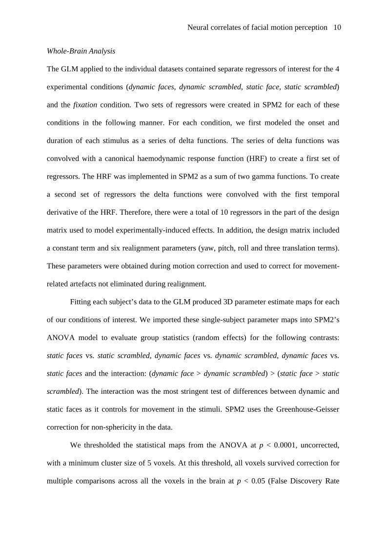
Whole-Brain Analysis
The GLM applied to the individual datasets contained separate regressors of interest for the 4
experimental conditions (dynamic faces, dynamic scrambled, static face, static scrambled)
and the fixation condition. Two sets of regressors were created in SPM2 for each of these
conditions in the following manner. For each condition, we first modeled the onset and
duration of each stimulus as a series of delta functions. The series of delta functions was
convolved with a canonical haemodynamic response function (HRF) to create a first set of
regressors. The HRF was implemented in SPM2 as a sum of two gamma functions. To create
a second set of regressors the delta functions were convolved with the first temporal
derivative of the HRF. Therefore, there were a total of 10 regressors in the part of the design
matrix used to model experimentally-induced effects. In addition, the design matrix included
a constant term and six realignment parameters (yaw, pitch, roll and three translation terms).
These parameters were obtained during motion correction and used to correct for movement-
related artefacts not eliminated during realignment.
Fitting each subject’s data to the GLM produced 3D parameter estimate maps for each
of our conditions of interest. We imported these single-subject parameter maps into SPM2’s
ANOVA model to evaluate group statistics (random effects) for the following contrasts:
static faces vs. static scrambled, dynamic faces vs. dynamic scrambled, dynamic faces vs.
static faces and the interaction: (dynamic face > dynamic scrambled) > (static face > static
scrambled). The interaction was the most stringent test of differences between dynamic and
static faces as it controls for movement in the stimuli. SPM2 uses the Greenhouse-Geisser
correction for non-sphericity in the data.
We thresholded the statistical maps from the ANOVA at p < 0.0001, uncorrected,
with a minimum cluster size of 5 voxels. At this threshold, all voxels survived correction for
multiple comparisons across all the voxels in the brain at p < 0.05 (False Discovery Rate

Neural correlates of facial motion perception 11
FDR, Genovese et al. 2002) and all clusters survived cluster-wise multiple corrections at p <
0.05 (Friston et al. 1994).
Figure 2 (activations rendered on inflated brain) was created using the spm_surfrend
toolbox (http://spmsurfrend.sourceforge.net/) and displayed using Neurolens software
(www.neurolens.org) on the inflated template brain from the Freesurfer toolbox
(http://surfer.nmr.mgh.harvard.edu).
Regions Of Interest Analysis
In addition to our whole-brain, voxel-wise group analysis, we performed analyses on
individually-defined face sensitive regions of interest (ROI). These ROIs were identified
using the contrast static faces > static scrambled, as follows. We searched in each subject’s
individual GLM analysis for clusters whose peak response was located less than 10 mm away
from the peak response of the clusters found in the group ANOVA. The single-subject GLMs
were thresholded at the lower p<0.001 uncorrected threshold during this ROI search (1)
because we were looking in regions of a-priori interest which had already survived whole-
brain correction in the group ANOVA and (2) to increase the likelihood of finding significant
clusters in as many of the individual subjects as possible.
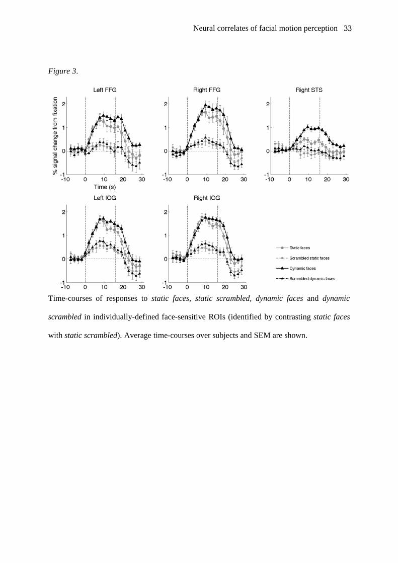
After identifying these individual ROIs, we computed their block-averaged response
time-courses to each condition, as follows. Raw BOLD signal data were extracted and
filtered by removing low frequencies (cutoff = 128 seconds) and movement artefacts (using
the realignment parameters calculated by SPM2), then averaged over voxels in each ROI. For
each run of each participant, the time-series were converted into percent signal change from
average activity by dividing the signal measured at each time point by the average signal
during the run, subtracting 1, and then multiplying by 100. The block-related responses to
each condition were then averaged across all participants from 10s before to 30s after each

Neural correlates of facial motion perception 12
block onset. The signal from the fixation condition was then used as a baseline and subtracted
from each of the four other conditions. Therefore, the “0” point on the y axis of Figure 3
corresponds to the mean activity in the fixation condition across all runs, and positive and
negative values respectively represent relative increases and decreases from the mean signal
intensity in the fixation condition.
In each ROI, group statistics were assessed as follows. For each block of trials, the
magnitude of the response to each condition was calculated by averaging the signal time-
course in the period between 7.5 and 19 seconds after block onset. The response to static
faces and dynamic faces was then compared using 2-tailed paired-samples t-tests over
subjects. To assess the robustness of the magnitude effects to differences in low-level
stimulus characteristics, these tests were computed again after subtracting from the response
time-course to each faces condition the response to the matching phase-scrambled faces
conditions. This effectively tests the following interaction: (dynamic face > dynamic
scrambled) > (static face > static scrambled). Note: Our ROIs were defined by comparing
static faces to static scrambled, and thus the response to dynamic faces (or to dynamic
scrambled) did not play any role in the definition of these ROIs (i.e., the voxels of our ROI
could respond more, less or similarly to dynamic faces compared to static faces). As the way
we defined the ROIs did not influence the outcome of the contrasts testing for responses to
dynamic faces vs. other conditions, it is perfectly valid to statistically compare responses to
static faces and dynamic faces without a-priori biases introduced through the ROI definition
method. In effect, instead of performing a separate localiser experiment, we used some of the
conditions of our experiment as a localiser contrast to define regions in which we
subsequently tested other contrasts (Friston et al. 2006).
Neural correlates of facial motion perception 13
Results
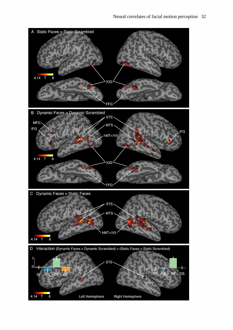
Whole-Brain Statistics
Clusters of voxels responding more to static faces than to static scrambled were found in
fusiform gyrus (FFG) bilaterally, in inferior occipital gyrus (IOG) bilaterally and in the right
STS. Given their anatomical location (see coordinates in Table 1), the clusters in FFG and
IOG most likely correspond respectively to the fusiform face areas (FFA, Kanwisher et al.
1997) and the occipital face areas (OFA, Halgren et al. 1999; Gauthier et al. 2000; Hoffman
and Haxby 2000). As we did not define these clusters by contrasting faces against objects as
was done in the studies defining FFA and OFA, we prefer to use the terms FFG and IOG.
Figure 2A shows these results thresholded at p<0.0001 uncorrected (Note: right STS survived
the threshold of p<0.05, whole-brain corrected but not p<0.0001 uncorrected and thus does
not appear in Figure 2). Clusters of voxels responding more to dynamic faces than to dynamic
scrambled were found bilaterally in the following structures: fusiform gyri (FFG), inferior
occipital gyrus (IOG), in the posterior and middle parts of the superior temporal sulcus (STS)
extending into middle (MTS) and inferior temporal sulci, including the anatomical location of
area hMT+/V5 (Dumoulin et al. 2000), as well as in middle prefrontal (MFG), medial
prefrontal and medial orbitofrontal cortex, inferior frontal gyrus (IFG) and posterior cingulate
gyrus (see Figure 2B). A higher response to dynamic faces than to static faces was found
bilaterally in STS (extending into middle temporal gyrus and sulcus, MTS), in the inferior
temporal sulcus (hMT+/V5), and in a small cluster in the precentral gyrus (see Figure 2C).
No areas were found that responded more to static than dynamic faces. The interaction
(dynamic face > dynamic scrambled) > (static face > static scrambled) yielded significant
effects exclusively in bilateral STS (Figure 2D). Details of the peaks of these activations are
reported in Table 1.
Neural correlates of facial motion perception 14
-----------------------------------
Insert Table 1 about here
Insert Figure 2 about here
-----------------------------------
Individual Face-Sensitive Regions Of Interest
We located the following ROIs in 8 to 10 out of our 10 subjects: left and right FFG, left and
right IOG, and right STS. As stated in the previous paragraph, FFG and IOG most likely
correspond to FFA and OFA respectively (see coordinates in Table 2). As reported in Table 2
and shown in Figure 3, all ROIs except the right IOG responded more to dynamic faces than
to static faces when both conditions were compared with fixation. In addition, right FFG and
right STS also showed increased activation for dynamic compared to static faces when both
were contrasted with their matched phase-scrambled controls (i.e., (dynamic faces > dynamic
scrambled) > (static faces > static scrambled)). No ROI showed a higher response to static
faces than to dynamic faces.
Note: almost identical time-courses were found in fusiform and occipital ROIs
identified using the contrast dynamic faces > dynamic scrambled, which is an indication of
the great overlap between ROIs identified using both methods.
-----------------------------------
Insert Table 2 about here
Insert Figure 3 about here
-----------------------------------
Neural correlates of facial motion perception 15
Discussion
In this study, we investigated brain activation in response to dynamic face stimuli using
natural video sequences of facial motion and directly compared it to activation in response to
static face images. Using ROI analyses, we found that in most of the classic face-sensitive
areas (bilateral FFG, left IOG and the right STS), the BOLD response to dynamic faces was
higher than to static faces. In right FFG and right STS, these effects survived even when
controlling for low-level visual properties of the stimuli using matched phase-scrambled
controls. In addition, our analyses confirmed that STS is the brain region most sensitive to
dynamic faces when controlling for stimulus motion. No clusters of the whole-brain analysis
or any ROI showed greater response to static than dynamic faces. Taken together, these
results show higher brain activation for dynamic than static faces not only in areas that have
been related to the processing of changeable aspects of faces but also in areas that have been
previously attached to the processing of invariant aspects of faces, i.e., the processing of
facial form rather than facial motion (Haxby et al., 2000). This is particularly interesting
given that face recognition, a process thought to involve mainly areas sensitive to invariant
aspects of faces, can be facilitated by facial motion (O'Toole et al. 2002; Pilz et al. 2006).
These results suggest an integration of form and motion information in a network of areas
including STS, as has been proposed in models of the recognition of biological motion (Giese
and Poggio, 2003). In addition, our results provide a strong argument for the use of dynamic
stimuli to localize areas related to the processing of human faces, supporting an argument put
forward by Fox and colleagues (Fox et al. 2008).
Higher BOLD responses to Dynamic than Static Faces
In almost all face-sensitive ROIs, the BOLD response to dynamic faces was higher than to
static faces. This is consistent with previous results directly comparing dynamic and static

Neural correlates of facial motion perception 16
faces (Kilts et al. 2003; Sato et al. 2004) and with a recent study showing a stronger
differential response in these areas between faces and objects when shown in motion rather
than statically (Fox et al. 2008). However, the same contrast performed in the whole-brain
analysis did not show significant activation in FFG or IOG (except after lowering the
threshold to p<0.01 uncorrected; data not shown). This suggests that the analysis done on
individually-defined ROIs is more sensitive, which can be due to several reasons. First, the
ROIs were identified individually which compensates for the between-subjects variation in
location of functionally-defined regions. Second, the much smaller number of tests being
performed in ROI analyses compared to testing all voxels in the brain reduces the multiple
comparisons problem and allows more sensitive thresholds to be used in ROI analyses (Saxe
et al. 2006).
The higher activation we found for dynamic faces are compatible with the idea that
more neurons are tuned to these stimuli because they are more familiar and behaviorally
relevant stimuli, as has been suggested by several research groups (Kilts et al. 2003; Bartels
and Zeki 2004; Pelphrey et al. 2007; Fox et al. 2008); for example, more neurons have been
found that are tuned to frontal views as compared to side views of faces, which could be
related to the fact that we have greater experience with frontal view faces (Perrett et al.
1998). We encounter moving faces frequently every day when interacting with other people.
Therefore, our visual system is probably more familiar with seeing moving than static faces.
As a result, more neurons might be sensitive to dynamic than static faces.
However, the additional number of frames present in the dynamic face stimuli lead to
two alternative explanations of our findings: First, an explanation on the level of a single
population of neurons sensitive to both static and dynamic faces is that neurons responding to
faces might show response adaptation during the presentation of static faces. Because nothing
changes during the presentation of a single static face, the neuronal response would be

Neural correlates of facial motion perception 17
smaller at the end than at the beginning of each trial, as neural activity and the related BOLD
signal are known to decrease when there is no stimulus change (Grill-Spector and Malach
2001). This explanation was also put forward recently by others (Fox et al. 2008). Those
same neurons might not adapt during the presentation of the dynamic faces, because the face
undergoes subtle changes between successive frames shown during each trial. Less neuronal
adaptation during dynamic face presentations might therefore lead to higher metabolic
demands and thus to the higher BOLD signal we observe. Given the slow dynamics of the
BOLD signal, this difference in neuronal adaptation might also account for the bigger
difference in BOLD response we observed at the end of the blocks of trials compared to the
beginning.
Second, one could propose an explanation on the level of different neuronal
populations that each responds to a particular static frame of the dynamic face stimuli. In this
case, all these populations would be active during presentation of our dynamic face stimuli,
but only a subset of them would respond to our static face stimuli. This mechanism has also
been suggested recently by Fox and colleagues (2008). The difference in the number of
static-face-sensitive neuronal populations involved would then explain the difference in
BOLD signal we observed, without any involvement of neurons sensitive to face motion per
se.
Disproving these alternative hypotheses requires the use of control stimuli with the
same number of frames as the dynamic stimuli but not perceived as facial motion. These
stimuli are very difficult to create, because simply frame-scrambling our movies yields
stimuli perceived as strange, unnatural speeded-up motion, and these perceptual effects
probably involve unnatural responses of the face-processing system, leading to further
difficulties in experimental design and interpretation. We are currently addressing this
question in further experiments.
Neural correlates of facial motion perception 18
Interestingly, the difference in response to dynamic and static faces was not only
found in the right STS which is known to respond to biological motion and facial motion
(e.g., Haxby et al. 2000; Bartels and Zeki 2004; Hasson et al. 2004), but also in the areas
classically known to process invariant aspects of the faces: FFG and IOG. A recent study by
Fox and colleagues (2008) also reported a greater difference in response to dynamic faces
versus dynamic objects in these areas. But in their study, the responses to dynamic and static
faces could not be compared directly. As recognition of facial identity is thought to be mainly
accomplished by those latter areas, their higher response to dynamic faces might be linked to
the increased recognition performance observed for dynamic faces (O'Toole et al. 2002; Pilz
et al. 2006). This will have to be investigated further in purposefully-designed experiments.
Our findings constitute evidence that both motion- and form-related areas participate
in the processing of dynamic faces and suggest that temporal and spatial aspect of faces seem
to be processed in an integrated fashion in higher level visual brain areas. Those findings are
particularly interesting given that the different face identities and expressions in the stimulus
set were the same for static and dynamic faces, as were their presentation schedule, and that
in some ROIs, these effects even survived when the responses to the phase-scrambled control
stimuli was subtracted. Therefore, the effects are not related to face identity or expression
differences, and are not simply related to the fact that something was moving in the dynamic
face blocks or that each trial was composed of a series of different frames.
Other Regions Responding to Dynamic Faces
Contrasting the parameter estimates for dynamic faces to those for dynamic scrambled, we
found, in addition to activations in the face- and motion-sensitive areas discussed above,
higher activation in inferior and middle frontal gyrus (IFG and MFG) as well as medial
prefrontal and orbitofrontal cortex and posterior cingulate gyrus. In their recent paper, Fox
Neural correlates of facial motion perception 19
and colleagues (2008) found similar results by comparing dynamic faces to dynamic objects.
Recent neuroimaging studies have shown that the IFG, prefrontal and inferior parietal areas
are important for action observation and imitation (Molnar-Szakacs et al. 2005; Vogt et al.
2007). Iacoboni and colleagues (Iacoboni et al. 2005) found that the ventral premotor cortex
responds more to actions observed in an action-related context than in the absence of such a
context. They suggest that the human mirror system does not only provide an action
recognition mechanism, but also constitutes a neural system for coding the intentions of
others. This is supported by studies showing impairment in the recognition of emotional
stimuli and attribution of personality traits in patients with lesions in frontal cortex (Damasio
et al. 1991; Heberlein et al. 2004). Our stimuli show expressive faces that have a high
relevance when interacting in social situations. Therefore, it is reasonable that watching
dynamic expressive faces activates areas related to processing of emotional stimuli and
observing relevant actions of other people.
The posterior cingulate gyrus has been found to respond more to familiar faces, voices
and words (Kim et al. 1999; Leveroni et al. 2000; Shah et al. 2001) and shows an increasing
response during acquisition of facial familiarity (Kosaka et al. 2003). Its activation when
watching dynamic faces might reflect the fact that dynamic faces are more familiar and / or
that dynamic faces automatically trigger processes leading to their familiarization.
Conclusion
This study shows that dynamic faces elicit more activation than both static faces or phase-
scrambled controls in form-related face-processing areas (FFG and IOG) and in motion-
related face-processing (STS). These results are consistent with the hypothesis that our brain
contains mechanisms that are especially tuned to dynamic aspects of faces, and further reveal
that regions tuned to invariant aspects of faces respond more to dynamic than static faces. In

Neural correlates of facial motion perception 20
addition, our results show that dynamic stimuli provide an excellent tool for robustly
localizing areas related to the processing of facial form and motion information (also shown
by Fox et al. 2008).
Acknowledgments
The work was conducted while both authors were employed at the Max Planck Institute for
Biological Cybernetics, Tübingen, Germany. The authors would like to thank Heinrich H.
Bülthoff for support. Conflict of Interest: None declared.

Neural correlates of facial motion perception 21
References
Allison T, Puce A, McCarthy G (2000) Social perception from visual cues: role of the STS
region. Trends Cogn Sci 4: 267-278
Ashburner J, Friston K (1997) The role of registration and spatial normalization in detecting
activations in functional imaging. Clinical MRI/Developments in MR 7: 26-28
Ashburner J, Friston KJ (1999) Nonlinear spatial normalisation using basis functions. In:
Human Brain Mapping, vol 7 pp 254-266
Bartels A, Zeki S (2004) Functional Brain Mapping During Free Viewing of Natural Scenes.
Hum.Brain Mapp. 21: 75-85
Bassili JN (1976) Temporal and spatial contingencies in the perception of social events. J
Pers Soc Psychol 33: 680-685
Blake R, Shiffrar M (2007) Perception of Human Motion. Annu Rev Psychol 58: 47-73
Blakemore SJ, Frith CD, Wolpert DM (2001) The cerebellum is involved in predicting the
sensory consequences of action. Neuroreport 12: 1879-1884
Bonda E, Petrides M, Ostry D, Evans A (1996) Specific involvement of human parietal
systems and the amygdala in the perception of biological motion. J Neurosci 16:
3737-3744
Brainard DH (1997) The Psychophysics Toolbox. Spat Vis 10: 433-436
Campbell R, MacSweeney M, Surguladze S, Calvert G, McGuire P, Suckling J, Brammer
MJ, David AS (2001) Cortical substrates for the perception of face actions: an fMRI
study of the specificity of activation for seen speech and for meaningless lower-face
acts (gurning). Brain Res.Cogn Brain Res. 12 233-243
Costen NP, Parker DM, Craw I (1996) Effects of high-pass and low-pass spatial filtering on
face identification. Percept Psychophys 58: 602-612

Neural correlates of facial motion perception 22
Damasio AR, Tranel D, Damasio H (1991) Somatic markers and the guidance of behavior:
Theory and preliminary testing. Oxford University Press, New York
Dumoulin SO, Bittar RG, Kabani NJ, Baker CL, Jr., Le Goualher G, Bruce PG, Evans AC
(2000) A new anatomical landmark for reliable identification of human area V5/MT:
a quantitative analysis of sulcal patterning. Cereb Cortex 10: 454-463
Eger E, Henson RNA, Driver J, Dolan RJ (2004) BOLD Repetition Decreases in Object-
Responsive Ventral Visual Areas Depend on Spatial Attention. J Neurophysiol 92:
1241-1247
Fox CJ, Iaria G, Barton JJS (2008) Defining the face processing network: Optimization of the
functional localizer in fMRI. Human Brain Mapping Online in advance of print
Friston KJ, Ashburner J, Frith CD, Poline JB, Heather JD, Frackowiak RS (1995a) Spatial
registration and normalisation of images. Hum Brain Mapp 2: 165-189
Friston KJ, Holmes A, Poline JB, Price CJ, Frith CD (1996) Detecting activations in PET and
fMRI: levels of inference and power. Neuroimage 4: 223-235
Friston KJ, Holmes AP, Worsley KJ (1999) How Many Subjects Constitute a Study?
Neuroimage 10: 1-5
Friston KJ, Holmes AP, Worsley KJ, Poline JB, Frith CD, Frackowiak RS (1995b) Statistical
parametric mapping in functional imaging: a general linear approach. Hum Brain
Mapp 2: 189-210
Friston KJ, Rotshtein P, Geng JJ, Sterzer P, Henson RNA (2006) A critique of functional
localisers. Neuroimage 30: 1077-1087
Friston KJ, Worsley KJ, Frackowiak R, Mazziotta J, Evans AC (1994) Assessing the
significance of focal activations using their spatial extent. Hum Brain Mapp 1: 210-
220

Neural correlates of facial motion perception 23
Gauthier I, Curby KM, Skudlarski P, Epstein RA (2005) Individual differences in FICA
activity suggest independent processing at different spatial scales. Cogn Affect Behav
Neurosci 5: 222-234
Gauthier I, Skudlarski P, Gore JC, Anderson AW (2000) Expertise for cars and birds recruits
brain areas involved in face recognition. Nat Neurosci 3: 191-197
Genovese CR, Lazar NA, Nichols T (2002) Thresholding of statistical maps in functional
neuroimaging using the false discovery rate. Neuroimage 15: 870-878
Giese MA, Poggio T (2003) Neural mechanisms for the recognition of biological movements.
Nat Rev Neurosci 4: 179-192
Grill-Spector K, Malach R (2001) fMR-adaptation: a tool for studying the functional
properties of human cortical neurons. Acta Psychol 107: 293-321
Grossman E, Donnelly M, Price R, Pickens D, Morgan V, Neighbor G, Blake R (2000) Brain
areas involved in perception of biological motion. J Cogn Neurosci 12: 711-720
Halgren E, Dale AM, Sereno MI, Tootell RBH, Marinkovic K, Rosen BR (1999) Location of
human face-selective cortex with respect to retinotopic areas. Hum Brain Mapp 7: 29-
37
Hall DA, Fussell C, Summerfield AQ (2005) Reading Fluent Speech from Talking Faces:
Typical Brain Networks and Individual Differences. Journal of Cognitive
Neuroscience 17: 939-953
Hasson U, Nir Y, Levy I, Fuhrmann G, Malach R (2004) Intersubject Synchronization of
Cortical Activity During Natural Vision. Science 303: 1634
Haxby JV, Hoffman EA, Gobbini MI (2000) The distributed human neural system for face
perception. Trends Cogn Sci. 4: 223-233
Neural correlates of facial motion perception 24
Heberlein AS, Adolphs R, Tranel D, Damasio H (2004) Cortical Regions for Judgments of
Emotions and Personality Traits from Point-light Walkers. J. Cogn. Neurosci. 16:
1143-1158
Hoffman EA, Haxby JV (2000) Distinct representations of eye gaze and identity in the
distributed human neural system for face perception. Nat Neurosci 3: 80-84
Iacoboni M, Molnar-Szakacs I, Gallese V, Buccino G, Mazziotta JC, Rizzolatti G (2005)
Grasping the intentions of others with one's own mirror neuron system. PLoS Biology
3: e79
Jacques C, Rossion B (2007) Early electrophysiological responses to multiple face
orientations correlate with individual discrimination performance in humans.
Neuroimage 36: 863-876
Jellema T, Perrett DI (2003) Cells in monkey STS responsive to articulated body motions and
consequent static posture: a case of implied motion? Neuropsychologia 41: 1728-
1737
Kamachi M, Bruce V, Mukaida S, Gyoba J, Yoshikawa S, Akamatsu S (2001) Dynamic
properties influence the perception of facial expressions. Perception 30: 875-887
Kanwisher N, McDermott J, Chun MM (1997) The fusiform face area: a module in human
extrastriate cortex specialized for face perception. J Neurosci 17: 4302-4311
Kanwisher N, Tong F, Nakayama K (1998) The effect of face inversion on the human
fusiform face area. Cognition 68: B1-B11
Kilts CD, Egan G, Gideon DA, Ely TD, Hoffman JM (2003) Dissociable Neural Pathways
Are Involved in the Recognition of Emotion in Static and Dynamic Facial
Expressions. Neuroimage 18: 156-168

Neural correlates of facial motion perception 25
Kim JJ, Andreasen NC, O'Leary DS, Wiser AK, Ponto LL, Watkins GL, Hitchwa RD (1999)
Direct comparison of the neural substrates of recognition memory for words and
faces. Brain 122: 1069-1083
Kosaka H, Omori M, Iidaka T, Murata T, Shimoyama T, Okada T, Sadato N, Yonekura Y,
Wada Y (2003) Neural substrates participating in acquisition of facial familiarity: an
fMRI study. Neuroimage 20: 1734-1742
Kovacs G, Zimmer M, Banko E, Harza I, Antal A, Vidnyanszky Z (2006)
Electrophysiological Correlates of Visual Adaptation to Faces and Body Parts in
Humans. Cereb Cortex 16: 742-753
LaBar KS, Crupain MJ, Voyvodic JT, McCarthy G (2003) Dynamic perception of facial
affect and identity in the human brain. Cereb Cortex 13: 1023-1033
Leveroni CL, Seidenberg M, Mayer AR, Mead LA, Binder JR, Rao SM (2000) Neural
systems underlying the recognition of familiar and newly learned faces. J Neurosci
20: 878-886
Molnar-Szakacs I, Iacoboni M, Koski L, Mazziotta JC (2005) Functional segregation within
pars opercularis of the inferior frontal gyrus: Evidence from fMRI studies of imitation
and action observation. Cereb. Cortex 15: 986-994
Morrison DJ, Schyns PG (2001) Usage of spatial scales for the categorization of faces,
objects, and scenes. Psychon Bull Rev 8: 454-469
Näsänen R (1999) Spatial frequency bandwidth used in the recognition of facial images.
Vision Res 39: 3824-3833
O'Toole AJ, Roark DA, Abdi H (2002) Recognizing moving faces: a psychological and
neural synthesis. Trends Cogn Sci 6: 261-266
Neural correlates of facial motion perception 26
Oram MW, Perrett DI (1996) Integration of form and motion in the anterior superior
temporal polysensory area (STPa) of the macaque monkey. J Neurophysiol 76: 109-
129
Pelli DG (1997) The VideoToolbox software for visual psychophysics: Transforming
numbers into movies. Spat Vis 10: 437-442
Pelphrey KA, Morris J, Michelich C, Allison T, McCarthy G (2005) Functional anatomy of
biological motion perception in posterior temporal cortex: an fMRI study of eye,
mouth and hand movements. Cereb Cortex 15: 1866-1876
Pelphrey KA, Morris JP, McCarthy G, LaBar KS (2007) Perception of dynamic changes in
facial affect and identity in autism. Social Cognitive and Affective Neuroscience
Advance Access
Perrett DI, Oram MW, Ashbridge E (1998) Evidence accumulation in cell populations
responsive to faces: an account of generalisation of recognition without mental
transformations. Cognition 67: 111-145
Pilz KS, Buelthoff HH, Vuong QC (2008) Learning influences the encoding of static and
dynamic faces and their recognition across different spatial frequencies. Mem Cog in
press
Pilz KS, Thornton IM, Bulthoff HH (2006) A search advantage for faces learned in motion.
Exp Brain Res 171: 436-447
Puce A, Allison T, Bentin S, Gore JC, McCarthy G (1998) Temporal cortex activation in
humans viewing eye and mouth movements. J Neurosci 18: 2188-2199
Puce A, Syngeniotis A, Thompson JC, Abbott DF, Wheaton KJ, Castiello U (2003) The
human temporal lobe integrates facial form and motion: evidence from fMRI and
ERP studies. Neuroimage. 19: 861-869

Neural correlates of facial motion perception 27
Rotshtein P, Vuilleumier P, Winston J, Driver J, Dolan R (2007) Distinct and convergent
visual processing of high and low spatial frequency information in faces. Cereb
Cortex 17: 2713-2724
Rousselet GA, Husk JS, Bennett PJ, Sekuler AB (2007) Single-trial EEG dynamics of object
and face visual processing. Neuroimage 36: 843-862
Sadr J, Sinha P (2004) Object recognition and Random Image Structure Evolution. Cognitive
Sci 28: 259-287
Sato W, Kochiyama T, Yoshikawa S, Naito E (2004) Enhanced neural activity in response to
dynamic facial expressions of emotion: an fMRI study. Cognitive Brain Research 20:
81-91
Saxe R, Brett M, Kanwisher N (2006) Divide and conquer: A defense of functional
localizers. Neuroimage 30: 1088-1096
Shah NJ, Marschall JC, Zafiris O, Schwab A, Zilles K, Markowitsch K, Fink GR (2001) The
neural correlates of person familiarity: a functional magnetic resonance imaging study
with clinical implications. Brain 124: 804-815
Thompson JC, Hardee JE, Panayiotou A, Crewther D, Puce A (2007) Common and distinct
brain activation to viewing dynamic sequences of face and hand movements.
Neuroimage 37: 966-973
Vogt S, Buccino G, Wohlschlager AM, Canessa N, Shah NJ, Zilles K, Eickhoff SB, Freund
H-J, Rizzolatti G, Fink GR (2007) Prefrontal involvement in imitation learning of
hand actions: Effects of practice and expertise. Neuroimage 37: 1371-1383
Vuilleumier P, Armony JL, Driver J, Dolan RJ (2003) Distinct spatial frequency sensitivities
for processing faces and emotional expressions. Nat Neurosci 6: 624-631

Neural correlates of facial motion perception 28
Tables
Table 1. Anatomical and statistical details of the peaks of significant activations revealed by
the contrasts performed in the ANOVA group analysis. All activations survive correction for
multiple comparisons across the whole brain.
Coordinates Anatomy Hemisphere
X, Y, Z
T Z
Static faces > static scrambled
Left -42, -48, -24 5.72 4.79 Fusiform gyrus (FFG)
Right 39, -57, -18 5.62 4.73
Left -39, -72, -12 5.65 4.75 Inferior occipital gyrus (IOG)
Right 45, -75, -12 5.79 4.84
Superior temporal sulcus (STS) Right 51, -48, 21 4.98 4.31
Dynamic faces > dynamic scrambled
Left -54, -48, 6 8.09 6.07 Superior temporal sulcus (STS)
Right 50, -36, 0 7.21 5.64
Left -45, -51, -21 6.51 5.26 Fusiform gyrus (FFG)
Right 39, -54, -18 5.67 4.75
Left -39, -72, -12 5.27 4.50 Inferior occipital gyrus (IOG)
Right 45, -69, -12 5.71 4.79
Left -39, 30, 3 5.42 4.60 Middle prefrontal cortex
Right 51, 33, 0 7.28 5.67
Medial orbitofrontal cortex Right 3, 42, -15 5.45 4.62
Posterior cingulate cortex Right 6, -54, 33 5.39 4.58
Left -48, 18, 24 5.05 4.36 Inferior frontal gyrus
Right 45, 24, 18 5.18 4.45

Neural correlates of facial motion perception 29
Superior medial prefrontal gyrus Left -6, 51, 30 4.66 4.09
Dynamic faces > static faces
Left -54, -58, 6 8.17 6.10 Superior temporal sulcus (STS)
Right 63, -27, 0 7.13 5.71
Left -51, -69, 9 8.66 6.33 hMT+/V5
Right 45, -66, 3 7.35 5.71
Left -39, -3, 51 4.58 4.04 Precentral gyrus
Right 54, 0, 51 4.63 4.07
Interaction: (Dynamic faces > dynamic scrambled) > (Static faces > static scrambled)
Left -57, -42, 6 5.91 4.14 Superior temporal sulcus (STS)
Right 66, -27, 0 4.56 4.02
Note: Coordinates indicate local maxima in MNI space. T and Z column respectively indicate
T values and Z scores from whole-brain ANOVA analysis.
Neural correlates of facial motion perception 30
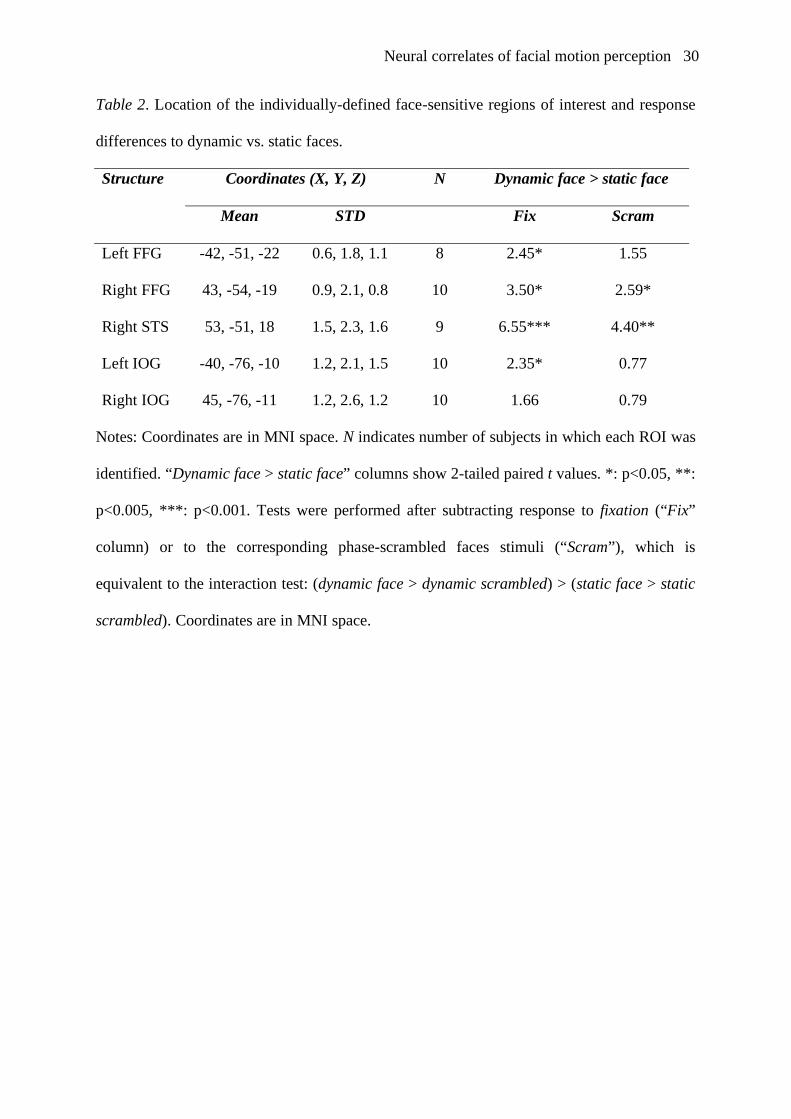
Table 2. Location of the individually-defined face-sensitive regions of interest and response
differences to dynamic vs. static faces.
Coordinates (X, Y, Z) N Dynamic face > static face Structure
Mean STD Fix Scram
Left FFG -42, -51, -22 0.6, 1.8, 1.1 8 2.45* 1.55
Right FFG 43, -54, -19 0.9, 2.1, 0.8 10 3.50* 2.59*
Right STS 53, -51, 18 1.5, 2.3, 1.6 9 6.55*** 4.40**
Left IOG -40, -76, -10 1.2, 2.1, 1.5 10 2.35* 0.77
Right IOG 45, -76, -11 1.2, 2.6, 1.2 10 1.66 0.79
Notes: Coordinates are in MNI space. N indicates number of subjects in which each ROI was
identified. “Dynamic face > static face” columns show 2-tailed paired t values. *: p<0.05, **:
p<0.005, ***: p<0.001. Tests were performed after subtracting response to fixation (“Fix”
column) or to the corresponding phase-scrambled faces stimuli (“Scram”), which is
equivalent to the interaction test: (dynamic face > dynamic scrambled) > (static face > static
scrambled). Coordinates are in MNI space.

Neural correlates of facial motion perception 31
Figures with Captions
Figure 1.
Example stimulus images. Top: all 26 frames of an example face movie stimulus (dynamic
face). Bottom: all 26 frames of an example phase-scrambled face movie stimulus (dynamic
scrambled). In the static conditions, only the last frame of each movie was shown, for the
same duration as the dynamic stimuli. Stimuli were shown in color.
Figure 2 (next page)
Results of the whole-brain ANOVA group statistics projected on the surface of an inflated
standard structural scan. Panel A shows clusters responding more to static faces than static
scrambled. Panel B shows clusters responding more to dynamic faces than dynamic
scrambled. Panel C shows clusters responding more to dynamic faces than static faces. Panel
D shows clusters with a significant interaction effect: (dynamic faces > dynamic scrambled)
> (static faces > static scrambled). Insets in D show percent signal change from fixation
(mean & SEM over subjects) for static faces (SF), static scrambled (SS), dynamic faces (DF)
and dynamic scrambled (DS) in left and right STS clusters (left and right insets respectively).
Maps are thresholded at p<0.0001 uncorrected, but all activations survive whole-brain
correction at p<0.05. Gradient bar shows T values.
Neural correlates of facial motion perception 32
Neural correlates of facial motion perception 33
Figure 3.
Time-courses of responses to static faces, static scrambled, dynamic faces and dynamic
scrambled in individually-defined face-sensitive ROIs (identified by contrasting static faces
with static scrambled). Average time-courses over subjects and SEM are shown.