national tse surveillance program (ntsesp) national ... · national tse surveillance program...
TRANSCRIPT

National TSE Surveillance Program (NTSESP)
National Guidelines for Field Operations
JULY 2015 TO JUNE 2016
(JULY 2015)

Published July 2015
© Animal Health Australia 2015
Disclaimer
This publication is published by Animal Health Australia for information purposes
only. Information contained in it is drawn from a variety of sources including some
external to Animal Health Australia. Although reasonable care was taken in its
preparation, Animal Health Australia does not guarantee or warrant the accuracy,
reliability, completeness, or currency of the information or its usefulness in achieving
any purpose.
To the fullest extent permitted by law, Animal Health Australia will not be liable for
any loss, damage, cost or expense incurred in or arising by reason of any person
relying on the information in this publication. Persons should accordingly make and
rely on their assessments and enquiries to verify the accuracy of the information
provided.
Copyright
This publication is protected by copyright. Information or material from this
publication may be reproduced in unaltered form for personal, non-commercial use.
All other rights are reserved. Information or material from this publication may be
used for the purposes of private study, research, criticism or review permitted under
the Copyright Act 1968.
Any reproduction permitted in accordance with the Copyright Act 1968 must
acknowledge Animal Health Australia as the source of any selected passage, extract,
diagram or other information. Any reproduction must also include a copy of the
original copyright and disclaimer notice as set out here.
Commercial and other use
No part of this publication may be reproduced, stored in a retrieval system, distributed
or commercialised in any form without prior written approval from Animal Health
Australia. The contents of the publication may not be used to sell a product or service
for commercial reasons such as advertising.
Trade marks
Any trade marks or logos contained in this publication may not be used without the
prior written permission of Animal Health Australia.

TSEFAP – National Guidelines for Field Operations 2015/16 3
CONTENTS
CONTENTS 3
GLOSSARY 4
INTRODUCTION 5
TSE SURVEILLANCE IN AUSTRALIA 6
Background 6 National Surveillance Design 7 Field, laboratory and reporting procedures 8
BSE AND SCRAPIE: THE NATURE OF THE DISEASES 12
Aetiology 12 Clinical Signs 12 Pathology 13 Modes of Transmission 13
APPENDIX 1 - SAMPLING DESIGN 16
Sampling Design for BSE 16 Sampling Design for Scrapie 17
APPENDIX 2 - TSE SURVEILLANCE IN CLINICALLY CONSISTENT CATTLE AND
SHEEP 18
Incentive Eligibility Criteria - Clinically Consistent 18 Differential Diagnoses of Nervous Disease in Australian Cattle and Sheep 19 Instructions for Submitters - Clinically Consistent 22 Guidelines for the Provision of Producer Incentives 30 Guidelines for the Provision of Veterinary Incentives 30 Guidelines for the Provision of Veterinary Laboratory Incentives 31
APPENDIX 3 - TSE SURVEILLANCE IN FALLEN AND CASUALTY SLAUGHTER
CATTLE AND SHEEP 34
Incentive Eligibility Criteria - Fallen and Casualty Slaughter 34 Instructions for Submitters - Fallen and Casualty slaughter 36
APPENDIX 4 - CLINICAL HISTORY AND POST-MORTEM REPORT 45
APPENDIX 5 - NTSESP CONTACTS 46
Web Site 46

TSEFAP – National Guidelines for Field Operations 2015/16 4
GLOSSARY
AAHL Australian Animal Health Laboratory, Commonwealth Scientific and
Industrial Research Organisation, Geelong, Victoria
AHA Animal Health Australia
AHO Government Animal Health Officer (includes stock inspectors and
government veterinarians)
ANZSDP Australian and New Zealand Standard Diagnostic Procedure
AUSVETPLAN Australian Veterinary Emergency Plan
BSE bovine spongiform encephalopathy
CNS central nervous system
DA Australian Government Department of Agriculture
NAHIS National Animal Health Information System
NATA National Association of Testing Authorities
NTSESP National Transmissible Spongiform Encephalopathies Surveillance
Program
OIE World Organisation for Animal Health
PrP prion protein
SAF scrapie associated fibrils
TSE transmissible spongiform encephalopathy
UK United Kingdom
vCJD variant Creutzfeldt-Jakob disease

TSEFAP – National Guidelines for Field Operations 2015/16 5
INTRODUCTION
These Guidelines for Field Operations provide field and laboratory workers in both the private
and public sectors with the information they need to participate in Australia’s National
Transmissible Spongiform Encephalopathies Surveillance Program (NTSESP). It contains
background information on the need to undertake national surveillance for animal transmissible
spongiform encephalopathies (TSEs) and describes the clinical signs and present understanding
of the modes of transmission of BSE and scrapie.
The guidelines do not cover atypical scrapie, although cases of this rare disease in sheep may be
an incidental finding from scrapie surveillance. The OIE Terrestrial Animal Health Code
Chapter 14.9 states that ‘the chapter does not cover so-called ‘atypical’ scrapie which is
clinically, pathologically, biochemically and epidemiologically unrelated to ‘classical’ scrapie,
may not be contagious and may, in fact, be a spontaneous degenerative condition of older
sheep’.
The NTSESP provides for payments to be made to primary producers, private veterinarians and
government agencies for submitting animals for examination where certain requirements are
fulfilled. These requirements relate to case definition, specimen collection and documentation.
The relevant requirements and their standards and criteria are described herein.

TSEFAP – National Guidelines for Field Operations 2015/16 6
TSE SURVEILLANCE IN AUSTRALIA
Background
Australia is recognised as meeting World Organisation for Animal Health (OIE) requirements
for a bovine spongiform encephalopathy (BSE) Negligible Risk and classical scrapie free
country. Countries with this BSE and scrapie status must have implemented defined
surveillance programs, as recommended by the OIE, for both diseases. It is important that
Australia meets this requirement to assure continued access to export markets.
Australia's status for BSE has been confirmed by national and international risk assessments -
for example those conducted by the OIE, Food Standards Australia New Zealand, the United
States of America, and Japan.
Australia has operated passive and active surveillance programs for BSE and scrapie for many
years. Passive surveillance occurs through routine investigations of disease by private and
government veterinarians and stock inspectors, the ante - and post-mortem examination of all
cattle and sheep at slaughter, and the inspection of animals destined for live export.
Active surveillance for BSE was commenced in Australia in 1990 and was modified in 1997
under the NTSESP to comply with recommendations in the chapter on BSE and what was then
the draft chapter on scrapie in the OIE Terrestrial Animal Health Code.
The NTSESP is an integrated national program jointly funded by governments and livestock
industries to demonstrate Australia’s ongoing freedom from BSE and scrapie and to provide
early detection of those diseases should they occur. The program is managed by Animal Health
Australia.
The objectives of the NTSESP are:
trade support - to maintain a TSE surveillance system consistent with the OIE Terrestrial
Animal Health Code that assures all countries that import cattle and sheep commodities
from Australia can be assured that Australia remains free of these diseases
protect public and animal health – to ensure the early detection of BSE should it occur in
Australia’s cattle so that an appropriate, early response can be mounted under
AUSVETPLAN to protect the health of Australia’s people and animals.
The NTSESP comprises:
field investigations by government and private veterinarians of animals where there is
suspicion of a TSE on the grounds of clinical signs
veterinary pathologists screening the case histories of all laboratory submissions, with a
clinical history of nervous disease, to consider whether they can exclude the diagnosis of
a TSE
veterinary histopathologists, trained in the diagnosis of a TSE, screening the brains of
submitted specimens from cattle over 30 months and less than nine years of age, and from
sheep over 18 months of age (but preferably less than five years) with a clinical history of
nervous disease to detect lesions of TSE
further laboratory testing of screen test results that are not conclusively negative
the collection of samples by Australian Government Department of Agriculture
(Department of Agriculture) from fallen and casualty slaughter cattle and sheep for testing
with rapid TSE tests – see Appendix Three
maintenance of NATA accreditation for rapid TSE tests by the Australian Animal Health

TSEFAP – National Guidelines for Field Operations 2015/16 7
Laboratory (AAHL).
participation in external proficiency testing for TSEs (AAHL)
an easily accessible and up-to-date national database of TSE surveillance information
supported by case records for at least 7 years
publication of NTSESP results in the National Animal Health Information System’s
(NAHIS) quarterly newsletter - Animal Health Surveillance, its annual report - Animal
Health in Australia, and on the Animal Health Australia website-
http://www.animalhealthaustralia.com.au/programs/disease-surveillance/national-animal-
health-information-system/.
Each state/territory animal health agency, and the commonwealth, participates in the NTSESP
with a national coordinating role provided through the TSEFAP National Advisory Committee
(NAC) and National Technical Committee (NTC). Awareness and training programs on TSE
surveillance are carried out through industry peak bodies, state/territory animal health agencies
and Department of Agriculture.
The NTSESP includes a financial incentive scheme to maximise reporting and investigation of
eligible clinically consistent animals by producers and private veterinarians.
Eligibility criteria for the financial incentives are shown in Appendix 3 (fallen and casualty
slaughter) and Appendix 2 (clinically consistent).
National Surveillance Design
The NTSESP is structured to comply with the OIE Terrestrial Animal Health Code – for BSE,
Chapter 11.5, and for scrapie, Chapter 14.9. The NTSESP involves submission of samples from
clinically consistent1, fallen2 and casualty slaughter3 cattle and sheep.
Appendix 1 provides details about the application of the OIE TSE sampling principles to the
NTSESP.
BSE surveillance design (Appendix 1)
The OIE recommends that BSE Negligible Risk countries conduct Type B surveillance to
“confirm the conclusions of the risk assessment, for example by demonstrating the effectiveness
of the measures mitigating any risk factors identified, through surveillance targeted to maximise
the likelihood of identifying failures of such measures”.
Type B surveillance as recommended by the OIE “will allow the detection of BSE around a
design prevalence of at least one case per 50,000 in the adult cattle population in the country,
zone or compartment of concern, at a confidence level of 95%”. The basis of Type B
surveillance, and therefore of the design of BSE surveillance within the NTSESP, is shown in
Appendix 1.
1 A clinically consistent animal is defined as “an animal that is found with clinical signs considered consistent with
BSE”. This is analogous with the term “clinical suspect” used in the OIE Terrestrial Animal Health Code Chapter
11.5 on BSE. 2 Fallen cattle are defined by the OIE Terrestrial Animal Health Code Chapter 11.5 as “cattle over 30 months of
age which are found dead or killed on farm, during transport or at an abattoir”. 3 Casualty slaughter cattle are defined by the OIE as “cattle over 30 months of age that are non-ambulatory,
recumbent, unable to rise or to walk without assistance; cattle over 30 months of age sent for emergency slaughter
or condemned at ante-mortem inspection”.

TSEFAP – National Guidelines for Field Operations 2015/16 8
The OIE’s recommended BSE surveillance approach takes into account the OIE’s general
principles of surveillance and the epidemiology of BSE. Points are assigned to each animal
according to the likelihood of detecting BSE infection based on the age of the animal sampled
and the subpopulation from which the sample was collected (clinically consistent, fallen or
casualty slaughter cattle). Surveillance points are valid within a seven-year moving window.
As recommended by the OIE, the NTSESP is primarily focused on sampling clinically
consistent cattle because this sub-population is recognised as the most sensitive sub-population
to target and therefore is allocated the most surveillance points per sample. Also as
recommended by the OIE, the NTSESP includes sampling of fallen and casualty slaughter sub-
populations and that all clinically consistent cattle1 should be investigated, regardless of the
number of points accumulated.
To ensure the funds of the NTSESP incentive scheme are used efficiently to promote
submission of animals with the highest potential for TSE detection, incentive payments are only
available for submission of clinically consistent animals that satisfy the eligibility criteria
(Appendix 2).
The OIE considers seven years to be the 95th percentile of the BSE incubation period.
Consequently the likelihood of BSE is relatively low in cattle 8 years and older. Allowing for a
margin of error in cattle ageing, and to be consistent with the OIE age groupings for
surveillance points, financial incentives are not available to cattle aged nine years or older.
Scrapie surveillance design (Appendix 1)
The scrapie surveillance program is designed so that annually there is at least a 99% probability
of detecting scrapie if this disease accounted for 1% of the cases of neurological disease in
sheep in Australia. This surveillance design meets the general requirements for countries that
are historically free from scrapie in accordance with the OIE Terrestrial Animal Health Code
Chapter 14.9. By targeting neurological cases, the designed minimum detection limit is one in a
million adult sheep. The minimum number of eligible sheep required each year and details of
sample design calculations are shown in Appendix 1. Numbers for each State/Territory may
change annually based on official livestock statistics. The program also includes sampling of
fallen and casualty slaughter sheep (see below).
Field, laboratory and reporting procedures
All procedures associated with the collection, processing, testing, reporting and invoicing of
TSE submissions should be consistent with these guidelines.
Clinical and post-mortem examination of eligible animals is carried out by private veterinarians,
and officers of state/territory animal health agencies and Department of Agriculture Biosecurity
through existing networks and samples submitted to participating animal health laboratories.
All TSE submissions must be accompanied by completed laboratory submission form/s.
Initial screen testing to specifically exclude TSEs is performed by veterinary pathologists and
laboratory technicians trained in TSE diagnostic techniques. If required, further diagnostic
investigations are undertaken by trained personnel located at AAHL.
The information obtained from eligible animals is recorded in NAHIS and presented as
quantitative evidence to Australia’s international trading partners of Australia’s freedom from
BSE and scrapie.

TSEFAP – National Guidelines for Field Operations 2015/16 9
Clinically consistent animal sampling (Appendix 2)
A clinically consistent animal is defined as “an animal that is found with clinical signs
considered consistent with BSE”. This is analogous with the term “clinical suspect” used in the
OIE Terrestrial Animal Health Code Article 11.5.20 on surveillance for BSE.
To claim financial payments from NTSESP incentive scheme, eligibility criteria for clinically
consistent cattle and sheep must be met (Appendix 2), including that all submissions of
clinically consistent animals must include a completed ‘Clinical history and post-mortem
report’ (Appendix 4).
Procedures for animal destruction, sample collection and dispatch, guidelines for the provision
of producer and veterinary incentives, and details on laboratory diagnosis are included in
Appendix 2
Fallen and casualty slaughter sampling (Appendix 3)
Fallen stock are defined by the OIE Terrestrial Animal Health Code Article 11.5.20 as “cattle
over 30 months of age and less than nine years of age which are found dead or killed on farm,
during transport, or at an abattoir”.
Casualty slaughter cattle are defined by the OIE as “cattle over 30 months of age that are non-
ambulatory, recumbent, unable to rise or to walk without assistance; cattle over 30 months of
age sent for emergency slaughter or condemned at ante-mortem inspection”.
Brainstem samples from fallen and casualty slaughter cattle (300) and sheep (100) are collected
at export abattoirs and submitted to AAHL.
To claim financial payments from NTSESP incentive scheme, eligibility criteria for fallen and
casualty slaughter cattle and sheep must be met (Appendix 3).
Procedures for animal destruction, sample collection and dispatch, guidelines for the provision
of incentives, and details on laboratory diagnosis are included in Appendix 3
Laboratory examination
Laboratory examinations must comply with the current version of the Australian and New
Zealand Standard Diagnostic Procedure – Transmissible Spongiform Encephalopathies ,
available from www.scahls.org.au/procedures/anzsdps. Brain specimens (paraffin embedded
tissue blocks and fresh CNS) for TSE surveillance must be retained until a report of TSE
exclusion has been issued by the relevant laboratory, after which they may be discarded or
destroyed in accordance with normal laboratory policy.
Where appropriate, examinations will be conducted to investigate differential diagnoses.
Animal health laboratory examination for TSEs includes:
histopathological examination of selected brain sections, chosen because of the known
distribution of BSE and scrapie lesions
examination of extracts of brain by electron microscopy for TSE associated fibrils of the
protease-resistant prion protein
inoculation of suspect prion containing material into mice to detect development of TSE
disease
examination of histological sections using antiserum and appropriate histochemical
markers for immunohistochemical demonstration of prion protein

TSEFAP – National Guidelines for Field Operations 2015/16 10
immunoblot using antiserum after detergent extraction and protease digestion removes the
normal prion protein to identify abnormal prion protein
enzyme linked immunosorbent assay (ELISA) analysis using antibodies to detect
abnormal prion protein after protease digestion to remove normal prion protein
State/Territory animal health laboratories must refer samples with non-negative TSE screen test
results to AAHL for further examinations4.
Laboratory findings are reported to the submitter and to the state NTSESP coordinator or
delegate.
Laboratories invoice Animal Health Australia through state NTSESP coordinators for the cost
of laboratory examinations.
National Reporting
The database is the responsibility of the NAHIS Database Administrator. The NAHIS Database
Administrator has the responsibility of keeping it functional and providing summary data to
NAHIS publications each quarter.
State NTSESP coordinators have responsibility to ensure that records are entered into the
national database as results become available from animal health laboratories. State/territory
primary records must be maintained for at least 7 years using existing systems.
The NTSESP summary database information can be found at:
www.animalhealthaustralia.com.au/programs/disease-surveillance/national-animal-health-
information-system/
Quality Assurance and Reporting Standards
Veterinary laboratories performing screening and confirmatory TSE testing must hold relevant
NATA accreditations.
Laboratory testing of clinically consistent cattle and sheep is processed individually.
Targets for providing final diagnostic reports for TSE submissions (from the final laboratory if
serial testing) are:
50% of animals within 4 weeks from the date of sample collection
90% within 8 weeks
100% within 12 weeks.
Laboratory testing of fallen and casualty slaughter cattle and sheep is batched. Typically
samples are processed in quarterly test runs using rapid TSE testing kits.
All (100%) diagnostic reports for fallen and casualty slaughter submissions should be finalised
within 16 weeks of the date of collection.
4 A suspect case of BSE is defined by the AUSVETPLAN Manual for BSE as “An animal of the genus Bos (cattle) or Bubalus
(buffalo) with history, clinical signs and histological changes consistent with BSE, until BSE is confirmed or excluded; or An
animal with a positive result from a sensitive and specific screening test such as an ELISA for transmissible spongiform
encephalopathies, until BSE is confirmed or excluded.”

TSEFAP – National Guidelines for Field Operations 2015/16 11
Figure 1: Specimen and information flows in the NTSESP
State /Territory
Coordination Commonwealth Coordination

TSEFAP – National Guidelines for Field Operations 2015/16 12
BSE AND SCRAPIE: THE NATURE OF THE DISEASES
BSE and scrapie are two of the TSE or “prion” diseases, which are characterised by progressive
neurodegenerative disease of adult animals, long incubation periods and the accumulation in the
central nervous system of an abnormal isoform of a host-encoded protein (PrP).
Three known strains of bovine spongiform encephalopathy (BSE) have been identified in cattle:
classical BSE, low-type (L-type) BSE and high-type (H-type) BSE. L-type BSE and H-type
BSE are also collectively called ‘atypical BSE’. Atypical BSE is a very rare disease that has
been recognised in a number of countries for less than 10 years.
Aetiology
A protease–resistant isoform (PrPsc
) of a normal cellular prion protein (PrPc
) has a pivotal role
in the pathogenesis of TSEs and, according to the prion hypothesis, is the sole component of the
TSE infectious agent. Scrapie has been recognised as a distinct neurological disease of sheep for
centuries and is distributed widely in Europe and North America, with sporadic occurrence in
some African and Asian countries.
BSE was first recognised in the UK in 1986 and the associated epidemic arose through the use
of MBM feed contaminated with the BSE agent. The origin of the BSE agent itself is uncertain
and various hypotheses have been considered. These include the possibility that BSE is derived
from the scrapie agent, that it is a new prion agent from cattle or that it originated from some
other mammalian species. Atypical BSE has been detected in a number of countries during
large-scale surveillance for BSE in cattle. The origin of this rare condition is not yet known, but
a spontaneous, non-contagious origin cannot be excluded.
Clinical Signs
BSE
Due to the long incubation period, signs usually appear when cattle are older than 30 months of
age, but less than nine years of age. BSE usually has an insidious onset and a slowly
progressive clinical course extending over weeks to months. Apprehension, hyperaesthesia, and
ataxia are the main signs, and at least one of these signs is present in most BSE cases; they
represent the most frequent changes in mental status, sensation, and posture and movement
respectively. Changes in mental status affect behaviour and temperament; the first sign of BSE
may be when a normally placid animal becomes aggressive and kicks in the milking shed.
Hypersensitivity can be to touch, sound and light. Ataxia affects mainly hind limbs. Other
abnormalities of posture and movement include falling, tremor, and abnormal head carriage. In
advanced cases, generalised weakness and loss of condition can cause recumbency, and signs of
altered mental status and hyperaesthesia may no longer be obvious, but should be carefully
sought in the clinical history of any recumbent animal. Loss of body weight and reduced milk
yield often accompany the nervous signs as the disease progresses.
In BSE affected countries, BSE is considered in the differential diagnosis of “sudden” death or
cases of purported misadventure. It is noteworthy that a higher incidence of BSE has been
found in Europe in emergency slaughter cattle than in animals passing pre-slaughter inspection;
when BSE has been diagnosed in either circumstance, there is often a history of overlooked
clinical signs of BSE. The AAHL video, A Tale of Transmission, clearly demonstrates the
clinical signs of BSE.

TSEFAP – National Guidelines for Field Operations 2015/16 13
All natural cases of atypical BSE have been reported in cattle that are at least 8 years of age.
Clinical signs of atypical BSE (when present) can be similar to those of classical BSE;
experimentally, they have included mental dullness and amyotrophy.
Scrapie
A gradual onset of clinical signs is observed in sheep between two and five years of age. Signs
observed may include hyper excitability; tremor, especially of the head and neck and in
response to stimulation; pruritus leading to rubbing of wool and secondary skin trauma; and
excessive nibbling or licking following tactile stimulation of the skin. In the later stages of the
disease, ataxia, posterior paresis and constitutional signs such as emaciation or obesity may be
noted. The clinical course can vary from weeks to months depending on the breed of sheep
affected and the strain of scrapie agent.
Pathology
There are no gross changes in BSE or scrapie. The three characteristic histological TSE
changes in the central nervous system (CNS) are vacuolation of grey matter neuropil
(spongiform change) and /or vacuolation of neurons, astrocytosis, and neuronal degeneration.
These changes have a predilection for certain neuroanatomical nuclei, particularly within the
brainstem, and occur bilaterally and generally in a symmetrical distribution. These features of
the pathological lesion mean that it is particularly important to submit the entire brainstem for
histological assessment. Accumulation of PrP can be demonstrated within these lesions using
immunohistochemical techniques.
Modes of Transmission
BSE
There is no evidence for maternal transmission or for horizontal spread of BSE between cattle,
either directly or indirectly. This is consistent with the restriction of its infectivity largely to
CNS tissue.
Epidemiological investigations suggest the BSE epidemic occurred as a result of ingestion of
feed containing MBM contaminated with high concentrations of the BSE agent. Most cases
were the result of calf-hood exposure to the agent. As carcasses of BSE-infected cattle were
recycled through rendering plants, the contamination level in MBM and incidence of cattle
infection were amplified, to produce the BSE epidemic. Since bans on the feeding of risk
materials to ruminants were progressively introduced, BSE incidence in affected countries has
dramatically declined (see figure 2).

TSEFAP – National Guidelines for Field Operations 2015/16 14
Figure 2: BSE cases reported in the UK and Europe
Source : Report on the monitoring and testing of ruminants for the presence of TSE in the EU in 2009
and 2011.
Latest numbers of BSE cases can be viewed at: www.oie.int/animal-health-in-the-world/bse-
specific-data/
The OIE recognises that BSE infectivity in cattle is largely confined to the brain, spinal cord,
eyes, trigeminal and dorsal root ganglia, tonsils and distal ileum. The BSE agent is not known to
be transferred in semen or embryos when collected to specified standards. TSE agents could

TSEFAP – National Guidelines for Field Operations 2015/16 15
potentially be spread by inoculation of biologically derived therapeutic products (iatrogenic
spread) such as biological products derived from central nervous system extracts.
Fomites, including surgical and veterinary instruments have not been a recognised method of
BSE spread to cattle in the UK outbreak. The potential for transmission of BSE by fomites is
limited, because contamination requires exposure to CNS tissue from affected cattle. However
care is required in the disposal or decontamination of equipment used for the post-mortem
removal of brain tissue from suspected BSE cases. Surgical instruments used for procedures
with CNS exposure (e.g. eye ablation) may also be contaminated if the animal is incubating
BSE but such procedures are rare and this form of transmission is very unlikely.
Scrapie
The scrapie agent is found in medium to high titre in the brain, spinal cord, pituitary, spleen,
tonsil, lymph nodes, ileum, colon, and rectum, of naturally infected sheep and goats. With
scrapie, infection with the prion agent probably occurs via ingestion. In many cases this may be
at or around the time of birth. The early appearance of the agent in tonsil, retropharyngeal and
mesenteric lymph nodes and intestine suggests that oral infection plays a major role in primary
infection; this could occur prenatally through ingestion of contaminated foetal fluids or
postnatally via milk or a contaminated environment.
The pathogenesis of scrapie includes an initial replication phase in gut-associated lymphoid
tissues before infection spreads to the central nervous system.
Susceptibility and resistance to the development of clinical scrapie is associated with several
polymorphisms within the PrP gene.
Further information on BSE and scrapie can be found within their respective Australian
Veterinary Emergency Plan (AUSVETPLAN) Disease strategies5.
5 www.animalhealthaustralia.com.au/programs/emergency-animal-disease-preparedness/ausvetplan/

TSEFAP – National Guidelines for Field Operations 2015/16 16
APPENDIX 1 - SAMPLING DESIGN
Sampling Design for BSE
The NTSESP sampling design for BSE is based on Chapter 11.5 of the OIE Terrestrial Animal
Health Code, 2011 Edition. Relevant sections from the chapter are modified and shown below.
Australia is a country assessed by the OIE as BSE Negligible Risk and therefore should
implement OIE Type B surveillance. The application of OIE Type B surveillance is designed to
allow the detection of at least one BSE case per 50,000 in the adult cattle population at a
confidence level of 95%.
From Table 1 below, Australia’s target is to achieve a minimum of 150,000 surveillance points
during a seven-year moving window. Australia should also meet OIE recommendations to
investigate all clinically consistent cattle regardless of the number of points accumulated and
ensure that cattle from the fallen and casualty slaughter subpopulations are also tested.
Table 1: Points targets for different adult cattle population sizes in a country
Adult cattle population size
(24 months and older) Type A surveillance Type B surveillance
>1,000,000 300,000 150,000
800,000-1,000,000 240,000 120,000
600,000-800,000 180,000 90,000
400,000-600,000 120,000 60,000
200,000-400,000 60,000 30,000
100,000-200,000 30,000 15,000
50,000-100,000 15,000 7,500
25,000-50,000 7,500 3,750
Table 2 ‘Surveillance point values for samples collected by subpopulation and age’ is used to
determine the OIE point values of each BSE surveillance sample collected. Points are assigned
to each animal’s sample according to the animal’s age and cattle subpopulation from which it
was collected. The point values reflect the relative likelihoods of expressing BSE by age and
sub-population, according to scientific knowledge of the disease. The OIE recommends that
samples should be collected from at least three of the four subpopulations, but that ages and
sub-populations sampled should reflect the demographics of the cattle herd.
The total points for samples collected may be accumulated over a maximum of 7 consecutive
years to achieve the target number of points determined in Table 1. Surveillance points remain
valid for 7 years (the 95th percentile of the incubation period).

TSEFAP – National Guidelines for Field Operations 2015/16 17
Table 2: Surveillance point values for samples collected by subpopulation and age
Routine
slaughter
Fallen
stock
Casualty
slaughter
Clinically
consistent
Age ≥ 1 year and <2years
0.01 0.2 0.4 N/A
Age ≥ 2 years and <4 years (young adult)
0.1 0.2 0.4 260
Age ≥ 4 years and <7 years (middle adult)
0.2 0.9 1.6 750
Age ≥ 7 years and <9 years (older adult)
0.1 0.4 0.7 220
Age ≥ 9 years (aged)
0.0 0.1 0.2 45
Sampling Design for Scrapie
The NTSESP scrapie sampling design is consistent with meeting the OIE Terrestrial Animal
Health Code, 2013 recommendations for a classical scrapie free country and is based on
detecting scrapie with 99% confidence if it comprised 1% of neurological cases.
It is assumed that there are about 80 million sheep in Australia and that 50 million of these
would be over 18 months of age. Thus the reference population of interest comprises the 5000
expected neurological cases from this group. This results in a recommendation to examine a
minimum of 440 eligible neurological cases each year assuming perfect sensitivity and
specificity of the diagnostic system.
It is further assumed that neurological cases in sheep are uniformly distributed throughout
Australia. The sampling fraction is therefore the same for each State and is applied to each
State’s sheep population to reach the numbers specified in Table 3. Numbers are based on the
three-year rolling average of 2009-12 (ABS figures).
While scrapie can occur in both sheep and goats, the NTSESP only applies to sheep. Scrapie in
goats would only be seen in Australia as a ‘spill-over infection’ from sheep.
Table 3: Number of clinically consistent sheep required each year
State/Territory Number required
QLD 22
NSW 160
VIC 93
TAS 14
SA 63
WA 88
ACT and NT 0
Total 440

TSEFAP – National Guidelines for Field Operations 2015/16 18
APPENDIX 2 - TSE SURVEILLANCE IN CLINICALLY CONSISTENT CATTLE AND SHEEP
Incentive Eligibility Criteria - Clinically Consistent
Payments for submission (producer incentive, vet fee rebate, collect and document, and freight),
TSE laboratory tests and other laboratory tests to investigate alternate diagnoses may be
claimed from the NTSESP incentive scheme for clinically consistent submissions that meet the
eight incentive eligibility criteria below.
Species – only cattle and sheep are eligible for payment.
Live examination – the animal must be examined while alive by the submitting veterinarian
or biosecurity officer to independently establish the clinical state prior to euthanasia and
sampling
Age -
o cattle must be at least 30 months but less than nine years of age
o sheep must be 18 months of age or older (preferably less than 5 years)
Clinically consistent animal – the animal must have at least two clinical signs
consistent with BSE or scrapie as per Appendix 4 (Clinical history and post-mortem
report form)
Sample type and quality – submitters must submit complete samples of the correct
tissues (brain and spinal cord) of diagnostic quality for TSE evaluation to a participating
laboratory as per these national guidelines
Completion of required forms - each submission must include completed
documentation as per the policy and procedures of the relevant agency including a
competed laboratory submission form and Clinical history and post-mortem report from
(Appendix 4) or equivalent.
1 or 2 animals only per disease incident per property.
Conflict of interest – the submitting veterinarian or biosecurity officer must not have an
actual or perceived conflict of interest with the recipient of a payment arising from the
NTSESP (e.g. a financial or family link to them);
or must satisfy the relevant state agency that a clinically consistent animal could not
have been reasonably submitted by an alternative veterinarian or biosecurity officer
without an actual or perceived conflict of interest.

TSEFAP – National Guidelines for Field Operations 2015/16 19
Differential Diagnoses of Nervous Disease in Australian Cattle and Sheep
It is important, wherever possible, to establish a diagnosis for the condition under investigation.
Differential Diagnoses for Australian CATTLE Include:
- trauma
brain and spinal cord
- musculoskeletal diseases
- nutritional myopathy (vitamin E or selenium deficiency)
- metabolic diseases
hypomagnesaemia/hypocalcaemia
nervous acetonaemia
hepatic (e.g. pyrrolizidine alkaloidosis) and renal encephalopathy
polioencephalomalacia
heat stress
- infectious diseases
brain or spinal abscess (including cranial or vertebral osteomyelitis)
listeriosis
thromboembolic meningo-encephalomyelitis
cerebral babesiosis (type 1.3 – BHV 1.3)
bovine herpes virus encephalitis
sporadic bovine encephalomyelitis
bovine malignant catarrhal fever
focal symmetrical encephalomalacia (Clostridium perfringens)
bovine ephemeral fever
rabies (exotic)
- toxicoses
lead toxicosis
plant toxicoses
perennial rye grass staggers (Acremonium sp endophyte on Lolium perenne)
Annual rye grass staggers, blown grass staggers/flood plain staggers (Clavibacter toxicus on
seed heads)
paspalum staggers (ergotism: Claviceps paspali on Paspalum dilatatum)
phalaris staggers
Swainsona spp toxicosis
Zamia (palm) staggers
Xanthorrhoea spp (grasstrees) toxicity
botulism
urea toxicoses
snakebite

TSEFAP – National Guidelines for Field Operations 2015/16 20
- genetic diseases
cerebellar hypoplasia (Shorthorn, Brahman cattle)
cerebellar abiotrophy (Angus cattle)
progressive ataxia (Charolais cattle)
progressive spinal myelinopathy (Murray Grey cattle)
neuronal ceroid-lipofuscinosis (Devon cattle)
tomaculous-like neuropathy (Santa Gertrudis cattle)
- neoplasia

TSEFAP – National Guidelines for Field Operations 2015/16 21
Differential Diagnoses for Australian SHEEP Include:
- pruritus
lice, keds, itch mite, cutaneous myiasis
- metabolic diseases
hypomagnesaemia/hypocalcaemia
polioencephalomalacia
hepatic (e.g. pyrrolizidine alkaloidosis) and renal encephalopathy
copper deficiency
- infectious diseases
brain or spinal abscess
bacterial meningitis
listeriosis
melioidosis
focal symmetrical encephalomalacia (Clostridium perfringens)
rabies (exotic)
- genetic diseases
ovine segmental axonopathy (Murrurundi disease, Mudgee ataxia)
cerebellar abiotrophy (Yass ataxia)
thalamic-cerebellar neuropathy
cervicothoracic vertebral subluxation and ataxia
- toxicoses
lead poisoning
plant poisoning
“humpy back” (suspected Solanum esuriale)
perennial rye grass staggers (Acreonium sp endophyte on Lolium perenne)
annual rye grass staggers, blown grass staggers/flood plain staggers (Clavibacter sp on seed heads)
paspalum staggers (ergotism: Claviceps paspali on Paspalum dilatatum)
phalaris staggers
Swainsona poisoning
Sorghum spp associated neuraxonal degeneration
Tribulus terrestris staggers (Coonabarabran ataxia)
Tribulus micrococcus ataxia (Narrabri ataxia)
Swainsona toxicosis
botulism
Ixodes paralysis
delayed organophosphate toxicosis
urea poisoning
- neoplasia

TSEFAP – National Guidelines for Field Operations 2015/16 22
Instructions for Submitters - Clinically Consistent
These instructions support assessment of cases, specimen collection and submission of
clinically consistent animals to be examined, tested and reported under the NTSESP.
It is valuable to establish an alternate presumptive or definitive diagnosis in clinically consistent
animals that do not have a TSE. As clinical signs consistent with TSE may arise from lesions
affecting tissues other than the brain, submitters should collect and submit a range of samples
that will support a full laboratory investigation, and not simply exclude a TSE. It is
recommended that submitters discuss what additional samples to submit with a veterinary
pathologist.
To receive incentive payments from the NTSESP, all submitters (private veterinarians and
government officers) must ensure that each clinically consistent case meets the eligibility
criteria.
Submitter assessment of cases as suitable for submission for TSE exclusion and eligible for
incentive payments.
While all cattle and sheep that meet the OIE definition of ‘clinically consistent’ should be
submitted for TSE exclusion – only those that meet all eight eligibility criteria are eligible for
payments under the NTSEP financial incentive scheme.
Payments will not be made for TSE exclusion of ineligible animals unless otherwise agreed
with AHA on a case-by-case basis.
Submitters must establish that the case is ‘clinically consistent’ with BSE or scrapie by
carefully examining affected live animals and establishing a full history of the circumstances of
occurrence of the disease, and any treatments and response to treatment.
Clinical assessment of ‘nervous system disease’ and ‘clinically consistent’ with BSE or scrapie
should be considered in the broadest sense. Animals suitable for inclusion in the program may
have subtle abnormalities like lip or ear paralysis, or more obvious general disturbances like
head pressing or convulsions. Animals with signs that could be consistent with BSE or scrapie
should be assessed as ‘clinically consistent’ – even though these clinical signs may be primary
or secondary to disease involving other organ systems.
CAUTION - the likelihood of infection from Salmonella, Listeria, Leptospira or other zoonotic
organisms requires all persons conducting post-mortem examinations to undertake appropriate
precautions.

TSEFAP – National Guidelines for Field Operations 2015/16 23
Cattle - clinical signs consistent with BSE
Cattle may be submitted as ‘clinically consistent’ if at least two of the following clinically signs
are verified by examination of live cattle by an appropriate veterinarian (private or government)
or government officer.
Mental status
• altered consciousness
• apprehension
• behaviour change
• excitability
• frenzy
• hesitation at doors, gates,
barriers
• herd hierarchy change
• moribund without evidence of
infection or trauma
• teeth grinding
Sensation
• blindness
• excessive licking of nose
and flank
• head rubbing or pressing
• head shyness
• hyperaesthesia (sound, touch)
• hypoaesthesia (sound, touch)
• kicking persistently when
milked
Posture and movement
• abnormal ear position
• abnormal head carriage
• ataxia
• circling
• falling
• fetlock knuckling
• paralysis/paresis
• recumbency
• tremor
Sheep - clinical signs consistent with scrapie
Sheep may be submitted as ‘clinically consistent’ if at least two of the following clinically signs
are verified by examination of live sheep by an appropriate veterinarian (private or government)
or government officer.
Mental status
• altered consciousness
• apprehension
• behaviour change
• frenzy
• moribund without evidence of
infection or trauma
• temperament change
Sensation
• blindness
• hyperaesthesia (sound,
touch)
• hypoaesthesia (sound,
touch)
• rubbing/itching
• wool loss (flank and hind
quarter)
Posture and movement
• abnormal head carriage
• ataxia
• circling
• falling
• fetlock knuckling
• paralysis/paresis
• recumbency
• tremor

TSEFAP – National Guidelines for Field Operations 2015/16 24
Animal Destruction
The specimens are best removed from an animal killed by intravenous injection, commonly a
barbiturate anaesthetic. Euthanasia performed in this way, may constitute a danger to animals
that might consume the carcass or offal and this should always be borne in mind.
Alternative techniques such as captive bolt, shooting or the use of other injectable agents can be
considered. A range of techniques are outlined in the AUSVETPLAN Destruction Manual.
Care needs to be taken to avoid excessive damage to the cerebellum and brain stem. When
shooting an animal, please consider the location of the brain in the skull (figure 3). A poll shot
may be less likely to damage the brain stem than the routine frontal shot. Consult the relevant
state/territory authority regarding recommendations for humane methods for animal destruction
appropriate for the collection of diagnostic TSE exclusions specimens.
In the case of a strong clinical suspicion of BSE or scrapie, it is essential the euthanasia
method used enables collection of the whole undamaged brain and spinal cord samples.
Figure 3: euthanasia procedure: consideration of cerebellum and brain stem
Brain Removal Techniques
There are a number of methods to remove the brain in the field without damaging the brain stem
and cerebellum so that it is suitable for TSE examination.
Method 1 - removing the skullcap. This is achieved with 3 carefully placed cuts with a saw or
axe (Figure 4). The two most important cuts are along the sides of the skull and must cut the
occipital condyles. This allows the brainstem and first section of the spinal cord to be removed
undamaged. The 3rd cut is placed transversely across the front of the skull, midway between
the eye and the horn site. The lateral cuts must intersect with this transverse cut to form a
triangle of cuts around the skull. The loosened skullcap can then be levered off using a
sharpening steel or axe blade. After removing the dura mater (the white tough membrane that
surrounds the brain) the brain can then easily be “rolled” out from front to rear using a boning
knife to cut the nerve roots so that the caudal brainstem can be removed intact. Take particular
care to remove all of the dura mater reflection (tentorium cerebelli) that projects downwards to
provide a transverse partition between the cerebral hemispheres and cerebellum. This may
ossify, particularly in cattle, and failure to excise it may result in damage to critical anatomic
sites in the brainstem when the brain is subsequently lifted from the cranial cavity.

TSEFAP – National Guidelines for Field Operations 2015/16 25
Figure 4: brain removal: cranial capping technique
Method 2 - removing the brain in pieces by cutting through the skull and brain transversely with a saw (single transverse craniotomy). The cut is made dorsoventrally in a transverse plane
that is perpendicular to the frontal surface, just cranial (1-1.5 cm) to the external ear canal, and
parallel to the caudal border of the mandible as illustrated in Figure 5. The dorsoventral cut
should angle forward rather than backwards. Some laboratories discourage this technique
because there is a risk that such a cut, if not placed sufficiently cranially, will damage a key
diagnostic site in the brainstem. Also, if the cut is oblique, bilateral histological assessment is
not possible at that site.
Figure 5: brain removal: transverse craniotomy technique
Method 3 - splitting the skull (but not the brain) ventrally and dorsally along its longitudinal
axis with a saw or axe (Figure 6). The axe can be hit with a small sledgehammer for greater
control and safety. The two halves can be levered open from the nose end to expose the intact
brain. This method requires practice. It has the advantage of exposing the pituitary gland. The
brain should not be removed by longitudinally sectioning the skull and brain. Certain key
diagnostic sites along the brainstem are located near the midline. These can be damaged with
this technique as well as allow distortion of the brainstem during fixation and prevent bilateral
histological assessment.
TSEFAP – National Guidelines for Field Operations 2015/16 26
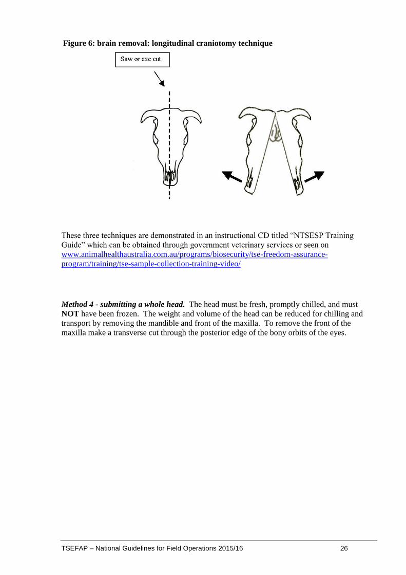
Figure 6: brain removal: longitudinal craniotomy technique
These three techniques are demonstrated in an instructional CD titled “NTSESP Training
Guide” which can be obtained through government veterinary services or seen on
www.animalhealthaustralia.com.au/programs/biosecurity/tse-freedom-assurance-
program/training/tse-sample-collection-training-video/
Method 4 - submitting a whole head. The head must be fresh, promptly chilled, and must
NOT have been frozen. The weight and volume of the head can be reduced for chilling and
transport by removing the mandible and front of the maxilla. To remove the front of the
maxilla make a transverse cut through the posterior edge of the bony orbits of the eyes.

TSEFAP – National Guidelines for Field Operations 2015/16 27
Specimens to Collect
Essential specimens
It is essential to submit both formalin fixed and unfixed (fresh) tissues from the central nervous
system. Fixed brain tissue is used for histopathology and immunohistochemistry, which are the
frontline tests to exclude TSEs. Unfixed cervical spinal cord (plus cerebellum for sheep) is
collected in case testing of the fixed brain tissue cannot exclude TSE, and may be used for
electron microscopy, immunoblot and mouse bioassay, as well as to further characterise a TSE
if detected. In particular, the cerebellum of sheep is collected to facilitate further differentiation
of scrapie and atypical/Nor98 scrapie by Western blotting if required.
The brain should be removed as soon as possible after death, with the brainstem attached and
intact.
Fresh tissues: Samples of fresh brain should be collected and refrigerated without fixation, as
soon as possible, either at refrigeration temperatures (4oC) or frozen (-20oC).
Cattle: 2 - 3 cm length of cervical spinal cord and/or medulla caudal to the
obex (see Figure 7a).
Sheep: (i) 2 - 3 cm length of cervical spinal cord and/or medulla caudal to the
obex, plus
(ii) the dorsal (top) third of the cerebellum sampled via a
coronal/horizontal approach (CAUTION - remove only just the top third
of the cerebellum, deeper sampling may damage TSE Standard Site 2
and compromise histological evaluation). See figure 7b.
These specimens must be collected and submitted for frozen storage at the state/territory
laboratory at a consistent temperature of -20C or less. They should be retained until a report of
TSE exclusion has been issued by the relevant laboratory, after which they may be discarded or
destroyed in accordance with normal laboratory policy.
Figure 7a: Cattle TSE exclusion – one unfixed tissue sample (blue shaded area)
1. Unfixed section of cervical spinal cord and/or medulla caudal to the obex

TSEFAP – National Guidelines for Field Operations 2015/16 28
Figure 7b: Sheep TSE exclusion – two unfixed tissue samples (blue shaded areas)
1. Unfixed section of cerebellum CAUTION – remove only top one-third.
Removing more may damage diagnostic sites below.
2. Unfixed section of cervical spinal cord and/or medulla caudal to the obex
Fixed tissues: The remainder of the brain from both cattle and sheep, with residual cerebellum
and brainstem attached, should be then be submerged in 10% buffered formalin after removal.
Prompt fixation helps to avoid artefactual vacuolation associated with decomposition that may
mimic BSE/scrapie. It is important to ensure adequate volumes of 10% buffered formalin
solution are used. Cattle brains need to be fixed in a minimum of 2 litres of 10% buffered
formalin and sheep brains in a minimum of 1 litre of 10% buffered formalin for a minimum of 7
days at room temperature (do not chill or freeze).
Use a sufficiently large container so that the brain “floats” and does not fix in a distorted
position. The brain should be suspended with the cerebrum resting on the bottom of the
container so that the caudal brainstem (midbrain and medulla) is not distorted through contact
with the bottom of the container.
Figure 8. Dorsal view of bovine brainstem and the three standard sites for histological
exclusion of TSE (cerebellum has been removed in this view).
Optional specimens for laboratory investigations of differential diagnoses
Microbiological sampling of central nervous system tissue before fixation may be appropriate.
Any other tissues with lesions should be preserved without delay in 10% buffered formalin.
The range of other specimens that should be considered (but are not mandatory) and the uses to
which they may be put are as follows:
• formalin fixed tissues - slices of lumbar spinal cord, liver, kidney, heart and lung of a
maximum thickness of 5 mm (can be fixed in the same container as the brain). Ensure
adequate volumes of formalin are used.
• serum - serology (IBR, SBE, botulism toxin), biochemistry (Ca, Mg, ketones),
serum - bank (if required for future investigation)

TSEFAP – National Guidelines for Field Operations 2015/16 29
• EDTA blood and smear - haematology, plasma cholinesterase
• organ smears of brain, kidney - tick fever organisms
• faeces - bacteriology, helminth egg and oocyst count
• fresh liver and kidney - lead, copper assay
• gastrointestinal content - rumen (botanical analysis), small and large intestine (botulism
toxin) 2x50 ml samples
In addition to these standard samples, other samples may be appropriate on a case-by-case
basis. If you are not sure which would be the most appropriate specimens for laboratory
analysis, please telephone a pathologist at your veterinary laboratory.
Documentation
Adequate documentation of the case details and history is required to meet national and
international reporting obligations and would be critical in a response to detection of a TSE.
All required forms must be completed for the case to be eligible for payments from the
NTSESP financial scheme.
The required forms include:
laboratory submission form provided by the approved State/Territory laboratory
Clinical history-post mortem report (Appendix 4) or equivalent as provided by the
relevant State/Territory.
Dispatch of Specimens
Ensure that:
• the specimens are securely and correctly packaged for transport
• all relevant details accompany the specimens, especially the required laboratory
submission form and the Clinical history and post mortem report (Appendix 4)
• the laboratory is notified if the specimens have to be picked up from transport terminals
after regular hours, as this may be essential to prevent deterioration of specimens.
Fresh Specimens - some fresh/chilled specimens (i.e. for microbiological culture, blood for
biochemistry and haematology) should be dispatched on the day of collection to ensure
handling at the laboratory as soon as practicable after death. The specimens must be sent with
the laboratory submission form and the Clinical history and post mortem report (Appendix 4)
with a clear indication that fixed specimens will follow.
The cervical spinal cord and/or medulla caudal to the obex, and for sheep the dorsal third of the
cerebellum, if kept chilled can be submitted with the fixed brain a few days after collection.
Fixed Specimens - to avoid transporting heavy and dangerous volumes of formalin, adequately
fixed tissues can be kept moist during transport by placing fixed tissues in a securely tied,
double plastic bag with either:
50 ml of 10% formalin, or
wrapped in formalin-soaked high absorbency paper towels
The brain and other fixed specimens should be sent to the laboratory with a submission form
but indicating on the submission form that fresh specimens had been previously sent.

TSEFAP – National Guidelines for Field Operations 2015/16 30
Guidelines for the Provision of Producer Incentives
Background
Producers will be paid incentives by participating State/Territory agencies under the NTSESP.
Incentives are provided to encourage the reporting and investigation of suitable animals and the
submission of suitable specimens for examination. It is essential that producers recognise and
report cattle and sheep with suspicious signs to a private veterinarian or government officer.
The producer incentive is available from the NTSESP via the relevant State/Territory agencies
to producer/s/owners for reporting and permitting euthanasia and sampling of cases that meet
the Eligibility Criteria for Clinically Consistent Cattle and Sheep (see page 18). No payment
will made if the incentive eligibility criteria have not been met, except as agreed with AHA.
Producer Incentives
$300 per eligible cattle
$100 per eligible sheep.
Administration
A claim for payment of the TSE incentive should be made through a government animal health
officer using an official claim form approved by the Chief Veterinary Officer of the relevant
State or Territory.
Guidelines for the Provision of Veterinary Incentives
Background
Private veterinarians and government agencies6 may claim payment under the NTSESP for
conducting post-mortem examinations and submitting reports and diagnostic samples. The
payments assist government agencies to cover costs and provide incentive to veterinary
practitioners to ensure suitable and appropriate reports are presented, specimens are collected,
packaged and submitted to an approved laboratory to enable TSE exclusion and establish a
likely alternative diagnosis so national targets for testing are met.
Veterinary incentives are available from the NTSESP for cases that meet the Incentive
Eligibility Criteria for Clinically Consistent Cattle and Sheep (see page 18). No payment will
made if the incentive eligibility criteria have not been met, except as agreed with AHA.
Veterinary fee rebates
$300 (GST ex) for eligible cattle ($200 for vet fees (cattle) and $100 collect and document)
$200 (GST ex) for eligible sheep ($100 for vet fees (sheep) and $100 collect and document)
Freight of $25 (GST ex) for each eligible animal.
Administration
Private veterinarians must claim for payment of the TSE rebate through a government
animal health officer using an invoice or the relevant claim form provided by the relevant
agency.
State/Territory agencies submit claims for veterinary incentives to AHA.
6 Submitters include private and government veterinarians and animal health officers.

TSEFAP – National Guidelines for Field Operations 2015/16 31
Guidelines for the Provision of Veterinary Laboratory Incentives
Background
Payments are available to government agencies under the NTSESP for laboratory investigation
of eligible ‘clinically consistent’ animals. The payments assist government agencies to cover
costs and provide incentive to test for TSEs and investigate differential diagnoses in clinically
consistent animals so national targets for testing are met.
Veterinary laboratory incentives are available from the NTSESP for cases that meet the
Incentive Eligibility Criteria for Clinically Consistent Cattle and Sheep (see page 18).
No payment will be made if the incentive eligibility criteria have not been met, except as agreed
with AHA.
Veterinary laboratory Incentives – State/Territory laboratories
Actual costs for TSE histopathology
Actual costs for laboratory investigation of differential diagnoses.
Veterinary laboratory Incentives – AAHL
$182 per immunohistochemistry test
$495 per scrapie associated fibril test.
Administration
Claims for payment of laboratory costs should be submitted to AHA.

TSEFAP – National Guidelines for Field Operations 2015/16 32
Histological Diagnosis and Reporting of Clinically Consistent Animals
TSE Positive
Characteristic vacuolation of grey matter neuropil (spongiform change) and/or of neurons,
usually with a bilaterally symmetrical distribution. Other forms of neuronal degeneration and
an astrocytic reaction support the diagnosis, if associated with grey matter vacuolation.
Scrapie
Neuronal vacuolation, particularly in the dorsal vagal nucleus at the obex, is usually
more common than neuropil vacuolation. Occasional vacuolated neurons (1 or 2 in a
section of medulla) without associated spongiform change in grey matter neuropil may
be found in normal sheep brains.
Bovine Spongiform Encephalopathy
Neuropil vacuolation, particularly in the solitary tract nucleus and spinal tract nucleus of
the trigeminal nerve at the obex, and in the periventricular grey matter of the midbrain,
is more prominent than neuronal vacuolation, which is most frequent in the vestibular
nuclear complex. In well-preserved material, a positive finding consists of more than 3
neuropil vacuoles at a neuroanatomical profile area.
Neuronal vacuolation in the red nucleus is a common incidental finding in normal cattle
brains.
Diffuse vacuolation of white matter (myelinic vacuolation) is not a feature of natural
scrapie or BSE. Distinction must be made between true spongiform change within grey
matter, and vacuolation, which is an artefact of fixation or processing.
TSE Pending
These are specimens with equivocal vacuolation of grey matter neuropil and/or neurons.
TSE Unsuitable Specimen
These are specimens with severe autolytic change or inadequate representation of the standard
sites for TSE exclusion, or of the neuroanatomical profile areas at these sites.
Where “TSE unsuitable specimen” is reported to the submitter, they should be notified that
submitted tissues will be subjected to further testing and results advised as soon as possible but
may take some weeks.
To minimise the number of TSE unsuitable specimens with the associated delays and extra
laboratory costs, it is advisable that advice and extension materials on specimen collection be
made freely available to submitters through their local government animal health staff.
TSE Negative
These are specimens with no vacuolation of grey matter neuropil or neurons at the three
standard sites.
TSE Confirmatory Testing
Cases identified histologically as “TSE Positive”, “TSE Pending”, or “TSE unsuitable
specimen” and lacking lesions in brain or other organs to account for the neurological signs,
should be further tested at AAHL by immunohistochemistry as well as other diagnostic
techniques if required.

TSEFAP – National Guidelines for Field Operations 2015/16 33
Report of TSE Exclusion
In all cases of progressive neurological disease in animals where TSE lesions have been
excluded by histological examination of the standard brain sites, the laboratory report should
include one of the following statements of TSE exclusion depending on histology screening
results:
“TSE Negative - No histological lesions suggestive of transmissible spongiform
encephalopathy (TSE) detected at the brain sites specified in the Australian and New Zealand
Standard Diagnostic Protocols for Animal Diseases –Transmissible Spongiform
Encephalopathies”
OR
"TSE negative - histological lesions with a low degree of suspicion for Transmissible
Spongiform Encephalopathy (TSE) were detected at the brain sites specified in the Australian
Standard Techniques for Animal Diseases - TSE's; specifically the medulla at the level of the
obex, the medulla through the caudal cerebellar peduncles and midbrain through the rostral
colliculus. Samples were provided to AAHL for further testing, with TSE's excluded by
immunohistochemistry on specified sites"
OR
"TSE negative - histological lesions with a moderate degree of suspicion for Transmissible
Spongiform Encephalopathy (TSE) were detected at the brain sites specified in the Australian
Standard Techniques for Animal Diseases - TSE's; specifically the medulla at the level of the
obex, the medulla through the caudal cerebellar peduncles and midbrain through the rostral
colliculus. Samples were provided to AAHL for further testing, with TSE's excluded by
immunohistochemistry on specified sites"

TSEFAP – National Guidelines for Field Operations 2015/16 34
APPENDIX 3 - TSE SURVEILLANCE IN FALLEN AND CASUALTY SLAUGHTER CATTLE AND SHEEP
Overview
Each year, brainstem samples from fallen and casualty slaughter cattle (300) and sheep (100)
are collected at Australian export abattoirs. These samples are submitted to AAHL and
contribute to meeting OIE surveillance requirements.
Incentive Eligibility Criteria - Fallen and Casualty Slaughter
Submission (collect and document) and TSE laboratory test costs may be claimed from the
NTSESP incentive scheme for fallen and casualty slaughter submissions that meet all the
incentive eligibility criteria below.
Species – only cattle and sheep are eligible for payment.
Age - Cattle
o at least 30 months but less than nine years of age, or
o with middle (I1) permanent incisors erupted and in wear plus at least one of the
second incisors (I2) erupted (see photograph below), but less than nine years of
age
Age - Sheep
o at least 18 months of age or older (preferably less than 5 years); or
o with middle (I1) permanent incisors erupted and in wear plus at least one of the
second incisors (I2) erupted
Class of animal, either
o fallen stock as defined by the OIE sampled at an abattoir i.e. found dead or killed
at an abattoir; or during transport to an abattoir
o casualty slaughter as defined by the OIE sampled at an abattoir
i.e. non-ambulatory, recumbent, unable to rise or to walk without assistance, sent
for emergency slaughter or condemned at ante-mortem inspection
Sample type and quality
o samples (as described in Appendix 1) of diagnostic quality are submitted to
AAHL.
Adequate documentation
o owner and animal identification information has been provided for tracing
purposes
o all relevant laboratory submission form/s have been completed in full
NOTE
In the case of clinical suspicion of BSE or scrapie, or if the animal meets the eligibility criteria
for a clinically consistent submission, every effort should be made to submit samples as a
clinically consistent (rather than fallen or casualty) submission, as per Appendix 2, including
collection of fresh and fixed undamaged brain and cord samples and other relevant samples for
laboratory investigation of alternative diagnoses.

TSEFAP – National Guidelines for Field Operations 2015/16 35
Cattle of eligible age by dentition
Figure 10: Beef and veal language summary
Figure 9: An example of dentition of an
animal meeting the eligibility criteria
showing middle (I1) incisors fully
developed and in wear and both I2 incisors
erupted. Even if this animal had one of its
I2 incisors erupted, it would meet
minimum age eligibility criteria.
Source:
www.fsis.usda.gov/OFO/TSC/bse_informa
tion.htm)

TSEFAP – National Guidelines for Field Operations 2015/16 36
Instructions for Submitters - Fallen and Casualty slaughter
Animal Destruction
The specimen is best removed from an animal killed by intravenous injection, commonly a
barbiturate anaesthetic. Euthanasia performed in this way, may constitute a danger to animals that
might consume the carcass or offal and the producer should be advised on the correct method of
disposing of the animal e.g. by burial or complete burning.
Alternative techniques such as captive bolt, shooting or the use of other injectable agents can be
considered. A range of techniques are outlined in the AUSVETPLAN Destruction Manual. Care
needs to be taken to avoid excessive damage to the cerebellum and brain stem. When shooting an
animal, please consider the location of the brain in the skull (Figure 11). A poll shot may be less
likely to damage the brain stem than the routine frontal shot. Check with your state/territory
authority regarding their recommendations for appropriate humane methods for animal destruction
to collect brains for this project.
In the case of a strong clinical suspicion of BSE or scrapie, it is essential to collect the whole
undamaged brain and spinal cord samples (refer to Appendix Two).
Figure 11: euthanasia procedure: consideration of cerebellum and brain stem
Sample collection and storage - Procedure for fallen and casually slaughter submissions
The preferred sample for ELISA testing, and subsequent confirmatory testing where indicated,
comprises a portion of the brainstem including the obex. The sample is collected from the detached
head using a modified spoon via the foramen magnum. Cleaning and decontamination of the spoon
between samples is not essential. Where brainstem specimens are not available, such as might occur
following certain methods of euthanasia, anterior cervical spinal cord is also acceptable.
Samples should be stored at refrigeration temperatures (4°C) prior to dispatch to the laboratory.
If sample receipt at the primary testing laboratory is expected to take longer than 3 days after the
death of the animal then the sample should be stored at freezer temperatures (-20°C or lower) prior
to dispatch.

TSEFAP – National Guidelines for Field Operations 2015/16 37
Figure 12: Segment of brainstem and cranial spinal cord required for sampling fallen
and casualty slaughter cattle and sheep
Documentation
All samples must be accompanied by the relevant specimen submission form (e.g. AAHL
Specimen Advice Note as illustrated below).
IMPORTANT - The form should be completed fully and the owner and animal details
confirmed before the samples are submitted. In the event of a positive result this information
will be vital for international reporting and follow up investigations.
Despatch of specimens
When reasonable and appropriate, samples should be dispatched to arrive at the primary testing
laboratory within three days of the death of the animal
Do not send samples to arrive at the laboratory out of business hours or at the weekend without
checking with the laboratory first to ensure that they can be received. Liaise with the laboratory
regarding the most appropriate time to send samples.
Specimens must be securely packaged and kept chilled during transport in accordance with
national guidelines. The laboratory must be notified if the specimens have to be picked up from
transport terminals after regular hours, as this may be essential to prevent deterioration of
specimens.
Obex
Brain stem and
cord segment
required

TSEFAP – National Guidelines for Field Operations 2015/16 38
Diagnosis and reporting
One of four outcomes will be reported against submissions. The four outcomes are:
1. Unsuitable specimen: The specimen was
unsuitable for examination under the fallen and
casualty slaughtered stock program in the view of the
duty veterinary pathologist. Reasons may include a
tissue sample being too small, the way the specimen
has been preserved (may have putrefied in transport),
etc.
2. TSE Pending: This is an interim result indicating that more test
results are to follow.
3. TSE Negative: The result of the rapid screening test is
a. Negative; OR
b. The screening test is positive or inconclusive but
the confirmatory testing is negative.
4. TSE Positive: The testing indicates that the sample contains material
consistent with BSE infection with the screening test
being positive and confirmatory testing also positive.
TSE Confirmatory testing
Cases identified as “TSE Positive”, “TSE Pending”, or “TSE unsuitable specimen” should be
further tested at AAHL by other diagnostic techniques.
Report of TSE Exclusion
In all cases where a TSE has been excluded, the laboratory report should include one of the
following statements of TSE exclusion as appropriate:
“TSE Negative – the result of the rapid screening test is negative”
OR
“TSE Negative – the result of the rapid screening test is positive or inconclusive but the
confirmatory testing is negative”.

TSEFAP – National Guidelines for Field Operations 2015/16 39
FIGURE 13: Sample Submission Form for AAHL

TSEFAP – National Guidelines for Field Operations 2015/16 40
Figure 14: Sampling Fallen and Casualty Slaughter Cattle
Prionic sampling involves the removal of the
brainstem from the head using a specially
modified spoon called a ‘prionics spoon’ as
seen in Figure A.
Place the head on a flat stable surface with the
dorsal surface facing down. Clear any excess
muscle or fat away from the occipital condyles
to allow easy access to the foramen magnum.
Separate the dura mater and any connective
tissue from the brainstem before the prionics
spoon is inserted. An index finger, as in
Figure C, can be used.
Figure D shows final placement of spoon. It
should be placed firmly, caudal to the occipital-
sphenoid crest. If placed on the rostral surface
of the crest, severing and removal of the
brainstem is very difficult.
Cattle
Sheep
Ventral
Dorsal
Occipital
condyles
Foramen
Magnum
Dura
mater
Brain Stem
D
C
B
A
Occipital sphenoid crest

TSEFAP – National Guidelines for Field Operations 2015/16 41
Insert the spoon slowly between the dura
mater and the dorsal surface of the brain
stem, until the junction of the handle and
blade is level with the occipital condyles.
Once the blade is fully inserted, push the blade
forward ventrally and firmly against the
occipital-sphenoid crest and rotate from left to
right to sever any connective tissue. To
remove the brainstem, drag the spoon caudally
along the ventral surface.
INCORRECT TECHNIQUE
The brainstem cannot be severed if the spoon is
inserted incorrectly. There is no cutting surface
available to sever the sample. The tip of the
spoon pushes into the mid brain and does not
allow removal of brain stem.
The brainstem should be made up of a small
section of the spinal cord, obex and part of the
medulla and should be placed in a yellow top
container, details recorded on the side and
chilled to 4°C, or frozen to less than – 20°C as
soon as possible if receipt at the primary
testing laboratory is expected to be longer than
3 days from the death of the animal.
Ventral
Dorsal
Brain Stem
Occipital
condyles
Occipital sphenoid
crest
Dorsal
Occipital
condyle
Ventral
E
F
H
G

TSEFAP – National Guidelines for Field Operations 2015/16 42
Figure 15: Sampling Fallen and Casualty Slaughter Sheep
Prionics sampling involves the removal of the
brain stem from the head using a specially
modified spoon called a ‘prionics spoon’ as
seen in Figure A.
After removing the head, hold it with the
rostral (nose) end pointing down as in Figure
B. If necessary, clear away any excess muscle
or fat from the occipital condyles to allow clear
access to the foramen magnum and brain stem.
Figure D shows an example of the final
placement of the spoon minus the brain. Note
the tip firmly touching the occipital-sphenoid
crest and the spoon forming an arc from the
occipital condyles down to the occipital-
sphenoid crest.
Aim to slide the prionics spoon through the
occipital condyles and over the dorsal surface
of the brain stem. Removal of dura mater is
not necessary however connective tissue
surrounding the brain stem can be broken with
the index finger.
Cattle
Sheep
Foramen magnum
Rostrum
Occipital Condyles
Brain Stem
Occipital Condyle
A
B
Occipital Sphenoid
crest
C
D

TSEFAP – National Guidelines for Field Operations 2015/16 43
INCORRECT TECHNIQUE
Incorrect technique - The brain stem cannot be
severed if the spoon is inserted incorrectly.
Figures G and H show an example of the
spoon being used upside down. There is no
cutting surface available to sever the brain
stem. The tip of the spoon pushes into the mid
brain and does not allow removal of brain
stem.
Once the spoon is in the foramen magnum, it is
important to keep the tip of the spoon pointing
dorsally until ¾ of the blade is past the
occipital condyles. Then, push the blade
ventrally and firmly against the occipital-
sphenoid crest. Ensure the tip remains caudal
to the crest (Figure E) to provide a cutting
surface for the spoon. Move the tip of the
spoon from side to side to insure a proper cut.
The brain stem should be made up of a small
section of the spinal cord, obex and part of the
medulla and should be placed in a yellow top
container, details recorded on the side and
chilled to 4°C, or frozen to less than – 20°C as
soon as possible if receipt at the primary
testing laboratory is expected to be longer than
3 days from the death of the animal.
Occipital Sphenoid
crest
D
G
H
F
E

TSEFAP – National Guidelines for Field Operations 2015/16 44
Guidelines for Provision of Financial Incentives - Fallen and Casualty Slaughter
Claims for payment of costs associated with sample submission and testing should be sent to
Animal Health Australia, preferably within one month of the end of each quarter.
Australian Animal Health Laboratory
Payments are available for laboratory testing eligible fallen or casualty slaughter samples.
Prionics testing $117.15 per sample
Bio-Rad testing $98.30 per sample
Department of Agriculture
Payment of $15 per sample (plus GST) is available to DA for the collection of samples from
eligible fallen or casualty stock.
Freight will be charged directly to AHA as a special freight service has been arranged for the
shipping of these samples.

TSEFAP – National Guidelines for Field Operations 2015/16 45
APPENDIX 4 - CLINICAL HISTORY AND POST-MORTEM REPORT
Date examined Owner/manager
PIC Property Address
NLIS/RFID Number
Animal type (please circle) BOVINE or OVINE Age estimate in months or years
Enterprise Type (please circle) Meat or Milk or Fibre or Feedlot
Imported Animal? (please circle) YES or NO
Home bred? (please circle) YES or NO
Clinical history including treatment (if administered) and post mortem findings
Provisional diagnosis
What samples have been submitted?
Unfixed, frozen cervical spinal cord (2 – 3 cm)
Fresh dorsal third of cerebellum (sheep)
Whole, undistorted brain (preferably fixed)
Other tissue specimens (optional) – recommended to support alternate diagnosis
Tick minimum of two (2) neurological and behavioural changes consistent with BSE or scrapie shown by this case
Mental Status Sensation Posture/Movement
Altered consciousness Blindness Abnormal ear position
Apprehension Excessive licking of nose or flank Abnormal head carriage
Behaviour change Head rubbing or pressing Ataxia
Excitability Head shyness Circling
Frenzy Hyperaesthesia (sound, touch) Falling
Hesitation at doors, gates, barriers Hypoaesthesia (sound, touch) Fetlock knuckling
Herd hierarchy change Rubbing/Itching Paralysis/paresis
Moribund (without infection/trauma) Kicking persistently when milked Recumbency
Teeth grinding Wool loss (flank & hind quarter) Tremor
Temperament change
Name of submitter (AHO/Veterinarian) (print)
Business name and address (print)
Incentives are not paid where:
inadequate reports or specimens are submitted
the animal does not meet eligibility criteria 7
Maximum payment = 2 animals per disease outbreak SUBMITTER SIGNATURE DATE
Office Use Only DATE FORM RECIEVED
Recommendation
Clinically consistent animal (eligible for subsidy)
Fallen (Dead) animal (NOT eligible for subsidy)
Casualty (Down) slaughter animal NTSESP COORDINATOR SIGNATURE DATE
Eligible cattle are older than 30 months of age and less than 9 years, that display at least two (2) behavioral changes or neurological signs without evidence of infectious disease Eligible sheep are 18 months of age or more (but preferably not more than five years old), that display at least two (2) clinical signs compatible with scrapie

TSEFAP – National Guidelines for Field Operations 2015/16 46
APPENDIX 5 - NTSESP CONTACTS
The relevant National, State or Territory coordinator below should be contacted if further
details are required on the NTSESP.
Name Organisation Phone Fax e-mail
Robert Barwell TSEFAP Manager
02 6203 3947 02 6232 5511 [email protected]
Rowena Bell Tas. State
Coordinator 03 6165 3258 03 6777 2135 [email protected]
Cameron Bell Vic. State
Coordinator 03 5430 4545 03 5430 4520 [email protected]
Deborah Middleton
Australian Animal Health
Laboratory 03 5227 5016 03 5227 5555 [email protected]
Katie Webb WA State
Coordinator 08 9780 6255 08 9780 6136 [email protected]
Susanne Fitzpatrick
NT State Coordinator
08 8999 2123 [email protected]
Dermot McNerney
NSW State Coordinator
03 5019 8400 03 5027 4319 [email protected]
Diana Miller SA State
Coordinator 08 8207 7837 [email protected]
Janine Barrett Qld State
Coordinator 07 3087 8017 07 3087 8001 [email protected]
Christine Coulson
DA Coordinator
02 6272 4167 [email protected]
Web Site
The BSE AUSVETPLAN, the national TSE database, these guidelines, and other information
on the NTSESP and TSEFAP in general can be found on the Animal Health Australia website:
http://www.animalhealthaustralia.com.au