national science state/local advanced lab and section ...web1.tvusd.k12.ca.us/gohs/myoung/bio....
TRANSCRIPT

Section ObjectivesNational Science State/Local Advanced Lab and
Standards Standards Demo Planning
End of Chapter AssessmentStudent Edition
Study Guide, p. 217Content Assessment, pp. 218–219
UCP.1–3, UCP.5;A.1, A.2; B.6; C.1,C.5
UCP.1–3, UCP.5;A.1, A.2; B.2, B.3;C.1, C.5; G.1–3
UCP.1, UCP.2; A.1,A.2; C.1, C.6; E.1,E.2; F.1, F.4, F.5, F.6;G.1, G.2
Student Labs:MiniLab 8.1, p. 198: plastic bag, starch, twisttie, beaker, 50 mL iodine solutionAdditional Lab, p. 196: potato, 2 100-mLbeakers or paper cups, measuring spoon, salt,graduated cylinder, label, pen, stirring rod,balance, plastic wrap or aluminum foil
Teacher Demonstration:Quick Demo, p. 196: microprojector, dilutesolution of India ink, microscope slide,water, lightbulb
Student Labs:Problem-Solving Lab 8.1, p. 203Problem-Solving Lab 8.2, p. 204MiniLab 8.2, p. 209: fish mitosis slide, microscope
Teacher Demonstration: Quick Demo, p. 202: insulated electricalcord, string, rubber band
Student Lab:Problem-Solving Lab 8.3, p. 212Investigate BioLab, p. 214: See materialsbelow.
Student Lab: Investigate BioLab, p. 214: prepared slide ofonion root tip, microscope
Level 1 activitiesshould be appropriatefor students withlearning difficulties.
Level 2 activitiesshould be within theability range of all students.
Level 3 activitiesare designed forabove-average students.
ELL activitiesshould be within theability range ofEnglish LanguageLearners.
Cooperative Learningactivities are designedfor small group work.
P
COOP LEARN
P
ELLL3
P
L2
P
L1
P
2 sessions, 1 block
1. Explain how the processes of dif-fusion, passive transport, andactive transport occur and whythey are important to cells.
2. Predict the effect of a hypotonic,hypertonic, or isotonic solutionon a cell.
2 sessions, 2 blocks
3. Sequence the events of the cellcycle.
4. Relate the function of a cell to itsorganization into tissues, organs,and organ systems.
2 sessions, 1/2 block
5. Describe the role of enzymes inthe regulation of the cell cycle.
6. Distinguish between the events ofa normal cell cycle and the abnor-mal events that result in cancer.
7. Identify ways to potentiallyreduce the risk of cancer.
194A
Biology/LifeSciences1aInvestigation &Experimentation1a
Investigation &Experimentation1i

Reproducible Masters Technology
and Transparencies
Unit 3 FAST FILE ResourcesMiniLab Worksheet, p. 79Reinforcement and Study Guide in English, p. 85Reinforcement and Study Guide in Spanish, p. 89Concept Mapping, p. 93Transparency Worksheets, pp. 95, 99–102,
105–108Laboratory Manual, pp. 43–44Section Focus Transparency 18Basic Concepts Transparencies 8, 9Reteaching Skills Transparencies 11, 12
Unit 3 FAST FILE ResourcesMiniLab Worksheet, p. 80BioLab Worksheet, pp. 81–82Reinforcement and Study Guide in English,
pp. 86–87Reinforcement and Study Guide in Spanish,
pp. 90–91Transparency Worksheets, pp. 96, 103–104,
109–110Laboratory Manual, pp. 45–48Section Focus Transparency 19Basic Concepts Transparency 10Reteaching Skills Transparency 13
Unit 3 FAST FILE ResourcesReinforcement and Study Guide in English, p. 88Reinforcement and Study Guide in Spanish, p. 92Critical Thinking/Problem Solving, p. 94Transparency Worksheets, p. 97
Section Focus Transparency 20
Unit 3 FAST FILE ResourcesChapter Assessment, pp. 111–116Student Recording Sheet, p. 117
Reviewing Biology, pp. 15–16
Interactive Chalkboard CD-ROM: Section 8.1 PresentationTeacherWorks™ CD-ROMGuided Reading Audio Summaries MP3
Interactive Chalkboard CD-ROM: Section 8.2 PresentationTeacherWorks™ CD-ROMGuided Reading Audio Summaries MP3Virtual Labs CD-ROMVirtual Lab: Cell Reproduction
Interactive Chalkboard CD-ROM: Section 8.3 PresentationTeacherWorks™ CD-ROMGuided Reading Audio Summaries MP3
Interactive Chalkboard: Chapter 8 AssessmentMindJogger Videoquizzes DVD/VHSExamView® Pro Test Bank CD-ROM TeacherWorks™ CD-ROM
Transparency CD-ROM MP3 Videocassette DVD
Legend
/self_check_quiz/vocabulary_puzzlemaker/chapter_test/standardized_test
194B
ca.bdol.glencoe.com
Succeeding on National Standards CD-ROM
Indicates materials created specifically for California.

Short on Time?
The BioDigest at theend of this unit can be used as a(n):• preview to introduce important
unit concepts.• overview if time does not per-
mit teaching the entire chapter.• review of key unit concepts.
Understandingthe PhotoThe image of this plant cell inanaphase was taken using a lightmicroscope that diffracts the light.The chromosomes, colored yellowin this photo, become most visibleduring the middle and later stagesof the cell cycle. Students can learnmore about the stages of the cellcycle beginning on page 204.Refer to the accompanying illus-tration to help locate the featuresof the photo.
194
Activity Have students observe osmo-sis by placing a few raisins in warmwater for several minutes. After remov-ing the raisins from the water, ask stu-dents about their observations. Howdoes the appearance of the raisins
change after several minutes in thewater? The raisins will swell somewhatbecause water has entered the cells byosmosis, thus making them larger. L1
P
194Jean Claude Revy/Phototake, NYC
What You’ll Learn■ You will discover how mole-
cules are transported acrossthe plasma membrane.
■ You will sequence the stagesof cell division.
■ You will identify the relation-ship between the cell cycleand cancer.
Why It’s ImportantTransportation of molecules andparticles through the plasmamembrane and cell reproductionare two important functionsthat help cells maintain homeo-stasis and keep you healthy.
Cellular Transport and the Cell CycleCellular Transport and the Cell Cycle
Visit to• study the entire chapter
online• access Web Links for more
information and activities onthe cell cycle
• review content with theInteractive Tutor and self-check quizzes
This photo shows a cell in aplant’s root tip in one stage ofthe cell cycle. Color enhance-ment helps distinguish the chro-mosomes, which appear yellowin this photo.
Understandingthe Photo
ca.bdol.glencoe.com
Color-enhanced TEM Magnification: 1600�
Cell wall
Chromosomes
Spindle fibers

195
BIOLOGY: The Dynamics of Life SECTION FOCUS TRANSPARENCIES
Use with Chapter 8,Section 8.1
11
22
Transparency Water in the Cell18 SECTION FOCUS
Cop
yrig
ht ©
Gle
ncoe
/McG
raw
-Hill
, a d
ivis
ion
of T
he M
cGra
w-H
ill C
ompa
nies
, Inc
.
Figure A Figure B
How do the plant cells in figures A and B differ?
What is the effect of this difference on the plants?
Unit 3 FAST FILE ResourcesMiniLab Worksheet, p. 79Reinforcement and Study Guide in
English, p. 85Reinforcement and Study Guide in
Spanish, p. 89Concept Mapping, p. 93Transparency Worksheets, pp. 95,
99–102, 105–108
Reading Essentials for Biology,Section 8.1
Laboratory Manual, pp. 43–44
Section Focus Transparency 18Basic Concepts Transparencies 8, 9Reteaching Skills Transparencies 11, 12
1 FocusBellringerSection Focus Transparency 18
Cellular Transport
Osmosis: Diffusion of WaterAlthough the plasma membrane of a cell can act as a dam or pump for
water-soluble molecules that cannot pass freely through the membrane, itdoes not limit the diffusion of water. Recall that diffusion is the move-ment of particles from an area of higher concentration to an area of lowerconcentration. In a cell, water always moves to reach an equal concentra-tion on both sides of the membrane. The diffusion of water across a selec-tively permeable membrane is called osmosis (ahs MOH sus). Regulatingthe water flow through the plasma membrane is an important factor inmaintaining homeostasis within the cell.
What controls osmosis?If you add sugar to water, the water becomes sweeter as you add more
sugar. If a strong sugar solution and a weak sugar solution are placed indirect contact, water molecules diffuse in one direction and sugar mole-cules diffuse in the other direction until all molecules are evenly distrib-uted throughout.
osmosis from theGreek word osmos,meaning “push-ing”; Osmosis canpush out a cell’splasma membrane.
8.1 CELLULAR TRANSPORT 195
Answer Questions Before you read Chapter 8, write under each tab what you already know about how osmosis affects cells. After you read the chapter, list what you learned about how osmosis affects cells in each type of solution listed on your Foldable.
Fold a vertical sheet of paper from side to side. Make the back edge about 2 cm longer than the front edge.
Turn lengthwise and fold into thirds.
Unfold and cut only the top layer along both folds to make three tabs.
Label each tab.
Osmosis Make the following Foldable to help identify what you already know about osmosis, and what you learned about how osmosis affects cells.
STEP 1
STEP 3
STEP 2
STEP 4
IsotonicSolution
HypotonicSolution
HypertonicSolution
How osmosis affects cells in...
SECTION PREVIEWObjectivesExplain how the processesof diffusion, passive trans-port, and active transportoccur and why they areimportant to cells.Predict the effect of ahypotonic, hypertonic, orisotonic solution on a cell.
Review Vocabularyplasma membrane: the
boundary between thecell and its environment(p. 175)
New Vocabularyosmosisisotonic solutionhypotonic solutionhypertonic solutionpassive transportfacilitated diffusionactive transportendocytosisexocytosis
8.1Standard 1a Students know cells are enclosed within semipermeable
membranes that regulate their interaction with their surroundings.California Standards
This CD-ROM is an editableMicrosoft® PowerPoint®
presentation that includes:• Section presentations• Section checks• Image bank• Hot links to Biology Online• All transparencies
FOLDABLES™For an additional Foldablesactivity idea, see the Chapter 8Foldables page in Unit 3 FAST
FILE Resources.
Pages 194–195: Biology/Life Sciences 1a

EnrichmentThe kinetic theory of matterexplains the transport of mole-cules from one place to another.Elicit what the major differences,at the molecular level, are amonga solid, a liquid, and a gas.Students should recognize thatthe freedom of random particlemovement is the only difference.
196
Observing OsmosisPurposeStudents will observe and measure theeffect of osmosis on a potato.Safety Precautions Students should wear goggles and anapron.
Materials2-cm potato cubes, two 100-mL beakers orpaper cups, measuring spoon, salt, gradu-ated cylinder, label, pen, stirring rod, bal-ance, plastic wrap or aluminum foil
ProcedureGive the following directions to students.1. Label one beaker Water and the other
Salt. Place 100 mL of water into each.
2. Place 3 tablespoons of salt into the saltbeaker and stir until the salt is dissolved.
3. Measure and record the mass of eachpotato piece. Then place one piece inthe water beaker and the other in thesalt beaker. Record the texture of thepotato cubes before soaking.
4. Cover the beakers with plastic wrap oraluminum foil and allow them to situndisturbed for two days.
If the two solutions are separated bya selectively permeable membranethat allows only water to diffuse acrossit, water flows to the side of the mem-brane where the water concentrationis lower. The water continues to dif-fuse until it is in equal concentrationon both sides of the membrane, asshown in Figure 8.1. Therefore, weknow that unequal distribution of par-ticles, called a concentration gradient,is one factor that controls osmosis.
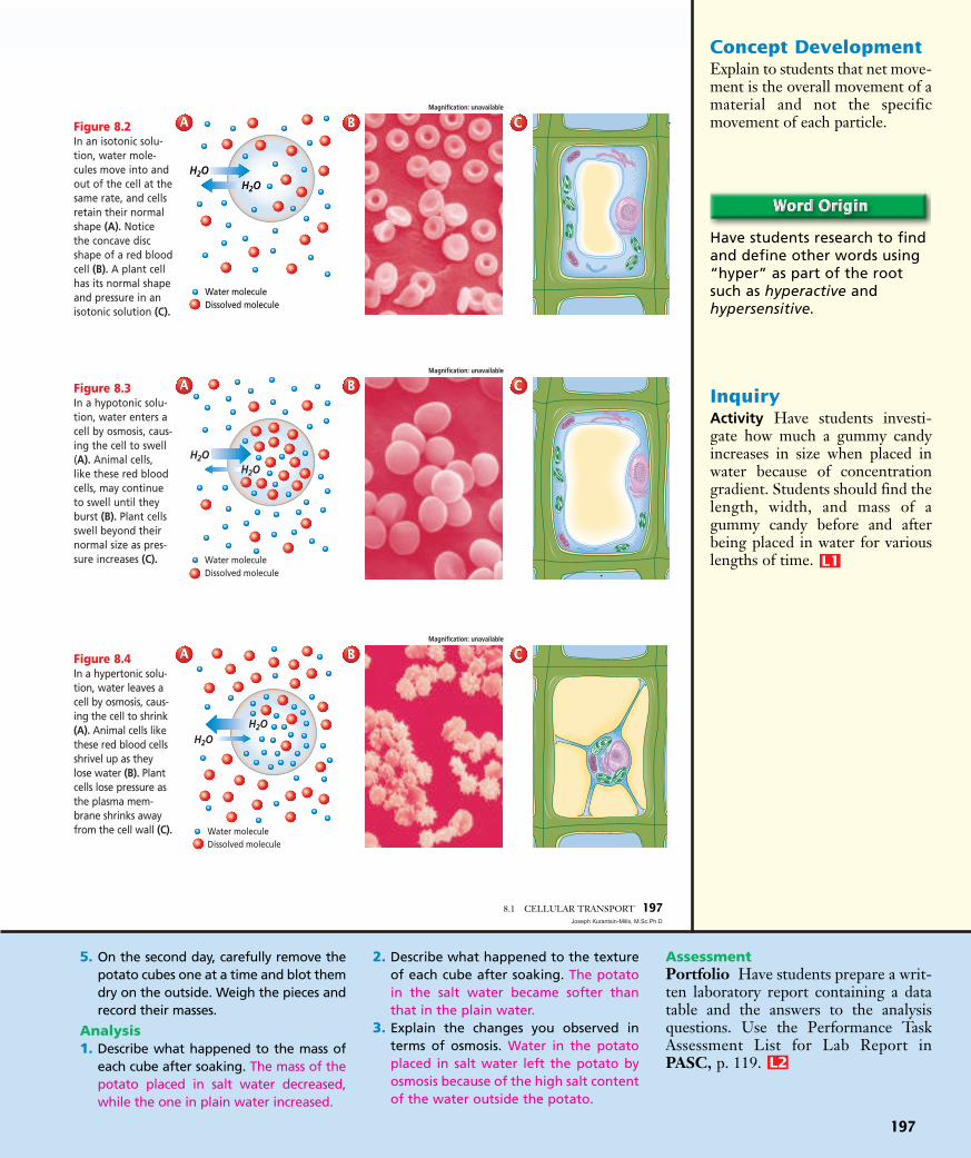
Cells in an isotonic solution It is important to understand how
osmosis affects cells. Most cells,whether in multicellular or unicellu-lar organisms, are subject to osmosisbecause they are surrounded by watersolutions. In an isotonic solution,the concentration of dissolved sub-stances in the solution is the same asthe concentration of dissolved sub-stances inside the cell. Likewise, theconcentration of water in the solutionis the same as the concentration ofwater inside the cell.
Cells in an isotonic solution doexperience osmosis, but because waterdiffuses into and out of the cells at thesame rate, the cells retain their normalshape, as shown in Figure 8.2.
Cells in a hypotonic solutionIn the hypotonic solution in
Figure 8.3A, the concentration of dissolved substances is lower in the solution outside the cell than the concentration inside the cell.Therefore, there is more water out-side the cell than inside. Cells in ahypotonic solution experience osmo-sis. Water moves through the plasmamembrane into the cell. The cellswells and its internal pressureincreases.
As the pressure increases inside animal cells, the plasma membraneswells, like the red blood cells shownin Figure 8.3B. If the solution isextremely hypotonic, the plasma mem-brane may be unable to withstand thispressure and may burst.
Because plant cells contain a rigidcell wall that supports the cell, theydo not burst when in a hypotonicsolution. As the pressure increasesinside the cell, the plasma membraneis pressed against the cell wall, asshown in Figure 8.3C. Instead ofbursting, the plant cell becomes morefirm. Grocers keep produce lookingfresh by misting the fruits and vegeta-bles with water.
Cells in a hypertonic solutionIn a hypertonic solution, the
concentration of dissolved sub-stances outside the cell is higher thanthe concentration inside the cell.Cells in a hypertonic solution experi-ence osmosis that causes water toflow out.
Animal cells in a hypertonic solu-tion shrivel because of decreasedpressure in the cells.
196 CELLULAR TRANSPORT AND THE CELL CYCLE
Water moleculeSugar molecule
Selectivelypermeablemembrane
Before osmosis After osmosis
Figure 8.1During osmosis, water diffuses across a selectively permeable membrane.Notice that the number of sugar molecules did not change on each sideof the membrane, but the number of water molecules on either side ofthe membrane did change.
iso-, hypo-, hyper-from the Greekwords isos, mean-ing “equal,” hypo,meaning “under,”and hyper, mean-ing “over,” respectively.
Jiggling MoleculesDemonstrate Brownianmovement using a micro-projector and a very dilutesolution of India ink. Theeffects of collisions of waterand ink molecules (jiggling)can be seen when you placea wet mount over a lightbulb and quickly refocus theslide. Students should beable to see increased kineticenergy with increased tem-perature.
2 Teach
Pages 196–197: Biology/Life Sciences 1a
Concept DevelopmentExplain to students that net move-ment is the overall movement of amaterial and not the specificmovement of each particle.
Have students research to findand define other words using“hyper” as part of the rootsuch as hyperactive and hypersensitive.
InquiryActivity Have students investi-gate how much a gummy candyincreases in size when placed inwater because of concentrationgradient. Students should find thelength, width, and mass of agummy candy before and afterbeing placed in water for variouslengths of time. L1
P
5. On the second day, carefully remove thepotato cubes one at a time and blot themdry on the outside. Weigh the pieces andrecord their masses.
Analysis1. Describe what happened to the mass of
each cube after soaking. The mass of thepotato placed in salt water decreased,while the one in plain water increased.
2. Describe what happened to the textureof each cube after soaking. The potatoin the salt water became softer thanthat in the plain water.
3. Explain the changes you observed interms of osmosis. Water in the potatoplaced in salt water left the potato byosmosis because of the high salt contentof the water outside the potato.
AssessmentPortfolio Have students prepare a writ-ten laboratory report containing a datatable and the answers to the analysisquestions. Use the Performance TaskAssessment List for Lab Report inPASC, p. 119. L2
P
197
8.1 CELLULAR TRANSPORT 197Joseph Kurantsin-Mills, M.Sc.Ph.D
Water moleculeDissolved molecule
H2OH2O
Figure 8.2In an isotonic solu-tion, water mole-cules move into andout of the cell at thesame rate, and cellsretain their normalshape (A). Noticethe concave discshape of a red bloodcell (B). A plant cellhas its normal shapeand pressure in anisotonic solution (C).
Figure 8.3In a hypotonic solu-tion, water enters acell by osmosis, caus-ing the cell to swell(A). Animal cells,like these red bloodcells, may continueto swell until theyburst (B). Plant cellsswell beyond theirnormal size as pres-sure increases (C).
Figure 8.4In a hypertonic solu-tion, water leaves acell by osmosis, caus-ing the cell to shrink(A). Animal cells likethese red blood cellsshrivel up as theylose water (B). Plantcells lose pressure asthe plasma mem-brane shrinks awayfrom the cell wall (C).
H2OH2O
Water moleculeDissolved molecule
H2OH2O
Water moleculeDissolved molecule
Magnification: unavailable
Magnification: unavailable
AA BB CC
AA BB CC
Magnification: unavailable
AA BB CC

198
PurposeStudents will determine if a plasticmembrane is selectively permeable.
Process Skillsformulate models, draw conclu-sions, observe and infer, recog-nize cause and effect
Safety PrecautionsHave students wear gloves,aprons, and goggles. Remind stu-dents to rinse immediately ifiodine gets on skin or clothing. Ifiodine gets in eyes, rinse thor-oughly at the eyewash station.
Teaching Strategies■ Allow students to work in
teams of two or three.■ See p. 18T of the Teacher
Guide for preparation of starchand iodine solutions. Light-weight, inexpensive bags workbest for this lab.
Expected ResultsThe inside of the bag will be pur-ple, indicating passage of iodineinto the bag. The outside of thebag will be a rust color, indicatingstarch did not pass out of the bag.
Analysis1. start—starch was clear, iodine
was rust; end—starch waspurple, iodine was rust
2. iodine; The liquid inside thebag changes color.
3. Students may suggest that theplastic bag is an adequatemodel because it allowsiodine to cross, but not thestarch. Others may suggestthat real membranes are morecomplex in their response.Both observations are correct.
AssessmentSkill Ask students to make a dia-gram depicting pore size of theplastic membrane in relation tomolecule size of iodine andstarch. Use the Performance TaskAssessment List for ScientificDrawing in PASC, p. 127. L2
P
Plant cells in a hypertonic environ-ment lose water, mainly from the cen-tral vacuole. The plasma membraneand cytoplasm shrink away from thecell wall, as shown in Figure 8.4C. Lossof water in a plant cell results in a dropin pressure and explains why plants wilt.
Passive TransportSome molecules, like water, can pass
through the plasma membrane by sim-ple diffusion, as shown in Figure 8.5A.The cell uses no energy to move theseparticles; therefore, this movement ofparticles across the membrane is classi-fied as passive transport. You caninvestigate passive transport by per-forming the MiniLab on this page.
Passive transport by proteinsRecall that transport proteins help
substances move through the plasmamembrane. Passive transport of mate-rials across the membrane using trans-port proteins is called facilitateddiffusion.
Some transport proteins, calledchannel proteins, form channels thatallow specific molecules to flowthrough, as illustrated in Figure 8.5B.
198 CELLULAR TRANSPORT AND THE CELL CYCLEKS Studio
Figure 8.5Passive transportcan occur by (A)simple diffusion,(B) facilitated dif-fusion by channelproteins, and (C)facilitated diffusionby carrier proteins.
Plasmamembrane
Concentrationgradient
Plasmamembrane
Concentrationgradient
Carrier proteins
Step 1 Step 2
Plasmamembrane
Concentrationgradient
Channelproteins
AA BB
CC
Formulate ModelsCell Membrane Simulation In thisexperiment, a plastic bag is used tomodel a selectively permeable mem-brane. Starch is placed inside of thebag. When iodine and starch mole-cules come in contact with oneanother, a dark purple color results.
Procedure! Fill a plastic bag with 50 mL of
starch. Seal the bag with a twist tie.
@ Fill a beaker with 50 mL of iodine solution. CAUTION:Rinse with water if iodine gets on skin. Iodine is toxic.
# Note and record the color of the starch and iodine.$ Place the bag into the beaker. CAUTION: Wash your hands
with soap after handling lab materials.% Note and record the color of the starch and iodine
24 hours later.
Analysis1. Describe Compare the color of the iodine and starch at
the start and at the conclusion of the experiment.2. Observe Which molecules crossed the membrane? What
is your evidence?3. Think Critically Evaluate whether or not a plastic bag is
an adequate model of a selectively permeable membrane.
Learning Disabled: Visual-Spatial Usingcolored chalk, draw a U-tube on thechalkboard similar to the “before osmo-sis” diagram in Figure 8.1. Draw mole-cules in at least two colors, showing onethat cannot cross the selectively perme-able membrane. Have students make a
similar diagram using colored pencils.Challenge students to diagram the“after osmosis” stage. Walk around theroom, checking to see if students under-stand the concept of osmosis as theydraw. Help students as needed.
P
ELLL1
P

199
Passive trans-port involves moving mole-cules with the concentrationgradient from an area ofhigher concentration to anarea of lower concentration,and requires no energy fromthe cell. Active transportallows molecules to moveagainst the concentrationgradient from an area oflower concentration to anarea of higher concentra-tion, and does require ener-gy from the cell.
Caption Question Answer Figure 8.6 Molecules tend tomove from higher to lower con-centration. Active transportreverses the trend requiringenergy input.
EnrichmentKinesthetic Channel proteinsare often called ion channels. Ionchannels have “gates” that openbriefly in response to certain con-ditions such as mechanical stimu-lation. Using this information,help students create a modeldescribing the gate analogy anddemonstrate the transport of mol-ecules through channel proteins.
The movement is with the concentra-tion gradient, and requires no energyinput from the cell.
Carrier proteins are another typeof transport protein. Carrier proteinschange shape to allow a substance topass through the plasma membrane,as shown in Figure 8.5C. In facili-tated diffusion by carrier protein, themovement is with the concentrationgradient and requires no energy inputfrom the cell.
Active TransportA cell can move particles from a
region of lower concentration to aregion of higher concentration, but itmust expend energy to counteract theforce of diffusion that is moving theparticles in the opposite direction.Movement of materials through amembrane against a concentrationgradient is called active transportand requires energy from the cell.
How active transport occursIn active transport, a transport
protein called a carrier protein firstbinds with a particle of the substanceto be transported. In general, eachtype of carrier protein has a shapethat fits a specific molecule or ion.When the proper molecule bindswith the protein, chemical energy
allows the cell to change the shape ofthe carrier protein so that the particleto be moved is released on the otherside of the membrane, something likethe opening of a door. Once the par-ticle is released, the protein’s originalshape is restored, as illustrated inFigure 8.6. Active transport allowsparticle movement into or out of acell against a concentration gradient.
Transport of substances across thecell membrane is required for cells tomaintain homeostasis. The types oftransport are summarized in Table 8.1.
Compare and contrast active and passive trans-port across the cell membrane.
8.1 CELLULAR TRANSPORT 199
Carrier proteins
Cellularenergy
Step 1 Step 2
Plasmamembrane
Concentrationgradient
+
Table 8.1 Transport Through the Cell Membrane
Type of Transport Direction of Requires Energy ClassificationTransport Protein Used? Movement Input from Cell? of Transport
Simple No With No PassiveDiffusion concentration
gradient
Facilitated Yes—channel With No PassiveDiffusion proteins or concentration
carrier proteins gradient
Active Yes—carrier Against Yes ActiveTransport proteins concentration
gradient
Figure 8.6Carrier proteins are used in active transport to pickup ions or molecules from near the cell membrane,carry them across the membrane, and release themon the other side. Think Critically Why does activetransport require energy?
Channel Proteins Students whoneed an additional challenge canresearch the channel proteins in cys-tic fibrosis. Have them create amodel that explains normal chlorideion transport through channel pro-teins and abnormal transport in thedisease. L3
P
Pages 198–199: Biology/Life Sciences 1aInv. & Exp. 1a

3 Assess
Knowledge Place a recipe formaking pickles on an overhead.Ask students to explain in theirjournals the role of osmosis inmaking pickles. When cucum-bers are placed in salty brine,the water inside the cucumberflows out of the cucumber andinto the salty brine by osmosis.This occurs because of the ten-dency of substances to flowfrom areas of higher concentra-tion to areas of lower concen-tration. In this case, the saltybrine has a lower water concen-tration. So, water will flow outof the cucumber and into thesalty brine. L1
P
AssessmentAssessmentMODIFIED
1. The concentration of water on eitherside of the membrane and the perme-ability of the membrane.
2. In an animal cell, the extra water maycause the plasma membrane to burst.In a plant cell, the plasma membranepushes against the cell wall, providingadded support.
3. Facilitated diffusion and active trans-port use carrier proteins. Facilitateddiffusion does not require energy;active transport does.
4. Carrier proteins move substances thatcannot diffuse through the plasmamembrane from an area of higher tolower concentration.
5. The organism is in a hypotonic envi-ronment and the concentration gradi-ent is from outside to inside.
6. Increasing temperature will increasethe rate of osmosis, but it will notchange the final outcome because itcannot change the membrane perme-ability to other solutes.
200
Transport of LargeParticles
Some cells can take in large mole-cules, groups of molecules, or evenwhole cells. Endocytosis is a processby which a cell surrounds and takes inmaterial from its environment asshown in Figure 8.7. This materialdoes not pass directly through themembrane. Instead, it is engulfed andenclosed by a portion of the cell’splasma membrane. That portion of themembrane then breaks away, and theresulting vacuole with its contentsmoves to the inside of the cell.
Figure 8.7 also shows the reverseprocess of endocytosis, called exocy-tosis. Exocytosis is the expulsion orsecretion of materials from a cell.Cells use exocytosis to expel wastes.They also use this method to secretesubstances, such as hormones pro-duced by the cell. Because endocyto-sis and exocytosis both move massesof material, they both requireenergy.
With the various mechanisms thecell uses to transport materials in andout, cells must also have mechanismsto regulate size and growth.
Understanding Main Ideas1. What factors affect the diffusion of water
through a membrane by osmosis?
2. How do animal cells and plant cells react differently in a hypotonic solution?
3. Compare and contrast active transport and facilitated diffusion.
4. How do carrier proteins facilitate passive transportof molecules across a membrane?
Thinking Critically5. A paramecium expels water when it is in fresh-
water. What can you conclude about the concen-tration gradient in the organism’s environment?
6. Observe and Infer What effect do you think atemperature increase has on osmosis? For morehelp, refer to Observe and Infer in the SkillHandbook.
SKILL REVIEWSKILL REVIEW
200 CELLULAR TRANSPORT AND THE CELL CYCLE
Exocytosis
Nucleus
Wastes
Digestion
Endocytosis
Figure 8.7Some unicellular organisms ingest food byendocytosis and release wastes or cell prod-ucts from a vacuole by exocytosis.
endo-, exo- fromthe Greek wordsendon, meaning“within,” and exo,meaning “out”;Endocytosis movesmaterials into thecell; exocytosismoves materialsout of the cell.
ca.bdol.glencoe.com/self_check_quiz
Check for UnderstandingEvaluate students’ under-standing of the followingterms: passive transport, activetransport, diffusion, facilitateddiffusion, isotonic, hypotonic,and hypertonic. Have studentspredict the direction of move-ment between cells and solutions.
ReteachVisual-Spatial Place severalcelery sticks in salt water andseveral in tap water. Ask stu-dents to describe any changesthey observe in the celery.
ResourcesFor more practice, useReading Essentials forBiology, Section 8.1.
P
ELLL1
P
L2
P

201
BIOLOGY: The Dynamics of Life SECTION FOCUS TRANSPARENCIES
Glucose molecule
Oxygen molecule
Carbon dioxide molecule
Use with Chapter 8,Section 8.2
What materials move through this cell by diffusion?
How might increasing the size of the cell affect the cell?
11
22
SECTION FOCUS
Cop
yrig
ht ©
Gle
ncoe
/McG
raw
-Hill
, a d
ivis
ion
of T
he M
cGra
w-H
ill C
ompa
nies
, Inc
.
Transparency Diffusion andCell Size19
Unit 3 FAST FILE ResourcesMiniLab Worksheet, p. 80BioLab Worksheet, pp. 81–82Real World BioApplications, pp. 83-84Reinforcement and Study Guide in
English, pp. 86–87Reinforcement and Study Guide in
Spanish, pp. 90–91
Transparency Worksheets, pp. 96, 103–104, 109–110
Reading Essentials for Biology,Section 8.2
Laboratory Manual, pp. 45–48Section Focus Transparency 19Basic Concepts Transparency 10Reteaching Skills Transparency 13
1 FocusBellringerSection Focus Transparency 19
8.2 SECTION PREVIEWObjectivesSequence the events ofthe cell cycle.Relate the function of acell to its organization intissues, organs, and organsystems.
Review Vocabularyorganelle: the membrane-
bound structures withineukaryotic cells (p. 173)
New Vocabularychromosomechromatincell cycleinterphasemitosisprophasesister chromatidcentromerecentriolespindlemetaphaseanaphasetelophasecytokinesistissueorganorgan system
8.2 CELL GROWTH AND REPRODUCTION 201
Cell Size LimitationsThe cells that make up a multicellular organism come in a wide variety
of sizes and shapes. Some cells, such as red blood cells, measure only8 �m (micrometers) in diameter. Other cells, such as nerve cells in largeanimals, can reach lengths of up to 1 m but have small diameters. The cellwith the largest diameter is the yolk of an ostrich egg measuring 8 cm.Most living cells, however, are between 2 and 200 µm in diameter.Considering this wide range of cell sizes, why then can’t most organismsbe just one giant cell?
Diffusion limits cell sizeYou know that the plasma membrane allows nutrients to enter the
cell and wastes to leave. Within the cell, nutrients and wastes move bydiffusion.
Although diffusion is a fast and efficient process over short distances, itbecomes slow and inefficient as the distances become larger. Imagine amitochondrion at the center of a cell with a diameter of 20 cm. It wouldhave to wait months before receiving molecules entering the cell. Becauseof the slow rate of diffusion, organisms can’t be just one giant-sized cell.
What makes up your body?Using an Analogy Where do you live? This question sounds simpleenough, but it has many answers. You live at a certain address, whichis a part of a city. Many cities and towns form the state in which youlive. The states form a country. Some tasks are performed by thecountry as a whole, while others are performed by states, cities, orindividuals. In thesame way, your bodycells are parts of tis-sues, organs, organsystems, and the bodyas a whole. Compare and ContrastCells in multicellular andunicellular organismsundergo cell division.Which type of cells do youthink is more specialized?
Cell Growth andReproduction
Standard 1l Students will analyze situations and solve problems thatrequire combining and applying concepts for more than one area of science.
California Standards
Using an Analogy Compare and Contrast Cells inmulticellular organisms aremore specialized.
Biology JournalLimits to Cell Size Using whatthey know about the relationshipof surface area to the volume of anobject, ask students to write aparagraph explaining why theexistence of a single-celled giantcreature, such as the one in themovie The Blob, would be impos-sible. L2
P
Pages 200–201: Biology/Life Sciences 1a
202
DNA limits cell sizeYou have learned that the nucleus
contains blueprints for the cell’s pro-teins. Proteins are used throughoutthe cell by almost all organelles toperform critical cell functions. Butthere is a limit to how quickly theblueprints for these proteins can becopied in the nucleus and made intoproteins in the cytoplasm. The cellcannot survive unless there is enoughDNA to support the protein needs ofthe cell.
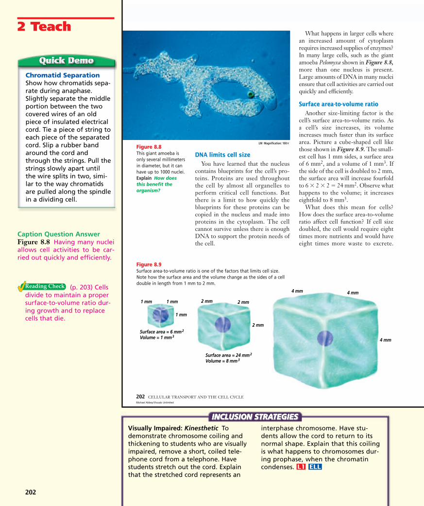
What happens in larger cells wherean increased amount of cytoplasmrequires increased supplies of enzymes?In many large cells, such as the giantamoeba Pelomyxa shown in Figure 8.8,more than one nucleus is present.Large amounts of DNA in many nucleiensure that cell activities are carried outquickly and efficiently.
Surface area-to-volume ratioAnother size-limiting factor is the
cell’s surface area-to-volume ratio. Asa cell’s size increases, its volumeincreases much faster than its surfacearea. Picture a cube-shaped cell likethose shown in Figure 8.9. The small-est cell has 1 mm sides, a surface areaof 6 mm2, and a volume of 1 mm3. Ifthe side of the cell is doubled to 2 mm,the surface area will increase fourfoldto 6 � 2 � 2 � 24 mm2. Observe whathappens to the volume; it increaseseightfold to 8 mm3.
What does this mean for cells?How does the surface area-to-volumeratio affect cell function? If cell sizedoubled, the cell would require eighttimes more nutrients and would haveeight times more waste to excrete.
202 CELLULAR TRANSPORT AND THE CELL CYCLEMichael Abbey/Visuals Unlimited
4 mm
4 mm4 mm
2 mm
2 mm2 mm
1 mm
1 mm1 mm
Surface area = 6 mm2
Volume = 1 mm3
Surface area = 24 mm2
Volume = 8 mm3
Figure 8.9Surface area-to-volume ratio is one of the factors that limits cell size.Note how the surface area and the volume change as the sides of a celldouble in length from 1 mm to 2 mm.
Figure 8.8This giant amoeba isonly several millimetersin diameter, but it canhave up to 1000 nuclei.Explain How doesthis benefit theorganism?
LM Magnification: 100�
Chromatid SeparationShow how chromatids sepa-rate during anaphase.Slightly separate the middleportion between the twocovered wires of an oldpiece of insulated electricalcord. Tie a piece of string toeach piece of the separatedcord. Slip a rubber bandaround the cord andthrough the strings. Pull thestrings slowly apart untilthe wire splits in two, simi-lar to the way chromatidsare pulled along the spindlein a dividing cell.
2 Teach
Caption Question AnswerFigure 8.8 Having many nucleiallows cell activities to be car-ried out quickly and efficiently.
(p. 203) Cellsdivide to maintain a propersurface-to-volume ratio dur-ing growth and to replacecells that die.
Visually Impaired: Kinesthetic Todemonstrate chromosome coiling andthickening to students who are visuallyimpaired, remove a short, coiled tele-phone cord from a telephone. Havestudents stretch out the cord. Explainthat the stretched cord represents an
interphase chromosome. Have stu-dents allow the cord to return to itsnormal shape. Explain that this coilingis what happens to chromosomes dur-ing prophase, when the chromatincondenses.
P
ELLL1
P

203
PurposeStudents will compare the in-crease in volume of an object withthe increase in its surface area.
BackgroundCell volume increases much fasterthan cell surface area. In cells, thisfact contributes to cell size limita-tion because cells do not have suf-ficient surface area to accommo-date the influx of nutrients to sup-port the volume of a large size.
Process Skillsmeasure in SI, use numbers, rec-ognize cause and effect, interpretdata, analyze
Teaching StrategiesAsk students if they have everwondered why cells can’t contin-ue to grow larger and larger tobecome giant cells. Then askthem to consider the fact thatmost cells, whether from an ele-phant or an earthworm, aremicroscopic in size.
Thinking Critically1. 642. As the cell surface area grows,
its volume increases dramati-cally. More resources are need-ed by organelles and morewaste is produced.
3. As the cell grows, it reaches apoint where the surface areais not large enough to trans-port resources and wastes toallow the cell to survive.
AssessmentPerformance Students shouldwrite a summary of the MiniLab,including the Analysis questions,for their journals. Use thePerformance Task AssessmentList for Lab Report in PASC, p. 119. L2
P
8.2 CELL GROWTH AND REPRODUCTION 203
The surface area, however, wouldincrease by a factor of only four.Thus, the plasma membrane wouldnot have enough surface areathrough which oxygen, nutrients, andwastes could diffuse. The cell wouldeither starve to death or be poisonedfrom the buildup of waste products.You can investigate surface area-to-volume ratios yourself in theProblem-Solving Lab shown here.
Because cell size can have dramaticand negative effects on a cell, cellsmust have some method of maintain-ing optimum size. In fact, cells dividebefore they become too large to func-tion properly. Cell division accom-plishes other purposes, too, as youwill read next.
Cell ReproductionRecall that the cell theory states
that all cells come from preexistingcells. Cell division is the process bywhich new cells are produced fromone cell. Cell division results in twocells that are identical to the origi-nal, parent cell. Right now, as youare reading this page, many of thecells in your body are growing,dividing, and dying. Old cells on the soles of your feet and on thepalms of your hands are being shedand replaced, cuts and bruises arehealing, and your intestines are pro-ducing millions of new cells eachsecond. New cells are produced astadpoles become frogs, and as an ivyvine grows and wraps around agarden trellis. All organisms growand change; worn-out tissues arerepaired or are replaced by newlyproduced cells.
Explain two reasonswhy cell division is a required cellprocess.
The discovery of chromosomesEarly biologists observed that just
before cell division, several short,stringy structures suddenly appearedin the nucleus. Scientists also noticedthat these structures seemed to vanishsoon after division of a cell. Thesestructures, which contain DNA andbecome darkly colored when stained,are called chromosomes (KROH muhsohmz).
Eventually, scientists learned thatchromosomes are the carriers of thegenetic material that is copied andpassed from generation to generationof cells. This genetic material is cru-cial to the identity of the cell.Accurate transmission of chromo-somes during cell division is critical.
chromosome fromthe Greek wordschroma, meaning“colored,” andsoma, meaning“body”; Chromo-somes are dark-staining structuresthat containgenetic material.
Draw ConclusionsWhat happens to the surface area of a cell as its volumeincreases? One reason cells are small is that they need alarge surface area as compared to volume so nutrients can dif-fuse in and wastes can diffuse out.
Solve the Problem Look at the cubes shown below. Note the size and magnitudeof difference in surface area and volume.
Thinking Critically1. Estimate How many small cubes (1 mm) do you think it
would take to fill the largest cube (4 mm)?2. Use Models Using the cubes as models, describe how a
cell is affected by its size. 3. Infer Explain how a small change in cell size can have a
huge impact on cellular processes.
4 mm4 mm
4 mm
2 mm
2 mm
2 mm1 mm
1 mm
1 mm
Surface area � 6 mm2
Volume � 1 mm3Surface area � 24 mm2
Volume � 8 mm3
Surface Area Using a few smallboxes, one large box (approximatelythe same size as the small boxes com-bined), and wrapping paper, ask thestudents to figure out which set willneed more paper, the large box orthe small boxes each wrapped sepa-rately. L1
P

204
PurposeStudents will compare cell cyclesof two different types of cells.
BackgroundMitosis requires the same relativeamount of time no matter whattype of cell is dividing. Theamount of time spent in inter-phase determines the length ofthe cell cycle.
Process Skillscompare and contrast, interpretdata, analyze
Teaching StrategiesRelate this lab to the uncontrolledgrowth of cancer cells, whichspend a very short time in inter-phase.
Thinking Critically1. The first part of interphase, in
which the cell is growing, isthe most variable in length.
2. The cell with the longer peri-od of growth would carry onmore metabolic activitiesthan the more rapidly divid-ing cell.
3. Students should justify theiranswers with statements suchas that certain types of cellsare always being damagedand need to be replaced.
Modified AssessmentPerformance Have studentswrite three questions related tothis lab in their journals. Use thePerformance Task AssessmentList for Asking Questions inPASC, p. 91. L1
P
The structure of eukaryotic chromosomes
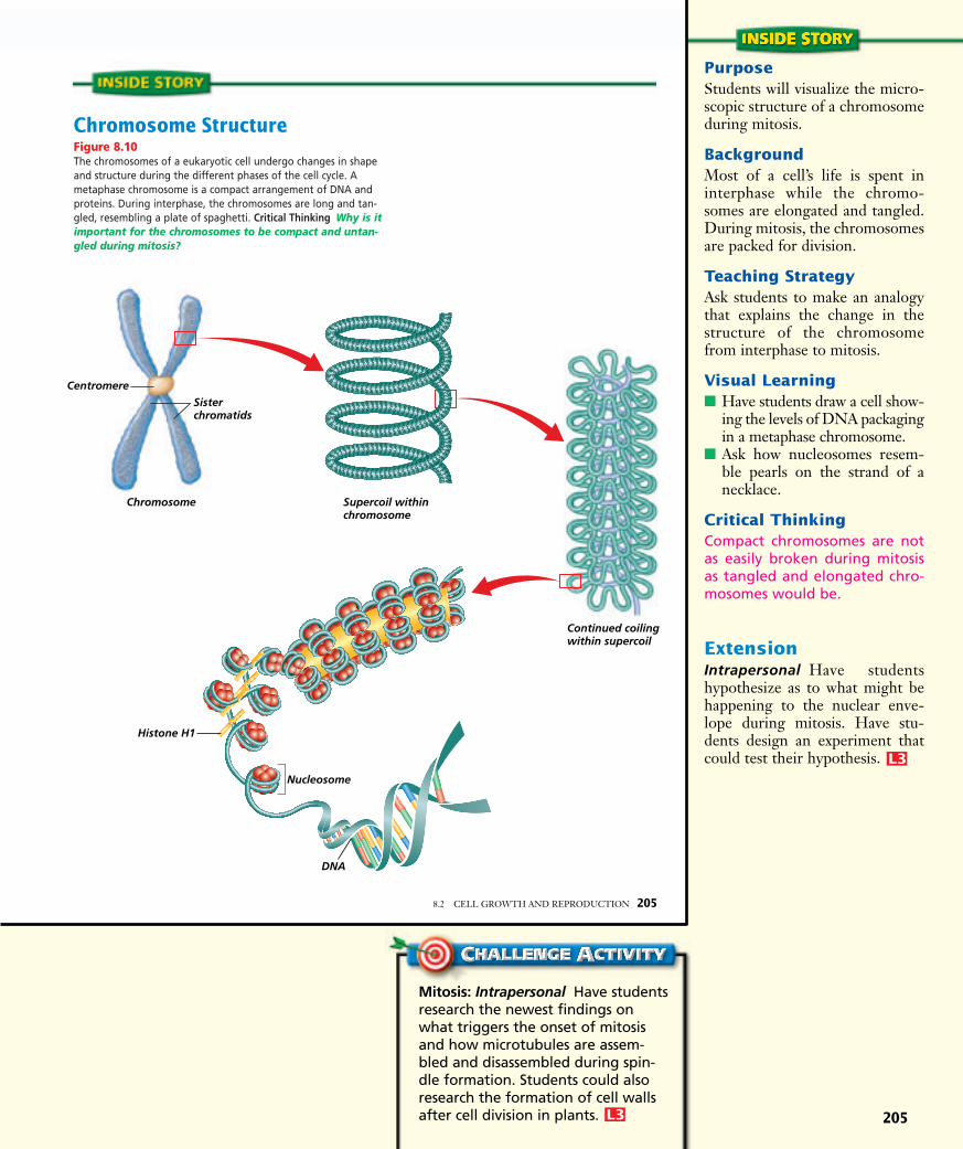
For most of a cell’s lifetime, chro-mosomes exist as chromatin, longstrands of DNA wrapped around
proteins called histones. Under anelectron microscope, chromatinlooks like beads on a string. Eachbead is a group of histones called anucleosome. Before a cell can divide,the long strands of chromatin mustbe reorganized, just as you would coila long strand of rope before storingit. As the nucleus begins to divide,chromosomes take on a differentstructure in which the chromatinbecomes tightly packed. Look atFigure 8.10 for more information onchromosome structure.
The Cell CycleFall follows summer, night follows
day, and low tide follows high tide.Many events in nature follow a recur-ring, cyclical pattern. Living organismsare no exception. One cycle commonto most living things is the cycle of thecell. The cell cycle is the sequence ofgrowth and division of a cell.
As a cell proceeds through its cycle,it goes through two general periods: aperiod of growth and a period of divi-sion. The majority of a cell’s life isspent in the growth period known asinterphase. During interphase, a cellgrows in size and carries on metabo-lism. Also during this period, chromo-somes are duplicated in preparationfor the period of division.
Following interphase, a cell entersits period of nuclear division calledmitosis (mi TOH sus). Mitosis is theprocess by which two daughter cellsare formed, each containing a com-plete set of chromosomes. Interphaseand mitosis make up the bulk of thecell cycle. Following mitosis, thecytoplasm divides, separating the twodaughter cells. You can use theProblem-Solving Lab on this page andthe BioLab at the end of this chapterto investigate the rate of mitosis.
204 CELLULAR TRANSPORT AND THE CELL CYCLE
Observe and InferHow does the length of the cell cycle vary? The cell cyclevaries greatly in length from one kind of cell to another. Somekinds of cells divide rapidly, while others divide more slowly.
Solve the ProblemExamine the cell cycle diagrams of two different types of cells.Observe the total length of each cell cycle and the length oftime each cell spends in each phase of the cell cycle.
Thinking Critically1. Make and Use Graphs Which part of the cell cycle is
most variable in length? 2. Infer What can you infer about the functions of these
two types of cells? 3. Think Critically Why do you think the cycle of some
types of cells is faster than in others? Explain your answer.
7 hours 3 hours
Mitosis1 hour
11 hours
Interphase
Total = 22 hours
37 hours
7 hours
Mitosis1 hourInterphase
Total = 48 hours
3 hours
Jane Cooke Wright Discuss with stu-dents the role of Jane Cooke Wright inthe development of chemotherapytechniques to treat cancer. Wright’swork in the 1950s and 1960s involvedtesting various anticancer drugs on peo-ple with different kinds of cancer. In the1970s, Wright found that examinations
of cancer cells grown in tissue culturecould help predict which drugs wouldbe most effective against that type ofcancer. Since that time, Wright has beenan active publisher of work in the field,and in 1975 she was honored by theAmerican Association for CancerResearch for her contributions.
Pages 204–205: Inv. & Exp. 1i
PurposeStudents will visualize the micro-scopic structure of a chromosomeduring mitosis.
BackgroundMost of a cell’s life is spent ininterphase while the chromo-somes are elongated and tangled.During mitosis, the chromosomesare packed for division.
Teaching StrategyAsk students to make an analogythat explains the change in thestructure of the chromosomefrom interphase to mitosis.
Visual Learning■ Have students draw a cell show-
ing the levels of DNA packagingin a metaphase chromosome.
■ Ask how nucleosomes resem-ble pearls on the strand of anecklace.
Critical ThinkingCompact chromosomes are notas easily broken during mitosisas tangled and elongated chro-mosomes would be.
ExtensionIntrapersonal Have studentshypothesize as to what might behappening to the nuclear enve-lope during mitosis. Have stu-dents design an experiment thatcould test their hypothesis. L3
P
205
Histone H1
Nucleosome
DNA
Centromere
Sisterchromatids
Chromosome Supercoil within chromosome
Continued coilingwithin supercoil
Chromosome StructureFigure 8.10The chromosomes of a eukaryotic cell undergo changes in shapeand structure during the different phases of the cell cycle. Ametaphase chromosome is a compact arrangement of DNA andproteins. During interphase, the chromosomes are long and tan-gled, resembling a plate of spaghetti. Critical Thinking Why is itimportant for the chromosomes to be compact and untan-gled during mitosis?
8.2 CELL GROWTH AND REPRODUCTION 205
Mitosis: Intrapersonal Have studentsresearch the newest findings onwhat triggers the onset of mitosisand how microtubules are assem-bled and disassembled during spin-dle formation. Students could alsoresearch the formation of cell wallsafter cell division in plants. L3
P

206
EnrichmentKinesthetic Have students workin groups to design models of thefour stages of mitosis. Studentsmight use colored macaroni orlicorice for chromosomes. Havestudents include their completedmodels in their portfolios.
The BioLab atthe end of thechapter can beused at thispoint in the lesson.
PortfolioCell Cycle The average humancell that is capable of dividing hasa cell cycle of 20 hours. Have stu-dents calculate how many cellsthere would be in one week if theystart with one cell at time zero.Students should show their workand place the pages in their port-folios. There are 168 hours inone week. If a cell divides every20 hours, there would be 256cells at the end of one week. At20 hours, there would be 2 cells;40 hours, 4 cells; 60 hours, 8 cellsand so on, until 160 hours whenthere would be 256 cells. L2
P
P
COOP LEARN
P
ELLL1
P
Interphase: A Busy Time
Interphase, the busiest phase of thecell cycle, is divided into three partsas shown in Figure 8.11. During thefirst part, the cell grows and proteinproduction is high. In the next part ofinterphase, the cell copies its chro-mosomes. DNA synthesis does notoccur all through interphase but is confined to this specific time. After the chromosomes have been
duplicated, the cell enters anothershorter growth period in which mito-chondria and other organelles aremanufactured and cell parts neededfor cell division are assembled.Following this activity, interphaseends and mitosis begins.
The Phases of MitosisCells undergo mitosis as they
approach the maximum cell size atwhich the nucleus can provide blue-prints for proteins, and the plasmamembrane can efficiently transportnutrients and wastes into and out ofthe cell.
Although cell division is a continu-ous process, biologists recognize fourdistinct phases of mitosis—each phasemerging into the next. The four phasesof mitosis are prophase, metaphase,anaphase, and telophase. Refer toFigure 8.13 to help you understandthe process as you read about mitosis.
Prophase: The first phase of mitosis
During prophase, the first andlongest phase of mitosis, the long,stringy chromatin coils up into visiblechromosomes. As you can see inFigure 8.12, each duplicated chro-mosome is made up of two halves.The two halves of the doubled struc-ture are called sister chromatids.Sister chromatids and the DNA theycontain are exact copies of each otherand are formed when DNA is copiedduring interphase. Sister chromatidsare held together by a structure calleda centromere, which plays a role inchromosome movement during mito-sis. By their characteristic location,centromeres also help scientists iden-tify and study chromosomes.
As prophase continues, the nucleusbegins to disappear as the nuclear enve-lope and the nucleolus disintegrate.
206 CELLULAR TRANSPORT AND THE CELL CYCLE
DNA synthesisand replication
Rapid growthand metabolic activity
Centrioles replicate; cell preparesfor division
Interphase
Mitosis
CytokinesisFigure 8.11In preparation formitosis, most of thetime spent in the cellcycle is in interphase.The process of mitosis,represented here bythe yellow wedge, isshown in detail inFigure 8.13.
Sister chromatids
Centromere
Figure 8.12The two sister chromatidsare held together by acentromere.
Mitosis Use models of plant or ani-mal cell mitosis (available from bio-logical supply houses) to visualize andreinforce the discussion of mitosis. L1
P
Pages 206–207: Inv. & Exp. 1i

207
DiscussionLinguistic Discuss the details ofthe timing mechanism of the cellcycle with the class.
The DNA synthesis and repli-cation phase of interphase andmitosis may be triggered by anenzyme called “cdc2 kinase.”Another protein called “cyclin” isalso involved in cell division con-trol. In a new cell, cyclin is syn-thesized continuously and once itbuilds up to a critical level, it linksup with cdc2 kinase protein toform a complex.
Several other biochemicalsteps then serve to activate thecdc2–cyclin complex, transform-ing it into maturation-promotingfactor (MPF). Mitosis then takesplace.
Now ask the students to writeabout another process (biologicalor otherwise) and the timingmechanism that governs the stepsor phases of the process. Studentsshould include a discussion ofwhat happens if one portion ofthe process breaks down. L3
P
8.2 CELL GROWTH AND REPRODUCTION 207(t ct b)Ed Reschke/Peter Arnold, Inc., (cb)Carolina Biological Supply/Phototake, NYC
Figure 8.13Mitosis begins after interphase. Follow the stagesof mitosis as you read the text. The diagrams andthe photos show mitosis in plant cells.
AnaphaseThe centromeressplit and the sisterchromatids arepulled apart toopposite poles ofthe cell.
CC
Stained LM Magnification: 500�
Stained LM Magnification: 360�
Spindle fibers
Disappearingnuclear envelope
Doubled chromosome
Centromere
Sister chromatids
Nuclear envelopereappears
Two daughtercells are formed
ProphaseThe chromatin coilsto form visiblechromosomes.
AA
TelophaseTwo distinctdaughter cells areformed. The cellsseparate as the cellcycle proceeds intothe next interphase.
DD
MetaphaseThe chromosomesmove to the equatorof the spindle.
BB
Stained LM Magnification: 400�
Stained LM Magnification: 640�
Biology in Practice: Interpersonal Aska cell biologist to talk to the classabout how basic cell biology is criticalto advanced research. Ask the speakerto describe how his or her investiga-tions revolve intricately around whatthe class is learning in this unit. Havethe class prepare some questions inadvance to ask the guest. L2
P
CD-ROM Students will explore the phases of a cell cycle in CellReproduction.
208
208 CELLULAR TRANSPORT AND THE CELL CYCLEBarry King, University of California, School of Medicine/Biological Photo Service
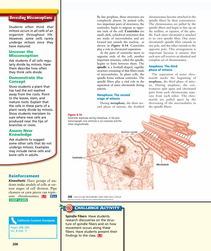
Figure 8.14Centrioles duplicate during interphase. In the pho-tomicrograph, one centriole is cut crosswise and theother longitudinally.
Microtubule
Magnification: unavailable
By late prophase, these structures arecompletely absent. In animal cells,two important pairs of structures, thecentrioles, begin to migrate to oppo-site ends of the cell. Centrioles aresmall, dark, cylindrical structures thatare made of microtubules and arelocated just outside the nucleus, asshown in Figure 8.14. Centriolesplay a role in chromatid separation.
As the pairs of centrioles move toopposite ends of the cell, anotherimportant structure, called the spindle,begins to form between them. Thespindle is a football-shaped, cagelikestructure consisting of thin fibers madeof microtubules. In plant cells, thespindle forms without centrioles. Thespindle fibers play a vital role in theseparation of sister chromatids duringmitosis.
Metaphase: The second stage of mitosis
During metaphase, the short sec-ond phase of mitosis, the doubled
chromosomes become attached to thespindle fibers by their centromeres.The chromosomes are pulled by thespindle fibers and begin to line up onthe midline, or equator, of the spin-dle. Each sister chromatid is attachedto its own spindle fiber. One sisterchromatid’s spindle fiber extends toone pole, and the other extends to theopposite pole. This arrangement isimportant because it ensures thateach new cell receives an identical andcomplete set of chromosomes.
Anaphase: The third phase of mitosis
The separation of sister chro-matids marks the beginning ofanaphase, the third phase of mito-sis. During anaphase, the cen-tromeres split apart and chromatidpairs from each chromosome sepa-rate from each other. The chro-matids are pulled apart by theshortening of the microtubules inthe spindle fibers.
Centriole
Students often think thatmitosis occurs in all cells of anorganism throughout life.However, some cells rarelyundergo mitosis once theyhave matured.
Uncover theMisconceptionAsk students if all cells regu-larly divide by mitosis. Havethem describe how oftenthey think cells divide.
Demonstrate theConceptShow students a plant thathas had the soil washedaway from the roots. Pointto the leaves, stem, andmature roots. Explain thatthe cells in these parts of aplant rarely divide by mitosis.Show students meristem tis-sues where new cells areproduced near the tips ofbranches or roots.
Assess NewKnowledgeAsk students to suggestsome other cells that do notundergo mitosis. Examplesmay include nerve cells andbone cells in adults.
ReinforcementKinesthetic Have groups of stu-dents make models of cells at var-ious stages of cell division. Pipecleaners or yarn pieces can repre-sent chromosomes.
P
COOP LEARN
P
ELLL1
P
Spindle Fibers Have studentsresearch discoveries on the struc-ture of spindle fibers and on howmovement occurs along thesefibers. Have students present theirfindings to the class. L3
P
Pages 208–209: Inv. & Exp. 1i

209
Telophase: The fourth phase of mitosis
The final phase of mitosis istelophase. Telophase begins as thechromatids reach the opposite poles ofthe cell. During telophase, many of thechanges that occurred during prophaseare reversed as the new cells prepare fortheir own independent existence. Thechromosomes, which had been tightlycoiled since the end of prophase, nowunwind so they can begin to direct themetabolic activities of the new cells.The spindle begins to break down, thenucleolus reappears, and a new nuclearenvelope forms around each set of chro-mosomes. Finally, a new double mem-brane begins to form between the twonew nuclei.
CytokinesisFollowing telophase, the cell’s cyto-
plasm divides in a process calledcytokinesis (si toh kih NEE sus).Cytokinesis differs between plants andanimals. Toward the end of telophasein animal cells, the plasma membranepinches in along the equator as shownin Figure 8.15. As the cell cycle pro-ceeds, the two new cells are separated.Find out more about mitosis in animalcells in the MiniLab.
Plant cells have a rigid cell wall, so the plasma membrane does notpinch in. Rather, a structure known asthe cell plate is laid down across thecell’s equator. A cell membrane formsaround each cell, and new cell wallsform on each side of the cell plate untilseparation is complete.
8.2 CELL GROWTH AND REPRODUCTION 209(t)Ed Reschke/Peter Arnold, Inc., (b)David M. Phillips/Visuals Unlimited
Compare and ContrastSeeing Asters The result of theprocess of mitosis is similar in plantand animal cells. However, animalcells develop structures calledasters that are thought to serve asa brace for the spindle fibers,while plant cells do not developasters.
Procedure! Examine a slide showing fish mitosis
under low- and high-power magnification. CAUTION: Use care when handling prepared slides.
@ Find cells that are undergoing mitosis. You will be able tosee dark-stained rodlike structures within certain cells.These structures are chromosomes.
# Note the appearance and location of asters. They willappear as ray or starlike structures at opposite ends ofcells that are in metaphase.
$ Asters may also be observed in cells that are in otherphases of the cell cycle.
Analysis1. Describe What is the location of asters in cells that are in
prophase?2. Infer How do you know that asters are not critical to
mitosis?3. Use Models Sketch and label a plant cell and an animal
cell in prophase.
Asters
Stained LM Magnification: 250�
Figure 8.15The furrow, created when proteins positioned underthe plasma membrane at the equator of this frogcell contracted and slid past each other, will deepenuntil the cell is pinched in two.
Color-enhanced SEM Magnification: 25�
Learning Disabled To help studentsbetter understand the phases of mito-sis, have them research the prefixesinter, pro, meta, ana, and telo. Havethem relate the meanings of the pre-fixes to the phases of mitosis. L1
P
PurposeStudents will observe animal cellsthat are undergoing mitosis andwill note the location and appear-ance of the aster.
Process Skillscompare and contrast, observeand infer, critical thinking
Teaching Strategies■ Advise students that the slide
material is taken from fish blas-tulas. Ask students why thismaterial is ideal for the study ofcells undergoing mitosis.
■ To reduce cost of purchasing aclass set of prepared slides,purchase one slide and use amicrovideo camera or a micro-projector if available. Altern-atively, purchase 35 mm slidesand project them onto thescreen with a slide projector.
■ If necessary, review the stagesof mitosis with students.
Analysis1. Asters are starlike projections
of microtubules associatedwith centrioles. Asters arefound at the cell poles inprophase.
2. Asters are not critical becauseplant cells undergo mitosiswithout the structures.
3. Sketches should show thechromatin coils forming visi-ble chromosomes. Asters canbe included for the animalcell.
AssessmentPortfolio Have students researchthe role and function of the asterand summarize the scientific opin-ion in their portfolios. Use thePerformance Task Assessment Listfor Writing in Science in PASC,p. 159. L3
P

3 Assess
1. As volume increases, surface area doesnot increase sufficiently to supportlarge cells.
2. Each daughter cell must get an identi-cal copy of the set of chromosomes.
3. Cells are the basic level of organiza-tion. Similar cells that perform thesame function make up tissues. Organs
are made up of tissues. Several organsmake up an organ system.
4. Possible answers include tissues, organs,organ systems.
5. The cell is not resting, but growing, producing proteins, and replicating its chromosomes in preparation fordivision.
6. Interphase: chromosomes are copied;prophase: nuclear envelope disappears;metaphase: chromosomes line up; ana-phase: chromatids separate; telophase:chromosomes move to the poles
210
Understanding Main Ideas1. Describe how a cell’s surface area-to-volume ratio
limits its size.
2. Why is it necessary for a cell’s chromosomes to bedistributed to its daughter cells in such a precisemanner?
3. Relate cells to each level of organization in a multicellular organism.
4. In multicellular organisms, describe two cellularspecializations that result from mitosis.
Thinking Critically5. At one time, interphase was referred to as the
resting phase of the cell cycle. Why do you thinkthis description is no longer used?
6. Get the Big Picture Make a table sequencingthe phases of the cell cycle. Mention one impor-tant event that occurs at each phase. For morehelp, refer to Get the Big Picture in the SkillHandbook.
SKILL REVIEWSKILL REVIEW
210 CELLULAR TRANSPORT AND THE CELL CYCLE
Tissue (muscle tissue)
Cell (muscle cell)
Organ (stomach)
Organ system (digestive system)
Organism (Florida panther)
Figure 8.16Cells of complex multicellular organisms are organized into tissues,organs, and organ systems. Sequence What levels of organizationis a human blood cell a part of?
Results of MitosisMitosis is a process that guarantees
genetic continuity, resulting in theproduction of two new cells withchromosome sets that are identical tothose of the parent cell. These newdaughter cells will carry out the samecellular processes and functions asthose of the parent cell and will growand divide just as the parent cell did.
When mitosis is complete, unicellu-lar organisms remain as single cells—the organism simply multiplied. Inmulticellular organisms, cell growthand reproduction result in groups ofcells that work together as tissue toperform a specific function. Tissuesorganize in various combinations toform organs that perform more com-plex roles within the organism. Forexample, cells make up muscle tissue,
then muscle tissue works with othertissues in the organ called the stomachto mix up food. Multiple organs thatwork together form an organ system.The stomach is one organ in the diges-tive system, which functions to breakup and digest food.
All organ systems work togetherfor the survival of the organism,whether the organism is a fly or ahuman. Figure 8.16 shows an exam-ple of cell specialization and organiza-tion for a complex organism. Inaddition to its digestive system, thepanther has a number of other organsystems that have developed throughcell specialization. It is important toremember that no matter how com-plex the organ system or organismbecomes, the cell is still the mostbasic unit of that organization.
ca.bdol.glencoe.com/self_check_quiz
Check for Understanding Test the students’ ability torecognize the various phasesof mitosis. Place photomicro-graphs on an overhead pro-jector and ask the class toidentify each stage.
ReteachVisual-Spatial Review thephases of mitosis, emphasiz-ing that the process is contin-uous and that one phaseblends into the next. Usephotomicrographs and dia-grams to help students identifythe phases and learn theterms associated with struc-tures in mitosis.
Resources For more practice, useReading Essentials forBiology, Section 8.2.
P
ELLL2
P
L2
P
ExtensionEncourage students to researchthe stages of the cell cycle. Theymay find information on howlong each stage lasts for variousspecies and what events occur ateach stage.
Performance Call out variousstages of mitosis and have stu-dents find and show that stage totheir lab partners using onionroot slides under the microscope.Walk around the room to checktheir results.
Caption Question Answer Figure 8.16 A blood cell is apart of every level.
L1
P
AssessmentAssessment
L3
P

211
BIOLOGY: The Dynamics of Life SECTION FOCUS TRANSPARENCIES
Use with Chapter 8,Section 8.3
The weeds in this garden are spreading rapidly. Whateffect might this have on the flowers in the garden?
Suppose a change in a cell’s genes causes the cell toreproduce very rapidly. How might this increased rateof reproduction affect surrounding cells?
11
22
SECTION FOCUS
Cop
yrig
ht ©
Gle
ncoe
/McG
raw
-Hill
, a d
ivis
ion
of T
he M
cGra
w-H
ill C
ompa
nies
, Inc
.
Transparency UncontrolledCell Division20
Unit 3 FAST FILE ResourcesReinforcement and Study Guide in
English, p. 88Reinforcement and Study Guide in
Spanish, p. 92Critical Thinking/Problem Solving, p. 94Transparency Worksheets, p. 97
Section Focus Transparency 20Reading Essentials for Biology,
Section 8.3
1 FocusBellringerSection Focus Transparency 20
8.3SECTION PREVIEWObjectivesDescribe the role ofenzymes in the regulationof the cell cycle.Distinguish between theevents of a normal cellcycle and the abnormalevents that result in cancer.Identify ways to poten-tially reduce the risk ofcancer.
Review Vocabularyprotein: a large complex
polymer composed ofcarbon, hydrogen, oxygen, nitrogen, andsometimes sulfur (p. 160)
New Vocabularycancergene
8.3 CONTROL OF THE CELL CYCLE 211
Normal Control of the Cell CycleWhy do some types of cells divide rapidly, while others divide slowly?
What tells a cell when it is time to leave one part of the cell cycle andbegin the next?Proteins and enzymes control the cell cycle
The cell cycle is controlled by proteins called cyclins and a set ofenzymes that attach to the cyclin and become activated. The interactionof these molecules, based on conditions both in the cell’s environmentand inside the cell, controls the cell cycle. Occasionally, cells lose controlof the cell cycle. This uncontrolled dividing of cells can result from thefailure to produce certain enzymes, the overproduction of enzymes, orthe production of other enzymes at the wrong time. Cancer is a malig-nant growth resulting from uncontrolled cell division. This loss of con-trol may be caused by environmental factors or by changes in enzymeproduction.
Enzyme production is directed by genes located on the chromosomes.A gene is a segment of DNA that controls the production of a protein.
Many studies point to the portion of interphase just before DNA repli-cation as being a key control period in the cell cycle. Scientists have iden-tified several enzymes that trigger DNA replication.
Getting ControlFinding Main Ideas As you read through the sec-tion on control of the cellcycle, answer the followingquestions.
Study Organizer 1. Enzymes control the cell
cycle. What controlsenzyme production?
2. What are two environ-mental factors that contribute to the develop-ment of cancer? List anypossible ways you caninfluence these factors.
3. How does a person’s dietrelate to the chances ofgetting cancer?
Luis M. De La Maza, PhD.M.D./Phototake, NYC
Color-enhanced SEM Magnification: 7500�
Control of the Cell Cycle
This tumor is developing due to a mistake inthe cell cycle.
Finding Main IdeasStudy Organizer1. Enzyme production is con-
trolled by feedback from thecellular environment and theDNA in the nucleus.
2. UV radiation and cancer caus-ing chemicals are possibleanswers. Avoid exposure toUV radiation from suntanbooths and sunlight. Do notsmoke.
3. Many dietary experts agreethat a diet low in fat andhigh in fiber can reduce therisk of certain kinds of cancer.Foods that contain certainvitamins and minerals alsocan reduce the risk of gettingcancer.
Pages 210–211: Inv. & Exp. 1i

212
2 Teach
PurposeStudents will analyze a graphshowing incidence of certain bodyorgan cancers.
Process Skillsinterpret data, think critically,analyze information, sequence,make and use graphs
Teaching Strategies■ Allow students to work in small
groups.■ Explain the difference between
skin melanoma and basal cellor squamous skin cancers.
■ Help student to determine per-cent survival if they are having difficulty.
Thinking Critically1. basal cell and squamous; skin
melanoma2. lung; basal cell and squamous3. calculation: new cases �
deaths � survivors; so (sur-vivors � new cases) �100 � % survival. 180 000 �40 000 � 140 000. Then (140000 � 180 000) � 100 � 78%
4. approximately nine percent
AssessmentPortfolio Have students gatherinformation on a specific type ofcancer and prepare a brief oralreport. Use the Performance TaskAssessment List for Oral Presen-tation in PASC, p. 143.
The cancermay have spread to vitalorgans and damaged thembeyond repair.
L2
P
Cancer: A Mistake in the Cell Cycle
Currently, scientists consider can-cer to be a result of changes in one ormore of the genes that produce sub-stances that are involved in control-ling the cell cycle. These changes areexpressed as cancer when somethingprompts the damaged genes intoaction. Cancerous cells form masses
of tissue called tumors that deprivenormal cells of nutrients. In laterstages, cancer cells enter the circula-tory system and spread throughoutthe body, a process called metastasis,forming new tumors that disrupt thefunction of organs, organ systems,and ultimately, the organism.
Cancer is the second leading causeof death in the United States,exceeded only by heart disease. Can-cer can affect any tissue in the body. Inthe United States, lung, colon, breast,and prostate cancers are the mostprevalent types. Use the Problem-Solving Lab on this page to estimatethe number of people in the UnitedStates who will develop these kinds ofcancers in this decade, and how manypeople are expected to die from can-cers. The Connection to Health featureat the end of this chapter further dis-cusses skin cancer.
Infer why cancer isdifficult to treat in later stages.
The causes of cancerThe causes of cancer are difficult to
pinpoint because both genetic andenvironmental factors are involved.The environmental influences of can-cer become obvious when you con-sider that people in differentcountries develop different types of cancers at different rates. Forexample, the rate of breast cancer isrelatively high in the United States,but relatively low in Japan. Similarly,stomach cancer is common in Japan,but rare in the United States.
Other environmental factors, suchas cigarette smoke, air and water pollution, and exposure to ultra-violet radiation from the sun, are allknown to damage the genes thatcontrol the cell cycle. Cancer mayalso be caused by viral infections thatdamage the genes.
212 CELLULAR TRANSPORT AND THE CELL CYCLE
Interpret DataHow does the incidence of cancer vary? Cancer affectsmany different body organs. In addition, the same body organ,such as our skin, can be affected by several different types ofcancer. Some types of cancer are more treatable than others.Use the following graph to analyze the incidence of cancer.
Thinking Critically1. Make and Use Graphs Which cancer type is most com-
mon? Least common?2. Interpret Data Which cancer type seems to be least treat-
able? Most treatable?3. Interpret Data Using breast cancer as an example, calcu-
late the percent of survival for this cancer type. 4. Use Numbers Approximately what percentage of new
cancer cases in the United States in 2000 were lung cancer?
20 00060 000100 000140 000180 000220 000260 000300 000340 000760 000800 000840 000
Kind of cancer
Num
ber o
f cas
es
Breas
tLu
ng
Prosta
teSk
in:
bas
al ce
ll and
squam
ous
Skin
:
mela
noma
Colon
Cancer Rates in the United States (2000)
Estimated new casesEstimated deaths
Cancer Have students interview theschool nurse or other healthcareofficial to find out the warningsigns of cancer. Have students com-pile their results on a poster tohang in the classroom. L2
P

213
3 Assess
ExtensionIntrapersonal Ask students tofind out how chemotherapy drugswork. Ask them to find out whyhair follicles and the lining of thedigestive system are affected bythese drugs, resulting in hair lossand nausea.
Skill Ask students to make aposter that sequences the eventsthat regulate the cell cycle.
Caption Question AnswerFigure 8.17 fruits, vegetables,and grain as well as any foodslow in fat or high in fiber content
L1
P
AssessmentAssessmentMODIFIED
L3
P
1. No, the rate of completion varieswidely depending on the cell and itsfunction.
2. Under the proper environmental con-ditions inside a cell, enzymes activateby attaching to cyclins.
3. Uncontrolled cell division could causetumor formation and cancer.
4. Cancer cells form masses of tissue thatdeprive normal cells of nutrients.
5. People in different countries get can-cer at different rates.
6. Many cancers are a result of environ-mental factors. Teens who protect theirskin and do not smoke can prevent themajority of skin and lung cancers.
Cancer preventionFrom recent and ongoing investi-
gations, scientists have established aclear link between a healthy lifestyleand the incidence of cancer.
Physicians and dietary expertsagree that diets low in fat and high infiber content can reduce the risk ofmany kinds of cancer. For example,diets high in fat have been linked toincreased risk for colon, breast, andprostate cancers, among others.People who consume only a minimalamount of fat reduce the potentialrisk for these and other cancers andmay also maintain a healthy bodyweight more easily. In addition,recent studies suggest that diets highin fiber are associated with reducedrisk for cancer, especially colon can-cer. Fruits, vegetables, and grainproducts are excellent dietary optionsbecause of their fiber content andbecause they are naturally low in fat.The foods displayed in Figure 8.17illustrate some of the choices that areassociated with cancer prevention.
Vitamins and minerals may alsohelp prevent cancer. Key in this cate-gory are carotenoids, vitamins A, C,and E, and calcium. Carotenoids arefound in foods such as yellow andorange vegetables and green leafyvegetables. Citrus fruits are a great
source of vitamin C, and many dairyproducts are rich in calcium.
In addition to diet, other healthychoices such as daily exercise and notusing tobacco also are known toreduce the risk of cancer.
Understanding Main Ideas1. Do all cells complete the cell cycle in the same
amount of time?2. Describe how enzymes control the cell cycle.3. How can disruption of the cell cycle result in cancer?4. How does cancer affect normal cell functioning?Thinking Critically5. What evidence shows that the environment influ-
ences the occurrence of cancer?
6. Recognize Cause and Effect Although not all cancers are preventable, some lifestyle choices, such as a healthy diet and regular exercise, can decrease your cancer risk. Give a summary of how these two lifestyle choices could be implemented by teens. For more help, refer to Recognize Cause and Effect in the Skill Handbook.
SKILL REVIEWSKILL REVIEW
8.3 CONTROL OF THE CELL CYCLE 213KS Studios/Bob Mullenix
Figure 8.17A healthy diet mayreduce your risk of cancer. Classify Whattypes of food makeup a diet thatreduces the risk of cancer?
ca.bdol.glencoe.com/self_check_quiz
Check for UnderstandingHave students compare anormal and a cancer cellcycle.
ReteachWrite the following cell repro-duction times (in minutes) onthe board. Normal chickenstomach cells: interphase 120,prophase 60, metaphase 10,anaphase 3, telophase 12.Cancerous chicken stomachcells: interphase 16, prophase15, metaphase 2, anaphase 1,telophase 3. Ask students tosuggest possible reasons whythey are different.
ResourcesFor more practice, useReading Essentials forBiology, Section 8.3.
L2
P

214
214 CELLULAR TRANSPORT AND THE CELL CYCLE
Y
X
AA
Before You BeginMitosis and the resultingmultiplication of cells areresponsible for the growthof an organism. Does mito-sis occur in all areas of anorganism at the same rate,or are there certain areaswithin an organism wheremitosis occurs more often?You will answer this ques-tion in this BioLab. Yourorganism will be an onion,and the areas you aregoing to investigate willbe different locations in its root.
Where is mitosis mostcommon?
ProblemDoes mitosis occur at the same rate in all of the parts of anonion root?
ObjectivesIn this BioLab, you will:■ Observe cells in two different root areas.■ Identify the stages of mitosis in each area.
Materialsprepared slide of onion root tipmicroscope
Skill HandbookIf you need help with this lab, refer to the Skill Handbook.
Safety PrecautionsCAUTION: Report any glass breakage to your teacher.
1. Using diagram A as a guide, locate area X on a prepared slide of onion root tip.
2. Place the prepared slide under your microscope and uselow power to locate area X. CAUTION: Use care when han-dling prepared slides.
3. Switch to high power. 4. Using diagram B as a guide:
a. Identify those cells that are in mitosis and those cellsthat are in interphase.
b. Create a data table. Record the number of cellsobserved in each phase of mitosis and interphase forarea X. Note: It will be easier to count and keep trackof cells by following rows. See diagram C as a guide tocounting.
5. Using diagram A again, locate area Y on the same pre-pared slide.
PROCEDUREPROCEDURE
PREPARATIONPREPARATION
214
PROCEDUREPROCEDURE
Teaching Strategies■ Review the phases of mitosis with
students. ■ Allow students to look at the onion
root tip slide macroscopically orthrough a dissecting microscope firstto locate areas X and Y.
■ You may wish to verify that studentsare looking at the correct areas beforethey begin their counts.
■ If materials are in short supply, consid-er doing the lab using a microprojectoror with a video camera setup.
■ Advise students that the cells on theprepared slide are not alive.
TroubleshootingRemind students that when slides areplaced on the microscope stage, everythingis reversed. The tip end of the root shouldbe facing away from the student, and whenmoving from area X to Y, the slide must bemoved away from the student.
Time Allotment one class period
Process Skillscollect data, compare and contrast,apply concepts, think critically,interpret data, observe and infer
Safety PrecautionsStudents must handle preparedslides, glass slides, and coverslipswith care.
Obtain prepared slides of Allium(onion) root tip from a biologicalsupply house.
PREPARATIONPREPARATION
Phase Area X
Interphase
Prophase
Metaphase
Anaphase
Telophase
95
15
5
2
4
36
0
0
0
0
Area Y
Data Table
Pages 214–215: Inv. & Exp. 1a, 1c

215
High-power view
Startcountinghere
Endcountinghere
Interphase
Prophase
Metaphase
Anaphase
Telophase
8.3 CONTROL OF THE CELL CYCLE 215
BB
CC
6. Place the prepared slide under your microscope and use low power to locatearea Y.
7. Switch to high power.8. Using diagram B as a guide:
a. Identify those cells that are in mitosis and those that are in interphase. b. Record in the data table the number of cells observed in each phase of
mitosis and interphase for area Y.9. Clean all equipment as instructed by your teacher,
and return everything to its proper place.CLEANUP AND DISPOSAL
ANALYZE AND CONCLUDEANALYZE AND CONCLUDE
1. Observe Which area of the onion root tip (X or Y) had the greatest per-centage of cells undergoing mitosis? The lowest? Use specific totals fromyour data table to support your answer.
2. Predict If mitosis is associated with rapidgrowth, where do you believe is the locationof most rapid root growth, area X or Y?Explain your answer.
3. Apply Where might you look for cells in thehuman body that are undergoing mitosis?
4. Think Critically Assume that you were notable to observe cells in every phase of mito-sis. Explain why this might be, consideringthe length of each phase.
5. What factors might causemisleading results? How could you avoidthese problems?
ERROR ANALYSIS
Make and Use Graphs Prepare a circlegraph that shows the total number of cellscounted in area X and the percentage of cellsin each phase of mitosis.
Web Links To find out more about mitosis, visitca.bdol.glencoe.com/mitosis
1. X, Y; Student totals will varyto support the conclusions.
2. X; This was the area showingcells undergoing mitosis atthe highest rate.
3. an area of rapid growth suchas skin, hair follicles, intes-tine lining
4. Answers may vary: the phasehas already occurred, thephase has not yet occurred,area of view is not rapidlygrowing, incorrect observa-tion or recording stage
5. E r r o r smight result from countingcells out of the area of great-est growth, or by not count-ing enough cells.
AssessmentKnowledge Ask students toexplain the steps that could betaken to make the gathering ofdata more accurate. Use thePerformance Task AssessmentList for Designing an Experimentin PASC, p. 95. L2
P
ERROR ANALYSIS
ANALYZE AND CONCLUDEANALYZE AND CONCLUDE
Data and ObservationsStudent data will vary but the sampledata on page 214 can be used as a guide:Mitosis should be observed in cells inarea X but not in area Y.
■ Remind students that all cells will notbe viewed from the side, and they maynot be able to classify the stage ofmitosis for each cell.
Make and Use Graphs Havestudents prepare their ownslides of onion mitosis. Kitsthat provide the onion tips,stain, and directions areavailable through biologicalsupply houses. L2
P
Make sure microscopes are handledproperly and that they are stored
with the low-power objective lens inplace.
CLEANUP AND DISPOSAL
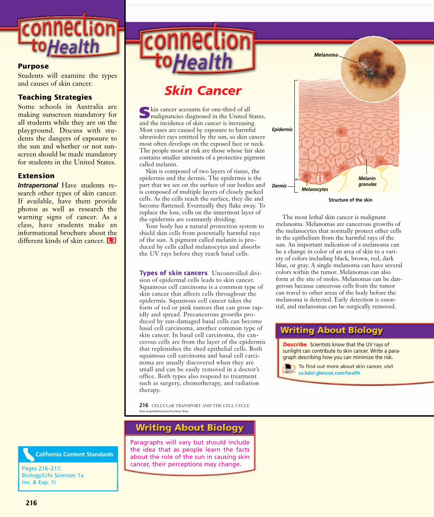
Paragraphs will vary but should includethe idea that as people learn the factsabout the role of the sun in causing skincancer, their perceptions may change.
216
Purpose Students will examine the typesand causes of skin cancer.
Teaching StrategiesSome schools in Australia aremaking sunscreen mandatory forall students while they are on theplayground. Discuss with stu-dents the dangers of exposure tothe sun and whether or not sun-screen should be made mandatoryfor students in the United States.
ExtensionIntrapersonal Have students re-search other types of skin cancer.If available, have them providephotos as well as research thewarning signs of cancer. As aclass, have students make aninformational brochure about thedifferent kinds of skin cancer. L3
P
216 CELLULAR TRANSPORT AND THE CELL CYCLE
Skin Cancer
Skin cancer accounts for one-third of allmalignancies diagnosed in the United States,
and the incidence of skin cancer is increasing.Most cases are caused by exposure to harmfulultraviolet rays emitted by the sun, so skin cancermost often develops on the exposed face or neck.The people most at risk are those whose fair skincontains smaller amounts of a protective pigmentcalled melanin.
Skin is composed of two layers of tissue, theepidermis and the dermis. The epidermis is thepart that we see on the surface of our bodies andis composed of multiple layers of closely packedcells. As the cells reach the surface, they die andbecome flattened. Eventually they flake away. Toreplace the loss, cells on the innermost layer ofthe epidermis are constantly dividing.
Your body has a natural protection system toshield skin cells from potentially harmful raysof the sun. A pigment called melanin is pro-duced by cells called melanocytes and absorbsthe UV rays before they reach basal cells.
Types of skin cancers Uncontrolled divi-sion of epidermal cells leads to skin cancer.Squamous cell carcinoma is a common type ofskin cancer that affects cells throughout theepidermis. Squamous cell cancer takes theform of red or pink tumors that can grow rap-idly and spread. Precancerous growths pro-duced by sun-damaged basal cells can becomebasal cell carcinoma, another common type ofskin cancer. In basal cell carcinoma, the can-cerous cells are from the layer of the epidermisthat replenishes the shed epithelial cells. Bothsquamous cell carcinoma and basal cell carci-noma are usually discovered when they aresmall and can be easily removed in a doctor’soffice. Both types also respond to treatmentsuch as surgery, chemotherapy, and radiationtherapy.
The most lethal skin cancer is malignantmelanoma. Melanomas are cancerous growths ofthe melanocytes that normally protect other cellsin the epithelium from the harmful rays of thesun. An important indication of a melanoma canbe a change in color of an area of skin to a vari-ety of colors including black, brown, red, darkblue, or gray. A single melanoma can have severalcolors within the tumor. Melanomas can alsoform at the site of moles. Melanomas can be dan-gerous because cancerous cells from the tumorcan travel to other areas of the body before themelanoma is detected. Early detection is essen-tial, and melanomas can be surgically removed.
Dixie Knight/Medichrome/The Stock Shop
Describe Scientists know that the UV rays of sunlight can contribute to skin cancer. Write a para-graph describing how you can minimize the risk.
To find out more about skin cancer, visit ca.bdol.glencoe.com/health
Epidermis
Melanocytes
MelaningranulesDermis
Structure of the skin
Melanoma
Pages 216–217: Biology/Life Sciences 1aInv. & Exp. 1i

217
Use the ExamView®Pro Test Bank CD-ROM to:• Create multiple versions of tests• Create modified tests with one mouse click for inclusion students• Edit existing questions and add your own questions• Build tests aligned with state standards using built-in
State Curriculum Tags• Change English tests to Spanish with one mouse click and vice versa
Key ConceptsSummary statements can beused by students to review themajor concepts of the chapter.
For additional helpwith vocabulary,have students
access the VocabularyPuzzleMaker online atca.bdol.glencoe.com/vocabulary_puzzlemaker
Section 8.1STUDY GUIDESTUDY GUIDE
CHAPTER 8 ASSESSMENT 217Luis M. De La Maza, PhD.M.D./Phototake, NYC
Section 8.2
Section 8.3
To help you reviewosmosis, use the Organizational StudyFold on page 195.
ca.bdol.glencoe.com/vocabulary_puzzlemaker
Key Concepts■ Osmosis is the diffusion of water through a
selectively permeable membrane.■ Passive transport moves a substance with
the concentration gradient and requires noenergy from the cell.
■ Active transport moves materials againstthe concentration gradient and requiresenergy to overcome the flow of materialsopposite the concentration gradient.
■ Large particles may enter a cell by endo-cytosis and leave by exocytosis.
Vocabularyactive transport (p. 199)endocytosis (p. 200)exocytosis (p. 200)facilitated diffusion (p. 198)hypertonic solution
(p. 196)hypotonic solution
(p. 196)isotonic solution (p. 196)osmosis (p. 195)passive transport
(p. 198)
CellularTransport
Key Concepts■ Cell size is limited largely by the diffusion
rate of materials into and out of the cell,the amount of DNA available to programthe cell’s metabolism, and the cell’s surfacearea-to-volume ratio.
■ The life cycle of a cell is divided into twogeneral periods: a period of active growthand metabolism known as interphase, and aperiod that leads to cell division known asmitosis.
■ Mitosis is divided into four phases:prophase, metaphase, anaphase, andtelophase.
■ The cells of most multicellular organismsare organized into tissues, organs, andorgan systems.
Vocabularyanaphase (p. 208)cell cycle (p. 204)centriole (p. 208)centromere (p. 206)chromatin (p. 204)chromosome (p. 203)cytokinesis (p. 209)interphase (p. 204)metaphase (p. 208)mitosis (p. 204)organ (p. 210)organ system (p. 210)prophase (p. 206)sister chromatid (p. 206)spindle (p. 208)telophase (p. 209)tissue (p. 210)
Cell Growth andReproduction
Key Concepts■ The cell cycle is controlled by key enzymes
that are produced at specific points in thecell cycle.
■ Cancer is caused by genetic and environ-mental factors that change the genes thatcontrol the cell cycle.
Vocabularycancer (p. 211)gene (p. 211)
Color-enhanced SEMMagnification: 7500�
Control of theCell Cycle
Visit /self_check_quiz/vocabulary_puzzlemaker/chapter_test/standardized_test
ca.bdol.glencoe.com
FOLDABLES™Have students use theirFoldables to review the contentof Section 8.1. On the back ofthe paper, analyze what wouldhappen to a cell that has lost itsability to transport ions againsta concentration gradient.
Questions andanswers have been
aligned and verified by ThePrinceton Review.
1. cancer 4. true2. centrioles 5. mitosis3. osmosis
6. B 10. D7. B 11. D8. C 12. B9. D 13. D
14. Cells that carry on a greatdeal of active transportwould have more mito-chondria.
15. By being able to control theenzymes, scientists may beable to modify the rapidcell division in cancer cells.
16. The solution outside thebag will turn blue-black.
17. The paramecium, being in ahypertonic solution, wouldprobably die because waterdiffuses out.
18. Student essays may indicatethe incidence of the disor-der, that it is caused by arecessive allele, the therapyto treat the disorder, as wellas other information.
Evaluating Open-Ended QuestionsOpen-ended assessment questions aregraded by using a multilevel rubricthat guides you in assessing a stu-dent’s knowledge of a particular con-cept. The following rubric is a samplescoring device.
218
Review the Chapter 8 vocabulary words listed inthe Study Guide on page 217. Determine if each statement is true or false. If false, replace theunderlined word with the correct vocabularyword.
1. Mitosis is the result of uncontrolled divisionof cells.
2. Small, dark cylindrical structures that aremade of microtubules and located just out-side the nucleus are called genes.
3. Diffusion of water across a selectively per-meable membrane is called cytokinesis.
4. In a hypotonic solution, the concentrationof dissolved substances inside cells is higherthan the concentration outside the cell.
5. Cancer is a period of nuclear division in a cell.
6. What kind of environment is described whenthe concentration of dissolved substances isgreater outside the cell than inside?A. hypotonic C. isotonic B. hypertonic D. saline
7. How is osmosis defined?A. as active transportB. as diffusion of water through a selectively
permeable membraneC. as an example of facilitated diffusionD. as requiring a transport protein
8. An amoeba ingests large food particles bywhat process?A. osmosis C. endocytosisB. diffusion D. exocytosis
9. Of what are chromosomes composed?A. cytoplasm C. RNA and proteinsB. centrioles D. DNA and proteins
10. Which of the following does NOT occurduring interphase?A. excretion of wastesB. cell repairC. protein synthesisD. nuclear division
11. During metaphase, the chromosomes move to the equator of what structure (shown here)?A. polesB. cell plateC. centrioleD. spindle
12. All but which of the following factors limitcell size?A. time required for diffusionB. elasticity of the plasma membraneC. presence of only one nucleusD. surface area-to-volume ratio
13. Which of the following is NOT a knowncause of cancer?A. environmental influencesB. certain virusesC. cigarette smokeD. bacterial infections
14. Open Ended How would you expect thenumber of mitochondria in a cell to berelated to the amount of active transport itcarries out?
15. Open Ended Suppose that all of theenzymes that control the normal cell cyclewere identified. Suggest some ways that thisinformation might be used to fight cancer.
16. Open Ended Substance A’s molecules aresmall. Substance B’s molecules, which reactwith substance A to produce a blue-blackcolor, are larger in comparison. If a solutionof substance A is placed inside a selectivelypermeable bag, and the bag is placed in asolution of substance B, what will happen?
17. Predict What do you think will happenwhen a freshwater paramecium is placed insalt water?
218 CHAPTER 8 ASSESSMENTEd Reschke/Peter Arnold, Inc.
ca.bdol.glencoe.com/chapter_test
Stained LM Magnification: 250�
219
19. B 23. A20. A 24. B21. C 25. D22. D 26. B27. Student responses will vary
but should contain atestable hypothesis and rea-sonable steps to test thehypothesis.
28. Ocean water contains highconcentrations of salts. Inorder to excrete the excesssalt, the body excretes morewater than it takes in andyou dehydrate.
Answer Sheet A practiceanswer sheet can be found on p. 117 of Unit 3 FAST FILE
Resources.
California StandardsPractice
Multiple ChoiceUse the following illustration to answer questions19–23.
19. Which drawing indicates a cell in metaphaseof mitosis?A. A C. CB. B D. D
20. During which stage do the chromatids ofchromosomes separate?A. A C. CB. B D. D
21. Which drawing indicates a cell whosenuclear membrane is dissolving?A. A C. CB. B D. D
22. Which of the following indicates the correctorder of mitosis in animal cells?A. A-B-C-D C. C-A-D-BB. B-C-A-D D. C-B-A-D
23. Which drawing shows a cell in anaphase?A. A C. CB. B D. D
24. A biologist notes that some cells are growingfaster than others in a tissue culture. A weeklater, the fast-growing cells have tripled innumber. This observation is a clue that thefast-growing cells ________.A. have killed the slow-growing cellsB. might be unable to control mitosisC. were exposed to radiation or chemicalsD. contain an unknown enzyme
25. Which parts of the nucleosome are made ofDNA?A. 1 and 2 C. 2 and 3B. 1 and 3 D. 2 and 4
26. Which parts of the nucleosome are made ofprotein?A. 1 and 2 C. 2 and 3B. 1 and 3 D. 2 and 4
2 3
Nucleosome
14
A B C D
18. Cystic fibrosisis a genetic disorder that results from theinability of cells to properly transport somematerials. Visit
to investigate cystic fibrosis. Write an essay that explains what you have learnedabout cystic fibrosis and present it to yourclass.
REAL WORLD BIOCHALLENGE
CHAPTER 8 ASSESSMENT 219
ca.bdol.glencoe.com
ca.bdol.glencoe.com/standardized_test
Constructed Response/Grid InRecord your answers on your answer document.
27. Open Ended The cell cycle can be affected by internal and external factors. Injury to a tissuecan prompt changes in the cell cycle of the cells near the injury site. Formulate a testablehypothesis concerning a specific type of cell’s response to injury. State your hypothesis, planan investigative procedure to test your hypothesis, and list the steps.
28. Open Ended Explain why drinking quantities of ocean water is dangerous to humans.
The assessed California standard appears next to the question.
STP to come
CHAPTER ASSESSMENT CHAPTER 8 Cellular Transport and the Cell Cycle 119
Cop
yrig
ht ©
by
Gle
ncoe
/McG
raw
-Hill
, a d
ivis
ion
of th
e M
cGra
w-H
ill C
ompa
nies
, Inc
.
AssessmentStudent Recording Sheet
Name Date Class
Chapter
8Chapter Chapter AssessmentChapter Assessment
Use with pages 218–219of the Student Edition
Standardized Test Practice
Vocabulary ReviewWrite true if the statement is true. If the statement is false, write the vocabulary word that makes the sentence true.
1. _________________________ 4. _______________________
2. _________________________ 5. _______________________
3. _________________________
Understanding Key ConceptsSelect the best answer from the choices given and fill in the corresponding oval.
6. 10.
7. 11.
8. 12.
9. 13.
Constructed ResponseRecord your answers for Questions 14–16 on a separate sheet of paper.
Thinking CriticallyRecord your answer for Question 17 on a separate sheet of paper.
18. Follow your teacher’s instructions for presenting your BioChallenge answer.REAL WORLD BIOCHALLENGE
Part 1 Multiple ChoiceSelect the best answer from the choices givenand fill in the corresponding oval.
19.
20.
21.
22.
23.
24.
25.
26.
Part 2Constructed Response/Grid InRecord your answers for Questions 27 and 28 on a separate sheet of paper.
A B C D A B C D
A B C D A B C D
A B C D
A B C D
A B C D
A B C D
A B C D
A B C D
A B C D
A B C D
A B C D
A B C D
A B C D
A B C D
SCORE CRITERIA4 A correct solution that is sup-
ported by well-developed,accurate explanations.
3 A generally correct solution,but may contain minor flaws inreasoning.
SCORE CRITERIA2 A partially correct interpretation
and/or solution to the problem.1 A correct solution with no sup-
porting evidence or explanation. 0 An incorrect solution or no
solution is given.
Pages 218–219: Biology/Life Sciences 1aInv. & Exp. 1i