nasal cavity and paranasal sinuses the angle of the mandible, represented by ohngren’s line, ......
TRANSCRIPT

Nasal Cavity and Paranasal Sinuses 69
6
6 Nasal Cavity and Paranasal Sinuses
(Nonepithelial tumors such as those of lymphoid tissue, soft tissue, bone, and cartilage are not included. Staging for mucosal melanoma of the nasal cavity and paranasal sinuses is not included in this chapter – see Chap. 9.)
At-A-Glance
SUMMARY OF CHANGES
! T4 lesions have been divided into T4a (moderately advanced local disease) and T4b (very advanced local disease), leading to the stratifi cation of Stage IV into Stage IVA (moderately advanced local/regional disease), Stage IVB (very advanced local/regional disease), and Stage IVC (distant metastatic disease)
ANATOMIC STAGE/PROGNOSTIC GROUPS
Stage 0 Tis N0 M0
Stage I T1 N0 M0
Stage II T2 N0 M0
Stage III T3 N0 M0 T1 N1 M0 T2 N1 M0 T3 N1 M0
Stage IVA T4a N0 M0 T4a N1 M0 T1 N2 M0 T2 N2 M0 T3 N2 M0 T4a N2 M0
Stage IVB T4b Any N M0 Any T N3 M0
Stage IVC Any T Any N M1
ICD-O-3 TOPOGRAPHY CODES C30.0 Nasal cavity C31.0 Maxillary sinus C31.1 Ethmoid sinus
ICD-O-3 HISTOLOGY CODE RANGES 8000–8576, 8940–8950, 8980–8981
ANATOMY
Primary Sites. Cancer of the maxillary sinus is the most common of the sinonasal malignancies. Ethmoid sinus and nasal cavity cancers are equal in frequency but considerably less common than maxillary sinus cancers. Tumors of the sphenoid and frontal sinuses are rare.
The location as well as the extent of the mucosal lesion within the maxillary sinus has prognostic signifi cance. His-torically, a plane, connecting the medial canthus of the eye
to the angle of the mandible, represented by Ohngren’s line, is used to divide the maxillary sinus into an anteroinferior portion (infrastructure), which is associated with a good prognosis, and a posterosuperior portion (suprastructure), which has a poor prognosis (Figure 6.1 ). The poorer outcome associated with suprastructure cancers refl ects early invasion by these tumors to critical structures, including the eye, skull base, pterygoids, and infratemporal fossa.
For the purpose of staging, the nasoethmoidal complex is divided into two sites: nasal cavity and ethmoid sinuses.

70 American Joint Committee on Cancer • 2010
The ethmoids are further subdivided into two subsites: left and right, separated by the nasal septum (perpendicular plate of ethmoid). The nasal cavity is divided into four subsites: the septum, fl oor, lateral wall, and vestibule.
Site Subsite Maxillary sinus Left/right Nasal cavity Septum Floor Lateral wall Vestibule (edge of naris to mucocutane-
ous junction) Ethmoid sinus Left/right
Regional Lymph Nodes. Regional lymph node spread from cancer of nasal cavity and paranasal sinuses is relatively uncommon. Involvement of buccinator, submandibular, upper jugular, and (occasionally) retropharyngeal nodes may occur with advanced maxillary sinus cancer, particularly those extending beyond the sinus walls to involve adjacent struc-tures, including soft tissues of the cheek, upper alveolus, pal-ate, and buccal mucosa. Ethmoid sinus cancers are less prone to regional lymphatic spread. When only one side of the neck is involved, it should be considered ipsilateral. Bilateral spread may occur with advanced primary cancer, particularly with spread of the primary beyond the midline.
In clinical evaluation, the physical size of the nodal mass should be measured. Most masses over 3 cm in diameter are not single nodes but, rather, are confl uent nodes or tumor in soft tissues of the neck. There are three categories of clini-cally positive nodes: N1, N2, and N3. The use of subgroups a, b, and c is required. Midline nodes are considered ipsilat-eral nodes. In addition to the components to describe the N category, regional lymph nodes should also be described according to the level of the neck that is involved. Pathologic examination is necessary for documentation of such disease extent. Imaging studies showing amorphous spiculated margins of involved nodes or involvement of internodal fat resulting in loss of normal oval-to-round nodal shape strongly suggest extracapsular (extranodal) tumor spread. No imaging study (as yet) can identify microscopic foci in regional nodes or distinguish between small reactive nodes and small malignant nodes without central radiographic inhomogeneity.
For pN, a selective neck dissection will ordinarily include six or more lymph nodes, and a radical or modifi ed radical neck dissection will ordinarily include ten or more lymph nodes. Negative pathologic examination of a lesser number of lymph nodes still mandates a pN0 designation.
Distant Metastases. Distant spread usually occurs to lungs but occasionally there is spread to bone.
RULES FOR CLASSIFICATION
Clinical Staging. The assessment of primary maxillary sinus, nasal cavity, and ethmoid tumors is based on inspec-tion and palpation, including examination of the orbits, nasal and oral cavities, and nasopharynx, and neurologic evaluation of the cranial nerves. Nasal endoscopy with rigid or fiberoptic flexible instruments is recommended. Radiologic assessment with magnetic resonance imaging (MRI) or computed tomography (CT) is mandatory for accurate pretreatment staging of malignant tumor of the sinuses. If available, MRI more accurately depicts skull base and intracranial involvement and the differentiation of fluid from solid tumor, and helps define local exten-sion of disease. Neck nodes are assessed by palpation +/! imaging. Imaging for possible nodal metastases is probably unnecessary in the presence of a clinically negative neck. Examinations for distant metastases include appropriate imaging, blood chemistries, blood count, and other rou-tine studies as indicated.
Pathologic Staging. Pathologic staging requires the use of all information obtained in clinical staging and histologic study of the surgically resected specimen. The surgeon’s evaluation of gross unresected residual tumor must also be included. Specimens that are resected after radiation or chemotherapy need to be identifi ed and considered in con-text. The pathologic description of the lymphadenectomy specimen should describe the size, number, and level of the
FIGURE 6.1. Sites of origin of tumors of the paranasal sinuses.

Nasal Cavity and Paranasal Sinuses 71
6
involved node(s) and the presence or absence of extracapsu-lar spread (ECS).
PROGNOSTIC FEATURES
In addition to the importance of the TNM factors outlined previously, the overall health of these patients clearly infl uences outcome. An ongoing effort to better assess prognosis using both tumor and nontumor related factors is underway. Chart abstraction will continue to be performed by cancer registrars to obtain important information regarding specifi c factors related to prognosis. This data will then be used to further hone the predictive power of the staging system in future revisions.
Comorbidity can be classifi ed by specifi c measures of additional medical illnesses. Accurate reporting of all illnesses in the patients’ medical record is essential to assessment of these parameters. General performance measures are helpful in predicting survival. The AJCC strongly recommends the clinician report performance status using the ECOG, Zubrod, or Karnofsky performance measures along with standard staging information. An interrelationship between each of the major performance tools exists.
Zubrod/ECOG Performance Scale
0. Fully active, able to carry on all predisease activities without restriction (Karnofsky 90–100).
1. Restricted in physically strenuous activity but ambu-latory and able to carry work of a light or sedentary nature. For example, light housework, offi ce work (Karnofsky 70–80).
2. Ambulatory and capable of all self-care but unable to carry out any work activities. Up and about more than 50% of waking hours (Karnofsky 50–60).
3. Capable of only limited self-care, confi ned to bed or chair 50% or more of waking hours (Karnofsky 30–40).
4 . Completely disabled. Cannot carry on self-care. Totally confi ned to bed (Karnofsky 10–20).
5. Death (Karnofsky 0).
Lifestyle factors such as tobacco and alcohol abuse nega-tively infl uence survival. Accurate recording of smoking in pack years and alcohol in number of days drinking per week and number of drinks per day will provide important data for future analysis. Nutrition is important to prognosis and will be indirectly measured by weight loss of >10% of body weight. Depression adversely impacts quality of life and sur-vival. Notation of a previous or current diagnosis of depres-sion should be recorded in the medical record.
Figure 6.2A, B presents observed and relative survival rates for sinonasal carcinomas (all histologies) for 1998–1999, classifi ed by the AJCC staging classifi cation.
Mucosal Melanoma. Mucosal melanoma of all head and neck sites is staged using a uniform classifi cation as discussed in Chap. 9.
DEFINITIONS OF TNM
Primary Tumor (T)TX Primary tumor cannot be assessedT0 No evidence of primary tumorTis Carcinoma in situ
Maxillary SinusT1 Tumor limited to maxillary sinus mucosa with no
erosion or destruction of boneT2 Tumor causing bone erosion or destruction
including extension into the hard palate and/or middle nasal meatus, except extension to poste-rior wall of maxillary sinus and pterygoid plates
T3 Tumor invades any of the following: bone of the posterior wall of maxillary sinus, subcutaneous tissues, fl oor or medial wall of orbit, pterygoid fossa, ethmoid sinuses
T4a Moderately advanced local diseaseTumor invades anterior orbital contents, skin of cheek, pterygoid plates, infratemporal fossa, cri-briform plate, sphenoid or frontal sinuses
FIGURE 6.2. ( A ) Five-year, observed survival by “combined” AJCC stage sinonasal carcinomas (all histologies), 1998–1999. (*95% confi dence intervals correspond to year-5 survival rates.) ( B ) Five-year, relative survival by “combined” AJCC stage sinonasal carcinomas (all histologies), 1998–1999. (*95% confi dence intervals correspond to year-5 survival rates.)

72 American Joint Committee on Cancer • 2010
Primary Tumor (T) (continued)T4b Very advanced local disease
Tumor invades any of the following: orbital apex, dura, brain, middle cranial fossa, cranial nerves other than maxillary division of trigeminal nerve (V
2), nasopharynx, or clivus
Nasal Cavity and Ethmoid SinusT1 Tumor restricted to any one subsite, with or with-
out bony invasionT2 Tumor invading two subsites in a single region or
extending to involve an adjacent region within the nasoethmoidal complex, with or without bony invasion
T3 Tumor extends to invade the medial wall or fl oor of the orbit, maxillary sinus, palate, or cribriform plate
T4a Moderately advanced local diseaseTumor invades any of the following: anterior orbital contents, skin of nose or cheek, minimal extension to anterior cranial fossa, pterygoid plates, sphenoid or frontal sinuses
T4b Very advanced local diseaseTumor invades any of the following: orbital apex, dura, brain, middle cranial fossa, cranial nerves other than (V2), nasopharynx, or clivus
Regional Lymph Nodes (N)NX Regional lymph nodes cannot be assessedN0 No regional lymph node metastasisN1 Metastasis in a single ipsilateral lymph node, 3 cm
or less in greatest dimensionN2 Metastasis in a single ipsilateral lymph node, more
than 3 cm but not more than 6 cm in greatest dimension, or in multiple ipsilateral lymph nodes, none more than 6 cm in greatest dimension, or in bilateral or contralateral lymph nodes, none more than 6 cm in greatest dimension
N2a Metastasis in a single ipsilateral lymph node, more than 3 cm but not more than 6 cm in great-est dimension
N2b Metastasis in multiple ipsilateral lymph nodes, none more than 6 cm in greatest dimension
N2c Metastasis in bilateral or contralateral lymph nodes, none more than 6 cm in greatest dimen-sion
N3 Metastasis in a lymph node, more than 6 cm in greatest dimension
Distant Metastasis (M)M0 No distant metastasisM1 Distant metastasis
ANATOMIC STAGE/PROGNOSTIC GROUPS
Stage 0 Tis N0 M0
Stage I T1 N0 M0
Stage II T2 N0 M0
Stage III T3 N0 M0 T1 N1 M0 T2 N1 M0 T3 N1 M0
Stage IVA T4a N0 M0 T4a N1 M0 T1 N2 M0 T2 N2 M0 T3 N2 M0 T4a N2 M0
Stage IVB T4b Any N M0 Any T N3 M0
Stage IVC Any T Any N M1
PROGNOSTIC FACTORS (SITE-SPECIFIC FACTORS)(Recommended for Collection)
Required for staging
None
Clinically signifi cant
Size of lymph nodesExtracapsular extension from lymph nodes for head and neckHead and neck lymph nodes levels I–IIIHead and neck lymph nodes levels IV–VHead and neck lymph nodes levels VI–VIIOther lymph nodes groupClinical location of cervical nodesExtracapsular spread (ECS) clinicalExtracapsular spread (ECS) pathologicHuman papillomavirus (HPV) statusTumor thickness
HISTOLOGIC GRADE (G)
Grade is reported in registry systems by the grade value. A two-grade, three-grade, or four-grade system may be used. If a grading system is not specifi ed, generally the following sys-tem is used:
GX Grade cannot be assessed G1 Well differentiated G2 Moderately differentiated G3 Poorly differentiated G4 Undifferentiated

Nasal Cavity and Paranasal Sinuses 73
6
HISTOPATHOLOGIC TYPE
The predominant cancer is squamous cell carcinoma. The staging guidelines are applicable to all forms of carcinoma. Mucosal melanoma of the head and neck is very rare but has unique behavior warranting a separate classifi cation as dis-cussed in Chap. 9. Other nonepithelial tumors such as those of lymphoid tissue, soft tissue, bone, and cartilage are not included. Histologic confi rmation of diagnosis is required. Histopathologic grading of squamous carcinoma is recom-mended. The grade is subjective and uses a descriptive as well as a numerical form (i.e., well differentiated, moderately differentiated, and poorly differentiated), depending on the degree of closeness to or deviation from squamous epithe-lium in mucosal sites. Also recommended where feasible is a quantitative evaluation of depth of invasion of the primary tumor and the presence or absence of vascular invasion and perineural invasion. Although the grade of the tumor does not enter into the staging of the tumor, it should be recorded. The pathologic description of any lymphadenectomy specimen should describe the size, number, and level of the involved node(s) and the presence or absence of ECS.
BIBLIOGRAPHY
Bridger GP, Mendelsohn MS, Baldwinn M, et al. Paranasal sinus cancer. Aust N Z J Surg. 1991;61:290–4.
Cantu G, Solero CL, Mariani L, et al. A new classifi cation for malignant tumors involving the anterior skull base. Arch Otolaryngol Head Neck Surg. 1999;125:1252–7.
de Leeuw JR, de Graeff A, Ros WJ, Blijham GH, Hordijk GJ, Winnubst JA. Prediction of depressive symptomatology after treatment of head and neck cancer: the infl uence of pre-
treatment physical and depressive symptoms, coping, and social support. Head Neck. 2000;22(8):799–807.
Jiang GL, Ang KA, Peters LJ, et al. Maxillary sinus carcinomas: Natural history and results of postoperative radiotherapy. Radiother Oncol. 1991;21:194–200.
Jiang GL, Morrison WH, Garden AS, et al. Ethmoid sinus car-cinoma: Natural history and treatment results. Radiother Oncol. 1998;49:21–7.
Kondo M, Horiuchi M, Shiga H, et al. CT of malignant tumors of the nasal cavity and paranasal sinuses. Cancer. 1982;50:226–31.
Le QT, Fu KK, Kaplan M, et al. Treatment of maxillary sinus carcinoma. A comparison of the 1997 and 1977 American Joint Committee on Cancer Staging Systems. Cancer. 1999;86: 1700–11.
Nandapalan V, Roland NJ, Helliwell TR, Williams EM, Hamilton JW, Jones AS. Mucosal melanoma of the head and neck. Clin Otolaryngol Allied Sci. 1998;23(2):107–16.
Patel SG, Prasad ML, Escrig M, et al. Primary mucosalmalignant mel-anoma of the head and neck. Head Neck. 2002;24(3):247–57.
Patel SG, Singh B, Polluri A, et al. Craniofacial surgery for malig-nant skull base tumors: Report of an international collabora-tive study. Cancer. 2003;98(6):1179–87.
Paulino AFG, Singh B, Carew J, et al. Epstein-Barr virus in squamous carcinoma of the anterior nasal cavity. Ann Diagn Pathol. 2000;4:7–10.
Piccirillo JF. Inclusion of comorbidity in a staging system for head and neck cancer. Oncology. 1995;9:831–6.
Sisson GA, Toriumi DM, Atiyah RH. Paranasal sinus malignancy: A comprehensive update. Laryngoscope. 1989;99:143–50.
Som PM, Dillon WP, Sze G, et al. Benign and malignant sinona-sal lesions with intracranial extension: Differentiation with MRI imaging. Radiology. 1989;172:763–6.
Van Tassel P, Lee YY. GD-DTPA enhanced MR for detecting intracranial extension of sinonasal malignancies. J Comput Assist Tomogr. 1991;15:387–92.

Nasal Cavity and Paranasal Sinuses 75
(continued on next page)
y clinical– staging completed after neoadjuvant therapy but before subsequent surgery
y pathologic – staging completed after neoadjuvant therapy AND subsequent surgery
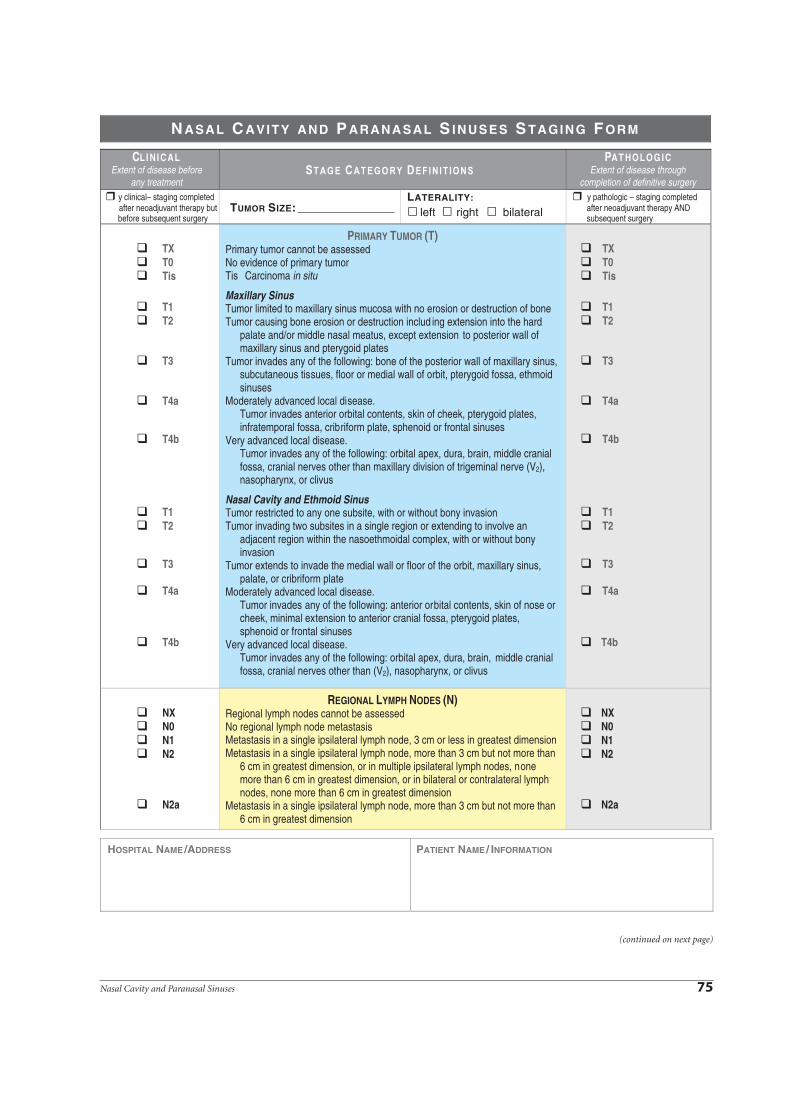
TXT0Tis
T1T2
T3
T4a
T4b
T1T2
T3
T4a
T4b
PRIMARY TUMOR (T)Primary tumor cannot be assessedNo evidence of primary tumorTis Carcinoma in situ
Maxillary SinusTumor limited to maxillary sinus mucosa with no erosion or destruction of boneTumor causing bone erosion or destruction includ ing extension into the hard
palate and/or middle nasal meatus, except extension to posterior wall of maxillary sinus and pterygoid plates
Tumor invades any of the following: bone of the posterior wall of maxillary sinus, subcutaneous tissues, floor or medial wall of orbit, pterygoid fossa, ethmoid sinuses
Moderately advanced local disease.Tumor invades anterior orbital contents, skin of cheek, pterygoid plates, infratemporal fossa, cribriform plate, sphenoid or frontal sinuses
Very advanced local disease.Tumor invades any of the following: orbital apex, dura, brain, middle cranial fossa, cranial nerves other than maxillary division of trigeminal nerve (V2), nasopharynx, or clivus
Nasal Cavity and Ethmoid SinusTumor restricted to any one subsite, with or without bony invasionTumor invading two subsites in a single region or extending to involve an
adjacent region within the nasoethmoidal complex, with or without bony invasion
Tumor extends to invade the medial wall or floor of the orbit, maxillary sinus, palate, or cribriform plate
Moderately advanced local disease.Tumor invades any of the following: anterior orbital contents, skin of nose or cheek, minimal extension to anterior cranial fossa, pterygoid plates, sphenoid or frontal sinuses
Very advanced local disease.Tumor invades any of the following: orbital apex, dura, brain, middle cranial fossa, cranial nerves other than (V2), nasopharynx, or clivus
TXT0Tis
T1T2
T3
T4a
T4b
T1T2
T3
T4a
T4b
NXN0N1N2
N2a
REGIONAL LYMPH NODES (N)Regional lymph nodes cannot be assessedNo regional lymph node metastasisMetastasis in a single ipsilateral lymph node, 3 cm or less in greatest dimensionMetastasis in a single ipsilateral lymph node, more than 3 cm but not more than
6 cm in greatest dimension, or in multiple ipsilateral lymph nodes, none more than 6 cm in greatest dimension, or in bilateral or contralateral lymph nodes, none more than 6 cm in greatest dimension
Metastasis in a single ipsilateral lymph node, more than 3 cm but not more than 6 cm in greatest dimension
NXN0N1N2
N2a
CLI NI CALExtent of disease before
any treatment
PAT HOLOG ICExtent of disease through
completion of definitive surgery
N ASAL C AVITY AND P ARANASAL S INUSES S TAGING F ORM
left right bilateralLATERALITY:
TUMOR SIZE:
HOSPITAL NAME/ADDRESS PATIENT NAME/ INFORMATION

76 American Joint Committee on Cancer • 2010
(continued from previous page)
N2b
N2c
N3
Metastasis in multiple ipsilateral lymph nodes, none more than 6 cm in greatest dimension
Metastasis in bilateral or contralateral lymph nodes, none more than 6 cm in greatest dimension
Metastasis in a lymph node, more than 6 cm in greatest dimension
N2b
N2c
N3
M0M1
DISTANT METASTASIS (M)No distant metastasis (no pathologic M0; use clinical M to complete stage group)Distant metastasis M1
CLINICALGROUP T N M
0 Tis N0 M0I T1 N0 M0II T2 N0 M0III T3 N0 M0
T1 N1 M0T2 N1 M0T3 N1 M0
IVA T4a N0 M0T4a N1 M0T1 N2 M0T2 N2 M0T3 N2 M0T4a N2 M0
IVB T4b Any N M0Any T N3 M0
IVC Any T Any N M1
PATHOLOGICGROUP T N M
0 Tis N0 M0I T1 N0 M0II T2 N0 M0III T3 N0 M0
T1 N1 M0T2 N1 M0T3 N1 M0
IVA T4a N0 M0T4a N1 M0T1 N2 M0T2 N2 M0T3 N2 M0T4a N2 M0
IVB T4b Any N M0Any T N3 M0
IVC Any T Any N M1
Stage unknown Stage unknown
PROGNOSTIC FACTORS (SITE-SPECIFIC FACTORS)REQUIRED FOR STAGING: NoneCLINICALLY SIGNIFICANT:
Size of Lymph Nodes ___________________________________________Extracapsular Extension from Lymph Nodes for Head & Neck ___________Head & Neck Lymph Nodes Levels I-III _____________________________Head & Neck Lymph Nodes Levels IV-V ____________________________Head & Neck Lymph Nodes Levels VI-VII ___________________________Other Lymph Nodes Group ______________________________________Clinical Location of cervical nodes _________________________________Extracapsular spread (ECS) Clinical _______________________________Extracapsular spread (ECS) Pathologic _____________________________Human Papillomavirus (HPV) Status _______________________________Tumor Thickness ______________________________________________
General Notes: For identification of special cases of TNM or pTNM classifications, the "m" suffix and "y," "r," and "a" prefixes are used. Although they do not affect the stage grouping, they indicate cases needing separate analysis.
m suffix indicates the presence of multiple primary tumors in a single site and is recorded in parentheses: pT(m)NM.
N ASAL C AVITY AND P ARANASAL S INUSES S TAGING F ORM
A N A T O M I C S T A G E • P R O G N O S T I C G R O U P S
HOSPITAL NAME/ADDRESS PATIENT NAME/ INFORMATION

Nasal Cavity and Paranasal Sinuses 77
(continued on next page)
Histologic Grade (G) (also known as overall grade)
Grading system
2 grade system
GradeGrade I or 1
3 grade system Grade II or 2
4 grade system Grade III or 3
No 2, 3, or 4 grade system is available Grade IV or 4
General Notes (continued):
y prefix indicates those cases in which classification is performed during or following initial multimodality therapy. The cTNM or pTNM category is identified by a "y" prefix. The ycTNM or ypTNM categorizes the extent of tumor actually present at the time of that examination. The "y" categorization is not an estimate of tumor prior to multimodality therapy.
r prefix indicates a recurrent tumor when staged after a disease-free interval and is identified by the "r" prefix: rTNM.
a prefix designates the stage determined at autopsy: aTNM.
surgical margins is data field recorded by registrars describing the surgical margins of the resected primary site specimen as determined only by the pathology report.
neoadjuvant treatment is radiation therapy or systemic therapy (consisting of chemotherapy, hormone therapy, or immunotherapy) administered prior to a definitive surgical procedure. If the surgical procedure is not performed, the administered therapy no longer meets the definition of neoadjuvant therapy.
ADDITIONAL DESCRIPTORSLymphatic Vessel Invasion (L) and Venous Invasion (V) have been combined into Lymph-Vascular Invasion (LVI) for collection by cancer registrars. The College of American Pathologists’ (CAP) Checklist should be used as the primary source. Other sources may be used in the absence of a Checklist. Priority is given to positive results.
Lymph-Vascular Invasion Not Present (absent)/Not IdentifiedLymph-Vascular Invasion Present/IdentifiedNot ApplicableUnknown/Indeterminate
Residual Tumor (R)The absence or presence of residual tumor after treatment. In some cases treated with surgery and/or with neoadjuvant therapy there will be residual tumor at the primary site after treatment because of incomplete resection or local and regional disease that extends beyond the limit of ability of resection.
RX Presence of residual tumor cannot be assessedR0 No residual tumorR1 Microscopic residual tumorR2 Macroscopic residual tumor
Clinical stage was used in treatment planning (describe):
National guidelines were used in treatment planning NCCN Other (describe):
Physician signature Date/Time
N ASAL C AVITY AND P ARANASAL S INUSES S TAGING F ORM
HOSPITAL NAME/ADDRESS PATIENT NAME/ INFORMATION

78 American Joint Committee on Cancer • 2010
(continued from previous page)
IllustrationIndicate on diagram primarytumor and regional nodesinvolved.
N ASAL C AVITY AND P ARANASAL S INUSES S TAGING F ORM
HOSPITAL NAME/ADDRESS PATIENT NAME/ INFORMATION