nasa workshop on biological adaptation technical memorandum 89468 nasa workshop on biological...
TRANSCRIPT

- NASA Technical Memorandum 89468
NASA Workshop on Biological Adaptation
(IASA-TH-89466) E B S B OCBBSbCE CE BIOLOGICAL NB8- 18 174 A L A P Z A I I C B ( N A Z A ) 105 p CSCL 06B
Unclas 63/51 0125855
February 1988
National Aeronautics and Space Administration
https://ntrs.nasa.gov/search.jsp?R=19880008790 2018-06-02T13:15:02+00:00Z

~
NASA Technical Memorandum 89468
NASA Workshop on Biological Adaptation Chaired by: Dr. Emily Morey-Holton, Ames Research Center, Moffett Field, California Dr. Thora Halstead, NASA Headquarters, Washington, DC
Edited by: Dr. Emily Morey-Holton, Ames Research Center, Moffett Field, California Dr. Marc Tischler, University of Arizona, Tucson, Arizona
February 1988
t
National Aeronautics and Space Administration
Ames Research Center Moffett Field, California 94035

TABLE OF CONTENTS
Page . 'L
PREFACE ............................................................................ 1
t
.
EXECUTIVE SUMMARY .................................................................. 3
GRAVITATIONAL EFFECTS ON STRUCTURE AND BIOMINERALIZATION
r INTRODUCTION ........................................................................ OVERVIEW ........................................................................... 6
Current Knowledge .................................................................. 6 Spaceflight ..................................................................... 6 Rat Model Simulating Certain Aspects of Spaceflight-- Bone/Endocrine Regulation .................................................................... 7
Electromechanics, Electrophysiology, and Ion Fluxes ............................. 9 Cellular Responses to Structural or Mechanical Stimuli ......................... 12 Crystal/Matrix Energetics ...................................................... 14 Biomineralization .............................................................. 16
Relationship of Current Research to the Program ................................... 22 Bone/Endocrine Regulation ...................................................... 22 Electromechanics, Electrophysiology, and Ion Fluxes ............................ 22 Cellular Responses to Structural or Mechanical Stress .......................... 23 Crystal/Matrix Energetics ...................................................... 23 Biomineralization .............................................................. 23
BASIC SC I ENCE QUESTIONS . .......................................................... 24
RESEARCH PRIORITIES ............................................................... 25
REFERENCES ........................................................................ 27
GRAVITY AFFECTED REGULATORY MECHANISMS
INTRODUCTION ...................................................................... 37
OVERVIEW .......................................................................... 37
Current Knowledge ................................................................. 37 Endocrine Mechanisms ........................................................... 37 Neural Mechanisms .............................................................. ~8 Mechanisms of Metabolic Adaptations ............................................ 53 Systemic Responses ............................................................. 55
WECEDING PAOE BLANK NOT F U Z D

Page Environmental Responses ........................................................ 59 Ionic Mediators ................................................................ 62
Relationship of Current Research to the Program. .................................. 63
Neural Mechanisms .............................................................. 65 Mechanisms of Metabolic Adaptations ............................................ 65 Systemic Responses ............................................................. 66 Environmental Responses ........................................................ 67
Endocrine Mechanisms ........................................................... 63
BASIC SCIENCE QUESTIONS ........................................................... 67
RESEARCH PRIORITIES ............................................................... 68
REFERENCES ........................................................................ 69
.
1
APPENDIX A: ABSTRACTS ............................................................ 83
8
iv

PREFACE
A workshop on NASA's Space Biology Biological Adaptation Research was convened April 28-30, 1986, to review the current program and its objectives and to identify future research directions. Gravitational effects on structures and biomineralization and gravity-affected regulatory mechanisms. The participants also recommended that research concentrate on rapidly growing animals, since gravity effects may be more pronounced during growth and development. tions were identified. The recommendations of the workshop will assist the NASA's Life Sciences Division in its assessment and long-range planning of these areas of space biology. Equally important, the workshop was intended to stimulate thought and research among those attending the workshop so that they would, in turn, inter- est, excite, and involve other members of the academic community in research efforts relevant to these programs. The workshop was comprised of two panels that defined programs for each of the two research areas. assist in preparing the workshop report. The report on gravitational effects on structures and biomineralization was organized by Dr. Emily Holton, NASA Ames Research Center with contributions by Dr. Bernard Halloran, University of California (UC) San Francisco (bone/endocrine regulation); Dr. Mark Cooper, UC Berkeley (elec- tromechanics, electrophysiology, and ion fluxes); Dr. Steve Doty, Columbia Univers- ity (cellular responses to structural or mechanical stimuli); Dr. Lelia Coyne, San Jose State University (crystalhatrix energetics); and Dr. Heinz Lowenstam, Califor- nia Institute of Technol.ogy (biomineralization). The report on gravity-affected regulatory mechanisms was organized by Dr. Marc Tischler, University of Arizona, Tucson, with contributions from Dr. Johnnie Underwood, Ames Research Center (vitamin D and parathyroid hormone); Dr. Ken Snowdowne, University of the Pacific Dental School (cytosolic calcium and hormone secretion); Dr. Wesley Hymer, Pennsylvania State University (growth hormone); Dr. Francis Canong, UC San Francisco (renin and vasopressin); Dr. John Horowitz, UC Davis (neural mechanisms); and Dr. Charles Fuller, UC Davis (thermoregulation, circadian timekeeping, sleep, and environmental
Two research areas emerged from these deliberations:
Both research areas were defined and future research direc-
Attendees were given assignments to
3
a responses).
The workshop was organized by Dr. Emily Holton, Ames Research Center and Dr. Thora Halstead, NASA Headquarters. Attendees included:
Dr. Claude Arnaud UC San Francisco Dr. Rod Ballard Ames Research Center Dr. Daniel Bik.le UC San Francisco Ms. Charlotte Cone Ames Research Center Dr. Mark Cooper UC Berkeley Dr. Lelia Coyne San Jose State University
1
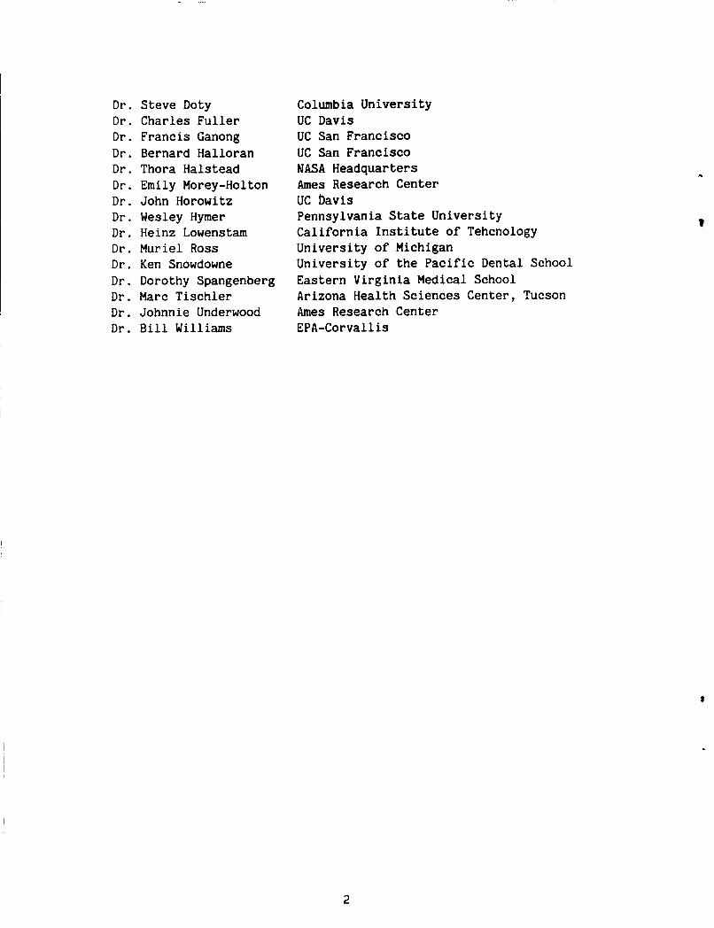
Dr . Dr . Dr . Dr . Dr . Dr . Dr . Dr . Dr . Dr . Dr . Dr . Dr . Dr . Dr .
Steve Doty Charles Fuller Francis Ganong Bernard Halloran Thora Halstead Emily Morey-Holton John Horowitz Wesley Hymer Heinz Lowenstam Muriel Ross Ken Snowdowne Dorothy Spangenberg Marc Tischler Johnnie Underwood Bill Williams
Columbia University UC Davis UC San Francisco UC San Francisco NASA Headquarters Ames Research Center UC bavis Pennsylvania State University California Institute of Tehcnology University of Michigan University of the Pacific Dental School Eastern Virginia Medical School Arizona Health Sciences Center, Tucson Ames Research Center EPA-Corvallis
2
EXECUTIVE SUMMARY
5
This report summarizes the results of a workshop which critiqued existir;; anc
Prior to Lie potential research in NASA's Space Biology/Biological Adaptation Program and made recommendations for future research projects and program directions. meeting, all participants received a copy of pertinent parts of the Gravitational & Space Biology Program Plan and the current Biological Adaptation research goals, objectives, and justifications. Participants were requested to submit an abstract which addressed the foll.owing points:
1 ) What is the ultimate goal of your research?
2) How does this goal relate to the Biological Adaptation program?
3 ) What techniques, expertise, and flight opportunities are necessary to accomplish your goal (for example, do you need animals, unique species, cell cul- ture, electrophoresis, RIAs, or special techniques)? If technology does not pres- ently exist (either techniques are too insensitive o r nonexistent) to allow comple- tion of your research goal, then what technology needs to be developed and when might such technology be available?
These abstracts are included in this report as appendix A. The workshop began with each participant addressing the major points of their abstract. discussion, the workshop focused on the existing Biological Adaptation research program. plan be divided into two programs. program focusi-g on gravitational effects on structures and biomineralization and another program focusing on gravity-affected regulatory mechanisms. objectives were defined for each program. based experiments were :summarized and research needs for advancement and future development were projected. Existing research efforts were placed in the appropriate program and necessary new efforts were identified. Since gravitational effects may be more pronounced during growth and development, research using animals should concentrate on growing species.
After a lively
All participants of the workshop strongly recommended that the research The logical division appeared to be a research
Hypotheses and Current knowledge from space and ground-
The program on gravitational effects on structure and biomineralization should concentrate on the cytoskeleton and subcellular support structures as well as the skeleton. Research in this program should focus on:
1) gravity-regulated structural and functional relationships including:
a) shape/size/composition of organ systems and cells.
b) cellular proliferation and maturation.
c) structural integrity
d) assembly/disassembly
and molecular organization.
and interaction of organics/inorganics.
3

e) stimulus-response coupling.
f) genetic changes producing altered organic matrix products.
g) endocrine alterations/interactions.
h) ionic alterations/interactions both intracellular and extracellular.
2) comparative biomineralization to understand the changes in mineral types from exoskeletal to endoskeletal systems and the influence of organic matrix/crystal interfaces.
3 ) understanding of the level of organization required for cell and organism structures to perceive gravity.
The program on gravity-affected regulatory mechanisms should concentrate on:
1 ) mechanisms by which gravity affects cells and organisms emphasizing neural, endocrine, metabolic adaptation, and systemic response mechanisms.
2)
3 ) importance of environmental parameters (e.g., temperature, light) in grav-
ionic mediators of gravity effects.
i ty responses.
Basic science questions and research priorities are found on pp. 24-26 and These questions and priorities focus almost entirely on gravitational 67-68.
responses. priority projects. Other research projects which propose to investigate ground- based problems not directly related to proven gravitational effects, although poten- tially scientifically highly meritorious, are considered lower priority.
Projects aimed at these questions and priorities are considered as high
Finally, the members of this workshop strongly recommended to NASA that contin- ued investment of time, talent, and monies for basic research in Space Biology is essential. Unexpected ideas which continuously appear in new fields of Biology should be anticipated and incorporated into existing programs. Young investigators with their fresh approaches and new ideas for quality research should be attracted to the NASA programs. To date, most existing data deal with short duration space- flight or acute ground experiments; chronic studies should be encouraged to prepare for experiments on space station. A complete understanding of the role of gravity in development of structural systems and regulatory mechanisms will only be achieved when long term experiments can be conducted in space.

GRAVITATIONAL EFFECTS ON STRUCTURE AND BIOMINERALIZATION
INTRODUCTION
All biological species on earth have evolved under the influence of gravity. In response to this force, organisms have developed structures to withstand gravity loads; structures may be optimized for a specific gravity level (i.e., 1 GI. Such structures may differ between species and may be due to the influence of gravity on a particular cell or organism. With the advent of routine spaceflight and the potential for continuous presence in space, many presently unknown gravity sensitive structures will be identified in multiple species. However, the scope of this research program will focus on systems presently known to be gravity responsive.
In focus with Space Biology Program goals (to use the unique characteristics of the space environment, especially microgravity, to increase our understanding of life and its processes; and to understand how gravity affects and has shaped life on Earth), the objectives of this research program are:
1 ) to identify, compare, and contrast support structures that living systems have evolved in response to gravity and to understand the influence of gravity on cellular proliferation and maturation, size, shape, composition, and metabolism of such structures.
2) to determine whether gravity directly affects the cells regulating struc- tural mass and/or exerts its effect extracellularly, and to elucidate the mecha- nism(s) involved.
3 ) to determine whether gravity is necessary to produce structurally sound support systems.
4 ) to determine mineral and matrix types of various organisms and whether their structural composition is gravity dependent.
5 ) to use the microgravity of spaceflight as a tool to understand how orga- nisms adapted to gravity during evolution and how gravity might regulate physiologi- cal structures on earth.
To accomplish the above objectives, a vigorous, well-planned research program involving definition of' parameters in earth-bound experiments is necessary. How- ever, most if not all the experiments in this program will require Space Shuttle or Space Station experimentation for definitive studies.
5

I OVERVIEW
! Current Knowledge
I Spaceflight
Very little information exists about the effects of spaceflight on cell and organism structure. Data have been obtained from growing rats, cell cultures, and plant tissues following flight on Soviet Cosmos missions or US Skylab and Space Shuttle missions. Young, male rats have flown on three Soviet Cosmos flights and one Space Shuttle mission; existing data are from rats either 8-9 or 12 wk of age in space for either 7 or 19 days. The most significant changes in bone structure in these young, growing rats were:
1 ) Indications of a suppression and possible cessation of bone growth in length and width; bone formation parameters were decreased, while resorption parame- ters appeared normal (Morey and Baylink, 1978; Wronski and Morey, 1983; Cann and
~ Adachi, 1983). I 2 ) An increase in bone mass with no increase in bone strength (Spengler
et al., 1983; Patterson-Buckendahl et al., 1985).
3 ) Indications that the organic matrix formed during flight did not mineralize I properly contributing to the strength problem (Turner et al., 1985).
4 ) Indication that mineral crystallites formed during flight were smaller and oriented with the collagen fibers (Turner et al., 1985).
5 ) about 8 days while mineralization time was increased from about 1.25 to about 2.1 days (Morey and Baylink, unpublished observations).
Indications that osteoid maturation rate was prolonged from about 4 to
6 ) Indications of a delayed maturation of bone (Simmons et al., 1983).
7) Indications of a lack of differentiation of bone progenitor cells (Roberts et al., 1981).
8) Indications that muscle attachments (in flight, but not on Earth) minimize bone loss (Simmons et al., 1983; Spector et al., 1983).
The degree of change appears to depend on the rate of growth; on SL3, younger ani- mals exhibited significant bone changes on the 7-day mission while older animals exhibited only trends suggesting suppression of growth in many bone parameters. In the 19-day flights, all animals showed highly significant differences whether 63 or 83 days old. Regardless of flight duration, these young animals showed no signs of bone loss; some components of bone continued to grow while other featuressuggested growth suppression or arrest.
V
6
Random hemolysis of red blood cells was approximately three times greater in rats aboard the early Cosmos missions (Leon et al., 1978) possibly indicating increased fragility due to cytoskeletal changes. In heart tissue, cytoskeletal disturbances were suggested by a lack of microtubules in the muscle of SL3 young flight rats (Philpott et al., 1985). Earlier studies using human embryonic lung cells in culture did not show any structural changes during flight (Montgomery et al., 1977), but more recent studies with lymphocytes suggested that cultured cells in flight do not respond to mitogens (Bechler and Cogoli, 1986; Cogoli et a1 1984). Inflight, cells might appear normal and only display abnormalities when stimulated.
Rat Model Simulating Certain Aspects of Spaceflight--Bone/Endocrine Regulation
Bone
In the growing animal, skeletal unloading does not cause a loss of bone per se, out rather, produces a temporary inhibition of bone formation (Landry and Fleish, 1964; Klein et al., 1983; Globus et al., 1985). This transitory reduction in bone formation results in an osteopenic bone compared to age-matched, normally loaded bones. Studies using the modified Morey model (Globus et al., 1985) to produce skeletal unloading in the growing rat indicate that within 5-7 days of unloading there is a significant inhibition of 45Ca and 3H-proline uptake by bone. formation rate at the tibiofibular junction and total bone calcium (Ca) are reduced by 50% and 10-40%, respectively (Globus et al., 1985; Halloran et al., 1986). Longitudinal bone growth and trabecular bone surface lined with osteoblasts are reduced by 21% and 32%, respectively, but percent bone surface lined with osteo- clasts is unchanged (Halloran et al., 1986). Between days 7-15 of unloading, uptake of and 3H-proline returns to normal and although total bone calcium remains low, the rate of Ca accumulation with time and the osteoblast population in the tibial metaphysis return to normal. fibular junction returns toward normal, it is still significantly suppressed. These data are consistent with the hypothesis that skeletal unloading in the growing animal produces inhibition of bone formation. With time and continued unloading, metabolism and bone growth in length return to normal. Total bone mass remains low because of the reduction.in bone formation and subtle defects in mineralization.
Bone
Although the bone formation rate at the tibio-
Endocrine Regulation
The period of inhibited bone formation during skeletal unloading is marked by a small but significant increase ("10%) in the serum concentration of Ca, a dramatic decrease (60%) in the serum concentration of 1,25-dihydroxyvitamin D (1,25(OH)2D), a small and insignificant increase in the serum.of 24,25-dihydroxyvitamin D (24,25(OH)2D), and no change in the serum concentration of 25-hydroxyvitamin D (250HD) (Halloran et al., 1986). Between days 5-15 of unloading the serum concen- trations of Ca, 1,25(OH)2D and 24,25(OH)2D all return to normal and presumably remain so indefinitely, although measurements in animals unloaded for periods longer than 15 days have not been made (Halloran et al., 1986). serum 1,25(OH)*D associated with unloading is prevented by continuous infusion of
If the transitory fall in
7
1,25~(0H)~D (Halloran et al., 19861, or if the serum concentrations of 1,25(OH)2D are manipulated by variation of dietary Ca (Globus et al., 19861, the bone changes still occur. with acute skeletal unloading is probably not the cause of the defect in bone forma- tion, but rather the result of changes in bone cell activity and demand for Ca. It is important to note that although variation of dietary Ca cannot prevent the bone changes induced by unloading, increasing dietary Ca can increase bone Ca and thereby provide some protection against the loss of structural support induced by unloading (Globus et al., 1986).
This suggests that the transitory decrease in serum 1,25(OH)2D associated
The serum concentration of parathyroid hormone (PTH) after 15 days of skeletal unloading is normal (Globus et al., 1986). This is consistent with the fact that the serum concentrations of Ca, inorganic phosphate (Pi), 1 ,25(OH)2D, 24,25(OH)2D, 250HD, and bone metabolism (indicated by osteoblast and osteoclast populations in the metaphysis) are also normal at this time. PTH immediately after skeletal unloading have not been measured, it is likely that they will exhibit a pattern very similar to that of the serum concentrations of 1,25(OH)2D (i.e., a transitory fall in response to the temporary hypercalcemia induced by unloading followed by a return to normal).
Although the serum concentrations of
Summary
Taken collectively, the available data suggest that inhibition of bone forma- tion induced by skeletal unloading is not the result of any abnormality in the serum concentrations of the vitamin D metabolites or PTH, but rather the direct result of the reduction in physical stress (or load) on the bone itself (i.e., a locally mediated process). (e.g., humerus) in the modified Morey model do not become osteopenic while those portions of the skeleton that are unloaded (e.g., tibia) do. Changes in the serum concentrations of the vitamin D metabolites and PTH most likely reflect the changing demand of the bone for Ca. When bone formation decreases, shortly after unloading, the need for Ca is reduced. This acute reduction in Ca demand would be expected to cause a transitory accumulation of Ca in the serum pool resulting in suppression of PTH release and 1,25(OH)2D synthesis. In effect, changes in the serum concentra- tions of the vitamin D metabolites and PTH are not likely the cause of the defect in bone formation but rather the result.
This is consistent with the fact that normally loaded bones
It is possible, however, that the defect in bone formation induced by unloading may still involve vitamin D and PTH. It is conceivable, for example, that the reduction in physical stress on the bone changes bone cell sensitivity to vitamin D or PTH by changing hormone receptor numbers or affinity. tor characteristics could change the responsiveness of the bone to a given hormonal environment and thereby induce a change in bone metabolism. Interestingly, PTH receptors have only been found on osteoblast-like cells to date. Thus, suppression of osteoblast function if followed by a decrease in surface receptors might also decrease PTH action on bone.
A change in hormone recep-
I It is also possible that factors other than the classical bone hormones (vita- min D and PTH) play a role in the inhibition of bone formation induced by skeletal
I 8
unloading. The importance of these factors should be investigated.
Likely candidates are the adrenal steroids and local growth factors.
. Finally, the ground-based model unloads only the rear hindlimbs, not the total
skeletal system; the loaded limbs might produce systemic factors which mask or minimize the responses in the unloaded bones. Animals flown in space show chronic suppression of bone growth for at least 19 days; however, a transient response lasting 3 wk or more cannot be ruled out (Morey and Baylink, 1978; Wronski and Morey, 1983). to date. An experiment which will determine the fidelity of the model for predict- ing at least spaceflight effects (e.g., simulation of young SL3 rats) is essential.
No inflight blood samples or tissues from rodents have been collected
Electromechanics, Electrophysiology, and Ion Fluxes
Altered current fluxes, asymmetric H+ efflux, and asymmetric intracellular Ca++ have been proposed to be transduction steps in stimulus-response sequence of plant gravitropism (Halstead and Scott, 1984). in bone growth under altered loads. Transient voltage gradients (electric fields) of 1-100 mV/cm are produced within bone when it is mechanically stressed. These stressed-induced potentials yield important information about the dynamics of bone matrix under mechanical load (Grodzinsky, 1983; Gross and Williams, 1982; Pollack et al., 1984; Erickson, 1976). They are also viewed as a possible coupling mechanism by which mechanical stress could stimulate cellular activities involved in bone remodeling (Erickson, 1976; Bassett, 1971 ) . Study of bone cell electrophysiology also contributes to an understanding of how environmental stimuli are transduced at the bone cell membrane by elucidating how factors produced by weightlessness (changes in circulating hormones, electrolyte shifts, lack of stress on bone matrix, etc.) are detected by bone cells.
Similar processes may also induce changes
Electrical Dynamics of Bone Matrix
Two physically independent mechanisms are known to produce electrical fields in mechanically loaded bone: 1) piezoelectricity and 2) streaming potentials. Piezo- electricity occurs when charges fixed within a solid matrix are separated as the material becomes mechanically deformed (strained). In dry bone, the dominant source of piezoelectricity is the alignment of collagen fibers and their associated charges under mechanical stress (Erickson, 1976). As bone becomes hydrated, piezoelectric potentials disappear because of the appearance of mobile, hydrated counterions, such as K+ and Na+, which rapidly redistribute and shield deformation-induced polariza- tion charges. Wet bone, however, does produce an internal electric field when stressed, through an electrokinetic phenomena known as strain-induced streaming potentials. When a hydrated bone is loaded, fluid under hydrostatic pressure is forced through its interstices. This fluid movement displaces and separates posi- tive counterions from fixed negative charges located in the bone matrix, thus pro- ducing an electrical potential gradient in the direction of fluid flow (Gross and Williams, 1982; Pollack et al. , 1984; Erickson, 1976). Streaming potentials may persist in bone for several seconds after removal of a mechanical load, as mechani- cal stresses and internal fluid distributions within the bone reequilibrate.
4
9
Streaming potentials are also observed in articular cartilage. In this tissue, a high density of fixed negative charges associated with glycosaminoglycans causes the extracellular matrix to strongly attract positive counterions as well as their associated water (Grodzinsky, 1983; Grodzinsky et al., 1981; Lee et al., 1981). The osmotic pressure associated with the glycosaminoglycans is a major factor which allows cartilage to resist mechanical compression (Eisenberg and Grodzinskv, 1985). Presently, it is not known whether strain-induced streaming potentials actually affect bone cell physiology. These extracellular electric fields, which are typically 1-100 mV/cm, will induce hyperpolarizations and depolarizations of t O . 1 - 1 0 mV in long cells (e.g., neurons) or electrically coupled tissues with elec- trical length constants of 1 nun (Cooper, 1984). Such membrane potential perturba- tions are sufficient to alter Ca-channel conductances in certain cells (Eckert and Chad, 1984).
Electrophysiology of Living Bone Cell Membranes
Very little is known about bone cell electrophysiology from direct microelec- We know nothing about changes that might occur in bone electro- trode recordings.
physiology during or following weightlessness. which have been performed indicate that osteoblast-like cells have low resting membrane potentials -4 to -17 mV (Chow et al., 1984; Jeansonne et al., 1978), -19 to -55 mV in odontoblasts (Winter et al., 1963). One study on osteoclasts indicates that PTH depolarizes the cell membrane, whereas calcitonin (CT) hyperpolarizes the cell (Mears, 1971 ) . Although PTH and CT are known to alter Ca++ fluxes across osteoclasts and osteoblast cell membranes, how these hormones specifically influence Ca-channel kinetics is not known. Beside exhibiting steep voltage-sensitive conduc- tances, the activation/inactivation of Ca-channels are also regulated by phosphorylation/dephosphorylation in many cells (Eckert and Chad, 1984). Future studies employing whole-cell patch clamp techniques are needed to understand the mechanisms of Ca-channel regulation in bone cells, as well as the role of Ca-channels in exocytosis, motility and cell proliferation. Verapamil, a Ca-channel antagonist, has been reported to block bone resorption and lysosomal release from osteoclasts (Lerner et al., 1985).
The few electrophysiology studies
Tissue Electrophysiology
Gap junctions have been established in several bone preparations (periosteum and cortical bone) using electron microscopy, dye transfer, and current coupling measurements (Matthews et al., 1973; Doty, 1981; Jeansonne et al., 1979). These junctions, which have been observed in osteocytes and osteoblasts, may be involved in coordinating cellular activities via the exhange of small molecules and ions. By establishing electrical continuity between adjoining cytoplasms, gap junctions also increase the sensitivity of bone cells by a factor of 10-100 to transmembrane poten- tial perturbations induced by external electric fields (Cooper, 1984). Future research is needed to determine whether gap junctional coupling is altered in the periosteum and endosteum in unloaded bones, since both tissue layers are sites of active bone remodeling.
10

i
1
Active ion transport has been reported to occur in the periosteum (K+) and endosteum (Na+,Cl' ) (Trumbore et al., 1980; Borgens, 1984). In the endosteum, Na+ and C1' transport produces an electrogenic current which has been measured using an extracellular vibrating electrode (Borgens, 1984). Since this current represents a coordinated tissue activity, it may provide a sensitive means of detecting rapid changes in the endosteum when bone is exposed to hormonal, pharmacological, and mechanical stimuli. As a real-time measurement technique, the extracellular vibrat- ing electrode (Jaffe and Nuccitelli, 1974) could be easily adapted to spaceflight experiments, where it could also be used in studying the transmembrane ion fluxes associated with plant geotropism.
Neural Innervation of Bone
Several types of neurons have been detected in bone. These include C-fibers in the Haversian canals (Cooper et al., 1966) as well as proprioceptive and peptidergic (substance P) nerves in the periosteum (Aro et al., 1985; Cronblad et al., 1984). Myelinated nerves are numerous in the marrow cavity and terminate in the endosteum as delicate fibrils running along blood vessels (McLean and Urist, 1968). ceptive and pain-sensing nerves may play important roles in maintaining posture while the skeleton is load-bearing. Periosteal proprioceptive nerve receptors may also act as mechanoreceptors of long bones during adaptive remodeling after frac- ture. Fractures fail to unite in rats if these nerves are removed (Aro et al., 1985). The proprioceptive nerves may contribute to coordinated activity of frac- tured limbs to prevent harmful overloading of the fracture callus (Aro et al., 1985). Future research is needed to determine the role of these nerves; in particu- lar if they sense muscular tension (a possible trophic influence on bone growth) transmitted tq the periosteum. Besides sensory nerves, the bone maintains a comple- ment of autonomic nerves, such as C-fibers in the Haversian systems (Cooper et al., 1966) to regulate blood supply. The neurotransmitter, dopamine, produces a con- striction of the nutrient artery, whereas apomorphine induces a relaxation of the intraosseous vasculature (Tran, 1981). Norepinephrine and CT also induce smooth muscle of the nutrient artery to constrict (Driessens and Vanhoutte, 1981). research is needed to understand the role of the autonomic nervous system in regu- lating blood flow and blood vessel permeability in the intraosseous network of loaded and unloaded bones.
Proprio-
Future
Effect of Electrical Stimuli on Bone .
Early electrical treatments of nonunion bone fractures used current delivered from metallic electrodes inserted directly into tissue (Becker et al., 1977; Brighton, 1981). Although these treatments induced callus formation and fracture healing, it is very probable that the stimulation of cell growth was provoked by electrochemical products, such as H+ and H202, released at the electrode surface, rather than to the electrical current directly (Brighton et al., 1975; Black and Brighton, 1979). (Electrode specific reaccions, such as bone resorption near an anode and deposition near a cathode, are indicators of electrochemical effects. Individual cells cannot sense their position in an electrical field, only the volt- age drop across their own length.) Two alternative methods which apply electrical
1 1

current to bone noninvasively have been developed. netic field, delivered by paired Helmholtz coils, to induce electrical current within the bone and surrounding tissue (Bassett, 1984). strengths associated with this induced current are typically 1-10 mV/cm. A second method of applying current uses high-frequency (60 kHz) electric current delivered from electrodes contacting the skin (Brighton and Pollack, 1984). quency, current is transmitted capacitively across the skin to underlying tissues, producing calculated peak internal field strengths of 16.5 1985). clinically to treat nonunion bone fractures (Bassett, 1984; Brighton and Pollack, 1984). In several laboratories, disuse osteoporosis has been successfully prevented or reversed using these same methods (Cruess et al., 1983; Brighton, et al., 1985; Rubin and Lanyon, 1985). In adult turkeys, ulnas normally exhibit a cortical thin- ning of 20% after 8 wk of immobilization. With pulsing electromagnetic stimulation, the loss of bone is not only prevented, but bone material is actually deposited to a greater extent than in control contralateral limbs (Rubin and Lanyon, 1985). major question surrounding these effects is the cellular site(s) of interaction with the electrical field. Pulsing electric fields will hyperpolarize and depolarize the membranes of all cells, the magnitude of the perturbations being dependent upon the length of the cell or electrically coupled tissue and its associated electrical length constant (Cooper, 1984). Pulsing electromagnetic fields have been shown to increase the release of noradrenaline from neurons in culture (Dixey and Rein, 1982). capillary permeability to circulating growth hormones. Alternatively, the electro- magnetic fields may operate directly on bone cells. In organ culture and in tissue culture, reactions of osteoblasts to PTH are inhibited by electromagnetic fields similar to those used in the treatment of nonunion fractures and disuse osteoporosis (Luben et al., 1982).
The first uses a pulsing mag-
The electric field
At this fre-
V/cm (Brighton, et al., Both inductively and capacitively coupled electrical stimuli have been used
A
In bone, such transmitter release from nerves could alter blood flow and
Cellular Responses to Structural or Mechanical Stimuli
At the cellular level, many different cell types have been shown to respond to mechanical stress or structural deformation. These cellular activities can be categorized into three groups.
The Effect of Structural Change on Cell-Substrate Interactions
Cells are attracted by and adhere to specific extracellular proteins (Roth, 1984) such as fibronectin, collagen, or glycosaminoglycans. In connective tissues, noncollagenous proteins (Butler, 1981) such as phosphoproteins, glycoproteins, and serum proteins, are useful for cell attachment and mobility. Some of these proteins are important for tissue or organ development (Weiss and Reddi, 1981) or for miner- alization of the connective tissues (Weinstock, 1979). There are indications that these extracellular proteins as well as the metabolism of the attached cells are altered by mechanical stretching of the tissue (Meikle, et al., 1980; Meikle, et al., 1982; Leung et al., 1976). This may be caused by transmembrane connections (Singer and Paradiso, 1981) between the extracellular proteins and the cell membrane
12

or by alterations in the cytoskeleton within the cell as it is altered by mechanical deformation.
The Effect of Structural Change on Intracellular Organelles and the Cytoskeleton
If the tension on a ligament is reduced, collagen mass within that ligament will be significantly reduced (Amiel et al., 1983). collagen degradation surpasses new collagen synthesis. New collagen synthesis in connective tissues and bone is stimulated by, if not dependent upon, a cetain level of mechanical deformation (Meikle et al., 1980; Meikle et al., 1982; Jaworski et al., 1980; Schock et al., 1975). When external tension or deformation is removed, intracellular degradation of newly synthesized collagen can occur through lysosomal activity (Gallagher et al., 1982; Wang, 1982; Bienkowski, 1983) or within the Golgi or nonlysosomal compartment (Bienkowski, 1983; Wheatley, 1984; Cho and Garant, 1985). The cytoskeleton preserves normal intracellular relationships; e.g., it can prevent organelle stratification during centrifug Tion (Moroz, 1984). Because the cytoskeleton forms attachments between the cell membrane and the nucleus (Scott, 1984; Geiger, 1985), the preservation or stability of this structure permits organized cellular functions to occur. When the cytoskeleton is disorganized, for example, by administration of colchicine or by alterations in internal calcium levels, cellular functions are disturbed (Cho and Garant, 1985; Rennard et al., 1982; Holzapfel et al., 1983; Freed and Lebowitz, 1970; Schatten et al., 1985). The effects on cellular metabolism can be postulated to occur via the cytoskeleton and its relationship with the extracellular environment through either the attachment of the cytoskeleton to the cell membrane (Scott, 1984) or activation of transmembrane receptors (Singer and Faradiso, 198 1 ; Geiger , 1985 ) .
This result occurs because
The Effect of Structural Change on Cell-Cell Interactions
Chondrocytes (DeWi.tt et al., 1984) and muscle cells (Leung et al., 1976) when grown in culture and exposed to cylic stresses, show increased cell metabolism and increased DNA synthesis. We know that mechanical deformation can alter cell metab- olism. However many cell types, including bone cells (Doty, 1981) maintain a com- munication pathway with each other. Therefore, local alterations in cyclic-AMP or calcium ion concentrations (Spray et al., 1981; Pitts, 1980; Hennings and Holbrook, 1983) can influence surprisingly large numbers of cells because of the existing communication network. These networks can be broken up by severe mechanical injury as well as by altered cytosolic pH or calcium ion concentration (Doty, 1981). We do not yet know the influence of moderate structural change on cell-cell communica- tion. Nor do we know t o what extent structural deformation affects individual cells without normal cell-cell contact, such as macrophages or blood borne cells.
Summary
The effects just mentioned briefly describe how Earth-based studies offer some insight into the effect of mechanical or structural deformation on cellular activi- ties. Whether the absence of gravity is exactly comparable to these studies is obviously unknown until we have the option to experiment in a weightless
environment. We have not offered specific examples for study, such as whether gravity affects a specific cell type, but rather provided a broad perspective. We have not described all the metabolic changes which could be studied such as collagen synthesis, CAMP changes, lysosomal enzyme synthesis and secretion, membrane receptor function, etc. These specific measurements are only a means to understand the basic phenomenon of the influence of gravitational or mechanical forces on biological processes. The usefulness of these specific measurements will change as the ques- tions change and become more directed toward our goal of understanding the role of
I gravity in cell evolution and metabolism.
Crystal/Matrix Energetics
The effects of microgravity on mineralized tissue are unpredictable because the mechanisms of hard tissue crystallization/dissolution and the relationships between mineral structure, stability, strength and reactivity are only incompletely under- stood even under normal gravity. Although it is important to observe alterations in mineralization and its regulation under conditions of reduced gravity, correct interpretatons of these results can evolve only if a strong data base and a theoret- ical framework in which t o intrepret it are simultaneously developed using ground- based models. Such ground-based studies of mineralization and mineral properties in relationship to structural insights at the molecular level will provide a perspec- tive for assigning correctly the driving factors for changes observed in a micro- gravity environment, most particularly if this environment is produced in flight.
Extraterrestrial environments differ from terrestrial ones in more factors than just the changes in gravity, most notably in the atmospheric constituents (or lack of them, as the case may be) and the prevailing energy flux. Minerals interact with interfacial gases or solutions, radiation and energetic particles in a manner which, like the interaction of mineralization with gravity, is also only partly under- stood. Therefore, an important aspect of ground-based truth studies is to elucidate the interaction of minerals with interfacial fluids and radiation. Unless this is done, unraveling the effects of microgravity and those of radiation using data collected in flight is not feasible.
I I
Recent investigations have shown a controlling influence of organic material on crystallization/dissolution and crystal habits of biominerals. Biominerals typi- cally are templated by organic matrices (Weiner and Traub, 1980). Solubilization and precipitation of marine carbonates is delayed by organic coatings (Chave, 1971; Pytkowicz, 1971; Mitterer and Cunningham, 1986). The morphologies of geologically and biologically formed minerals are widely divergent, despite similar structural parameters (Lowenstam, 1974; Lowenstam, 1980). Spiny lobsters solubilize their own
I shells in preparation for moult and remineralization (Travis, 1955). Mysids scav- I I enge minerals of rare composition for statolith formation (Lowenstam, 1981).
Biomineralization is clearly biologically regulated. However, the possibili- ties that the intrinsic reactivity of minerals may influence their own alterations and that minerals may reciprocally affect organic processes should not be ignored. Minerals themselves are surface-active catalysts and/or reagents for multitudinous organic reactions. Evidence can be drawn from geochemical theories of petroleum and
I
I 14
.
coal formation (Tissot and Welte, 19841, from chemical evolutionary models which use minerals as catalysts for the synthesis and polymerization of biologically signifi- cant molecules (Cairns-Smith, 19821, from the ubiquity of surface-active minerals in carbonaceous meteorites (Barber, 1985), and from the photo- (Inoue et al., 1979; Schrauzer and Guth, 1977) and fracture-induced (Freund, et al., 1985) reactivity of minerals in carbon and nitrogen fixation.
It is certainly to be expected that the general factors affecting crystal growth, stability, strength and mineral/organic interactions would be influential also in the growth, properties, and surface-mediated processes of biominerals as well.
One very important factor in the formation and properties of a material is its degree of structural isotropy. Carbonates are more isotropic than apatities, which are more isotropic than two-dimensional layered structures, such as clays. Any property o r interaction of a material which is directionally dependent will show directionally dependent responses in interfacial interactions to mechnical stress and to radiaton. These directionally dependent properties and interactions also need to be understood and compared between minerals with widely differing degrees of structural isotropy.
Changes in the pattern of crystal growth and crystal properties can be affected by the presence of focal defect centers (i.e., microscopic structural factors, as well as macroscopic ones) such as the structural isotropy of the bulk crystal lat- tice (Brice, 1986). been of disproportionate importance during crystallization/decrystallization, because they would have been on the surface during formation. Therefore, defect centers (e.g., substitution of F- for OH' or Mn substitution or inclusion in an apatite crystal) represent "fossil" remnants of the conditions of growth.
Bulk focal idiosyncracies in a crystalline material would have
An almost totally undeveloped aspect of the influence of defect centers on crystal stability is the extent to which new defects can be introduced, or the properties of intrinsic defects altered for an extended period of time by stored energy resulting from electronic excitation of the material by penetrating energy sources. Such epergy sources include radioactivity and, perhaps, mechanical stress. This stored energy has been proposed to be capable of altering the surface reactivity of the material (Coyne, 1985). The energetic history of a material may produce significant focal idiosyncratic differences betweeen structurally similar materials. Thus, environmental factors during mineral formation can influence the response of the formed mineral to radiation which then can influence surface- mediated processes in which the mineral is engaged.
Minerals share a number of structural and electronic properties which poten- tially may be highly influential in their self- and surface-reactivity, particularly that part mediated by defect centers. penetrating environmental sources and store it for long durations as trapped sepa- rated charge pairs. stored energy on surface activity are: a) high energy of electronic excitation, b) high density of structural defects, c) high internal (or external) electrical
They can absorb and moderate energy from
The major attributes predisposing t o the potential influence of
15
fields, and d) small particle size. Most biominerals possess one or more of these attributes. The composite of these features would predict efficient separation of ion pairs formed by electronic excitation, the trapping of potentially chemically significant numbers of high-energy excitations for extended periods of time and interaction between these bulk stored excitations and the mineral surfaces where chemical reactions occur.
Significantly underdeveloped is our understanding of the spectroscopic and surface chemical manifestations of energy storage in biominerals and the nature of the interaction between these materials and penetrating energy sources, such as natural radioactive decay and mechanical stress. In an extraterrestrial environ- ment, the most significant energy source would likely be cosmic radiation. In any case, no process concerned with dissolution or surface reactivity can be thorougly explicated in ignorance of the environmental history of a mineral, because of the long-term nature of energy storage in these materials and the dependence of storage sites on the conditions of mineral formation.
The role of crystal defect centers and energy storage in the formation, disso- lution and reactivity of biominerals, whether regulated chemically or biochemically, is not well investigated. A variety of spectroscopic and surface chemical tech- niques should prove to be helpful in understanding the relationship between the electronic structure of minerals and their biological formation, utility, and durability.
Among spectroscopic techniques expected to be of particular utility in studies of bulk and surface electronic properties of minerals and their impact on absorbed organics, are luminescence and thermal-luminescence, diffuse reflectance spectros- copy throughout the near infrared, visible and ultraviolet, electron spin resonance, nuclear magnetic resonance spectroscopy, and a variety of surface spectroscopies with and without depth profiling.
Biomineralization
With the advent of space exploration and exploitation the human skeleton will be uniquely challenged to adapt to a gravity-free enviroment. Analysis of the different pathways of the evolution of biomineralization traceable in prokaryotes and eukaryotes should provide a clearer concept of adaptive modifications that evolved in response to having been subjected to terrestrial gravity. With this information at hand, it should then be possible to predict with greater confidence the adaptive changes that would likely take place in mineralized skeletal and other hard parts of humans and other organisms subjected to long-term weightlessness o r low gravity in space.
To interpret evolutionary trends of biomineralization, it seems in order to consider the following attributes of present-day biomineralization products and their modes of formation.
Extant biomineralization products are far more widely formed by organisms than has been recognized in the past. They also encompass a far greater diversity in
16
L
mineral species than was previously known. been identified to date, with specific gravities ranging from 1.7 to 7.6.
Some 50 different mineral species have
Certain general characteristics of biominerals are emerging: close to half of them are Ca++-based minerals containing H2 and OH; and 25% are amorphous in nature. with multiple mineralization sites, with as many as five different minerals. When viewed in summary, the distribution of the minerals within the phyla of the five kingdoms confirms the long-established fact that carbonate minerals are by far the most widely used bioinorganic constituents. Silica (in the form of opal) emerges as the second most extensively formed biogenic mineral. Ferrihydrite and related ferric oxide minerals rank third, and magnetite may well prove to be the fourth most extensively formed biogenic mineral (Lowenstam, 1981).
Organisms usually form only one kind of mineral, but there are some species
This distribution relation of the minerals does not hold true when the phyla of different kingdoms are compared. The most noticeable differences are in evidence when one compares the carbonate and silica abundances with those of other biomin- erals in the phyla of the monerans and protoctists. In the monerans the carbonate minerals form only about 30% of the minerals, whereas in the two eukaryote kingdoms they are in excess of 50% of the minerals formed within these phyla. widely used by eukaryotes and among them, in particular, by the protoctists. It seems to be absent as a bioinorganic constituent in the monerans. Other distinc- tions are beginning to emerge: in the monerans slightly over 60% of the minerals are unique to this kingdom, and the same monerans form extremely deverse mineral types which often reflect the specific environment in which they live, whereas in the eukaryotes this phenomenon is extremely rare.
Silica is
Two biomir,?ralization processes have been recognized: one in which mineral formation is induced by organisms as a result of interaction between biologically produced metabolic end products and cations present in the external environment, termed "biologically induced" mineralization (Lowenstam, 1961 ) ; and the other, a process in which mineral formation is under rigorous control by the biochemistry of the organism, hence termed llbiologically controlled" mineralization (Mann, 1983; Weiner, 1986; Lowenstam, 1986). The two processes are end members of an integrating spectrum, in which the organism exercises increasing control over the mineral species to be deposited, as well as crystal growth. mineralization (Lowenstam, 1981) would then refer only to those processes within biologically controlled mineralization in which control is exercised by means of a prior construction of an organic frame work or matrix into which or onto which crystals form (Mann, 1983; Lowenstam, 1986; Weiner, 1986).
"Organic matrix mediated"
In biologically induced mineralization the mineral precipitates adopt crystal habits similar to those formed by inorganic processes, and the orientation of the crystallites, in the case of aggregates, is essentially random. Furthermore, in some cases, different minerals are formed by t h e same organisms at the same cell site, presumably because the minerals are not formed under genetic controls (Hallberg, 1972; Lowenstam, 1986). In contrast, in the organic matrix mediated processes of biological controlled mineralization, minerals adopt unique crystal habits, their size distribution falls within a narrow range, and the crystallites
have a well-defined orientation. Moreover, in some cases the mineral deposited is one that cannot be formed in the environment where the organisms live by inorganic processes alone (summarized in Lowenstam, 1981).
The biologically induced mineralization process is dominant in the monerans. Among the plants and fungi of the eukaryotes, this mode of mineralization is wide- spread, but less common in the protoctists and rarely encountered in animals.
Biologically controlled mineralization processes are rarely encountered in the monerans. In the eukaryotes they are dominant in animals, fairly widespread in the protoctists, rarely encountered in the plants, and so far unknown in the fungi.
Biologically induced and biologically controlled mineralization processes are utilized among the eukaryotes by one animal species at different tissue sites and in another at the same tissue site (elaborated in Lowenstam and Weiner, 1983).
Information from the fossil record on the evolution of biomineralization was until quite recently limited to body fossils (i.e., skeletal remains) and in the case of compound skeletons, their dismembered component constituent parts. This gave the impression that biomineralization came into existence only close to 600 million years ago, that it occurred almost simultaneously within a wide range of invertebrate phyla, and that bio-mineralization was initiated in the form of bio- logically controlled processes.
It is now known that many biogenic minerals have unique crystal habits as well as an overprint of disequilibrium chemical signatures with respect to the environ- ment. Exploitation of these properties, in particular disequilibrium chemical signatures, has already contributed materially to a more complete picture of the evolution of biomineralization. least as old as 2.7 billion years show dissimilatory fractionating values similar to those produced by extant sulfate reducing bacteria (Goodwin et al., 1976). together with the discovery of 1.6 billion-year-old manganese-enervating bacteria (Muir, 1978), it now appears that biologically induced mineralization had already evolved in late Archaean time. The fact that this type of mineralization is very common in living monerans is consistent with this view of the fossil record.
Thus the 34S/32S ratios of sedimentary pyrites at
Seen
Some recent monerans are known to produce minerals by a biologically controlled process. Significantly, two of the four examples known are iron minerals. One is a ferric-ferrous mineral and the other a ferric mineral. This mineral composition may still reflect the progression of the buildup of atmospheric oxygen in Proterozoic time. If indeed these observations are indicative of the fact that biologically controlled mineralization did evolve early during the Precambrian, then the wide- spread exploitation of this process for skeleton building towards the end of the Precambrian may represent the culmination of a long history of biologically con- trolled evolution and not the initiation of the biologically controlled mineraliza- tion strategy.
Close t o the Precambrian-Cambrian boundary, the fossil record clearly shows that biologically controlled mineralization was widely used by the eukaryotes for
18

the purpose of building mineralized skeletons and supporting structures (Lowenstam and Margulis, 1980). Aspects of the biochemistry of organic matrices from various eukaryote phyla show similar properties (Weiner et al., 1983), an observation con- sistent with the notion that biologically controlled mineralization know-how was inherited from some common Precambrian ancestral stock. On the other hand, if biologically controlled mineralization evolved only at the end of the Precambrian, then it would appear that each phylum independently evolved this ability, since hard parts appear to have evolved after the divergence of the eukaryotes into individual phyla.
There is still no agreement on the meaning of the explosive diversification of invertebrate phyla with mineralized hard parts of the biologically controlled miner- alization type at the beginning of the Phanerozoic (Stanley, 1973, 1976; Lowenstam and Margulis, 1980; Runegar, 1982). Further, there is now some question whether calcium phosphate was the dominant biomineralization product at the beginning of the Cambrian (Bengston, private communication). A major development during the Phanero- zoic was the increasing use of amorphous silica by members of various eukaryotic phyla. Other evolutionary trends during the Phanerozoic included the development of multiple mineralization sites in protoctists and animals. Finally, skeletal demin- eralization occurred in cephalopods and fishes.
The extension of mineral forming processes to gravity receptors such as stato- cysts and otocysts is clear evidence of the effects of gravity on the evolution of biomineralization. products and modification of the physical relation of the mineral grains. Another expression of one effect of gravity (i.e., unloading expressed as neutral buoyancy) seems to be indicated in extant marine organisms.
Evolutionary changes extend to the specificity of the mineral
Mineralization products in gravity receptors have been located to date only in the eukaryotes, and there only in protoctists and animals. In the protoctists, two algal genera, Chara and Spirogyra, are known to form statoconia composed of barite crystals (BaS04) in their rhizoids (Schroter et al., 1975; Kreger and Boere, 1969). receptors. In the inverterbrates they occur in the form of statoconia and stato- liths and as otoconia and otoliths in vertebrates.
By contrast, many animal phyla are known to have mineral-containing gravity
Table I shows that precise determinations of the mineral constituents are so far confined to very few animal classes. produced as gravity receptors by animals are Ca++ throughout, including those for which only elemental determinations are available. In the protoctists the minerals are Ba++. The animals which produce Ca++ minerals have either a nectonic o r plank- tonic mode of life, whereas the protoctid algae belong to the sessile benthos. The difference in specific gravity of their mineral products may well be at least in part related to the organisms' different modes of life. In other words, there seems to be no selection pressure on sessile benthos, whereas in the plankton and necton
The table further shows that minerals
lighter minerals may
The descendants time have statoconia
be
of or
selected for and maintained from the start.
cephalopod and fish groups which evolved earlier in geologic otoconia ( i .e. , "crystal sand" ) in their gravity
19

TABLE I.- MINERAL COMPONENTS OF GRAVITY RECEPTORS
Anatomical Specific Kingdom Phylum site Mineral gravity References
Protoctista Camophyta
Charophyta
I Animalia Cn idar ia
Ctenophora
Mollusca
Arthropoda
Chorda ta
Rhizoids
Rhizoids
Statocysts
Apical ’’ s i?ns e organ ‘I
Statocysts
Statocysts
Otocysts
Barite 4.5
Barite ( Bas04 1
Gypsum 2 . 3 (CaS04-H20)
(Ca,Mg a phosphates)
( Ca a phosphates
(Ca,Mg a phosphate )
Aragonite 2.9-3 .O ACP‘ a
(CaF2 1 Fluorite 3.18
Vaterite 2.65
Calcite 2.7 ( CaC03 1
( CaC03 1
Aragonite 2.9-2.95 ( CaC03 )
( CaC03 1 Vater i te
ACPC <2.9
Kreger and Boere
Schrofer et al. 1969)
(1975)
Spangenberg and Beck (1968)
Singla (1975)
Chapman ( 1985
Chapman ( 1985)
Lowenstam et al. (1984)
Lowenstam and McConnell (1968)
Ariani et al. (1983)
Carlstrom (1963) Lowenstam and Fitch (1978)
‘Specific gravity unknown. bMineral undetermined. ‘Amorphous (hydrous) calcium phosphate.
20

c
receptors. In representatives of geologically later appearing groups, there is a progression in crystal aggregation leading to a single statolith or otolith in the statocyst or otocyst (Carlstrom, 1963; Lowenstam et al., 1964; Lowenstam and Fitsh, 1978). control on the species or genus level (Lowenstam, unpublished). This parallel trend in two genetically unrelated class representatives seems to serve primarily to achieve more rapid nerve transmission of such signals as linear acceleration, sound reception, and hydrostatic pressure changes and only indirectly, if at all, of more precise gravity cognition.
The surface geometry of the statolith and otolith is under strict genetic
In viewing the great diversity of biominerals and the various functions they seem to perform, it appears that aside from the role played by a few as gravity receptors, neutral buoyancy has had little effect in determining what kind of min- erals were usually formed in the course of evolution in general and that of biomin- eralization in particular. However, a far greater role of neutral buoyancy emerges when one compares minerals and their specific gravity from extant marine plankton with representatives from the same classes having a nectonic and particularly benthic mode of life. Data in support of this view are presented in table 11. They show that nearly four times as many minerals are formed by sessile benthic as com- pared with planktonic organisms. Significantly, the specific gravity of minerals formed by the plankton fall into the range of the lighter n-inerals produced by the sessile benthos. benthic organisms extend from values which are slightly lower to values considerably higher than those of the plankton. Since sessile benthic organisms are neutrally buoyant, this indicates that minerals can be formed irrespective of their specific gravity, whereas in case of plankton, there is rigid selection pressure for light minerals.
The specific gravity range of the minerals formed by sessile
TABLE 11.- MODE OF LIFE AND GRAVITY RECEPTORS
Number of Specific gravity Most widely Mode of life minerals range of minerals used mineral
Plank ton 5 2.10-3.97 Opal Necton 10 2.10-3.96 Calcite Vagrant benthos 17 1.94-5.18 Calcite Sessile benthos 22 1.71-7.50 Calcite
An exploratory survey of the evolutionary changes from a benthic to a plank-
These adaptive changes to a planktonic existence tonic mode of life reveals five different pathways in gravity compensational adapta- tions at class to order levels. are: ( 1 ) skeletal suppression; (2 ) skeletal demineralization with retention of the organic skeletal constituents; ( 3 ) reduction of multiple mineralization sites to a single one, significantly located in the gravity receptors; (4) formation of miner- alized skeletons with a high surface to volume ratio, mineral constituents of low specific gravity, and minimal trace element contents; and (5 ) mineralized hard parts with designs as in benthic species with gravity compensational devices in the form
21 I

of a raft with air-filled cells. Stages 1 to 4 contain buoyancy-enhancing devices in the form of gas- and lipid-containing vacuoles.
Search of the fossil record to document times of inception and details of the modes of suggested gravity-compensating adaptations is still in progress. As of now gravity receptors of teleost fishes have been traced back in geologic time t o Late Jurassic time, and some fish otoliths of unknown taxonomic affinities have been reported from Carboniferous 'deposits. The time of apparent inception and subsequent adaptive changes to a planktonic mode of life is well-documented for some protoc- tists. The fossil record further shows that among echinoderms, crinoids developed a planktonic mode of life between about 230 and 85 million years B.C. There is fur- ther suggestion that trilobites, a group of arthropods which became extinct at the end of the Paleozoic, repeatedly developed planktonic habits.
4
Examining mineral suppression and mineral selectivity that occurred in extant marine organisms during evolutionary changes from sessil benthic to planktonic should establish the adaptive modifications of hard parts related to gravitational loading. gravity-compensating modifications in fossil skeletal remains. Reliable biomineral information may assist in predicting the adaptive changes which might occur in mineralized tissues during extended spaceflights.
Such data should provide.criteria for a more reliable detection of similar
Relationship of Current Research to the Program
Bone/Endocrine Regulation
Both spaceflight and ground-based research have documented changes in the growing rat skeleton. of the changes are being studied. Major research in progress by Morey-Holton, Doty, Roberts, and Bikle and Halloran involves the ground-based rat model to identify and characterise bone/endocrine changes with unloading. Two middeck flight experiments (Morey-Holton and Roberts) and two experiments accepted in response to Announcements of Opportunity (Morey-Holton and Bikle) aboard the Space Shuttle have been approved to look at short duration flight on bone changes in the growing rat skeleton and are scheduled for flight in the 1990s. Comparative studies in multiple species should be initiated to determine whether the changes in rat skeleton are species spe- cific. A major undertaking in the coming year will be a high-fidelity repeat of the Spacelab 3 rat experiment using the ground-based model rather than the Space Shuttle; this experiment should define the effectiveness of the partially unloaded rat model in predicting space flight changes. during partial unloading of the growing rat on Earth, but such changes in calcio- tropic hormones appear to be secondary to the bone changes; in-flight measurements on growing rats have not be made to date.
The mechanisms of these changes and the duration and extent
Endocrine changes have been reported
I Electromechanics, Electrophysiology, and Ion Fluxes I Grants in this area should be initiated only if funds are added to this program
so that research in this important area is possible. Study of bone cell
I 22

electrophysiology will contribute to an understanding of how environmental stimuli are transduced at the bone cell membrane and may elucidate a possible coupling mechanism by which mechanical stress could stimulate cellular activities involved in bone remodeling. matrix, electrophysiology of living bone cell membranes, tissue electrophysiology, neural innervation of bone, and electrical stimuli and bone responses. The role of autonomic nerves in regulating bone-blood flow should be defined, but quantifiable bone-blood flow techniques for small animals need to be developed. The interaction of bone-nerve-muscle also should be studied. Initial efforts involve the affect of electrical fields on bone cells in vitro and unloaded bones in vivo.
Major research areas should include electrical dynamics of bone
Cellular Responses to Structural or Mechanical Stress
Many cell types respond to mechanical stress or structural deformation. One project (Doty) is concentrating on cell-substrate interactions, intracellular organ- elles and the cytoskeleton and cell-cell interactions. ferentiation and maturation have been reported in the ground-based model and growing rats in space, but the duration of such changes and the mechanisms involved have not been identified. Gap junctions have been found in bone cells; these structures may be altered during unloading (Doty). A tissue culture system to determine affects on a bone forming system should be developed to learn whether changes with gravity are directly on bone cells or indirect through other systemic changes. culture system should synthesize collagen and mineralize at a rate consistant with in vivo studies.
Changes in osteoblast dif-
However, this
Crystal/Matrix Energetics
No projects are presently funded in this important area and funds should be made available to initiate such investigations. and energy storage in the formation, dissolution and reactivity of biominerals should be investigated. Geochemical techniques for investigating petroleum and coal formation, chemical evolution, carbonaceous meteorites, and carbon and nitrogen fixation should be used to investigate whether minerals may reciprocally affect organic processes. ization. be made available to initiate such investigations.
The role of crystal defect centers
This research should interface closely with that in biomineral- No projects are presently funded in this important area and funds should
Biomineralization
No projects specifically related to comparative studies of biomineralization Under-
Biomineralization processes
are presently funded and proposals for such research should be solicited. standing of extant biomineralization products may help interpret evolutionary trends and predict possible spaceflight biomineral species. should be studied and related to gravity-compensating modifications.
23
BASIC SCIENCE QUESTIONS
The following basic science questions are related to priority research; grants addressing any of these questions should have funding priority in this program.
1. What role does gravity play in development (formation, size, shape, func- tion, and metabolism) of support structures that exist in living systems?
I 2. Is the response of bone to unloading a local or systemic response? If systemic, what is the role of the calciotropic hormone system in eliciting the response?
~ 3 . Does gravity directly or indirectly affect cells or subcellular elements I regulating structural mass? What mechanism(s) elicits such effect(s)? I
4. Is the "normal" structural association between cells and their extracellu- lar matrix a prerequisite for response to gravity, or does gravity "direct" the proper cell-matrix interaction and arrangement?
5. Does gravity have a greater effect on cell populations when they are junc- tionally coupled to each other? Do isolated epithelial cells which are normally coupled respond the same when uncoupled? How do cells which are normally uncoupled (e.g., macrophages or blood cells) respond to gravity? Is their response different than that of an epithelial sheet of cells?
I
I 6. What is the influence of gravity on proliferation and maturation of cells of support structures? What mechanism(s) is(are) involved; does calcium or another ion mediate such responses?
I 7. Is gravity necessary for bone strength to increase as bone grows?
8. Is it possible to find a mechanical or electrical perturbation to which structures will respond which can be used as a substitute for gravity? For example, would local mechanical loading, vibration, stretching, or ultrasound, cause a cellu- lar response wh ch could offset the hypogravity effects? I
9. What role does gravity play in biomineralization? Does gravity determine mineral size, shape, and composition.
I 10. Are there gravity receptors at the cell level? If so, what are they, how can they be studied, and where are they located (at the cell membrane, at the organ- elle level, or at the nucleus)?
11. Does the force of gravity limit species or cell size? What is the smallest structure that demonstrates gravity responses?
12. What is the gravity threshold for support structures and biomineraliza- tion? Is it below or above 1 g or is it linear from 0 through hypergravity levels?
, 24
RESEARCH PRIORITIES
1. Determine the effectiveness of the partially unloaded rat model in predict- ing spaceflight changes in bone structure (e.g., with high fidelity repeat the SL3 bone experiments on the rat model).
2. Study calcium metabolism and the dynamic role of calcium in gravity medi- ated responses. Determine the relationship of endocrine responses t o bone changes during skeletal unloading.
3. Investigate the role of cellular receptors, hormone receptors, systemic factors, and local factors on bone formation in growing systems.
4. Determine how muscle tension or mechanical strain influences bone growth during skeletal unloading.
5. Determine the influence of unloading bones on differentiation and matura- tion of bone forming cells in vivo.
6. Develop a tissue culture system for hypogravity studies to determine
This in vitro system should synthesize collagen and mineralize at rates com- effects on bone forming systems, cellular differentiation, mineralization rates, etc. parable to in vivo environments.
7. Determine how Ca-channels are regulated in osteoblasts and osteoclasts and whether gravity loads are important in the regulation.
8. Determine whether gravity directly affects cells regulating structural mass or exerts its effect extracellularly. Elucidate the mechanism(s) involved.
9 . Study the physics and effects of applied electric and electromagnetic fields on systemic (e.g., blood flow) and cellular bone physiology in the unloaded skeleton.
10. Determine whether stress-induced streaming potentials influence bone cell physiology and whether induction of such potentials can substitute for gravity in growing systems.
11. Compare and contrast biomineralization in vertebrates and invertebrates to determine whether gravity played a role in evolution of biominerals.
12. Use the microgravity environment of space to understand how organisms have adapted structure and biomineralization to withstand the gravitational force of Earth during evolution.
13. Determine whether bone crystal size, form, or defect sites are altered during unloading.
14. Compare and contrast support structures that various living systems have evolved in response to gravity.
25
15. Study the structure and function of skeletal systems, and the mechanisms of regulation from conception to senescence in multiple species at various gravity levels.
16. Determine the role of autonomic nerves in regulating bone-blood flow at various gravity levels.
17. Develop electrophysiology techniques (e.g., patch clamping) to study the reactions of bone cells to hormonal, mechanical and electrical stimuli during appli- cation of strain or gravity loads.
18. Determine the minimum-size cell or organism structure that responds to gravity, and identify the mechanisms that control the response.
26
REFERENCES
Amiel, D., W.H. Akeson, F.L.Harwood, and C.B. Frank. 1983. Stress deprivation effect on metabolic turnover of the medial collateral ligament collagen. Clin. Orthop. 172 : 265-270.
Aro, H., E. Eerola and A.J. Aho. 1985. Development of nonunions in the rat fibula after removal of periosteal neural mechanoreceptors. Clin. Orthop. 199: 292-299.
Barber, D.J. 1985. Phyllosilicates and other layer-structured materials in stony meteorites. Clay Minerals. 20: 415-454.
Bassett, C.A.L. 1971. Biophysical principles affecting bone structure. In: The Biochemistry and Physiology of Bone (G.H. Bourne, ed. ) , vol. 3, Academic Press, NY, . pp. 1-76.
Bassett, C.A.L. 1984. The development and application of pulsed electromagnetic fields (PEMFs) for ununited fractures and arthrodeses. Orth. Clin. N. Amer. 15: 61-87.
Becker, R.O., J.A. Spadaro and A.A. Marino. 1977. Clinical experiences with low intensity direct current stimulation of bone growth. Clin. Orthop. 124: 75-83.
Bechler, B. and A. Cogoli. 1986. Lymphozyten sind schwerkraftempfindlich. Naturwissenschaften. 73 : 400-403.
Bienkowski, R.S. 1983. Intracellular degradation of newly synthesized secretory proteins. Bi0chem.J. 214:l-10.
Black, J. and C.T. Brighton. 1979. Mechanism of stimulation of osteogenesis by direct current. In: Electrical Properties of Bone and Cartilage (C.T. Brighton, J. Black and S.R. Pollack, eds.), Grune and Stratton, NY, pp. 215-224.
Borgens, R.B. 1984. Endogenous ionic currents traverse intact and damaged bone. Science. 225: 478-482.
Brice, J . C . 1986. Crystal growth processes. Blackie, London.
Brighton, C.T. 1981. The treatment of non-unions with electricity. J. Bone Jt. Surg. 63A: 847-851.
Brighton, C.T., S . Adler, J. Black, N. Itada and Z.B. Friendenberg. 1975. Cathodic oxygen consumption and electrically induced osteogenesis. Clin. Orthop. 107: 277-282.
27
Brighton, C.T., M.J. Katz, S.R. Goll, C.E. Nichols, 3rd, and S.R. Pollack. 1985. Prevention and treatment of sciatic denervation disuse osteoporosis in the rat tibia with capacitively coupled electrical stimulation. Bone. 6: 87-97.
Brighton, C.T., and S.R. Pollack. 1984. Treatment of nonunion of the tibia with a capacitively coupled electrical field. J. Trauma. 24: 153-155.
Burkhart J.M. , and Jowsey J. 1967. ment of immobilization osteoporosis. Endocrinology. 81: 1053-1062.
Parathyroid and thyroid hormones in the develop-
Butler, W.T. 1981. Concepts and horizons for research in non-collagenous proteins of dentin. In: The Chemistry and Biology of Mineralized Connective Tissues (A. Veis, ed.), Elsevier, NY, pp. 373-375.
Cairns-Smith, G. 1982. Genetic takeover and the mineral basis for the origin of life. Cambridge University Press, Cambridge, England.
Cann, C.E. and R.R. Adachi. 1983. Bone resorption and mineral excretion in rats during spaceflight. Am. J. Physiol. 244: R327-331.
Carlstrom, D.A. 1963. A crystallographic study of vertebrate otoliths. Biol. Bull. I 125: 441-463.
Chave, K. 1971. Chemical reactions and the composition of sea water. J. Chem. Ed. I 48: 148-151.
Cho, M. and P.R. Garant. 1985. Ultrastructural and cytochemical studies of the fate of unsecreted collagen precursors after administration of colchicine to mice. Anat. 121: 205-215.
Acta.
Chow, S.Y., Y.C. Chow, W.S.S. Jee, and D.M. Woodbury. 1984. Electrophysilogical
long bones. Calcif. Tissue Int. 36: 401-408. I properties of osteoblastlike cells from the cortical endosteal surface of rabbit
Cooper, M.S. 1984. Gap junctions increase the sensitivity of tissue cell.; to , exogenous electric fields, J. Theor. Biol. 111 : 123-130.
Cooper, R.H., J.W. Milgrim and R.A. Robinson. 1966. Morphology of the osteon: an electron microscope study. J. Bone Jt. Surg. 48A: 1239-1271.
I Coyne, L. 1985. A possible energetic role of mineral surfaces in chemical evolu- tion. Origins of Life. 15: 161-206.
Cruess, R.L., K.Kan, and C.A.L. Bassett. 1983. The effect of pulsing electromagnetic fields on bone metabolism in experimental disuse osteoporosis. Clin. Orthop. 173:
I 245-250.
28

DeWitt, M.T., C.J. Handley, B.W. Oakes, and D.A.Lowther. 1984. In vitro response of chondrocytes to mechanica.1 loading: the effect of short term mechanical tension. Conn. Tiss. Res. 12: 97-103.
Dixey, R. and G.Rein. 1982. cell line by low intensity pulsed magnetic fields. Nature. 296: 253-255.
3H-noradrenaline release potentiated in a clonal nerve
Donaldson C.L., S.B. Hulley, J.M. Vogel, R.S. Hattner, J.H. Bayers, and D.E. McMillan. 1970. Effect of prolonged bed rest on bone mineral. Metabolism. 19: 107 1 - 1084.
Doty, S.B. 1981. Morphological evidence of gap junctions between bone cells. Calcif. Tissue Int. 33: 509-512.
Driessens, M. and P.M. Vanhoutte. 1981. Effect of calcitonin, hydrocortisone, and parathroid hormone on canine bone blood vessels. Am. J. Physiol. 241: H91-94.
Eckert R. and J.E. Chad. 1984. Inactivation of calcium channels. Prog. Biophys. Molec. Biol. 44: 215-267.
Eisenberg S.R. and A.J. Grodzinsky. 1985. Swelling of atricular cartilage and other connective tissues : electromechanochemical forces. J . Orthop. Res. 3 : 148- 159.
Erickson, C. 1976. Electrical properties of bone. In: The Biochemistry and Physiol- ogy of Bone (G.H. Bourne, ed. 1, vol. 4, Academic Press, NY, pp. 329-384.
Fitch, J.E. i969. Fossil lanternfish otoliths of California, with notes on fossil myctophidae of North America. Los Angeles County Mus. Nat. Hist. Contribs. Science, 173: 1-20.
Freed, J.J. and M.M. Lebowitz. 1970. The association of saltatory movements with micortubules in cultured cells. J. Cell Biol. 45: 334-354.
Freund, F., J.T. Dickinson, C.H. Becker, M.M. Freund, and S. Chang. 1985. Organic molecules released from olivine by impact fracture. Abstract 066, Fifth ISSOL Meet- ing and the Eighth International Conference on the Orgin of Life, July 21-25, Berkeley, California, p. 115.
Gallagher, J.A., H.L. Guenther, and H.A. Fleisch. 1982. Rapid intracellular degrada- tion of newly synthesized collagen by bone cells. Biochim. Biophys. Acta. 719: 349-355.
Geiger, B. 1985. Membrane-cytoskeleton interaction. Biochim. Biophys. Acta. 737: 305-34 1.
Geiser, M. and J. Trueta. 1958. Muscle action, bone rarefaction, and bone formation, an experimental study. J. Bone Joint Surg. 40B: 282-311.
29
Globus, R.K., D.D. Bikle, B.P. Halloran, and E. Morey-Holton. 1986. Skeletal response to dietary calcium in a rat model simulating weightlessness. J. Bone Min. Res. 1: 191-197.
Globus, R.K., D.D. Bikle, and E.R. Morey-Holton. 1984. Effects of simulated weight- less on bone mineral metabolism. Endocrinology. 114: 2264-2270.
Globus, R.K., D.D. Bikle, and E.R. Morey-Holton. 1986. The temporal response of bone to unloading. Endocrinology. 118: 733-742.
Goodwin, A.M., J. Munster, and H.G. Thode. 1976. Carbon and sulfur isotope abun- dances in Archean iron-formations and early Precambrian life. Econ. Geol. Bull. SOC. Econ . Geol . ( Lancaster ) 7 1 : 870-89 1 . Grodzinsky, A.J. 1983. Electromechanical and physiochemical properties of connective tissue. CRC Crit. Rev. Biomed. Eng. 9: 133-139.
Grodzinsky, A.J., V. Roth, E. Myers, W.D. Grossman, and V.C. Mow. 1981. The signif- icance of electromechanical and osmotic forces in the nonequilibrium swelling behav- ior of articular cartilage in tension. J. Biomechan. Eng. 103: 221-231.
Gronblad, M., P. Liesi, 0. Korkala, E. Karaharju, and J. Polak. 1984. Innervation of human bone periosteum by peptidergic nerves. Anat. Rec. 209: 297-299.
Gross, D. and W.S. Williams. 1982. Streaming potential and the electromechanical response of physiologically-moist bone. J. Biomech. 15: 277-295.
Hallberg, R. 1972. Iron and zinc sulfides formed in a continuous culture of sulfate-reducing bacteria. N. Jahrbuch f. Min. Monatshefte. 481-500.
Halloran, B.P., D.D. Bikle, T.J. Wronski, R.K. Globus, M.J. Levens, and E. Morey-Holton. 1986. The role of lT25-dihydroxyvitamin D in the inhibition of bone formation induced by skeletal unloading. Endocrinology. 118: 948-954.
Hardt, A.B. 1972. Early metabolic responses of bone to immobilization. J. Bone Joint Surg. 54A: 119-124.
Heaney R.P. 1962. Radiocalcium metabolism in disuse osteoporosis in man. Am. J. Med. 33: 188-200.
Hennings, H. and K.A. Holbrook. 1983. Calcium regulation of cell-cell contact and differentiation of epidermal cells in culture. Exp. Cell Res. 143: 127-142.
Holzapfel, G., J. Wehland, and K. Weber. 1983. Calcium control of actin-myosin based contraction in triton models of mouse 3T3 fibroblasts is mediated by the myosin light chain kinase (MLCK)-calmodulin complex. Exp. Cell Res. 148: 117-126.

Inoue, T., A. Fujishima, S. Konishi, and K. Honda. 1979. Photoelectrocatalytic reduction of carbon dioxide in aqueous suspension of semiconductor powders. Nature. 277: 637-638.
Jaffe, L.F. and R. Nuccitelli. 1974. An ultrasensitive vibrating probe for measuring steady extracellular currents. J. Cell Biol. 63: 614-628.
Jaworski, Z.F.G., M. Liskova-Kiar, and H.K. Uhthoff. 1980. Effect of long-term immo- bilisation on the pattern of bone loss in older dogs. J.Bone Jt. Surg. 62B: 104-110.
Jeansonne, B.G., F.F. Feagin, R.W. McMinn, R.L. Shoemaker, and W.S. Rehm. 1979. Cell-to-cell communication of osteoblasts. J. Dent. Res. 58: 1415-1423.
Jeansonne, B.G., F.F. Feagin, R.L. Shoemaker and W.S. Rehm. 1978. Transmembrane potentials of osteoblasts. J. Dent. Res. 57: 361-364.
Jee, W.S.S, T.J. Wronski, E.R. Morey, and D.B. Kimmel. 1983. Effects of spaceflight on trabecular bone in rats. Am. J. Physiol. 244: R310-314.
Klein, L., K.G. Heiple, and B.V. Stromberg. 1983. Comparison of rowth-induced resorption and denervation-induced resorption on the release of ?H-tetracycline, 45calcium, and 3 H-collagen from whole bones of rats. J. Orthopaed. Res. 1 : 50-56.
Kreger, D.R. and H. Boere. 1969. Some observations on barium sulfate in spirogyra. Acta bot. neerl. 18: 143.
Landry, M. and H. Fleisch. 1964. The influence of immobilization on bone formation as evaluated by osseous incorporation of tetracyclines. J. Bone Joint Surg. 46B: 764-771.
Lee, R.C., E.H. Frank, A.J. Grodzinsky and D . K . Roylance. 1981. Oscillatory com- pressional behavior of articular cartilage and its associated electromechanical properties. J. Biomechan. Eng. 103: 280-292.
Leung, D.Y., S. Glagov, and M.B. Mathews. 1976. Cyclic stretching stimulates synthe- sis of matrix components by arterial smooth muscle cells in vitro. Science. 191: 475-477.
Lerner, U., B.B. Fredholm, and L. Hanstrom. 1985. Diphenylhydantoin inhibits para- thyroid hormone and prostaglandin E2-stimulated bone resorption in mouse calvaria without affecting cyclic AMP formation. J . Oral. Pathol. 14: 644-653.
Lowenstam, H.A. 1974. Impact of life on chemical and physical processes. In: Marine Chemistry, Vol . 5, The Sea (E. D. Goldberg, ed . ) , Wiley-Interscience, 7 19-796. Lowenstam, H.A. 1980. Bioinorganic Constituents of Hard Parts. In: Biogeochemistry of Amino Acids (R.E. Hare, T.C. Hoering, and K. King Jr., ed.), Wiley, 3-16.
31

Lowenstam, H.A. 1981. Minerals formed by organisms. Science. 211: 1125-1131.
Lowenstam, H.A. 1986. In: Biomineralization in the lower invertebrates and plants, Oxford Univ. Press, p. 1.
Lowenstam, H.A. and L Margulis. 1980. Evolutionary prerequisites for early phanero- zoic calcareous skeletons. BioSystems. 12: 27-41.
Lowenstam, H.A. and S. Weiner. 1983. Mineralization by organisms and the evolution of biomineralization. In: Biomineralization and biological metal accumulation (P. Westbroek and E.D. DeJong, eds.), Reidel, Dordrecht, pp. 191-203.
Luben, R.A., C.D. Cain, M. C.-Y. Chen, D.M. Rosen, and W.R. Adey. 1982. Effects of electromagnetic stimuli on bone and bone cells in vitro: parathyroid hormone by low-energy low-frequency fields. Proc. Natl. Acad. Sci. USA.
inhibition of responses t o
79: 4180-4184.
Mann, S. 1963. Mineralization in biological-systems. Structure and Bonding. 54: 125- 174.
Matthews, J.L., J.H. Martin, J.W. Kennedy, 3rd, and E.J. Collins. 1973. An ultra- structural study of calcium and phosphate deposition and exchange in tissues. In: Hard Tissue Growth, Repair and Remineralization, Ciba Foundation Symp., Elsevier, NY, pp. 187-211.
Mears, D.C. 1971. Effects of parathyroid hormone and thyrocalcitonin on the membrance potential of osteoclasts. Endocrinology. 88: 1021-1028.
McLean, F.C. and M.R. Urist. 1968. Bone Fundamentals of the Physiology of Skeletal I Tissue, 3rd ed., Univ. of Chicago Press, Chicago, p . 1 7 .
Meikle, M.C., J.K. Heath, R.M. Hembry, and J.J. Reynolds. 1982. Rabbit cranial suture fibroblasts under tension express a different collagen phenotype. Archs. Oral Biol. 27: 609-613.
Meikle, M.C., A. Sellers, and J.J. Reynolds. 1980. Effect of tensile mechanical stress on the synthesis of metalloproteinases by rabbit coronal sutures in vitro. Calcif. Tiss. Int. 30: 77-82.
Minaire P., P. Meunier, C. Edouard, J. Bernard, P. Courpron, and J. Bourret. 1974. Quantitative histological data on disuse osteoporosis. Calcif. Tiss. Res. 17: 57-73.
I Mitterer, R. and R. Cunningham. 1985. The interaction of natural organic matter with grain surfaces: implications for calcium carbonate precipitation. SEPM Spec. Publ. in Carbonate Cements (P.M. Harris and N. Schneidermann, eds), pp. 17-31.
I Morey, E.R. and D.J. Baylink. 1978. Inhibition of bone formation during space I flight. Science. 201: 1138-1141.
32
Moroz, P.E. 1984. The cell in the field of gravity and the centrifugal field. J. Theoretical Biol. 107: 303-321.
Muir, M.D. 1978. Microenvironments of some modern and fossil iron- and manganese- oxidizing bacteria. metals, and assessment (W.E. Krumbein, ed.), vol. 3, Ann Arbor Science, Ann Arbor,
In: Environmental biogeochemistry and geomicrobiology: methods,
MI. 3 : 937-944.
Patterson-Buckendahl, P.E., R.E. Grindeland, R.B. Martin, C.E. Cann, and S.B. Arnaud. 1985. Osteocalcin as an indicator of bone metabolism during spaceflight. The Physiologist. 28: S227-228. i Pitts, J.D. 1980. The role of junctional communication in animal tissues. In Vitro. 16: 1049-1056.
Pollack, S.R., R. Salzstein, and D. Pienkowski. 1984. The electric double layer in bone and its influence on stress-generated potentials, Calcif. Tissue Int. 3 6 : S77-81.
Pytkowics, R. 1971. Sand-seawater interactions in Bermuda beaches. Geochim. et I Cosmochim. Acta. 35: 509-515. I
Rennard, S.I., L.E. Stier, and R.G. Crystal. 1982. Intracellular degradation of newly synthesized collagen. J. Invest. Dermatol. 79 : s77-82. i Roberts, W.E., P.G. Mozsary, and E.R. Morey. 1981. Suppression of osteoblast dif- ferentiation during weightlessness. The Physiologist. 24: S75-76.
Roth, S. 1984. The biochemistry of cell adhesion in vertebrates. In: Cell Interac- tions and Development (K.M. Yamada, ed.), J. Wiley and Sons, NY, pp. 78-98.
I
Rubin, C.T. and L.E. Lanyon. 1985. Prevention of disuse osteoporosis by pulsed electromagnetic field. Trans. Orthop. Res. SOC., p. 70.
Runnebar, B. 1982. The Cambrian explosion; animals or fossils? J. Geol. SOC. Australia. 29: 395-411.
Schatten, C., T. Bestor, R. Balczon, J. Henson, and H. Schatten. 1985. Intracellu- lar pH shift leads to microtubule assembly and microtubule mediated motility during sea urchin fertilization. Europ. J. Cell. Biol. 36: 116-127.
Schock, C.C., F.R. Noyes, M.M. Crouch, and C.H.E. Mathews. 1975. The effects of immobility on long bone remodelling in the Rhesus monkey. Henry Ford Hosp. Med. J. 23: 107-115.
Schrauzer, G.N. and T.D. Guth. 1977. Photolysis of water and photoreduction of nitrogen on titanium dioxide. J. Am. Chem. SOC. 99: 7189-7190.
33

Schroter, K., A. Lachli, and A. Sievers. 1975. Microanalytical identification of barium sulfacte crystals in statoliths of Chara rhizoids (Ch. fragilis, Desv.). Planta. 122: 213-226.
Scott, J.A. 1984. The role of cytoskeletal integrity in cellular transformation. J. Theor. Biol. 106: 183-188.
I
Simmons, D.J., J.E. Russell, F. Winter. P. Tran Van, A. Vignery, R. Baron, C.D. Rosenberg, and W.V. Walker. 1983. Effect of spaceflight on the non weight bearing bones of rat skeleton. Am. J. Physiol. 244: R319-326.
Singer, 1.1. and P.R. Paradiso. 1981. A transmembrane relationship between fibro- nectin and vinculin (130 kd protein): serum modulation in normal and transformed hamster fibroblasts. Cell. 24: 481-492.
Spector M., R.T. Turner, E. Morey-Holton, D.J. Baylink, and N.H. Bell. 1983. Arrested bone formation during space flight results in a hypomineralized skeletal defect. The Physiologist. 26: S111-112.
Spengler , M., E.R. Morey, D.R. Carter, R.T. Turner, and D.J. Baylink. 1983. Effects of spaceflight on structural and material strength of growing bone. 2xper. Biol. Med. 174: 224-228.
Proc. SOC. -
3ray, D.C., A.L. Harris, and M.V.L. Bennett. 1981. Gap junctional conductance is a Limple and sensitive function of intracellular pH. Science. 211: 712-715.
Stanley, S.M. 1973. An ecological theory for the sudden origin of multicellular life in the late Precambrian. Proc. Nat. Acad. Sci. U.S.A. 70: 1486-1489.
Stanley, S.M. 1976. Fossil data and the Precambrian-Cambrian evolutionary transi- tion. Amer. J. Sci. 276: 56-76.
Tissot, B.P. and D.H. Welte. 1984. Petroleum Formation and Occurrence. Spr Verlag, N . Y .
Tran, M.A. 1981. Effect of dopamine and apomorphine on bone circulation. J Pharmacol . 12 : 4 17-426.
nger-
Travis, D.F. 1955. Moulting cycle of spiny lobster, Panulirus argus Latreille I1 Pre-ecdysial, histological and histochemical changes in the hepatopancreas and integumental tissue. Biological Bulletin 108: 88-112.
Trumbore, D.C., W.J. Heideger, and K.W. Beach. 1980. Electrical potential difference across bone membrane. Calcif . Tissue. Int . 32 : 159- 168. Turner, R.T., N.H. Bell, P. Duvall, J.D. Bobyn, M.Spector, E.Morey-Holton and D.J. Baylink. 1985. Spaceflight results in formation of defective bone. Proc. SOC. Exp. Biol. Med. 180: 544-549.
34

Uhthoff, H.K. and Z.F.G. Jaworski. 1978. Bone loss in response to long term immobil- ization. J. Bone Joint Surg. 60B: 420-429.
Vogel, J.M., M.C. Whittle, M.C. Smith, and P.C. Rambaut. 1977. Bone mineral measure- ment experiment M078. In: Biomedical Results from Skylab (R.S. Johnston and L.F. Dietlein, eds.), NASA SP-377, pp. 183-190.
Wang, H.M. 1982. Detection of lysosomal enzymes derived from pig periodontal liga- ment fibroblasts and their ability to digest collagen fibrils and proteoglycan. Archs. Oral Biol. 27: 715-720.
Weiner, S. 1986. Organization of extracellularly mineralized tissues: a comparative study of biological crystal growth. CRC Crit. Rev. Biochem. 20: 365-408.
Weiner, S. and W. Traub. 1980. X-Ray diffraction study of the insoluble organic matrix of mollusk shells. FEBS Letters. 1 1 1 : 311-316.
Weiner, S., W. Traub, and H.A. Lowenstam. 1983. Organic matrix in calcified exo- skeletons. In: Biomineralization and biological metal accumulation (P. Westbroek and E. W. DeJong , eds . ) , Reidel, Dordrecht , pp. 205-224. Weinstock, M. 1979. Radioautographic visualization of 3 H-fucose incorporation into glycoprotein by osteoblasts and its deposition into bone matrix. Calcif. Tiss. Int. 27: 177-185.
Weiss, R.E. and A.H. Reddi. 1981. Appearance of fibronectin during the differentia- tion of cartilage, bone and bone marrow. J. Cell Biol. 88: 630-636.
Wheatley, D.N. 1984. Intracellular protein degradation: basis of a self-regulating mechanism for the proteolysis of endogenous proteins. J. Theoretical Biol. 107: 127- 149.
Winter, H.F., J . G . Bishop, and D.L. Dorman. 1963. Transmembrane potentials of odon- toblasts. J. Dent. Res. 42: 594-598.
Wronski, T.J. and E.R. Morey. 1982. Skeletal abnormalities in rats induced by simulated weightlessness. Metab. Bone Dis. Rel. Res. 4 : 69-75.
Wronski, T.J. and E.R. Morey. 1983. Effect of spaceflight on periosteal bone forma- tion in rats. Am. J. Physiol. 244: R305-309.
35
GRAVITY AFFECTED REGULATORY MECHANISMS
INTRODUCTION
A major objective of research in biological adaption is to understand how gravity affects the physiology and behavior of organisms. effects on physiology of the animal, it is necessary to focus on a variety of sys- tems which are likely to be altered. responses, neural adaptations, metabolic alterations, general systemic responses (e. g. , temperature regulation) and interaction of other environmental factors (e. g . , temperature or light) with gravity. of animal function, microgravity in space, a.s well as, of readaptation to gravity upon return to Earth.
In considering the
These areas of focus include hormonal
The study of how gravity affects these facets should include the mechanisms of response and adaptation t o
OVERVIEW
Current Knowledge
Endocrine Mechanisms
What effect(s) (if any) microgravity might have on hormone production/release are still poorly underst,ood. indicate that important effects are likely. endocrine system is far more complex than depicted in the textbooks of the recent past. regulatory molecules, currently categorized as "growth factors," are appropriately considered as par t of the endocrine system. In fact, these factors often share chemical and biological characteristics that are common with the classic hormones. These factors can (but do not necessarily have to) be carried in the circulation. In some cases they have been shown to modify activity of a cell type residing in the tissue of their origin. The tools of cell biology and biochemistry now permit study of hormonal regulatory molecules with regard to: synthesis and processing, b) cellular/subcellular sites of activity, and c) secre- tion. The extent to which microgravity might affect these processes is completely unknown.
Yet sufficient information has been collected to Current research reveals that the
In addition to the "classic" hormones, it is now quite clear that a myriad of
a) steps in their intracellular
Vitamin D3 and Parathyroid Hormone
Calcium homeostasis under normal conditions is main- tained primarily by actions and interactions of parathyroid hormone (PTH) and the vitamin D hormone, 1,25-dihydroxyvitamin D3 ( 1 ,25(OH)*D3). these two hormones are bone, kidney and intestine. low plasma calcium levels, PTH is released into the circulation. PTH increases reabsorption of calcium from the kidney tubules and stimulates synthesis of
Earth-Based Knowledge.
The known targets of When the system is stressed by

1,25(OH)2D3 (Carabedian et al., 1974). becomes abnormally elevated, the synthesis of 1,25(OH)2D3 is inhibited. Simultane- ously, release of calcitonin occurs to promote calcification of bone. Thus, plasma calcium levels are maintained in the normal physiological range. Possibly other minerals such as phosphorus may also play important roles in the regulatory process.
On the other hand, when plasma calcium
The vitamin D hormone and PTH interact directly with target tissues and in this The mechanisms manner maintain normal circulating levels of calcium and phosphorus.
by which PTH and 1,25(OH)2D3 act at cellular and subcellular levels remains unclear. Evidence suggests that 1,25(OH) D acts like a steroid hormone on its target tissues. According to this hypothesis, 1,25(OH)*D
vitamin D-receptor complex then inhibits or promotes production of specific messen- ger RNA. and Haussler, 1975) evidence to support the movement of 1,25(OH)2D3 and a clearly
(Genovese et al., 1984).
2 3 binds to a specific receptor in the cytosol and moves to the chromatin (Hauss ? er, 1986). The
There is autoradiographic (Zile et al., 1978) and biochemical (Brumbaugh l
I demonstrated decrease in collagen mRNA production in response to this hormone
Spaceflight Knowledge. Calcium homeostasis does not appear to be maintained normally during prolonged periods of spaceflight. apparent bone loss and high levels of calcium excretion in urine (Morey-Holton and Arnaud, 1985). During periods of spaceflight, it is probable that either the regu- lation of the concentration of these hormones or their interaction with target tissues is somehow disturbed.
Some astronauts have demonstrated
Circulating levels of vitamin D metabolites were measured in four astronauts who flew on Spacelab-2. Samples were obtained from all the astronauts 24, 18, and 3 days prior to launch, immediately after landing, and 3 and 10 days postmission. Inflight samples were taken from two astronauts at 18 h, and at 6 days, 19 h, and from two other astronauts at 1 day, 7 h, and at 7 days, 7 h into the mission. Metabolite levels appeared to be elevated after 31 h of flight, but returned to or below preflight levels by day 7 of the mission (Schnoes, DeLuca, Morey-Holton, and Phelps, unpublished observations). PTH levels exhibited a slight depression during flight (Klein, Nissenson, and Arnaud, unpublished observations). Plasma calcium and phosphorus did not change significantly. These data suggest that the bone loss noted in longer missions is probably not due to elevated circulating levels of these hormones early in flight. Although these data are critical for our understanding of the interaction of the calcium endocrine system and bone during flight, hormone levels should be more closely monitored during flight to obtain a better understand- ing of the adaptation of calcium homeostasis during spaceflight.
The mechanism of interaction between vitamin D and PTH probably involves recep- Vitamin D receptors have been isolated from a number of tissues and partially tors.
characterized (Simpson and DeLuca, 1982; Pike et al., 1983). Alterations in the I numbers of receptors or the receptor itself could be responsible for calcium losses I or bone abnormalities seen in spaceflight. Therefore, cellular interactions with
hormones need to be characterized during flight to determine whether there is a defect occurring at this level.
38

A change in the expression of specific genes could occur under conditions of microgravity. Evidence suggests that vitamin D exerts some of its effects by alter- ing expression of specific genes. Specific mRNA has been isolated from the intes- tine of chickens and rats in response to 1,25(OH)2D3 (Kessler and DeLuca, 1985). It has been suggested that this mRNA codes for a calcium binding protein which aids in the transport of calcium across the wall of the intestine. Also there is a signifi- cant decrease in the production of type I procollagen mRNA in response to I l,25(OH)2D3 in both organ and culture systems (Genovese et al., 1984). Using simple systems and techniques developed to study gene expression, investigators can deter- mine whether microgravity is exerting an effect at the level of transcription. Northern blot analysis has been used to localize tissue specific responses to hor- mones or conditions of microgravity (Angerer et al., 1984). Future studies should explore the possibility that changes in gene expression and mRNA production occur in response to microgravity.
I
Cytosolic Calcium and Hormone Secretion I Earth-Based Knowledge. Hormonal secretion encompasses the synthesis, storage
and release of hormones into the extracellular space upon stimulation. It must occur in an organized, systematic series of steps to be effective. These steps include: (1) binding of the stimulating molecule, the secretogogue, to a compatible receptor on the surface membrane; (2) changing the electrical properties of the surface membrane; ( 3 ) producing cyclic nucleotides; (4) activating phospholipid metabolism; (5) increasing in cytosolic free calcium; and ( 6 ) phosphorylating effec- tor proteins and the release of hormone.
This discussion focuses on the CH3 continuous cell line that secretes prolactin and growth hormone and has been reviewed recently (Gourdji et al, 1982). several reasons for this selection: (1) for many practical reasons it makes good sense to investigate the effects of space flight on cellular activity using a con- tinuous cell line kept under tissue culture conditions; (2) growth hormone secretion is known to be altered by spaceflight (see next endocrine section); and ( 3 ) the G H 3 continuous cell line is fairly typical of all secretory cells.
There are
I
Binding of the Secretogogue: Secretion is initiated by binding of a secreta- gogue to its receptor on the surface membrane; in the case of GH3 that secretogogue is thyrotropin releasing hormone (TRH). 10-40 nM, and there are between 10,000 and 130,000 receptors per cell (Gourdji et al., 1973; Hinkle et al., 1980). Once bound, the TRH-receptor complex is endo- cvtosed (Hinkle and Kinsella, 1982). The number of TRH receptors can be altered by hormones; hydrocortisone and beta-estradiol increasing, and TRH and triiodothyronine decreasing them (Perrone and Hinkle, 1978).
The TRH binds to its receptors with a Kd of
Electrical Properties of the Surface Membrane: Initial electrophysiological measurements of GH3 cells showed that many of the cells were difficult to impale and tended to have low resting-membrane potentials (Kidokoro, 1975). Cells with ade- quate membrane potentials exhibited spontaneous waves of depolarization of approxi- mately 4 to 10 mV. each <2 msec duration.
Atop these waves were often a series of 5-10 action potentials Most of these action potentials were calcium-dependent since
39 I

they were unaffected by the removal of sodium or tetrodotoxin but were blocked by lanthanum o r magnesium (Bailes et al., 1977). TRH causes a decrease in the potassium conductance of the surface membrane resulting in more waves of depolarization (Ozawa, 1981), and an increase in the number and duration of the calcium-dependent action potentials (Ozawa and Kimura, 1974). The consequence of these TRH-evoked action potentials is an enhancement of calcium influx that increases the concentration of cytosolic calcium and thereby modulates the amount of hormone released.
The sudden exposure of GH3 cells to
Production of Cyclic AMP: The TRH causes an increase in the intracellular concentration of cyclic AMP by activating its synthesizing enzyme, adenylate cyclase, which is located in the surface membrane of the GH3 cell (Gautvik et al., 1982). Other laboratories, on the other hand, have observed no increase in adeny- late cyclase activity (Hinkle and Tashjian, 1977) so that the role of cyclic AMP in secretion in GH3 cells remains controversial. I
I
Activation of Phospholipid Metabolism: The importance of a minor component of the phospholipids of the surface membrane, namely the phosphatidylinositol 4,5 bis-phosphate (PI4,5P), to secretion has only become known within the last few years (Berridge, 1984). The binding of certain receptors by their secretogogues activates phospnolipase C that catabolizes PI4,5P into the water soluble inositol 1,4,5 tris- phosphate (IP3) and the lipid soluble component 1,2 diacylglycerol (DAG). The IP3 diffuses to the endoplasmic reticulum and activates the release mechanism for cal- cium ions resulting in a transient increase in cytoplasmic free calcium (Cai). The DAG remains within the membrane where it activates calcium dependent protein kinase C or is metabolized to the calcium ionophore phosphatidic acid and arachidonic acid. including prostaglandins, leukotrienes and thromboxanes.
Arachidonic acid is the precursor for many possible endogenous modulators
One of the earliest reported actions of TRH was an increase in the incorpora-
Sutton and Martin showed that TRH decreased the 32P label of PIP within The half maximal dose of TRH for 32P mobilization was similar to its binding
Furthermore, it was not blocked by lowering extracellular calcium o r by
tion of 32P into the phosphatidylinositols (PIP) of GH3 cells (Sutton and Martin, 1982). 2 min. constant and the degree of 32P mobilized was greater for TRH than for its weaker analogs. blocking influx with verapamil (a typical calcium channel blocker), and was, there- fore, independent of the TRH-evoked mobilization of calcium. Using chromatographic techniques to separate reactants and products, it soon became clear that TRH evoked the breakdown of PI4,5P into DAG, IP3 and inositol 1,4 bisphosphate (Rebecchi and Gershengorn, 1983; Martin, 1983). TRH is present. During this time prolactin secretion continues at two to four times above basal rate (Schlegel et al., 19841, which suggests that high IP3 continues f o r the duration of secretion. Finally it is pertinent to note that an analog of DAG that effectively penetrates animal cells and stimulates protein kinase C, also causes the release of prolactin from GH3 cells (Martin and Kowalchyk, 1984).
The TRH-evoked increase in IP3 lasts as long as
I Increase in Cytosolic Free Calcium (Cai): The TRH-evoked increases have been measured using the fluorometric indicator quin2 (Gershengorn and Thaw, 1983; Albert
l and Tashjian, 1984) and the photometric indicator aequorin (Snowdowne and Borle,
I 40

1984). Basal or resting Cai has been estimated to be 120 nM (Gershengorn and Thaw, 1983) or 380 nM (Albert and Tashjian, 1984) using quin2 or 90 nM (Snowdowne and Borle, 1984) using aequorin. In all cases 0.1 I.IM TRH caused a dramatic increase in Cai that continued for 1 min and was often followed by a secondary increase. TRH-evoked rise in Cai represents a release from an intracellular pool that is depletable and requires at least 30 min t o become totally replenished (Snowdowne and coworkers). The secondary rise appears to be dependent on extracellular calcium and is probably due to the opening of calcium channels in the surface membrane.
This
Phosphorylation of Effector Proteins and Release: The result of all of the above responses is that TRH evokes a rise in cyclic AMP, an increase in IP3 that releases calcium ions, and the opening of calcium channels in the surface membrane which leads to both a rise in Cai and an increase in DAG and arachidonic acid and its metabolites. The consequence of these changes is an activation of cyclic AMP dependent kinases, calmodulin dependent kinase and phosphatase, and protein kinase C that alters the pattern of phosphorylation of effector proteins. tion controls the movement of secretory vesicles to the surface membrane and the process of fusion, and enhances the rate of hormone synthesis (Burnham and Williams, 1984). Just how all of these factors work together to support secretion is an area of intense research.
Such phosphoryla-
Spaceflight Knowledge. The effects of spaceflight on cell physiology have been reviewed by Cogoli (1985). few dramatic effects. For example, a culture of human embryonic lung cells flown in a 59-day mission aboard Skylab 3 were identical to their earthbound control in morphology, chromosome appearance, proliferation and nuclear density, but they did consume 20% less glucose. Motile protozoa, Paramecium aurelia, flown on the Salyut 6 mission exhibited an increase in the rate of proliferation and cell volume, but a decreae in the rate of protein synthesis and in the intracellular concentrations of calcium, magnesium and phosphate. Another experiment aboard Salyut showed that human lymphocytes exhibited a five-fold increase in interferon production after exposure to interferon inducers in vitro. Finally lymphocytes flown on the first Spacelab mission lost 90% of their ability to be activated by concanavalin A, com- pared to earthbound controls of cells taken from the same donor (Bechler and Cogoli, 1986). et al. (1985) that spaceflight apparently damaged the ability of pituitary somato- trophic cells to secrete growth hormone (see next section).
In surveying the results of these studies he found
More pertinent t o the problem of secretion is the recent report of Hymer
Growth Hormone
One of the hormone systems that has been studied in microgravity, at least in a preliminary way, is that of pituitary growth hormone. Results of these investiga- tions are summarized in the following subsections. They are based, in part, on the hypothesis that the mammalian anterior pituitary synthesizes and releases a high molecular-weight variant of growth hormone that is enriched in bioactivity, but depleted in immunoreactivity.
Earth-Based Knowledge. Of the six well characterized hormones secreted by the anterior pituitary gland, growth hormone plays a prominent role in bone and muscle
4 1

function. Specifically, growth hormone: a) increases skeletal growth; b) increases protein synthesis (amino acid transport, ribosome number, mRNA synthesis); c ) decreases uptake of carbohydrate into cells; d) increases breakdown of fat; and e) enhances mitogenesis in bone marrow, spleen, muscle, and liver tissue.
While both the structure of the hormone molecule and the gene coding for the hormone are now reasonably well understood, many problems regarding structural relationships to physiological action remain unresolved. For instance, it is still unclear in what form(s) the hormone circulates in human plasma. Additionally, it is important to know which hormone form(s) is (are) responsible for the different activities of growth hormone. Also unresolved are molecular details of growth hormone processing within the somatotroph. Finally, researchers need to study the significance and relationship of growth hormone molecular heterogeneity to growth hormone cell subpopulations known to exist in the pituitary.
Earlier work by Ellis and Grindeland (1974) pointed to the existence of a high molecular weight growth hormone form which had, relative to its immunological potency, elevated biological activity. The structure of this high molecular-weight variant remains unknown. However, details of transcriptional and translational processing of growth hormone leading to variants of this hormone are becoming more clear (Lewis, 1984).
Cell separation studies by Hymer and Hatfield (1983) established that two subtypes of growth hormone producing cells in the rat pituitary could be separated on the basis of differences in their density. A major morphological difference between these two subtypes lies in their complement of growth hormone-containing secretory granules. More recent data have established that the growth hormone released from the heavily granulated cells has a much higher biological to immuno- logical activity ratio (B/I) than that released from their less dense counterparts (Grindeland et al., 1982). These findings were the result of both in vitro and in vivo testing. Especially interesting was the observation that implantation of the high bioactive, growth-hormone-producer cells into hypophysectomized rats resulted in significant increases in bone length and muscle mass. Finally, it could also be shown that the amount of bioactive hormone released depended upon the physiological state of the pituitary donor.
With the advent of growth hormone bioassays of high sensitivity (i.e., in t h e nanogram range), the problem of isolation of a growth hormone form with high biopo- tency is once again being addressed. The 3T3 mouse fibroblast growth hormone bio- assay of Nixon and Green (1984) is based on the observation that fibroblasts differ- entiate into adipocytes in the presence of growth hormone. This assay is quantified by measurement of glycerophosphate dehydrogenase activity in cells previously exposed to the hormone. Ongoing research in Hymer's laboratory indicates that both human and rat pituitary glands contain a high-molecular-weight growth hormone form with high biopotency. This can be partially purified by high-pressure liquid chro- matography and/or continuous flow electrophoresis.
Spaceflight Knowledge. Not surprisingly, effects of spaceflight on the pitui- tary gland specifically and on the endocrine system in general are less well
42

understood. The early data collected on astronaut blood from the Skylab mission would suggest that plasma levels of immunoreactive growth hormone, relative to preflight levels, fluctuate in flight, and sometimes significantly so (Leach and Rambaut, 1974). Thus, plasma human growth hormone levels were about double those of preflight on mission days 3-4 (p < 0.05), but were only one half of preflight controls after 73 days in microgravity. Interestingly, on days 0-4 into recovery, hormone levels rebounded such that they were about twice those of preflight. It is emphasized that some of these data (e.g., serum growth hormone) were made on samples collected under nonideal conditions. For example, diet, activity, and pulsatile hormone release are all variables that could affect these growth hormone measure- ments. To date, it has not been possible to do well-controlled growth hormone secretion studies during spaceflight. Nevertheless, the tendency for significant suppressions of circulating growth hormone in the Skylab astronauts is consistent with results of bedrest studies done at Ames Research Center. In these latter studies, two weeks of complete bedrest, under strict diet and activity control, results in significant reductions of growth hormone in the plasma (Grindeland,
I I
unpublished). ~
In one sense, this subproject in "Endocrine Mechanisms" concerns itself with issues centering about pituitary growth hormone and microgravity. sense it also addresses the possibility that microgravity affects cell structure/ function directly at the cellular level. latter idea is interesting and worth considering since it may help us to appreciate what is currently known (or not known) about this, as yet, poorly understood phenomenon.
In a broader
The historical background underlying the
The careful and thorough study by Montgomery et al. (1978) set the tone for the thought that microgravity-driven effects were probably not operating directly at the cellular level. Indeed this tone was set by the authors themselves. It is, how- ever, more than a little curious that analysis of metabolites in spent culture media from both flight and ground based WI-38 cells revealed that glucose concentrations in the former group were 93 mg%, whereas those in the latter were 75 mgs. While a majority of the endpoints of the Montgomery study were structural, this metabolic endpoint was downplayed by the authors. Was cellular metabolism affected by flight?
It seems clear that lymphocytes are the best studied cells in terms of microgravity-driven effects on cell function. Data from Cogoli (1981) offers the qualitative impression that T-lymphocyte reactivity is often modified in space- flight. The dramatic result by Cogoli documenting a near complete loss in the ability of Con-A to stimulate lymphocyte mitogenesis in vitro during spaceflight offers positive evidence for direct microgravity effect at the cellular level. It is extremely interesting that this same result was again obtained by Cogoli on the D-1 mission in 1985, and that exposure of lymphocytes to Con-A in a 1-C centrifuge on Space Shuttle partially obviated the inhibitory response (Bechler and Cogoli, 1986). conclusively establish direct microgravity effects at the cellular level.
It is obvious that this latter experimental approach will be the best way to
Recent results from rats flown on Spacelab 3 showed that the pituitary glands removed from rats previously exposed to microgravity for one week were significantly
43

affected relative to ground controls (Hymer et al., 1985; Grindeland et al, 1987). Glands from the flight animals contained more growth hormone cells and more hormone/cell. However? these cells released significantly less hormone in cell culture. When implanted into hypophysectomized rats, cells from the flight rats did not stimulate long bone growth to the same extent as cells from ground-based rats. Finally, the flight cells, in culture, did not release a molecular variant of a high-molecular-weight growth hormone, while those from controls did. Taken together, the results from the pituitary studies of Spacelab 3 rats show that growth hormone cells, in the animal in microgravity, may experience a "secretory lesion."
To what extent might microgravity directly affect growth hormone cell func- tion? after flight, were unable to release as much growth hormone in vitro as cells treated in similar fashion, but kept on the ground (Hymer et al., 1987). It is considered significant that prolactin levels in these same flight media were increased about four fold. While preliminary, this important result suggested that some pituitary cell types per se may be directly responsive to gravity changes.
The results of a preliminary experiment on STS-8 showed that pituitary cells,
Results from other cell culture experiments in microgravity have given con- flicting information with regard to the possibility that microgravity directly affects cell structure/function. The recent findings of Cogoli et a1 (1984) docu- ment dramatic reductions in the ability of lymphocytes to respond to a mitogenic stimulus (Con-A) during spaceflight. The earlier experiments of Montgomery et al. (1978) on WI-38 human embryonic lung cells showed that chromosome numbers and struc- ture, as well as general cell structure, were unchanged in flight. The two flight experiments by the Hungarian-Soviet (1980) and Romanian-Soviet (1981) crews showed that lymphocytes exposed to "interferon inducers" (e.g., poly I :C , poly G:C, NDV) in microgravity produced/released four fold to eight fold more interferon than the corresponding ground-based control cells (Talas et al., 1983). Furthermore, in these same two experiments lymphocytes prepared from the blood of astronauts after spaceflight did not respond to interferon inducers as well as they did before flight. Reports of Tixador et al. (1980) and Plane1 et al. (1981) describe increased multiplication rates of Paramecium aboard Solyut-6 and altered membrane permeability to K, P, Ca++ and Mg++ ions. Finally, the number of human kidney cells that could attach to collagen-coated microcarrier beads in microgravity (STS-7 and STS-8) actually increased in spaceflight (Tschoop et al., 1984).
Renin and Vasopressin
Earth-Based Knowledge. There is evidence that serotonergic neurons, in the dorsal raphe nucleus of the midbrain, trigger increases in renin secretion by a pathway which passes from the nucleus to the ventral part of the hypothalamus (Karteszi et al., 1982). detecting how the signal gets from the hypothalamus to the kidneys to increase renin secretion, and at exploring the possibility that the serotonergic pathway, and more generally, the hypothalamus, are important in mediating the alterations in plasma renin activity.
Research in Ganong's laboratory has been directed at
Such alterations may be produced by gravitationally and
I 114

spaceflight-related stimuli such as tilting, alterations in extracellular fluid volume, and psychological stress.
A pharmacologic study was first conducted to determine whether the pathway from the hypothalamus to the kidneys was sympathetic. In rats, the increase in plasma renin activity produced by the serotonin-releasing drug p-chloroamphetamine (PCA) was found to be blocked when beta-adrenergic receptors on the renin-secreting juxta- glomerular cells were blocked by L-propranolol, but not when the relatively inactive D-isomer of propranolol idas injected as a control. The effect of chlorisondamine, a drug that blocks conduction in sympathetic ganglia, was also investigated. This drug increased plasma renin activity by itself, in all probability because it lowered blood pressure. However, PCA given to rats treated with chlorisondamine failed to produce any further increase in plasma renin activity. cate that the pathway from the hypothalamus to the kidneys is indeed sympathetic (Alper and Ganong, 1984).
These data indi-
Other recent accomplishments in understanding the control of renin secretion are (Ganong, 1985):
( 1 ) The response to PCA previously shown to be abolished by lesions of the dorsal raphe nucleus and the mediobasal hypothalamus was abolished by lesions limited to the paraventricular nuclei and their immediate environs, but not by sham operations or lesions in various other parts of the hypothalamus. hand, the response in Brattleboro rats, which are congenitally unable to make vaso- pressin in the hypothalamus, was potentiated rather than inhibited.
On the other
(2) Immobilization was shown to be a reproducible and relatively potent stimu- lus to renin secretion. Immobilization was carried out by permitting rats to crawl into lucite tubes, then preventing them from crawling out of the tubes.
( 3 ) The renin response to immobilization was found to be unaffected by lesions of the dorsal raphe nucleus, and it is normal or perhaps potentiated in Brattleboro rats, which are unable to make vasopressin in their brains. However, the response was markedly reduced or abolished by paraventricular lesions and, most recently, by knife cuts behind the paraventricular nuclei.
( 4 ) Head-up tilting of anesthetized rats was shown to produce a reproducible
In the meantime, partial removal of the paraventric- increase in plasma renin activity. raphe lesions are under way. ular nucleus also seem t o inhibit the renin response.
Studies of the effects of hypothalamic and
(5) In a preliminary experiment with rats suspended in such a way that their hindlimbs did not touch the ground for one week, plasma renin activity and vasopres- sin remained at normal levels. However, for one day there was an increase in plasma corticosterone.
The experiments with PCA are in effect a mapping expedition designed to provide the details of the pathway from the raphe nuclei to the kidneys that is responsible for the increase in renin secretion produced by PCA. At present, the serotonergic
45
fibers from the raphe nut-ci appear to feed into a brainstem-hypothalamic mechanism that mediates renin secretion in a broader sense. Thus, it is highly significant that the response to the psychological stress of immobilization is blocked by para- ventricular lesions and, most recently, by removing the paraventricular nuclei, but is unaffected by dorsal raphe lesions. This suggests that there is a basic brain- stem mechanism that is responsible for renin responses to a variety of different stimuli, and that serotonergic pathway is merely one input to this system. Further- more, at least on the basis of the very preliminary experiment with removal of the paraventricular nuclei, the response to tilting is also mediated by the hypothalamus.
There is a prominent vasopressinergic pathway passing from the hypothalamus to the medulla oblongata, and it has been argued that it plays a role in the regulation of cardiovascular responses. However, in neither the PCA nor the immobilization experiments, was the renin response reduced in animals in which this pathway was presumably nonfunctional because they could not make vasopressin. This suggests that some other transmitter is involved.
Spaceflight Knowledge. It is relatively well established that weightlessness d u r i n g spaceflight leads to accumulation of fluids in the upper part of the body, and it has been postulated that this fools receptors in this region, leading to decreased secretion of renin and vasopressin and loss of salt and water (Leach et al., 1983). However, this postulate has not been proven. In addition, there is a lack of knowledge about fundamental neural mechanisms that adjust the secretion of renin and to a lesser extent the secretion of vasopressin in response to postural and other gravitational stimuli. A separate, but related area in which current knowledge is deficient is the exact anatomy of the neural pathways and the transmit- ters involved in increasing the secretion of corticotropin releasing hormone and, hence, of ACTH in response to stress.
The relation of renin, aldosterone, and other salt and water regulating hor- mones to spaceflight is obvious. Alterations in these hormones are probably respon- sible for salt and water loss during the early stages of spaceflight, for example, and although this loss does not appear to be disabling over the time spans that have been studied, we should know more about it. A troubling problem on return to Earth is cardiovascular deconditioning, with failure to maintain blood pressure upon standing (Blomqvist, 1983). With further knowledge, it may be possible to use regulation of extracellular fluid volume to minimize this deconditioning.
Spaceflight is also confining, and under certain circumstances it must be stressful. Although we know in a general way that stress induces ACTH secretion, the details of the neural pathways that converge on the hypothalamus to .-.gulate ACTH secretion via corticotropin releasing hormone are not known. Recent data suggest that the brain renin-angiotensin system does not maintain ACTH secretion (Buckner et al., 1986). With increased information in this area, it should be possible to tailor drugs which will alter the stress response when it is potentially narmful .
46

Corticosteroids, Catecholamines, and Insulin
Earth-Based Knowledge. Because of the importance of the stress response, some studies have considered the potential effects and role of glucocorticoids in various models of reduced activity of rat hindlimbs produced by non-weight-bearing (unload- ing). vated at least during the first 3 days of restraint (Popovic et al., 1982; Tischler, unpublished observations). This rise in plasma corticosterone is preceded by increased plasma ACTH (Popovic et al., 1982). In certain muscles, unloading by harness suspension leads to increased glucocorticoid cytosolic binding capacity (Steffen and Musacchia, 1982), just as occurs with immobilization of the gastroc- nemius (DuBois and Almon, 1980). In contrast, the thymus shows a fall in the con- centration of these hormone receptors in harness-suspended rats (Steffen and Musacchia, 1984).
In both harness and tail-cast-suspended rats, plasma corticosterone is ele-
In light of elevated plasma corticosterone, increased glucocorticoid receptors in the soleus, and the known marked effects of these hormones on protein and amino acid metabolism, their effects on soleus muscle response to unloading have been considered. Recent evidence suggests that corticosterone is not responsible for the loss of soleus protein in tail-suspended rats, but that this hormone could augment the response to unloading (Jaspers and Tischler, 1986). Neither does corticosterone seem to be responsible for the decrease in bone mass of the tibia of suspended rats (Bikle et al., 1985). response of branched-chain amino acid metabolism in soleus to unloading, while causing the only response in the extensor digitorum longus, a anterior muscle which is unresponsive to unloading per se (Jaspers et al., unpublished observations). Glutamine metabolism in muscle is clearly affected directly by elevated amounts of this hormone, wllich induces glutamine synthetase (Jaspers et al., 1985).
Elevated levels of this hormone also seem to augment the
Some attention also has focused on the potential changes and altered effects of catecholamines and insulin. Unloading of hindlimbs of rats for 1 week, and to a much greater extent after 3 weeks, led t o increased plasma epinephrine and norepi- nephrine (Poppei e t al., 1983). Thereafter, these plasma hormones declined. In another study (Popovic, 1983), plasma catecholamines were elevated by day 1 of hindlimb suspension with head-down tilt. by 7 days of harness suspension (Steffen et al., 1985), the response of certain muscles to insulin may be altered. Analysis of overall perfused hindlimb muscle response of glucose uptake to insulin, following 7 days of harness hindlimb suspen- sion, suggested a lowered sensitivity (Fell et al., 1985). Investigation of the soleus muscle only, after 6 days of tail-cast suspension, revealed a greater response to insulin because of an increased capacity of insulin binding relative to muscle mass (Henriksen et al., 1986). This latter response to unloading is indica- tive of loss of myofibrillar, but not sarcoplasmic, proteins.
Although insulin levels are not affected
Spaceflight Knowledge. Studies on the role of corticosterone in responses to Although humans show elevated blood levels of cortisone spaceflight are limited.
(Leach and Rambaut, 19771, a study from rats flown on Spacelab 3 showed no differ- ences in blood corticosterone (Patterson-Buckendah1 et al., 1985). However, it is important to note that plasma levels of ground control animals were somewhat
47

elevated. Catecholamine and insulin levels in the plasma of Skylab crewmen were found generally to be depressed during flight (Leach et al., 1983).
Neural Mechanisms
The few studies done to date dealing with the effects of gravity on neural mechanisms indicate that neural function is altered in the microgravity environ- ment. In addition, neural systems exhibit altered responses to gravitational fields greater than 1 G. By combining experiments in space with experiments utilizing animal centrifuges to provide hyperdynamic environments, it is possible to examine neural response as a function of incremental changes in gravity.
Virtually every aspect of neural function could form the subject of such studies. Three topics are outlined here to point out areas where gravity dependent changes have been reported. First, cellular studies are illustrated by considera- tion of a specific type of receptor on a nerve cell involved in receiving signals from another nerve cell and by consideration of neural metabolism. higher level of complexity, the vestibular system is considered as an example of a sensory system, specifically a system used in part for detecting the orientation of the animal in space. Third, the neural control of temperature regulation is selected as an example of a homeostatic system which integrates a large number of effectors to control the level of heat production, l o s s , o r conservation,
Second, at a
Cell Biology of the Neuron
Earth-Based Knowledge. The transfer of a signal from one nerve cell to another in mammals often involves the release of a compound which diffuses from one nerve cell and binds for a short time at a receptor on an adjacent nerve cell. One type of receptor is specialized to recognize the neurotransmitter serotonin. This recep- tor is of particular interest because the number of these receptors on nerve cells in the hippocampus has been shown to be altered as a function of gravity whereas their affinity for the neurotransmitter is not altered. Thus, gravity alters the size of the receptor population without changing inherent receptor properties (Miller et al., 1985).
The synthesis, storage, release, binding, and reuptake of neurotransmitters have been well studied under 1-G conditions. In the hippocampus several neurotrans- mitters have been identified including norepinephrine, acetylcholine, and GABA as well as serotonin. Studies on monoamine neurons (specifically those releasing norepinephrine and serotonin) have been reviewed by Moore (1975). Data have been obtained using autoradiographic tracing techniques which demonstrate that the sero- tonin innervation of the hippocampal formation essentially overlaps the norepi- nephrine innervation in its terminal distribution. The serotonin innervation of the hippocampus originates from neurons whose cell bodies lie far outside the hippo- campus within the median raphe nuclei in the brain stem. Signals thus travel over nerve fibers from the midbrain to the hippocampus where the axons branch to dif- fusely innervate cells in this structure.
48

Studies using hippocampal brain slices (Segal, 1980) have shown that hippo- campal pyramidal cells have receptors for serotonin. Microelectrode studies show that the pyramidal cell membrane conductance is increased following the binding of serotonin. This shows that channels are opened allowing ions to move down their electrochemical gradients. The substitution of sulfate for chloride ions, in the bathing solutions over the slice, still results in a hyperpolarization of the cell on the addition of serotonin. Thus it appears that the shift in membrane potential cannot be accounted for in terms of a chloride ion flux, but more likely is due to the movement of potassium. ions through the channel. Additional experiments (Segal, 1980) add to the evidence that serotonin binds with a receptor to open an ion chan- nel so that potassium ions can then move out of the cell driven by the electro- chemical gradient for potassium. Pharmacological studies have served to identify different subclasses of serotonergic receptors (5HT1 and 5HT2), and 5HT1 has been recently identified as the specific type found on hippocampal pyramidal cells ( Andrade and Nicoll , 1985).
Gravitationally induced changes in the activity of neurons suggest changes in cellular metabolism. Several techniques are currently available to study such changes. Methods include 2-deoxyglucose and cytochrome oxidase staining proce- dures. Such procedures have the advantage that large areas of the central nervous system can be surveyed to identify specific regions where mekabolic activity is modified by changes in the gravitational field.
To study receptor number above 1 C, rats were placed on an animal centrifuge with the field set to 2 GI (Miller et al., unpublished results). Brain regions were examined after exposure to the hypergravic field and compared with the corresponding regions of the central nervous system in control animals housed in the same room at 1 G. There was a 27% decrease in the number of hippocampal serotonergic receptors in the rats exposed to the hypergravic field. These studies indicate that gravitational field modification of receptor number is specific to particular regions of the brain. The importance of this study is that it shows a gravity dependent change a t the cellular level, a change that most likely reflects altered neural activity over neural networks that is in some way sensitive to fields greater than 1 G. Studies using hippocampal slice techniques on rats exposed to 2 G can be designed to further describe pyramidal cell response to serotonin. Many additional receptors associated with a wide variety of neurotransmitters in different areas of the brain could also be examined following hypergravic exposure to determine in greater detail which areas of the central nervous system and which transmitters are affected by hyper- gravitational fields.
The 5HT1 binding was determined using the ligand [ 5 HI-5-hydroxytryptamine.
Spaceflight Knowledge. Recent experiments on Spacelab-3 have shown that the number of receptors on cells in a particular portion of the central nervous system, namely the hippocampus, increases in a microgravity environment (Miller et al., 1985). With studies at 2 G , 1 G , and 0 G it appears that the number of 5HT1 recep- tors on hippocampal cells increases as the field decreases. affinity does not change with gravitational field strength. animals exposed to a microgravity environment the change in serotonergic receptor
Furthermore, receptor In experiments on
number appears to be localized to specific regions of the brain (e.g., the hippo- campus), since no changes in serotonergic receptor number were observed in the lateral frontal. cortex. Finally, the changes were receptor sub-type specific, since changes in 5HT2 binding were not oberved in the prefrontal cortex, amygdala, or suprachiasmatic nucleus. Such terminal field specificity cannot be explained in terms of changes in impulse flow in the presynaptic (median raphe) neuron. such changes probably reflect alterations in transmitter releaser re-uptake in specific 5HT terminal fields.
Rather
Additional data from Spacelab-3 (Miller et al., 1985) suggest a decrease in dopamine receptor number in the striatum although studies on noradrenergic, m u - carinic and GABA receptors in various regions of the brain have as yet not revealed significant changes in receptor number. These studies are preliminary and only a few regions of the central nervous system have been examined to date. The observa- tion that there is a change at the cellular level opens the way for many further experiments.
The studies described here suggest a further examination of neurotransmitter and neurotransmitter metabolite levels is needed in varying gravitational fields. For instance, the levels of 5HT in the hippocampus and its major metabolite, SHIAA, should be examined in varying gravitational fields. Transmitter levels may prove t he controlling factor in the receptor changes previously discussed. Low levels of neurotransmitter typically induce receptor upregulation (a compensatory increase in receptor number). Similar, high transmitter levels down regulate the receptor (leading to a decrease in receptor number). Since only a few areas of the brain have been examined to date, a variety of neurotransmitters in different regions merit further study.
The changes in hippocampal 5HT1 receptors with gravitational fields suggest that the activity of the postsynaptic neuron may likewise be altered. to the further investigation of underlying mechanisms involves hippocampal slice techniques to measure changes in transmembrane potential. Another procedure uses chronic recording of single-cell activity in freely behaving animals.
One approach
In addition to more detailed experiments related to serotinin, many other cellular properties can be examined in greater detail. For example, changes in neuronal activity were assessed by measuring cytochrome oxidase, a major metabolic marker, in Spacelab-3 experiments (Murakami et al., 1985). Activity in the paraven- tricular and supraoptic nuclei of rats flown on Spacelab-3 were compared with rats at 1 C. The flight animals had a reduced metabolic activity in these brain regions. This suggests a reduction in neurosecretory activity, and is consistent with the proposal that fluid-shifts in space modify neural activity involved in the regulation of fluid balance.
Sensory Systems
Earth-Based Knowledge. The vestibular system is phylogenetically old, having been identified in the earliest known vertebrate fossils. This sensory system is comprised of both peripheral and central nervous system components, which act
50

together to provide information about the position of the head in space. This is achieved in part by recoding of signals by the macula, a peripheral sensory struc- ture consisting of sensory cells covered by a layer of calcium carbonate o r calcite crystals (called the otoconia). Since otoconia have a greater specific gravity than does the surrounding endolymph, gravity and linear acceleration can produce a shear- ing deformation of sensory cells. Thus there are a specific set of receptors that are responsive to terrestrial gravity, 1 G (Ross and Pote, 1984; Ross, 1985). Sensory cells in turn excite vestibular neurons that relay a barrage of signals over the eighth cranial nerve to the vestibular nuclei within the brainstem. also peripheral receptors in the semicircular canals that are extremely sensitive to angular acceleration.)
(There are
large variety of mechanisms to maintain their core temperature within narrow limits. Many areas of the central nervous system, as well as the peripheral nervous system, are involved in this control of temperature. These regions of the central nervous are variously involved in temperature reception, information transfer,
The difference in specific gravity of calcite crystals and the surrounding endolymph has led to a wi.de variety of experiments at 1 G on the response of vestib- ular organ neurons. For example, small changes in the tilt of the animal's head are encoded as alteredneural signals. A common feature of vestibular neurons is their high resting discharge. At 1 G the resting discharge of first order neurons in the otolith organs in the squirrel monkey is approximately 40 spikes/sec. of these high resting firing rates is the ability to encode a bidirectional response. discharge; linear acceleration in the opposite direction decreases discharge, a disfacilitation. provides the basis for a tonic influence exerted by the first-order vestibular neurons on nuclei in the brainstem. This resting firing rate may also decrease the magnitude of the sensory threshold. Thus at 1 G there is an angling drive applied to central vestibular nuclei and tilts modulate this central drive. Elidan et al. (1982) showed that in the cat the response of vestibular nuclei to pulse accelera- tions can be monitored by recording far-field potentials (weak electrical signals recorded at the surface of the animal's head). Far-field responses have also been recorded in rats (Hoffman and Horowitz, 1984).
A consequence
Linear acceleration in one direction (or a gravitational field) increases
Goldberg and Fernandez (1971) argue that this resting discharge
Spaceflight Knowledge. The microgravity of space provides a unique opportunity to study both peripheral and central mechanisms in the vestibular system. appears that after a time the system can adapt. experiments that could use the space environment to clarify basic vestibular mecha- nisms and to greatly advance our understanding of sensory physiology.
It ~
There are a great many possible
I Central Neural Function
51
thermoregulatory mechanisms are controlled by distinct neurocontrollers arranged in parallel and independently activated. For example, there are several controllers that activate heat producing mechanisms and heat conservation mechanisms when an animal is cold exposed at Earth gravity to maintain core temperature near 37°C.
One mode of thermogenesis is shivering. Shivering reflects activation of pattern generators within the spinal cord and the transmission of signals via motor- neurons to activate skeletal muscle cells. A second thermogenic mode is nonshiver- ing thermogenesis (NST), a process whereby chemical energy is converted to heat without skeletal muscle contraction. tissue and its level of heat production is under the control of the sympathetic
and this tissue can thus produce more heat in these cold acclimated animals. In addition to thermogenic modes, heat conservation is achieved by control of blood
serves t o maintain core temperature when an animal is cold exposed.
A primary effector of NST is brown adipose
I nervous system. Brown adipose tissue mass is increased in rats cold exposed at 5"C,
I flow via the nervous system. The integration of these modes by parallel controllers
Oyama et al. (1971) were the first to show a marked fall in core temperature, Tc, Tc, was followed by a period of relatively unchanged body temperature lasting sev- eral hours, and then by a return of Tc toward normal values over a period of a few days. 1977; Giacchino et al., 1979; Schertel et al., 1980; Monson et al., 1983) and forms
impaired activation of thermogenic effectors. The fall in Tc on exposure to hyper- dynamic fields has been observed not only in rats, but also in dogs and monkeys.
in rats exposed to centrifugation for a period of several days. This fall in
This observation has been verified by many studies (e.g., Fuller et al.,
I the basis for more detailed investigations on altered neural control leading to
I Further experiments in hypergravic fields show that the control of thermogenic mechanisms can be interpreted as an uncoupling of three parallel neurocontrollers, two for heat production (shivering and nonshivering thermogenesis) and one for heat conservtion (Monson et al., 1983). In addition, it appears that in a hypergravic field rats and monkeys regulate their temperature at a lowered setpoint.
Spaceflight Knowledge. During the flight of Cosmos 1514, body temperature of a Microgravity monkey was monitored via biotelemetry implants (Sulzman et al., 1986).
appears to alter the steady-state regulation of body temperature since over the course of the 5-day flight there was a reduction in Tc. maintained but the phase and/or period was altered. Similar observations were made in rats flown on Spacelab-3 (Fuller, 1985). Such alterations were not observed in heart rate or activity. These observations lend support to the hypothesis that at least two neural oscillators are involved in the generation of circadian rhythms and furthermore suggest that the temperature oscillator is gravity sensitive. These experiments are provocative, but further experiments are needed to provide more detailed information on the nature of thermoregulatory deficits in microgravity. Thus it seems that the thermoregulatory system functions optimally at 1 G, and is both impaired and reset to a lower level in hypergravity environments. Core temper- ature also appears to be reset to a lower level in microgravity.
The circadian rhythm was
52

Mechanisms of Metabolic Adaptations
Earth-Based Knowledge. The force of gravity at the Earth's surface is unchanb- ing; therefore, the weight-to-mass ratio is perhaps the most constant of all envi- ronmental factors experienced by life. One of the many homeostatic adaptations of living organisms to this force is the ability to convert substrates in the biosphere into energy for physical support, movement and behavior. Animals may be viewed as a collection of biochemical and physiological mechanisms that all work in an organized fashion to accomplish these functions. Such organization requires a continuous expenditure of energy and continuous regulation of that energy. is described as metabolism. These adaptive systems shape many of the actions ana behaviors of the organism, yet all metabolic changes must ultimately be manifested at the cellular level. Thus, the study of metabolism can be approached at many levels, from biochemical to the whole organism. An understanding of metabolism requires an appreciation f o r diversity. environment (e.g., gravity, ambient temperature, etc.), circadian and circannual rhythms, body mass, nutrition, activity, and training/detraining. Collectively, this broad area of research is referred to as "metabolic regulation," and includes studies of the magnitude, efficiency, regulation and control of energy utiliza- tion.
This use o? energy
Metabolism is impacted by such things as
As an example of one level of organization, consider muscle metabolism.
Although study of some skeletal muscles (e.g., soleus) are appropriate under the category of supporting structure, the overall response of muscle best fits into considerations of metabolic adaptations, just as does studies of other organs (e.g., liver), which have diverse metabolic pathways. Suspension models have been used to study the adaptation of muscle and bone to lack of weight bearing. models provide a ground-based system to study the unloading portion of a micrograv- ity environment, as well as cardiovascular changes caused by fluid shifts, other possible responses to microgravity cannot be achieved with such models. Indeed a recent attempt with the model to reproduce some changes in hepatic metbolism proved unsuccessful (Merrill and co-workers, unpublished observations).
While such
In rat hindquarters unloaded by tail cast (Morey, 1979) or harness (Musacchia et al., 1980) suspension, the soleus shows the greatest response by undergoing significant atrophy over a one week period (Feller et al., 1981; Morey-Holton and Wronski, 1981; Musacchia et al., 1983; Jaspers and Tischler, 1984). In general, other posterior muscles are less responsive while anterior hindlimb muscles may show little or no response of protein and mass (Musacchia et al., 1983; Jaspers and Tischler, 1984). Associated physiological changes have been summarized in a recent brief review (Tischler et al., 1985b).
The loss of muscle mass, by certain hindlimb muscles, is associated with increased urinary excretion of 3-methylhistidine, an in vivo qualitative indicator of myofibrillar protein degradation (Musacchia et al., 1983). Accordingly, in vitro measurements of muscle protein content and metabolism suggest a preferential loss of myofibrillar proteins (Jaspers et al., 1985) and a relatively greater rate of pro- tein degradation (Jaspers and Tischler, 1984). metabolism after 6 days of unloading show both slower protein synthesis and faster protein degradation (Jaspers et al., 1987).
In vivo measurements of protein
In line with greater protein
53

degradation, unloading of the soleus increases activity of acid protease (Templeton et al., 1984) and calcium-activated protease (Ellis and Nagainis, 1984). Preliminary data from Tischler's laboratory suggests that enhancement of proteolysis via the calcium-activated protease may be largely responsible for the rise in degradation of myofibrillar proteins with unloading (Tischler et al., unpublished observations).
Other muscle studies using suspension models have focused on adaptations of Fell et al. (1985) and Henriksen et al. carbohydrate and amino acid metabolism.
(1986) showed that muscles which lose mass during 7 days suspension show increased glycogen concentrations, which is likely due to decreased use and a fall in glycogen phosphorylase activity ratio, and leads to a fall in the glycogen synthase activity ratio (Henriksen et al., 1986). Likely the increase in muscle glycogen leads to diminution of the glycogen synthase activity ratio. More recent data indicate that the increase in glycogen concentration occurs within the first 24 hr after unloading and remains constant thereafter (Henriksen and Tischler, unpublished observa- tions). In addition, perfused hindlimb muscles of harness-suspended rats show decreased uptake of glucose (Fell et al., 1985), in contrast to the soleus of tail- suspended rats which shows greater uptake (Henriksen et al., 1986). Metabolic changes of amino acids include increased output of tyrosine due to protein break- down, and reduced metabolism of aspartate and glutamine as a result of decreased energy use by the muscle (Jaspers et al., 1986).
Spaceflight Knowledge. Exposure to microgravity produces marked metabolic changes in rats just as in man (for reviews see Leach et al., 1983; Herbison and Talbot, 1985). Studies on Cosmos biosatellites showed biochemical evidence of atrophy of the soleus and triceps brachii with loss of both myofibrillar and sarco- plasmic proteins in the soleus (Gayeskaya et al., 1976; Gazenko et al., 1980; Oganov, 1981). In contrast to Earth-based studies using suspension models, muscle of spaceflown rats showed no changes in glycogen content or glycogen phosphorylase activity (Nesterov et al., 1979). erase also did not differ from controls. 936, however, microgravity led to increased glycogen concentration in the soleus (Daranski et al., 1979).
Activities of adenylate cyclase and phosphodiest- In a different Russian study on Biosputnik
Results from several laboratories on the effects of microgravity on skeletal muscles of rats flown on Spacelab-3 have helped to provide some additional data on the adaptation of muscle in space. 36%) lower mass for posterior hindlimb muscles of 250-g animals, with only slightly lower mass for anterior hindlimb muscles than in ground controls (Grindeland et al., 1985; Henriksen et al., 1985). In terms of mass and other data, the soleus was clearly most responsive. Atrophy of the soleus was associated with a 40% diminution of fiber area (Martin et al., 1985; Riley et al., 19851, increased DNA but decreased RNA concentrations (Steffen and Musacchia, 1985) and decreased total protein content which paralleled the loss in mass (Henriksen et al., 1985; Steffen and Musacchia, 1985). The loss of soleus muscle protein was reflected in increased tyrosine, an indicator of muscle turnover (Tischler et al., 1985a). Riley et al. (1985) showed elevated calcium-activated protease in flown soleus, suggesting a possible role of
Muscle mass data showed considerably (219 to
I 54
proteolysis in the cytosol, and contributing to the idea that primary degradation of myofibrils appears to involve focal breakdown of myofilaments rather than lysosomal autophagy. cast suspension model (Tischler et al., unpublished observations). the flight soleus is also altered as it shifts from oxidative to glycolytic (Riley et al., 1985) and shows changed metabolism of aspartate and glutamine (Tischler et al., 1985a). In several respects, data with muscles of flown animals and those suspended on Earth agreed well suggesting that whole body suspension models seem to simulate the unloading effects of weightlessness and thereby help t o generate ques- tions to be tested in further experiments in space.
Such conclusions are supported by preliminary studies using the tail- Metabolism in
I
Additional results from rats flown on Spacelab-3 focused on metabolic adapta- tions in some other tissues. (Metabolic studies on bone are considered under Grav- ity Effect on Structure and Biomineralization.) increased amount of glycogen, accumulation of lipid droplets, and significantly reduced specific activity of CAMP-dependent protein kinase (Philpott et al., 1985). phosphodiesterase and adenylate cyclase were unaltered. suggest beta-adrenergic responses were affected during space flight primarily because of modification of intracellular signed processing (Philpott et al., 1985). Cyclic-AMP-dependent protein kinase activation ratiJs were lower in sublin- gual gland cell fractions, of flown rats while absolute enzyme activity was unaf- fected (Mednieks and Hand, 1985). As in the heart, the holoenzyme form of this enzyme seemed to be extensively dissociated. Finally, liver of flown rats showed some interesting adaptations. For instance, glycogen concentration was 20 fold greater and cytochrome P450 was 50% lower (Hargrove and Jones, 1985), and serine palmitoyltra,?sferase, a microsomal enzyme showed lower activity (Merrill et al., 1985). This latter enzyme catalyzes the initial reaction in sphingolipid biosynthe- sis. in this study, in contrast with an earlier report from a Cosmos spaceflight (Abraham et al., 1983). Certain cytosolic hepatic enzymes tyrosine and aspartate aminotrans- ferase, glutathione-S-transferase) also were unaffected (Hargrove and Jones, 1985).
Studies on the heart revealed an
Although high Km phosphodiesterase in the heart was also decreased, low Km These and other results
Glycerol 3-phosphate acyltransferase activity was not altered by spaceflight
Systemic Responses
Earth-Based Knowledge. Alterations in gravity have profound effects on many physiological and behavioral regulatory systems. Such physiological systems include growth, body composition, food and water intake, body temperature regulation, cir- cadian rhythmicity, cardiovascular function, and sleep. At present, our understand- ing of the responsiveness of these and other physiological systems to changes in gravity is defined, but not understood. Our lack of understanding results from both paucity of long-term studies providing us with the understanding of the kinetics and adaptive timing of these responses, and a lack of detailed information regarding the mechanism of these responses.
The study of physiology and behavior is frequently divided into the examination of specific control systems. studied independently of other systems. While such studies are important, it is
Similarly, the control of such systems is usually
55
also vital to recognize that these systems are integrated and function together interdependently. tion, one must view control of temperature regulation at various levels. For example, temperature regulation is known to interact with a variety of other sys- tems, including : ( 1 ) sleep, (2) respiration, ( 3 ) endocrine , and ( 4 ) cardiovascu- lar. Moreover, there is a prominent temporal component; i.e., a circadian tempera- ture rhythm. While this system is conceptually a rather simple physiological sys- tem, it is complexly integrated with these other control systems and its responses are modified by them. These integrated systems are discussed in the following sections as examples of gravity-sensitive physiological systems.
Thus, to fully understand a function such as temperature regula-
~
Thermoregulation: Numerous physiological changes occur during exposure of animal to increased acceleration fields by means of centrifugation (cf. Smith, 1975 f o r review). Typically these responses are triphasic (Oyama, 1971) in that an initial response is followed by a recovery period and then acclimation. Included in the response of primates (Fuller et al., 1981; Fuller and Williams, 19821, rats (Oyama et al. , 1971; Fuller et al. , 1977b; Schertel et al. , 1980) and dogs (Oyama, 1975) to increased acceleration fields is a substantial decline in colonic tempera- ture. In rats, this initial decrease occurs immediately, reaching a minimum within 2 hr. The recovery period, however, can take up to several days. During this recovery period the circadian rhythm in body temperature is also markedly depressed (Oyama, 1975).
The initial decrease in colonic temperature observed in rats is accompanied by an increased heat loss (Fuller et al., 1977b). Additionally, decreases in oxygen consumption (heat production) have been observed in rats (Oyama and Chan, 1973). These changes in heat production and loss were determined not to be thermoregulatory responses elicited by changes in temperature at the sites of temperature recep- tion. This was determined by comparison of the skin (tail), hypothalamic, and spinal cord temperatures (sites of receptors which, in the rat, serve as inputs for the neural control of core temperature (Fuller et al., 1975, 1977a) which showed depression similar to core temperature (Fuller et al., 1977b). The increased heat loss found to occur during the fall in body temperature would not have been pre- dicted on the basis of thermoreceptor inputs to a normally functioning controller.
Among the factors which may have altered the thermoregulatory controller is the
In support of the possibility that such mechanical forces affect the activity mechanical stimulus on the ventral surface of the brain resulting from accelera- tion. of the ventral portion of the neural axis are several experimental observations: a) accelerating rats in an inverted position reduced or eliminated the initial temperature decrease (Fuller et al., 1977b); b) the magnitude of the initial decline in body temperature is a function of the intensity of the acceleration field over the range of 1.12 to 2.5 G (Oyama et al., 1971) and is independent of ambient tem- perature over the range of 20-30°C; and c) the responses of other physiological systems which are controlled, at least in part, by the ventral portion of the CNS show similar responses in time of onset and recovery after acceleration. responses include: depression of circadian rhythms in body temperature (Oyama, 1975) and oxygen consumption (Wunder, 1974) during recovery; changes in sleep-wake
Such
I 56 I
behaviors; and depression of the hypothalamically controlled food-intake level (Oyama, 1971; Smith, 1975).
During recovery from acceleration fields (hypergravity) of the temperature response, there is a marked depression in the ability of rats to maintain body temperature during acute cold exposures (Fuller et al., 1977b; Giachino et al., 1979; Horowitz et al., 1979). clear. in: (1) central and/or peripheral thermosensitivity; (2) the ability to gain o r lose heat; or (3) integrative ability of the central neural controller for thermo- regulation.
The cause of this thermoregulatory impairment is not It may be the result of changes in any of several factors including changes
Circadian Timekeeping: Oscillations in biological processes, such as body temperature, sleep-wake cycles and other phsiological variables that can be entrained by 24-hr light-dark (LD) cycles and persist with approximately 24-hr periods in constant conditions are called circadian rhythms. period of these rhythms in the absence of external temporal information indicates the presence of an endogenous circadian timekeeping system within the organism (Pittendrigh, 1974).
The approximate 24-hr
In the qyperdynamic environment, the amplitude of the body tempature rhythm of rats and primates is markedly reduced for an extended period of time. appears to be due, in part, by a differential sensitivity of temperature regulation system over the 24-hr day to changes in gravitational loading. active phase, the body temperature is depressed more in the hyperdynamic environment than the body temperature during the animals' rest period. Thus, the net result is a smaller amplitude body temperature rhythm at lower overall mean. It also appears that the hyperdynamic environment influences the free-running period of the endoge- nous oscillator that times these various biological rhythms. Squirrel monkeys chronically exposed to 2 G had a longer circadian feeding rhythm period in constant light than they did at 'I G either before or after centrifugation suggesting an influence of gravity on circadian timekeeping.
This response
During the animals
Sleep: The circadian rhythm of which we are most aware is our daily cycle of sleep and wakefulness. The dramatic changes from consciousness to unconsciousness dominate our lives like no other rhythm in body function. We are much more aware of a change in timing of sleep and wakefulness because of nocturnal insomnia or daytime sleepiness than of a comparable change in the thermoregulatory rhythms.
Sleep itself is not a single state. Shortly after methods to record the elec- trical potentials of the human brain cortex (EEG) were developed by Hans Berger in 1930, it was recognized that variations in the electrical potentials were associated with changes in the depth of sleep (Loomis et al., 1935). nized specific changes in the frequency and amplitude of the EEG waveform during sleep. Aserinsky and Kleitman (1953) reported that bursts of rapid eye movements (REM) periodically occur during sleep. tone, because the activity of spinal motor neurons is inhibited (Morrison and Pomeiano, 1965). Further, the alteration between REM and non-REM sleep is cyclic.
These researchers recog-
REM sleep is accompanied by a loss of muscle
57
Evidence demonstrating the sensitivity of this control mechanism to the dynamic environment are relatively scarce. In a hyperdynamic environment, squirrel monkeys (Fuller, 1984) demonstrate an enhanced wake state during acute daytime centrifuga- tion.
Cardiovascular: be reproduced by the use of head-down tilt models (Hargens et al., 1983). such conditions in humans, initial loss of leg volume occurs due to a passive shift of venous blood toward the head (Hargens, 1983). Additionally, increasing degrees of inversion lead to a gradual increase of overall blood pressure and a decrease in heart rate and normal capillary flow (Sfakianos et al., 1985). versy under consideration is whether the ability of the cardiovascular system to handle the fluid overload results specifically from the fluid shift (Blomqvist, 1983). lar system (Nixon et al., 1979; Blomqvist et al., 1983).
The effects of weightlessness on body fluid redistribution can Under
One major contro-
These altered conditions bring about a rapid adaptation of the cardiovascu-
The models of suspension developed by Musacchia and coworkers and Holton and coworkers also provide the opportunity to study the effects of head-down tilt in an animal model. is that the rat can be maintained readily either in an antiorthostatic or ortho- static position. In such antiorthostatic rats, elevated blood pressure is sustained for up to 7 days (Musacchia and Steffen, 1982, 1984). These elevated measurements included mean arterial pressure, as well as systolic and diastolic values. Hence, it seems clear that head-down tilt models of either humans or rats may be useful models for studying the specific effects of fluid redistribution on cardiovascular function in 0 G and in recovery from this state.
One advantage of Musacchia and coworkers' harness suspension system
Spaceflight Knowledge.
Thermoregulation and Circadian Rhythms: The Bioflights in the late 1950's provided the first temperature data from a primate (Graybiel et al., 1959). Although many problems exist in interpretation of the data (i.e., axillary instead of core temperature, short flight duration, etc.), there is a suggestion of a gen- eral decline in body temperature of squirrel monkeys during suborbital flight.
The most complete record of primate temperature data during spaceflight is that of Biosatellite I11 (Hahn et al., 1971). Three applicable observations are apparent from this data: (1 ) there was a depression in body temperature during the flight; ( 2 ) the circadian rhythm of body temperature persisted but was "free-running" and not entrained to the ambient light-dark cycle; and (3 ) there were substantial changes in the sleep/wake behavior, including shifts in the phase-angle relationship to the LD cycle (with synchrony to 24 hr maintained) and fragmentation of the sleep/wake cycle (Hanley and Adey, 1971; Hoshizaki et al., 1971). The loss of synchronization of the temperature rhythm, while the sleep rhythm remained synchro- nized, further suggests that the altered gravitational field had a selective effect on the circadian timekeeping system. These data are consistent with our current understanding of the mammalian circadian timekeeping system. That is, the circadian clock timing the temperature rhythm is different from the clock timing behaviors such as drinking and rest-activity (Fuller et al., 1981). The recent Soviet Cosmos
58

(Cosmos 1514) monkeys also showed a decrease in body temperature of 0.5-0.75"C, with a suggestion of a loss of entrainment of the temperature rhythm to the light dark cycle.
Sleep: Several lines of evidence demonstrate the sensitivity of the sleep control mechanism to the dynamic environment. The early Gemini flights showed changes in sleep duration and spectral power density of the EEG early in the flight (Adey et al., 1967). On the Apollo and Skylab missions, sleep was also modified during initial exposure to spaceflight (Nicogossian and Parker, 1982). Sleep onset has been a problem both for some Soviet cosmonauts and American astronauts (Calvin and Gazenko, 1975), sometimes requiring the use of sleeping pills. Early reports on sleep stages assumed that slow wave sleep content is increased and REM sleep by a well-documented rebound phenomenon. temperature were those of the Biosatellite monkey. This animal showed fragmentation of the sleep periods into shorter episodes, similar to the astronauts (Hanley and Adey, 1971). ground controls (Hoshizaki et al., 1971). This would suggest an influence of micro- gravity on the circadian pacemaker controlling the sleep/wake cycle.
The only 24-hr recordings of sleep and body
The sleep/wake cycle phase was also phase-shifted by 2 hr from the
Cardiovascular: As just described for the head-down tilt models, cardiovascu- lar responses in zero gravity can be attributed to redistribution of vascular fluid (Thornton et al., 1974). The cardiovascular responses to spaceflight simply repre- sent adaptation of the system to the fluid shifts towards the head. Therefore, cardiovascular function is normal for the conditions to which it adapts. However, it is this adaptation of the system which accounts for the subsequent dysfunction upon return to normal gravity (Nicogossian et al., 1976).
Environmental Responses
Earth-Based Knowledge. The environment produces two classes of stimuli which in turn affects the control systems differently. One type of response in these systems is to the steady-state or tonic presentation of the stimulus. The general response is to alter the steady-state level of the control systems. The second type of stimulus is of a discontinuous nature with an abrupt or phasic shift in the level of the stimulus. These stimuli will not only elicit new homeostatic responses in the control systems, but they are also important in conveying temporal information to the circadian timekeeping system. Under natural conditions, an organism is exposed to a combination of these two types of stimuli by both the day/night cycle of the environment and movement of the organism in its microenvironment (Sulzman et al., 1979, 1981, 1982).
Gravity: When animals are exposed over sufficiently long periods to altered acceleration fields, they exhibit the sequence of biological stress and physiologi- cal adaptation that also is exhibited by organisms exposed to other extreme environ- mental conditions (hypoxia, thermal extremes, etc.). The physiological changes observed in high-gravity-adapted organisms indicate the adaptational responses to a simulated increase in Earth gravity. A similar stress and adaptation sequence has not been observed in plants (Brown et al., 1975).
59
Many of the adaptive responses to chronic acceleration can be directly related The metabolic require- to the increased load (the increased weight-to-mass ratio).
ments for maintenance of posture and locomotion are increased by the gravitational load and this results in an increased energy turnover which has been observed in several species (Smith and Burton, 1971; Smith et al., 1974; Katovich and Smith, 1978; Smith, 1978; Pace and Smith, 1981, 1983).
Characteristics of the load-bearing system (bone and muscle) also increase in response to the gravitational load. (extensor) muscles (Burton et al., 1967), and they exhibit a greater contractile strength (Matthews, 1953) and resistance to fatigue (Canonica, 1966). Although the geometry of bones doesn't increase, there is an increased bone mineral mass (Pace et al., 1985) and an increased breaking strength (Wunder et al., 1979). There also is an enhanced functional activity of the vasomotor apparatus, presumably in response to the increased intravascular hydrostatic pressures (Duling, 1967). The myogenic resistance of the circulatory system and the responses of the baroreflexes are doubled. There also is an increased plasma volume which appears t o result from the gravitational displacement of blood and the action of the Henry-Gauer reflex (Burton and Smith, 1969).
There is a selective increase in antigravity
There are other physiological changes observed in animals adapted to chronic acceleration in which the relationship to the gravitational load is not apparent. Such adaptive changes have been rationalized as resulting from the load-stimulation of some gravitationally sensitive tissue or organ which transduces the stimulus and secondarily produces the observed adaptive response. This concept implies that animals have gravity receptors that are as yet unidentified.
One such adaptive response which cannot be related to the gravitational load is a decrease in growth rate and in mature body size, which is proportional to species body size and field strength (Oyama and Platt, 1965, 1967; Smith and Burton, 1967; Pitts, 1977; Pitts et al., 1975;). This growth repression appears to be a regulated phenomenon since an additional loss of body substance, by a superimposed fast, is readily regained upon realimentation (Smith and Burton, 1967). the acceleration-induced repression of growth does not result from an inability to acquire feed or from any metabolic insufficiency.
This indicates that
Most of this reduction in body size results from a decreased body fat content (Oyama and Daligcon, 1967; Keil, 1969; Miller and Wise, 1975; Smith et al., 1975), with only a minor reduction in lean body mass. proportional to body size as well as field strength, has been observed in all studies of mammals and birds with the exception of monkeys (Smith et al., 1974; Pace et al., 1985). Studies of liver enzymes of chronically centrifuged rats and chickens have indicated a decreased activity for those involved in fat synthesis (Feller and Neville, 1965; Feller et al., 1965; Oyama and Daligcon, 1967; Evans et al., 1969; Daligcon and Oyama, 1975). Otherwise the mediation and regulatory mechanism of the gravitational defatting is not known; however, it has not been reported in studies using mass loading of animals.
This loss of body fat, which is
60

Ambient Temperature: Experiments on both restrained (Oyama et al., 1971; Fuller et al., 1977; Monson et al., 1983) and unrestrained rats (Giacchino et al., 1979; Schertel et al., 1980) show a fall in core temperature, Tc, when an animal is placed in a hypergravic field. loss from the animal to the environment not from a marked decrease of heat produc- tion. Results of experiments involving the measurement of oxygen consumption (and hence thermogenesis), tail temperature and core temperature in rats during 3 hr of 3 - G exposure are consistent with this proposal that during hypergravic exposure a transient impairment of heat conservation mechanisms is primarily responsible for the initial drop of Tc (Monson and Horowitz, 1984).
The fall of Tc has been attributed to increased heat
Tc falls over a period of approximately one hour and then stabilizes for rats maintained in a 2- to 3-G field. Thus, thermogenic mechanisms are not immediately activated to return Tc back to 37°C. studied by comparing oxygen consumption at 1 G and 3 G during exposure to cold. At 1 G, both shivering and nonshivering thermogenesis (heat production by means other than shivering) are activated on exposure to cold. The two modes are activated by signals over different neural pathways; shivering is controlled by motor neurons to skeletal muscles while nonshivering is stimulated primarily via sympathetic activa- tion of brown adipose tissue (Smith and Horwitz, 1969; Foster and Frydman, 1978). At 1-G, rats acclimated to room temperature (RT rats) generate about 80% of their cold-induced thermogenesis by shivering and 20% by nonshivering thermogenesis. However, in cold-acclimated rats (CA rats) at 1 G, shivering accounts for only about 20% of the cold-induced thermogenesis with 80% caused by nonshivering thermogenesis (Horwitz and Smith, 1972). When exposed to cold in a 3-G field, RT rats consumed 35% less oxygen than they did when exposed to cold at 1 G. The CA rats (acclimated for 6 wk to 5°C) consumed oxygen at the same rate at 3 G as at 1 G. In addition, the fall in Tc occurring during cold exposure at 3 G was less in the CA versus the RT rats. These studies show that cold exposure of RT rats is accompanied by a partial decrease of shivering at 3 G compared to 1 G, while nonshivering thermogene- sis of cold-exposed CA rats was the same at 3 G and 1 G (Horowitz et al., 1983; Monson et al., 1983). Thus, on one hand, a low ambient temperature serves as a stimulus t o activate thermogenic mechanisms either fully (CA rats) or at least by 65% (RA rats). spinal cord and hypothalamic temperature receptors) does not activate thermogene- sis. a selective impairment of thermogenic mechanisms to approximately respond to the fall in Tc.
Thermogenic mechanisms have been further
On the other hand, a fall in Tc (and thus a fall in temperature at
Failure to activate heat production in rats at 2 or 3 G at 24°C thus reflects
The impaired thermoregulation observed in mammals exposed to hypergravic fields lasts several days, and rats in time acclimate to the hypergravic environment. The effect of a 1-hr period of cold exposure on Tc has been measured at various times during centrifugation at; 3 G. exposure show a fall of Tc of 2.5OC over the period of cold exposure while rats at 3 G for 8 days show a fall of only 1.1" (Schertal et al., 1980). Moreover, the response of rats born and raised at 2.1 G (belonging to the twelfth generation of rats living continuously on a centrifuge at 2.1 G) to cold exposure at 3 G is greatly enchanced over that of nonacclimated rats.
Rats at 3 G for 3 hr prior to the onset of cold
The Tc of control animals falls
61

off sharply while those of acclimated rats falls only slightly at the end of 3 hr at 2.1 G and an ambient temperature of 9°C. Thus rats acclimated to a 2 . 1 4 field show unimpaired thermoregulatory responses in a 2.1-G field even when challenged by cold (Horowitz et al., 1985).
Spaceflight Knowledge.
Gravity: The principal physical change in orbiting vehicles is the absence of the effects of Earth gravity--weightlessness, and understanding this phenomenon is of critical importance to the continuing development of gravitational biology. The environment of space may also have other factors that modify biological function, such as solar and cosmic radiation, forces and materials produced in the space station (noise, vibration, environmental contaminants, etc.), illumination schedule, etc. These secondary factors may produce separate biological effects, or modify the effects of weightlessness. An interaction of the effects of ionizing radiation and gravitational fields has been demonstrated in rats (Edwards, 1963; Casey et al., 1967). In short term orbital experiments these extraneous factors may not signifi- cantly affect results; however, in the protracted exposure anticipated with she Space Station, their cumulated effects may seriously interfere with research. It is, therefore, of critical importance that any influence of these extraneous factors upon biological experiments in space be identified so they can be separated and not be confused with the effect of weightlessness. For this, provision must be made for a suitable control as a part of space experiments, which can only be fulfilled by an on-board centrifuge operating at 1 G, as was done on the European D-1 flight. If the only biologically significant factor in the space environment is weightlessness, the responses of in-flight 1-G controls should be the same as those exhibited by simultaneous ground-based controls. The use of on-board 1-G controls during space- flight experiments should be continued until all variables in the space environment are identified and determined not to have interfering biological effects.
Temperature: To date, no spaceflight experiments have been performed examining the physiological influence of ambient temperature in microgravity. It has been commented on by various crews, however, that a thermoneutral temperature, which is comfortable on the ground feels cold in the early phases of spaceflight. This has been noted particularly at night.
Light: Data from three separate flight experiments have suggested major changes in the responses of the circadian timekeeping system during exposure to microgravity (Biosatellite 111, COSMOS 1514, and Spacelab 3 ) . In all three instances, the rhythms of body temperature appeared to be no longer synchronized to the 24-hr light dark cycle (cf. Systemic Responses). Beyond this change in respon- siveness to temporal information, no data are currently available regarding the homeostatic influences of light intensity on physiological regulation.
Ionic Mediators
This area of work encompasses primarily studies on nonhormone mediators of gravitational responses in plants. Research on this problem has been described by Halstead and Scott (1984).
62

Relationship of Current Research to the Program
Endocrine Mechanisms
Vitamin D3, Parathyroid Hormone, and Calcium
Calcium homeostasis does not appear to be maintained normally during prolonged periods of spaceflight. Some astronauts have demonstrated apparent bone loss and high levels of calcium excretion in urine (Lutwak et al., 1969; Rambaut et al., 1975; Rambaut et al., 1979; Rambaut and Johnston, 1979; Leach et al., 1983). The 1,25-dihydroxyvitamin D3, parathyroid hormone and calcitonin are hormones which interact to maintain normal calcium homeostasis. Other minerals such as phosphorus may also play an important role in calcium homeostasis. flight or microgravity, it is possible that either the regulation of the concentra- tion of these hormones or their interaction with target tissues is somehow dis- turbed. Future studies should include the following. First, the effects of micro- gravity on the circulating levels of calcium regulating hormones. Future studies should include close monitoring of concentrations of all of the calcium regulating hormones in conjunction with plasma calcium and phosphorus levels so that changes that occur during spaceflight can be clearly catalogued. The next step should be to determine whether changes are the result of an effect on synthesis or degradation of those hormones. Second, the effects of microgravity at the target tissues. The 1,25-dihydroxyvitamin D3 exerts a direct effect on intestine to increase calcium absorption. In the presence of PTH, 1,25(0H)*D3 acts directly on bone to release calcium and phosphorus into the blood stream (Raisz et al., 1972). hormones interact directly with target tissues and in this manner maintain normal circulating levels of calcium and phosphorus. Abnormal interactions at the cellular level could be responsible for calcium loss or bone abnormalities. cellular interactions with hormones need to be characterized during spaceflight to determine whether or not; the defect is at this level. One possible defect at this level would be a change in receptor number or type. Third, the effects of micro- gravity at the subcellular or molecular level. Evidence suggests that hormones such as PTH and vitamin D exert some of their effects at the molecular level. mRNA has been isolated from the intestine of chickens and rats in response to l,25(OH)2D3 (for review see Haussler, 1986). codes for a calcium-binding protein which aids in the transport of calcium across the wall of the intestine. Future studies should explore the possibility that changes in gene expression and mRNA production occur in response to microgravity.
During periods of space-
These two
Therefore,
Specific
It has been suggested that this mRNA
Growth Hormone
A major research activity required in the study of growth hormone is the con- tinued use and development of assay systems/probes that recognize biological activi- ties of isolated growth hormone variant form(s) of the hormone molecule. probes must be sensitive and specific, and near term goals include: of antisera which recognize potent bioactive forms of growth hormone variants and b) streamlining of current in vitro assay systems (e.g., 3T3 mouse fibroblast and
These a) generation
63

rat chondrocyte assay) to improve assay turnaround timeand sensitivity. it is always worthwhile remembering that results from this kind of research must be relevant to the living organism. If a goal is to understand the activity/ mode of action of a growth hormone variant form, then that goal should incorporate measures of these activities in the living animal.
Of course
Many approaches to the study of molecular mechanisms of hormone/growth factor action require use of cells to obtain biologically meaningful answers. that complement those just described, but in addition utilize cells, help keep the overall goals of the NASA program in perspective. foreseen at this time. First, use of cell separation to search for subpopulations of cell classes that produce hormones/factors with high specific activities. Sec- ond, developing experiments aimed at probing transcriptional control of hormone/ factor processing. Third, studying posttranslational control of the production of hormone/factor processing. Fourth, investigation of receptor mediated control for regulatory molecules in producer cells as well as target cells. Finally, developing procedures for long-term maintenance of producer cells in vitro.
Experiments
A variety of approaches can be
Renin and Vasopressin
I The NASA-supported research under the direction of William F. Ganong includes a project aimed at describing in detail the temporal alterations in plasma renin activity, vasopressin, ACTH, corticosterone, and aldosterone produced in rats sub- jected to hindquarter suspension as a model of spaceflight. through angiotensin I1 regulates aldosterone secretion and thirst, and plays a major role in the fluid and electrolyte changes associated with alterations in posture, other gravitational stimuli, and spaceflight. Also under study are mechanisms involved in the renin and ACTH responses to stress, an important, but neglected facet of NASA's research program.
Of course, renin
Exploration of fundamental mechanisms underlying the neuroendocrine responses to gravitational stimuli is even more important because it fills gaps in our knowl- edge and permits us to design suitable pharmacological and other techniques to control and adjust the responses. For this reason, Dr. Ganong is also studying the role of the paraventricular nuclei of the hypothalamus and related nuclei and path- ways in the increases in renin secretion produced by the gravitational stress of head-up tilt and the psychological stress of immobilization in rats. A practical end result of the fundamental research could well be drugs which act through the nervous system to modify the fluid and electrolyte shifts related to spaceflight.
l
Corticosteroids, Catecholamines and Insulin
In attempting to understand the mechanisms of biological adaptations, it is important to define the potential role of glucocorticoids, catecholamines, and insulin. Adaptation to any physiological perturbation necessarily involves a stress response of varying magnitude be it due to anxiety, fear, or trauma. Typically, a stress response will be characterized by increased secretion of glucocorticoids and catecholamines through mechanisms originating in the sympathetic nervous system. turn, catecholamines can suppress secretion of insulin, while glucocorticoids
In
64

potentially can induce resistance to insulin. Therefore, ongoing work in studying to what extent variations in these hormones will affect a variety of processes possibly can better help us to understand the mechanisms involved in biological adaptations to gravitational forces.
Neural Mechanisms
NASA-supported research covers the three topics highlighted in the overview under Current Knowledge. At the cellular level serotonergic receptor number in the hippocampus has been found to increase in a microgravic environment, and to decrease in a hypergravic environment. Cytochrome oxidase levels in selected areas of the central nervous system have been shown to be sensitive to changes in gravity. These observations of a gravity dependent change at the cellular level opens the way to many additional studies on neurochemical, metabolic and physiological properties. Second, at a higher level of complexity, the vestibular system has been the subject of many studies supported by NASA. studies in terrestrial gravity provide background information for studies to micro- gravity. The environment of space provides a unique opportunity to study the ves- tibular system, and information on basic mechanisms of vestibular function will greatly enhance understanding of an important sensory modality. Third, the neural control of hemeostatic systems (i.e., circadian rhythms and thermoregulatory sys- tems) have been shown to be affected by both hypergravic and microgravic environ- ments. The three areas *just cited all deal with neurobiological mechanisms that are now known to be affected by changing gravitational fields. While much additional work can and should be carried on in these areas, NASA-supported studies to date have led to several thorough studies that have added to our basic understanding of neurobiological mechanisms.
This emphasis seems well placed, as baseline
Mechanisms of Metabolic Adaptations
The ongoing research is focusing on metabolic adaptations in a variety of tissues. Ground-based models can be used to simulate the unloading and fluid s h i f t aspects of microgravity, and thus permit investigators to compare how these particu- lar changes in microgravity produce differences from normal conditions. Such analy- sis will provide a better understanding of how weight bearing and normal fluid distribution as components of gravitational forces, establish normal metabolic steady-state conditions. Additionally, these models will permit investigators to determine the key questions to be addressed regarding the adaptation of these metabolic processes in microgravity.
The smattering of informative data obtained to date on metabolic adaptations of animals to spaceflight point to the likelihood of extensive new findings in the future. While some measurements showed no differences, there were other findings which could prove of considerable interest. For instance, the general finding that five hindlimb muscles, the heart, and the liver all accumulated glycogen abnormally to varying degrees points to some general systemic response found in spaceflight, but not with Earth-based models. Hence, much of the data compiled in recent years,
65
and which are related to ongoing research, will likely serve as the basic work which will help guide future endeavors in this aspect of gravitational studies.
In addressing issues relevant to metabolic adaptation it will be important to consider responses which occur at the cellular level. For instance, in experiments conducted through the European Space Agency on flight D-1, Bechler and Cogoli (1986) showed that the activation of human lymphocytes by concanavalin A was markedly depressed in space whether or not the cells were subjected to 1 C in an on-board centrifuge. This response was evident in cells obtained at 1-hr postflight as well and the activation response did not return to normal until 7- to 13-days post- flight. cell movement in microgravity. Paramecium showed increased proliferation possibly because energy normally required for movement in 1 G could be channeled to the former process (Richoilley et al., 1986). In space, E. coli showed increased con- jugation without altered transduction possible because of a reduced frequency of mating interruption in microgravity (Ciferri et al., 1986). the importance of carefully analyzing data obtained in microgravity to dissect out those effects caused specifically by microgravity per se and those which are secon- dary to the adaptation.
Other responses of individual motile cells may be related to the ease of
Such studies illustrate
Systemic Responses
The ongoing research is focusing on physiological responses in a variety of physiological systems. Such systems currently include temperature regulation, circadian timekeeping, sleep physiology, the cardiovascular system, and more. These studies have included ground based (both 1 G and hypergravity) as well as space- flight research. Such studies have proved ample evidence for the role of gravity in influencing physiological systems, their regulation, and adaptation.
Such research, however, has not extensively developed an understanding of the mechanisms behind these responses. The sensors perceiving the changes in gravity have yet to be identified. Further, the long-term consequences of such responses are likewise undefined. gravity continum (microgravity to hypergravity) is only now beginning to be under- stood. These relationships are not only present, but vary with different physio- logical systems. Thus, while NASA is currently actively pursuing research in these, and other areas of systemic responses t o gravity, much still remains to be learned.
Finally, the relationships of these responses over the
Cardiovascular responses to not seem to pose a problem in space per se, but
In light of this problem of rather upon return to normal gravity when this system becomes deconditioned in attempting to readapt to normal fluid distribution. cardiovascular deconditioning, it seems essential to continue studies with the head-down tilt animal model. response of this system is extremely difficult in humans, so that this area of space physiology is understood poorly. that this problem will be better understood.
The ability to study the regulatory mechanisms of the
Hence it may be only through such animal models
66
Environmental Response?
NASA-sponsored research covers the three topics highlighted in the overview. Gravity is recognized as the chief geophysical change in the environment of an Earth-orbiting vehicle. As sucn, it has received much scrutiny. Ground-based models, simulating aspects of both microgravity (immersion, suspension, etc.) and hypergravity (centrifugation, mass loading, etc.) have been developed and have proved successful, at least to sane degree. Research investigating the influence of other environmental parameters is also currently under way, but not nearly as advanced. We are beginning to recognize that an organism's physiological responses to temperature, light, and possibly even radiation may be altered in non-Earth gravity environments. on in these areas, NASA-supported studies to date have led to several studies that have added to our basic understanding of environmental physiology,
Thus, while much additional work can be and should be carried
BASIC SCIENCE QUESTIONS
1. What are the gravity thresholds of molecular, cellular, and systemic regulatory responses?
2. How does gravity effect neuro-endocrine mechanisms?
3 . What roles do endocrine and neural systems play in controllinghodifying adaptation to gravity?
4. What is the role of calcium and other mediators of hormone action in the response to gravity?
5. Does gravity influence the subcellular distribution and concentration of calcium and other mediators of cellular processes?
6 . What is the influence of gravity on the formation, turnover, and metabo- lism of support structures?
7. What is the relative importance of load bearing and gravitational force in metabolic adaptations of muscle and bone?
8. What intracellular processes are affected by gravity and what is the mechanism of these responses?
9. What is the influence of gravity on the regulation of genetic expression?
10. What are the smallest functional units (e.g., cells, oranisms) that demon- strate gravity responses?
11. How does gravity influence behavior and biological rhythms?
6 7

12. How does gravity interact with other environmental factors to control physiology and behavior?
13. How does gravity influence physiological functions (e.g., temperature regulation, metabolism)?
14. Does gravity influence size of the organism?
15. Are responses to microgravity reversible on return to 1 G?
16. Are responses to an artificial 1-G environment in space equivalent to 1-C responses on Earth?
17. How does protein matrix produced under normal conditions differ from that produced in microgravity?
18. If the collagen is different, what are factors which regulate its production?
RESEARCH PRIORITIES
1. Determine whether neural and endocrine mechanisms, including the function of calcium and other mediators, are gravity dependent, and if these processes must adapt to deviations from 1 G.
2. Determine whether gravity regulates metabolism directly at the cellular level or exerts its effect(s) extracellularly through systemic modification. Eluci- date the mechanism(s) involved in these adaptations.
3. Establish the threshold of responses to gravity and the smallest func- I tional unit which is responsive.
4. Dissect out the possible components of the gravitational influence (e.g. unloading versus force per se) to determine which is the major contributing factor in each adaptive response.
5. Determine to what extent gravity is important in regulating animal behav- ior, biological rhythms, and regulation of physiological processes. Learn whether other environmental factors augment or attenuate these effects of gravity.
6. Use the microgravity environment of space to understand how organisms have adapted their regulatory mechanisms to gravity during evolution and how they adapt to changes in their gravitational environment (i.e. from 1 G t o microgravity and vice versa).
6 8
REFERENCES
Abraham, S., C.Y. Lin, C.M. Volkmann, and H.P. Klein. 1983. Biochemical changes in rat liver after 18.5 days of spaceflight. Proc. SOC. Exp. Biol. Med. 172:334-339.
Adey, W.R., R.T. Kado, and D.O. Walter. 1967. Computer analysis of EEG data from Gemini flight GT-7. Aerospace Med. 38:346-359.
Albert, P.R., and A.H. Tashjian. 1986. Thyrotropin-releasing hormone-induced spike and plateau in cytosolic free Ca++ concentrations in pituitary cells. Chem. 259:5827-5832.
J. Biol.
Alper, R.H. and W.F. Ganong. 1984. Pharmacological evidence that the sympathetic nervous system mediates the increase in secretion of renin produced by p-chloroamphetamine. Neuropharmacol. 23:1237-1240.
Andrade, R., and R.A. Nicoll. 1985. The novel aniolytic buspirone elicits a small hyperpolarization and reduces serotonin responses at putative 5-HT1 receptors on hippocampal CA1 pyramidal cells. SOC. for Neurosci. Abs. 11:597.
Aserinsky, E., and N. Kleitman. 1953. Reglilarly occurring periods of eye motility and concomitant phenomena during sleep. Science 118:273-274.
Bailes, B., M.A. Dichter, and A. Tischler. 1977. Sodium and calcium action poten- tial in pituitary cells. Nature 267:172-174.
Baranski, S., W. Baranska, M. Marciniak, and E.I. Ilyina-Kakueva. 1979. Ultra- structural investigations of the soleus muscle after space flight in the Biosputnik 936. Aviat. Space Environ. Med. 50:930-934.
Bechler, B., and A. Cogoli. 1986. Lymphozyten sind schwerkraftemfindlich. Naturwissenschaften 73:400-403.
Berridge, M.J. 1984. Inositol trisphosphate and diacylglycerol as second messen- gers. Biochem. J . 220 : 345-360.
Bikle, D.D., B.P. Halloran, C.M. Cone, and E. Morey-Holton. 1985. Bone loss during simulated weightlessness: is it glucocorticoid mediated? Physiologist 28:S123-S124.
Blomqvist, C.G. 1983. Cardiovascular adaptation to weightlessness. Med. Sci. Sports Exerc. 15:428-431. I
Blomqvist, C.G., J.V. Ni.xon, R.L. Johnson, Jr., and J.H. Mitchell. 1980. Early cardiovascular adaptation to zero gravity, simulatzd by head-down tilt. Acta Astro- nautica 7:543-553. I
I
Brown, A.H., D.O. Dahl, and D.K. Chapman. 1975. Morphology of Arabidopsis grown under chronic centrifugation and on the chemostat. Plant Physiol. 57:358-364.
Brumbaugh, P.F., and M.R. Haussler. 1975. Specific binding of 1-alpha, 25-dihydroxycholecalciferol to nuclear components of chick intestine. J. Biol. Chem. 250: 1588-1594.
Buckner, F.S., F.-N. Chen, C.E. Wade, and W.F. Ganong. 1986. Centrally adminis- tered inhibitors of the generation and action of angiotensin I1 do not attenuate the increase in ACTH secretion produced by ether stress in rats. Neuroendocrinol. 42:97-101.
I Burnham, D.B., and J.A. Williams. 1984. Stimulus-secretion coupling in pancreatic acinar cells. J. Pediatr. Gastroenterol. Nutr. 3:Sl-S10.
Burton, R.R., E.L. Besch, S.J. Sluka, and A.H. Smith. 1967. Differential effects of chronic acceleration upon skeletal muscles. J. Appl. Physiol. 23:80-84.
Burton, R.R., and A.H. Smith. 1969. Hematological findings associated with chronic acceleration. Space Life Sci. 1:501-513.
Calvin, M., and O.G. Gazenko. 1975. Foundations of Space Biology and Medicine, Vol. 111. NASA, Washington, DC, pp. 1-520.
Canonica, P.C. 1966. Effects of Prolonged Hypergravity Stress on Myogenic Proper- ties of Gastrocnemius Muscle. Dissertation (MS, Biology), Univ. South Carolina.
~ Casey, H.W., D.R. Cordy, M. Goldman, and A.H. Smith. 1967. Influence of chronic acceleration on the effects of whole body irradiation in rats. Aerospace Med. 38~451-457.
Ceferri, O., 0. Tiboni, G. DiPasquale, A.M. Orlandoni, and M.L. Marchesi. 1986. Effects of microgravity on genetic recombination in Escheria coli. Naturwissenschaften 73:418-421.
Cogoli, A. 1981. Hematological and immunological changes during space flight. Acta Astronautica 8:995-1002.
Cogoli, A. 1985. Gravity sensing in animal cells. Physiologist. 28:S47-S50.
I Cogoli, A., A. Tschoop, and P. Fuchs-Bislin. 1984. Cell sensitivity to gravity. I Science 225:228-230.
Daligcon, B.C., and J. Oyama. 1975. Increased uptake and utilization of glucose by diaphragm of rats exposed to chronic centrifugation. Am. J. Physiol. 228:742-746.
DuBois, D.C., and R.R. Almon. 1980. Disuse atrophy of skeletal muscle is associa- ted with an increase in number of glucocorticoid receptors. Endocrinology 107:1649-1651.
Duling, B.R. 1967. Effects of chronic centrifugation at 3 GIs on cardiovascular reflexes of the rat. Am. J. Physiol. 213:466-472.
Edwards, B.F. 1963. Effects of radiation and supragravitational forces on growth. Doctoral Thesis, Emory Univ.
Elidan, J., S. Lev, H. Sohmer and I. Gay. 1984. Short latency vestibular evoked response to acceleration stimuli recorded by skin electrodes, Ann. Otol. Rhin. Laryngol . 93 : 257-26 1 . Ellis, S., and R.E. Grindeland. 1974. Dichotomy between bioassayable and immuno- assayable growth hormone. In: Advance in Human Growth Hormone Research. Washington, D.C., V.S. Government Printing office, DHEW Publication No. (NIH) 74-612, 409-433.
Ellis, S., and P.A. Nagainis. 1984. Activity of calcium activated protease in skeletal muscles and its changes in atrophy and stretch. Physiologist 27:S73-S74.
Evans, J.W., A.H. Smith, and J.M. Boda. 1969. Fat metabolism and chronic accelera- tion. Am. J. Physiol. 216:1468-1471.
Fell, R.D., .M. Steffen, and X.J. Musacchia. 1985. Effect of hypokinesia- hypodynamia on rat muscle oxidative capacity and glucose uptake. Am. J. Physiol. 249:R308-R312.
Feller, D.D., H.S. Ginoza, and E.R. Morey. 1981. Atrophy of rat skeletal muscles in s imula ted we igh t l e s sness . Phys io log i s t 24:S9-S10.
Feller, D.D., and E.D. Neville. 1965. liver of rats exposed to acceleration stress. Am. J. Physiol. 208:892-895.
Conversion of acetate to lipids and C02 by
Feller, D.C., E.D. Neville, J. Oyama, and E. G. Averkin, 1965. Chemical and meta- bolic changes of hepatic lipids from rats exposed to chronic radial acceleration. Proc. SOC. Exp. Biol. Med. 19:522-525.
Foster, D.O., and M.L. E+ydman. 1978. Nonshivering thermogenesis in the rat. 11. Measurements of blood flow with microspheres point to brown adipose tissue as the dominant site of the calorigenesis induced by noreadrenaline. Can. J. Physiol. Pharrnacol. 56:llO-122.
Fuller, C.A. 1985. Homeostasis and biological rhythms in the rat during space- flight. Physiologist 28:S199-S200.
71

Fuller, C.A., J.M. Horowitz, and B.A. Horwitz. 1977a. Spinal cord thermosensitivity and sorting of neural signals in cold-exposed rats. J. Appl. Physiol. 42:154-158.
Fuller, C.A., J.M. Horowitz, and B.A. Horwitz. 1977b. Effects of acceleration on the thermoregulatory responses of unanesthetized rats. J. Appl. Physiol. 42:74-79.
Fuller, C.A., B.A. Horwitz, and J.M. Horowitz. 1975. Shivering and non-shivering thermogenic responses of cold-exposed rats to hypothalamic warming. Am. J. Physiol. 228~1519-1524.
Fuller, C.A., F.M. Sulzman, and M.C. Moore-Ede. 1981. Shift-work and the jet-lag syndrome: Conflicts between environmental and body time. In: Biological Rhythms, Sleep and Shift Work. Johnson, L.C., D.I. Tepas, W.P. Colquhoun, and M.J. Colligam, eds. Spectrum, New York, pp. 241-256.
Fuller, C.A., and B.A. Williams. 1982. Short hyperdynamic profiles influence pri- mate temperature regulation. Physiologist 25:S91-S92.
Ganong, W.F. 1985. Neural mechanisms by which gravitational stimuli and stress affect the secretion of renin and other hormones. In: NASA Space Biology. Program-Annual Symposium NASA conference publication 2336, pp. 87-88.
Garabedian, M., M.F. Holick, H.F. DeLuca, and I.T. Boyle. 1972. Control of 25-hydrocycholecalciferol metabolism by parathyroid glands. Proc. Natl. Acad. Sci. U S A 69 : 1673- 1676.
Garabedian, M., Y. Tanaka, M.F. Holick, and H.F. DeLuca. 1974. Response of intestinal calcium transport and bone calcium mobilization to 1,25-dihydroxyvitamin D3 in thyroparathyroidectomized rates. Endocrinology 94:1022-1027.
Gautvik, K.M., K. Mojmir, T. Jahnsen, E. Haug, and V. Hansson. 1982. Relationship between stimulated prolactin release from GH cells and cyclic AMP degradation and formation. Mol. Cell. Endo. 26 :295-308.
Gautvik, K . M . , K. Mojmir, T. Jahnsen, E. Haug, and V. Hansson. 1982. Relationship between stimulated prolactin release from GH cells and formation. Mol. Cell. Endo. 26 :295-308.
cyclic AMP degradation and
Gayeskaya, M.S., A.S. Ushakov, R.A. Belitskaya, N.A. Veresotskaya, T.F. Vlasova, E.V. Kolchina, N.S. Kolganova, L.M. Kurkina, E.A. Nosova, and N.P. Rassolova. 1976. Influence of a 22-day spaceflight on tissue metabolism of skeletal muscles in rats. Space Biol. Aerosp. Med. 10:34-40. [Translation of Kosm. Biol. Aviak. Med. 10:25-30.]
Gazenko, O.G., A.I. Grigor'yev, and A.V. Natochin. 1980. Fluid electrolyte homeo- stasis and weightlessness. Space Biol. Aerosp. Med. 14:l-11. [Translation of Kosm. Biol. Aviak. Med. 14:3-10. I
72
Genovese, C., D. Rowe, and B. Kream. 1984. Construction of DNA sequences complemen- tary to rat 1 and 2 collagen mRNA and their use in studying the regulation of type T collagen synthesis by 1,25dihydroxyvitamin D. Biochemistry 23:6210-6216.
Gershengorn, M.C., and C. Thaw. 1983. Calcium influx is not required for TRH to elevate free cytosolic calcium in GH3 cells. Endocrinology 113:1522-1524.
Giacchino, J., B.A. Horwitz and J.M. Horowitz. 1979. Thermoregulation in unre- strained rats during and after exposure to 1.5-4 G. J. Appl. Physiol. 46:1049-1053.
Gourdji, D., A. Tixier-Vidal, A. Morin, P. Pradelles, J.L. Morgat, P. Fromageot, and B. Kerdelhue. 1973. Binding of a tritiated thyrotropin-releasing factor to a prolactin secreting clonal cell line (GH3). Exptl. Cell Res. 82:39-46.
Gourdji, D., C. Tougard, and A. Tixier-Vidal. 1982. Clonal prolactin strains as a tool in neuroendocrinology. In: Frontiers in Neuroendocrinology. Vol. 7. Ganong, W.F. and L. Martini, eds. Raven Press, New York.
Graybiel, A., R.H. Holmes, D.E. Besicher, G.E. Champlin, G.P. Pedigo, W.C. Hixson, T.R.A. Davis, N.L. Barr, W.G. Kistler, J.I. Niven, E. Wilbarger, D.E. Stullken, W.S. Augerson, R. Clark, and J.H. Berrian. 1959. An account of experiments in which two monkeys were recovered unharmed after ballistic spaceflight. Aerospace Med. 30:871-931.
Grindeland, R., T. Fast, M. Ruder, M. Vasques, P. Lundgren, S. Scibetta, J. Tremor, P. Buckendahl, L. Keol, 0. Chee, T. Reilly, B. Dalton, and P. Callahan. 1985. Rodent body, organ, and muscle weight responses to seven days of microgravity. Physiologist 28:375 (Abstract).
Grindeland, R. W.C. Hymer, M. Farrington, T. Fast, C. Hayes, K. Motter, L. Patil, and M. Vasques. 1987. Changes in pituitary growth hormone cells prepared from rats flown on Space Lab 3 . Am. J. Physiol. 252:R209-R215.
Grindeland, R., W.C. Hymer, P. Lundgren, and C. Edwards. 1982. Differential secre- tion of bioassayable GH by two types of rat somatotrophs. (Abstract).
Physiologist 25:262
Halstead, T.W., and T.K. Scott. 1984. Plant Gravitational and Space Research. American Society of Plant Physiologists, Rockville, MD, pp. 22-29.
Hanley, J., and W.R. Adey. 1971. Sleep and wake states in the Biosatellite I11 monkey: from earth orbital flight. Aerospace Med. 42:204-313.
Visual and computer analysis of telemetered electroencephalographic data
Hargens, A.R. 1983. Fluid shifts in vascular and extravascular spaces during and after simulated weghtlessness. Med. Sci. Sports Exerc. 15:421-427.
73

Hargens, A.R., C.M. Tipton, P.D. Gollnick, S.J. Mubarak, B.J. Tucker, and W.H. Akeson. weightlessness. J. Appl. Physiol. 54:1003-1009.
1983. Fluid shifts and muscle function in humans during acute simulated
Hargrove, J . L . , and D.P. Jones. 1985, Hepatic enzyme adaptation in rats after spaceflight. Physiologist 28:S230.
Haussler, M.R. 1986. Vitamin D receptors:nature and function. Ann. Rev. Nutr. 6~527-562.
Henriksen, E.J., M.E. Tischler, S. Jacob, and P.H. Cook. 1985. Muscle protein and glycogen responses to recovery from hypogravity and unloading by tail-cast suspen- sion. Physiologist 28:S193-S194.
Henriksen, E.J., M.E. Tischler, and D.G. Johnson. insulin of glucose metabolism in the six-day unloaded soleus muscle. J. Biol. Chem.
1986. Increased response to
261:10707-10712.
Herbison, G.J., and J.M. Talbot. 1985. Muscle atrophy during space flight: research needs and opportunities. Physiologist 28:520-527.
Hinkle, P.M., and P.A. Kinsella. 1982. Rapid temperature-dependent transformation of the thyrotropin-releasing hormone-receptor complex in rat pituitary tumor cells. J. Biol. Chem. 257:5462-5470.
Hinkle, P.M., D.G. Lewis, and T.L. Creer. 1980. Thyrotropin-releasing hormone- receptor interaction in GH3 pituitary cells. Endocrinology 106:lOOO-1005.
Hinkle, P.M., and A.H. Tashjian. 1977. Adenylyl cyclase and cyclic nucleotide phosphodiesterases in GH-strains of rat pituitary cells. Endocrinology 100:934-944.
Hoffman, L. and J. Horowitz. 1984. Wide band noise masks brainstem response evoked by clicks more effectively than response evoked by angular acceleration in rats. SOC. for Neurosci. Abs. 10:112.
I Horowitz, J.M., B.A. Horwitz and C.B. Monson, 1983. Thermoregulation in cold- and noncold-acclimated rats cold exposed in hypergravic fields. Physiologist 26 :s169-s172.
Horowitz, J., S. Patterson, and C. Monson. 1985. Oxygen consumption during cold exposure at 2.1 G in rats adapted to hypergravic fields. Physiologist 28:S141-S142.
Horowitz, J.M., E.R. Schertel, and B.A. Horwitz. 1979. Centrifuge high-g effects on temperature regulation in unanesthetized rats. Physiologist 22:S57-S58.
I Horwitz, B.A., and R. Em. Smith. 1972. Function and control of brown fat thermo- genesis during cold exposure. Proc. Intern. Symp. Environ. Physiol., Dublin, Ireland, pp. 134-40.
~ 74

Hoshizaki, T., R. Durham, and W.R. Adey. 1971. Sleep/wake patterns of a Macaca nemestrina monkey during nine days of weightlessness. Aerospace Med. 42:288-295.
Hymer, W.C., R. Grindeland, M. Farrington, T. Fast, C. Hayes, K. Motter and L. Patil. 1985. Microgravity associated changes in pituitary growth hormone (GH) cells prepared from rats flown on space lab 3.
Hymer, W.C., R. Grindeland, C. Hayes, J.W. Lanhan, and D. Morrison. 1987. Life sciences, biotechnology and microgravity. 33rd Annual Conf. Proc. Amer. Astro. SOC., in press.
Hymer, W.C. and J. Hatfield. 1983. Purification of cells from the anterior pitui- tary. Methods Enzymol. 103:257-287.
Jaspers, S. R., J.M. Fagan, S. Satarug, P.H. Cook, and M.E. Tischler. 1987. Effects of immobilization of rat hind limb muscles under non-weight bearing conditions. Muscle Nerve, in press.
Jaspers, S.R., J.M. Fagan, and M.E. Tischler. 1985. Biochemical response to chronic shortening in unloaded soleus muscles. J. Appl. Physiol . 59 : 1 159- 1 163. Jaspers, S.R., S. Jacob, and M.E. Tischler. 1986. Metabolism of amino acids by the atrophied soleus of tail-casted, suspended rats. Metabolism 35:216-223.
Jaspers, S.R., and M.E. Tischler. 1984. Atrophy and growth failure of rat hindlimb muscles in tail-cast suspension. J. Appl. Physiol. 57: 1472-1479.
Jaspers, S.R., and M.E. Tischler. 1986. Role of glucocorticoids in the response of rat leg muscles to reduced activity.
Karteszi, M., L.D. Van de Kar, G.B. Makara, E. Stark, and W.F. Ganong. 1982. Evidence that the mediobasal hypothalamus is involved in serotonergic stimulation of renin secretion. Neuroendocrinology 34:323-326.
Katovich, M.J., and A.H. Smith. 1978. Body mass, composition and food intake in rabbits during altered acceleration fields. J. Appl. Physiol. 45:51-55.
Physiologist 25:S197-S198.
Muscle Nerve 9:554-561.
Keil, L.C. 1969. Changes in growth and body composition of mice exposed to chronic centrifugation. Growth 33:83-88.
Kessler, M.A., and H.F. DeLuca. on rat small intestine: changes in translatable mRNA species. Arch. Biochem. Biophys. 236:17-25.
1985. The action of 1-alpha, 25-hydroxyvitamin D3
Kidokoro, Y. 1975. Spontaneous calcium action potentials in a clonal pituitary cell line and their relationship to prolactin secretion. Nature 258:741-742.
Kleitman, N. 1963. Sleep and Wakefullness. Univ. Chicago Press.
75
Leach, C.S., S.I. Altchuler, and N.M. Cintron-Trevino. 1983. The endocrine and metabolic responses to space flight. Med. Sci. Sports Exerc. 15:432-440.
Leach, C.S. and P. Rambaut. 1974. Biochemical responses of the Skylab crewman. NASA Technical Memorandum X-58154, 11.
Leach, C.S., and P.C. Rambaut. 1977. Biochemical responses of the Skylab crew- men: an overview. In: Biomedical Results from Skylab. Johnston, R.S. and L.F. Dietlein, editors. NASA, Washington, D.C., pp. 204-216.
Lewis, V.J. 1984. Variants of growth hormone and prolactin and their posttransla- tional modifications. Ann. Rev. Physiol. 46:33-42.
Loomis, A.L., E.N. Harvey, and G.A. Horbart. 1935. Electrical potentials of the human brain. J. Exp. Psychol. 19:249-279.
Lutwak, L., G.D. Whedon, P.A. Lachance, J.M. Reid, and H.S. Lipscomb. 1969. Mineral, electrolyte, and nitrogen balance studies of the Geminii VI1 fourteen day orbital space flight. J. Clin. Endocrinol. 29:1140-1156.
Martin, T.F.J. 1983. Thyrotropin-releasing hormone rapidly activates the phospho- diester hydrolysis of polyphosphoinositides in G H 3 pituitary cells. J. Biol. Chem. 258:14816-14822.
Martin, T.F.J. and J.A. Kowalchyk. 1984. Evidence for the role of calcium and diacylglycerol as dual secondary messengers in thyrotropin-releasing hormone action. Endocrinology 115:1517-1526.
Martin, T.P., V.R. Edgerton, and R.E. Grindeland. 1985. The influence of space flight on the rat soleus. Physiologist 28:379 (Abstract).
Matthews, B.H.C. 1953. Adaptation to centrifuge acceleration. J. Physiol. 122 :31 P.
Mednieks, M.I., and A.R. Hand. 1985. Biochemical and morphological evaluation of the effects of space flight on rat salivary glands. Physiologist 28:S215-S216.
I Merrill Jr., A.H., E. Wang, and J.L. Hargrove. 1985. Hepatic enzymes of sphingo- lipid and glycerolipid biosynthesis in rats from Spacelab 3. Physiologist 28:S229.
Miller, D.S., and A. Wise. 1975. Effects of prolonged centrifugation on body-weight [sic] regulation. Proc. Nutr. SOC. 34:19A. ~
Miller, J.D., B.A. McMillen, D.M. Murakami, M.M. McConnaughey, H.L. Williams, and C.A. Fuller. 1985. Effects of weightlessness on neurotransmitter receptors in selected brain areas. Physiologist 28:S203-S204.
76
Monson, C.B., and J.M. Horowitz. 1984. Impairment of thermogenesis and heat conser- vation in rats during 3 hours of 3 G exposure. Aviat. Space Environ. Med. 55~542-545.
Monson, C.B., J.M. Horowitz and B.A. Horwitz, 1983. Hypergravic fields and parallel controllers for thermoregulation. J. Appl. Physiol.: Respirat. Environ. Exercise Physiol. 55:990-995.
Montgomery Jr., P.O.B., J.E. Cook, R.C. Reynolds, J.S. Paul, L. Hayflick, D. Stock, W.W. Shults, S. Kimsey, R.G. Thirolf, T. Rogers, and D. Campbell. 1978. The response of single human cells to zero gravity. In Vitro 14:165-173.
Moore, R.Y. 1975. Monoamine neurons innervating the hippocampal formation and septum: organization and response to injury. In: The Hippocampus. Vol. 1.: Structure and Function. Isaacson, R.L. and K.H. Pribram, editors. Plenum Press, New York.
Morey, E.R. 1979. Space flight and bone turnover: correlation with a new rat model of weightlessness. Bioscience 29:168-172.
Morey-Holton, E., and T.J. Wronski. Animal models for simulating weightlessness. Physiologist 24 :S45-S46.
Morrison, A.A., and 0. Pompeiano. 1965. An analysis of the supraspinal influences acting on motor neurons during sleep in the unrestrained cat: Responses of the alpha motor neurons to direct electrical stimulation during sleep. Arch. Ital. Biol. 103:497-516.
Murakami, D.M., J.D. Miller and C.A. Fuller. 1985. Changes in functional metabo- lism in the rat centra.1 nervous system following spaceflight. 28:S143-S144.
Physiologist
Musacchia, X.J., D.R. Deavers, G.A. Meininger, and T.P. Davis. 1980. A model for hypokinesia: effects on muscle atrophy in the rat. J. Appl. Physiol. 48:479-486.
Musacchia, X.J. and J.M. Steffen. 1982. Short term 1 and 3 day) cardiovascular adjustments to suspension antiorthostasis in rats. Physiologist 25:S163-S164.
Musacchia, X.J., and J.M. Steffen. 1984. Cardiovascular and hormonal (aldosterone) responses in rat model which mimics responses to weightlessness. Physiologist 27:S41-S42.
Musacchia, X.J., J.M. Steffen, and D.R. Deavers. 1983. Rat hindlimb muscle responses to suspension hypokinesia/hypodynaia. Aviat. Space Environ. Med. 54:1015-1020.
Nesterov, V.P., Z.P. Zhelvdkova, and L.A. Kuznetsova. 1979. Effect of Dynamic Factors of Spaceflight, on the Animal Body. Nauka, Moscow.
77
Neville, E.D., and D.D. Feller. 1969. trifuged rats. Aerospace Med. 40: 164-167.
In vivo utilization of Acetate-1-C in cen-
I Nicogossian, A.E., and J.F. Parker. Space Physiology and Medicine. NASA, Washington, DC, pp. 1-324.
Nixon, J.V., R.G. Murray, C. Bryant, R.L. Johnson, Jr., J.H. Mitchell, O.B. Holland, C. Gomez-Sanchez, P. Vergne-Marini, and C.G. Blomqvist. 1979. Early cardiovascular adaptation to zero gravity. J. Appl. Physiol. 46:541-548.
Nixon, R. and H. Green. 1984. Contribution of growth hormone to the adipogenic activity of serum. Endocrinology 114:527-532.
Oganov, V.S. 1981. Results of biosatellite studies of gravity-dependent changes in the musculoskeletal system of mammals. Physiologist 24:S55-S58.
Oyama, J. 1971. Effect of artificial gravity on thermoregulation, respiratory metabolism and intermediary metabolism of animals. In: Regulatory Biology: Depressed Metabolic States. NASA Tech. Mem. TM-X-69354, pp. 27-51.
Oyama, J., and L. Chan. 1973. Oxygen consumption and carbon dioxide production in rats during acute centrifugation stress and after adaptation to chronic centrifuga- tion. Fed. Proc. 32:392 (Abstract).
Oyama, J., and B.C. Daligcon. 1967. Liver glycogenesis glycogen synthetase and adrenal responsiveness of rats exposed to acceleration stress. Endocrinology 80~707-713.
Oyama, J., and W.T. Platt. 1965. Effects of prolonged centrifugation on growth and organ development of rats. Am. J. Physiol. 209:611-615.
Oyama, J., W.T. Platt, and V.B. Holland. 1971. Deep-body temperature changes in rats exposed t o chronic centrifugation. Am. J. Physiol. 221:1271-1277.
I Ozawa, S. 1981. Biphasic effect of thyrotropin-releasing hormone on membrane K+ I permeability in rat clonal pituitary cells. Brain Res. 209:240-244.
Ozawa, S., and N. Kimura. 1979. Membrane potential changes caused by thyrotropin- releasing hormone in the clonal GH3 cell and their relationship to secretion of pituitary hormone. Proc. Natl. Acad. Sci. USA 76:6017-6020.
Pace, N., D.F. Rahlmann, A.M. Kodama, and A.H. Smith. 1978. Changes in the body composition of monkeys during long-term exposure to high acceleration fields. COSPAR Life Sciences and Space Research XVI:71-76.
Pace, N., and A.H. Smith. 1981. Gravity and metabolic scaling effects in mam- mals. Physiologist 24:S27-S30.
I 78

Pace, N., and A.H. Smith. 1983. Scaling of metabolic rate on body mass in small animals at 2.0 G. Physiologist 26:S125-S126.
Pace, N., A.H. Smith, and D.F. Rahlmann. 1985. Skeletal mass change as a function of gravitational loading. Physiologist 28:S17-S20.
Patterson-Buckendahl, P.E., R.E. Grindeland, R.B. Martin, C.E. Cann, and S.B. Arnaud. 1985. Osteocalcin as an indicator of bone metabolism during spaceflight. Physiologist 28:S227-S228.
Perrone, M.H. and P.M. Hinkle. 1978. Regulation of pituitary receptors for thyrotropin-releasing hormone by thyroid hormones. J. Biol. Chem. 253 :5 168-5 173.
Philpott, D.E., A. Fine, K. Kato, R. Egnor, L. Cheng, and M. Mednieks. 1985. Microgravity changes in heart structure and cyclic-AMP metabolism. Physiologist 28:S209-S210.
Pittendrigh, C.S. 1974. Circadian oscillations in cells and the circadian organi- zation of multicellular systems. Smith, F.O., and F.G. Worden, editors. MIT Press, Cambridge, pp. 437-58.
In: The Neurosciences: Third Study Program.
Pitts, G.C. 1977. Body size and chronic acceleration. COSPAR Life Scis. Space Res. XV:251-55.
Pitts, G.C., L.S. Bull, ;and J. Oyama. 1975. Regulation of body mass in rats exposed to chronic acceleration. Am. J. Physiol. 228:714-717.
Planel, H., R. Tixador, Y . G . Nefedov, C. Grechko, G. Richoilley, R. Bassler, and E. Monrozies. 1981. Spaceflight effects on Paramecium tetraurelia flown aboard Salyut 6 in the Cytos I and Cytos in experiments. Adv. Space Res. 1:95-100.
Popovic, V.P. 1983. Circulation in the rat. In: NASA Space Biology Program Annual Symposium Program and Abstracts. NASA Conference Publication 2299, pp. 46-47.
Popovic, V., P. Popovic, and C. Honeycutt. 1982. Hormonal changes in anti- orthostatic rats. Physiologist 25:S77-S78.
Poppei, M., K. Hecht, and M. Grasse. 1983. Chronic hypokinesis and 3 periods of the stress reactivity in rats. Physiologist 26 :S153-S154.
Raisz, L.G., C.L. Trummel, M.F. Holick, and H.F. DeLuca. 1972. 1,25-dihydroxycholecalciferol--potent stimulator of bone resorption in tissue culture . Science 175 : 768
Rambaut, P.C., and R.S. Johnston. 1979. Prolonged weightlessness and calcium loss in man. Acta Astronautica 6:1113-1122.
79
Rambaut, P.C., C.S. Leach, and P.C. Johnson. 1975. Calcium and phosphorus changes of the Apollo 17 crewmembers. Nutr. Metab. 18:62-69.
Rambaut, P.C., C.S. Leach, and G.D. Whedon. 1979. A study of metabolic balance in crewmembers of Skylab IV. Acta Astronautica 6:1313-1322.
Rebecchi, M.J., and M.C. Gershengorn. 1983. Thyroliberin stimulates rapid hydroly- sis of phosphatidylinositol 4,5-bisphosphate by a phosphodiesterase in rat mammo- tropic pituitary cells. Biochem. J. 216:287-294.
Richoilley, G., R. Tixador, G. Gasset, J. Templier, and H. Planel. 1986. Preliminary results of the Paramecium experiment. Naturwissenschaften 73:404-406.
Riley, D.A., S. Ellis, G.R. Slocum, T. Satyanarayana, J.L.W. Bain, and F.R. Sedlak. 1985. Morphological and biochemical changes in soleus and extensor digi- torum longus muscles of rats orbited in spacelab 3. Physiologist 28:S207-S208.
ROSS, M.D. and K.G. Pcte. 1984. Some properties of otoconia. Phil. Trans. R. S O C . , (Lond.) B Series 304:445-452.
Ross, M . D . 1985. Anatomic evidence for peripheral neural processing in mammalian graviceptors. Aviat . Space Environ. Med. 56 338-343. Satinoff, E. 1978. Neural organization and evolution of thermal regulation in mammals. Science 201:16-22.
Schertel, E.R., J.M. Horowitz and B.A. Horwitz. 1980. Effects of gravitational profiles on the rat's thermoregulatory responses to cold. J. Appl. Physiol.: Respirat. Environ. Exercise. Physiol. 49:663-668.
Schlegel, W., F. Wuarin, C.B. Wollheim, and G.R. Zahnd. 1984. Somatostatin lowers the cytosolic free Ca2+ concentration in clonal rat pituitary cells (GH3 cells). Cell Calcium 5:223-236.
Segal, M. 1980. The action of serotonin in the rat hippocampal slice prepara- t ion. J. Physiol. (Lond) . 303:423-439.
Simpson, R.U., and H.F. DeLuca. 1982. Purification of chick intestinal receptor for 1-alpha, 25-dihydroxyvitamin D3 to apparent homogeneity. Proc. Natl. Acad. Sci. USA 79~16-20.
Smith, A . H . 1975. Principles of Gravitational Biology. In: Foundations of Space Biology and Medicine. Calvin, M., and 0. Gazenko, editors. NASA, Washington, DC, pp. 125-72.
Smith, A.H. 1978. The roles of body mass and gravity in determining energy requirements of homoiotherms. COSPAR Life Scis. Space Res. XVI :83-88.
80

Smith, A.H., and R.R. Burton. 1971. Chronic acceleration of animals. In: Gravity and The Organism, Gordon, S., and M. Cohen, editors. Univ. Chicago Press, pp. 371-88.
Smith, A.H., and R.R. Burton. 1967. The influence of the ambient acceleration force on mature body size. Growth 31:317-329.
Smith, R.Em., and B.A. Idorwitz. 1969. Brown fat and thermogenesis. Physiol. Rev. 49:330-425.
Snowdowne, K.W. and A.B. Borle. 1984. Changes in cytosolic ionized calcium induced by activators of secret.ion in GH3 cells. Am. J. Physiol. 246:E198-E201.
Steele, T.H., J.E. Engle, Y. Tanaka, R.S. Lorenc, K.L. Dudgeon, and H.F. DeLuca. 1975. Phosphatemic action of 1,25-dihydroxyvitamin D3. Am. J. Physiol. 229~489-495.
Steffen, J.M., and X.J. Musacchia. 1982. Effect of suspension hypokinesia/ hypodydnamia on glucocorticoid receptor levels in rat hindlimb muscles. Physiolo- gist 25:S151-S152.
Steffen, J.M., and X.J. Musacchia, 1984. Thymic involution in the suspended rat model for weightlessness: decreased glucocorticoid receptor concentration. Physi- ologist 27:S39-S40.
Steffen, J.M., and X.J. Musacchia. 1985. Effect of seven days of spaceflight on hindlimb muscle protein, RNA and DNA in adult rats. Physiologist 28:S221-S222.
Sulzman, F.M., C.A. Fuller, and M.C. Moore-Ede. 1979. Tonic effects of light on the circadian system of the squirrel monkey. J. Comp. Physiol. 129:43-50.
Sulzman, F.M., C.A. Fuller, and M.C. Moore-Ede. 1981. Effects of phasic and tonic light inputs on the circadian organization of the squirrel monkey. Photochem. Phot ibiol. 34 :249-256.
Sulzman, F.M., C.A. Fuller, and M.C. Moore-Ede. 1982. Circadian entrainment of the squirrel monkey by extreme photoperiods: Interactions between the phasic and tonic effects of light. Physiol. Behav. 29:637-641.
Sulzman, F.M., C.A. Fuller, M.C. Moore-Ede, V. Klirnovitsky, V. Magedov and A.M. Alpatov. 1986. Synchronization of primate circadian rhythms in space. In: Final Reports of U.S. 1514. Mains. R.C. and E.W. Gomersall, eds. NASA TM 88223, pp. 71-102.
Monkey and Rat Experiments Flown on the Soviet Satellite Cosmos
Sutton, C.A., and T.F.J. Martin. 1982. Thyrotropin-releasing hormone (TRH) selec- tively and rapidly stimulates phosphatidylinositol turnover in GH pituitary cells: a possible second step of TRH action. Endocrinology 110: 1273-1280.
Talas, M., L. Batkai, I. Stoger, K. Nagy., L. Hiros, I. Konstantinova, M. Rykova, I. Mozgovaya, 0. Guseva and V. Kozharinov. 1983. Results of space experiment program "Interferon". feron preparations and interferon inducers. Acta Microbiol. Hung. 3053-6 1 .
11. Influence of spaceflight conditions on the activity of inter-
Templeton, G.H., M. Padalino, J. Manton, M. Glasberg, C.J. Silver, P. Silver, G. DeMartino, T. LeConey, G. Klug, H. Hagler, and J.L. Sutko. 1984. Influence of suspension-hypokinesia on rat soleus muscle. J. Appl. Physiol. 56:278-286.
Tischler, M.E., E.J. Henriksen, S. Jacob, and P.H. Cook. 1985a. Responses of amino acids in hindlimb muscles to recovery from hypogravity and unloading by tail-cast suspension. Physiologist 28:S191-S192.
Tischler, M.E., S.R. Jaspers, E.J. Henriksen, and S. Jacob. 1985b. Responses of skeletal muscle to unloading - a review. Physiologist 28:S13-S16.
Tixador, R., G. Richoilley, H. Planel, R. Bassler, A. Lepoint, F. De Parmentier, P. Moatti, and E. Monrozies. 1980. Effect of features of the space environment on Paramecium cultures (Cytos experiment): Cytochemical study and measurement of the electrolyte content of culture media. C.R. Hebd. Seances Acad. Sci., Ser. D 2901183-185.
Tschoop, A., A. Cogoli, M. Lewis and D. Morrison. 1984. Bioprocessing in space: human cells attach to beads in microgravity. J. Biotech. 1:287-293.
Wunder, C.C. 1974. Gravitational Considerations With Animal Rhythms. In: Chrono- biology. Scheving, L.E., F. Halberg, and J. E. Pauly, editors. Igaku Shoin, Tokyo, pp. 460-465.
Wunder, C.C., R.C. Welch, and K.M. Cook. 1979. Femur strength as influenced by growth, bone length and gravity with the male rat. J. Biomech. 12:501-507.
Zile, M., E.C. Bunge, L. Barsness, S. Yamada, H.K. Schnoes, and H.F. DeLuca. 1978. Biochem. Biophys. 186: 15-24.
Localization of 1,25-dihydroxyvitamin D3 in intestinal nuclei in vivo. Arch.
82

A P P E N D I X A
ABSTRACTS
83

Claude D. Arnaud, M.D. Principal Investigator
Space Biology/Biological Adaptation Research Plan
April 28-30, 1986
Abstract
The ultimate goal of our research is to determine the mechanisms responsible of the increased urinary and fecal calcium excretion and bone mineral loss observed in manned spaceflight. Initial studies will be performed on SLS-1 and SLS-2. They include the measurement of circulating concentrations of the calciotropic hormones (parathyroid hormone, calcitonin, and the active vitamin D metabolites) and ionized calcium pre-, during and post-flight as well as intestinal absorption of calicum (using stable calcium isotopes) pre- and during flight.
It is anticipated that adaptation to the unloading of the weight bearing skeleton under near zero-gravity conditiorw will cause an increase in bone resorption. This will be reflected in a slight increase in the ionized calcium which should cause a decrease in serum levels of PTH and lY25(OH)2D3 and a decrease in intestinal absorption of calcium. Based on available data from animal studies, humans immobilized because of a paraplegia or quadraplegia, and previous spaceflight studies, these changes are expected to occur within the first week of flight and continue at least through 12 weeks.
Although the results of these studies will be helpful in developing a conceptual framework for the adaptive changes of osseous tissue to near zero-gravity conditions, they cannot provide information about the rate or degree of bone loss that occur in man as a result of exposure to this environmental change. Thus, it will be important in the future to develop practical means for accurately and precisely measuring mineral content at various sites in the skeleton during spaceflight . Such measurements can now be made on earth using either dual photon absorptiometry or computer assisted tomography. Therefore, the next logical step in assessing skeletal adaptation to spaceflight in vivo will be the construction of similar instrumentation which is compatable with the constraints of spaceflight technology .
85
Emily R. Morey-Holton
aaAL: The ultimate goal of my research is t o learn what turns Lane cells on and off and if/how bone cells comnunicate w i t h each o t k x and wi th their errvironnent. The primry emphasis of t h i s research is to &+ternhe how gravity directs the shape and function of the growiw skelet.c.r>; sljclr direction requires mdulations a t the cellular level.
REIATION To BIOUXICAL AINEW4TION: The research is directly related t o the f i r s t two objectives of the Biological Adaptation Research Plan: 1) t o compare and contrast support structures that living sys- tems have evolved i n response t o gravity and t o understand both struc- tural function and regulation and 2) t o determine whether gravity directly affects the cells reyulatirq structural n u s or exerts it ef- fect extracellularly and t o elucidate the mechanism(s) involved.
W.C"IQUES, EXPEHTISE, AND FLIGKT OppoRT[lNTTIES: Multiple techniques are necessary for specimens ranging from whole animals t o cell and or- gan c u l t u r e . Much of the present research plan cerii-prs a r o u d a gm.- ing ra t model which simulates certain aspects of w e i g h t l e s s r ~ ~ ~ (although we would l i k e t o expand into cell cu l ture work using such animals, present techniques have Iirilitatiors of harvesting necessary cell numbers and possible damage t o isolated cells). We use biochemi- cal, histological, and physiological techniques for a multidisci- plinary approach t o study this mdel system. Within the next year, we h o p t o s t a r t sore cjerletic prde studies on bone matrix production, specifically collagen. The primary problem with nnst bone measure- rents is that the majority of the bone studied is present prior t o the keginning of an experiment; probes which allow dissectirq out only those bone coroponents formed or los t during the experjnlerit ~ ~ 1 2 greatly eidiancx the sensit ivity of the measurements and interpretation of data. We have been fortunate i n obtaining specimens from nultiple f l igh ts lasting from 1 t o 3 weeks, but would prefer longer duration missions. Dramatic changes do occur i n growing rats i n as few as 7 days of f l igh t but such changes m y reflect acute transitjorlal altera- tions. HOweVer, short f l igh t do allow a determination of the adaptive stages i n structures which are known t o be load sensitive. Inforria- tion obtained t o date suggests that the growing skeleton is signifi- cantly altered when exposed t o spaceflight and that unl ike Cjroud- base8 nocjele, t h e alteration m y be chronic, rather than acute. &v- ever, rrany more experiments are recessary, both ground and f l ight , t o fully understand how gravitational loading has shaped skeletal form and function.
86

ORIGINAL PAGE IS OF POOR QUALITY
REG’JLATION OB COLLAGEN SIOSPNTHESIS
Johnnie L. Underwood, University of Califor?-lz Sarr Francisco. Departnent of 3.adiology
The biosynthesis of collagen, an integral part of bone growth, has,beerr worked out in detail. 3ut those factors whic:? regulate collagen synthesis during bone growth still remain a mystery . The goal of my research is to 2d;entify those factors which normally regulate collagen syntnesis and determine whether o r not these things are altered during spaceflight.
It is known that 1,25dihydroxyvitamin D and TTII bot!? inhibit collagen synthesis in cell culture and organ culture systerns. Evidence suggests that this inhibition is occuring at the level of transcription. We =ow have the tools with which to examirre the role of specific hormones in the regulation of coilzger. synthesis. c9NA for r\,uzerous collagen gerres are available. Labeled probes can be prepared from cDNA an6 these car? be =sed to neasure the gfoductiorr of mRNA under a variety of conditlons including susperrsion and weightiessness.
Techniques are available that allow us to examine Sinding characteristics of DNA-protein interactions. Protein bindinG to sFecific DNA sequences suggests a regulatory role for that pro- tein. If there is binding to a specific collagen sequence, then this would be evidence for a direct effect of that 3ormone or proteiri on collagen synthesis at the level of transcription. If there is no protein-DYA interaction, then the effect of the hormone must be indirect and occuring through another factor or altered ionic conditions.
3y using s o m e of the techniques of zucleic a c i d bioche3istry and molecular biology, we can 1) identify the level of ssecific regulatory events necessary for normal 5oFe growth ar?ci 2 ! Iderr- tify those factors which have a direct effect on collagen synthe- sis and hence on bone growth.
87

Stud ies of Hypogravity E f f e c t s on t h e S k e l e t a l System of Mammals.
I Steve Doty, Columbia Un ive r s i ty , NY, NY 10032
Statement of G o a l :
upon p r e s e n t a t i o n of mechanical fo rce t o cells of t h e ske le ton . T h i s mechanical force is somehow t r ansmi t t ed dur ing w e i g h t bear ing i n a normal g r a v i t a t i o n a l f i e l d , e i ther through the d a s e connect ive t i s s u e t o t h e cel ls , o r a long s u r f a c e s between t i s s u e i n t e r f a c e s . Th i s t r ansmi t t ed energy i s monitored or '!sensed" by t h e cel ls w h i c h then respond by inc reas ing or decreas ing their numbers and/or t h e i r metabolic a c t i v i t i e s . Our major goal t h e r e f o r e i s t o determine how mechanical fo rce ( induced by mechanical stress, weight bea r ing , o r g r a v i t y ) can in f luence c e l l u l a r d i f f e r e n t i a t i o n and/or c e l l u l a r func t ion w i t h i n t h e s k e l e t a l system.
The development and maintenance of ske l e t a l i n t e g r i t y i s dependent
B i o l o g i c a l Adapta t ion of t h e Skele ton:
1) To provide a l e v e r a r m for accomplishing "work" (wa lk ing , l i f t i n g ,
2 ) To provide a r e s e r v o i r f o r calcium, phosphate, and o t h e r i no rgan ic s
3 ) A s a p r o t e c t i v e reservoir f o r m a r r o w cel ls and va r ious " s t e m " cel ls . Skeletal t i s s u e and i t s func t ions have a l l evolved i n t h e presence
of g r a v i t y and t h e r e f o r e gravi ty i s a n a t u r a l f a c t o r i n development and/or adap ta t ion of t h i s t i s s u e . For example, we know t h a t i n t h e absence of weight bear ing , as w e l l a s i n l o w gravi ty environments, t h e r e i s a loss of bone m a s s . This seems t o occur by a reduct ion of new bone formation b u t a cont inued normal bone removal o r remodelling. O t h e r factors which are unknown regard ing t h e adap ta t ion of t h e ske le ton dur ing reduced w e i g h t bear ing are : (1) What vascu la r changes o r a l t e r a t i o n s i n blood flow occur w i t h i n t h e ske le ton , e s p e c i a l l y i n in s t ances of reduced muscular a c t i v i t y ? ( 2 ) How does t h e nervous system respond t o changes i n w e i g h t bear ing of t he ske le ton? Recent s t u d i e s i n d i c a t e t h a t mechanoreceptors i n t h e long bones may in f luence the a b i l i t y t o syn thes i ze new bone. ( 3 ) D o normal l e v e l s of c i r c u l a t i n g hormones (e.g. c a l c i t o n i n , PTH, t hy ro id hormones,etc) cause a d i f f e r e n t response i n ce l l s i n a non-weight bea r ing bone compared t o normal bone? And a r e o the r c i r c u l a t i n g f a c t o r s now important t o bone ce l l s which , i n t h e g r a v i t y loaded cond i t ion , w e r e no t prev ious ly important (e.g. serum f a c t o r s , growth f a c t o r s , enzyme levels, etc. ) ?
The s k e l e t a l system h a s t h r e e major func t ions :
pushing, wit ing g r a n t s , e t c ) .
necessary for l i f e processes.
A t t h e c e l l u l a r level , bone cells are inf luenced by t h e skeletal mat r ix on which t h e y r e s i d e and i n t u r n , t h e y a f f e c t t h e formation of t h i sma t r ix . Osteoblasts are connected by gap j unc t ions so that a cohor t of cel ls , by concerted e f f o r t , depos i t ma t r ix i n the proper p lace
I 88

S. Doty Page 2
and o r i e n t a t i o n . When o s t e o c l a s t s r e s o r b bone t h e y do so i n response t o unknown s ignals , however, t h e o s t e o c l a s t is always "po la r i zed" wi th r e s p e c t t o t h e mat r ix s u r f a c e . I n e f f e c t , t h e o s t e o b l a s t i s po la r i zed t o send products & t.h.e.matrix s u r f a c e and t h e o s t e o c l a s ' i s po la r i zed t o send products f . r - 0 ~ the matr ix s u r f a c e . Does the s a m e mechanical/gravitational f o r c e c o n t r o l both t h e s e c e l l u l a r even t s even though t h e y are oppos i te func t ions? Is the s i g n a l t r a n s m i t t e d mechanically/electrically through t h e mat r ix t o t h e ce l l s o r by some hormonal/chemical f a c t o r through the e x t r a c e l l u l a r f l u i d s surrounding the ce l l s and mat r ix? D o t h e s e cells con ta in a " g r a v i t y sensing" mechanism? W e have new evidence t o demonstrate t h a t s e c r e t o r y g ranu le s i n t he bone forming ce l l s a r e de l ive red t o t h e mat r ix s i d e of t h e ce l l . What i s t h e mechanism re spons ib l e f o r t h i s and i s it a f f e c t e d by hypo- g r a v i t y ?
Future N e e d s :
t o determine e f f e c t s on bone forming systems, c e l l u l a r d i f f e r e n t i a t i o n , m i n e r a l i z a t i o n r a t e s , e tc . W e need a respons ive i n v i t r o system w h i c h s y n t h e s i z e s co l l agsn and mine ra l i zes a t r a t e s comparable t o i n v ivo environments. However, t i s s u e c u l t u r e techniques a r e a l r e a d y i n p l ace t o s tudy b a s i c c e l l u l a r processes such a s c e l l adhesion, c e l l m o b i l i t y , a ? l l s y n t h e s i s of s p e c i f i c pLoteins , c e l l d i v i s i o n , e tc .
Immunological and molecular b io logy technology now permi ts new experiments and ana lyses t o be done which w e r e improbable a f e w y e a r s ago. For example, i n s i t u h y b r i d i z a t i o n can p e r m i t l o c a l i z a t i o n i n t i s s u e s e c t i o n s of a s p e c i f i c messenger W A populat ion. T h i s technology i s probably s e v e r a l y e a r s away i n a p p l i c a t i o n t o bone t i s s u e , y e t t h e poss ib l i t y does now e x i s t . O n the other hand, p r o t e i n species deter- minat ions by one and two dimensional g e l e l e c t r o p h o r e s i s can be done on extremely s m a l l sample s i z e s . W e have not y e t taken advantage of t h i s technology.
done. However our real answers concerning g r a v i t y can on ly come from t h e one l a b o r a t o r y which can provide u s w i t h long term environments -- space.
T i s sue c u l t u r e h a s no t been app l i ed w e l l t o hypogravi ty s t u d i e s
There is s t i l l a tremendous amount of ground based s tudy t o be
l o w g r a v i t y
89

!IXE EFFECT OF SKELETAL UNLOADING ON BONE FORMATION
Daniel D. Bikle, M.D., Ph.D. Bernard Halloran, Ph.D. Department of Medicine, University of California Veterans Administration Medical Center San Francisco, California
The ultimate goal of our research is to determine the molecular mechanisms(s) underlying the response of bone cells to weight-bearing in the hope of preventing bone loss during temporary weightlessness. model, we have demonstrated that cessation of bone formation occurs in the hind limbs within a few days of the weightless state, but that after a week of continued nonweight-bearing, bone formation returns toward normal. The bone formed during this recovery period is not normal, however, in that it is less well mineralized than bone that continues to bear weight. This indicates that biological adaptation is incomplete from the perspective of an animal that bears weight normally. However, adaption is occurring, since with continued hind limb elevation, the bones in the hind limbs reach a new steady state and do not continue to decrease in size relative to other bones. The major thrust of our work is to understand the molecular switch that turns bone formation off and on again and that dictates the degree of mineralization achieved in the bone that is formed.
The concept that bone formation is turned off initially, but then recovers with continued weightlessness, has been developed with the hind limb-elevated rat model. However, we do not know whether such a concept will apply to the weightlessness in space. Therefore, we need to repeat these studies in space. Our current resources shoul enable us to obtain appropriately labeled (tetracycline, j5Ca, and [ 3H]proline) bones from rats after various periods of spaceflight. Such data will enable us to determine the temporal sequence of the cessation and reappearance of bone formation. investigating the prevention of weightlessness-induced cessation of bone formation with electromagnetic fields are successful, we will also want to use this approach with whole animals in future spaceflights.
We now have no knowledge about the effects of hypogravity on the function of bone cells in culture. However, that is a model we are currently using to study changes in bone cell function induced by electromagnetic fields. used to determine the effects of weightlessness on collagen production and alkaline phosphatase activity by such bone cell cultures. If these factors are affected by weightlessness, the ability of electromagnetic fields to reverse the effects of hypogravity then could be studied.
Using the hind limb-elevated rat
If our current experiments
Current techniques can be
I
90
Mark S. Cooper Department of Zoology University of California Berkeley, CA 94720
My research deals with several aspects of bone electrophysiology: ( 1 ) the physiologlcal reactions of bone tissues to electrical stimulation; (2) ionic transport (Na', Cl') properties of the endosteum (an epithelium which lines the marrow cavity in long bones); (3)the types of ion channels within bone cell membranes and how they mediate cellular reactions to environmental stimuli such as hormones, mechanical stress and electrical current. The goal of this research is to understand the electrical controls of bone formation and how bone growth, in terrestrial or weightless environments, might be augmented or modulated with either electrical or pharmacological stimuli.
Recently, several investigators have reported that pulsed electric fields can stimulate the healing of non-union bone fractures as well as counteract disuse osteoporosis in bones which have been mechanically immobilized. Utilizing core conductor and electrical cable theory, I have analyzed the spatial and temporal electrical responses (induced membrane hyperpolarizations and depolarizations) of bone cells to these applied fields. The calculations also provide estimations of the electrical pulse durations and magnitudes necessary to effectively stimulate osteo- blasts and nerves within bone.
Presently, we are constructing a vibrating extracellular electrode to mointbr ionic currents in the endosteum of isolated long bones from the rat. This technique allows the measurement of Na+ and C1- transport across the epithelium, which itself is a progenitive layer for osteoblasts. The transporting properties of the endosteum are of interest since they reflect a coordinated cellular activity dependent upon intercellular communication. We are interested in constrasting the physiological activity of loaded versus un-loaded bones to determine changes associated with disuse osteo- porosis.
91
TEE EFFECT OF mVIl'Y 08 TEE BOLE OF CYTOSOLIC CALCIIM IR TEE IHITIATIOIJ AED c0l"lROL OF CELLULAR PROCESSES. Kenneth W. Snowdowne, Ph.D., Un ive r s i ty of t h e P a c i f i c School of Den t i s t ry , Department of Biochemistry, 23155 Webster S t r e e t , San Francisco, CA 94523
~ It is w e l l known t h a t t h e cytoplasmic concen t r a t ion of calcium ( C a i )
p lays important r o l e s i n t h e i n i t i a t i o n and c o n t r o l of many biochemical phenomena inc luding hormonal s e c r e t i o n , n e u r a l t ransmiss ion , c o n t r a c t i o n t r a n s e p i t h e l i a l t r a n s p o r t of i ons and t h e rate of c e l l u l a r metabolism. Because t h e normal func t ion ing of t h e s e c e l l u l a r phenomena i s v i t a l t o t h e long term adapt ion of man t o space , i t is important t o measure t h e p u t a t i v e e f f e c t s of space t ravel on t h e a b i l i t y of ce l l s t o c o n t r o l t h e i r C a i and t h e i r Cai-dependent processes . Indeed t h e r e s u l t s of experiments conducted onboard space l a b sugges t s t h a t two of t h e s e Cai-dependent processes , hormonal s e c r e t i o n and sodium exc re t ion , were a l t e r e d by some a s p e c t of space t r a v e l , perhaps g rav i ty .
The r e c e n t development of probes f o r Cai h a s made i t p o s s i b l e t o i n v e s t i g a t e t h e p u t a t i v e e f f e c t s of g r a v i t y i n a most s t r a t i g h t f o r w a r d manner. For example, C a i c an be measured photometr ica l ly with aequor ln - a photopro te in t h a t luminesces as i t b inds calcium ions. It i s p o s s i b l e t o load aequor in i n t o t h e cytoplasm of cel ls , t hen e n t r a p them i n s i d e a per fus ion c u v e t t e of a n u l t r a s e n s i t i v e photometer. The pe r fusa t e can be c o l l e c t e d wi th a f r a c t i o n c o l l e c t o r and each a l i q u o t e can be assayed f o r s e c r e t o r y products pe rmi t t i ng t h e c o r r e l a t i o n of "real-time" changes i n C a i w i th t h e rate of s e c r e t i o n . The low r a t e of aequor in consumption a l lows experiments t o con t inue f o r 12 hours so t h a t many tests can be made wi th t h e same group of cells, thereby reducing cross -popula t iona l v a r i a b i l i t y . The in s t rumen ta t ion is l i g h t enough t o be e a s i l y placed i n t h e cab in of a c e n t r i f u g e t o measure t h e e f f e c t s of 2G on Cai and t h e r o l e of C a i i n s e c r e t o r y a c t i v i t y . Should dramat ic e f f e c t s be observed a t 2G, then t h e in s t rumen ta t ion can be redesigned t o work under Og cond i t ions aboard the s h u t t l e o r t h e f u t u r e space s t a t i o n . It i s hoped t h a t such obse rva t ions w i l l i n d i c a t e p o s s i b l e remedies of t h e untowards e f f e c t s of space t r a v e l on s e c r e t o r y a c t i v i t y .
92

Biomineralization in Recent and Fossil Prokaryotes and Eukaryotes
Heinz A. Lowenstam, California Institute of Technology
Continued research has been directed toward establishing the kinds of minerals formed by organisms, characterization of their physical and chemical properties , the proc.esses involved in their formation, and the tracing of evolutionary mineralogic changes in the fossil record. Extended to mineralized hard parts, primary emphasis has been placed on defining the mor- phology, microarchitecture and crystal morphology of minute to submicroscopic structures which are formed by many groups of organisms which are generally considered "soft bodied" and hence as a rule are either very poorly known from the fossil record or more often have not been reported at all from the sedimentary rocks of the geologic past.
The broader objectives of these studies are, first, to make possible the tracing of the history of groups of organisms, which seem at present to lack a fossil record; and second, to elaborate on the progression and modes of the impact of life on physical and chemical processes on the biosphere of the last 3.5 x io9 years.
Toward the first objective we have obtained SEM micrographs of the morphology and microarchitecture of the minute hard parts from a wide range of bacteria, Protista and animals and also determined their mineralogy, crystal habits and the biogeochemistry of the minerals. The recently published results of such a study dealing with the hard parts of cephalopods shows that it will now be possible to trace in greater detail the hard part evolution of the ancestors of ext:inct Nautilus, determine the inception of their benign kidney stones and above all trace via statoliths the evolutionary history of "soft bodied cephalopods". As to the impact of life on the biosphere we have obtained data on the wide range of silica precipitating protists, which will allow us to assess more precisely the progression of life's substitution of inorganic processes in silica precipitation in the hydrosphere.
Dr. S. Weiner, of the Weizmann Institute of Science i n Rehovot, Israel, continued collaboration with Lowenstam in characterizing the structure of organic matrices of mineralized hard parts from fossil cephalopods, 300 million years in age.
Gravity receptors in adult animals differ widely in mineralogy, yet they are all calcium minerals. The same phenomenon is seen in vertebrate species where the mineralogy undergoes ontogenetic changes. However, in the two protoctists where mineralized gravity receptors are developed, the mineral is barite. More data are needed on the mineralogy of the gravity receptors in embryos and juveniles of invertebrates and in particular of protoctists to assess the possibility of evolutionary mineralogic changes. Organisms with multiple mineralization sites, when exposed to microgravity, may aid in isolating specific effects of weightlessness on biomineralization processes. As to the interaction of other environmental factors, consideration of magnetoreception under conditions of weightlessness seems desirable.
93
RESEARCH INTERESTS - INACTIVE Lelia M. Coyne
Investiqation of Biomineralization Defects as Active Foci for the Formation and Dissolution of Mineralized Bone
Background and Proposed Approach:
As mineralized bone appears to be a microcrystalline material, it is to be expected that general factors affecting crystal growth and stability would be influential also in the formation and dissolution of bone mineral. For other materials, changes in the pattern of crystal growth can be effected by the development of crystal defect centers and by the presence of organic material, Spectroscopic techniques are of general utility in revealing defect centers and mineral/organic interactions. More specif icaly , there is literature precedent for the utility o f thermoluminescence, (TL) and electron spin reson- ance (ESR) in examining focal mineralization anomalies in minerals of hydroxy- apatite and related calcium phosphate structures. TL studies with bone minerals have been equivocal, but have not been extensively developed. ESR has shown promise for synthetic and natural minerals and bone.
Drawing on the concepts and experimental techniques under development in my clay work, I have done some preliminary studies of TL and ESR as companion probes in studying the comparison between normal and osteoporotic bone. I have determined that bone slices from rats and monkeys and decollagenized monkey bone powder have TL signals which are qualitatively similar. It also appears that the pretreatments known to effect stored energy - heating and gamma- irradiation - have promise for understanding defect structure in bone as well. Additionally, I have found that the thermoluminescence yield is sensitive to chemical pretreatments used to remove collagen before analysis by TL, The implication of this observation is that energy storing, or energy transducing sites exist on the surface of the bone. The energetic properties of these sites can thus be used as a probe of the details of molecular structural anomalies. Clearly, thorough investigation of the surface chemistry of bone formation and dissolution will also involve aspects of mineral/organic inter- facial chemistry, as well as spectroscopic characterization of the bulk material. These same spectroscopic methods can be expected also to illuminate the details of the interaction between mineral and organic phases.
I I would like to develop more extensively my studies of TL and ESR as companion techniques for studying mineralization defects in whole and decolla- genized bone and inorganic mineral analogs. The short-term objectives of such work would be to use these spectroscopic methods, in addition to the more standard compositional and crystallographic methods, to identify better natural, or synthetic, models f o r these biological minerals. The longer term goals would be to characterize the role of focal mineralization defects in osteo- porosis of various types, and perhaps to find new ways to predict risk factors for osteoporosis and means by which it can be prevented or reversed.
I 9 4

Mammalian Gravity Receptors: Structure and Metabolism
Muriel D. Ross
The ultimate goal of my research is to shed some light on how gravity receptors work and on the role gravity played in their evolution. This is to be accomplished in part by Earth-bound research on selected species to define the morphology of gravity receptors more completely than was accomplished before, by use of computer-assisted 3-dimensional reconstructions; and to localize sites of calcium accumulation in the system under 1-g conditions. This research on adult tissue will be used as a basis for study of inner ears from animals of equivalent age collected in space and post-flight, 1) to learn whether morphological changes can be detected in otoconia or the neurosensory areas to account for adaptation to microgravity; and 2) to learn whether readaptation to Earth's gravitational field occurs rapidly post-flight. An essential part of this work is the collection of ---- tissue in-flight. Readaptation possibly occurs within a short time after return to Earth, although the actual period involved is unknown. A further focus of this research will be study of the development of rat inner ear under microgravity conditions. An ordinary flight of 7 days duration will provide initial information about whether genetic and environme-ita1 factors interact during maturation of maculas (mammalian gravity receptors) and otoconia in rat pups. Work on the space station will be essential in the long run, however, to learn whether successive generations of rats (or other animals) will retain gravity receptor organization as observed on Earth; and return to
' Earth of animals adapted to microgravity over several generations will be necessary, to learn how readily they can readapt to a 1-g environment. This research is related to evolutionary considerations. Therefore, several different species, chosen for evolutionary significance, should be tested although only work on rat is featured here. Work on otoconia, which are biomineralized particles used as a test mass in many vertebrate gravity receptors, and on the development and evolution of gravity receptors, are all related to the goals of the part of the Space Biology Program dealing w i t h biological adaptation. I believe that the basic technology to carry out the tasks outlined already exists. All that is required is opportunity.
95
Effects of microgravity on Aurelia ephyra development and behavior Dorothy B.Spangenberg, Eastern Virginia Medical School, Norfolk,VA.
exposure of the polyps to microgravity. 3. Flight opportunities needed: two shuttle flights and space station experiment. Technics are developed for the shuttle experiments. Whole organism nutrient media research is needed for the space station
1. Ultimate Goal: a. To determine whether the microgravity of outer space will modify: the development of ephyrae from polyps; the formation or demineralization of statoliths of rhopalia; or the swimming/pulsing behavior of ephyrae. b. By comparing the features listed above in ephyrae which developed in space with those that developed on earth, to discover the role that gravity plays in the development of ephyrae, their graviceptors, and their behavior.
2. This goal relates to the Biological Adaptation program in the following way: a. One of the research priorities of this program is to study the mechanisms of biomineralization in support structures, and what causes demineralization in microgravity. Jellyfish make statoliths during ephyra develoment and mechanisms of biomineralization and demineralization have been studied in them on earth. It is now possible to compare these results obtained on earth with ephyrae which had developed in the microgravity of outer space. Size, number, and habit of the statoliths will be observed and total calcium in the ephyrae quantitated. Of particular interest will be the effect of high levels of thyroxine on statolith demineralization in space as compared with ground-based controls. (An earlier study revealed that thyroxine accelerates demineralization of unfed ephyrae over an 8 day period). This study also relates to the question as to whether biomineralization is a gravity-dependent process.
b. Understand what physiological functions are influenced by gravitational forces. In order for ephyrae to pulse, their striated muscle must contract and their neuromuscular system must be properly co-ordinated. In order for ephyrae to swim directionally their neuromuscular system must be further coordinated with their graviceptor structures. Normal swimming/pulsing behavior, therefore, is directly related to physiological functions geared to function at lg. Recent studies of ephyra swimming/pulsing behavior during Og induced by parabolic flight demonstrated that ephyrae do not pulse or swim normally. A planned shuttle flight experiment will permit recording of pulsing/swimming behavior over a period of several days and provide the opportunity to determine whether these organisms adapt to Og behaviorally.

SKELETAL MUSCLE METABOLISM I N HYPOKINETIC RATS
Marc E. Tischler, Ph.D.
Research i n my l a b o r a t o r y h a s f o c u s e d on t h e m e t a b o l i c r e s p o n s e s t o u n l o a d i n g o f m u s c l e s w h i c h u n d e r g o a t r o p h y or r e d u c t i o n i n g rowth . U l t i m a t e l y , we are s t r i v i n g t o d e l i n e a t e t h e sequence o f e v e n t s which lead t o m u s c l e a t r o p h y and t r i g g e r , i n p a r t i c u l a r , t h e breakdown o f m y o f i b r i l l a r p r o t e i n s . I n t h e c o u r s e of t h i s work, we h a v e found remarkable d i f f e r e n c e s i n m u s c l e s whose a t r o p h y i s e l i c i t e d by u n l o a d i n g v e r s u s d e n e r v a t i o n . These r e s u l t s p o i n t t o t h e p o s s i b i l i t y of d i s t i n c t mechanisms of a t r o p h y i n t h e two systems. The p o t e n t i a l i n f l u e n c e of c i r c u l a t i n g f a c t o r s , p a r t i c u - l a r l y c o r t i c o s t e r o i d s , a l s o has r e c e i v e d some a t t e n t i o n .
T h i s work h a s p a r t i c u l a r r e l e v a n c e t o t h e i n f l u e n c e o f g r a v i t y on s u p p o r t s t r u c t u r e s . While bone p r o v i d e s t h e major s o u r c e o f s u p p o r t , there may b e i m p o r t a n t p h y s i c a l i n t e r a c t i o n s b e t w e e n b o n e s , w h i c h a r e l o s i n g c a l c i u m , and a d j a c e n t musc le s , which a r e e i ther undergoink a t r o p h y (e.g., s o l e u s ) o r f a i l i n g t o grow (e.&, p l a n t a r i s ) . Within t h i s c o n t e x t , it w i l l be i m p o r t a n t t o d e t e r m i n e w h e t h e r t h e s e r e s p o n s e s o f m u s c l e e i t h e r ( a ) augmen t b o n e l o s s , ( b ) a r e a l t e r e d by l o s s of bone o r (c ) o c c u r e n t i r e l y independen t o f t h e bone response. j i n c e Ca-ac t iva t ed p r o t e a s e o f m u s c l e p o t e n t i a l l y c o u l d p l a y a c e n t r a l r o l e i n t he breakdown o f m u s c l e p r o t e i n , we must c o n s i d e r whether t h e Ca released from bone is of any consequence i n t h e r e s p o n s e o f m u s c l e . D e t e r m i n i n g t h e mechan i sm o f a t r o p h y c o u l d be i m p o r t a n t i n d e f i n i n g a r o l e f o r t h i s Ca. In a d d i t i o n , i f m u s c l e d o e s augment bone loss, t h e n it is d o u b l y i m p o r t a n t t o unde r s t and t h e mechanism f o r t h e musc le a t r o p h y o r growth r e d u c t i o n .
The p r e l i m i n a r y r e s u l t s f r o m m u s c l e s of r a t s f l o w n on SL-3 s u g g e s t t h a t t h e t a i l cast s u s p e n s i o n model is p o t e n t i a l l y u s e f u l f o r t e s t i n g t h e v a l i d i t y and d e s i g n o f e x p e r i m e n t s to be ca r r i ed out i n space. More e x t e n s i v e m i c r o g r a v i t y expe r imen t s , where a n i m a l s are k i l l e d and t i s s u e s f r o z e n i n s p a c e are needed t o f u r t h e r e s t a b l i s h t h e u s e f u l n e s s of t h e model f o r m u s c l e and bone s t u d i e s . C l e a r l y e x p e r i m e n t s w i t h i n t a c t a n i m a l s are e s s e n t i a l , b u t i t migh t a l s o be w o r t h w h i l e t o t e s t t h e e f f e c t s of micro- g r a v i t y on muscle and bone c e l l s i n c u l t u r e . Such d a t a would h e l p t o show whe the r t h e l a c k o f g r a v i t y p e r se, r a t h e r s i m p l y t h e l a c k o f w e i g h t b e a r i n g , is t h e s i g n i f i c a n t d e t e r m i n a n t of m u s c l e and bone response.
97

P i t u i t a r y Growth Hormone a#d Microgravi ty W. C. Hymer
The Pennsylvania S t a t e Univers i ty U n i v e r s i t y Park, PA 16802
ULTIMATE RESEARCH GOAL(S). It is l i k e l y t h a t t he mammalian a n t e r i o r p i t u i t a r y g land syn thes i zes and releases a po ten t , high molecular weight growth hormone (GH) v a r i a n t form which is d i s t i n c t from the w e l l cha rac t e r i zed 22 kD spec ie s . Our u l t i m a t e goa ls are twofold. F i r s t is the p u r i f i c a t i o n and s t r u c t u r a l i d e n t i f i c a t i o n of t h i s as y e t poorly charac te r ized va r i an t . Second is t h e s e p a r a t i o n of t h e p i t u i t a r y cel l type respons ib le f o r product ion of t h i s GH v a r i a n t .
RELATIONSHIP OF GOALS TO BIOLOGICAL ADAPTATION PROGRAM. Resu l t s from previous and ongoing s t u d i e s from our l a b o r a t o r i e s show a ) t h a t a subpopulat ion of rat p i t u i t a r y GH cells release a GH form t h a t s t imu la t e s long bone growth and muscle mass, b) t h a t GH cells prepared from rats flown on SL-3 con ta in more GH/somatotroph, but cannot release a s nuch hormone e i t h e r i n v i t r o or i n v ivo , c ) t h a t p i t u i t a r y cells flown on STS-8 were unable t o produce / re lease as much GH on Ear th as ground based c o n t r o l s (pre l iminary r e s u l t ) , d ) t h a t a h igh molecular weight (> lo0 kD) GH v a r i a n t wi th high bioactivity/immunoactivity r a t i o can be p a r t i a l l y p u r i f i e d from human and ra t p i t u i t a r y t i s s u e / c u l t u r e media and f i n a l l y e) t h a t cells prepared from p i t u i t a r i e s of rats flown on SL-3 d id not release t h i s h igh MW GH v a r i a n t i n c u l t u r e on Earth. Taken t o g e t h e r , t h e s e r e s u l t s sugges t t h a t p i t u i t a r y GH cells are s e n s i t i v e t o microgravi ty and may exper ience a " sec re to ry l e s ion"
TECHNIQUES/FLIGHT OPPORTUNITIES. To accomplish our u l t i m a t e g o a l ( s ) w e w i l l r e q u i r e use of procedures and reagents t h a t span t h e e n t i r e range of animals ( r a t ) t o molecules. For example, t o determine i f microgravi ty e f f e c t s on GH c e l l f u n c t i o n are d i r e c t , cel l c u l t u r e experiments are requi red . E f f o r t s t o overcome suspec ted s e c r e t o r y l e s i o n s w i l l involve supplementat ion of c u l t u r e media wi th hypothalamic pept ides . S imi l a r "replacement therapy" approaches t o rats i n microgravi ty would a l s o be appropr ia te . A s i g n i f i c a n t ground based r e s e a r c h e f f o r t w i l l be r e q u i r e d a) t o he lp de f ine t h e r o l e of t h i s high MW GH form i n m s c l e / b o n e physiology i n organisms on Ear th and b) t o help in des ign and execut ion of meaningful microgravity-based experiments. A current, h igh p r i o r i t y e f f o r t i n our l a b o r a t o r i e s is d i r e c t e d toward gene ra t ion of a n t i s e r a which recognize t h i s h igh MW v a r i a n t in both human and rat t i s s u e . Success fu l gene ra t ion of such a reagent should be u s e f u l i n t h e s t u d i e s descr ibed above.
In c o l l a b o r a t i o n wi th D r s . R. Grindeland (NASA-Ames), W. Lanham (McDonnell Douglas As t ronau t i c s Corp.) and D. Morrison (NASA-JSC).
*
98
"Neural Mechanisms by which G r a v i t a t i o n a l St imui and S t r e s s Affec t t h e Sec re t ion
of Renin and Other Hormones''
W i l l i a m F. Ganong, M.D.
The u l t i m a t e goa l of my NASA-supported r e sea rch i s t o understand t h e
fundamental n e u r a l mechanisms t h a t r e g u l a t e r en in s e c r e t i o n and t o d e l i n e a t e
t h e i r phys io logic func t ion .
The r e l a t i o n of t h e r e sea rch -- t o t h e B io log ica l Adaptation Program i s f i r s t ,
t h e e f f o r t > t o understand one of t h e many s t i l l unknown a s p e c t s of t h e r e g u l a t o r y
systems wi th which we must d e a l when engaging i n space f l i g h t . Of course , r e n i n
through ang io tens in I1 r e g u l a t e s a ldos t e rone s e c r e t i o n and t h i r s t , and p l a y s a
major r o l e i n t h e f l u i d and e l e c t r o l y t e changes a s s o c i a t e d wi th a l t e r a t i o n s i n
pos tu re , o t h e r g r a v i t a t i o n a l s t i m u l i , and space f l i g h t .
mechanisms involved i n t h e r e n i n and ACTH responses t o stress, and t o my mind,
stress is an important but neglec ted f a c e t of NASA's r e sea rch program. A s
p a r t of ou r s t u d i e s , w e a l s o c o l l a b o r a t e wi th D r . Lanny K e i l a t NASA-Ames t o
ob ta in d e t a i l e d d a t a on t h e endocrine e f f e c t s of headdowr. suspension i n rats.
A p r a c t i c a l end r e s u l t of t h e fundament21 r e sea rch could w e l l be drugs which
a c t through t h e nervous system t o modify t h e f l u i d and e l e c t r o l y t e s h i f t s
r e l a t e d t o space f l i g h t .
We are a l s o s tudying
Most of t h e techniques needed f o r t h e r e sea rch a re a t hand, a l though i t
would be h e l p f u l t o have methods t o measure hormones on smaller samples of
p lasma and o t h e r body f l u i d s . This means b e t t e r a n t i b o d i e s i n abundant supply.
A t t h e moment, no f l i g h t o p p o r t u n i t i e s are needed; i n f a c t , w e would p r e f e r
t o work wi th ground based models of space f l i g h t so t h a t w e can c o n t r o l t h e
v a r i a b l e s and n o t be f r u s t r a t e d by t h e de l ays and schedul ing problems inhe ren t
i n c u r r e n t space f l i g h t programs. However, once w e have more of t h e funda-
mental knowledge we seek and something l i k e t h e space s t a t i o n 2s i n ope ra t ion ,
it w i l l be important t o determine t h e s imi la r i tes and d i f f e r e n c e s between our
s imula t ions and a c t u a l microgravi ty .
99
HOHEOSTASIS IN THE BYPERDYHAUIC ENVIRONMENT Charles A. Fuller
University of California at Davis
lying mechanisms of these responses. Further, this program has focused on the responses of the whole animal (primates and rodents) to further understand the interaction between these physiological
100
.
BILL WILLIAMS CORVALLIS ENVIRONMENTAL RESEARCH LABORATORY U.S.E.P.A. CORVALLIS, OREGON
WILDLIFE TOXICOLOGY RESEARCH
The emphasis of the research underway in our laboratory is directed toward understanding the effects and the mechanisms of action of several classes of pesticides and toxic substances on peripheral systems of wildlife.
Our current laboratory test species include the bobwhite quail, mallard duck, and several species of small, feral mammals, (including hibernators). The U.S.E.P.A. is required to determine the effects of field applications of commercial pesticides on these species in order to develop risk prediction capabilities for specific use of these products. Although we, at the Corvallis Laboratory, are not interested in the efficacy of the chemicals in question, we are interested in the potential effects on non-target wildlife known to utilize the habitats where the chemicals are used.
Most of the chemicals of interest to our laboratory are in the class of organophosphates and carbamates. These chemicals are not persistent and do not bioaccumulate to any degree, but are known to have acute toxic effects on the acetylcholinesterase system of birds and mammals. We have been actively pursuing the mechanism of action of this class of chemicals on the central nervous system and the resulting effects on thermoregulation, metabolism, and behavior.
The impact of chemicals on the central nervous system is more acute, but similar, to the effects seen by environmental stressors, including temperature and possibly zero-gravity. Understanding the mechanism of temperature control is important in both cases. It is likely that the disruption of normal rhythmic CNS activity is a secondary effect of both of these stressors, but the resultant disruption of metabolism and thermoregulation is the same.
The effect of zero-gravity on thermoregulation may also be more pronounced on some of the small hibernating mammals. Disruption of the central nervous control of thermoregulation in these animals could be more dramatic than regular homeotherms.
101

THE EFFECTS OF GIUiUITFiTIONRL FIELDS ON NElML PROCESSING
John Horoppitz Department o f hima1 physiology
University o f California Davis, hI i f o m i a -16.
The ultimate goal o f my NASA supported research program is t o
develop an understonding of the altered activity of #e central
neruous system in micrograuity d hlJperQrovic environments.
There are a series of e x p e r i m t s which point toward altered
thermoregulatory a c t i v i t y in rats in 2 to 4 G emrironarents, ond
scattered experiments a t 8 0 . f k an example of the latter,
H i l l e r CMd his ccwuor+kers have shown cm increase in serotonin
binding in the hippocampus in rats aboard Spacelab-3. The goal
o f understanding neural function is d i rec t l y related t o the
Biologicul kbaptatim program, as prelimimry experiments
indicate that the centrol nervous sy5tear can adapt to altered
grauitational f ields, but the extent and nature o f this
adaptation have yet to be dc i r i f ied . F l i g h t opportunities are
needed to accomplish #is goal as animals must be exposed to Q
microgravity environment. To determine how neural function is
dependent on grwi t a t i o r d fields, minds nust be exposed to a
range o f g rw i ta t ionaI f ie lds fm 8 t o 1 G and f m 1 G to 4 G.
FI wide variety o f techniques to record neuraI activity or
effector function together would then serv~ to describe transient
effects of changes in grouitationcll f ie lds as rue11 as long term
adaptive changes. 102

.
1. Report No. NASA TM-89468
Report Documentation Page 2. Government Accession No.
17. Key Words (Suggested by Authods)) Biological adaptation Gravity Structural support Regulatory mechanisms Biomineralization
I
4. Title and Subtitle
18. Distribution Statement Jnclassified-Unlimited
Subject Category - 51
NASA Workshop on Biological Adaptation
19. Security Classif. (of this report) 20. Security Classif. (of this page) 21. No. of pages
Unclassified Unclassified 103
7. Editors:
Emily Morey-Holton and Marc Tischler"
22. Price
A04
9. Performing Organization Name and Address
Ames Research Center Noffett Field, CA 94035
12. Sponsoring Agency Name and Address
National Aeronautics and Space Administration Washington, DC 20546-0001
3. Recipient's Catalog No.
5. Report Date February 1988
6. Performing Organization Code
8. Performing Organization Report No. A - 8 7 2 4 8
10. Work Unit No. 199-4042
11. Contract or Grant No.
13. Type of Report and Period Covered
Technical Memorandum
14. Sponsoring Agency Code
15. Supplementary Notes
Point of Contact: Emily Morey-Holton, Ames Research Center, MS 236-7
*Dept. of Biochemistry, University of Arizona, Tucson, A2 85724 Moffett Field, CA 94035 (415) 694-5247 o r FTS 464-5247
16. Abstract A workshop on NASA's Space Biology Biological Adaptation Research was convened
April 28-30, 1986, to review the current program and its objectives and to identify future research directions. Two research areas emerged from these deliberations: Gravitational effects on structures and biomineralization and Gravity affected regulatory mechanisms. The participants also recommended that research concentrate on rapidly growing animals, since gravity effects may be more pronounced during growth and development. Both research areas were defined and future research direc- tions were identified. The recommendations of the workshop will assist the Life Sciences Division of the National Aeronautics and Space Administration (NASA) in its assessment and long range planning of these areas of Space Biology. tant, the workshop was intended to stimulate thought and research among those attending the workshop so that they would, in turn, interest, excite, and involve other members of the academic community in research efforts relevant t o these programs.
Equally impor-
NASA FORM 1626 OCT 86 For sale by the National Technical Information Service, Springfield, Virginia 2216 1