nanotoxicology diego a gómez-gualdrón final project nanotechnology chen 689-602 texas a&m...
Post on 19-Dec-2015
217 views
TRANSCRIPT

NanotoxicologyDiego A Gómez-Gualdrón
Final ProjectNanotechnology CHEN 689-602
Texas A&M University

OUTLINE
Overview of the Toxicity Problem of Nanoparticles
Methodology for Toxicity Studies on Nanoparticles
Toxicology of Carbon Nanoparticles
Toxicology of Metal Nanoparticles
Conclusions

PART IOverview of the Toxicity
Problem of Nanoparticles

Nanotechnology Products Consumer products using nanoscale materials have an increasingly presence in the market

Properties of Nanoscale Materials
• Increased surface to mass ratio
• Enhanced reactivity
• Enhanced permeation
• Relevant quantum effects
• Previously unknown forms of common materials
The same properties making nanomaterials so interesting can make them potentially harmful
Gold Nanoparticle Catalyst
Previously known as a fairly inert material, gold is highly active in its nanoparticle form
www.sciencedaily.com
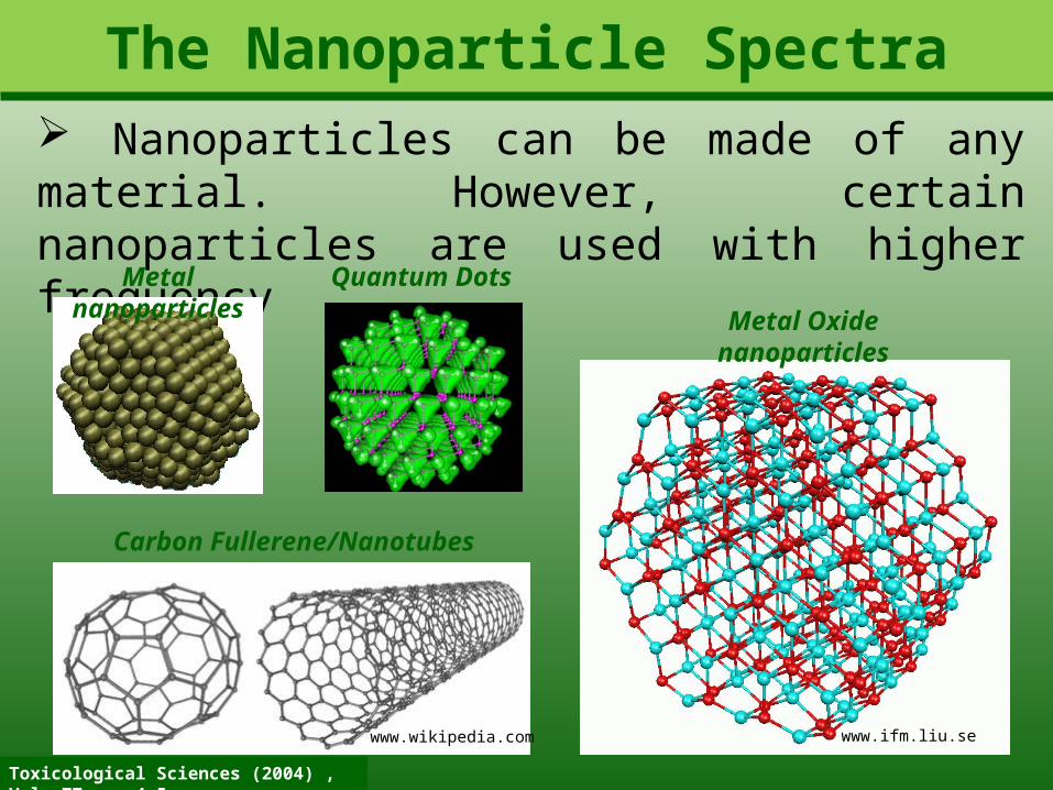
The Nanoparticle Spectra Nanoparticles can be made of any material. However, certain nanoparticles are used with higher frequencyMetal nanoparticles
Carbon Fullerene/Nanotubes
www.wikipedia.com
Metal Oxide nanoparticles
www.ifm.liu.se
Quantum Dots
Toxicological Sciences (2004) , Vol. 77, pp 4-5
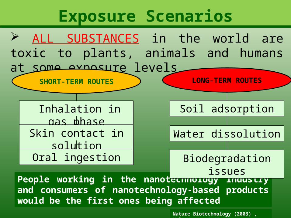
Exposure Scenarios ALL SUBSTANCES in the world are toxic to plants, animals and humans at some exposure levels
Inhalation in gas phase
Skin contact in solution
Oral ingestion
SHORT-TERM ROUTES LONG-TERM ROUTES
Soil adsorption
Water dissolution
People working in the nanotechnology industry and consumers of nanotechnology-based products would be the first ones being affected
Biodegradation issues
Nature Biotechnology (2003) , Vol. 21, pp 1165

Nanotechnology Issues
• Lack of specific regulations on nanotechnology
• Non-mandatory reports on toxicity of products
The risk evaluation for exposure to nanotechnology products is hindered by the law-protected secrecy of product formulations
• Old criteria and methods becoming obsolete
Exposure Measurement Initiatives
Also:
Researchers from the National Institute for Occupational Safety and Health determine the concentration of nanoparticles in the air while unloading a reactor for producing metal oxide nanoparticle
Nature nanotechnology (2007) , Vol. 3, pp 635

Research Uncertainty
Fate Model Template for Nanoparticles
A high number of uncertainties in the data might make studies as the one below meaningless
•A study made on Swiss waters have determined the risk quotient for Ag and TiO2 nanoparticles, and carbon nanotubes
Case study
•The risk quotient is the ratio of current concentration to a threshold concentration expected to be harmful
•Contrary to TiO2, Ag nanoparticles and carbon nanotubes showed quotients less than one
Nature nanotechnology (2008) , Vol. 3, pp 322
Fate models calculate the concentration of nanoparticles in different environmental media and the mass flux between them to assess risks

International Initiative Recently, some agencies have taken some actions to establish regulations to nanotechnology
• US National Nanotechnology Initiative (NNI)
• International Standards Organization (ISO)
• British Standards Institute (BSI)
Nature nanotechnology (2007) , Vol. 3, pp 635
• Environmental Protection Agency (EPA)
Cover of a nanotechnology Safety workshop proceedings
All these agencies have published reports and guidelines related to the handling of nanomaterials and the research approach to nanotoxicology. However, all of them are voluntary to follow

USA Agencies Efforts
NSF Toxic effects of nanoparticles: nanoparticles in air pollution, water purification, nanoscale processes in the environment
DoDToxicological properties of nanomaterials: computational models that will predict toxic, salutary and biocompatible effects based on nanostructured features
Toxicology of manufactured nanomaterials: fate, transport and transformation, human exposure and bioavailability
Transport and transformation of nanoparticles in the environment: exposure and risk analysis, health effectsDoE
NIH Nanomaterials in the body: cell cultures and laboratory use for diagnostic and research tools
NIST Developing measurement tools: tests and analytical methods
EPA
NTP Potential toxicity of nanomaterials: titanium dioxide, several types of quantum dots, and fullerenes
EPA and Nanotechnology: strategy, responsibilities and activities, April 2006

Toxicological Sciences (2004) , Vol. 77, pp 4-5
Research Questions An overview to these reports reveal that the following are the critical issues worth of research
exposure assessment of manufactured nanoparticles
environmental and biological fate, transport, persistence,
and transformation of manufactured nanoparticles
ability to extrapolate manufactured nanoparticle
toxicity using existing particle and fiber toxicological
databases
toxicology of manufactured nanoparticles
recyclability and overall sustainability of manufactured
nanomaterials

PART IIMethodology for Toxicity Studies of Nanoparticles

Considerations A number of points arises when determining the method to test for toxicity:
Duration and route of exposure
Dosage and formulation of test
material
References and standard materials for
comparisons
Biological species to be subject of tests
What to measure?
Nature nanotechnology (2009) , Vol. 4, pp 395

Designing a realistic test
Nature nanotechnology (2009) , Vol. 4, pp 395
Meaningful results on the toxicity of nanomaterials are achieved when the conditions of possible exposure are reproduced accurately
How the nanoparticles would enter the body?
ACCIDENTALLY DELIBERATELY INTRODUCED
• Environmental contamination
• Work place exposure
• Medicine applications
Different methods of exposure of the nanoparticle might produce different results

Rats that were instilled with high doses of SWCNT’s died of respiratory blockage rather than pulmonary intoxication
Example I: Instillation of CNT’s in Rats
Micrograph of Lung Tissue in Rats
Toxicological Sciences (2004) , Vol. 77, pp 117
The picture shows that the respiratory airways are mechanically blocked by carbon nanotubes. This led to the asphyxiation of 15% of the test population
Methods
• Four kind of particles including SWCNT’s
• Pressurized Intratraqueal instillation
• Tracking of alveolar response
• Observation periods at 24h, 1 week, 1 month and 3 months
Results Inflammation, no cytotoxicity

Example II: Inhalation of CNT’s in Rats
Nature nanotechnology (2009) , Vol. 4, pp 451
Exposing rats to air contaminated with CNT’s led to immune-suppression Mechanism for Immune-suppression by
CNT’s
A signal, likely TGFβ, is released when the carbon nanotube is inhaled. This was tested by isolating the BALF protein from both exposed and control rats. It was shown that the protein from exposed mice cause immune-suppression
Methods• Air contaminated with low
concentration CNT’s• Exposure 6h per day during 14 days
• Tracking of proteins and immune response
ResultsImmune-suppression

Test AssessmentsThe methodology used led to different conclusions on the toxicity of CNT’s. Several criteria must be used to select the meaningful results
What are the realistic conditions of CNT exposure in the work place?
Nature nanotechnology (2009) , Vol. 4, pp 409
QUESTIONS
• What test simulates a more realistic exposure?
• Are these results applicable to all nanotubes?
• Are exposure concentration realistic?
•Do animal subjects respond similar to humans?
Reliable data to answer these questions can make the difference between deeming CNT’s unsafe or safe

Need for Updating MethodsThe methods to determine toxicity have to keep up with nanotechnology development. Immunological evaluation is an example of an area where nanoparticles interfere with conventional assays
Nature nanotechnology (2009) , Vol. 4, pp 411
Sources of interference for several assays specific to nanoparticle formulation
The more complex structures and physicochemical properties typical of nanoparticles increases the probability of interference , even more so than traditional pharmaceuticals

Consequence: Modified Tests Developed
Schematics of Rabbit Pyrogen Test
Relevant to nanoparticle-based medical treatments is the potential of fever that would induce organ damage
The test is conducted in two phases. If one animal shows T increase larger than 0.5 K, phase 2 follows suit Nature nanotechnology (2009) , Vol. 4, pp 411
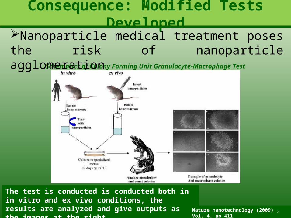
Schematics of Colony Forming Unit Granulocyte-Macrophage Test
Nanoparticle medical treatment poses the risk of nanoparticle agglomeration
The test is conducted is conducted both in in vitro and ex vivo conditions, the results are analyzed and give outputs as the images at the right
Consequence: Modified Tests Developed
Nature nanotechnology (2009) , Vol. 4, pp 411

Switching to In Vitro TestsTraditionally
In vivoIn vitro
Ex vivo
Current Trend
In vitroIn vivo
Ex vivo
advantages
•Cost effective
•Time effective
•No ethical issues
Primary Assay
Primary Assay

A necessity: Increasing the Diversity
Formulation Tests
Methods Subjects
• Type of nanoparticle
• Concentrations
• Solvent, pH,
• Exposure mode
• Exposure time
• Targeted organs• Additives, Coatings
• Rodents• Canines• Swine• Feline• Human Cells
• End points
•In Vitro
•In Vivo
•Ex vivo
Several studies conclude that the response to nanoparticles is too diverse. For instance, dogs and pigs have a higher immune-response to nanoparticles

Improving In Vitro TechniquesSubstituting 2-D in vitro assays for 3-D ones increases the accuracy of the tests. 3-D assays could potentially reproduce better the 3-D structures of cells in in vivo tests
3-D structure of a liver sectionSchematics of a 3-D spheroid scaffold
• Pores (white circles) are created in a hydrogel matrix using a colloidal crystal template. The 5µm spheroid liver cells (grey circles) are grown in the scaffold structure forming a 3-D network of cells
• Concurrent advances in tissue engineering can help to develop more advanced and realistic 3-D assays (introduction of veins, conducts, macrophages, etc…)
Nature nanotechnology (2009) , Vol. 4, pp 342
advantages•Representation of complex cell-cell and cell-matrix
interactions

3D vs. 2D assays: Quantum Dot ToxicityQuantum dot toxicity in liver cells was lower when measured using the 3D scaffold technique, as determined by the cell death rate after exposure to CdTe nanoparticles
Small 2009, 5, pp 1213-1221
3D scaffold micrograph3D 2D
Dead cells are identified with red color after a staining assay. Despite some cell death in the spheroid surface, the death rate is much higher in the 2D tissue
Results in 3D culture correlate much better to animal in vivo studies

Assay Unification IssuesNo consensus in the toxicity particular nanoparticles calls has been achieved due to the lack of unification of toxicity tests.
Summary of toxicity results in CdSe QD
Environmental Health Perspectives 2006, 114, pp 165-172

Other Alternatives: Ab initio SimulationsThe toxicity of nanoparticles (NP) depends on its physical chemical interactions with gases, liquids and other nanoparticles surrounding them. This can be studied using molecular simulations
Interaction of Water Molecules with NP
Nature nanotechnology (2009) , Vol. 4, pp 332
Recent Example Studies
• Surface reactivity of ferrihydride NP assembled in
ferritin (and iron storage protein)
• Nanotoxicological implications of oxygen adsorption at silver
surfaces
The specific and quantitative knowledge obtained from theory and simulation can help building predictive models and algorithms for assessing the likelihood of toxicity in various natural environments

Example I: Anatase (TiO2) toxicityTitanium dioxide (TiO2) a powerful photocatalyst coming in three forms; rutile, anatase and brookite. Anatase form has raised concerns on toxicity
Photocatalytic activity of anatase produces the
reactive (toxic) oxygen species
The (001) anatase surface has been shown to be particularly reactive
The number of (001) facets in an anatase NP
depends on NP size and T
Density Functional Theory (DFT) can predict the
number of these facets depending on size and T
Thought ProcessResults
Number of (001) Facets vs. T and Size
ACS Nano (2008) , Vol. 2, pp 2237
The toxicity of the anatase NP is expected to increase with T and decrease with size

PART IIIToxicology of Carbon
Nanoparticles

Fullerene Disruption of Cell MembranesSome studies has suggested the penetration of fullerenes aggregates through cells, and blood and brain barriers. However, the mechanism is poorly understood
Diffusion Coefficient of Fullerene as It Moves through the Membrane
The results reveal a higher permeability of fullerene through the lipid bilayer is higher than water but lower than hydrocarbon molecules
Nature nanotechnology (2008) , Vol. 3, pp 363
Proposed Toxic Mechanisms
•Membrane Damage
•Disruption of Membrane Elastic Properties
•Chemical Interaction

Fullerene Disruption of Cell Membranes
Migration of a Single Fullerene
Migration of a Fullerene Aggregate
Unbiased MD simulations show that the fullerenes easily pass through the lipid head group, to further diffuse slowly within the bilayer region
Nature nanotechnology (2008) , Vol. 3, pp 363
Average penetration time is 500 ps
Average penetration time is 1 µs
Pore formation appears not to be induced by the
presence of fullerene

Fullerene Disruption of Cell MembranesThe presence of fullerenes inside the membrane appears to barely affect the structure of lipid bilayer
Snapshot of Fullerene Positions Inside the Membrane
Change in the Order Parameter at Different Positions of the Fullerenes
Nature nanotechnology (2008) , Vol. 3, pp 363
Outside of Membrane
Fullerene and fullerene aggregate are kinetically and thermodynamically favored to locate near the center of the membrane
The mechanism of cell disruption due to mechanistic damage of the cell membrane by the fullerene is discarded
Possible mechanism of disruption of cell function is through the change of elastic properties of the
membrane

CNT Disruption of Pleural TissueDue to similarities with asbestos fibers, carbon nanotubes toxicity is usually related to pulmonary illnesses
Setup for aerolization of MWCNT’sBefore and After Aerolization TEM image
of MWCNT’s
Nature nanotechnology (2009) , Vol. 4, pp 747
MWCNT’s are aerolized as mice inhale the aerosol to reproduce doses of 0.2 – 4 mg (MWCNT)/kg (mice)
The exposure time was 6h, and the subsequent retrieval of pulmonary tissue was made after 1 day, 2 weeks, 6 weeks and 14 weeks

CNT Disruption of Pleural Tissue
Macrophages (MØ) ‘eating’ the CNT’s TEM image of CNT’s in sub-pleural cells
After just one day of exposure, carbon nanotubes make their way to several lung structures. They are typically engulfed by macrophages
Nature nanotechnology (2009) , Vol. 4, pp 747
The presence of the carbon nanotubes in the lung tissues is likely to trigger and immune response. Since the nanotubes are trapped by the macrophages the migration of the latter along the lung tissues spread the nanotube to other locations

CNT Disruption of Pleural Tissue
Different Transversal Sections of Lung Tissue
The immune response to the presence of the nanotubes was evaluated by counting the number of mononuclear cell aggregates (macrophages, lymphocytes, etc)
Nature nanotechnology (2009) , Vol. 4, pp 747
Aggregate Counts
There is a correlation between the number of aggregate counts and the dosage of carbon nanotube to the mice. Also, it is seen that carbon black (CB) does not trigger as a strong response as CNT’s

CNT Disruption of Pleural Tissue
Stain Assay Revealing Fibrosis
The immune response to the presence of the nanotubes was evaluated by counting the number of mononuclear cell aggregates (macrophages, lymphocytes, etc)
Nature nanotechnology (2009) , Vol. 4, pp 747
Fibrosis Detailed Images
After two weeks of exposure fibrosis is detected in the sub pleural tissue, as opposed to the control. The detailed image of the fibrosis lesion reveals the correlation with the macrophages and the presence of the CNT’s
control

Carbon Nanotubes: Germ Killers Carbon nanotubes were shown to reduce the viability of E. Coli culture, revealing the germicide effect of pristine carbon nanotubes
SEM image of a normal E. Coli Culture
SEM image of a E. Coli Culture with SWCNT’s
The carbon nanotubes were grown using a cobalt-containing catalyst on a silica support . The nanotubes were washed and stripped of metal traces and used for the culture. Staining assays are able to tell the live cells from the dead cells
Langmuir (2007) , Vol. 23, pp 8671

Carbon Nanotubes: Germ Killers Staining assays reveals the high percentage of dead cells due to the presence of carbon nanotubes
Carbon Nanotube Patch
It is apparent the correlation of the location of the nanotube in the culture and the location of dead cells. Comparison of the fluorescent images for the totality of the cells and the ones dead shows a very high rate of mortality, which was determined to be around 80 %
Fluorescent Image of Culture PI Stained Dead Cells
Langmuir (2007) , Vol. 23, pp 8671

Biodegradation of CNT’s and Toxicity Nanotubes are biodegraded using the human myeloperoxidase hMPO, and further used to evaluate their toxicity to evaluate the impact of biodegradationNanotube Solutions Time-evolution of IR spectra Time-evolution of Raman spectra
The carbon nanotube characteristic bands in the IR and Raman spectra are seen fading as time progresses and their biodegradation advances. The change of these peaks is related to drastic changes in morphology of the nanotubes
Nature nanotechnology (2010) April -Advanced Online Publication

Biodegradation of CNT’s and Toxicity Molecular simulations are used to get insights on the biodegradation mechanism of hMPO on the nanotube.
Nature nanotechnology (2010) April -Advanced Online Publication
A site-localized reaction in which hMPO positive charges favor the binding of nanotubes and radical-supporting aromatic groups participate in the cleavage of the nanotubes
protein
CNT
Attachment of CNT to the protein
Residual Groups Attacking the Nanotube
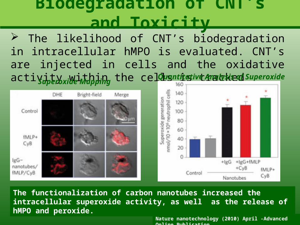
Biodegradation of CNT’s and Toxicity The likelihood of CNT’s biodegradation in intracellular hMPO is evaluated. CNT’s are injected in cells and the oxidative activity within the cells is tracked
Nature nanotechnology (2010) April -Advanced Online Publication
The functionalization of carbon nanotubes increased the intracellular superoxide activity, as well as the release of hMPO and peroxide.
Superoxide MappingQuantitative Analysis of Superoxide

Biodegradation of CNT’s and Toxicity It was demonstrated that biodegraded carbon nanotubes did not cause inflammatory response in pulmonary tissue of mice traqueally instilled with CNT’s
Nature nanotechnology (2010) April -Advanced Online Publication
The images show the formation of granulomas seven days after exposure to pristine carbon nanotubes. On the other hand , the graphs reveal a healthy tissue when the exposure was before biodegraded CNT’s
NanotubesBiodegraded Nanotubes Control

PART IVToxicology of Metal
Nanoparticles
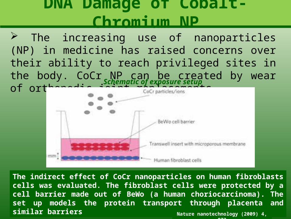
DNA Damage of Cobalt-Chromium NP The increasing use of nanoparticles (NP) in medicine has raised concerns over their ability to reach privileged sites in the body. CoCr NP can be created by wear of orthopedic joint replacements
Schematic of exposure setup
The indirect effect of CoCr nanoparticles on human fibroblasts cells was evaluated. The fibroblast cells were protected by a cell barrier made out of BeWo (a human choriocarcinoma). The set up models the protein transport through placenta and similar barriers
Nature nanotechnology (2009) 4, 873

DNA Damage of Cobalt-Chromium NP Despite the presence of the cell barrier the cell underneath were affected by the presence of the metallic nanoparticles as determined by various methods
Electrophoresis of fibroblast cells
Electrophoresis results reveal DNA damage of the fibroblast cells, as revealed by the difference e in the bands of the control hydrogel and the one corresponding to the assay with CoCr NP. Other functions were also affected such as the frequency of mitosis
Nature nanotechnology (2009) 4, 873

DNA Damage of Cobalt-Chromium NP Metal was observed to be internalized in the barrier, but curiously there were not morphological signs of cell death in the barrier
TEM image
Accumulation of nanoparticles is revealed by TEM images
XEDS
XEDS shows cobalt concentration inside the
barrier decreases
No cellular death
Ions are found to trespass the barrier, and small concentrations of metal are also found past the barrier. However, the damage to the cells underneath is larger when the barrier is present. Therefore, a mechanism involving the barrier must exist to cause the DNA damage
Nature nanotechnology (2009) 4, 873

DNA Damage of Cobalt-Chromium NP
The DNA damage of the cells below the barrier occurs through a chain of events starting with the damage of the mitochondria in the top layer of the cell barrier which end up in secretion of ATP from the bottom layer to the fibroblasts
DNA Damage Mechanism Schematics
Nature nanotechnology (2009) 4, 873

Silver Nanoparticles Toxicity The toxicity of silver nanoparticles was tested using embryos of zebra fish
Nanotechnology (2008) 4, 873
Two kind of nanoparticles were used. One capped with BSA and
the other one with starch
TEM images of Ag Nanoparticles
starch BSA
The coating of the nanoparticle confer them the desired solubility and stability properties in water
Extent of toxicity is to be measure in term of mortality rate, hatching, heart rate and
abnormal phenotypes
Optical characterization

Silver Nanoparticles Toxicity The toxicity of silver nanoparticles was tested using embryos of zebra fish
Nanotechnology (2008) 4, 873
The zebra fish eggs were taken to a 96-well plate, and a solution of silver nanoparticles at different concentrations was added to each well.
Normal Embryo
The images show the appearance of normal, malformed and dead embryos. Visual counting was made
Dead EmbryoMalformed Embryo

Silver Nanoparticles Toxicity It was found that the silver nanoparticles were able to trespass the embryo barrier and settle inside, thus causing the effects to be observed
Nanotechnology (2008) 4, 873
It is possible that the nanoparticles may enter the cells through many routes. Among them, endocytosis through the embryo wall is more likely
TEM Mitochondria TEM NucleusEDS of embryo
Nuclear deposition is believed to create a cascade of toxic
events leading to DNA damage and similar ones

Silver Nanoparticles Toxicity Toxicity End Points
Toxicity end points reveal a concentration-dependent
occurrence of negative events such as death
Nanoparticle deposition in the central nervous system could have adverse effects in the control of cardiac rhythm, respiration and body movements
Exposure to silver nanoparticles resulted as well in accumulation of blood causing edema and necrosis
Nanotechnology (2008) 4, 873

Model of Au-NP Transfer in Sea Ecosystems It is shown that gold nanorods can easily pass from the water column to the marine food web. The study raises awareness due to the debatable toxicity of gold nanoparticles
TEM Gold Nanorods Tidal Marsh Creek Simulated Mesocosmos
A series of estuarine mesocosmos were modeled using tanks containing sea water, sediment, biofilm, plant species and dosed with gold nanorods
Nature Nanotechnology (2009) 4, pp 441-444
The tides were driven using submersible pumps set to timers

Model of Au-NP Transfer in Sea Ecosystems Results shows the recovery of nanoparticle by the biofilm and sediment. This occurs despite the fact the biofilm accounts for less than 0.5 % of the bio mass of the system
Nano-Gold Distribution
A possible explanation for the affinity of marine biofilms and gold is the latter affinity for the negative charge of monomers constituting the polysaccharides and proteins of their structure

PART VConclusions
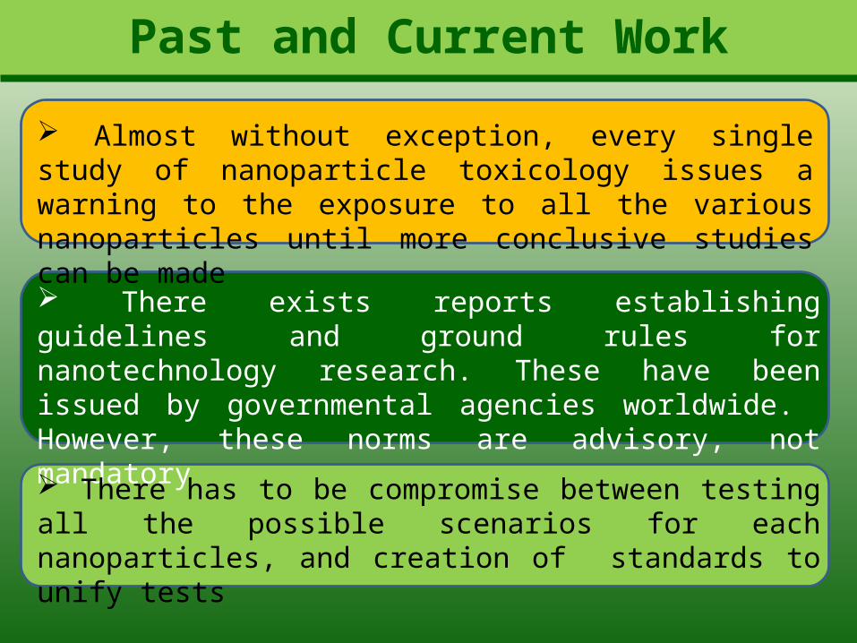
Past and Current Work
Almost without exception, every single study of nanoparticle toxicology issues a warning to the exposure to all the various nanoparticles until more conclusive studies can be made
There exists reports establishing guidelines and ground rules for nanotechnology research. These have been issued by governmental agencies worldwide. However, these norms are advisory, not mandatory
There has to be compromise between testing all the possible scenarios for each nanoparticles, and creation of standards to unify tests

Past and Current Work
Joint efforts by in vivo, ex vivo and in vitro tests, and theoretical studies can help putting together all the pieces of the puzzle to evaluate and understand the mechanisms behind harmful effects of nanoparticles
Nanotechnology is a double-edge sword, the same novel properties making nanoparticles attractive, makes them potentially toxic. Particular care must be taken in nanomedicine, since in this area is where greater exposure would be present

Future Work and Challenges
Completing the whole spectrum of toxicity for all the existent (and potentially new) nanoparticles
Modifying existent and create new assays that are up to the challenge of determining nanoparticle-related toxicity
Issuing new laws that compel companies the exertion of ‘responsible’ nanotechnology that facilitate the implementation of accurate fate models
Determine the exact origin of the toxicity of each nanoparticle to make the pertinent modifications in order to obtain safer products and technologies

?QUESTIONS