nanoparticles for nucleic acid delivery applications in ... · immunotherapy has recently emerged...
TRANSCRIPT

Contents lists available at ScienceDirect
Cancer Letters
journal homepage: www.elsevier.com/locate/canlet
Nanoparticles for nucleic acid delivery: Applications in cancerimmunotherapy
Alvin J. Mukalela, Rachel S. Rileya, Rui Zhanga, Michael J. Mitchella,b,c,∗
a Department of Bioengineering, University of Pennsylvania, Philadelphia, PA, USAbAbramson Cancer Center, Perelman School of Medicine, University of Pennsylvania, Philadelphia, PA, USAc Institute for Immunology, Perelman School of Medicine, University of Pennsylvania, Philadelphia, PA, USA
A R T I C L E I N F O
Keywords:CRISPRmRNADNAGene editingDrug deliveryOncologyNanotechnology
A B S T R A C T
Immunotherapy has recently emerged as a powerful tool for cancer treatment. Early clinical successes fromcancer immunotherapy have led to a growing list of FDA approvals, and many new therapies are in clinical andpreclinical development. Nucleic acid therapeutics, including DNA, mRNA, and genome editing systems, holdsignificant potential as a form of immunotherapy due to its robust use in cancer vaccination, adoptive T-celltherapy, and gene regulation. However, these therapeutics must overcome numerous delivery obstacles to besuccessful, including rapid in vivo degradation, poor uptake into target cells, required nuclear entry, and po-tential in vivo toxicity in healthy cells and tissues. Nanoparticle delivery systems have been engineered toovercome several of these barriers as a means to safely and effectively deliver nucleic acid therapeutics toimmune cells. In this Review, we discuss the applications of nucleic acid therapeutics in cancer immunotherapy,and we detail how nanoparticle platforms have been designed to deliver mRNA, DNA, and genome editingsystems to enhance the potency and safety of these therapeutics.
1. Introduction
Despite advances in understanding the underlying mechanisms ofcancer progression, chemotherapy, radiation, and surgery remain thecurrent standards-of-care for many cancers [1]. The use of these stra-tegies has become more focused and personalized based on the type andstage of disease, which has led to a decline in cancer-related mortalityover the past three decades [2]. However, these therapies are oftenhighly invasive, have substantial adverse side effects, and therapeuticresults are variable [3–5]. Thus, there is a dire need to develop non-invasive, minimally toxic, and highly specific alternatives. Towards thisgoal, cancer immunotherapy has emerged as a powerful alternative toconventional therapies, and substantial research efforts are ongoing toimprove upon their efficacy and safety.
The overarching goal of cancer immunotherapy is to introduce thenecessary molecular tools to harness the immune system to halt diseaseprogression. Thus, immunotherapy can be personalized for specifictypes and stages of cancer, with higher safety profiles and longertherapeutic windows compared to traditional cancer therapeutics [6].The field encompasses several classes of immunotherapy, includinggene therapy, cellular vaccines, checkpoint inhibitors, agonistic anti-bodies, and cytokines [7]. Of these, checkpoint inhibitors and cytokines
are the most widely studied to date, and multiple therapies are cur-rently used in the clinic [7]. More recently, nucleic acid therapeuticsincluding DNA, mRNA, and CRISPR/Cas9 gene editing systems haveemerged as an important branch of cancer immunotherapy. The vastpotential of nucleic acids for treating cancer can be demonstrated bythe use of CRISPR/Cas9 to inactivate PD-1/PD-L1 interactions betweencancer cells and T-cells [8]. In one example of this, CRISPR/Cas9genome editing was used to generate PD-1 deficient anti-CD19 chimericantigen receptor (CAR) T-cells, resulting in enhanced killing of PD-L1+tumor xenografts [8]. Similarly, gene therapy approaches are highlyprevalent in adoptive T-cell immunotherapy to induce T-cells to expressCARs. In 2017, Novartis gained the first FDA approval for a cell-basedgene therapy, Kymriah, which utilizes CAR T-cells to treat leukemia[9,10]. The early success of Kymriah and the ability for CRISPR/Cas9 toenhance T-cell-mediated killing form the basis for the development ofother types of gene therapy to treat cancer, with reduced adverse effectsand higher success rates than traditional approaches [9,10].
Although the examples described above demonstrate the ther-apeutic potential of nucleic acid therapeutics, their translation into theclinic is hindered by several delivery challenges for both ex vivo and invivo applications. Nucleic acids are highly unstable, and they degradequickly in the presence of nucleases before reaching the desired tissues
https://doi.org/10.1016/j.canlet.2019.04.040Received 3 February 2019; Received in revised form 17 April 2019; Accepted 30 April 2019
∗ Corresponding author. Department of Bioengineering, University of Pennsylvania, Philadelphia, PA, USA.E-mail address: [email protected] (M.J. Mitchell).
Cancer Letters 458 (2019) 102–112
0304-3835/ © 2019 Elsevier B.V. All rights reserved.
T

[11]. Further, nucleic acids are unable to enter cells alone, requiring theuse of transfection reagents or physical techniques (such as electro-poration) that are highly toxic to cells ex vivo and are not feasible for invivo use [12,13]. Several nucleic acid therapeutics, such as gene editingcomponents and DNA, are faced with another delivery barrier ofcrossing the nuclear membrane to be transcribed in the nucleus [14].Thus, there is great interest in developing novel delivery platforms thatcan encapsulate and protect nucleic acids, as well as mediate theirdelivery into the desired tissues and cells, in order to exploit theirpowerful therapeutic potential.
Nanoparticles (NPs), which are typically defined as particles thatare 1–1000 nm in diameter, are being developed to overcome the de-livery barriers faced by nucleic acids (Fig. 1). NPs can be comprised of arange of materials such as lipids, polymers, or metals, all of which offerunique delivery advantages that have been thoroughly reviewed else-where [11,15,16]. Importantly, NP features such as material composi-tion, size, and surface chemistry can be carefully engineered for nucleicacid delivery. NPs can encapsulate or bind to nucleic acid therapeuticsvia electrostatic interactions or chemical conjugation to overcome thetherapeutic challenges faced by unbound nucleic acids [6,7]. Ad-ditionally NPs can reduce therapeutic toxicity, by promoting site-spe-cific accumulation and reducing off-target effects. Further, NPs offerprotection over the therapeutic cargo, to avoid nuclease degradationand to extend circulation half-life. In addition to protecting nucleicacids, NPs can be engineered to respond to environmental cues, such asthe acidic environment within solid tumors or within the endosomes ofcells, to degrade and release therapeutic cargo on-demand [17]. Byenabling control over nucleic acid delivery, NPs can minimize toxicityin healthy tissues while maximizing delivery to cancer cells, which maybe highly beneficial for solid tumor immunotherapy [18]. Lastly, NPscan be modified with targeting ligands and other molecules, to promoteboth cellular and nuclear uptake to targeted tissues that overexpress thetargeted protein [19].
Here, we review the design of NP platforms for nucleic acid delivery- including mRNA, DNA, and genome editing therapies – and theirapplications in cancer immunotherapy. Several NP platforms have de-monstrated preclinical success in delivering nucleic acids to target cells,and significant efforts are now underway to translate these technologiesinto the clinic. Of note, lipid NPs (LNPs) complexed with mRNA arecurrently being evaluated in clinical trials of melanoma(NCT02410733) [20]. Further, Alnylam Pharmaceuticals received thefirst FDA approval of an RNA therapeutic for their lipid-siRNA NP,Onpattro, in 2018 [7,21]. Below, we overview applications of NPs fordelivering DNA, mRNA, and genome editing systems for cancer im-munotherapy, and we discuss future directions of gene therapy towardsthe goal of clinical translation.
2. Nanoparticles for DNA delivery
DNA vaccine-based cancer immunotherapy, in which cells aretransfected with plasmid or chemically synthesized DNA to elicit im-mune responses against the encoded antigen, is a powerful tool to en-gage the immune system to attack cancer cells [22]. Early studies inmice demonstrated the ability of DNA plasmids to drive immune re-sponses against transgene products related to influenza, human im-munodeficiency virus-1, and cancer, which established DNA as a pro-mising immunization platform [23]. However, initial clinicalapplications of DNA vaccines revealed only low levels of immunity,indicating that naked DNA was not feasible as an independent vacci-nation strategy largely due to the delivery barriers discussed above(Fig. 1) [23]. For example, the negatively charged DNA typically cannotcross the anionic cell membrane without an exogenous transfectionreagent or delivery vehicle [24], and once within cells, DNA needs tosurpass the nuclear membrane and enter the nucleus [24]. Lastly, it iscritical that DNA is transfected into the desired cells with minimal off-target expression [25–28]. Several physical techniques to improve DNA
delivery including gene guns, electroporation, and sonoporation arecommonly used ex vivo and in small animals [29,30], but they are eithernot feasible for in vivo use, or they are limited to local delivery [24].Utilizing NPs as DNA delivery vehicles can overcome the aforemen-tioned limitations, and several unique applications are described below[24].
Several types of LNPs, including liposomes, ionizable lipids, andpolymer-lipid NPs, have been developed to deliver DNA to target cells.Liposomes were among the first DNA delivery systems and are thefurthest in clinical development, as they are currently used clinically totreat cancer [31]. Liposomes are composed of materials with polar headgroups and non-polar tails, and they spontaneously self-assemble intovesicles at low concentrations [32,33]. Cationic lipids, such as DOTMA,DOTAP and zwitterionic DOPE, are commonly used to form cationicliposomes by exploiting electrostatic interactions between lipids andnegatively-charged nucleic acids (Fig. 2a). When used to encapsulateDNA or other drugs, these cationic liposomes induce stronger ther-apeutic effects than free drug, which has led to several cationic lipo-somal drug formulations advancing into clinical trials [34–38]. How-ever, the use of cationic liposomes is limited due to toxicity at the site ofadministration [39–41], undesired immune responses [42], and clotformation [43], all of which can limit the allowable administered dose[41,44–48].
As an alternative to traditional cationic liposomes, ionizable lipidsthat are neutral at physiologic pH (∼7.4) but ionize under acidicconditions, such as those found within endosomes, have been devel-oped for nucleic acid delivery [49–51]. The ability of these lipids tobuffer endosomal compartments by taking on positive charges canpromote endosomal escape and enable processing of the nucleic acids inthe cytosol [51,52]. Ionizable LNPs typically have 3 components inaddition to the ionizable lipid itself; a fusogenic helper phospholipid(DSPC, DOPE, DOTC, DOTMA, POPC) [53,54], cholesterol to increasestability and membrane fusion [55,56], and a lipid-anchored poly(ethylene glycol) (PEG) to extend their circulatory half-life and de-crease non-specific protein adsorption (Fig. 2a and b) [54]. IonizableLNPs have been used for DNA cancer immunotherapy by encapsulatingCpG (a TLR-9 agonist) oligodeoxynucleotides (ODNs). In this applica-tion, CpG-NPs were subcutaneously co-administered with tumor asso-ciated antigens in murine models of thymoma and melanoma [57]. NPsexhibited preferential accumulation and uptake by immune cells inlymph nodes and augmented antigen-specific immune cell and cyto-kine/chemokine responses, ultimately leading to greater tumor rejec-tion in a murine EG7-OVA tumor model [57]. Although ionizable LNPshave been shown to effectively load nucleic acids of relatively small size(e.g. short synthetic DNA, siRNA, and microRNA), encapsulating largecargo (e.g. pDNA) is challenging [58–60]. Thus, new classes ofpolymer-based NPs, such as polyplexes [61–63], chitosan-based NPs[64,65], and poly(beta-amino esters (PBAEs) [66–68], have been de-veloped to effectively condense pDNA into NPs and enhance transgeneexpression as described below.
Cationic polymeric NPs can be engineered to possess specific phy-sicochemical properties, such as hydrophobicity and charge, due to thediverse range of available polymers and chemical modifications [27].This chemical diversity allows researchers to utilize polymeric NPs fordelivery to a wide array of cell types [69]. Poly(L-lysine) (PLL) is ahomopolymer of the amino acid lysine that has been shown to effec-tively condense DNA (Fig. 2c) [70]. Studies indicate that PLL generallyhas low transfection success, likely due to its low rate of endosomalescape [24]. However, one study used PLL-coated polystyrene NPs todeliver pDNA encoding OVA antigen as a model for a DNA-based pro-phylactic cancer vaccine against EG7 tumor cells [70]. Two vaccina-tions with these NPs inhibited tumor growth following a EG7 tumor cellchallenge in mice [70]. Notably, immature dendritic cells (DCs) hadhigher levels of NP uptake compared to mature DCs [70], which islikely due to the reduced endocytic and phagocytic rates in mature DCsthat lowers their capacity to internalize and process antigens [70].
A.J. Mukalel, et al. Cancer Letters 458 (2019) 102–112
103

Similar to PLL, polyethylenimine (PEI) is another cationic polymerthat is often used as a “gold-standard” for transfection efficacy (Fig. 2c)[24,71]. PEI exerts a high charge density at low pH, which enhancesendosomal escape and makes it a potent transfection reagent, but it alsoconfers high cytotoxicity [72–76]. Longer chain and higher chargedensity PEIs tend to have damaging interactions with cellular mem-branes that lead to potent cytotoxicity, and several strategies haveemerged to address this including branched architectures, biodegrad-ability, and PEG-grafting [74,75,77]. In one instance, modified bran-ched PEI was synthesized to improve upon the cytocompatibility andtransfection efficiency of unmodified PEI [74]. Of note, succinylatedPEI induced better siRNA-mediated knockdown and 10-fold lower
polymer toxicity compared to unmodified PEI [74]. This demonstratesthe importance of balancing transfection efficiency and biocompat-ibility when designing PEI-based delivery vehicles. The high transfec-tion ability of PEI was exploited for cancer immunotherapy by con-densing IL-12-encoding pDNA [78]. This therapy was administered asan aerosol to mice bearing SAOS-LM7 tumors in a murine model ofosteosarcoma lung metastasis [78]. Mice that received aerosolized PEI-IL-12 gene therapy exhibited IL-12 expression only in the lungs and hadsignificantly fewer lung metastases than untreated controls [78]. Theability of PEI to condense DNA is also applicable to newer polymer-based NP delivery platforms, such as PBAEs, described below.
PBAEs are simple to synthesize and they provide an additional
Fig. 1. The role of NPs in overcoming extracellular and intracellular barriers for nucleic acid delivery. In the circulation, NPs need to protect nucleic acids from serumendo- and exo-nucleases, evade immune detection, and avoid non-specific protein interactions within the blood. Further, NPs must avoid renal clearance (achievedthrough size modulation), while also promoting extravasation from the blood and into target tissues, upon which they promote cellular uptake and localization intothe cytosol or nucleus. Adapted with permission from Ref. [24].
A.J. Mukalel, et al. Cancer Letters 458 (2019) 102–112
104

benefit of having tunable biodegradation (Fig. 2c) [79]. A major ad-vantage of their simple, parallelizable synthesis is the ability to gen-erate diverse libraries of PBAE structures that can be screened for DNAdelivery to identify key structures for potent gene delivery [80]. In thecontext of immunotherapy, PBAE NPs functionalized with an anti-CD3eT-cell-targeting antibody fragment were used to deliver leukemia spe-cific CD194-1BBz CAR pDNA to T-cells in situ in a murine leukemiamodel (Fig. 3a) [81]. These NPs also contained microtubule-associatedsequence (MTAS) and nuclear localization signal (NLS) peptides tomediate nuclear translocation of the therapeutic pDNA cargo [81]. NP-programmed CAR T-cells generated tumor regression similar to that oftraditionally prepared CAR T-cells, with only a small portion of NPstransducing phagocytic cells, likely due to successful antibody targetingto T-cells [81]. PBAEs have also been used to deliver cyclic dinucleo-tides (a STING agonist) or CpG nucleic acid adjuvants (Fig. 3b) [82,83].Notably, results from these studies indicated that the PBAE:DNA ratio isa critical factor for NP stability and in vivo functionality. Specifically,PBAE-NPs with higher PBAE:DNA ratios yielded better protection of theCpG cargo. However, lower ratios exhibited better CpG uptake andactivation of tumor-specific T-cells, resulting in improved survival in amouse melanoma model [82,83].
With the aid of NP delivery systems, DNA-based therapeutics have
shown great promise in the field of cancer immunotherapy [84]. Al-though using pDNA as an antigen source has shown encouraging out-comes in many preclinical studies, the same success has not been foundin human clinical trials, and interest in using DNA as antigen sourceshas decreased [84,85]. However, there is substantial ongoing work todevelop DNA NPs in immunotherapy for CAR T-cells or as adjuvants[81,82,86]. More recently, mRNA has emerged as a potent tool for geneimmunotherapy for cancer, and several unique applications are de-scribed below.
3. Nanoparticles for mRNA delivery
Early interest in mRNA stemmed from its use as an alternative toconventional and DNA-based vaccines [87]. mRNA therapeutics are apromising alternative to DNA owing to their lower mutational risk,fewer intracellular delivery barriers, and transient expression[24,88,89]. Further, mRNA only needs to cross the cell membrane andreach the cytosol – in contrast to DNA which requires nuclear entry - toinduce protein translation [88–90]. Finally, protein expression inducedby mRNA is transient and does not require integration into the genome,thereby avoiding the risk of insertional mutagenesis that can occur fromDNA [24,88,89]. When used as a vaccine, mRNAs encoding for antigens
Fig. 2. Chemical structures of lipids and polymers used to engineer NPs for nucleic acid delivery. A. Common lipids used for liposomal formulations includingDOTMA, DOSPA, DOTAP, DMRIE and DC-cholesterol, which are used to condense and encapsulate nucleic acids. Structurally, cationic lipids are defined as having acationic head group, linker region, and hydrophobic tails. B. Ionizable lipid LNP formulations are comprised of four components: ionizable lipids, such as C12-200,phospholipids (DOPE, DSPC), cholesterol, and lipid-anchored PEG. C. Cationic polymers and biopolymers used as vectors for nucleic acid delivery. PEI and PLL weretwo of the initial vectors used for DNA delivery but are faced with safety (PEI) and efficacy (PLL) concerns. PBAEs and pDMAEMA are newer polymer vectorsdeveloped for nucleic acid delivery with improved safety and efficacy. Adapted with permission from Ref. [24].
A.J. Mukalel, et al. Cancer Letters 458 (2019) 102–112
105

are delivered to antigen presenting cells, either through ex vivo trans-fection or under systemic administration. Antigen presenting cells thentranslate mRNA into its encoded cancer-associated antigen that is pre-sented to T-cells for activation and induction of cytotoxic T lymphocyteresponses [87]. However, the large size (103-105 nucleotides), negativecharge, and hydrophilicity of mRNA, combined with its susceptibility tonucleases, hinder the ability of naked mRNA to reach and enter targetcells upon systemic administration [14,24,88–90]. NPs can overcomethese barriers and facilitate its intracellular delivery, and several NPplatforms for mRNA delivery are described below.
Similar to DNA delivery, ionizable LNPs have also been used formRNA delivery. In one example of this, LNPs comprised of an ionizablelipid, a helper phospholipid, cholesterol, lipid-anchored PEG , andmRNA were designed to induce expression of luciferase and ery-thropoietin following systemic injection in BALB/c mice [91]. Thestudy utilized Design of Experiment (DoE) methodology to optimize atop-performing LNP for siRNA delivery to now deliver mRNA to theliver of mice [91]. In the context of immunotherapy, multilamellarionizable LNPs were used to deliver tyrosine-related protein 2 (TRP2)and glycoprotein 100 (gp100) tumor self-antigen mRNAs to antigenpresenting cells to induce a cytotoxic CD8 T-cell response (Fig. 3d).Subcutaneous administration of these LNPs led to reductions in tumorvolume, extended survival in a B16F10 tumor model, and yielded po-tent CD8+ activation [49]. Interestingly, these LNPs were able totransfect neutrophils, macrophages, and dendritic cells, demonstratingthat they may be useful to deliver mRNA to a range of immune cells.
Similar to DNA delivery, the chemical diversity of polymers and
polymer-lipid systems allows for identification and incorporation ofstructures that can improve biocompatibility and bioavailability of theencapsulated materials [27]. Polymers used for mRNA delivery arepositively charged and can condense mRNA into nanometer sizedelectrostatic complexes [88]. PBAEs are a major class of pH-responsiveand bioreducible polymers known for their biocompatibility, but theyhave had limited in vivo success due to their poor serum stability[92,93]. Recent PBAE work has explored the incorporation of PEG-li-pids and new PBAE architectures to improve uptake, stability, andbiodistribution [94–96]. In one approach, PBAEs were modified to in-clude internal alkyl tails to enable their formulation with PEG-lipidsand improve their stability under physiologic conditions [96]. mRNA-loaded PBAE terpolymer NPs formulated with PEG-lipids demonstratedgreater serum stability in vitro compared to those formulated withoutPEG-lipids, and they achieved selective luciferase expression in thelungs of mice following intravenous injection [96]. PBAEs have alsobeen used for mucosal immunization, where lipid-enveloped NPs withpH-responsive PBAE cores delivered mRNA to immune cells [97].Mucosal immunization has drawn interest for two reasons: 1) manypathogens invade through mucosal surfaces and 2) mucosal im-munizations can elicit both systemic and mucosal immunity [98,99].These NPs successfully delivered GFP mRNA to difficult-to-transfectdendritic cells in vitro as well as luciferase mRNA to mucosal tissuecompartments following intranasal administration in vivo [97].
Vaccination is a central application of mRNA for cancer im-munotherapy. Electroporation is typically used to introduce mRNA intoT-cells ex vivo, which can induce cellular toxicity [13]. Further, this ex
Fig. 3. NPs for nucleic acid delivery and their applications in cancer immunotherapy. A. A PBAE polymer functionalized with an MTAS-NLS peptide was used tocondense CAR-encoding plasmid DNA. In this application, an anti-CD3e-poly(glutamic acid) (PGA) conjugate was adsorbed to the surface of the PBAE core to enableT-cell targeting and in situ generation of CAR T-cells. Adapted with permission from Ref. [81]. B. PBAE polymer was used to deliver a Stimulator of InterferonReceptor Genes (STING) antagonizing cyclic dinucleotide (CDN) in combination with a PD-1 blocking antibody and demonstrated potent inhibition of tumor growth.Adapted with permission from Ref. [83]. C. A biodegradable ionizable lipid was used to co-deliver a modified sgRNA and Cas9 mRNA that achieved potent geneediting in the liver for 12 weeks. sgRNA was modified with phosphothiorate bonds at both ends of the strand (indicated by *) and 2′-O-methylation of nucleotides(shown in red). Adapted with permission from Ref. [126]. D. Multilamellar ionizable LNPs generated potent CD8 T-cell activation upon antigen delivery, and wereused to deliver tumor antigens gp100 and TRP2 that led to tumor shrinkage and elongated survival in a B16F10 melanoma mouse model. Adapted with permissionfrom Ref. [49]. (For interpretation of the references to color in this figure legend, the reader is referred to the Web version of this article.)
A.J. Mukalel, et al. Cancer Letters 458 (2019) 102–112
106

vivo cell engineering process is time, labor, and cost intensive, whichcreates significant challenges towards broader clinical translation[100]. Thus, NPs are ideal to deliver mRNA to T-cells without the needfor electroporation. An early mRNA cancer vaccine approach utilizedPEGylated histidine-rich polylysines mixed with L-histidine-(N,N-di-n-hexadecylamine)ethylamide (HDHE) and cholesterol liposomes, re-ferred to as histidylated lipopolyplexes, to deliver mRNA encodinghuman melanoma antigen MART1 to T-cells. Immunization withMART1 histidylated lipoplexes induced priming of B16-specific CD4+
and CD8+ T-cells, leading to a ∼10-fold reduction in tumor volumeand a 75% reduction in detectable lung metastases compared to controlmice in a B16/F10 melanoma model [101]. More recently, PBAE NPswere coated with CD3 or CD8 antibodies and used to target T-cells inorder to induce receptor-mediated endocytosis. Antibody-coated NPsimproved ex vivo T-cell transfection 10-fold compared to non-targetedPBAE NPs [12]. Building upon this success, this robust platform hasbeen used to deliver two different mRNAs. In one study, these NPsdelivered megaTAL nuclease mRNA to knockout endogenous T-cellreceptors that may cause graft-versus-host disease [12]. Separately, NPsloaded with an mRNA encoding the Foxo13A transcription factor wereused to guide CD62L+ T-cells away from terminal differentiation andsenescence, and towards a central memory phenotype [12]. Taken to-gether, these results indicate that this robust platform can be adapted togenerate several distinct immune responses.
Recently, mRNA vaccines have begun testing in clinical trials. Theclinical translation of mRNA vaccines is being led, in part, by Curevac(NCT03291002) with an RNA-based adjuvant that is being tested inpatients with melanoma and squamous cell carcinoma, among others.With the introduction of these vaccines into the clinic, researchers areincreasingly working towards introducing mRNA delivery systems aswell. LNPs complexed with NY-ESO-1, MAGE-A3, tyrosinase, and TPTEmRNA are currently being evaluated in clinical trials. Early phase 1dose escalation data has demonstrated that neutral or negativelycharged LNP-mRNA complexes are well tolerated, and there were dosedependent IFN-α and antigen-specific T-cell responses in three mela-noma patients [20]. Together with the preclinical studies describedabove, this demonstrates that NPs can aid in overcoming challengesassociated with mRNA delivery, and enable potential use for multipleforms of cancer immunotherapy.
4. Nanoparticles for gene editing
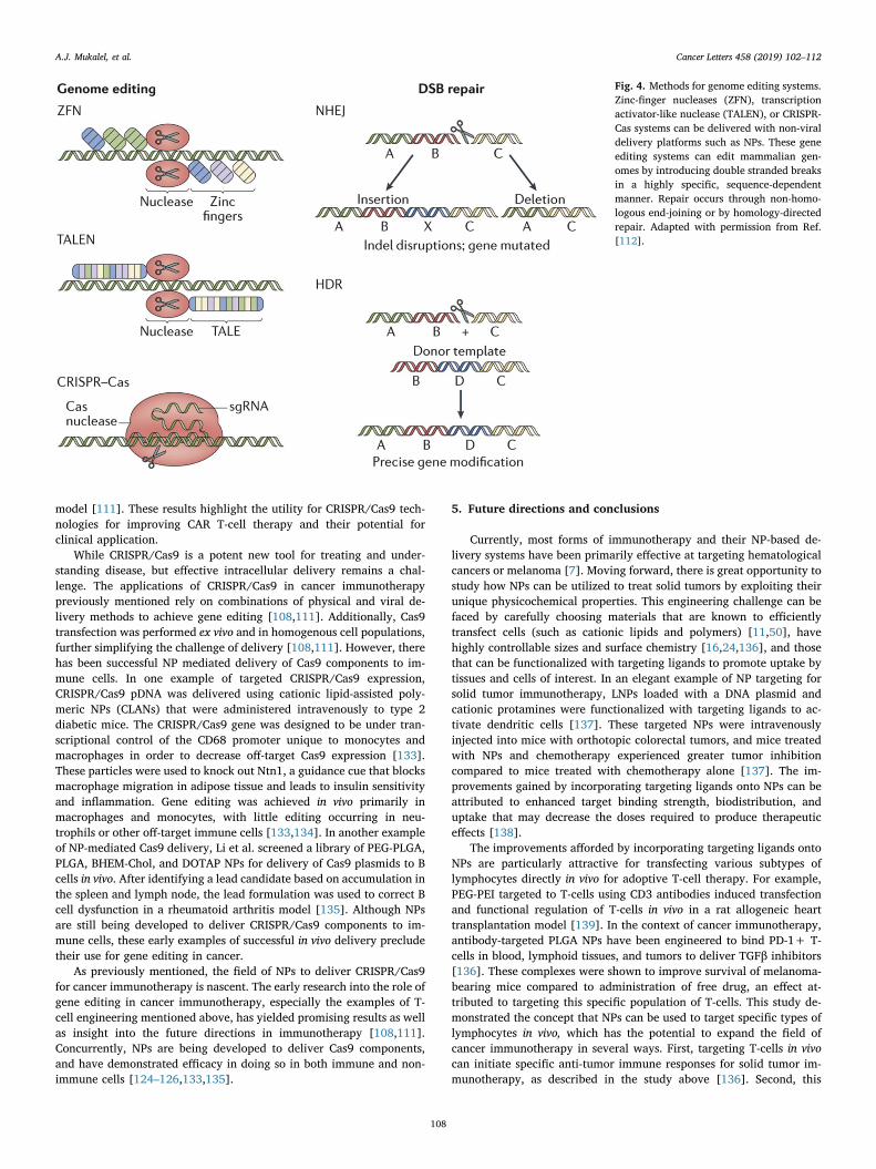
CRISPR/Cas9 has emerged as a powerful tool in understanding andtreating the genetic cause of various diseases (Fig. 4) [102,103]. In thecontext of cancer immunotherapy, CRISPR has been applied to diseasemodeling [104–106], target identification [107], and immune cell en-gineering [108–111]. CRISPR/Cas9 can be delivered as a nucleic acid-loaded protein (ribonucleoprotein, or RNP) or as nucleic acids[112,113]. Protein delivery presents several challenges, and strategiesfor overcoming these barriers have been reviewed elsewhere[114–116]. In contrast to nucleic acids, the chemical diversity and sizeof proteins often necessitates modifications to enable delivery with avector [117,118]. For example, a successful approach is to “super-charge” proteins through the addition of densely charged moieties toenable electrostatic complexation with carriers [117,118], and thisapproach has been particularly effective in the localized delivery ofCas9 RNP NPs [119–121].
Delivery of Cas9 protein and mRNA offer transient protein expres-sion, which is potentially beneficial because constitutive Cas9 expres-sion can increase the risk for off-target editing and stimulation of pre-existing adaptive immune responses to the Cas9 protein [122–124].However, the most successful attempts at formulating Cas9 RNP intoNPs have been limited to localized delivery, whereas Cas9 mRNA hasbeen successfully delivered via systemic administration[119–121,124–126]. Since CRISPR/Cas9 technology is still relativelynew, there have been few attempts to deliver Cas9 components using
NPs for the purpose of cancer immunotherapy. Here, we highlight NPdelivery systems that have delivered CRISPR/Cas9 to treat non-cancerdiseases, as well as viral delivery mechanisms for cancer im-munotherapy. Moving forward, we anticipate that the critical insightsgained from the gene editing studies discussed below will form the basisfor NP-mediated gene editing for cancer immunotherapy.
One of the first successful Cas9 NP approaches utilized viral andnon-viral NP delivery to achieve homology directed repair in hepato-cytes in a mouse model of hereditary tyrosinemia type 1 [124]. A li-pidoid-based LNP was used to encapsulate Cas9 mRNA and, due to sizeconstraints, the sgRNA expression cassette and homology directed re-pair template were delivered using an adeno-associated virus (AAV)[124]. After optimizing the timing of LNP and AAV administration tomaximize the overlap between peak Cas9 and sgRNA expression, thissystem achieved gene editing in 6% of hepatocytes at a 24.1% indel ratemeasured by deep sequencing of the target locus in total liver genomicDNA [124]. In a different proof-of-concept study, ionizable lipids andhelper lipids were used to co-deliver Cas9 mRNA and modified sgRNAto reduce serum concentrations of transthyretin, and achieved 70%gene editing and>97% knockdown in hepatocytes following a singleintravenous injection in mice (Fig. 3c) [126]. In another example of NP-mediated Cas9 delivery, Miller et al. synthesized a library of zwitter-ionic amino lipids (ZALs) to co-deliver Cas9 mRNA and Lox sgRNA tomice expressing a Lox-Stop-Lox tdTomato cassette, and demonstratedstable gene editing two months after NP administration [125]. ZALsdemonstrated potent protein expression with luciferase mRNA atdoses< 600 pM in vitro and l mg/kg in vivo [125]. Together, these earlyexamples of NP delivery platforms for genome editing preface their usefor CRISPR/Cas9 nucleic acid delivery for cancer immunotherapy.
In the preceding text, we highlighted successful preclinical appli-cations of NPs for the delivery of Cas9 components. Here, we describethe use of CRISPR/Cas9 for cancer immunotherapy mediated by viraland physical delivery methods. One important use of CRISPR/Cas9 inimmunotherapy is towards more robust T-cell engineering. AllogeneicCAR T-cells are an attractive alternative to traditional autologous CART-cells because they can be distributed “off-the-shelf” to patients[127,128]. However, allogeneic transplant T-cell receptors (TCRs) canbe reactive to host antigens in healthy tissues, leading to graft-versus-host disease (GVHD) [128,129]. Additionally, alloantigens present ontransplanted cells, such as human leukocyte antigen-1 (HLA-1), canelicit unwanted host immune responses [128,129]. CRISPR/Cas9 couldbe employed to knock-out surface molecules to improve the compat-ibility of allogeneic CAR T-cells [128–131]. An early study utilized acombination of CRISPR mRNA and gRNA to target the T-cell Receptoralpha-constant (TRAC) locus and knock out TCRs. Subsequent trans-fection with an AAV encoding CAR cDNA was used to induce expressionof CD19-specific CAR under transcriptional control of the TRAC pro-moter. These T-cells were more resistant to tonic signaling and haddelayed differentiation and exhaustion, ultimately leading to greatertumor rejection when compared to retrovirally transduced CARs, bothwith and without TCR knockout [108]. CRISPR/Cas9 targeting enabledthe identification of a specific transcriptional regulator that modulatedCAR expression to maximize therapeutic benefit.
While single gene editing has several applications, the simplicity ofCRISPR/Cas9 gives rise to the capability of multiplexed gene editing tosimultaneously knock-out several genes. In one example, Cas9 mRNAand gRNAs were delivered to primary T-cells via electroporation andused to knock out PD-1, a suppressor of CD8 T-cell activity, in additionto TCR and HLA-1 [111,132]. A lentivirus was used to transduce CD19or prostate-stem cell antigen (PSCA) CARs. Double knockout (TCR- andHLA-1-) CAR T-cells yielded reduced alloreactivity compared to singleknockout (TCR-) CAR T-cells while maintaining potent antitumor ac-tivity, measured by enhanced survival in a Nalm6 mouse tumor model[111]. Triple knockout (TCR-, HLA-1-, and PD-1-) CAR T-cells demon-strated quicker and complete elimination of tumor cells compared todouble-ablated CAR T-cells in an aggressive Nalm6-PDL1 leukemia
A.J. Mukalel, et al. Cancer Letters 458 (2019) 102–112
107
model [111]. These results highlight the utility for CRISPR/Cas9 tech-nologies for improving CAR T-cell therapy and their potential forclinical application.
While CRISPR/Cas9 is a potent new tool for treating and under-standing disease, but effective intracellular delivery remains a chal-lenge. The applications of CRISPR/Cas9 in cancer immunotherapypreviously mentioned rely on combinations of physical and viral de-livery methods to achieve gene editing [108,111]. Additionally, Cas9transfection was performed ex vivo and in homogenous cell populations,further simplifying the challenge of delivery [108,111]. However, therehas been successful NP mediated delivery of Cas9 components to im-mune cells. In one example of targeted CRISPR/Cas9 expression,CRISPR/Cas9 pDNA was delivered using cationic lipid-assisted poly-meric NPs (CLANs) that were administered intravenously to type 2diabetic mice. The CRISPR/Cas9 gene was designed to be under tran-scriptional control of the CD68 promoter unique to monocytes andmacrophages in order to decrease off-target Cas9 expression [133].These particles were used to knock out Ntn1, a guidance cue that blocksmacrophage migration in adipose tissue and leads to insulin sensitivityand inflammation. Gene editing was achieved in vivo primarily inmacrophages and monocytes, with little editing occurring in neu-trophils or other off-target immune cells [133,134]. In another exampleof NP-mediated Cas9 delivery, Li et al. screened a library of PEG-PLGA,PLGA, BHEM-Chol, and DOTAP NPs for delivery of Cas9 plasmids to Bcells in vivo. After identifying a lead candidate based on accumulation inthe spleen and lymph node, the lead formulation was used to correct Bcell dysfunction in a rheumatoid arthritis model [135]. Although NPsare still being developed to deliver CRISPR/Cas9 components to im-mune cells, these early examples of successful in vivo delivery precludetheir use for gene editing in cancer.
As previously mentioned, the field of NPs to deliver CRISPR/Cas9for cancer immunotherapy is nascent. The early research into the role ofgene editing in cancer immunotherapy, especially the examples of T-cell engineering mentioned above, has yielded promising results as wellas insight into the future directions in immunotherapy [108,111].Concurrently, NPs are being developed to deliver Cas9 components,and have demonstrated efficacy in doing so in both immune and non-immune cells [124–126,133,135].
5. Future directions and conclusions
Currently, most forms of immunotherapy and their NP-based de-livery systems have been primarily effective at targeting hematologicalcancers or melanoma [7]. Moving forward, there is great opportunity tostudy how NPs can be utilized to treat solid tumors by exploiting theirunique physicochemical properties. This engineering challenge can befaced by carefully choosing materials that are known to efficientlytransfect cells (such as cationic lipids and polymers) [11,50], havehighly controllable sizes and surface chemistry [16,24,136], and thosethat can be functionalized with targeting ligands to promote uptake bytissues and cells of interest. In an elegant example of NP targeting forsolid tumor immunotherapy, LNPs loaded with a DNA plasmid andcationic protamines were functionalized with targeting ligands to ac-tivate dendritic cells [137]. These targeted NPs were intravenouslyinjected into mice with orthotopic colorectal tumors, and mice treatedwith NPs and chemotherapy experienced greater tumor inhibitioncompared to mice treated with chemotherapy alone [137]. The im-provements gained by incorporating targeting ligands onto NPs can beattributed to enhanced target binding strength, biodistribution, anduptake that may decrease the doses required to produce therapeuticeffects [138].
The improvements afforded by incorporating targeting ligands ontoNPs are particularly attractive for transfecting various subtypes oflymphocytes directly in vivo for adoptive T-cell therapy. For example,PEG-PEI targeted to T-cells using CD3 antibodies induced transfectionand functional regulation of T-cells in vivo in a rat allogeneic hearttransplantation model [139]. In the context of cancer immunotherapy,antibody-targeted PLGA NPs have been engineered to bind PD-1+ T-cells in blood, lymphoid tissues, and tumors to deliver TGFβ inhibitors[136]. These complexes were shown to improve survival of melanoma-bearing mice compared to administration of free drug, an effect at-tributed to targeting this specific population of T-cells. This study de-monstrated the concept that NPs can be used to target specific types oflymphocytes in vivo, which has the potential to expand the field ofcancer immunotherapy in several ways. First, targeting T-cells in vivocan initiate specific anti-tumor immune responses for solid tumor im-munotherapy, as described in the study above [136]. Second, this
Fig. 4. Methods for genome editing systems.Zinc-finger nucleases (ZFN), transcriptionactivator-like nuclease (TALEN), or CRISPR-Cas systems can be delivered with non-viraldelivery platforms such as NPs. These geneediting systems can edit mammalian gen-omes by introducing double stranded breaksin a highly specific, sequence-dependentmanner. Repair occurs through non-homo-logous end-joining or by homology-directedrepair. Adapted with permission from Ref.[112].
A.J. Mukalel, et al. Cancer Letters 458 (2019) 102–112
108

technique can be used to generate CAR T-cells directly in vivo to over-come the many manufacturing limitations of in vitro CAR T-cell devel-opment, including high costs and production time [12]. Lastly, theability to activate specific subpopulations of immune cells in circulationmay be exploited to treat metastatic secondary tumors in addition toprimary tumors, as these engineered cells can target any cancer cellsthroughout the body that express the target protein [101].
Another means of utilizing nanotechnology for immunotherapy insolid tumors is by alleviating immunosuppressive signaling within themicroenvironment to improve native T-cell responses (Fig. 5) [140].For example, delivering siRNA against Snail, a critical transcriptionfactor that accelerates cancer metastasis by inducing immunosuppres-sion, to tumor cells was shown to promote the infiltration of tumor-specific lymphocytes into melanoma tumors [140]. This increase inlymphocyte infiltration led to inhibited primary and metastatic tumorgrowth [140]. By utilizing NPs to enhance anti-tumor immune re-sponses against solid tumors, researchers can exploit the inherentfunction of immune cells to attack cancer cells, which may be moreeffective for treating solid tumors compared to treating tumor cellsthemselves [141]. Moving forward, this technique to potentially makesolid tumors more susceptible to native immune activity could becombined with in vivo targeting of immune cells for a multi-prongedapproach to solid tumor immunotherapy [141].
In addition to developing delivery systems to enable solid tumorimmunotherapy, it is also critical to engineer NPs to deliver geneediting tools [24]. Previously, we discussed the use of NPs to deliverCRISPR/Cas9 gene editing material to immune cells to treat diabetesand rheumatoid arthritis, as well as the viral delivery of gene editingtechnology for cancer immunotherapy [108,111,133,135]. There areseveral challenges to address as researchers develop nanotechnology todeliver gene editing components in vivo for cancer immunotherapy. Acritical consideration is developing NPs that offer precise control overtiming of nucleic acid delivery and release, as successful gene editingrequires delivery of both guide RNA as well as the Cas9 protein ormRNA [124]. This challenge can be met by developing NPs using
materials with highly tunable degradation profiles to release the nucleicacid cargo on-demand [18]. Additionally, NPs carrying gene editingcomponents need to successfully edit a sufficient number of cells tomediate the desired therapeutic result, which can be challenge in vivowhere immune cells are circulating throughout the body. This re-quirement can be met by attaching targeting ligands to NPs to promotetheir binding and uptake to target cells [19,136]. Ultimately, NPs thatcan deliver gene editing technology for cancer immunotherapy canexploit the delivery capabilities of the carriers as well as the highlyspecific editing afforded by CRISPR/Cas9 systems.
The NP delivery platforms discussed in this article represent noveland recent developments in nucleic acid delivery, with several appli-cations in cancer immunotherapy. However, challenges remain beforethese systems can be used broadly in the clinic. For example, several ofthe examples discussed above utilize materials that have not yet beenused in clinical trials or are not yet approved by the FDA. NPs com-prised of FDA approved materials may have a simpler and expeditedpath towards clinical translation. Importantly, several NP-based genetherapeutics are in clinical trials or have been recently approved by theFDA. For example, in 2018 Alnylam Pharmaceuticals gained FDA ap-proval for their lipid-siRNA NP Onpattro to treat polyneuropathycaused by transthyretin amyloidosis [142]. This introduction of NP-nucleic acid complexes into the clinic is a critical milestone for theentry of other NP-based immunotherapies into clinical trials and ulti-mately for FDA approval. As new NP delivery systems enter the clinic,physicians and scientists can begin to shift the current paradigm ofcancer therapy towards potent and biocompatible nucleic acid deliverysystems for immunotherapy.
Author contributions
A.J.M, R.S.R., R.Z., and M.J.M. conceived the ideas, performed re-search for the manuscript, discussed the manuscript content and wrotethe manuscript. All authors reviewed and edited the manuscript beforesubmission.
Fig. 5. Sites of therapeutic intervention forNPs to generate anti-tumor immune re-sponses in solid tumors. Anti-tumor im-mune responses result from the presentationof tumor-associated antigens (TAAs), sti-mulating protective T-cell responses, andovercoming the immunosuppressive tumormicroenvironment (TME). NPs can be usedto activate these pathways to successfullydeliver immunotherapeutics to solid tumorsby: (1) enhancing delivery of nucleic acidsencoding TAAs to improve delivery to an-tigen presenting cells for immune activa-tion; (2) delivering nucleic acids to T-cellsto promote their survival, proliferation, andanti-tumor phenotypes; and (3) alleviatingthe immunosuppressive signaling within thetumor microenvironment. Figure adaptedwith permission from Ref. [141].
A.J. Mukalel, et al. Cancer Letters 458 (2019) 102–112
109

Competing interests
The authors have no conflicts of interest to declare.
Acknowledgements
The authors acknowledge support from a Burroughs Wellcome FundCareer Award at the Scientific Interface (CASI), a US National Institutesof Health (NIH) Director's New Innovator Award (DP2 TR002776), agrant from the American Cancer Society (129784-IRG-16-188-38-IRG),an Abramson Cancer Center (ACC)-School of Engineering and AppliedSciences (SEAS) Discovery Grant (P30 CA016520), and a 2018 AACR-Bayer Innovation and Discovery Grant, Grant Number 18-80-44-MITC(to M.J.M.). R.S.R. is supported by an NIH T32 multidisciplinarytraining grant (T32 HL007954). A.J.M. is supported by an NSFGraduate Research Fellowship.
References
[1] H.J. Burstein, L. Krilov, J.B. Aragon-Ching, N.N. Baxter, E.G. Chiorean,W.A. Chow, J.F. De Groot, S.M. Devine, S.G. DuBois, W.S. El-Deiry, Clinical canceradvances 2017: annual report on progress against cancer from the american so-ciety of clinical oncology, J. Clin. Oncol. 35 (2017) 1341–1367.
[2] R.L. Siegel, K.D. Miller, A. Jemal, Cancer statistics, 2018, CA A Cancer J. Clin. 68(2018) 7–30.
[3] N. Lameire, Nephrotoxicity of recent anti-cancer agents, Clinical Kidney Journal 7(2014) 11–22.
[4] T.M. Suter, M.S. Ewer, Cancer drugs and the heart: importance and management,Eur. Heart J. 34 (2012) 1102–1111.
[5] R. Baskar, J. Dai, N. Wenlong, R. Yeo, K. Yeoh, Biological response of cancer cellsto radiation treatment, Frontiers in Molecular Biosciences 1 (2014) 24.
[6] L. Milling, Y. Zhang, D.J. Irvine, Delivering safer immunotherapies for cancer,Adv. Drug Deliv. Rev. 114 (2017) 79–101.
[7] R.S. Riley, C.H. June, R. Langer, M.J. Mitchell, Delivery technologies for cancerimmunotherapy, Nat. Rev. Drug Discov. (2019) 175–196.
[8] L.J. Rupp, K. Schumann, K.T. Roybal, R.E. Gate, C.J. Ye, W.A. Lim, A. Marson,CRISPR/Cas9-mediated PD-1 disruption enhances anti-tumor efficacy of humanchimeric antigen receptor T cells, Sci. Rep. 7 (2017) 737.
[9] Drug and device news, PT : A Peer-Reviewed Journal for Formulary Management42 (2017) 608–651.
[10] A. Fisher, A. Stark, FDA Approval Brings First Gene Therapy to the united states,(2017).
[11] K.J. Kauffman, M.J. Webber, D.G. Anderson, Materials for non-viral intracellulardelivery of messenger RNA therapeutics, J. Control. Release 240 (2016) 227–234.
[12] H.F. Moffett, M.E. Coon, S. Radtke, S.B. Stephan, L. McKnight, A. Lambert,B.L. Stoddard, H.P. Kiem, M.T. Stephan, Hit-and-run programming of therapeuticcytoreagents using mRNA nanocarriers, Nat. Commun. 8 (2017) 389.
[13] M.P. Stewart, A. Sharei, X. Ding, G. Sahay, R. Langer, K.F. Jensen, In vitro and exvivo strategies for intracellular delivery, Nature 538 (2016) 183–192.
[14] M.A. McNamara, S.K. Nair, E.K. Holl, RNA-based Vaccines in cancer-immunotherapy, Journal of Immunology Research (2015) 7945282015.
[15] C. Wang, Y. Ye, Q. Hu, A. Bellotti, Z. Gu, Tailoring biomaterials for cancer im-munotherapy: emerging trends and future outlook, Adv. Mater. 29 (2017)1606036.
[16] R.S. Riley, E.S. Day, Gold nanoparticle-mediated photothermal therapy: applica-tions and opportunities for multimodal cancer treatment, WIREs NanomedNanobiotechnol 9 (2017) e1449.
[17] K.A. Whitehead, J.R. Dorkin, A.J. Vegas, et al., Degradable lipid nanoparticleswith predictable in vivo siRNA delivery activity, Nat. Commun. 5 (2014) 4277.
[18] N. Kamaly, B. Yameen, J. Wu, O.C. Farokhzad, Degradable controlled-releasepolymers and polymeric nanoparticles: mechanisms of controlling drug release,Chem. Rev. 116 (2016) 2602–2663.
[19] D.E. Large, J.R. Soucy, J. Hebert, D.T. Auguste, Advances in receptor-mediated,tumor-targeted drug delivery, Adv. Ther. 2 (2019) 1800091.
[20] L.M. Kranz, M. Diken, H. Haas, et al., Systemic RNA delivery to dendritic cellsexploits antiviral defence for cancer immunotherapy, Nature 534 (2016) 396–401.
[21] M.J. Mitchell, R.K. Jain, R. Langer, Engineering and physical sciences in oncology:challenges and opportunities, Nat. Rev. Canc. 17 (2017) 659–675.
[22] A. Tiptiri-Kourpeti, K. Spyridopoulou, A. Pappa, K. Chlichlia, DNA vaccines toattack cancer: strategies for improving immunogenicity and efficacy, Pharmacol.Ther 165 (2016) 32–49.
[23] M.A. Kutzler, D.B. Weiner, DNA vaccines: ready for prime time? Nat. Rev. Genet. 9(2008) 776–788.
[24] H. Yin, R.L. Kanasty, A.A. Eltoukhy, A.J. Vegas, J.R. Dorkin, D.G. Anderson, Non-viral vectors for gene-based therapy, Nat. Rev. Genet. 15 (2014) 541–555.
[25] Y. Chen, B. Groves, R.A. Muscat, G. Seelig, DNA nanotechnology from the test tubeto the cell, Nat. Nanotechnol. 10 (2015) 748–760.
[26] X. Shen, A. Rajapakse, F. Gallazzi, V. Junnotula, T. Fuchs-Knotts, R. Glaser,K.S. Gates, Isotopic labeling experiments that elucidate the mechanism of DNAstrand cleavage by the hypoxia-selective antitumor agent 1,2,4-benzotriazine 1,4-
di-N-oxide, Chem. Res. Toxicol. 27 (2014) 111–118.[27] D.W. Pack, A.S. Hoffman, S. Pun, P.S. Stayton, Design and development of poly-
mers for gene delivery, Nat. Rev. Drug Discov. 4 (2005) 581–593.[28] R. Zhang, B.D. Ulery, Synthetic vaccine characterization and design, J.
Bionanoscience 12 (2018) 1–11.[29] L. Pan, Z. Wang, Y. Li, F. Xu, Q. Zhang, C. Zhang, Nicking enzyme-controlled
toehold regulation for DNA logic circuits, Nanoscale 9 (2017) 18223–18228.[30] D. Ibraheem, A. Elaissari, H. Fessi, Gene therapy and DNA delivery systems, Int. J.
Pharm. 459 (2014) 70–83.[31] J.W. Park, Liposome-based drug delivery in breast cancer treatment, Breast Canc.
Res. 4 (2002) 95.[32] Y. Malam, M. Loizidou, A.M. Seifalian, Liposomes and nanoparticles: nanosized
vehicles for drug delivery in cancer, Trends Pharmacol. Sci. 30 (2009) 592–599.[33] M. Antonietti, S. Förster, Vesicles and liposomes: a self-assembly principle beyond
lipids, Adv. Mater. 15 (2003) 1323–1333.[34] D. Christensen, K.S. Korsholm, P. Andersen, E.M. Agger, Cationic liposomes as
vaccine adjuvants, Expert Rev. Vaccines 10 (2011) 513–521.[35] R.A. Schwendener, Liposomes as vaccine delivery systems: a review of the recent
advances, Therapeutic Advances in Vaccines 2 (2014) 159–182.[36] T.M. Allen, P.R. Cullis, Liposomal drug delivery systems: from concept to clinical
applications, Adv. Drug Deliv. Rev. 65 (2013) 36–48.[37] H. Chang, M. Yeh, Clinical development of liposome-based drugs: formulation,
characterization, and therapeutic efficacy, Int. J. Nanomed. 7 (2012) 49–60.[38] C.F. Bennett, M.Y. Chiang, H. Chan, J.E. Shoemaker, C.K. Mirabelli, Cationic lipids
enhance cellular uptake and activity of phosphorothioate antisense oligonucleo-tides, Mol. Pharmacol. 41 (1992) 1023–1033.
[39] C.R. Miller, B. Bondurant, S.D. McLean, K.A. McGovern, D.F. O'Brien,Liposome−Cell interactions in vitro: Effect of liposome surface charge on thebinding and endocytosis of conventional and sterically stabilized liposomes,Biochemistry 37 (1998) 12875–12883.
[40] E. Fröhlich, The role of surface charge in cellular uptake and cytotoxicity ofmedical nanoparticles, Int. J. Nanomed. 7 (2012) 5577–5591.
[41] H. Lv, S. Zhang, B. Wang, S. Cui, J. Yan, Toxicity of cationic lipids and cationicpolymers in gene delivery, J. Control. Release 114 (2006) 100–109.
[42] Z. Ma, J. Li, F. He, A. Wilson, B. Pitt, S. Li, Cationic lipids enhance siRNA-mediatedinterferon response in mice, Biochem. Biophys. Res. Commun. 330 (2005)755–759.
[43] J.H. Senior, K.R. Trimble, R. Maskiewicz, Interaction of positively-charged lipo-somes with blood: implications for their application in vivo, Biochim. Biophys.Acta Biomembr. 1070 (1991) 173–179.
[44] M.C. Filion, N.C. Phillips, Toxicity and immunomodulatory activity of liposomalvectors formulated with cationic lipids toward immune effector cells, Biochim.Biophys. Acta Biomembr. 1329 (1997) 345–356.
[45] K.B. Knudsen, H. Northeved, P. Kumar EK, et al., In vivo toxicity of cationic mi-celles and liposomes, Nanomed. Nanotechnol. Biol. Med. 11 (2015) 467–477.
[46] E. Jahnová, M. Ferenčík, Š. Nyulassy, F. Devínsky, I. Lacko, Amphiphilic de-tergents inhibit production of IgG and IgM by human peripheral blood mono-nuclear cells, Immunol. Lett. 39 (1993) 71–75.
[47] C.R. Dass, Biochemical and biophysical characteristics of lipoplexes pertinent tosolid tumour gene therapy, Int. J. Pharm. 241 (2002) 1–25.
[48] J.D. Tousignant, A.L. Gates, L.A. Ingram, C.L. Johnson, J.B. Nietupski, S.H. Cheng,S.J. Eastman, R.K. Scheule, Comprehensive analysis of the acute toxicities inducedby systemic administration of cationic lipid:Plasmid DNA complexes in mice,Hum. Gene Ther. 11 (2000) 2493–2513.
[49] M.A. Oberli, A.M. Reichmuth, J.R. Dorkin, M.J. Mitchell, O.S. Fenton, A. Jaklenec,D.G. Anderson, R. Langer, D. Blankschtein, Lipid nanoparticle assisted mRNAdelivery for potent cancer immunotherapy, Nano Lett. 17 (2017) 1326–1335.
[50] K.T. Love, K.P. Mahon, C.G. Levins, et al., Lipid-like materials for low-dose, in vivogene silencing, Proc. Natl. Acad. Sci. U.S.A. 107 (2010) 1864–1869.
[51] A. Akinc, A. Zumbuehl, M. Goldberg, et al., A combinatorial library of lipid-likematerials for delivery of RNAi therapeutics, Nat. Biotechnol. 26 (2008) 561–569.
[52] R.M. Schiffelers, M.C. Woodle, P. Scaria, Pharmaceutical prospects for RNA in-terference, Pharmaceut. Res. 21 (2004) 1–7.
[53] D.P. Siegel, R.M. Epand, The mechanism of lamellar-to-inverted hexagonal phasetransitions in phosphatidylethanolamine: implications for membrane fusion me-chanisms, Biophys. J. 73 (1997) 3089–3111.
[54] W. Li, F.C. Szoka, Lipid-based nanoparticles for nucleic acid delivery, Pharm. Res.(N. Y.) 24 (2007) 438–449.
[55] X. Cheng, R.J. Lee, The role of helper lipids in lipid nanoparticles (LNPs) designedfor oligonucleotide delivery, Adv. Drug Deliv. Rev. 99 (2016) 129–137.
[56] T.M. Allen, P.R. Cullis, Liposomal drug delivery systems: from concept to clinicalapplications, Adv. Drug Deliv. Rev. 65 (2013) 36–48.
[57] S. de Jong, G. Chikh, L. Sekirov, S. Raney, S. Semple, S. Klimuk, N. Yuan, M. Hope,P. Cullis, Y. Tam, Encapsulation in liposomal nanoparticles enhances the im-munostimulatory, adjuvant and anti-tumor activity of subcutaneously adminis-tered CpG ODN, Cancer Immunology, Immunotherapy 56 (2007) 1251–1264.
[58] K.A. Whitehead, R. Langer, D.G. Anderson, Knocking down barriers: advances insiRNA delivery, Nat. Rev. Drug Discov. 8 (2009) 129–138.
[59] X. Shen, D.R. Corey, Chemistry, mechanism and clinical status of antisense oli-gonucleotides and duplex RNAs, Nucleic Acids Res. 46 (2018) 1584–1600.
[60] J.M. McLendon, S.R. Joshi, J. Sparks, M. Matar, J.G. Fewell, K. Abe, M. Oka,I.F. McMurtry, W.T. Gerthoffer, Lipid nanoparticle delivery of a microRNA-145inhibitor improves experimental pulmonary hypertension, J. Control. Release 210(2015) 67–75.
[61] Z.P. Tolstyka, H. Phillips, M. Cortez, Y. Wu, N. Ingle, J.B. Bell, P.B. Hackett,T.M. Reineke, Trehalose-based block copolycations promote polyplex stabilization
A.J. Mukalel, et al. Cancer Letters 458 (2019) 102–112
110

for lyophilization and in vivo pDNA delivery, ACS Biomater. Sci. Eng. 2 (2016)43–55.
[62] B.R. Olden, Y. Cheng, J.L. Yu, S.H. Pun, Cationic polymers for non-viral genedelivery to human T cells, J. Control. Release 282 (2018) 140–147.
[63] G. Feng, H. Chen, J. Li, Q. Huang, M.J. Gupte, H. Liu, Y. Song, Z. Ge, Gene therapyfor nucleus pulposus regeneration by heme oxygenase-1 plasmid DNA carried bymixed polyplex micelles with thermo-responsive heterogeneous coronas,Biomaterials 52 (2015) 1–13.
[64] J. Yu, X. Xie, M. Zheng, L. Yu, L. Zhang, J. Zhao, D. Jiang, X. Che, Fabrication andcharacterization of nuclear localization signal-conjugated glycol chitosan micellesfor improving the nuclear delivery of doxorubicin, Int. J. Nanomed. 7 (2012)5079–5090.
[65] S.N. Tammam, H.M.E. Azzazy, H.G. Breitinger, A. Lamprecht, Chitosan nano-particles for nuclear targeting: the effect of nanoparticle size and nuclear locali-zation sequence density, Mol. Pharm. 12 (2015) 4277–4289.
[66] H. Guerrero-Cázares, S.Y. Tzeng, N.P. Young, A.O. Abutaleb, A. Quiñones-Hinojosa, J.J. Green, Biodegradable polymeric nanoparticles show high efficacyand specificity at DNA delivery to human glioblastoma in vitro and in vivo, ACSNano 8 (2014) 5141–5153.
[67] C.J. Bishop, R.L. Majewski, T.M. Guiriba, D.R. Wilson, N.S. Bhise, A. Quiñones-Hinojosa, J.J. Green, Quantification of cellular and nuclear uptake rates of poly-meric gene delivery nanoparticles and DNA plasmids via flow cytometry, ActaBiomater. 37 (2016) 120–130.
[68] A. Mangraviti, S.Y. Tzeng, K.L. Kozielski, et al., Polymeric nanoparticles for non-viral gene therapy extend brain tumor survival in vivo, ACS Nano 9 (2015)1236–1249.
[69] S.D. Xiang, C. Selomulya, J. Ho, V. Apostolopoulos, M. Plebanski, Delivery of DNAvaccines: an overview on the use of biodegradable polymeric and magnetic na-noparticles, WIREs Nanomed Nanobiotechnol 2 (2010) 205–218.
[70] G. Minigo, A. Scholzen, C.K. Tang, J.C. Hanley, M. Kalkanidis, G.A. Pietersz,V. Apostolopoulos, M. Plebanski, Poly-l-lysine-coated nanoparticles: a potent de-livery system to enhance DNA vaccine efficacy, Vaccine 25 (2007) 1316–1327.
[71] O. Boussif, F. Lezoualc'h, M.A. Zanta, M.D. Mergny, D. Scherman, B. Demeneix,J.P. Behr, A versatile vector for gene and oligonucleotide transfer into cells inculture and in vivo: Polyethylenimine, Proc. Natl. Acad. Sci. U.S.A. 92 (1995)7297–7301.
[72] A.K. Varkouhi, M. Scholte, G. Storm, H.J. Haisma, Endosomal escape pathways fordelivery of biologicals, J. Control. Release 151 (2011) 220–228.
[73] W.Y. Seow, K. Liang, M. Kurisawa, C.A.E. Hauser, Oxidation as a facile strategy toreduce the surface charge and toxicity of polyethyleneimine gene carriers,Biomacromolecules 14 (2013) 2340–2346.
[74] A. Zintchenko, A. Philipp, A. Dehshahri, E. Wagner, Simple modifications ofbranched PEI lead to highly efficient siRNA carriers with low toxicity, Bioconjug.Chem. 19 (2008) 1448–1455.
[75] Y. Yue, F. Jin, R. Deng, J. Cai, Z. Dai, M.C.M. Lin, H. Kung, M.A. Mattebjerg,T.L. Andresen, C. Wu, Revisit complexation between DNA and polyethylenimine— effect of length of free polycationic chains on gene transfection, J. Control.Release 152 (2011) 143–151.
[76] S.Y. Tzeng, J.J. Green, Polymeric nucleic acid delivery for immunoengineering,Current Opinion in Biomedical Engineering; Molecular and Cellular Engineering:Gene Ther. 7 (2018) 42–50.
[77] J.R. Melamed, N.L. Kreuzberger, R. Goyal, E.S. Day, Spherical nucleic acid ar-chitecture can improve the efficacy of polycation-mediated siRNA delivery, Mol.Ther. Nucleic Acids 12 (2018) 207–219.
[78] S. Jia, L.L. Worth, C.L. Densmore, B. Xu, X. Duan, E.S. Kleinerman, Aerosol genetherapy with PEI: IL-12 eradicates osteosarcoma lung metastases, Clin. Cancer Res.9 (2003) 3462–3468.
[79] J. Karlsson, H.J. Vaughan, J.J. Green, Biodegradable polymeric nanoparticles fortherapeutic cancer treatments, Annu. Rev. Chem. Biomol. Eng. 9 (2018) 105–127.
[80] J.J. Green, R. Langer, D.G. Anderson, A combinatorial polymer library approachyields insight into nonviral gene delivery, Acc. Chem. Res. 41 (2008) 749–759.
[81] T.T. Smith, S.B. Stephan, H.F. Moffett, L.E. McKnight, W. Ji, D. Reiman,E. Bonagofski, M.E. Wohlfahrt, S.P.S. Pillai, M.T. Stephan, In situ programming ofleukaemia-specific T cells using synthetic DNA nanocarriers, Nat. Nanotechnol. 12(2017) 813–820.
[82] S.J. Tsai, J.I. Andorko, X. Zeng, J.M. Gammon, C.M. Jewell, Polyplex interactionstrength as a driver of potency during cancer immunotherapy, Nano Research 11(2018) 5642–5656.
[83] D.R. Wilson, R. Sen, J.C. Sunshine, D.M. Pardoll, J.J. Green, Y.J. Kim,Biodegradable STING agonist nanoparticles for enhanced cancer immunotherapy,Nanomed. Nanotechnol. Biol. Med. 14 (2018) 237–246.
[84] R. Zhang, M.M. Billingsley, M.J. Mitchell, Biomaterials for vaccine-based cancerimmunotherapy, J. Control. Release 292 (2018) 256–276.
[85] B. Yang, J. Jeang, A. Yang, T.C. Wu, C. Hung, DNA vaccine for cancer im-munotherapy, Hum. Vaccines Immunother. 10 (2014) 3153–3164.
[86] G. Zhao, F. Steinhagen, T. Kinjo, D.M. Klinman, CpG DNA as a vaccine adjuvantAU - bode, christian, Expert Rev. Vaccines 10 (2011) 499–511.
[87] N. Pardi, M.J. Hogan, F.W. Porter, D. Weissman, mRNA vaccines — a new era invaccinology, Nat. Rev. Drug Discov. 17 (2018) 261–279.
[88] K.A. Hajj, K.A. Whitehead, Tools for translation: non-viral materials for ther-apeutic mRNA delivery, Nature Reviews Materials 2 (2017) 17056.
[89] D. Weissman, mRNA transcript therapy, Expert Rev. Vaccines 14 (2015) 265–281.[90] M.A. Kay, State-of-the-art gene-based therapies: the road ahead, Nat. Rev. Genet.
12 (2011) 316–328.[91] K.J. Kauffman, J.R. Dorkin, J.H. Yang, M.W. Heartlein, F. DeRosa, F.F. Mir,
O.S. Fenton, D.G. Anderson, Optimization of lipid nanoparticle formulations for
mRNA delivery in vivo with fractional factorial and definitive screening designs,Nano Lett. 15 (2015) 7300–7306.
[92] J.J. Green, R. Langer, D.G. Anderson, A combinatorial polymer library approachyields insight into nonviral gene delivery, Acc. Chem. Res. 41 (2008) 749–759.
[93] G.T. Zugates, W. Peng, A. Zumbuehl, S. Jhunjhunwala, Y. Huang, R. Langer,J.A. Sawicki, D.G. Anderson, Rapid optimization of gene delivery by parallel end-modification of poly(β-amino ester)s, Mol. Ther. 15 (2007) 1306–1312.
[94] A.K. Patel, J.C. Kaczmarek, S. Bose, K.J. Kauffman, F. Mir, M.W. Heartlein,F. DeRosa, R. Langer, D.G. Anderson, Inhaled nanoformulated mRNA polyplexesfor protein production in lung epithelium, Adv. Mater. 0 (2019) 1805116.
[95] J.C. Kaczmarek, K.J. Kauffman, O.S. Fenton, K. Sadtler, A.K. Patel, M.W. Heartlein,F. DeRosa, D.G. Anderson, Optimization of a degradable Polymer–Lipid nano-particle for potent systemic delivery of mRNA to the lung endothelium and im-mune cells, Nano Lett. 18 (2018) 6449–6454.
[96] J.C. Kaczmarek, A.K. Patel, K.J. Kauffman, O.S. Fenton, M.J. Webber,M.W. Heartlein, F. DeRosa, D.G. Anderson, Polymer–Lipid nanoparticles for sys-temic delivery of mRNA to the lungs, Angew. Chem. Int. Ed. 55 (2016)13808–13812.
[97] X. Su, J. Fricke, D.G. Kavanagh, D.J. Irvine, In vitro and in vivo mRNA deliveryusing lipid-enveloped pH-responsive polymer nanoparticles, Mol. Pharm. 8 (2011)774–787.
[98] Nasal vaccination: a non-invasive vaccine delivery method that holds great pro-mise for the future, Adv. Drug Deliv. Rev. 51 (2001) 1–3.
[99] J. Holmgren, C. Czerkinsky, Mucosal immunity and vaccines, Nat. Med. 11(2005) 45.
[100] C.H. June, S.R. Riddell, T.N. Schumacher, Adoptive cellular therapy: a race to thefinish line, Sci. Transl. Med. 7 (2015) 280ps7.
[101] M. Mockey, E. Bourseau, V. Chandrashekhar, A. Chaudhuri, S. Lafosse, E. Le Cam,V.F.J. Quesniaux, B. Ryffel, C. Pichon, P. Midoux, mRNA-based cancer vaccine:prevention of B16 melanoma progression and metastasis by systemic injection ofMART1 mRNA histidylated lipopolyplexes, Cancer Gene Ther. 14 (2007) 802–814.
[102] Y. Wu, D. Liang, Y. Wang, M. Bai, W. Tang, S. Bao, Z. Yan, D. Li, J. Li, Correction ofa genetic disease in mouse via use of CRISPR-Cas9, Cell Stem Cell 13 (2013)659–662.
[103] P. Hsu, E. Lander, F. Zhang, Development and applications of CRISPR-Cas9 forgenome engineering, Cell 157 (2014) 1262–1278.
[104] D. Heckl, M.S. Kowalczyk, D. Yudovich, R. Belizaire, R.V. Puram, M.E. McConkey,A. Thielke, J.C. Aster, A. Regev, B.L. Ebert, Generation of mouse models of mye-loid malignancy with combinatorial genetic lesions using CRISPR-Cas9 genomeediting, Nat. Biotechnol. 32 (2014) 941–946.
[105] M. Matano, S. Date, M. Shimokawa, A. Takano, M. Fujii, Y. Ohta, T. Watanabe,T. Kanai, T. Sato, Modeling colorectal cancer using CRISPR-Cas9–mediated en-gineering of human intestinal organoids, Nat. Med. 21 (2015) 256–262.
[106] R. Platt, S. Chen, Y. Zhou, et al., CRISPR-Cas9 knockin mice for genome editingand cancer modeling, Cell 159 (2014) 440–455.
[107] R.T. Manguso, H.W. Pope, M.D. Zimmer, et al., In vivo CRISPR screening identifiesPtpn2 as a cancer immunotherapy target, Nature 547 (2017) 413–418.
[108] J. Eyquem, J. Mansilla-Soto, T. Giavridis, d. S. van, M. Hamieh, K.M. Cunanan,A. Odak, M. Gönen, M. Sadelain, Targeting a CAR to the TRAC locus with CRISPR/Cas9 enhances tumour rejection, Nature 543 (2017) 113–117.
[109] S. Su, B. Hu, J. Shao, et al., CRISPR-Cas9 mediated efficient PD-1 disruption onhuman primary T cells from cancer patients, Sci. Rep. 6 (2016) 20070.
[110] K. Schumann, S. Lin, E. Boyer, et al., Generation of knock-in primary human T cellsusing Cas9 ribonucleoproteins, Proc. Natl. Acad. Sci. U.S.A. 112 (2015)10437–10442.
[111] J. Ren, X. Liu, C. Fang, S. Jiang, C.H. June, Y. Zhao, Multiplex genome editing togenerate universal CAR T cells resistant to PD1 inhibition, Clin. Cancer Res. 23(2017) 2255–2266.
[112] H. Yin, K.J. Kauffman, D.G. Anderson, Delivery technologies for genome editing,Nat. Rev. Drug Discov. 16 (2017) 387–399.
[113] C. Liu, L. Zhang, H. Liu, K. Cheng, Delivery strategies of the CRISPR-Cas9 gene-editing system for therapeutic applications, J. Control. Release 266 (2017) 17–26.
[114] R. Langer, N.A. Peppas, Advances in biomaterials, drug delivery, and bionano-technology, AIChE J. 49 (2003) 2990–3006.
[115] A.J. Almeida, E. Souto, Solid lipid nanoparticles as a drug delivery system forpeptides and proteins, Adv. Drug Deliv. Rev. 59 (2007) 478–490.
[116] V.P. Torchilin, A.N. Lukyanov, Peptide and protein drug delivery to and into tu-mors: challenges and solutions, Drug Discov. Today 8 (2003) 259–266.
[117] J.J. Cronican, D.B. Thompson, K.T. Beier, B.R. McNaughton, C.L. Cepko, D.R. Liu,Potent delivery of functional proteins into mammalian cells in vitro and in vivousing a supercharged protein, ACS Chem. Biol. 5 (2010) 747–752.
[118] D. Thompson, R. Villaseñor, B. Dorr, M. Zerial, D. Liu, Cellular uptake mechanismsand endosomal trafficking of supercharged proteins, Chem. Biol. 19 (2012)831–843.
[119] J.A. Zuris, D.B. Thompson, Y. Shu, J.P. Guilinger, J.L. Bessen, J.H. Hu,M.L. Maeder, J.K. Joung, Z. Chen, D.R. Liu, Cationic lipid-mediated delivery ofproteins enables efficient protein-based genome editing in vitro and in vivo, Nat.Biotechnol. 33 (2014) 73–80.
[120] X. Gao, Y. Tao, V. Lamas, et al., Treatment of autosomal dominant hearing loss byin vivo delivery of genome editing agents, Nature 553 (2017) 217–221.
[121] M. Wang, J.A. Zuris, F. Meng, et al., Efficient delivery of genome-editing proteinsusing bioreducible lipid nanoparticles, Proc. Natl. Acad. Sci. U.S.A. 113 (2016)2868–2873.
[122] C.T. Charlesworth, P.S. Deshpande, D.P. Dever, et al., Identification of preexistingadaptive immunity to Cas9 proteins in humans, Nat. Med. 25 (2019) 249–254.
[123] H. Wang, M. Li, C.M. Lee, S. Chakraborty, H. Kim, G. Bao, K.W. Leong, CRISPR/
A.J. Mukalel, et al. Cancer Letters 458 (2019) 102–112
111

Cas9-based genome editing for disease modeling and therapy: challenges andopportunities for nonviral delivery, Chem. Rev. 117 (2017) 9874–9906.
[124] H. Yin, C. Song, J.R. Dorkin, et al., Therapeutic genome editing by combined viraland non-viral delivery of CRISPR system components in vivo, Nat. Biotechnol. 34(2016) 328–333.
[125] J.B. Miller, S. Zhang, P. Kos, H. Xiong, K. Zhou, S.S. Perelman, H. Zhu,D.J. Siegwart, Non-viral CRISPR/cas gene editing in vitro and in vivo enabled bysynthetic nanoparticle co-delivery of Cas9 mRNA and sgRNA, Angew. Chem. 129(2017) 1079–1083.
[126] J.D. Finn, A.R. Smith, M.C. Patel, et al., A single administration of CRISPR/Cas9lipid nanoparticles achieves robust and persistent in Vivo genome editing, CellRep. 22 (2018) 2227–2235.
[127] C.L. Bonifant, H.J. Jackson, R.J. Brentjens, K.J. Curran, Toxicity and managementin CAR T-cell therapy, Molecular Therapy - Oncolytics 3 (2016) 16011.
[128] C. Graham, A. Jozwik, A. Pepper, R. Benjamin, Allogeneic CAR-T cells: more thanease of access? Cells 7 (2018) e155.
[129] W.A. Lim, C.H. June, The principles of engineering immune cells to treat cancer,Cell 168 (2017) 724–740.
[130] E.W. Petersdorf, J.A. Hansen, P.J. Martin, A. Woolfrey, M. Malkki, T. Gooley,B. Storer, E. Mickelson, A. Smith, C. Anasetti, Major-histocompatibility-complexclass I alleles and antigens in hematopoietic-cell transplantation, N. Engl. J. Med.345 (2001) 1794–1800.
[131] X. Liu, Y. Zhao, CRISPR/Cas9 genome editing: fueling the revolution in cancerimmunotherapy, Current Research in Translational Medicine 66 (2018) 39–42.
[132] M.E. Keir, M.J. Butte, G.J. Freeman, A.H. Sharpe, PD-1 and its ligands in toleranceand immunity, Annu. Rev. Immunol. 26 (2008) 677–704.
[133] Y. Luo, C. Xu, H. Li, Z. Cao, J. Liu, J. Wang, X. Du, X. Yang, Z. Gu, J. Wang,Macrophage-specific in vivo gene editing using cationic lipid-assisted polymeric
nanoparticles, ACS Nano 12 (2018) 994–1005.[134] B. Ramkhelawon, E.J. Hennessy, M. Ménager, et al., Netrin-1 promotes adipose
tissue macrophage retention and insulin resistance in obesity, Nat. Med. 20 (2014)377–384.
[135] M. Li, Y. Fan, Z. Chen, Y. Luo, Y. Wang, Z. Lian, C. Xu, J. Wang, Optimized na-noparticle-mediated delivery of CRISPR-Cas9 system for B cell intervention, NanoResearch 11 (2018) 6270–6282.
[136] D. Schmid, C.G. Park, C.A. Hartl, et al., T cell-targeting nanoparticles focus de-livery of immunotherapy to improve antitumor immunity, Nat. Commun. 8 (2017)1747.
[137] W. Song, L. Shen, Y. Wang, Q. Liu, T.J. Goodwin, J. Li, O. Dorosheva, T. Liu, R. Liu,L. Huang, Synergistic and low adverse effect cancer immunotherapy by im-munogenic chemotherapy and locally expressed PD-L1 trap, Nat. Commun. 9(2018) 2237.
[138] A. Akinc, W. Querbes, S. De, et al., Targeted delivery of RNAi therapeutics withendogenous and exogenous ligand-based mechanisms, Mol. Ther. 18 (2010)1357–1364.
[139] Y. Guo, W. Chen, W. Wang, J. Shen, R. Guo, F. Gong, S. Lin, D. Cheng, G. Chen,X. Shuai, Simultaneous diagnosis and gene therapy of immuno-rejection in ratallogeneic heart transplantation model using a T-cell-targeted theranostic nano-system, ACS Nano 6 (2012) 10646–10657.
[140] C. Kudo-Saito, H. Shirako, T. Takeuchi, Y. Kawakami, Cancer metastasis is ac-celerated through immunosuppression during snail-induced EMT of cancer cells,Cancer Cell 15 (2009) 195–206.
[141] I. Mellman, G. Coukos, G. Dranoff, Cancer immunotherapy comes of age, Nature480 (2011) 480–489.
[142] K. Garber, Alnylam Launches Era of RNAi Drugs, (2018).
A.J. Mukalel, et al. Cancer Letters 458 (2019) 102–112
112