nanoparticles: a new approach to upgrade cancer diagnosis
TRANSCRIPT

Yu et al. Nanoscale Res Lett (2021) 16:88 https://doi.org/10.1186/s11671-021-03489-z
NANO REVIEW
Nanoparticles: A New Approach to Upgrade Cancer Diagnosis and TreatmentZhongyang Yu4†, Lei Gao1†, Kehan Chen2, Wenqiang Zhang2, Qihang Zhang3, Quanwang Li1 and Kaiwen Hu1*
Abstract
Traditional cancer therapeutics have been criticized due to various adverse effects and insufficient damage to tar-geted tumors. The breakthrough of nanoparticles provides a novel approach for upgrading traditional treatments and diagnosis. Actually, nanoparticles can not only solve the shortcomings of traditional cancer diagnosis and treatment, but also create brand-new perspectives and cutting-edge devices for tumor diagnosis and treatment. However, most of the research about nanoparticles stays in vivo and in vitro stage, and only few clinical researches about nanopar-ticles have been reported. In this review, we first summarize the current applications of nanoparticles in cancer diag-nosis and treatment. After that, we propose the challenges that hinder the clinical applications of NPs and provide feasible solutions in combination with the updated literature in the last two years. At the end, we will provide our opinions on the future developments of NPs in tumor diagnosis and treatment.
Keywords: Nanoparticle, Nano-cryosurgery, Targeted delivery, Photothermal therapy, Cancer
© The Author(s) 2021. Open Access This article is licensed under a Creative Commons Attribution 4.0 International License, which permits use, sharing, adaptation, distribution and reproduction in any medium or format, as long as you give appropriate credit to the original author(s) and the source, provide a link to the Creative Commons licence, and indicate if changes were made. The images or other third party material in this article are included in the article’s Creative Commons licence, unless indicated otherwise in a credit line to the material. If material is not included in the article’s Creative Commons licence and your intended use is not permitted by statutory regulation or exceeds the permitted use, you will need to obtain permission directly from the copyright holder. To view a copy of this licence, visit http:// creat iveco mmons. org/ licen ses/ by/4. 0/.
IntroductionThe incidence and mortality of tumors remain high worldwide. Every year, there are nearly 14 million new cancer patients and 8 million people die of cancer-related diseases [1]. In recent years, traditional tumor treat-ments, such as chemotherapy, targeted therapy, radio-therapy, surgery, etc., are constantly criticized for being bogged down in progress and for many adverse reac-tions and unsatisfied treatment outcomes. Because of the shortcomings of traditional tumor therapies, more and more researches have begun to seek new tumor medi-cal methods with targeting ability, effective tumor stem cell killing ability and minor adverse reactions. New tumor treatment methods include, but are not limited to, immunotherapy, targeted therapy, physical ablation, gene therapy, photodynamics therapy (PDT) and photother-mal therapy (PTT) which have shown superior efficacy compared to traditional tumor therapy. The treatment
methods herein all have a common feature that requires carrier cooperation. Although viruses can be used as car-riers, viral vectors have been confirmed to cause inser-tional mutagenesis and immunogenicity [2]. Therefore, finding a safer and more effective carrier has become a top priority.
Due to nanoparticles’ small size, biosafety, drug load-ing, and physical properties can assist physical therapy, nanoparticles have been increasingly utilized as carri-ers in new tumor treatment methods. These nanoparti-cles-mediated therapies have virtues of multi-function, less adverse reactions and better curative effect [3]. In addition, many medical imaging technologies medi-ated by nanoparticles also have better clarity and accu-racy, which helps accurate tumor diagnosis [4]. With the development of nanotechnology and medical technol-ogy, metals and biological materials such as gold, silver, iron, liposomes, etc. have been widely applied in the production of medical nanoparticles (NPs) [5]. At pre-sent, many researchers utilize those materials based on their physical, chemical, and/or biological properties to embed drugs, imaging agents and even genes in nanopar-ticles, expanding the existing field of tumor diagnosis and
Open Access
*Correspondence: [email protected]†Zhongyang Yu and Lei Gao are co-first authors.1 Oncology Department, Dongfang Hospital, Beijing University of Chinese Medicine, Fangguyuan Rd, Fengtai District, Beijing 100078, ChinaFull list of author information is available at the end of the article

Page 2 of 17Yu et al. Nanoscale Res Lett (2021) 16:88
treatment such as drug targeted delivery, enhanced imag-ing, cryosurgery, PTT and PDT [6].
In addition, there is a phenomenon that most of the nanoparticles only stay in vivo and in vitro stage. How-ever, there is a lack of literature to summarize the reasons that deter the clinical application of NPs. Therefore, this article aims to not only summarize the application status of nanoparticles in the field of tumor diagnosis and treat-ment, but also to find the factors that inhibit the entry of nanoparticles into clinical applications and propose feasi-ble solutions.
Preparation and Characterization of Medical Functional NanoparticlesNanoparticles commonly used in medicine can be divided into three types: metal nanoparticles, non-metal nanoparticles and composite nanoparticles according to their constituent materials and functions, and their phys-ical and chemical properties are affected by parameters such as size and shape. Therefore, in view of the func-tional requirements of nanoparticles in different applica-tion directions, it is very important to choose a suitable preparation process. All the preparation methods of nan-oparticles can be classified into two methods: bottom up approaches and top-down approaches. The bottom-up approach is essentially through basic units (atoms, mol-ecules and even smaller particles can be used as the basis for assembling the required nanostructures) stacked on each other to form nanoparticles, while the top-down approach is essentially a whole solid material begins to decompose into nanoparticles [7]. Table 1 lists some examples of preparing medical nanoparticles.
Among the three types of nanoparticles commonly used in medicine, metal nanoparticles are the most widely used. Metal nanoparticle materials include metals and metal oxides. The most commonly used preparation process for metal nanoparticles is the sol–gel (Sol–Gel)
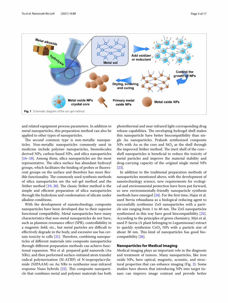
process proposed by Japanese scientist Sugimoto et al. in the 1990s, which is often used to prepare monodis-perse metal oxide particles in liquid phase. The sol–gel method is a bottom-up preparation process. The main principle of this method for preparing metal nanopar-ticles is to form a uniformly dispersed sol of metal ions through chemical and physical means, and then form a gel through redox reaction. The metal nanoparticles generated in the gel can controllably nucleate, grow and deposit. As long as the monodispersity of the metal col-loid used in the experiment, the concentration relation-ship of the metal ions and the oxidizing/reducing agent are controlled, the size of the synthesized metal nano-particles can be controlled. Figure 1 is the schematic dia-gram of the sol–gel method.
Commonly used bottom-up methods for preparing metal nanoparticles include co-precipitation, hydrother-mal approach, and photochemical method. The co-dep-osition method is a process of nucleation, growth and aggregation in a liquid environment at the same time. When the solution is oversaturated, a large number of small-sized particles insoluble products are obtained [15]. The hydrothermal method is a process performed in a liquid environment to control the morphology of the resulting nanoparticles by controlling the vapor pres-sure applied to the material in the solution. In addition, there are some top-down methods for preparing metal nanoparticles, such as electrical wire explosion and ball milling. The principle of electrical wire explosion is that in the process of electric explosion, the metal atoms are evaporated and quickly cooled in the electrolyte to form oxide nanoparticles. By controlling the electrolyte com-position and current intensity, finer and uniform nano-particles can be controlled. Ball milling is a method of quickly and large-scale production of nano-particles with controllable size using machining tools such as milling planetary gears by selecting appropriate grinding time
Table 1 Typical NPs preparation method
Synthesis approach Material Size (nm) Method Features Ref
Bottom up approaches TiO2 6–33 Sol–gel synthesis Continuous releasing of hydroxyl radicals and superoxide ions when exposed to ultraviolet rays
[8]
Fe3O4 10 Co-precipitation Fe3O4 can be excited by 808 nm infrared light to realize photo-thermal conversion
[9]
PEG-Fe-PDA NP 25–43 Microemulsions MRI imaging enhancement with pH activation, high photo-thermal efficiency and excellent biocompatibility
[10]
Magnetite NPs 39 Hydrothermal approach Small size magnetic nanoparticles with biocompatibility and superparamagnetism
[11]
Au NPs 8–300 photochemical method Enhanced medical diagnostic imaging [12]
Top-down approaches Cu-Sn oxides NPs 18–40.5 electrical wire explosion Ability to produce reactive oxygen species [13]
Magnetite NPs 12–20 Ball milling Small size magnetic nanoparticles with biocompatibility [14]
Page 3 of 17Yu et al. Nanoscale Res Lett (2021) 16:88
and related equipment process parameters. In addition to metal nanoparticles, this preparation method can also be applied to other types of nanoparticles.
The second common type is non-metallic nanopar-ticles. Non-metallic nanoparticles commonly used in medicine include polymer nanoparticles, biomolecules derived NPs, carbon-based NPs, and silica nanoparticles [16–18]. Among them, silica nanoparticles are the most representative. The silica surface has abundant hydroxyl groups, which facilitates the binding of probes or fluores-cent groups on the surface and therefore has more flex-ible functionality. The commonly used synthesis methods of silica nanoparticles are the sol–gel method and the Stöber method [19, 20]. The classic Stöber method is the simple and efficient preparation of silica nanoparticles through the hydrolysis and condensation of silicate under alkaline conditions.
With the development of nanotechnology, composite nanoparticles have been developed due to their superior functional compatibility. Metal nanoparticles have many characteristics that non-metal nanoparticles do not have, such as plasmon resonance effect (SPR), controllability in a magnetic field, etc., but metal particles are difficult to effectively degrade in the body, and excessive use has cer-tain toxicity to cells [21]. Therefore, combining nanopar-ticles of different materials into composite nanoparticles through different preparation methods can achieve func-tional expansion. Wei et al. prepared gold nanorods (Au NRs), and then performed surface-initiated atom transfer radical polymerization (SI-ATRP) of N-isopropylacryla-mide (NIPAAM) on Au NRs to synthesize near-infrared response Nano hybrids [22]. This composite nanoparti-cle that combines metal and polymer materials has both
photothermal and near-infrared light corresponding drug release capabilities. The enveloping hydrogel shell makes this nanoparticle have better biocompatibility than sin-gle Au nanoparticles. Prakash synthesized composite NPs with Au as the core and SiO2 as the shell through the improved Stöber method. The inert shell of the core–shell nanoparticles is beneficial to reduce the toxicity of metal particles and improve the material stability and drug-carrying capacity of the original single metal NPs [23].
In addition to the traditional preparation methods of nanoparticles mentioned above, with the development of nanotechnology science, new requirements for ecologi-cal and environmental protection have been put forward, so new environmentally-friendly nanoparticle synthesis methods have emerged [24]. For the first time, Hajar et al. used Stevia rebaudiana as a biological reducing agent to successfully synthesize ZnS nanoparticles with a parti-cle size ranging from 1 to 40 nm. The ZnS nanoparticles synthesized in this way have good biocompatibility [25]. According to the principles of green chemistry, Miri et al. used P. farcta (A plant belonging to Leguminosae) extract to quickly synthesize CeO2 NPs with a particle size of about 30 nm. This kind of nanoparticles has good bio-compatibility [26].
Nanoparticles for Medical ImagingMedical imaging plays an important role in the diagnosis and treatment of tumors. Many nanoparticles, like iron oxide NPs, have optical, magnetic, acoustic, and struc-tural properties that can enhance imaging (Fig. 2). Some studies have shown that introducing NPs into target tis-sues can improve image contrast and provide better
Fig. 1 Schematic diagram of the sol–gel method

Page 4 of 17Yu et al. Nanoscale Res Lett (2021) 16:88
image guidance for tumor surgery and diagnosis [27]. For example, in cryosurgery, NPs can enhance the imag-ing quality of the tumor and ice ball edges, which helps to cover the ice balls accurately and improve the therapeutic effect [28]. In addition, most of the nanoparticles used in imaging are made of metal. According to the difference of different imaging principles, nanoparticles will also be made of different metal materials. Table 2 lists some recent examples about NPs made by different materials for medical imaging.
Optical coherence tomography (OCT) is a non-inva-sive, micron-level resolution and biomedical imaging technology. OCT is useful in real-time diagnosis and surgical guidance. However, OCT cannot detect inelas-tic scattered light because this light is not coherent in the incident field [35]. Recently, many researches have proved the motion state of NPs can be able to change the amplitude of OCT, which may deal with this problem.
Interfering with the movement of NPs through the mag-netic field can cause local changes in light scattering. Some studies have pointed out that placing magnetic NPs in a magnetic field to control its motion can change the optical scattering in the area, so the originally incoherent inelastic scattered light can be detected. This new imag-ing method is magnetomotive optical coherence tomog-raphy (MMOCT) [36].
MRI is one of the most effective noninvasive tumor detection technology. Nevertheless, the lack of MRI signal comparison between biological background and cancer tissue often affects the clinical tumor diagnosis [37]. MRI is a scanning imaging method that measures the magnetization of hydrogen molecules in water mol-ecules. Each anatomical structure presents a different image since the protons of each tissue cause different changes in magnetization. The visibility of images can be improved through applying more contrast agents [38, 39].
Fig. 2 Diagrammatic illustration of imaging improved of NPs
Table 2 Typical NPs platforms made by different materials for medical imaging
US ultrasound, MSNs mesoporous silica nanoparticles, USMO ultrasmall manganese oxide, GEM Gemcitabine, OINPs oxygen/indocyanine green-loaded lipid nanoparticles, PA photoacoustic, MPI magnetic particle imaging, MRI magnetic resonance imaging, SPIO superparamagnetic iron oxide, USPIO ultra-small SPIO
NPs Size (nm) Targeting material Cell line Imaging technology Ref
MnO-TETT 6.7 ± 1.2 None C6 glioma cells Fluorescence/T1-MRI [29]
PLGA-mPEG 151.1 ± 1.3 cRGD SKOV-3 cells US [30]
USMO@MSNs 30–50 Dox HeLa cells MRI-guided chemotherapy [31]
OINPs 300 Folate SKOV3 ovarian cancer cells US/PA [32]
PEG-coated and Gd-loaded fluorescent silica
125.5 ± 9.9 YPSMA-1 LNCaP and PC3 prostate cancer cells MRI/fluorescence imaging [33]
SPIO/USPIO 50 None 4T1 murine breast cancer cells MRI/MPI [34]

Page 5 of 17Yu et al. Nanoscale Res Lett (2021) 16:88
The tumor-related EPR effect widely utilized in the early detection of tumors produces great contrast enhance-ment ability to magnetic NPs [40]. Iron oxide magnetic NPs (IONPs) which are currently the most common MRI nanoprobe contrast agents have certain cell target-ing [41]. For example, studies have found that IONPs could enter healthy liver Kupffer cells during the diag-nosis of liver cancer by using MRI but will be excluded from cancer cells, resulting in low-signal healthy tissue and high-signal tumor tissue [42]. Based on recent stud-ies, proper particle surface modification and appropriate tumor-specific bio-oligomer embedding of NPs can bet-ter fix NPs in tumors to achieve clearer imaging results and can even be used for early micro tumor imaging. For example, studies have found that AuNPs targeted for human transferrin can significantly enhance the imag-ing effect of brain tumors [43]. Gao et al. equipped with anti-epidermal growth factor receptor monoclonal anti-body (mAb) on the basis of paramagnetic NPs probes to achieve imaging of small tumors [44].
Nanoparticles for Targeted Drug DeliveryAlthough Chemotherapeutic drugs now are the most commonly used treatment for tumors, they still have the problem of poor target enrichment in malignant tumor areas and overaccumulation in healthy tissue [45]. This may cause the inhibition of cells hat divide vigorously, such as bone marrow, hair follicles, gastrointestinal cells and lymphocytes, leading to adverse reactions such as bone marrow suppression, mucositis, hair loss, and even death [46]. Targeted drug delivery which refers to active differentiation between normal cells and cancer cells for drug delivery has better efficacy and fewer adverse reac-tions than the conventional treatment [45].Many studies have confirmed that NPs can target chemotherapeutic drugs to tumor cells through active or passive targeting [47]. In addition, many experiments have found that NPs also play an important role in the targeted delivery of immune drugs [48].
As shown in Fig. 3, passive targeting often relies on some pathophysiological characteristics of tumor tis-sue, including abnormal blood vessels, temperature, pH and surface charge of tumor cells [49].For example, due to the enhanced permeability and retention effect (EPR) of blood vessels in the tumor tissue, NPs with a diame-ter of about 400 nm can be passively transferred to the tumor tissue [50]. However, there are many limitations on the passive targeting approach in terms of phys-icochemical properties of NPs such as diameter, surface charge, molecular weight, hydrophobicity, or hydrophi-licity. Besides, the passive targeting technique underper-forms in drug diffusion efficiency and shows insufficient EPR effect in tumor cells [51]. Due to the deficiencies of
passive targeting, in recent years, most research about the drug delivery NPs has shifted to active targeting (ligand targeting). Table 3 highlights some recent exam-ples about NPs used in drug delivery.
Active targeting (ligand targeting) NPs often carry some ligands of tumor-specific biomarkers [61]. As shown in Fig. 4 when the ligand contacts to the recep-tor on the tumor surface, NPs can be internalized by the tumor cells through receptor-mediated endocytosis, and the drugs can be released due to acidic pH and specific enzymes in the intracellular environment [62]. As for tar-geting ligands, folic acid, transferrin, epidermal growth factor receptor (EGFR) and glycoprotein are generally utilized in current research [62]. For example, Sandoval et al. obeserved significant drug enrichment and evi-dent efficacy in the treatment of mice with breast cancer through EGFR-targeted stearyl NPs equipped with gem-citabine [63]. Pandey et al. found that folic acid-targeted gold NPs carrying berberine hydrochloride (BHC) can effectively deliver drugs to human cervical cancer cells expressing folate receptor [64].
In recent years, compared with chemotherapy drugs, short interfering RNA (siRNA)-mediated gene silencing therapy has been regarded as a new prospect for tumor treatment [64]. Although viruses can be used as delivery vehicles for siRNA, viral vectors have been confirmed to cause insertional mutagenesis and immunogenic-ity [65]. By contrast, selenium NPs are reported to have great potential as siRNA carriers, because the trace ele-ment selenium itself can reduce tumor occurrence, lower drug toxicity, and regulate immune function [66]. In addition, the surface of selenium NPs can load vari-ous tumor- targeting moieties (such as folate, hyaluronic acid and RGD peptide) to enhance tumor targeting ability [67]. Xia et al. reported that selenium NPs (RGDfC-Se@siRNA) targeted by RGDfC peptide have excellent ability to target HeLa cervical cancer [60]. Meanwhile, because
Fig. 3 Diagrammatic illustration of passive targeting of NPs

Page 6 of 17Yu et al. Nanoscale Res Lett (2021) 16:88
RGDfC can specifically combine with αvβ3 integrin which is highly expressed by a variety of tumor cells, RGDfC-Se@siRNA NPs can be reused for targeted drug deliv-ery for a variety of tumors [68]. In terms of structure, RGDfC-SeNPs with positive charge can tightly package negatively charged siRNA through their electrostatic interaction [69]. Through animal experiments, RGDfC-Se@siRNA NPs show the ability to efficiently enter tumor cells through clathrin-associated endocytosis. In tumor cells, it can quickly release siRNA and efficiently silence related genes and promote the generation of reactive oxy-gen species (ROS) to inhibit tumor cells proliferate and promote tumor cells apoptosis [69]. Additionally, multi-ple SeNPs have demonstrated excellent biological safety and have no obvious toxic damage to liver, kidney, heart, lung, spleen and other major organs of mice [60, 70, 71].
At present, although there are many NPs used in tar-geted drug delivery, most applications still remain in the stage of cell or animal experiments, lacking potent clinical application support. In addition, many NPs are administered intratumorally, which limits the scope of NPs applicable to tumors and lacks special NPs drug delivery tools and other drug delivery methods.
Table 3 Typical NPs platforms used in drug delivery
DOX doxorubicin, 5-FU 5-fluorouracil, FA folic acid, PTX paclitaxel, ROS reactive oxygen species
Agent of NPs Vehicle Size (nm) Characters Effects Ref
DNA and RNA Exosomes 30–100 Small size, cellular origin, flexibility to incorporate macromolecules
Carrier for DNA, RNA and micro-RNA
Cross-stringent biological barriers, such as the blood–brain barrier
[52]
DOX Polymer-lipid encapsulated man-ganese dioxide
170 Bioreactive and multifunctional Downregulate TME-associated drug resistance and immuno-suppression
Enhancing chemotherapeutic efficacy and boosting antitumor immunity
[53]
5-FU Au-NPs/chitosan 100–400 Natural cationic, biodegradable and biocompatible
Enhance the curative effect for hepatocellular carcinoma cells (HepG2)
[54]
Tyrosinase-related protein 2 (Trp2) peptide
Layered double hydroxide (LDH) NPs
140–150 Provoking strong cell-mediated immune responses
Adjuvant multiple tumor-associ-ated antigen peptides
[55]
Cisplatin; ICG PLGA 90–100 Folate targetingControlled drug release
Promoting the apoptosis of McF-7 tumor cells
NIR sensitivity
[56]
IL-2 PEGylated liposomes with anti-CD137
80 Complete absence of systemic toxicity
Inducing intratumoural immune responses
Initial anti-tumor activity
[57]
FA Magnetic mesoporous silica 213 PH-sensitive drug release Inhibiting proliferation of HeLa cell lines higher cytotoxicity effect
[58]
PTX Peptide H7K(R2)2-modifiediron oxide NPs
168.3 ± 2.80 few side-effects Excellent MRI imagingInhibiting tumor growth
[59]
siRNA RGDfC-SeNPs 150 No toxicityMultiple tumor targeting
Carrier for siRNAInhibiting tumor cells proliferatePromoting the generation of ROS
[60]
Fig. 4 Diagrammatic illustration of active targeting of NPs
Page 7 of 17Yu et al. Nanoscale Res Lett (2021) 16:88
Therefore, exploring a better way to administer NPs may be a direction for the future research of targeted drug delivery NPs. According to the existing academic journals, vascular interventional administration may be a feasible way. In the assumption, first locate the position of tumor-feeding blood vessel with the help of imaging, and then use a guide wire to introduce NPs directly into the tumor-feeding blood vessel and control the move-ment of the NPs in a small range by applying a magnetic field simultaneously. Therefore NPs can be fixed at the proper position without being influenced by blood flow in the vessel. Otherwise, NPs targeted for drug delivery only have certain limitations. Targeting NPs will affect the systemic distribution of chemotherapeutic drugs and reduce the effect of chemotherapy on free tumor cells and micrometastasis. If they are equipped with targeted drugs, the targeting effect tends to be enhanced whereas the improvement is not evident based on the existing studies.. In addition, anti-tumor drugs are unlikely to eliminate all tumor stem cells by themselves. Neverthe-less, physical therapy based on the physical characteris-tics of NPs tends to be more effective against the tumor stem cells. Therefore, multifunctional NPs targeting drug carriers tend to be advisable in the future, such as cryo-surgery, photothermal therapy (PTT) and photodynam-ics therapy (PDT) etc., to form multi-functional NPs for tumor treatment.
Nanoparticles for CryosurgeryCryosurgery, the technique of destroying tumor tissue by freezing, has the advantages of low invasiveness, low cost, less intraoperative bleeding and less postopera-tive complications, but there are still disadvantages such as insufficient freezing efficiency and freezing damage to surrounding tissues [28]. Although protective agents such as antifreeze protein (AFP-1) have been utilized to assist cold ablation, the effect is still not ideal [72]. With the development of nanotechnology, the concept of nano-cryosurgery was proposed. The basic mechanism of nano-cryosurgery is to introduce NPs with specific physical or chemical properties into tumor tissues. By utilizing the properties of NPs, not only can the efficiency and effectiveness of freezing be improved, but the range adjustment and the direction of ice ball formation can be also controlled. Thus, the nano-cryosurgery is capable of killing tumor tissue and preventing surrounding healthy tissue from being frozen simultaneously [73]. The advan-tages of nano-cryosurgery have shown in Fig. 5.
In cryosurgery, intracellular ice formation is the key to tumor cell damage. Meanwhile, research proves that NPs can effectively induce intracellular ice formation [28]. NPs as external particles can induce heterogeneous nucleation. Studies have found that tissues enriched with
NPs freeze faster than conventional tissues and are more prone to heterogeneous nucleation. Under the same freezing conditions, ice formation of tissue with NPs is easier, indicating that NPs can significantly increase the speed and probability of ice formation in cells, which can kill tumor cells more effectively [74]. In addition, NPs with metal oxide will significantly improve the thermal conductivity in tumor tissue. For example, Liu and Deng compared the temperature response curve of pork tissues with and without NPs. They found that the tissues con-taining NPs cooled rapidly, and the lowest temperature could reach 115 ℃, which was much lower than that of the control group without NPs.
Since tumors are usually irregular in shape, the ice crystals produced by traditional cryosurgery tend not to cover all tumor tissue. Compared with traditional cryo-surgery, the nano-cryosurgery can deal with the problem easily. Because NPs can permeate into the intracellular fluid and have good physical properties like thermal con-ductivity, it is possible to control the growth direction and direction of the ice ball by the distribution of NPs [73].
In cryosurgery, insufficient freezing may not com-pletely destroy tumor tissue, and excessive freezing may damage adjacent healthy tissue. Especially when the tumor is in close contact with fragile organs, its location is deep, or its shape is irregular, the damage to the healthy tissue can be particularly serious. In recent years, phase change materials (PCMs) made from NPs have demon-strated excellent protective potential for surrounding healthy tissues during cryosurgery [75]. For example, Lv et al. microencapsulated phase change NPs with large latent heat and low thermal conductivity through liposomes, and before cryosurgery, injected microencap-sulated phase change NPs into healthy tissues around the tumor and found that avoided low temperature damage to healthy tissue [76].
Although NPs have been widely used in cryosurgery, there are still a series of deficiencies. First, it is still unable to control NPs in vitro, which results in uneven distribu-tion of NPs in tumor tissue and unsatisfactory expected function. Secondly, although there are a variety of mag-netic nanoparticles, the actual effect of in vitro magnetic field control NPs is still not ideal. In addition, the nano-cryosurgery is lack of clinical experimental research, and many NPs are still in the laboratory stage.
The application of NPs in cold ablation can be gener-ally divided into two types: synergistic effect and pro-tective effect, which are different in terms of the design requirements of NPs and the distribution in vivo. In the future, nano-cryosurgery may be assisted by a variety of NPs, viz, synergistic NPs are distributed inside the tumor while protective NPs are distributed around the

Page 8 of 17Yu et al. Nanoscale Res Lett (2021) 16:88
tumor. In addition, many nano-positioning devices, such as puncture-designed 3D printed coplanar tem-plate (3DPCT) which currently used for tumor posi-tioning before radioactive particle implantation may be used in cryosurgery. Prior to the cryosurgery, protec-tive NPs can be punctured and injected around tumor to protect the surrounding healthy tissue by 3D print-ing coplanar template (3DPCT) and CT guidance. The NPs are able to assist the cryosurgery ice balls to cover
the irregular edge of tumor. Then synergistic NPs will be introduced into the tumor tissue through the preset ablation site puncture or vascular intervention to per-form cold ablation. This nano-cryosurgery technique can not only overcome the difficulties of cold ablation of irregular tumors but also increase the effect of cold ablation and reduce the damage to healthy tissue. This method may become the future research direction of nano-cryosurgery. Table 4 highlights some recent examples about NPs used in cryosurgery.
Fig. 5 Diagrammatic illustration of NPs for cryosurgery. a NPs protect health cells during cryosurgery. b NPs enhance the freezing damage and control the freezing coverage. c With the help of NPs, more ice has been formed

Page 9 of 17Yu et al. Nanoscale Res Lett (2021) 16:88
Nanoparticles for PTT and PDTAt present, photothermal therapy (PTT) and photody-namic therapy (PDT) based on nanoparticles (NPs) have shown the virtues of strong efficacy, small invasion and mild adverse effects during tumor treatment (Fig. 6) [80]. In addition to killing tumor cells directly, fragments of dead tumor cells produced by PDT and PTT treatment can be used as potential antigens to trigger a continu-ous immune response, called photothermal and photo-dynamic immunotherapy [81]. Nanoparticles designed based on the PTT treatment concept are a new type of light-to-heat conversion nanomaterials, which can con-vert light energy into heat energy to kill cancer cells. Compared with traditional photothermal conversion materials, nanoparticles have many advantages. First, NPs can achieve the effect of tumor targeted aggrega-tion through particle surface modification, which con-tributes to higher enrichment ability of target tumor [82, 83]. Second, nanoparticles have better imaging capabili-ties than traditional photothermal materials, which can
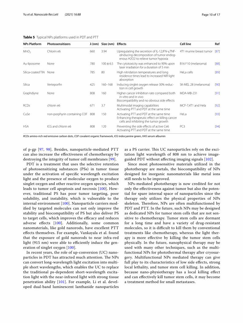
be accurately positioned by CT, MRI and photoacous-tic imaging [84, 85]. Targeted nanoparticles synthesized by Pan et al. can perform PTT under 0.2 W/cm2 NIR to induce tumor cell apoptosis by destroying the tumor cell nuclear DNA and inhibiting the DNA repair process [86]. Table 5 lists some recent examples about NPs used in PDT and PTT.
In addition, some studies have found that nanoparti-cle-mediated PTT can reverse tumor multidrug resist-ance (MDR). The overexpression of drug transporters, multidrug resistance-associated protein 1 (MRP1), and p-glycoprotein (p-gp) are generally believed to cause MDR in various tumors [95]. For example, multifunc-tional light-triggered nanoparticles designed by Li et al. can inhibit the expression of MRP1 in PTT, which con-sequently reverses the drug resistance of A549R cells [96]. Wang et al. reported that both gold nanoparticles and carbon-based nanoparticles can overcome DOX resistance by promoting the expression of heat shock factor trimer in PTT, thereby inhibiting the generation
Table 4 Typical NPs platforms used in cryosurgery
HCPN-CG H for hyaluronic acid, C for chitosan, P for PF127, N for PNIPAM-B, C for CPT, and G for ICG, PCM phase change materials
NPs Size (nm) Thermal conductivity (W/m K)
Heat capacity (J/m3 K)
Benefits Ref
PCM NPs 10–20 0.35 2.56 × 10⁶ Health tissue protection [76]
Fe3O4 NPs 8–14 7.1 3.2 × 10⁶ More intracellular ice formationHigh thermal conductivity
[77]
Au NPs 3 297.7 2.2 × 10⁶ Good biological compatibilityHigh thermal conductivity
[78]
HCPN-CG NPs 103.9 ± 1.5 None None Cold-responsive nanoparticle for controlled drug releaseNIR-induced photothermal effect
[79]
MgO NPs 50 34.3 3.2 × 10⁶ Nontoxic, biodegradable, and few side-effects [74]
Fig. 6 Diagrammatic illustration of NPs-mediated PDT and PTT. a NPs promote the generation of reactive oxygen. b NPs enhance tumor damage during PTT
Page 10 of 17Yu et al. Nanoscale Res Lett (2021) 16:88
of p-gp [97, 98]. Besides, nanoparticle-mediated PTT can also increase the effectiveness of chemotherapy by destroying the integrity of tumor cell membranes [99].
PDT is a treatment that uses the selective retention of photosensitizing substances (PSs) in tumor tissue under the activation of specific wavelength excitation light and the presence of molecular oxygen to produce singlet oxygen and other reactive oxygen species, which leads to tumor cell apoptosis and necrosis [100]. How-ever, traditional PS has poor tumor targeting, poor solubility, and instability, which is vulnerable to the internal environment [100]. Nanoparticle carriers mod-ified by targeted molecules can not only improve the stability and biocompatibility of PS but also deliver PS to target cells, which improves the efficacy and reduces adverse effects [100]. Additionally, some common nanomaterials, like gold nanorods, have excellent PTT effects themselves. For example, Vankayala et al. found that the exposure of gold nanorods to near infra-red light (915 nm) were able to efficiently induce the gen-eration of singlet oxygen [100].
In recent years, the role of up-conversion (UC) nano-particles in PDT has attracted much attention. The NPs can convert long-wavelength light excitation into multi-ple short wavelengths, which enables the UC to replace the traditional ps-dependent short-wavelength excita-tion light with the near-infrared light with strong tissue penetration ability [101]. For example, Li et al. devel-oped dual-band luminescent lanthanide nanoparticles
as a PS carrier. This UC nanoparticles rely on the exci-tation light wavelength of 808 nm to achieve image-guided PDT without affecting imaging signals [102].
Since most photosensitive materials utilized in the phototherapy are metals, the biocompatibility of NPs designed for inorganic nanomaterials like metal ions still needs to be improved.
NPs-mediated phototherapy is now credited for not only the effectiveness against tumor but also the poten-tial for spare internal space of nanoparticles since the therapy only utilizes the physical properties of NPs skeleton. Therefore, NPs are often multifunctioned by PDT and PTT. In the future, such NPs may be designed as dedicated NPs for tumor stem cells that are not sen-sitive to chemotherapy. Tumor stem cells are dormant for a long time and have a variety of drug-resistant molecules, so it is difficult to kill them by conventional treatments like chemotherapy, whereas the light ther-apy is more effective by killing the tumor stem cells physically. In the future, nanophysical therapy may be used with many other techniques, such as the multi-functional NPs for photothermal therapy after cryosur-gery. Multifunctional NPs mediated therapy can give full play to its characteristics of low side effects, strong local lethality, and tumor stem cell killing. In addition, because nano-physiotherapy has a local killing effect and can effectively kill tumor stem cells, it may become a treatment method for small metastases.
Table 5 Typical NPs platforms used in PDT and PTT
RCDs amino-rich red emissive carbon dots, COF covalent organic framework, ICG indocyanine green, HAS serum albumin
NPs Platform Photosensitizers λ (nm) Size (nm) Effects Cell line Ref
MnO2 Chlorin e6 660 3.94 Upregulating the secretion of IL-12,IFN-γ,TNF-αInducing decomposition of tumor endog-enous H2O2 to relieve tumor hypoxia
4T1 murine breast tumor [87]
Au-liposome None 780 100 ± 6.5 The cytotoxicity was enhanced to 90% upon laser irradiation for a duration of 5 min
B16 F10 (melanoma) [88]
Silica-coated TiN None 785 80 High nitridation temperatures and long residence times lead to increased NIR light absorption
HeLa cells [89]
Silica Verteporfin 425 160–168 Inducing singlet oxygen release 30% reduc-tion in cell growth
SK-MEL 28 (melanoma) [90]
Graphdiyne None 808 160 Higher cancer inhibition rate compared both in vitro and in vivo
Biocompatibility and no obvious side effects
MDA-MB-231 [91]
RCDs chlorin e6 671 3.7 Multimodal imaging capabilitiesActivating PTT and PDT at the same time
MCF-7,4T1 and Hela [92]
CuSe non-porphyrin containing COF 808 150 Activating PTT and PDT at the same timeEnhancing therapeutic effect on killing cancer
cells and inhibiting the tumor growth
HeLa [93]
HSA ICG and chlorin e6 808 120 Preventing the side effects of active Ce6Activating PTT and PDT at the same time
PC3 [94]

Page 11 of 17Yu et al. Nanoscale Res Lett (2021) 16:88
Nanoparticles for RadiotherapyRadiotherapy (RT) is a tumor treatment technique that kills local cells by ionizing radiation generated by rays and is currently an effective treatment for many primary and metastatic solid tumors [103]. Experiments prove that radiotherapy can effectively kill tumor stem cells [104].However, how to further improve the efficacy of radiotherapy is still a serious challenge. In recent years, nanoparticles in the field of radiotherapy have demon-strated strong radiosensitization capabilities, tumor-targeted delivery capabilities of radiosensitizing drugs, and imaging guidance enhancement capabilities [105]. At present, the most popular nanoparticles are made by high Z (atomic number) metal materials, which are fea-tured by chemical inertness and strong radiation absorp-tion capacity. They produce various reactions such as photoelectric effect and Compton effect after absorbing radiation, thereby releasing a variety of particles such as optoelectronics, Compton electrons, and Auger elec-trons. These electrons react with organic molecules or water in tumor cells to generate a large number of free radicals, leading to synergistic chemotherapy [106]. Common chemotherapy-sensitized NPs are currentlycat-egorized as precious metals, iron oxides, and semicon-ductors in terms of materials.
Precious metals NPs are made of high atomic number metal materials such as gold, silver, gadolinium, hafnium, platinum, bismuth, etc. [107]. Among them, gold nano-particles have become the most popular NPs due to their good biocompatibility, chemical stability, and relatively strong photoelectric absorption coefficient [108]. In 2000, Herold et al. discovered the chemosensitizing abil-ity of gold nanoparticles in kilovoltage X-rays. Nowadays, the specific mechanism of chemosensitization of gold nanoparticles is not yet clear, and the mainstream view believes that it depends on the photoelectric absorption capacity of high atomic number [109]. In addition to this, there are studies suggesting that the presence of gold nanoparticles improve the chemical sensitization of DNA to radiation, which increases the DNA damage induced by ionizing radiation (IR). At the same time, gold NPs can catalyze the mechanism of radiotherapy sensitization such as free radical production [105]. For instance, Liu found that AuNPs could significantly increase the pro-duction of hydroxyl radicals as well as the killing effect of x-rays and fast carbon ions on cells [110]. The hypothesis of the chemotherapy sensitization mechanism of other precious metals is similar to that of gold nanoparticles. Particularly, platinum NPs have an anti-tumor effect due to the inherent nature. Consequently, platinum NPs are expected to play the role of chemotherapy and radio-therapy simultaneously. However, the number of rele-vant research reports is insufficient, and the sensitizing
effect of platinum NPs is also questionable. For example, Charest et al. reported that liposomal formulation of cisplatin was able to increase the uptake of platinum by tumor cells, and could enhance the killing of F98 glioma cells by γ-rays at the same time [111]. On the contrary, Jawaid et al. reported that platinum NPs would reduce the generation of reactive oxygen species (ROS) and the efficacy of radiotherapy during chemotherapy [112].
Iron oxide nanoparticles (IONs), especially the super-paramagnetic magnet Fe3O4, have shown great poten-tial in image-guided tumor radiotherapy because they are capable of enhancing the dose of radiotherapy and MRI imaging, whereas its sensitization mechanism is not clear yet. Its sensitization mechanism is not yet clear. Some studies believe that iron oxide NPs mainly catalyze the generation of ROS through Fenton’s reac-tion and Haber–Weiss reaction. Then the highly reactive ROS will kill tumors [112–115]. Other studies propose that the mechanism depends on the radiation sensitiza-tion and synergistic effects of magnetic nanoparticles. As Khoei reported, iron oxide NPs can improve the radiosensitization of prostate cancer cells in vitro [116]. Huang et al. pointed out that cross-linked dextran-coated IONs (CLIONs) could be internalized by HeLa cells and EMT-6 mouse breast cancer cells, which enhances radia-tion therapy [117]. Although the synergistic effect of iron oxide NPs is obvious, its biological safety still needs to be improved. Many studies have proved that the biocompat-ibility and chemical stability of iron oxide NPs are ques-tionable, and it has certain toxicity [118].
Semiconductor NPs like silica NPs have also been found to have a synergistic effect on radiotherapy. For instance, Zhang et al. used flow cytometry analysis and MTT experiments to find that mesoporous silica NPs can effectively enhance the radiotherapy of glioblas-toma [119]. He et al. reported the mechanism of radio-active enhancement of silica NPs. He found that under X-ray irradiation, silica nanoparticles could produce fine hydroxyl radicals, which can effectively kill tumor cells [120].
At present, although many experiments have con-firmed that NPs were able to sensitize radiotherapy, the specific mechanism of sensitization is still unclear, which hinders the development of new sensitized NPs. There are some doctrines like sensitizing chemotherapy that promotes free radical production. Nevertheless, there is a lack of a quantitative relationship among the amount of free radical production, radiation intensity, and physical data of nanoparticles. In addition, most sensitized NPs are made of high atomic number metals. These metals have many disadvantages in human body such as diffi-culty in self-metabolism and biodegrading. Meanwhile, long-term accumulation of the metals will produce

Page 12 of 17Yu et al. Nanoscale Res Lett (2021) 16:88
toxicity, which limits the safe use of radiosensitized NPs. Moreover, compared with the radiotherapy sensitization NPs, fewer studies focused on NPs which can prevent the adverse reactions of radiotherapy and protect healthy tissues. The research on radiotherapy protective NPs is short in quantities.
In the future, searching for NPs material that can be metabolized by the kidney, biometabolized, biocompat-ible, stable in physicochemical properties, and inherently less toxic, or looking for surface modification that can help the body metabolize NPs may become a research direction for sensitized NPs. Moreover, although there have been many NPs studies on multi-function, namely simultaneous sensitization of radiotherapy and chemo-therapy, there are still many potentials in this field, which are worthy of focus in the future. The development of protective NPs that can protect normal tissues around radiotherapy and alleviate poor defense against radio-therapy may also become a research direction.
ConclusionThe poor curative effect, inefficient targeting ability, vari-ous side effects, and potential biological risk are some of the unfavorable attributes of conventional cancer therapy and diagnosis. In recent years, advanced nanotechnol-ogy and molecular cell biology have promoted the appli-cations of NPs in cancer field. Not only metal NPs, but also many lipid, nucleic acid and silicon NPs showed evi-dent outperformance in cancer diagnosis and treatment.. Moreover, new generation of NPs is no longer limited to solo but multiple functions. For example, gold-coated poly(lactic-co-glycolic acid) (PLGA) NPs equipped with PD-1 blockers which were designed by Luo et al. can not only target drug delivery but also mediate PTT therapy [121]. (Pd @ Au) / Fe3O4 Spirulina NPs with doxorubicin created by Wang et al. demonstrated the functions of photothermal therapy, delivery of chemotherapy drugs, and magnetic field control in cell experiments [122]. Multifunctional nanoparticles will become the trend of future research.
At present, we find that most of the nanoparticles only stay in vivo and in vitro stage. According to this review, we think the following reasons hinder the clinical appli-cation of NPs.
(i) Lack of injection routes and methods
Most NPs are injected into body via puncture or intra-venous injection. Therefore, the blood flow will take away NPs, making NPs difficult to stay in the target area for a long time, which leads to just few NPs that can be uptaked by tumor cells. Low-concentration drugs cannot produce the expected therapeutic effect, and low-concentration NPs also affect the physical killing
effects of PDT, PTT, cryosurgery, and radiotherapy. In our opinion, magnetic NPs platform may be a solution. There have been many in vitro and in vivo experiments that have proved the feasibility of using the three-dimensional magnetic field to control the movement of NPs against blood flow [122–125]. However, how to solve the interference of the human body to the mag-netic field, how to solve the impact of blood cells collid-ing with NPs, and how to control a large number of NPs in a group are still in discovery.
(ii) Difficulty in localization of NPs in vivo
Compared with the human body, the size of NPs is too tiny. Even if NPs are loaded with fluorescent proteins, it is still difficult for conventional imaging equipment (CT, X-ray, MRI) to locate the NPs in the human body in real time. To deal with this challenge, photoacous-tic computed tomography (PACT) may be a solution. Photoacoustic computed tomography (PACT) has attained high spatiotemporal resolution (125-μm in-plane resolution and 50-μs frame−1 data acquisition), deep penetration (48-mm tissue penetration in vivo), and anatomical and molecular contrasts [126]. Because of excellent performance, PACT has great poten-tial in NPs localization imaging in vivo. The PACT-guided microrobotic system designed by Wu et al. has achieved controlled propulsion and prolonged cargo retention in vivo of NPs with a diameter of 50 μm [127]. Although the current resolution and deep penetration of PACT are still insufficient, it is superior to conven-tional imaging equipment (CT, X-ray, MRI) in terms of NPs imaging positioning.
(iii) Difficulty of degrading in the human body
Although NPs are made of high biosafety materials, there is still a risk of damages to liver, kidney, and other organs if they stay in the body for a long time and can-not be degraded or excreted The use of materials that will be disintegrated after near-infrared light irradia-tion to fabricate NPs may be a solution to this problem. Recently, more and more NPs have been produced by these materials. Such NPs mediate PTT while loading drugs, meanwhile, the substances produced by the dis-integration of NPs can be rapidly metabolized by the human body. In addition, the use of more biocompat-ible and degradable materials for nanoparticle prepa-ration is also a solution. For example, the surface of chitosan is positively charged and can be broken down by the colonic flora, which facilitates interaction with specific tissues and can be metabolized by the body. The biocompatibility and degradability of chitosan has been proven to be non-toxic at appropriate drug con-centrations [128].

Page 13 of 17Yu et al. Nanoscale Res Lett (2021) 16:88
(iv) Difficulty in avoiding mononuclear phagocytic sys-tem (MPS)
In biofluids, NPs will adsorb proteins to form a corona layer referred to as “protein corona” in a broader sense giving biological identity to NPs and alters their biological characters, which will attract MPS especially macrophages to uptake NPs [129]. In order to avoid being uptaken by MPS, various polymer coatings such as forpolyether, polybetaine (PB) and polyolhave were investigated to cover NPs. For example, polyglycerol-grafting NPs are able to evade macrophage uptake by reducing protein adsorption [130]. In addition, there are two types of tumor-associated macrophages (TAM), M1 and M2. M1 macrophages inhibit tumor growth while M2 macrophages promote tumor growth. There-fore, no longer avoiding macrophages, but designing NPs targeted by macrophages, by regulating the func-tion of macrophages, and even using macrophages as new drug carriers to exert anti-tumor effects may become a novel solution. At present, common design strategies for such NPs include inhibiting macrophage recruitment, depleting TAM, reprogramming TAMs, and blocking CD47-SIRPα pathway [131]. Among them, following the design concept of reprogramming or blocking CD47-SIRPα pathway, NPs that repolarize M2 macrophages to M1 type have made a breakthrough in vivo experiments [132].
Considering the above difficulties and referencing to advanced researches, we come up with a new possible design of NPs. The NPs skeleton is made of pyrolytic material (spirulina, exosomes, et al.). Then, photother-mal materials (Au, Pd, etc.) are deposited on the NPs skeleton through electroless plating. After that the superparamagnetic iron oxide will be loaded on the surface of NPs through the sol–gel method. Then, suit-able polymers (polybetaine, polyglycerol, etc.) will coat the NPs. Finally, drug (like doxorubicin) will be loaded on the NPs. Afterwards, under the guidance of PACT, NPs will be injected into the upstream of tumor sup-plying blood vessel, and the tumor will be irradiated with NIR. At the same time, three-dimensional mag-netic field control is given to maximize the accumula-tion of NPs at the tumor site. Through this design, a large number of NPs will accumulate at the tumor site to ensure the drug concentration and PTT effect. At the same time, most NPs will be decomposed at the tumor site, and only a small number of NPs will circulate in the body.
Nowadays, anti-tumor therapy with NPs as the main body is still in the exploratory stage, and related tech-nologies and equipments need to be invented, so it is
unlikely to be clinically used in the short term. How-ever, NPs can change part of the function or structure of many actual technologies. The upgrade of actual technologies is expected to be applied in clinic quickly, which contributes to upgrading the diagnosis and treat-ment of tumors in consequence. For example, NPs can help to develop electrochemical devices based on the interaction between ions and conductive polymers, such as organic electrochemical transistors (OFETs), electrolyte gated field-effect transistors (FETs), fin field-effect transistor (FinFETs), tunneling field-effect tran-sistors (TFETs), electrochemical lab-on-chips (LOCs) [133]. These electrochemical devices are widely used in various tumor testing and diagnostic equipment. The use of NPs can help improve the accuracy of the equipment and reduce the detecting time. Many studies indicate that medical equipment using electronic com-ponents upgraded by NPs have been applied clinically [133–136].
Based on the evidence cited above, future research of NPs may not only focus on NPs themselves but also consider a feasible administration and efficacy assess-ing platform. In addition, the platform needs to be able to monitor immunotoxicity, the long-term toxicity, and neurotoxicity of NPs. As nanotechnology develops, if these problems were solved, NPs would be an ideal approach to upgrade cancer therapy and diagnosis.
AbbreviationsNPs: Nanoparticles; PDT: Photodynamics therapy; PTT: Photothermal therapy; SPR: Plasmon resonance effect; Au NRs: Gold nanorods; SI-ATRP: Surface-initiated atom transfer radical polymerization; NIPAAM: N-isopropylacrylamide; US: Ultrasound; MSNs: Mesoporous silica nanoparticles; USMO: Ultrasmall manganese oxide; GEM: Gemcitabine; OINPs: Oxygen/indocyanine green-loaded lipid nanoparticles; PA: Photoacoustic; MPI: Magnetic particle imaging; MRI: Magnetic resonance imaging; SPIO: Superparamagnetic iron oxide; USPIO: Ultra-small SPIO; OCT: Optical coherence tomography; MMOCT: Magnetomotive optical coherence tomography; mAb: Monoclonal antibody; DOX: Doxorubicin; 5-FU: 5-Fluorouracil; FA: Folic acid; PTX: Paclitaxel; ROS: Reactive oxygen species; EPR: Enhanced permeability and retention effect; EGFR: Epidermal growth factor receptor; BHC: Berberine hydrochloride; AFP-1: Antifreeze protein; PCMs: Phase change materials; 3DPCT: 3D printed coplanar template; RCDs: Amino-rich red emissive carbon dots; COF: Covalent organic framework; ICG: Indocyanine green; HSA: Serum albumin; MDR: Multidrug resistance; MRP1: Multidrug resistance-associated protein 1; p-gp: P-glyco-protein; PSs: Photosensitizing substances; UC: Up-conversion; RT: Radio-therapy; PLGA: Poly(lactic-co-glycolic acid); PACT : Photoacoustic computed tomography; MPS: Mononuclear phagocytic system; PB: Polybetaine; TAM: Tumor-associated macrophages; OFETs: Organic electrochemical transistors; FETs: Electrolyte gated field-effect transistors; FinFETs: Fin field-effect transistor; TFETs: Tunnelling field-effect transistors; LOCs: Electrochemical lab-on-chips.
AcknowledgementsNone.
Authors’ contributionsZY: Writing- Original draft preparation. ZY and KC: Performed the Literature Search of the Databases. LG: Writing- Reviewing and Editing. QZ: translation. QL and WZ: investigation. KH: Supervision. All authors read and approved the final manuscript.
Page 14 of 17Yu et al. Nanoscale Res Lett (2021) 16:88
Funding(1) National Key R&D Program of China (2018YFC1705102), which supported by Biological Center of Ministry of Science and Technology of China. (2) Capital Health Development Research Project (CFH2018-1-4201), which supported by Beijing Municipal Science and Technology Commission. (3) New Innovation Project-Yiqilin Leading Talent Project, which supported by Beijing Yizhuang Economic Development Zone Government. The fund doesn’t have code.
Availability of data and materialsAll data generated or analysed during this study are included in this published article.
Competing interestsNo benefits in any form have been received or will be received from a com-mercial party related directly or indirectly to the subject of this article.
Author details1 Oncology Department, Dongfang Hospital, Beijing University of Chinese Medicine, Fangguyuan Rd, Fengtai District, Beijing 100078, China. 2 College of Engineering, China Agricultural University, Tsinghua East Rd, Haidian District, Beijing 100083, China. 3 Department of Management, Fredericton Campus, University of New Brunswick, 3 Bailey Drive, Fredericton, NB E3B 5A3, Canada. 4 Beijing University of Chinese Medicine, 11 North Third Ring East Road, Chaoy-ang District, Beijing 100029, China.
Received: 13 August 2020 Accepted: 27 January 2021
References 1. Liu Y, Bhattarai P, Dai Z, Chen X (2019) Photothermal therapy and pho-
toacoustic imaging via nanotheranostics in fighting cancer. Chem Soc Rev 48:2053–2108
2. Herma R et al (2019) Carbosilane dendrimers with phosphonium terminal groups are low toxic non-viral transfection vectors for sirna cell delivery. Int J Pharmaceut 562:51–65
3. Woodman C, Vundu G, George A, Wilson CM (2021) Applications and strategies in nanodiagnosis and nanotherapy in lung cancer. Semin Cancer Biol 69:349–364
4. Israel LL, Galstyan A, Holler E, Ljubimova JY (2020) Magnetic iron oxide nanoparticles for imaging, targeting and treatment of primary and metastatic tumors of the brain. J Control Release 320:45–62
5. Singh H, Du J, Singh P, Mavlonov GT, Yi T (2018) Development of super-paramagnetic iron oxide nanoparticles via direct conjugation with gin-senosides and its in-vitro study. J Photochem Photobiol B 185:100–110
6. Tao Y, Li M, Ren J, Qu X (2015) Metal nanoclusters: novel probes for diagnostic and therapeutic applications. Chem Soc Rev 44:8636–8663
7. Sajid M, Potka-Wasylka J (2020) Nanoparticles: synthesis, characteris-tics, and applications in analytical and other sciences. Microchem J 154:104623
8. Priyanka KP, Sukirtha TH, Balakrishna KM, Varghese T (2016) Microbicidal activity of TiO2 nanoparticles synthesised by sol–gel method. IET Nano-biotechnol 10(2):81–86
9. Qi X, Xiong L, Peng J, Tang D (2017) Near infrared laser-controlled drug release of thermoresponsive microgel encapsulated with Fe3O4 nano-particles. RSC Adv 7:19604–19610
10. Liu F et al (2015a) Controllable synthesis of polydopamine nanopar-ticles in microemulsions with pH-activatable properties for cancer detection and treatment. J Mater Chem B 3:6731–6739
11. Daou TJ et al (2006) Hydrothermal synthesis of monodisperse magnet-ite nanoparticles. Chem Mater 18:4399–4404
12. McGilvray KL, Decan MR, Wang D, Scaiano JC (2006) Facile photochemi-cal synthesis of unprotected aqueous gold nanoparticles. J Am Chem Soc 128:15980–15981
13. Shahriyari F, Yaarali D, Ahmadi R, et al. (2020) Synthesis and characteri-zation of Cu-Sn oxides nanoparticles via wire explosion method with surfactants, evaluation of in-vitro cytotoxic and antibacterial properties. Adv Powder Technol 31(6):2337–2347
14. de Carvalho JF, de Medeiros SN, Morales MA, Dantas AL, Carriço AS (2013) Synthesis of magnetite nanoparticles by high energy ball milling. Appl Surf Sci 275:84–87
15. Mascolo MC, Pei Y, Ring TA (2013) Room temperature co-precipitation synthesis of magnetite nanoparticles in a large pH window with differ-ent bases. Materials (Basel, Switzerland) 6:5549–5567
16. Seidi F, Jenjob R, Phakkeeree T, Crespy D (2018) Saccharides, oligosac-charides, and polysaccharides nanoparticles for biomedical applica-tions. J Control Release 284:188–212
17. Chandler M, Afonin K (2019) Smart-responsive nucleic acid nano-particles (NANPs) with the potential to modulate immune behavior. Nanomaterials 9:611
18. Asadian E, Ghalkhani M, Shahrokhian S (2019) Electrochemical sensing based on carbon nanoparticles: a review. Sens Actuators B Chem 293(C):183–209
19. Wang J, Shah ZH, Zhang S, Lu R (2014) Silica-based nanocomposites via reverse microemulsions: classifications, preparations, and applications. Nanoscale 6:4418–4437
20. Xie C, Yang Z, Sun Y (2009) Synthesis and characterization of mono-dispersed SiO2@Y3Al5O12:Er3+ core–shell particles. J Fluoresc 19:623–629
21. Yu J, Yang L, Zhang L (2018) Pattern generation and motion control of a vortex-like paramagnetic nanoparticle swarm. Int J Robot Res 37(8):912–930
22. Wei Q, Ji J, Shen J (2008) Synthesis of near-infrared responsive gold nanorod/PNIPAAm core/shell nanohybrids via surface initiated ATRP for smart drug delivery. Macromol Rapid Commun 29:645–650
23. Nallathamby PD, Hopf J, Irimata LE, McGinnity TL, Roeder RK (2016) Preparation of fluorescent Au–SiO2 core–shell nanoparticles and nanorods with tunable silica shell thickness and surface modification for immunotargeting. J Mater Chem B 4(32):5418–5428
24. Gour A, Jain NK (2019) Advances in green synthesis of nanoparticles. Artif Cells Nanomed Biotechnol 47:844–851
25. Alijani HQ, Pourseyedi S, Torkzadeh Mahani M, Khatami M (2019) Green synthesis of zinc sulfide (ZnS) nanoparticles using Stevia rebaudi-ana Bertoni and evaluation of its cytotoxic properties. J Mol Struct 1175:214–218
26. Miri A, Sarani M (2018) Biosynthesis, characterization and cytotoxic activity of CeO2 nanoparticles. Ceram Int 44:12642–12647
27. Smith B, Gambhir SS (2017) Nanomaterials for in vivo imaging. Chem Rev 117:901–986
28. Hou Y, Sun Z, Rao W, Liu J (2018) Nanoparticle-mediated cryosurgery for tumor therapy. Nanomed Nanotechnol Biol Med 14:493–506
29. Lai J et al (2018) MnO nanoparticles with unique excitation-dependent fluorescence for multicolor cellular imaging and MR imaging of brain glioma. Mikrochim Acta 185:244
30. Huang H et al (2019) GSH-sensitive Pt(IV) prodrug-loaded phase-transitional nanoparticles with a hybrid lipid-polymer shell for precise theranostics against ovarian cancer. Theranostics 9:1047–1065
31. Wang D et al (2018a) Effective pH-activated theranostic platform for synchronous magnetic resonance imaging diagnosis and chemother-apy. ACS Appl Mater Interfaces 10:31114–31123
32. Liu Y et al (2019) Folate-Targeted and oxygen/indocyanine green-loaded lipid nanoparticles for dual-mode imaging and photo-sonody-namic/photothermal therapy of ovarian cancer in vitro and in vivo. Mol Pharm 16:4104–4120
33. Jiang W et al (2019) PEG-coated and Gd-loaded fluorescent silica nano-particles for targeted prostate cancer magnetic resonance imaging and fluorescence imaging. Int J Nanomed 14:5611–5622
34. Makela AV, Gaudet JM, Schott MA, Sehl OC, Contag CH, Foster PJ (2020) Magnetic particle imaging of macrophages associated with cancer: filling the voids left by iron-based magnetic resonance imaging. Mol Imaging Biol 22(4):958–968
35. Huang D et al (1991) Optical coherence tomography. Science 254:1178–1181
36. Odonnell M (2018) Magnetic nanoparticles as contrast agents for molecular imaging in medicine. Phys C 548:103–106
37. Gao Z et al (2016) Small is smarter: nano MRI contrast agents—advan-tages and recent achievements. Small 12:556–576

Page 15 of 17Yu et al. Nanoscale Res Lett (2021) 16:88
38. Estelrich J, Sanchezmartin MJ, Busquets MA (2015) Nanoparticles in magnetic resonance imaging: from simple to dual contrast agents. Int J Nanomed 10:1727–1741
39. Ito A, Shinkai M, Honda H, Kobayashi T (2005) Medical application of functionalized magnetic nanoparticles. J Biosci Bioeng 100:1–11
40. Albanese A, Tang PS, Chan WCW (2012) The effect of nanoparticle size, shape, and surface chemistry on biological systems. Annu Rev Biomed Eng 14:1–16
41. Oldenburg AL, Toublan FJJ, Suslick KS, Wei A, Boppart SA (2005) Mag-netomotive contrast for in vivo optical coherence tomography. Opt Express 13:6597–6614
42. Laconte LEW, Nitin N, Bao G (2005) Magnetic nanoparticle probes. Mater Today 8:32–38
43. Dixit S et al (2015) Transferrin receptor-targeted theranostic gold nanoparticles for photosensitizer delivery in brain tumors. Nanoscale 7:1782–1790
44. Liu C et al (2013) Magnetic/upconversion fluorescent NaGdF4:Yb, Er nanoparticle-based dual-modal molecular probes for imaging tiny tumors in vivo. ACS Nano 7:7227–7240
45. Bahrami B et al (2017) Nanoparticles and targeted drug delivery in cancer therapy. Immunol Lett 190:64–83
46. Maiyo F, Singh M (2017) Selenium nanoparticles: potential in cancer gene and drug delivery. Nanomedicine-UK 12:1075–1089
47. Zhang J, Sun H, Ma PX (2010) Host−guest interaction mediated polymeric assemblies: multifunctional nanoparticles for drug and gene delivery. ACS Nano 4:1049–1059
48. Surendran SP, Moon MJ, Park R, Jeong YY (2018) Bioactive nanoparticles for cancer immunotherapy. Int J Mol Sci 19:3877
49. Prabhu RH, Patravale VB, Joshi MD (2015) Polymeric nanoparticles for targeted treatment in oncology: current insights. Int J Nanomed 10:1001–1018
50. Smith AM, Duan H, Mohs AM, Nie S (2008) Bioconjugated quantum dots for in vivo molecular and cellular imaging. Adv Drug Deliv Rev 60:1226–1240
51. Ferreira DDS, Lopes SCDA, Franco MS, De Oliveira MC (2013) PH-sensitive liposomes for drug delivery in cancer treatment. Ther Deliv 4:1099–1123
52. Jiang L, Vader P, Schiffelers RM (2017) Extracellular vesicles for nucleic acid delivery: progress and prospects for safe RNA-based gene therapy. Gene Ther 24:157–166
53. Amini MA et al (2019) Combining tumor microenvironment modulat-ing nanoparticles with doxorubicin to enhance chemotherapeutic efficacy and boost antitumor immunity. J Natl Cancer Inst 111:399–408
54. Salem DS, Sliem MA, Elsesy MS, Shouman SA, Badr Y (2018) Improved chemo-photothermal therapy of hepatocellular carcinoma using chi-tosan-coated gold nanoparticles. J Photochem Photobiol B 182:92–99
55. Zhang L, Xie X, Liu D, Xu ZP, Liu R (2018) Efficient co-delivery of neo-epitopes using dispersion-stable layered double hydroxide nanoparti-cles for enhanced melanoma immunotherapy. Biomaterials 174:54–66
56. Gu L et al (2017) Folate-modified, indocyanine green-loaded lipid-pol-ymer hybrid nanoparticles for targeted delivery of cisplatin. J Biomater Sci Polym E 28:690–702
57. Zhang Y, Li N, Suh H, Irvine DJ (2018) Nanoparticle anchoring targets immune agonists to tumors enabling anti-cancer immunity without systemic toxicity. Nat Commun 9:6
58. Avedian N, Zaaeri F, Daryasari MP, Javar HA, Khoobi M (2018) PH-sensi-tive biocompatible mesoporous magnetic nanoparticles labeled with folic acid as an efficient carrier for controlled anticancer drug delivery. J Drug Deliv Sci Technol 44:323–332
59. Zheng X et al (2018) The theranostic efficiency of tumor-specific, ph-responsive, peptide-modified, liposome-containing paclitaxel and superparamagnetic iron oxide nanoparticles. Int J Nanomed 13:1495–1504
60. Xia Y et al (2020a) Functionalized selenium nanoparticles for targeted sirna delivery silence Derlin1 and promote antitumor efficacy against cervical cancer. Drug Deliv 27:15–25
61. Bae YH, Park K (2011) Targeted drug delivery to tumors: myths, reality and possibility. J Control Release 153:198–205
62. Farokhzad OC, Langer R (2009) Impact of nanotechnology on drug delivery. ACS Nano 3:16–20
63. Sandoval MA et al. (2012) EGFR-targeted stearoyl gemcitabine nanoparticles show enhanced anti-tumor activity. J Control Release 157:287–296
64. Pandey S et al (2013) Biogenic gold nanoparticles as fotillas to fire berberine hydrochloride using folic acid as molecular road map. Mater Sci Eng C 33:3716–3722
65. Li L et al (2019) Codelivery of DOX and siRNA by folate-biotin-quater-nized starch nanoparticles for promoting synergistic suppression of human lung cancer cells. Drug Deliv 26:499–508
66. Zhou Y et al (2016) Improving the anticancer efficacy of laminin receptor-specific therapeutic ruthenium nanoparticles (RuBB-loaded EGCG-RuNPs) via ROS-dependent apoptosis in SMMC-7721 cells. ACS Appl Mater Interfaces 8:15000–15012
67. Chen Q et al (2015) Multifunctional selenium nanoparticles: chiral selectivity of delivering MDR-siRNA for reversal of multidrug resist-ance and real-time biofluorescence imaging. Nanomed Nanotechnol Biol Med 11:1773–1784
68. Xia Y et al (2017) Novel functionalized nanoparticles for tumor-targeting co-delivery of doxorubicin and sirna to enhance cancer therapy. Int J Nanomed 13:143–159
69. Fu X et al (2016) RGD peptide-conjugated selenium nanoparticles: antiangiogenesis by suppressing VEGF-VEGFR2-ERK/AKT pathway. Nanomed Nanotechnol Biol Med 12:1627–1639
70. Xia Y et al (2020b) Silencing KLK12 expression via RGDfC-decorated selenium nanoparticles for the treatment of colorectal cancer in vitro and in vivo. Mater Sci Eng C 110:110594
71. Xia Y et al (2020c) Doxorubicin-loaded functionalized selenium nanoparticles for enhanced antitumor efficacy in cervical carcinoma therapy. Mater Sci Eng C 106:110100
72. Muldrew K et al (2001) Flounder antifreeze peptides increase the efficacy of cryosurgery. Cryobiology 42:182–189
73. Liu J, Deng Z (2009) Nano-cryosurgery: advances and challenges. J Nanosci Nanotechnol 9:4521–4542
74. Di D, He Z, Sun Z, Liu J (2012) A new nano-cryosurgical modality for tumor treatment using biodegradable MgO nanoparticles. Nanomed Nanotechnol Biol Med 8:1233–1241
75. Chua KJ, Chou SK, Ho JC (2007) An analytical study on the thermal effects of cryosurgery on selective cell destruction. J Biomech 40:100–116
76. Lv Y, Zou Y, Yang L (2012) Uncertainty and sensitivity analysis of prop-erties of phase change micro/nanoparticles for thermal protection during cryosurgery. Forsch Ingenieurwes 76:41–50
77. Ye P et al (2017) Fe3O4 nanoparticles and cryoablation enhance ice crystal formation to improve the efficiency of killing breast cancer cells. Oncotarget 8:11389–11399
78. Jelveh S, Chithrani DB (2011) Gold nanostructures as a platform for combinational therapy in future cancer therapeutics. Cancers 3:1081–1110
79. Wang H et al (2018b) Enhanced cancer therapy with cold-controlled drug release and photothermal warming enabled by one nanoplat-form. Biomaterials 180:265–278
80. Slovak R, Ludwig JM, Gettinger SN, Herbst RS, Kim HS (2017) Immuno-thermal ablations-boosting the anticancer immune response. J ImmunoTherapy Cancer 5:78
81. Hou X et al (2018) Nanoparticle-based photothermal and pho-todynamic immunotherapy for tumor treatment. Int J Cancer 143:3050–3060
82. Zhu X et al (2017a) Folic acid-modified and functionalized CuS nanocrystal-based nanoparticles for combined tumor chemo- and photothermal therapy. J Drug Target 25:425–435
83. Zhu H et al (2017b) Targeted delivery of siRNA with pH-responsive hybrid gold nanostars for cancer treatment. Int J Mol Sci 18:2029
84. Ju Y et al (2017) Monodisperse Au-Fe2C Janus nanoparticles: an attractive multifunctional material for triple-modal imaging-guided tumor photothermal therapy. ACS Nano 11:9239–9248
85. Song S et al (2017) Indocyanine green loaded magnetic carbon nanoparticles for near infrared fluorescence/magnetic resonance dual-modal imaging and photothermal therapy of tumor. ACS Appl Mater Interfaces 9:9484–9495

Page 16 of 17Yu et al. Nanoscale Res Lett (2021) 16:88
86. Pan L, Liu J, Shi J (2017) Nuclear-targeting gold nanorods for extremely low nir activated photothermal therapy. ACS Appl Mater Interfaces 9:15952–15961
87. Yang G et al (2017) Hollow MnO2 as a tumor-microenvironment-responsive biodegradable nano-platform for combination therapy favoring antitumor immune responses. Nat Commun 8:902
88. Singh SP et al (2017) NIR triggered liposome gold nanoparticles entrap-ping curcumin as in situ adjuvant for photothermal treatment of skin cancer. Int J Biol Macromol 110:375–382
89. Gschwend PM, Conti S, Kaech A, Maake C, Pratsinis SE (2019) Silica-coated TiN particles for killing cancer cells. ACS Appl Mater Interfaces 11:22550–22560
90. Rizzi M et al (2017) Verteporfin based silica nanoparticle for in vitro selective inhibition of human highly invasive melanoma cell prolifera-tion. J Photochem Photobiol B 167:1–6
91. Jin J et al (2018) Graphdiyne nanosheet-based drug delivery platform for photothermal/chemotherapy combination treatment of cancer. ACS Appl Mater Interfaces 10:8436–8442
92. Sun S et al (2019) Ce6-modified carbon dots for multimodal-imaging-guided and single-NIR-laser-triggered photothermal/photodynamic synergistic cancer therapy by reduced irradiation power. ACS Appl Mater Interfaces 11:5791–5803
93. Chunling H, Zhixiang Z, Sainan L, Xiangjian L, Maolin P (2019) Mono-dispersed CuSe sensitized covalent organic framework photosensitizer with an enhanced photodynamic and photothermal effect for cancer therapy. ACS Appl Mater Interfaces 11:23072–23082
94. Ji C et al (2019) Activatable photodynamic therapy for prostate cancer by NIR dye/photosensitizer loaded albumin nanoparticles. J Biomed Nanotechnol 15:311–318
95. Fletcher JI, Williams R, Henderson MJ, Norris MD, Haber M (2016) ABC transporters as mediators of drug resistance and contributors to cancer cell biology. Drug Resist Update 26:1–9
96. Li Y et al (2015) Multipronged design of light-triggered nanoparticles to overcome cisplatin resistance for efficient ablation of resistant tumor. ACS Nano 9:9626–9637
97. Wang L et al (2014) Novel insights into combating cancer chemo-therapy resistance using a plasmonic nanocarrier: enhancing drug sensitiveness and accumulation simultaneously with localized mild photothermal stimulus of femtosecond pulsed laser. Adv Funct Mater 24:4229–4239
98. Wang L et al (2015) Using hollow carbon nanospheres as a light-induced free radical generator to overcome chemotherapy resistance. J Am Chem Soc 137:1947–1955
99. Fay BL, Melamed JR, Day ES (2015) Nanoshell-mediated photothermal therapy can enhance chemotherapy in inflammatory breast cancer cells. Int J Nanomed 10:6931–6941
100. Moghissi K, Dixon K (2003) Is Bronchoscopic photodynamic therapy a therapeutic option in lung cancer. Eur Respir J 22:535–541
101. Gu T et al (2018) Upconversion Composite nanoparticles for tumor hypoxia modulation and enhanced near-infrared-triggered photody-namic therapy. ACS Appl Mater Interfaces 10:15494–15503
102. Li Y et al (2016) A versatile imaging and therapeutic platform based on dual-band luminescent lanthanide nanoparticles toward tumor metastasis inhibition. ACS Nano 10:2766–2773
103. Jin J, Zhao Q (2020) Engineering nanoparticles to reprogram radio-therapy and immunotherapy: recent advances and future challenges. J Nanobiotechnol 18:17–75
104. Baumann M et al (2016) Radiation oncology in the era of precision medicine. Nat Rev Cancer 16:234–249
105. Her S, Jaffray DA, Allen C (2017) Gold nanoparticles for applications in cancer radiotherapy: mechanisms and recent advancements. Adv Drug Deliver Rev 109:84–101
106. Kwatra D, Venugopal A, Anant S (2013) Nanoparticles in radiation therapy: a summary of various approaches to enhance radiosensitiza-tion in cancer. Transl Cancer Res 2:330–342
107. Liu Y et al (2018) Metal-based nanoenhancers for future radiotherapy: radiosensitizing and synergistic effects on tumor cells. Theranostics 8:1824–1849
108. Yang X, Yang M, Pang B, Vara M, Xia Y (2015) Gold nanomaterials at work in biomedicine. Chem Rev 115:10410–10488
109. Herold DM, Das IJ, Stobbe CC, Iyer RV, Chapman JD (2000) Gold micro-spheres: a selective technique for producing biologically effective dose enhancement. Int J Radiat Biol 76:1357–1364
110. Liu Y et al (2015b) The dependence of radiation enhancement effect on the concentration of gold nanoparticles exposed to low- and high-LET radiations. Phys Med 31:210–218
111. Charest G, Paquette B, Fortin D, Mathieu D, Sanche L (2010) Con-comitant treatment of F98 glioma cells with new liposomal platinum compounds and ionizing radiation. J Neuro-Oncol 97:187–193
112. Jawaid P et al (2014) Effects of SOD/catalase mimetic platinum nano-particles on radiation-induced apoptosis in human lymphoma U937 cells. Apoptosis 19:1006–1016
113. Ma P et al (2017) Enhanced cisplatin chemotherapy by iron oxide nanocarrier-mediated generation of highly toxic reactive oxygen spe-cies. Nano Lett 17:928–937
114. Hauser AK et al (2016) Targeted iron oxide nanoparticles for the enhancement of radiation therapy. Biomaterials 105:127–135
115. Titus D, Samuel EJJ, Roopan SM (2016) Current scenario of biomedical aspect of metal-based nanoparticles on gel dosimetry. Appl Microbiol Biotechnol 100:4803–4816
116. Khoei S, Mahdavi SR, Fakhimikabir H, Shakerizadeh A, Hashemian A (2014) The role of iron oxide nanoparticles in the radiosensitization of human prostate carcinoma cell line DU145 at megavoltage radiation energies. Int J Radiat Biol 90:351–356
117. Huang F et al (2010) Enhancement of irradiation effects on cancer cells by cross-linked dextran-coated iron oxide (CLIO) nanoparticles. Phys Med Biol 55:469–482
118. Revia RA, Zhang M (2016) Magnetite nanoparticles for cancer diagnosis, treatment, and treatment monitoring: recent advances. Mater Today 19:157–168
119. Zhang H, Zhang W, Zhou Y, Jiang Y, Li S (2017) Dual functional mesoporous silicon nanoparticles enhance the radiosensitivity of VPA in glioblastoma. Transl Oncol 10:229–240
120. He L, Lai H, Chen T (2015) Dual-function nanosystem for synergetic can-cer chemo-/radiotherapy through ros-mediated signaling pathways. Biomaterials 51:30–42
121. Luo L et al (2018) Laser immunotherapy in combination with perdur-able PD-1 blocking for the treatment of metastatic tumors. ACS Nano 12:7647–7662
122. Wang X et al (2019) Facile fabrication of magnetic microrobots based on spirulina templates for targeted delivery and synergistic chemo-photothermal therapy. ACS Appl Mater Interfaces 11:4745–4756
123. Jia G et al (2018) NRP-1 targeted and cargo-loaded exosomes facilitate simultaneous imaging and therapy of glioma in vitro and in vivo. Biomaterials 178:302–316
124. Hennig TL, Unterweger H, Lyer S, Alexiou C, Cicha I (2019) Magnetic accumulation of SPIONs under arterial flow conditions: effect of serum and red blood cells. Molecules (Basel, Switzerland) 24:2588
125. Manshadi MKD et al (2018) Delivery of magnetic micro/nanoparticles and magnetic-based drug/cargo into arterial flow for targeted therapy. Drug Deliv 25:1963–1973
126. Li L et al (2018) Small near-infrared photochromic protein for photoa-coustic multi-contrast imaging and detection of protein interactions in vivo. Nat Commun 9:2734
127. Wu Z et al (2019) A microrobotic system guided by photoacoustic computed tomography for targeted navigation in intestines in vivo. Sci Robot 4:x613
128. Cosco D et al (2016) Rutin-loaded chitosan microspheres: characteriza-tion and evaluation of the anti-inflammatory activity. Carbohydr Polym 152:583–591
129. Tavano R et al (2018) C1q-mediated complement activation and C3 opsonization trigger recognition of stealth poly(2-methyl-2-oxazoline)-coated silica nanoparticles by human phagocytes. ACS Nano 12:5834–5847
130. Zou Y et al (2020) Polyglycerol grafting shields nanoparticles from protein corona formation to avoid macrophage uptake. ACS Nano 14:7216–7226
131. Xia Y et al (2020d) Engineering macrophages for cancer immunother-apy and drug delivery. Adv Mater 32:2002054

Page 17 of 17Yu et al. Nanoscale Res Lett (2021) 16:88
132. Rong L et al (2019) Iron chelated melanin-like nanoparticles for tumor-associated macrophage repolarization and cancer therapy. Biomaterials 225:119515
133. Coluccio ML et al (2020) Emerging designs of electronic devices in biomedicine. Micromachines-Basel 11:123
134. Malara N et al (2018) Superhydrophobic lab-on-chip measures secretome protonation state and provides a personalized risk assess-ment of sporadic tumour. NPJ Precis Oncol 2:26
135. Xu X et al (2016) Detection EGFR exon 19 status of lung cancer patients by DNA electrochemical biosensor. Biosens Bioelectron 80:411–417
136. Amouzadeh Tabrizi M, Shamsipur M, Farzin L (2015) A high sensitive electrochemical aptasensor for the determination of VEGF165 in serum of lung cancer patient. Biosens Bioelectron 74:764–769
Publisher’s NoteSpringer Nature remains neutral with regard to jurisdictional claims in pub-lished maps and institutional affiliations.