nanoparticle/polyion assembly on microtemplates (lipid tubules and latex spheres)
TRANSCRIPT
Colloids and Surfaces B: Biointerfaces 23 (2002) 251–256
Nanoparticle/polyion assembly on microtemplates (lipidtubules and latex spheres)
Yuri M. Lvov a,*, Ronald R. Price b
a Institute for Micromanufacturing, Louisiana Tech Uni�ersity, PO Box 10137, Ruston, LA 71272, USAb Center for Biomolecular Science and Engineering, Na�al Research Laboratory, Washington, DC 20375, USA
Received 29 August 2000; accepted 2 January 2001
Abstract
This new approach to the nanofabrication of three-dimensional sub-micron structures utilizes charged nanoscalematerials and self-assembled lipid tubules as an approach to ordered assembly. Nanoparticle structures wereassembled onto lipid tubules through the sequential adsorption of oppositely charged polymers and 45-nm silicaspheres. For tubules of the zwitterionic 1,2 di-(10,12-pentacosadiynoyl)-sn-3-phosphatidylcholine)-DC8,11PC, thisprocess leads to the formation of caps on the ends of the tubules, with 50–100 silica spheres in each cap. For tubulesof DC8,11PC mixed with 2% of the charged lipid, the sequential adsorption leads to both end caps and helices ofnanoparticles winding around the interior surface of the walls of the microtubules. In another approach, the shellassembly of 75-nm diameter silica spheres and 35-nm latex onto 200-nm diameter spherical templates are presented.Utilizing modified and unmodified nanoparticles, results in flocculation and formation of a super-crystal, the largerparticles in it are separated by a layer of smaller particles. © 2002 Elsevier Science B.V. All rights reserved.
Keywords: Polymers; Nanoparticles; Lipid tubules
www.elsevier.com/locate/colsurfb
1. Introduction
A multilayer assembly via layer-by-layer ad-sorption of oppositely charged polyions was de-veloped in the early nineties [1–5]. This approachconsists of the charge re-saturation followingpolyion adsorption. The formed surface thus ex-presses the anionic or cationic charge of the ap-plied layer following sequential deposition. It ispossible to design multi-component films withthickness from 5 to 1000 nm, with the dimen-
sional precision of �1 nm and a definite knowl-edge of the structural composition. Self-assemblyis thus employed for the fabrication of ultrathinfilms from charged polymers (polyions), dyes,nanoparticles, proteins, and other supramolecularspecies [2–9]. Any of these species in any ordercan be adsorbed in monolayer-by-monolayer fash-ion. Oppositely charged species are held togetherby strong ionic bonds and form long lasting,uniform and stable films.
The assembly process elaborated for a planersolid support may be transferred to an assemblyon micro-sized cores, which was initially demon-* Corresponding author.
0927-7765/02/$ - see front matter © 2002 Elsevier Science B.V. All rights reserved.
PII: S 0927 -7765 (01 )00264 -8
Y.M. L�o�, R.R. Price / Colloids and Surfaces B: Biointerfaces 23 (2002) 251–256252
strated by Caruso and co-workers [10–12]. Theassembly of polyion, enzyme or silica shells onlatex spheres, and the production of hollow shellshas recently been demonstrated [10–14]. The as-sembly of organized nanoparticle/enzyme/polyionshells on microtemplates is a promising tool forthe creation of complex catalytic colloids.
This approach has been undertaken as a meansof developing a toolbox of techniques for theself-assembly of complex composites of nanopar-ticulates and cylindrical or spherical microtem-plates. First, we used this technique to explorevery small underlying charge distributions thatwere previously not observable. Each layer ofpolyion multiplies the charge of the layer below,yielding a significant amplification of the initialcharge. This approach of charge amplification andsurface decoration with electron dense nanopar-ticulates offers a means of exploring these weakcharges utilizing transmission electron microscopyas a probe. In addition, this technique offers oneapproach to the formation of complex three-di-mensional nanostructures. We assemblednanoparticle caps at the tubule ends, andnanoparticle helices winding along the charge de-fects located at the edges of helical lipid ribbons,which form the inside walls of the 500-nm diame-ter lipid tubules. Further, we formed ordered 75-nm diameter silica shells on 300-nm sphericalcores.
2. Materials and methods
Sodium poly(styrenesulfonate), MW 70 000(PSS, Aldrich) and poly(ethyleneimine), MW60 000 (PEI, Aldrich) were commercially availableand used without further purification at a concen-tration of 3 mg/ml. The pH of these solutions wasadjusted by adding aqueous NaOH or HCl. PEI(pKa 11) is a highly branched protonatedpolyamine, and PSS (pKa 1) is a linear polyanion.Polyions were used in aqueous solutions at pH6.5. SiO2 colloidal solution (231 mg/ml, NissanCo.) was diluted in water to provide a concentra-tion of 10 mg/ml, at pH 7. The diameters of thesilica particles for the two experiments were 45�5 or 75�7 nm.
Tubules formed from the zwitterionic di-acetylenic lipid 1,2 di-(10,12-pentacosadiynoyl)-sn-3-phosphatidylcholine) (DC8,11PC, GenzymePharm, UK) were used as microtemplates. Thesetubules were formed by dissolving the lipid in70:30 (v/v) ethanol/water solution at a concentra-tion of 5 mg/ml and temperature of 70 °C (abovethe phase transition temperature), followed byslowly cooling the solutions to room temperature[14]. The resulting tubules had a mean diameter of0.6 �m and length of 20–30 �m. In the experi-ments with helix formation, lipid tubules formedfrom the mixture of 98 wt.% zwitterionicDC8,11PC+2 wt.% negatively charged DC8,11PE–OH were used. An aqueous solution of carboxy-lated 300-nm, latex (Seradyn Inc.) was used asspherical microtemplates for the nanoparticle shellassembly.
2.1. Assembly process
2.1.1. TubulesFirst, the anionic PSS was added to DC8,11PC
tubules at a concentration of 1 mg/ml and al-lowed to stay for 15 min to reach saturatedadsorption. Unreacted polyions were removed bytriple 10 min centrifugation at 5000g, and precipi-tated tubules were re-dissolved by a short sonica-tion. Next, the same treatment was repeated withthe cationic PEI. Finally, the same treatment wasrepeated with anionic silica spheres of diameter 45or 75 nm. This procedure was performed at pH6.5. For tubules containing 2% of the negativelycharged lipids (98% DC8,11PC+2% DC8,11PE–OH), the helix assembly was performed in the twoadsorption steps beginning from the polycation.
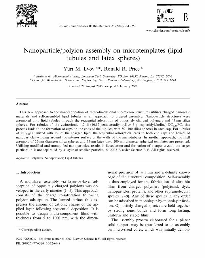
2.1.2. LatexIn the assembly, one adds the polyanion solu-
tion to the latex particles. After adsorption satu-ration is achieved, one has to separate the latexfrom the polyanion solution (by three cycles of5-min centrifugation at 10 000g). Then, themodified latex is exposed to the polycation solu-tion, and so forth (Fig. 1). Our carboxylated latexis negatively charged, and we used the followingcycles of adsorption: PEI+/PSS−/PEI+/75-nm sil-ica. The linear polyions with 3 mg/ml concentra-
Y.M. L�o�, R.R. Price / Colloids and Surfaces B: Biointerfaces 23 (2002) 251–256 253
Fig. 1. A scheme of polyanion/polycation/nanoparticle shell assembly on a spherical microtemplate.
tion, silica of 10 mg/ml, pH 7 and adsorption timeof 15 min were used. They are the same parame-ters as used for the formation of the same archi-tecture on a plane solid support [3,5]. The initialconcentration of the latex particles was 0.3 mg/ml,which gives �1010 particles in 1 cm3, and theaverage distance between the latex particles was�3000 nm (i.e., ten times more than their diame-ter). During the assembly, the latex concentrationgradually decreases. Each of the centrifugationcycles results in the loss of �5% of the latexmaterial.
2.1.3. Transmission electron microscopyIt was performed with LEO (Zeiss-Leica) in-
strument at voltage 60 kV.
3. Results and discussion
3.1. Assembly on tubular microtemplates
Lipid tubules are hollow cylinders made up ofhelical ribbons of diacetylenic lipid bilayers, withtypical diameters of 0.5–0.6 �m and lengths of20–100 �m. For our purposes, tubules served astemplates for the alternate adsorption of chargedpolymers and nanoparticles. By observing, wherethese components adsorb on the tubules, we cangain more information about the distribution ofcharge in the lipid helical ribbons, which form thewalls of the tubules, and we can take advantage ofthis helicity to build novel oriented structures ofnanoparticles. Nanoparticle structures were as-sembled onto lipid tubules through the sequentialadsorption of PSS− and PEI+ and 45-nm silica
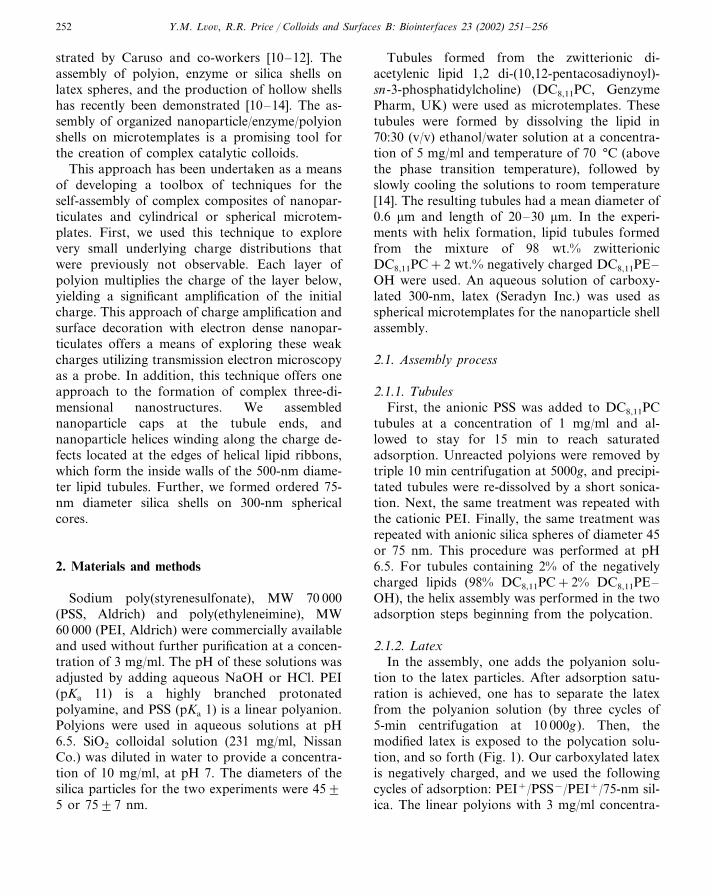
spheres. For tubules of the zwitterionic DC8,11PC,this process leads to the formation of caps at theends of the tubules, with 50–100 silica spheres ineach cap (Fig. 2). In a few cases, formation of the
Fig. 2. Nanoparticle cap at the end of DC8,11PC tubule afterthree-stage PSS−/PEI+/45-nm SiO2 treatment, (a) and (b).Transmission electron microscope LEO: Zeiss-Leica, 60 kV.
Y.M. L�o�, R.R. Price / Colloids and Surfaces B: Biointerfaces 23 (2002) 251–256254
caps was not completed and we observed acrown or halo formed instead. Nanoparticleswere concentrated only at the ends of thetubules, with no nanoparticle adsorption on thesurface or interior of the tubules. This indicatesthat the anchoring points for the polyion assem-bly were at the edge of the tubules where thelipid packing was not perfect. Formation ofcaps was also possible with a final treatmentutilizing larger particles (75-nm diameter silica)or with smaller particles (20-nm silica and 15-nm gold). It was necessary to begin the assem-bly from the polyanion.
Attempts to realize this assembly by applyingtwo-step PEI+/silica adsorption or just one-stepadsorption of silica were unsuccessful. At neu-tral or lower pH, the three-step PSS−/PEI+/sil-ica assembly resulted in cap formation on60–70% of tubules. At higher pH of 8–9, thecaps formed on only �5% of tubules. Thisshows that the excess of positive charges at theends on tubules at lower pH enhances the ad-sorption of negatively charged PSS and en-hances the formation of caps. Necessity of thethree-step assembly we explain by multiplicationof charges during PSS/PEI sequential adsorp-tion. Tsukruk et al. [15] demonstrated that dur-ing the initial two or three cycles of alternateadsorption the polyions spread from the initialbinding sites to form larger complexes. We be-lieve that this charge amplification is also appli-cable for our materials.
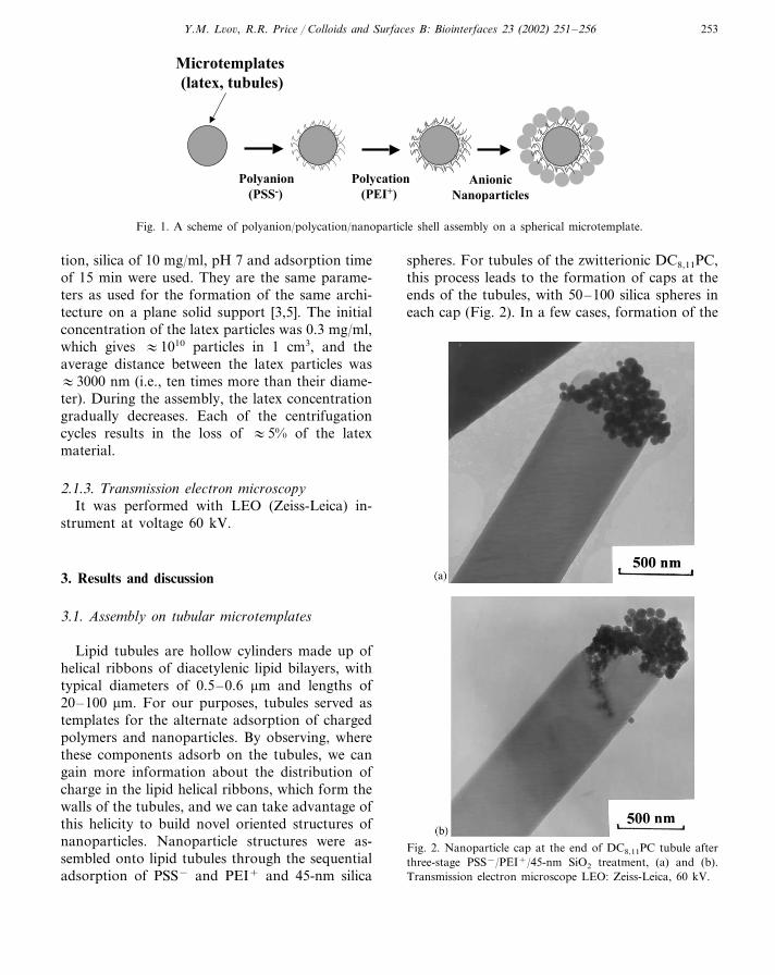
For the second set of experiments, we usedtubules of zwitterionic DC8,11PC lipid mixedwith 2% negatively charged lipids with the goalof using the layer-by-layer assembly to ‘develop’the distribution of DC8,11PE–OH. We intro-duced negative sites for the initial binding, andhad to begin the assembly from a polycation.An application of the four-step treatment, PEI+
/PSS−/PEI+/45 nm silica resulted in the forma-tion of nanoparticle caps and, additionally, thehelices inside the tubules (Fig. 3(a)). The se-quence of charged polymers and nanoparticlesadsorbs along charged line defects in the tubules(probably these lines are enriched by negativelycharged lipids). Thus, the sequential adsorptionmakes these charged sites visible in the transmis-
Fig. 3. Nanoparticle helix inside the tubules of DC8,11PC+2%DC8,9PE–OH after PEI+/PSS−/PEI+/45-nm SiO2 treatment(a), and part of the helix at higher magnification (b). TEMLEO: Zeiss-Leica, 60 kV, magnification 10 000× .
sion electron microscope. Fig. 3(b) shows a sec-tion of the nanoparticle helix at largermagnification. One can see the row of 45-nmdiameter silica spheres ordered along the invisi-ble charge line.
Y.M. L�o�, R.R. Price / Colloids and Surfaces B: Biointerfaces 23 (2002) 251–256 255
3.2. Assembly on spherical microtemplates
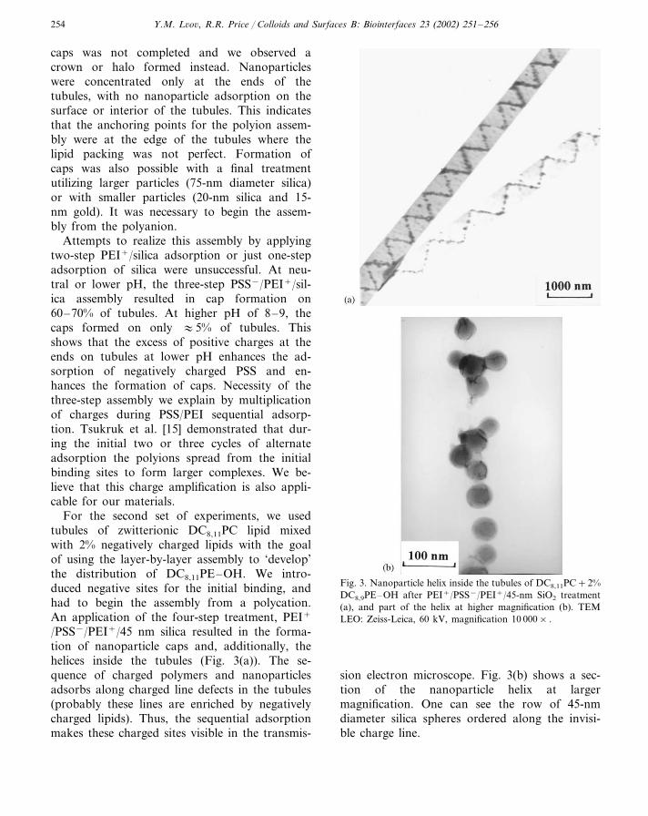
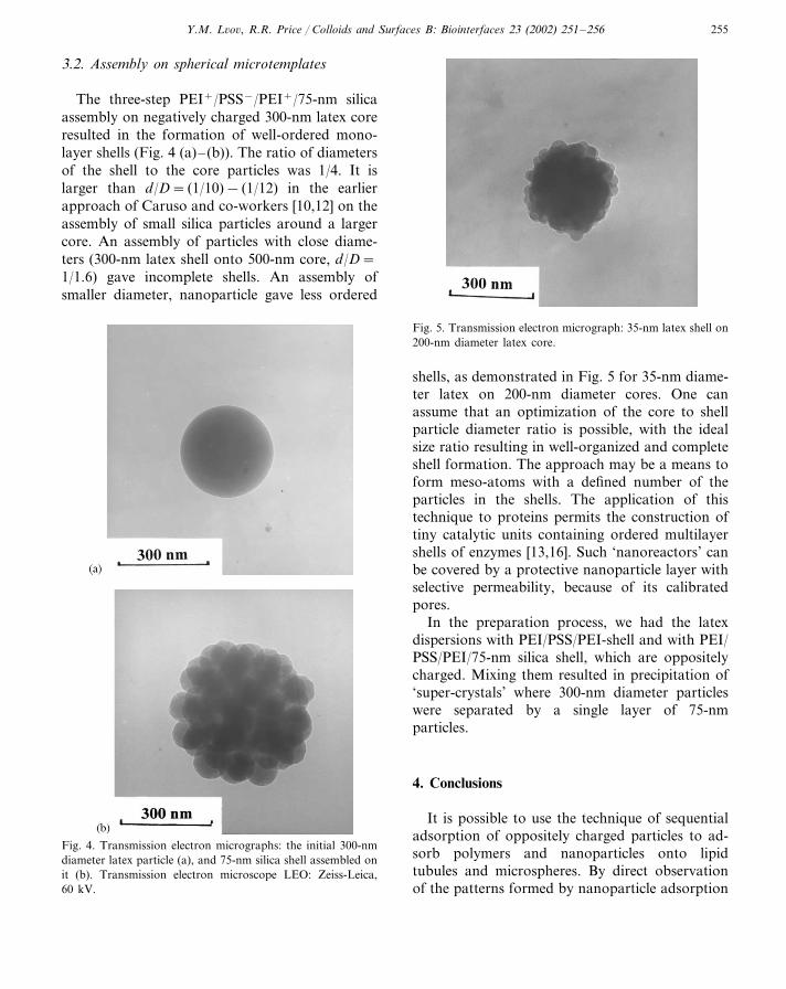
The three-step PEI+/PSS−/PEI+/75-nm silicaassembly on negatively charged 300-nm latex coreresulted in the formation of well-ordered mono-layer shells (Fig. 4 (a)– (b)). The ratio of diametersof the shell to the core particles was 1/4. It islarger than d/D= (1/10)− (1/12) in the earlierapproach of Caruso and co-workers [10,12] on theassembly of small silica particles around a largercore. An assembly of particles with close diame-ters (300-nm latex shell onto 500-nm core, d/D=1/1.6) gave incomplete shells. An assembly ofsmaller diameter, nanoparticle gave less ordered
Fig. 5. Transmission electron micrograph: 35-nm latex shell on200-nm diameter latex core.
Fig. 4. Transmission electron micrographs: the initial 300-nmdiameter latex particle (a), and 75-nm silica shell assembled onit (b). Transmission electron microscope LEO: Zeiss-Leica,60 kV.
shells, as demonstrated in Fig. 5 for 35-nm diame-ter latex on 200-nm diameter cores. One canassume that an optimization of the core to shellparticle diameter ratio is possible, with the idealsize ratio resulting in well-organized and completeshell formation. The approach may be a means toform meso-atoms with a defined number of theparticles in the shells. The application of thistechnique to proteins permits the construction oftiny catalytic units containing ordered multilayershells of enzymes [13,16]. Such ‘nanoreactors’ canbe covered by a protective nanoparticle layer withselective permeability, because of its calibratedpores.
In the preparation process, we had the latexdispersions with PEI/PSS/PEI-shell and with PEI/PSS/PEI/75-nm silica shell, which are oppositelycharged. Mixing them resulted in precipitation of‘super-crystals’ where 300-nm diameter particleswere separated by a single layer of 75-nmparticles.
4. Conclusions
It is possible to use the technique of sequentialadsorption of oppositely charged particles to ad-sorb polymers and nanoparticles onto lipidtubules and microspheres. By direct observationof the patterns formed by nanoparticle adsorption
Y.M. L�o�, R.R. Price / Colloids and Surfaces B: Biointerfaces 23 (2002) 251–256256
with an electron microscope, one can infer infor-mation about the distribution of charges on smallstructures, such as the tubules used in this study.In particular, the observation of nanoparticle heli-cal structures on tubules with a charged lipidshows that the charged lipid is concentrated alonghelical defect lines that are not otherwise visible.Formations of nanoparticle caps at the ends ofnanotubules and of the ordered shells on 300-nmspherical cores demonstrate a less selective assem-bly. These results show that charged nanopatternson microtemplates can serve as substrates for theordered construction from nanomaterial precur-sors of complex 3-D assemblies, an approach thatshows promise for future nanofabrication.
Acknowledgements
We are thankful to J. Schnur and J. Selinger foruseful discussion of results and A. Singh forproviding us with charged DC8,9PE–OH lipids.Acknowledge is made to the Donors of ThePetroleum Research Fund, administrated byAmerican Chemical Society, for partial support ofthis work (c 36066).
References
[1] G. Decher, Science 227 (1997) 1232.[2] Y. Lvov, G. Decher, H. Mohwald, Langmuir 9 (1993) 481.[3] Y. Lvov, K. Ariga, I. Ichinose, T. Kunitake, J. Am. Chem.
Soc. 117 (1995) 7114.[4] E. Kleinfeld, G. Ferguson, Science 265 (1994) 370.[5] Y. Lvov, K. Ariga, I. Ichinose, T. Kunitake, Langmuir 13
(1997) 6195.[6] T. Cassagneau, T. Mallouk, J. Fendler, J. Am. Chem. Soc.
120 (1998) 7848.[7] M. Leasche, J. Schmitt, G. Decher, W. Bouwman, K.
Kjaer, Macromolecules 31 (1998) 8893.[8] S. Joly, R. Kane, L. Radzilovski, T. Wang, A. Wu, R.
Cohen, E. Thomas, M. Rubner, Langmuir 16 (2000) 1354.[9] A. Mamedov, J. Ostander, F. Aliev, N. Kotov, Langmuir
16 (2000) 3941.[10] F. Caruso, R. Caruso, H. Mohwald, Science 282 (1998)
1111.[11] E. Donath, G. Sukhorukov, F. Caruso, S. Davis, H.
Mohwald, Angew. Chem. Int. 37 (1998) 2202.[12] F. Caruso, R. Caruso, H. Mohwald, Chem. Mater. 11
(1999) 3309.[13] F. Caruso, H. Mohwald, J. Am. Chem. Soc. 121 (1999)
6039.[14] Y. Lvov, R. Price, A. Singh, J. Selinger, M. Spector, J.
Schnur, Langmuir 16 (2000) 5949.[15] V. Tsukruk, V. Bliznyuk, D. Visser, A. Campbell, T.
Bunnig, W. Adams, Macromolecules 30 (1997) 6615.[16] F. Caruso, H. Fiedler, K. Hage, Coll Surf A 169 (2000) 287.