nanoparticle-based drug delivery system a target strategy
TRANSCRIPT

Review ArticleNanoparticle-Based Drug Delivery System—A Target Strategy forOsteoarthritis Treatment
Keda Liu , Dianjian Zhang , and Wei Wang
School and Hospital of Stomatology, China Medical University, Liaoning Provincial Key Laboratory of Oral Diseases,Shenyang 110001, China
Correspondence should be addressed to Wei Wang; [email protected]
Received 4 August 2021; Accepted 6 October 2021; Published 20 October 2021
Academic Editor: Hui Qi Xie
Copyright © 2021 Keda Liu et al. This is an open access article distributed under the Creative Commons Attribution License,which permits unrestricted use, distribution, and reproduction in any medium, provided the original work is properly cited.
Osteoarthritis (OA) is a bone and joint disease with pathological characteristics such as articular cartilage degeneration injury andsynovial and subchondral bone reactive hyperplasia. Once cartilage is damaged, it is difficult to repair it by itself. Current clinicalpractice is mainly limited to symptomatic treatment, not changing the degenerative process of osteoarthritis. The important goalof nanomedicine is targeted delivery. Nanoparticles (NPs) can provide many unique potential functions for the targeted treatmentof arthritis. This review summarizes the research progress of nanomaterials as a drug delivery system in the treatment ofosteoarthritis from three aspects: (1) the etiology of OA and the current research status of applying nanoparticles to treat OA,(2) the construction of osteoarthritis models, and (3) the classification of nanoparticle-based drug delivery systems.
1. Introduction
With the increase in life expectancy, obesity rates and sportsinjuries, the incidence of arthritis is rising steadily [1]. Thesereasons have promoted the research of tissue engineeringmaterials in orthopedics or joint surgery. Osteoarthritis, alsoknown as degenerative arthritis, senile arthritis, and hyper-trophic arthritis, is a bone and joint disease with main patho-logical characteristics such as articular cartilage degenerationinjury, joint edge and subchondral bone reactive hyperplasia,and synovial hyperplasia [2, 3]. Cartilage lacks nourishingpathways (such as blood vessels, nerves, and lymph), consist-ing of only a single type of cell with low proliferative activity(chondrocytes) [4]. Therefore, once damaged, it is extremelydifficult to repair it by itself. The current traditional methodsfor the treatment of cartilage defects mainly include autolo-gous cartilage transplantation, microfracture (bone marrowstimulation), and autologous chondrocyte transplantation[5]. Although these methods have certain curative effects,they have defects such as large damage to the donor site,inconsistent characteristics of the repaired area and sur-rounding cartilage, and poor interface healing. Current clin-ical practice is mainly limited to symptomatic treatment
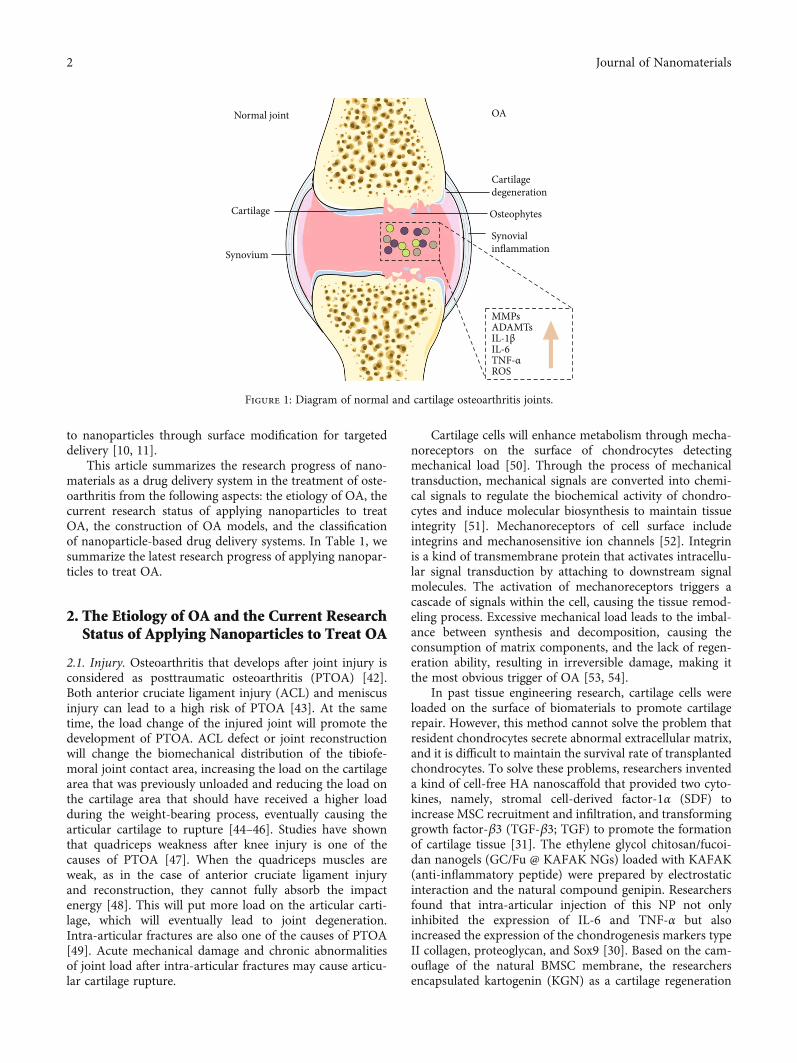
(pain relief and artificial joint replacement), not involvingthe underlying molecular cause of OA. This treatment doesnot change the degenerative process of osteoarthritis.Figure 1 shows the diagram of normal and cartilage osteoar-thritis joints.
Nanomaterials refer to materials whose structure is atthe nanometer scale in at least one dimension, or is com-posed of nanostructured units with special properties. Ithas been extensively studied in the field of tissue engineer-ing, including as a cell growth scaffold to promote bone tis-sue regeneration and as a transplant material for repairingperipheral nerves [6, 7]. Nanomaterials can perform biolog-ical imaging through spectral and fluorescent signals to pro-mote disease diagnosis [8]. At the same time, nanomaterialscan also be used as biosensors to effectively monitor diseaseprogression [9]. This review mainly focuses on the targeteddelivery function of nanomaterials in the field of OA. Nano-materials can provide many unique potential functions forthe targeted treatment of arthritis, and they include the fol-lowing: (1) maintaining the concentration of the drug inthe target area to maximize the effect of the drug, (2) carry-ing more drug molecules and increasing the solubility ofsome hydrophobic drugs, and (3) loading target molecules
HindawiJournal of NanomaterialsVolume 2021, Article ID 4064983, 15 pageshttps://doi.org/10.1155/2021/4064983
to nanoparticles through surface modification for targeteddelivery [10, 11].
This article summarizes the research progress of nano-materials as a drug delivery system in the treatment of oste-oarthritis from the following aspects: the etiology of OA, thecurrent research status of applying nanoparticles to treatOA, the construction of OA models, and the classificationof nanoparticle-based drug delivery systems. In Table 1, wesummarize the latest research progress of applying nanopar-ticles to treat OA.
2. The Etiology of OA and the Current ResearchStatus of Applying Nanoparticles to Treat OA
2.1. Injury. Osteoarthritis that develops after joint injury isconsidered as posttraumatic osteoarthritis (PTOA) [42].Both anterior cruciate ligament injury (ACL) and meniscusinjury can lead to a high risk of PTOA [43]. At the sametime, the load change of the injured joint will promote thedevelopment of PTOA. ACL defect or joint reconstructionwill change the biomechanical distribution of the tibiofe-moral joint contact area, increasing the load on the cartilagearea that was previously unloaded and reducing the load onthe cartilage area that should have received a higher loadduring the weight-bearing process, eventually causing thearticular cartilage to rupture [44–46]. Studies have shownthat quadriceps weakness after knee injury is one of thecauses of PTOA [47]. When the quadriceps muscles areweak, as in the case of anterior cruciate ligament injuryand reconstruction, they cannot fully absorb the impactenergy [48]. This will put more load on the articular carti-lage, which will eventually lead to joint degeneration.Intra-articular fractures are also one of the causes of PTOA[49]. Acute mechanical damage and chronic abnormalitiesof joint load after intra-articular fractures may cause articu-lar cartilage rupture.
Cartilage cells will enhance metabolism through mecha-noreceptors on the surface of chondrocytes detectingmechanical load [50]. Through the process of mechanicaltransduction, mechanical signals are converted into chemi-cal signals to regulate the biochemical activity of chondro-cytes and induce molecular biosynthesis to maintain tissueintegrity [51]. Mechanoreceptors of cell surface includeintegrins and mechanosensitive ion channels [52]. Integrinis a kind of transmembrane protein that activates intracellu-lar signal transduction by attaching to downstream signalmolecules. The activation of mechanoreceptors triggers acascade of signals within the cell, causing the tissue remod-eling process. Excessive mechanical load leads to the imbal-ance between synthesis and decomposition, causing theconsumption of matrix components, and the lack of regen-eration ability, resulting in irreversible damage, making itthe most obvious trigger of OA [53, 54].
In past tissue engineering research, cartilage cells wereloaded on the surface of biomaterials to promote cartilagerepair. However, this method cannot solve the problem thatresident chondrocytes secrete abnormal extracellular matrix,and it is difficult to maintain the survival rate of transplantedchondrocytes. To solve these problems, researchers inventeda kind of cell-free HA nanoscaffold that provided two cyto-kines, namely, stromal cell-derived factor-1α (SDF) toincrease MSC recruitment and infiltration, and transforminggrowth factor-β3 (TGF-β3; TGF) to promote the formationof cartilage tissue [31]. The ethylene glycol chitosan/fucoi-dan nanogels (GC/Fu @ KAFAK NGs) loaded with KAFAK(anti-inflammatory peptide) were prepared by electrostaticinteraction and the natural compound genipin. Researchersfound that intra-articular injection of this NP not onlyinhibited the expression of IL-6 and TNF-α but alsoincreased the expression of the chondrogenesis markers typeII collagen, proteoglycan, and Sox9 [30]. Based on the cam-ouflage of the natural BMSC membrane, the researchersencapsulated kartogenin (KGN) as a cartilage regeneration
Normal joint
Cartilage
Synovium
Synovialinflammation
MMPsADAMTsIL-1βIL-6TNF-αROS
OA
Cartilagedegeneration
Osteophytes
Figure 1: Diagram of normal and cartilage osteoarthritis joints.
2 Journal of Nanomaterials

drug into Fe3O4 nanoparticles. As a result, a nanomaterialwith excellent biocompatibility and good biosafety isobtained, achieving high-quality and fast cartilage regenera-tion [23]. Researchers synthesized cerium oxide nanoparti-cles (CeO2) with a particle size of about 120nm andcombined them with hyaluronic acid (HA) to construct
nanodelivery drugs. Through in vitro OA model studies, itwas found that the delivery system can protect chondrocytesfrom oxidative stress, and the expression of COL2a1 andACAN genes in chondrocytes was significantly increased[19]. Janus-based nanotubes (AAT) self-assemble from ana-logs of synthetic DNA bases (guanine-cytosine motif), which
Table 1: Research summary of nanoparticle-based drug delivery systems.
Type ofNPs
Carrier Cargo Outcome Ref
Liposomes(HA)-liposomal
Diclofenac,dexamethasone
Effectively relieving the pain of OA and having goodbiocompatibility
[12]
SLN systemIntegrin β1
overexpression pDNAReducing chondrocyte apoptosis and enhancing tissue
repair[13]
Micelles
PNIPAM-PMPC Diclofenac sodiumPNIPAM-PMPC nanospheres are biocompatible andupregulate anabolic genes, while downregulating
articular cartilage catabolism genes[14]
Polyethylene oxide- (PEO-) andpolypropylene oxide- (PPO-)based polymeric micelles
rAAV sox9Enhancing the deposition of ECM components and
cell survival levels, inhibiting inflammation[15]
Acid-activatable polymer CurcuminSuppression of tumor necrosis factor-alpha (TNF-α)and interleukin 1β (IL-1β). Potent antioxidant and
anti-inflammatory activities[16]
InorganicNPs
MnO2, gold-basednanoformulations, CeO2
COL2a1 and ACAN gene expression in chondrocyteswas significantly decreased
[17–19]
MSNs pSBMA, colchicineDue to the hydration lubrication mechanism, the wearresistance of the material is enhanced. Reducing nitric
oxide, malondialdehyde, COX2, and TNF-α[20, 21]
Zeolitic imidazolate framework-8S-Methylisothioureahemisulfate salt
Reducing the content of NO and H2O2, therebyinhibiting the production of HIF1α and M1
macrophages, alleviating mitochondrial function[22]
Membrane-disguised Fe3O4 Kartogenin Enabling rapid and high-quality cartilage regeneration [23]
PolymerNPs
CD-PMPC SilicaEnhancing penetration of dermal tissue and
lubrication, inducing drug release[24]
PLGA-PEG4MAL, kartogenin
(KGN)Prolonging IA drug retention for the treatment of OA.
Increasing sulfated glycosaminoglycans[25–27]
Electrostatic self-assembly heparinand ε-poly-l-lysine
Platelet lysate Platelet lysate is evenly distributed [28]
Hollow dextran/PNIPAM KAFAK In cartilage explants to suppress inflammation [29]
Glycol chitosan/fucoidannanogels
KAFAKInhibiting the expression of TNF-α and IL-6.Enhancing the expression of type II collagen,
proteoglycan, and Sox9[30]
Electro spun cell-free fibroushyaluronic acid
SDF-1α, TGF-β3Increasing recruitment and infiltration of MSCs to
enhance cartilage tissue formation[31]
PLGAAndrographolide,
p66shc siRNA, diacereinInhibiting osteoclast function and inflammation [32–34]
Poly(D,L-lactic acid)-poly(ethylene glycol)-poly(D,L-
lactic acid)BMP2
Inducing implant differentiation into cartilage andbone but also completely degraded without toxicity
[35]
LbL polymer microcapsule MnO2Eliminating hydrogen peroxide H2O2 and protecting
cells from oxidative stress[36]
Exosomes
miR-140, miR-9-5p,miR-100-5p, miR-135b,
miR-1405p, andlncRNA KLF3-AS1
Reducing inflammation and promoting cartilagemarker production
[37–41]
3Journal of Nanomaterials

can transport small RNA molecules to cells and tissues.Researchers encapsulated the nanotube AAT-packagedmiR365 antagomir in the yeast cell wall to construct nanoad-ministrative particles. This nanodrug delivery system inhib-ited the content of miR365 by oral administration to treatPTOA (mice) [55].
2.2. Inflammation. OA is also a disease caused by underlyingimmune response leading to bone remodeling and cartilagedegradation. Typical symptoms include swelling, pain, andstiffness [56]. By initiating the inflammatory process andinducing cartilage decomposition, the early changes in thecartilage surface are manifested as fibers extending distally,forming deep cracks, leading to cartilage delamination,forming calcified cartilage [57]. The thinning of articularcartilage is closely associated with dilation of basal calcifiedcartilage, which in turn leads to increased mechanical stressand further production of degrading factors [58]. Major sig-naling molecules involved in OA immune response are usu-ally divided into the anti-inflammatory and inflammatorygroups. The inflammatory cytokines include TNF-α, IL-1β,IL-6, IL-8, and IL-17, and the anti-inflammatory cytokinesinclude IL-13, IL-4, IL-10, and IL-1Ra [42, 49, 59]. Cytokinescan participate in the pathological process of cartilagedegeneration by mediating multiple signaling pathways,mainly including the mitogen-activated protein kinase(MAPK) signaling pathway [60], the AMPK signaling path-way [61], and the Wnt/β-catenin signaling pathways [62].They can also promote the synthesis of PGE2 and inducethe production of chondrocytes to synthesize metallopro-teinases (ADAMTS) [63], HIF2α, NOS2, MMPs, andCOX2, thereby promoting the inflammatory process andinhibiting the proliferation of chondrocytes [64, 65].
Therefore, implanting anti-inflammatory factors orenzymes into the joint cavity and maintaining the drug con-centration is another research direction for OA treatment.Hollow dextran/PNIPAM nanoparticles loaded withKAFAK effectively deliver therapeutic peptides to inhibitinflammation. These heat-responsive nanoparticles may bean effective drug delivery system that can deliver anti-inflammatory therapeutic peptides in an OA environment[29]. The researchers encapsulated three hydrophobic anti-inflammatory drugs (tenoxicam, dexamethasone, and cele-coxib) into core-shell terpolymer nanoparticles. Experimentshave shown that these loaded nanoparticles have the activityof acting as inflammatory mediator production regulatorsin vitro [66]. MnO2 nanoparticles, with excellent biocompat-ibility, can be used as artificial nanoenzymes to effectivelyeliminate reactive oxygen. Hollow MnO2 (H-MnO2) wassynthesized by the Stober method and modified with NH2-PEG-NH2, reducing the inflammatory response of OA [17].
2.3. Obesity. Obesity is a state of excessive accumulation offat in the body, which is believed to be directly related tosome metabolic diseases such as high blood pressure, dyslip-idemia, diabetes, and osteoarthritis [67–69]. Obesity plays animportant role in the occurrence and development ofweight-bearing and non-weight-bearing joint osteoarthritis.Increased joint load and systemic metabolic changes may
be important factors in the occurrence of obesity-relatedosteoarthritis. Moderate mechanical load can maintain thedynamic balance and integrity of articular cartilage. Com-pared with normal BMI people, tibiofemoral cartilage ofhigh BMI people withstand more compressive stress duringshort-term running tasks [70]. The proteoglycan content inobese subjects is reduced, indicating that the cartilage is ina “pre-osteoarthritis” state. Osteoarthritis caused by obesityis defined as a “metabolic osteoarthritis” phenotype [71].Metabolic osteoarthritis is associated with increased fatdeposits that release inflammatory cytokines/adipokines,leading to systemic inflammation, cartilage loss, and osteo-phyte formation [72].
In order to solve the abrasion caused by mechanical ero-sion, viscoelastic supplements based on hyaluronic acid(HA) have been widely used to treat knee joint injuries.However, current HA formulation cannot provide effectivehealing and recovery. Researchers have developed ananofiber-HA membrane system to protect arthritic carti-lage tissue from degeneration. The material has a uniquescaffold structure that can provide a 3D microenvironmentlike natural ECM, and deliver biologically active signals thatcan activate chondrocyte proliferation and functional colla-gen I synthesis. Researchers injected the ankle joint to fillthe joint cavity and found that this hybrid nanofiber mem-brane has a better therapeutic effect than the commerciallyavailable Hyalgan and Synvisc gels at a lower concentration,providing a simple and feasible alternative to OA treat-ment [73].
2.4. Age. Age is one of the main factors of osteoarthritis.There are literatures showing that the prevalence of kneearthritis increases almost linearly after the age of 40 [74].The incidence of osteoarthritis increase with age, whichmay be due to the accumulation of various risk factors andthe result of biological changes [75]. Osteoarthritis is charac-terized by the imbalance between catabolic and anabolicactivities in the joints. Aging chondrocytes do not respondwell to growth factor stimulation and cannot maintain thehomeostasis of articular cartilage, leading to the occurrenceof OA [76].
How to improve the response of aging chondrocytes togrowth factors has received extensive attention. Plateletlysate (PL) is a cost-effective mixture of growth factors. Elec-trostatic self-assembly heparin (Hep) and ε-poly-l-lysine(EPL) nanoparticles (NPs) were engineered to enhance thesustained release ability of PL. This nanodrug delivery mate-rial retained the initial gelling ability and showed long-termPL release ability, ameliorating cartilage degeneration in thelate stage of OA [28]. Researchers have prepared a cationiclipid nanoparticle (SLN) system that can efficiently deliverplasmid DNA (pDNA) into cells. This study reported thatthe overexpression of pDNA carrying integrin β1 was trans-ported into rat chondrocytes via liposomes, reducing IL-1β-stimulated chondrocyte apoptosis and enhancing tissuerepair [13]. Researchers developed a thermosensitive bifunc-tional nanosphere polymer (PNIPAM-PMPC) throughemulsion polymerization. The nanospheres can enhancelubrication by forming a hydration layer around the base
4 Journal of Nanomaterials

of the zwitterion head, and deliver local drug by wrappingdiclofenac sodium (an anti-inflammatory drug) [14, 77].
2.5. Genetic Factors. Epidemiological studies have confirmedthat genetic factors play a major role in the pathogenesis ofosteoarthritis. Studies based on the family history also con-firmed that OA has hereditary susceptibility [78, 79]. Themain mechanisms leading to OA were chemical modifica-tion of DNA, such as methylation, posttranslational modifi-cation of histones, and regulation of noncoding RNA.
(1) DNA methylation is a type of chemical modificationof DNA that can alter the performance of geneswithout changing the DNA sequence. It refers tothe covalent attachment of the methyl group to the5th carbon position of cytosine in the genomicCpG dinucleotide under the action of DNA methyl-transferase. Studies have shown that DNA methyla-tion can cause changes in chromatin structure,DNA conformation, DNA stability, and the wayDNA interacts with proteins, thereby controllingthe gene expression. The analysis of the wholegenome showed that the methylation of genesrelated to OA have changed, including genes encod-ing transcription factors, such as SOX9; genes encod-ing ECM protein and matrix degrading protease,including COL2a1, ACAN, and MMP13; and partic-ipating genes that signal growth factors and cyto-kines, such as GDF5 and BMP7 [80]. Someemerging trends indicate that methylation patternsmay differ between different OA joints and stagesof the disease [81]. For example, different methyla-tion patterns may occur between knee cartilage andhip cartilage, and between mild and severe cartilagefrom the same joint [82, 83]
(2) Gene expression is a complex process regulated bymultiple factors. Histones are an important part ofthe basic structure of chromosomes-nucleosomes,and their N-terminal amino acid residues canundergo acetylation, methylation, phosphorylation,and ubiquitination [84]
(3) Noncoding RNA (ncRNA) refers to RNA that doesnot encode protein. These include rRNA, tRNA,snRNA, snoRNA, microRNA, and other RNAs[85]. The common feature of these RNAs is that theycan be transcribed from the genome, performingtheir biological functions at the RNA level withoutbeing translated into proteins [86]. For example,miR-140 can inhibit the expression of harmful genesADAMTS5, MMP13, and IGFBP5 in OA [87]
Upregulating gene expression in target organs bydelivering specific signaling molecules is a kind of methodto treat OA caused by inheritance. Exosomes contain specificinformation about the source cell, with ability to delivermolecules targeting organs or tissues. The application ofexosomes and their derivatives by intra-articular injectionopen up new possibilities for the treatment of OA. Studies
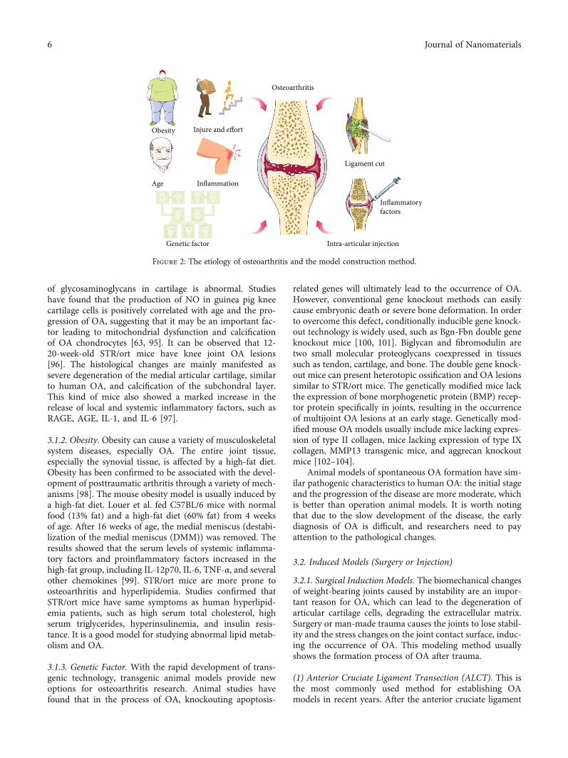
have shown that exosomes derived from primary chondro-cytes that overexpress miR-95-5p promoted cartilagedevelopment and cartilage matrix expression by directlytargeting histone deacetylase [88]. Exosomal miR-8485may regulate the expression of GSK-3 to suppress the pro-duction of glycogen synthase kinase, targeting the bindingantagonist of DACT1 to activate the Wnt/β-catenin pathwayand promote cartilage differentiation [89]. In Figure 2, weshow the common predisposing factors of osteoarthritis.
3. Method of Constructing an OAAnimal Model
Animal models are essential in studying the etiology of thedisease and the effectiveness of various treatment tools.The ideal model should include the following features: (1)all joint tissues (as the human body) are affected; (2) earlystages of the disease are included; (3) animal models cansimulate human disease; (4) animal species are easy to han-dle and raise; (5) the results can be evaluated in different bio-chemical, genetic, and imaging biomarkers, and should betransferable to the specificity of human medical therapeuticsand pathology. It is recommended to consider the followingpoints when selecting the OA model: the stage of OA thatneeds to be studied, the expansion of the lesion (focal or sys-temic), the therapeutic effect, and the target tissue to be stud-ied (cartilage, membrane, bones, or synovial fluid) [90].
OA models can be divided into induced models (surgeryor injection) and spontaneous models (natural developmentand genetic models). Spontaneous models can be used tostudy the pathogenesis of OA, with high economic costsand long time to achieve goals [91, 92]. On the contrary,the induction model achieves a reproducible and early OAmodel, but it prevents researchers from studying the possiblepathogenesis of diseases. Small mammals (mice, rats), rab-bits, and guinea pigs are the most common models forstudying the pathogenesis and pathophysiology of OA.
3.1. Spontaneous Models (Age, Obesity, and Genetic Factor).The main feature of spontaneous models is slow evolution,with a very long research cycle and high economic costs.However, they have an excellent correlation with naturalprocesses from a pathophysiological point of view. Sponta-neous models can be further subdivided into two subgroups:naturally occurring models or models produced by individ-ual genetic manipulation.
3.1.1. Age. OA tends to occur in the elderly. The animalmodel of spontaneous osteoarthritis is close to the progres-sion of human OA, as a valuable tool for studying the path-ogenesis of osteoarthritis. When a Hartly guinea pig about3 months old weighs 700 g, OA may appear on the medialtibial plateau of the knee joint. By the age of 18 months,the guinea pig’s medial tibial plateau had severe OA path-ological changes and there was no meniscus covering onthe surface [93, 94]. OA is manifested by cartilage degrada-tion, mainly histological changes in the cartilage in theweight-bearing area, which is similar to the occurrenceand development of human osteoarthritis. The distribution
5Journal of Nanomaterials
of glycosaminoglycans in cartilage is abnormal. Studieshave found that the production of NO in guinea pig kneecartilage cells is positively correlated with age and the pro-gression of OA, suggesting that it may be an important fac-tor leading to mitochondrial dysfunction and calcificationof OA chondrocytes [63, 95]. It can be observed that 12-20-week-old STR/ort mice have knee joint OA lesions[96]. The histological changes are mainly manifested assevere degeneration of the medial articular cartilage, similarto human OA, and calcification of the subchondral layer.This kind of mice also showed a marked increase in therelease of local and systemic inflammatory factors, such asRAGE, AGE, IL-1, and IL-6 [97].
3.1.2. Obesity. Obesity can cause a variety of musculoskeletalsystem diseases, especially OA. The entire joint tissue,especially the synovial tissue, is affected by a high-fat diet.Obesity has been confirmed to be associated with the devel-opment of posttraumatic arthritis through a variety of mech-anisms [98]. The mouse obesity model is usually induced bya high-fat diet. Louer et al. fed C57BL/6 mice with normalfood (13% fat) and a high-fat diet (60% fat) from 4 weeksof age. After 16 weeks of age, the medial meniscus (destabi-lization of the medial meniscus (DMM)) was removed. Theresults showed that the serum levels of systemic inflamma-tory factors and proinflammatory factors increased in thehigh-fat group, including IL-12p70, IL-6, TNF-α, and severalother chemokines [99]. STR/ort mice are more prone toosteoarthritis and hyperlipidemia. Studies confirmed thatSTR/ort mice have same symptoms as human hyperlipid-emia patients, such as high serum total cholesterol, highserum triglycerides, hyperinsulinemia, and insulin resis-tance. It is a good model for studying abnormal lipid metab-olism and OA.
3.1.3. Genetic Factor. With the rapid development of trans-genic technology, transgenic animal models provide newoptions for osteoarthritis research. Animal studies havefound that in the process of OA, knockouting apoptosis-
related genes will ultimately lead to the occurrence of OA.However, conventional gene knockout methods can easilycause embryonic death or severe bone deformation. In orderto overcome this defect, conditionally inducible gene knock-out technology is widely used, such as Bgn-Fbn double geneknockout mice [100, 101]. Biglycan and fibromodulin aretwo small molecular proteoglycans coexpressed in tissuessuch as tendon, cartilage, and bone. The double gene knock-out mice can present heterotopic ossification and OA lesionssimilar to STR/ort mice. The genetically modified mice lackthe expression of bone morphogenetic protein (BMP) recep-tor protein specifically in joints, resulting in the occurrenceof multijoint OA lesions at an early stage. Genetically mod-ified mouse OA models usually include mice lacking expres-sion of type II collagen, mice lacking expression of type IXcollagen, MMP13 transgenic mice, and aggrecan knockoutmice [102–104].
Animal models of spontaneous OA formation have sim-ilar pathogenic characteristics to human OA: the initial stageand the progression of the disease are more moderate, whichis better than operation animal models. It is worth notingthat due to the slow development of the disease, the earlydiagnosis of OA is difficult, and researchers need to payattention to the pathological changes.
3.2. Induced Models (Surgery or Injection)
3.2.1. Surgical Induction Models. The biomechanical changesof weight-bearing joints caused by instability are an impor-tant reason for OA, which can lead to the degeneration ofarticular cartilage cells, degrading the extracellular matrix.Surgery or man-made trauma causes the joints to lose stabil-ity and the stress changes on the joint contact surface, induc-ing the occurrence of OA. This modeling method usuallyshows the formation process of OA after trauma.
(1) Anterior Cruciate Ligament Transection (ALCT). This isthe most commonly used method for establishing OAmodels in recent years. After the anterior cruciate ligament
Osteoarthritis
Injure and effortObesity
Age Inflammation
Genetic factor Intra-articular injection
Inflammatoryfactors
Ligament cut
Figure 2: The etiology of osteoarthritis and the model construction method.
6 Journal of Nanomaterials

is cut, the number of cartilage cells in the superficial zonedecreases, swells, and becomes fibrotic [105, 106]. The deg-radation products of denatured type II collagen are signifi-cantly increased in the fibrotic area. It was reported thatafter cutting the anterior cruciate ligament of the canineknee joint, the synovial membrane of the canine joint wasthickened, the cartilage surface was eroded and osteophytesformed [107]. This phenomenon is the same as the patho-logical changes of natural joint instability caused by the rup-ture of the anterior cruciate ligament. This kind of model issimple to operate with less traumatic. It can fully reflect thepathological process of cartilage degeneration. However, thismethod takes a long time to successfully model—at least 6weeks. Some researchers fix the contralateral limb after therabbit anterior cruciate ligament is removed, so that the sur-gical limb is overloaded, and the time to make the OA modelis shorter [108].
(2) Meniscal Destabilization. The researchers removed partof the meniscus of the rabbit knee joint, causing the instabil-ity of the joint. The meniscus resection alone can reduce thedamage caused by the operation, but the modeling time iscorrespondingly prolonged. 12 weeks after removal of thelateral meniscus of the miniature pig’s knees, the numberof chondrocytes and the proteoglycan content decreased,the number of cells arranged in clusters increased, the thick-ness of cartilage became thinner, the surface of cartilagebecame fibrotic, and the femoral intercondylar notch osteo-phytes formed [109, 110].
(3) Hulth Model. Under sterile conditions, a longitudinal cutis made on the inside of the knee joint, the cruciate ligamentand medial collateral ligament are cut off, and the medialmeniscus is completely resected without damaging the artic-ular cartilage surface. After the operation, the injured limbwas not fixed and moved freely. After experimental animalsunderwent the Hulth method surgery, due to joint instabil-ity, increased friction on the articular surface, and the lackof cushioning effect of the meniscus, osteoarthritis can easilyoccur [111].
(4) Models for Generating Focal Defects. In the OA modelobtained through intra-articular surgery, joint instability isa factor that promotes the progress of OA. At the same time,in this type of OA model, the presence of synovial inflamma-tion leads to joint degeneration,whichwill affect the therapeu-tic effect of treatment measures for cartilage protection andrepair. An articular focal defect is a good model for observingthe pathology development. Researchers used sharp tools toscratch the articular cartilage in the weight-bearing area ofthe joint, but they did not damage the subchondral bone.This method would lead to pathological changes in the boneand joint. The joint focal defect method is an ideal model forobserving the early manifestations and treatment effects ofOA. It is particularly sensitive in observing the therapeuticeffects of cartilage protection and repair [112, 113].
3.2.2. Intra-Articular Injection. Toxic or inflammatory com-pounds were injected directly into the joint cavity of the
knee joint to induce disease by destroying the extracellularmatrix or articular cartilage cells. This method is simple tooperate, reproducible, and has a short cycle. It is suitable forOA pathology and antiosteoarthritis drug research. Com-monly used injections to induce osteoarthritis include papain,collagenase, and monosodium iodoacetate. Researchers havefound that collagenase can cause cartilage destruction andjoint tissue inflammation in rabbit knee joints in the earlystage and articular cartilage degradation in the late stage[114]. The glycolysis inhibitor monosodium iodoacetate(MIA) was injected into the knee joints ofWistar rats. Within1 week, chondrocytes became degenerated and necrotic, andfull-thickness changes of cartilage could also appear in thetibia and femoral condyles. MIA injection-induced osteoar-thritis is currently the most commonly used pharmaceuticalpreparation [115]. Vitamin A, hyaluronidase, cartilage frag-ments or foreign bodies, adrenal cortex hormones, etc.injected into the joint cavity of animals can also cause articu-lar cartilage degeneration and form an OA model [116, 117].
3.2.3. Joint Brake Modeling Method. The composition, struc-ture, and function of articular cartilage can be maintainedonly by ensuring the normal movement and function ofthe joints. Fixing limbs to break the joints can induce artic-ular cartilage atrophy, resulting in cartilage thinning, edema,and decreased proteoglycan content [118]. Because thesecartilage changes are similar to the pathological changes ofhuman OA, this model is especially used for the study of car-tilage degeneration. However, the use of this model hasgradually decreased in the research on the differences inthe basic morphology of cartilage. In the cartilage of OA,chondrocytes proliferate focally in cell clusters or cell nests,which are usually active at the later stage of OA. In thefixed-joint model, chondrocytes have no such changes.However, cell clusters often become necrotic, especiallywhen the splint is firmly fixed without any movement. Thischange in cartilage may be caused by the lack of nutrients incartilage cells. Moreover, if the immobilized limbs areallowed to move in a small range, the range of cartilagedegeneration will be significantly reduced [119, 120]. InTable 2, we compare the characteristics of several osteoar-thritis models.
4. Classification of Nanoparticle-Based DrugDelivery System
Nanomaterials have a close relationship with biological bod-ies in terms of size. For example, the ribonucleic acid proteincomplex, one of the elements of life, has a linearity between15 and 20nm. Nanobiomedical materials are the intersectionof nanomaterials and biomedical materials. Nanoparticlesand other materials are combined to make various compos-ite materials. With the further deepening of research, nano-materials have begun to penetrate into many disciplines,showing great potential application value. In recent years,the exuberant development of nanotechnology in drug deliv-ery systems has spawned new methods for treating OA. Thissection describes the current development and new applica-tions of NP-based drug delivery for the treatment of OA,
7Journal of Nanomaterials

including liposomes, micelles, polymeric nanoparticles, exo-somes, and inorganic nanoparticles [121].
4.1. Exosome. Exosomes are membrane vesicles with a diam-eter from 30 to 150nm, reflecting complex molecular pro-cesses that occur in parent cells. Exosomes, as endogenousdrugs, have diagnostic, drug delivery, and targeted therapycapabilities by transporting nucleic acids (DNA, mRNA,microRNA, and lncRNA), biologically active lipids, and pro-teins [122]. Exosomes do not require the same harsh storageconditions as seed cells, but can perform functions similar toseed cells. Therefore, it is more suitable for treatment anddelivering active substances [123, 124].
Exosomes used to treat OA are principally derived fromMSCs. Recent studies have shown that exosomal miRNAand lncRNA play a key role in anti-OA. It remains a majorchallenge to deliver a drug through the dense cartilagematrix. Liang et al. use lysosomal-membrane glycoproteinto fuse chondrocyte active peptide (CAP) on the surface ofchondrocyte-derived exosomes. In vivo studies have shownthat the delivery of miR-140 based on CAP exosomes signif-icantly reduces the development of OA in a rat model by IAinjection [37]. Several recent studies have shown thatmiRNA may participate in the regulation of several signalssuch as the SIRT1/p53 pathway, the Wnt/-catenin pathway,and the NF-κB pathway, thereby regulating the expression ofgenes involved in the decomposition and anabolism of OA.By delivering miRNA, exosomes can inhibit the productionof proteolytic enzymes and proinflammatory cytokines(e.g., IL-1, IL-6, and TNF-α) to affect the treatment of OA[125, 126].
Natural exosomes have their own therapeutic agentsand can be used for drug delivery. However, the numberof exosomes released by cells is uncertain due to the lackof clinical research. Obtaining a sufficient number of exo-somes for in vivo research is a technical challenge. In orderto increase the production of exosomes, some studies areexploring the use of three-dimensional spheres or tetrahe-drons to transport therapeutic substances. With the elucida-tion of exosomal mechanism and the maturity of exosomalmanufacturing technology, it will create a new field of OAtreatment [127].
4.2. Liposome. The liposome is an artificial membrane.When the hydrophilic head of the phospholipid moleculeis inserted into the water, the hydrophobic tail of the lipo-some extends to the air. After agitation, a spherical liposomewith a double layer of lipid molecules is formed, with adiameter ranging from 25 to 1000 nm [13]. Liposomes canbe used for genetically modified or prepared medicines.The liposomes can fuse with the cell membrane to deliverthe medicines into the cell. Liposomes are the first nanodrugcarrier approved by the FDA, deemed to be the most idealdrug delivery vehicle. Liposome formulations have beenextensively tested as drug delivery vehicles in OA, not onlybecause of their ability to carry hydrophobic and hydrophilicdrugs but also because of their excellent biocompatibility.
Adenosine is a key autocrine cytokine for maintainingcartilage homeostasis. The A2A receptor is a kind of recep-tor for adenosine. Researchers encapsulated adenosine andA2A receptor agonists in liposomes, and then they injectedthe liposomes to prevent the OA progression of obesity-
Table 2: Classification and characteristics of osteoarthritis animal models.
Spontaneous models Surgical induction models Intra-articular injection models
Principle oftheexperiment
The cause of OA is related to certaingenetic mutations
OA spontaneously occurs with ageGenetically modified animals
constructed using transgenic or geneknockout technologies can also
spontaneously form OA
Using surgery to cause disease in thejoint cavity to produce OA
Injecting toxic or inflammatorycompounds directly into the joint cavityof the joint to induce disease bydestroying extracellular matrix orarticular cartilage cells
Types ofcommonlyused models
Male Hartly guinea pig, STR/ort mice,Bgn-Fbn double gene knockout mice,
Cre-Gdf5/Bmpr1a floxP mice
The Hulth method, anterior cruciateligament transection (ACLT), partial orfull meniscus resection, medialmeniscus tear, joint mark method
Types of drugs: chemicals, enzymes, andhormonesCommon injection drugs: MIA,collagenase, papain, etc.
AdvantageAnimal models of spontaneous OA havesimilar pathogenic characteristics to
humans
Using aseptic technique to induce, theresults are highly reproducible and themodeling experiments are shorter
(1) The molding speed is fast(2) The pathological changes of cartilagein the end-stage OA can be observed ina short time(3) Less traumatic and easy to operate
Disadvantage
Early diagnosis of OA is difficult. Theresearch time is long. The model may berestricted by environmental factors,
ethical conditions, and high economiccost
(1) The trauma is large, andpostoperative infection is prone tooccur(2) Traumatic arthritis and synovialinflammation may occur during theoperation, which may affect theexperimental results
It is difficult to have a certain standardfor drug dosage. Different animals havedifferent doses of drugs injected.Therefore, a poor grasp of the drugdosage will cause errors in experiments
8 Journal of Nanomaterials

induced mouse and rat posttraumatic OA [128]. Recentstudies have shown that rapamycin encapsulated in lipo-somes has a notable anti-inflammatory effect in the sponta-neous OA model [129].
4.3. Micelle. Micelle refers to the orderly aggregates of mole-cules that begin to form in large quantities after the surfac-tant concentration in an aqueous solution reaches thecritical micelle concentration (CMC). In micelles, the hydro-phobic groups of surfactant molecules aggregate to form theinner core of the micelle, and the hydrophilic polar groupsform the outer layer of the micelle. Micelles can also be takenup by cells by binding to ligands, antibodies, or peptides.Because of its low CMC, polymer micelles have a longercycle time and are more stable, so they are the most widelyused in drug delivery systems [130, 131].
A study has reported that overexpression of the SOX9transcription factor by using polymer micelles as a carriercan readjust the metabolic balance in OA. In the presenceof inflammatory factors IL-1β and TNF-α, micellar-guidedrAAV-sox9 increases the level of extracellular cartilagematrix (ECM) deposition and chondrocyte survival [15].
4.4. Inorganic Nanoparticles. Inorganic nanoparticles (INPs)are synthesized from inorganic particles and biodegradablepolycations. Typical inorganic nanoparticles include metal,metal oxides, and carbon materials (such as fullerenes, nano-tubes, and fibers) and magnetic nanoparticles composed ofsuperparamagnetic iron oxide nanoparticles (SPION). Themore commonly used is mesoporous silica nanoparticles(MSN), which have the characteristics of uniform pores, easyfunctionalization, biocompatibility, high specific surfacearea, large pore volume, and biodegradability. In order to
improve the gene loading efficiency and cell absorption effi-ciency of MSN, its surface or inner pore will be coated with acationic polymer [132, 133].
The researchers prepared mesoporous silica nanoparti-cles (MSN) and used them as an encapsulant for colchicine.The free colchicine or encapsulated drug is then embeddedin the self-healing hydrogel. Treatment studies of this drugdelivery in a rat osteoarthritis model induced by monoio-doacetic acid (MIA) have shown that nanoparticle/hydrogelpatches have an effective and safe therapeutic potential forOA [21]. Oxidative stress is caused by the accumulation ofreactive oxygen and nitrogen substances (ROS and RNS) inthe cell microenvironment. These ROS and RNS can destroythe cell structure, leading to cell apoptosis and senescence.Researchers use the layer-by-layer (LbL) method to manu-facture polymer microcapsules that encapsulate manganesedioxide nanoparticles to exert antioxidant effects [36].
4.5. Polymer Nanoparticles. Cationic polymers have becomeanother major type of nonviral gene delivery vehicle due totheir ease of synthesis and flexibility. For example, syntheticor natural siRNA nanopolymers are colloidal solid materialsthat are specifically designed to degrade in the body withoutproducing toxic components. Polymers can be combinedwith nucleic acids to form multimeric complexes at physio-logical pH to facilitate gene delivery. Generally, polymernanoparticles have positively charged units to promote elec-trostatic binding with nucleic acids [134]. However, by usinga degradable linker (such as a disulfide bond or a sulfhydryl-maleimide bond), the covalent linkage of the nucleic acidand the polymer can also be achieved. Representatives in thiscategory are synthetic polymers, such as poly-L-lysine, poly-L-ornithine, linear and branched polyethyleneimine (PEI),
Liposome
Micelle
Exosome
Polymer NPs Inorganic NPs
Nanodeliverysystem
Cartilage homeostasis
Synovial inflammation
Subchondral bone remodeling
Figure 3: Classification and function of a nanoparticle-based drug delivery system.
9Journal of Nanomaterials

diethylaminoethyl-dextran, polyamide-amine dendrimer(PAMAM), and polydimethylaminoethyl methacrylate(PDMAEMA). In addition, natural polymers, such as chito-san (CS), dextran and gelatin, and complex synthetic sub-stances are currently under study [135, 136].
Studies have reported that polylactic-co-glycolic acid(PLGA), loaded with p66shc-siRNA, was injected into theknee joint of rats to inhibit the expression of p66shc andrelief the pain, cartilage damage, and inflammatory cytokineproduction induced by sodium iodoacetate (MIA) [33].Researchers designed the nanocomposite 4-arm-poly(ethyl-ene glycol)-maleimide (PEG-4 MAL) microgels. This kindof gel presents the effect of joint tuberculosis peptide andovercomes the adverse effects caused by the gel after rapidclearance. The gel can stay in the joint space for at least 3weeks without causing any joint-degenerative changes [26].In Figure 3, we show the classification and functions of fiveNP-based drug delivery systems.
5. Conclusion and Prospects
Although the NP-based drug delivery system shows signifi-cant targeting ability in OA treatment, there are still someshortcomings in clinical application. We have listed thecharacteristics of different NP-based drug delivery systemsin Table 3. PNP particles, such as PLGA, have been wildlyused because of the diverse forms. However, due to immunerejection, it may cause a new inflammatory response in vivo.Polymer nanoparticles modified by HA, chitosan, or othermaterials are a new direction to improve the biocompatibil-ity of nanomaterials [137]. On the contrary, as an endoge-nous substance, exosomes have good biocompatibility, butthe purification methods and the extremely low yield hinderits further development. Significantly improving the yield ofexosomes is an important direction for future exosomeresearch [123]. Although liposome-based drug delivery sys-tems have been extensively studied, they are not the bestchoice for hydrophilic drugs. For inorganic nanoparticles,the extremely low targeting and instability limit their furtherapplications.
Current clinical treatments for OA are only to delaythe symptom of the disease, relieve pain, and improve motorfunction [7]. There is still a major challenge in nanoparticle-based drug delivery systems for OA treatment. In addition to
the common problems of treatment drugs in the body(immune response, ethical challenges, and biocompatibilityissues), there is a fact that needs to be overcome: “residentchondrocytes cannot secrete matrix with the same character-istics as those formed during development [138].” The ulti-mate goal of NP-based drug delivery systems is thecomplete regeneration of the limbs, which requires “formingmultiple types of tissues at the same time and assemblingthese into complex organ systems.” As explained in thisreview, NP-based drug delivery systems are promising forthe treatment of OA [139].
Although this article outlines the developments of NP-based drug delivery systems, there are still many challengesahead: (1) Various nanomaterials need to be combined toproduce an effective therapeutic delivery system. For exam-ple, the use of nanoscaffold materials loaded with cytokinescan induce chondrocyte production while restoring cartilagedefects. (2) Nanomaterials can be studied to deliver drugs toother joint tissues, including fat pads, ligaments, and menis-cus, all of which contribute to the research of OA treatment.(3) NP-based drug delivery systems can be engineered bymodifying liposome membranes to improve the targetingability. NP-based drug delivery systems are one of the mostpromising methods in the field of tissue and organ repair.By improving new strategies and technologies, regenerativetissue engineering will eventually be able to deal with morecomplex tissue systems, organs, and limbs. The drug deliverysystem based on NPs may continue to push us to adopt acomprehensive treatment strategy and contribute to thetreatment of OA.
Conflicts of Interest
The authors declare that there are no conflicts of interestregarding the publication of this paper.
Acknowledgments
This study was supported by the National Natural ScienceFoundation of China (No. 81970980), the Liaoning ProvincialKey Research Plan Guidance Project (No. 2018225078), theLiaoning Provincial Natural Science Foundation GuidanceProject (No. 2019-ZD-0749), the Shenyang Major Scientificand Technological Innovation Research and Development
Table 3: Classification and characteristics of nanoparticle-based drug delivery system.
Nanocarriers Classification Advantage Disadvantage
Lipid basedLiposome
Operability, stability, low toxicity. Embeddinglipophilic and hydrophilic drug
Drug leakage, easily degraded
MicelleImproving the solubility of lipophilic drugs,
operability, releasing the drug in a controlled mannerNonencapsulated hydrophilic
drugs, CMC dependency, toxicity
PNPPLA, PLGA, PCL, chitosan,alginate, gelatin, albumin
Combining hydrophilic and hydrophobic drugs,stability, sustained release
Poor drug loading, toxicity
Exosome Natural source, biocompatibility, targeting Low yield, uncertain dose
InorganicNP
CeO2, MnO2, Pt Simple to prepare Toxicity, poor targeting ability
10 Journal of Nanomaterials

Plan (No. 19-112-4-027), and the Shenyang Young andMiddle-Aged Technological Innovation Talent Plan (No.RC200060).
References
[1] T. Takeuchi, H. Yoshida, and S. Tanaka, “Role of interleukin-6 in bone destruction and bone repair in rheumatoid arthri-tis,” Autoimmunity Reviews, vol. 20, no. 9, article 102884,2021.
[2] A. Kiadaliri and M. Englund, “Variability in end-of-lifehealthcare use in patients with osteoarthritis: a population-based matched cohort study,” Osteoarthritis and Cartilage,vol. 29, no. 10, pp. 1418–1425, 2021.
[3] B. Karaismailoglu, “High-intensity strength training, kneepain, and knee joint compressive forces in adults with kneeosteoarthritis,” JAMA, vol. 325, no. 22, pp. 2315-2316, 2021.
[4] W. Lin and J. Klein, “Recent progress in cartilage lubrica-tion,” Advanced Materials, vol. 33, no. 18, article 2005513,2021.
[5] H. Binder, L. Hoffman, L. Zak, T. Tiefenboeck, S. Aldrian,and C. Albrecht, “Clinical evaluation after matrix-associatedautologous chondrocyte transplantation: a comparison offour different graft types,” Bone & Joint Research, vol. 10,no. 7, pp. 370–379, 2021.
[6] W. Wang, Z. Wang, Y. Fu et al., “Improved osteogenic differ-entiation of human amniotic mesenchymal stem cells on gra-dient nanostructured Ti surface,” Journal of BiomedicalMaterials Research Part A, vol. 108, no. 9, pp. 1824–1833,2020.
[7] Y. Qian, W. Yuan, Y. Cheng, Y. Yang, X. Qu, and C. Fan,“Concentrically integrative bioassembly of a three-dimensional black phosphorus nanoscaffold for restoringneurogenesis, angiogenesis, and immune homeostasis,” NanoLetters, vol. 19, no. 12, pp. 8990–9001, 2019.
[8] C. D. Bocsa, V. Moisoiu, A. Stefancu, L. F. Leopold,N. Leopold, and D. Fodor, “Knee osteoarthritis grading byresonant Raman and surface-enhanced Raman scattering(SERS) analysis of synovial fluid,” Nanomedicine, vol. 20,article 102012, 2019.
[9] S. Khan, A. Dunphy, M. Anike et al., “Recent advances in car-bon nanodots: a promising nanomaterial for biomedicalapplications,” International Journal of Molecular Sciences,vol. 22, no. 13, p. 6786, 2021.
[10] G.-Z. Jin, “Current nanoparticle-based technologies for oste-oarthritis therapy,” Nanomaterials, vol. 10, no. 12, p. 2368,2020.
[11] S. Brown, S. Kumar, and B. Sharma, “Intra-articular targetingof nanomaterials for the treatment of osteoarthritis,” ActaBiomaterialia, vol. 93, pp. 239–257, 2019.
[12] M. C. Chang, P. F. Chiang, Y. J. Kuo, C. L. Peng, K. Y.Chen, and Y. C. Chiang, “Hyaluronan-loaded liposomaldexamethasone-diclofenac nanoparticles for local osteoar-thritis treatment,” International Journal of Molecular Sci-ences, vol. 22, no. 2, p. 665, 2021.
[13] Y. Zhao, H. Chen, L. Wang, Z. Guo, S. Liu, and S. Luo, “Cat-ionic solid lipid nanoparticles loaded by integrin β1 plasmidDNA attenuates IL-1β-induced apoptosis of chondrocyte,”Aging, vol. 12, no. 22, pp. 22527–22537, 2020.
[14] K. Zhang, J. Yang, Y. Sun et al., “Thermo-sensitive dual-functional nanospheres with enhanced lubrication and drug
delivery for the treatment of osteoarthritis,” Chemistry,vol. 26, no. 46, pp. 10564–10574, 2020.
[15] J. Urich, M. Cucchiarini, and A. Rey-Rico, “Therapeuticdelivery of rAAV sox9 via polymeric micelles counteractsthe effects of osteoarthritis-associated inflammatory cyto-kines in human articular chondrocytes,” Nanomaterials,vol. 10, no. 6, p. 1238, 2020.
[16] C. Kang, E. Jung, H. Hyeon, S. Seon, and D. Lee, “Acid-acti-vatable polymeric curcumin nanoparticles as therapeuticagents for osteoarthritis,” Nanomedicine, vol. 23, article102104, 2020.
[17] L. Chen, S. R. Tiwari, Y. Zhang, J. Zhang, and Y. Sun, “Facilesynthesis of hollow MnO2 Nanoparticles for reactive oxygenspecies scavenging in osteoarthritis,” ACS Biomaterials Sci-ence & Engineering, vol. 7, no. 4, pp. 1686–1692, 2021.
[18] P. Famta, M. Famta, J. Kaur et al., “Protecting the normalphysiological functions of articular and periarticular struc-tures by aurum nanoparticle-based formulations: an up-to-date insight,” AAPS PharmSciTech, vol. 21, no. 3, 2020.
[19] Y. W. Lin, C. H. Fang, F. Q. Meng, C. J. Ke, and F. H. Lin,“Hyaluronic acid loaded with cerium oxide nanoparticles asantioxidant in hydrogen peroxide induced chondrocytesinjury: an in vitro osteoarthritis model,” Molecules, vol. 25,no. 19, article 4407, 2020.
[20] L. Wan, X. Tan, T. Sun, Y. Sun, J. Luo, and H. Zhang, “Lubri-cation and drug release behaviors of mesoporous silica nano-particles grafted with sulfobetaine-based zwitterionicpolymer,” Materials Science & Engineering C, Materials forBiological Applications, vol. 112, article 110886, 2020.
[21] A. L. Mohamed, H. Elmotasem, and A. A. A. Salama, “Col-chicine mesoporous silica nanoparticles/hydrogel compositeloaded cotton patches as a new encapsulator system for trans-dermal osteoarthritis management,” International Journal ofBiological Macromolecules, vol. 164, pp. 1149–1163, 2020.
[22] F. Zhou, J. Mei, S. Yang et al., “Modified ZIF-8 nanoparticlesattenuate osteoarthritis by reprogramming the metabolicpathway of synovial macrophages,” ACS Applied Materials& Interfaces, vol. 12, no. 2, pp. 2009–2022, 2020.
[23] X. Zhang, J. Chen, Q. Jiang et al., “Highly biosafe biomimeticstem cell membrane-disguised nanovehicles for cartilageregeneration,” Journal of Materials Chemistry B, vol. 8,no. 38, pp. 8884–8893, 2020.
[24] W. Zhao, H. Wang, H. Wang et al., “Light-responsive dual-functional biodegradable mesoporous silica nanoparticleswith drug delivery and lubrication enhancement for the treat-ment of osteoarthritis,” Nanoscale, vol. 13, no. 13, pp. 6394–6399, 2021.
[25] L. Zerrillo, K. Gupta, F. Lefeber et al., “Novel fluorinatedpoly(lactic-co-glycolic acid) (PLGA) and polyethylene glycol(PEG) nanoparticles for monitoring and imaging in osteoar-thritis,” Pharmaceutics, vol. 13, no. 2, p. 235, 2021.
[26] L. M. Mancipe Castro, A. Sequeira, A. J. García, and R. E.Guldberg, “Articular cartilage- and synoviocyte-bindingpoly(ethylene glycol) nanocomposite microgels as intra-articular drug delivery vehicles for the treatment of osteoar-thritis,” ACS Biomaterials Science & Engineering, vol. 6,no. 9, pp. 5084–5095, 2020.
[27] B. Almeida, Y. Wang, and A. Shukla, “Effects of nanoparticleproperties on kartogenin delivery and interactions with mes-enchymal stem cells,” Annals of Biomedical Engineering,vol. 48, no. 7, pp. 2090–2102, 2020.
11Journal of Nanomaterials

[28] Q. Tang, T. Lim, L. Y. Shen et al., “Well-dispersed plateletlysate entrapped nanoparticles incorporate with injectablePDLLA-PEG-PDLLA triblock for preferable cartilage engi-neering application,” Biomaterials, vol. 268, article 120605,2021.
[29] Y. Song, T. Zhang, H. Cheng et al., “Imidazolium-based ionicliquid-assisted preparation of nano-spheres loaded with bio-active peptides to decrease inflammation in an osteoarthritisModel:Ex VivoEvaluations,” Journal of Biomedical Nanotech-nology, vol. 17, no. 5, pp. 859–872, 2021.
[30] T. Li, J. Yang, C.Weng et al., “Intra-articular injection of anti-inflammatory peptide-loaded glycol chitosan/fucoidan nano-gels to inhibit inflammation and attenuate osteoarthritis pro-gression,” International Journal of Biological Macromolecules,vol. 170, pp. 469–478, 2021.
[31] A. R. Martin, J. M. Patel, R. C. Locke et al., “Nanofibrous hya-luronic acid scaffolds delivering TGF-β3 and SDF-1α forarticular cartilage repair in a large animal model,” Acta Bio-materialia, vol. 126, pp. 170–182, 2021.
[32] T. Kulsirirat, K. Sathirakul, N. Kamei, and M. Takeda-Mor-ishita, “The in vitro and in vivo study of novel formulationof andrographolide PLGA nanoparticle embedded intogelatin-based hydrogel to prolong delivery and extend resi-dence time in joint,” International Journal of Pharmaceutics,vol. 602, article 120618, 2021.
[33] H. J. Shin, H. Park, N. Shin et al., “p66shc siRNA nano-particles ameliorate chondrocytic mitochondrial dysfunctionin osteoarthritis,” International Journal of Nanomedicine,vol. 15, pp. 2379–2390, 2020.
[34] J. H. Jung, S. E. Kim, H. J. Kim, K. Park, G. G. Song, and S. J.Choi, “A comparative pilot study of oral diacerein and locallytreated diacerein- loaded nanoparticles in a model of osteoar-thritis,” International Journal of Pharmaceutics, vol. 581, arti-cle 119249, 2020.
[35] R. Wu, G. Gao, and Y. Xu, “Electrospun fibers immobilizedwith BMP-2 mediated by polydopamine combined withautogenous tendon to repair developmental dysplasia of thehip in a porcine model,” International Journal of Nanomedi-cine, vol. 15, pp. 6563–6577, 2020.
[36] E. Marin, C. Tapeinos, S. Lauciello, G. Ciofani, J. R. Sarasua,and A. Larrañaga, “Encapsulation of manganese dioxidenanoparticles into layer-by-layer polymer capsules for thefabrication of antioxidant microreactors,” Materials Science& Engineering C, Materials for Biological Applications,vol. 117, article 111349, 2020.
[37] Y. Liang, X. Xu, X. Li et al., “Chondrocyte-targeted micro-RNA delivery by engineered exosomes toward a cell-freeosteoarthritis therapy,” ACS Applied Materials & Interfaces,vol. 12, no. 33, pp. 36938–36947, 2020.
[38] Z. Jin, J. Ren, and S. Qi, “Exosomal miR-9-5p secreted bybone marrow-derivedmesenchymal stem cells alleviates oste-oarthritis by inhibiting syndecan-1,” Cell and TissueResearch, vol. 381, no. 1, pp. 99–114, 2020.
[39] J. Wu, L. Kuang, C. Chen et al., “miR-100-5p-abundant exo-somes derived from infrapatellar fat pad MSCs protect artic-ular cartilage and ameliorate gait abnormalities via inhibitionof mTOR in osteoarthritis,” Biomaterials, vol. 206, pp. 87–100, 2019.
[40] S. Tao, T. Yuan, Y. Zhang, W. Yin, S. Guo, and C. Zhang,“Exosomes derived from miR-140-5p-overexpressinghuman synovial mesenchymal stem cells enhance cartilagetissue regeneration and prevent osteoarthritis of the knee
in a rat model,” Theranostics, vol. 7, no. 1, pp. 180–195,2017.
[41] Y. Liu, R. Zou, Z. Wang, C. Wen, F. Zhang, and F. Lin, “Exo-somal KLF3-AS1 from hMSCs promoted cartilage repair andchondrocyte proliferation in osteoarthritis,” The BiochemicalJournal, vol. 475, no. 22, pp. 3629–3638, 2018.
[42] R. Zhao, Z. Dong, X. Wei et al., “Inflammatory factors arecrucial for the pathogenesis of post-traumatic osteoarthritisconfirmed by a novel porcine model: “Idealized” anteriorcruciate ligament reconstruction and gait analysis,” Inter-national Immunopharmacology, vol. 99, article 107905,2021.
[43] Y. Kharaz, H. Birch, A. Chester et al., “The effect of exerciseon the protein profile of rat knee joint intra- and extra-articular ligaments,” Scandinavian Journal of Medicine & Sci-ence in Sports, 2021.
[44] J. Soul, M. Barter, C. Little, and D. Young, “OATargets: aknowledge base of genes associated with osteoarthritis jointdamage in animals,” Annals of the Rheumatic Diseases,vol. 80, no. 3, pp. 376–383, 2021.
[45] R. Tang, N. Harasymowicz, C. Wu et al., “Gene therapy forfollistatin mitigates systemic metabolic inflammation andpost-traumatic arthritis in high-fat diet-induced obesity,” Sci-ence Advances, vol. 6, no. 19, p. eaaz7492, 2020.
[46] J. Whittaker and E. Roos, “Infographic. Risk profile for sport-related post-traumatic knee osteoarthritis,” British Journal ofSports Medicine, vol. 54, no. 6, pp. 362-363, 2020.
[47] B. Tayfur, C. Charuphongsa, D. Morrissey, and S. Miller,“Neuromuscular function of the knee joint following kneeinjuries: does it ever get back to normal? A systematic reviewwith meta-analyses,” Sports Medicine, vol. 51, no. 2, pp. 321–338, 2021.
[48] T. Blackburn, D. Padua, B. Pietrosimone et al., “Vibrationimproves gait biomechanics linked to posttraumatic kneeosteoarthritis following anterior cruciate ligament injury,”Journal of Orthopaedic Research, vol. 39, no. 5, pp. 1113–1122, 2021.
[49] T. Pham, J. Erichsen, J. Kowal, S. Overgaard, and H. Schmal,“Elevation of pro-inflammatory cytokine levels followingintra-articular fractures—a systematic review,” Cells, vol. 10,no. 4, p. 902, 2021.
[50] N. Hirose, Y. Okamoto, M. Yanoshita et al., “Protectiveeffects of cilengitide on inflammation in chondrocytes underexcessive mechanical stress,” Cell Biology International,vol. 44, no. 4, pp. 966–974, 2020.
[51] Z. du, X. Feng, G. Cao et al., “The effect of carbon nanotubeson osteogenic functions of adipose-derived mesenchymalstem cells in vitro and bone formation in vivo compared withthat of nano-hydroxyapatite and the possible mechanism,”Bioactive Materials, vol. 6, no. 2, pp. 333–345, 2021.
[52] H. Jin, S. Jiang, R.Wang, Y. Zhang, J. Dong, and Y. Li, “Mech-anistic insight into the roles of integrins in osteoarthritis,”Frontiers in Cell and Developmental Biology, vol. 9, article693484, 2021.
[53] H. Wang, M. Zhang, Y. Zhang et al., “The decreased expres-sion of integrin αv is involved in T-2 toxin-induced extracel-lular matrix degradation in chondrocytes,” Toxicon, vol. 199,pp. 109–116, 2021.
[54] G. Zhen, Q. Guo, Y. Li et al., “Mechanical stress determinesthe configuration of TGFβ activation in articular cartilage,”Nature communications, vol. 12, no. 1, p. 1706, 2021.
12 Journal of Nanomaterials

[55] L. Zhang, H. Peng, W. Zhang, Y. Li, L. Liu, and T. Leng,“Yeast cell wall particle mediated nanotube-RNA deliverysystem loaded with miR365 antagomir for post-traumaticosteoarthritis therapy via oral route,” Theranostics, vol. 10,no. 19, pp. 8479–8493, 2020.
[56] J. von Heideken, S. Chowdhry, J. Borg, K. James, andM. Iversen, “Reporting of harm in randomized controlled tri-als of therapeutic exercise for knee osteoarthritis: a systematicreview,” Physical Therapy, 2021.
[57] D. Reece, T. Thote, A. Lin, N. Willett, and R. Guldberg, “Con-trast enhanced μCT imaging of early articular changes in apre-clinical model of osteoarthritis,”Osteoarthritis and Carti-lage, vol. 26, no. 1, pp. 118–127, 2018.
[58] S. Haysom, M. Vickers, L. Yu, C. Reynolds, E. Firth, andS. McGlashan, “Post-weaning high-fat diet results in growthcartilage lesions in young male rats,” PLoS One, vol. 12,no. 11, article e0188411, 2017.
[59] Z. Tian, F. Zeng, C. Zhao, and S. Dong, “Angelicin alleviatespost-trauma osteoarthritis progression by regulating macro-phage polarization via STAT3 signaling pathway,” Frontiersin Pharmacology, vol. 12, article 669213, 2021.
[60] C. Yu, H. Zang, C. Yang et al., “Study of chondroitin sulfate Eoligosaccharide as a promising complement C5 inhibitor forosteoarthritis alleviation,” Materials Science & EngineeringC, Materials for Biological Applications, vol. 127, article112234, 2021.
[61] D. Yi, H. Yu, K. Lu et al., “AMPK signaling in energy control,cartilage biology, and osteoarthritis,” Frontiers in Cell andDevelopmental Biology, vol. 9, article 696602, 2021.
[62] H. Yu, Y. Liu, X. Yang et al., “Strontium ranelate promoteschondrogenesis through inhibition of the Wnt/β-cateninpathway,” Stem Cell Research & Therapy, vol. 12, no. 1,p. 296, 2021.
[63] M. Abshirini, B. Ilesanmi-Oyelere, and M. Kruger, “Potentialmodulatory mechanisms of action by long-chain polyunsatu-rated fatty acids on bone cell and chondrocyte metabolism,”Progress in Lipid Research, vol. 83, article 101113, 2021.
[64] Z. Yang, L. Feng, J. Huang et al., “Asiatic acid protects artic-ular cartilage through promoting chondrogenesis and inhi-biting inflammation and hypertrophy in osteoarthritis,”European Journal of Pharmacology, vol. 907, article 174265,2021.
[65] D. Wilkinson, A. Falconer, H. Wright et al., “Matrixmetalloproteinase-13 is fully activated by neutrophil elastaseand inactivates its serpin inhibitor, alpha-1 antitrypsin:implications for osteoarthritis,” The FEBS Journal, 2021.
[66] G. M. Pontes-Quero, L. Benito-Garzón, J. Pérez Cano, M. R.Aguilar, and B. Vázquez-Lasa, “Modulation of inflammatorymediators by polymeric nanoparticles loaded with anti-inflammatory drugs,” Pharmaceutics, vol. 13, no. 2, p. 290,2021.
[67] M. Seward, L. Briggs, P. Bain, and A. Chen, “Preoperativenonsurgical weight loss interventions before total hip andknee arthroplasty: a systematic review,” The Journal ofArthroplasty, 2021.
[68] H. Yarizadeh, L. Setayesh, N. Majidi et al., “Nutrient patternsand their relation to obesity and metabolic syndrome in Ira-nian overweight and obese adult women,” Eating and WeightDisorders, 2021.
[69] J. Evans, S. Mouchti, A. Blom et al., “Obesity and revision sur-gery, mortality, and patient-reported outcomes after primary
knee replacement surgery in the National Joint Registry: a UKcohort study,” PLoS Medicine, vol. 18, no. 7, article e1003704,2021.
[70] I. Szilagyi, J. Waarsing, D. Schiphof, J. van Meurs, andS. Bierma-Zeinstra, “Towards sex-specific osteoarthritis riskmodels: evaluation of risk factors for knee osteoarthritis inmales and females,” Rheumatology, 2021.
[71] L. Kuusalo, D. Felson, N. Wang et al., “Metabolic osteoarthri-tis - relation of diabetes and cardiovascular disease with kneeosteoarthritis,” Osteoarthritis and Cartilage, vol. 29, no. 2,pp. 230–234, 2021.
[72] K. Collins, D. Hart, R. Reimer, R. Seerattan, and W. Herzog,“Response to diet-induced obesity produces time-dependentinduction and progression of metabolic osteoarthritis in ratknees,” Journal of Orthopaedic Research, vol. 34, no. 6,pp. 1010–1018, 2016.
[73] E. Arslan, M. Sardan Ekiz, C. Eren Cimenci et al., “Protectivetherapeutic effects of peptide nanofiber and hyaluronic acidhybrid membrane in in vivo osteoarthritis model,” Acta Bio-materialia, vol. 73, pp. 263–274, 2018.
[74] M. Thomas, T. Rathod-Mistry, E. Parry, C. Pope, T. Neogi,and G. Peat, “Triggers for acute flare in adults with, or at riskof, knee osteoarthritis: a web-based case-crossover study incommunity-dwelling adults,” Osteoarthritis and Cartilage,vol. 29, no. 7, pp. 956–964, 2021.
[75] A. Mohamadi, K. Momenzadeh, A. Masoudi et al., “Evolutionof knowledge on meniscal biomechanics: a 40 year perspec-tive,” BMC Musculoskeletal Disorders, vol. 22, no. 1, p. 625,2021.
[76] S. Poudel, M. Dixit, G. Yildirim et al., “Sexual dimorphicimpact of adult-onset somatopause on life span and age-induced osteoarthritis,” Aging Cell, no. article e13427, 2021.
[77] C. Wang, B. Yu, Y. Fan et al., “Incorporation of multi-walled carbon nanotubes to PMMA bone cement improvescytocompatibility and osseointegration,” Materials Science& Engineering C, Materials for Biological Applications,vol. 103, article 109823, 2019.
[78] B. Plotz, F. Bomfim, M. Sohail, and J. Samuels, “Current epi-demiology and risk factors for the development of hand oste-oarthritis,” Current Rheumatology Reports, vol. 23, no. 8,p. 61, 2021.
[79] Y. Badshah, M. Shabbir, H. Hayat et al., “Genetic markers ofosteoarthritis: early diagnosis in susceptible Pakistani popula-tion,” Journal of Orthopaedic surgery and Research, vol. 16,no. 1, p. 124, 2021.
[80] A. Miranda-Duarte, “DNAmethylation in osteoarthritis: cur-rent status and therapeutic implications,” The Open Rheuma-tology Journal, vol. 12, no. 1, pp. 37–49, 2018.
[81] M. Rushton, L. Reynard, M. Barter et al., “Characterization ofthe cartilage DNAmethylome in knee and hip osteoarthritis,”Arthritis & Rheumatology, vol. 66, no. 9, pp. 2450–2460,2014.
[82] F. Moazedi-Fuerst, M. Hofner, G. Gruber et al., “Epigeneticdifferences in human cartilage between mild and severeOA,” Journal of Orthopaedic Research, vol. 32, no. 12,pp. 1636–1645, 2014.
[83] W. den Hollander, Y. Ramos, S. Bos et al., “Knee and hiparticular cartilage have distinct epigenomic landscapes:implications for future cartilage regeneration approaches,”Annals of the Rheumatic Diseases, vol. 73, no. 12, pp. 2208–2212, 2014.
13Journal of Nanomaterials

[84] A. Michael and N. Thomä, “Reading the chromatinizedgenome,” Cell, vol. 184, no. 14, pp. 3599–3611, 2021.
[85] Y. Ramos and I. Meulenbelt, “The role of epigenetics in oste-oarthritis: current perspective,” Current Opinion in Rheuma-tology, vol. 29, no. 1, pp. 119–129, 2017.
[86] R. Vicente, D. Noël, Y. Pers, F. Apparailly, and C. Jorgensen,“Deregulation and therapeutic potential of microRNAs inarthritic diseases,” Nature Reviews Rheumatology, vol. 12,no. 4, pp. 211–220, 2016.
[87] G. Tardif, J. Pelletier, H. Fahmi et al., “NFAT3 and TGF-β/SMAD3 regulate the expression of miR-140 in osteoarthri-tis,” Arthritis Research & Therapy, vol. 15, no. 6, p. R197,2013.
[88] G. Mao, S. Hu, Z. Zhang et al., “Exosomal miR-95-5p regu-lates chondrogenesis and cartilage degradation via histonedeacetylase 2/8,” Journal of Cellular and Molecular Medicine,vol. 22, no. 11, pp. 5354–5366, 2018.
[89] Z. Li, Y. Wang, S. Xiang et al., “Chondrocytes-derived exoso-mal miR-8485 regulated the Wnt/β-catenin pathways to pro-mote chondrogenic differentiation of BMSCs,” Biochemicaland Biophysical Research Communications, vol. 523, no. 2,pp. 506–513, 2020.
[90] M. Choi, J. Jo, M. Lee, J. Park, and Y. Park, “Intra-articularadministration of cramp into mouse knee joint exacerbatesexperimental osteoarthritis progression,” International Jour-nal of Molecular Sciences, vol. 22, no. 7, p. 3429, 2021.
[91] W. Zeng, Y. Zhang, D. Wang et al., “Intra-articular injectionof kartogenin-enhanced bone marrow-derived mesenchymalstem cells in the treatment of knee osteoarthritis in a ratmodel,” The American Journal of Sports Medicine, vol. 49,no. 10, pp. 2795–2809, 2021.
[92] Y. Henrotin, S. Patrier, A. Pralus, M. Roche, and A. Nivoliez,“Protective actions of oral administration ofBifidobacteriumlongumCBi0703 in spontaneous osteoarthritis in dunkinhartley guinea pig model,” Cartilage, 2019.
[93] A. Spittler, M. Afzali, S. Bork et al., “Age- and sex-associateddifferences in hematology and biochemistry parameters ofDunkin Hartley guinea pigs (Cavia porcellus),” PLoS One,vol. 16, no. 7, article e0253794, 2021.
[94] A. Spittler, M. Afzali, R. Martinez et al., “Evaluation ofelectroacupuncture for symptom modification in a rodentmodel of spontaneous osteoarthritis,” Acupuncture in Med-icine, 2021.
[95] C. Bartholdy, S. Nielsen, S. Warming, D. Hunter,R. Christensen, and M. Henriksen, “Poor replicability of rec-ommended exercise interventions for knee osteoarthritis: adescriptive analysis of evidence informing current guidelinesand recommendations,” Osteoarthritis and Cartilage, vol. 27,no. 1, pp. 3–22, 2019.
[96] L. Ramos-Mucci, B. Javaheri, R. van ‘t Hof et al., “Meniscaland ligament modifications in spontaneous and post-traumatic mouse models of osteoarthritis,” Arthritis Research& Therapy, vol. 22, no. 1, p. 171, 2020.
[97] S. Kyostio-Moore, B. Nambiar, E. Hutto et al., “STR/ort mice,a model for spontaneous osteoarthritis, exhibit elevated levelsof both local and systemic inflammatory markers,” Compar-ative Medicine, vol. 61, no. 4, pp. 346–355, 2011.
[98] D. Bradley, “The intriguing intersection of type 2 diabetes,obesity-related insulin resistance, and osteoarthritis,” TheJournal of Clinical Endocrinology and Metabolism, vol. 106,no. 5, pp. e2370–e2372, 2021.
[99] C. Louer, B. Furman, J. Huebner, V. Kraus, S. Olson, andF. Guilak, “Diet-induced obesity significantly increases theseverity of posttraumatic arthritis in mice,” Arthritis andRheumatism, vol. 64, no. 10, pp. 3220–3230, 2012.
[100] L. van de Stadt, F. Kroon, F. Rosendaal et al., “Real-time-vsstatic scoring in musculoskeletal ultrasonography inpatients with inflammatory hand osteoarthritis,” Rheumatol-ogy, 2021.
[101] M. Kim, I. Koh, Y. Sung, D. Park, S. Yang, and Y. in, “Efficacyand safety of celecoxib combined with JOINS in the treat-ment of degenerative knee osteoarthritis: study protocol of arandomized controlled trial,” Therapeutic Advances in Mus-culoskeletal Disease, vol. 13, 2021.
[102] C. Woo, H. Kim, G. Jung et al., “Small extracellular vesiclesfrom human adipose-derived stem cells attenuate cartilagedegeneration,” Journal of Extracellular Vesicles, vol. 9, no. 1,article 1735249, 2020.
[103] Y. Ito, T. Matsuzaki, F. Ayabe et al., “Both microRNA-455-5pand -3p repress hypoxia-inducible factor-2α expression andcoordinately regulate cartilage homeostasis,” Nature commu-nications, vol. 12, no. 1, article 4148, 2021.
[104] H. Ling, Q. Zeng, Q. Ge et al., “Osteoking decelerates cartilagedegeneration in DMM-induced osteoarthritic mice modelthrough TGF-β/smad-dependent manner,” Frontiers inPharmacology, vol. 12, article 678810, 2021.
[105] Z. Liu, H. Wang, S. Wang, J. Gao, and L. Niu, “PARP-1 inhi-bition attenuates the inflammatory response in the cartilageof a rat model of osteoarthritis,” Bone & Joint Research,vol. 10, no. 7, pp. 401–410, 2021.
[106] J. Qian, Q. Xu, W. Xu, R. Cai, and G. Huang, “Expression ofVEGF-A signaling pathway in cartilage of ACLT-inducedosteoarthritis mouse model,” Journal of Orthopaedic Surgeryand Research, vol. 16, no. 1, p. 379, 2021.
[107] M. Teunissen, A. Miranda Bedate, K. Coeleveld et al.,“Enhanced extracellular matrix breakdown characterizes theearly distraction phase of canine knee joint distraction,” Car-tilage, 2021.
[108] S. Kamekura, K. Hoshi, T. Shimoaka et al., “Osteoarthritisdevelopment in novel experimental mouse models inducedby knee joint instability,” Osteoarthritis and Cartilage,vol. 13, no. 7, pp. 632–641, 2005.
[109] T. Ernest and P. Kondrashov, “The role of excessive bodyweight and meniscal instability in the progression of osteoar-thritis in a rat model,” The Knee, vol. 25, no. 6, pp. 1151–1156, 2018.
[110] K. Waller, K. Chin, G. Jay et al., “Intra-articular recombinanthuman proteoglycan 4 mitigates cartilage damage after desta-bilization of the medial meniscus in the Yucatan minipig,”The American Journal of Sports Medicine, vol. 45, no. 7,pp. 1512–1521, 2017.
[111] F. Liu, H. Yang, D. Li, X. Wu, and Q. Han, “Punicalaginattenuates osteoarthritis progression via regulating Foxo1/Pr-g4/HIF3α axis,” Bone, vol. 152, article 116070, 2021.
[112] G. Pattappa, J. Krueckel, R. Schewior et al., “Physioxiaexpanded bone marrow derived mesenchymal stem cells haveimproved cartilage repair in an early osteoarthritic focaldefect model,” Biology, vol. 9, no. 8, p. 230, 2020.
[113] R. Stefani, A. Lee, A. Tan et al., “Sustained low-dose dexa-methasone delivery via a PLGA microsphere-embedded aga-rose implant for enhanced osteochondral repair,” ActaBiomaterialia, vol. 102, pp. 326–340, 2020.
14 Journal of Nanomaterials

[114] J. Xu, L. Yan, B. Yan, L. Zhou, P. Tong, and L. Shan, “Osteo-arthritis pain model induced by intra-articular injection ofmono-iodoacetate in rats,” Journal of Visualized Experiments,no. 159, 2020.
[115] T. Pitcher, J. Sousa-Valente, and M. Malcangio, “The mono-iodoacetate model of osteoarthritis pain in the mouse,” Jour-nal of Visualized Experiments, no. 111, 2016.
[116] D. Apostu, O. Lucaciu, A. Mester et al., “Systemic drugs withimpact on osteoarthritis,” Drug Metabolism Reviews, vol. 51,no. 4, pp. 498–523, 2019.
[117] M. F. Hsueh, P. Önnerfjord, and V. B. Kraus, “Biomarkersand proteomic analysis of osteoarthritis,” Matrix Biology,vol. 39, pp. 56–66, 2014.
[118] X. Ouyang, J. Wang, S. D. Hong et al., “Establishment of a ratmodel for osteoarthritis resulting from anterior cruciate liga-ment rupture and its significance,” Experimental and Thera-peutic Medicine, vol. 10, no. 6, pp. 2035–2038, 2015.
[119] R. Beraud, M. Moreau, and B. Lussier, “Effect of exercise onkinetic gait analysis of dogs afflicted by osteoarthritis,” Veter-inary and Comparative Orthopaedics and Traumatology,vol. 23, no. 2, pp. 87–92, 2010.
[120] N. P. Fey, G. K. Klute, and R. R. Neptune, “Optimization ofprosthetic foot stiffness to reduce metabolic cost and intactknee loading during below-knee amputee walking: a theoret-ical study,” Journal of Biomechanical Engineering, vol. 134,no. 11, article 111005, 2012.
[121] L. Wang, C. Wang, S. Wu, Y. Fan, and X. Li, “Influence of themechanical properties of biomaterials on degradability, cellbehaviors and signaling pathways: current progress and chal-lenges,” Biomaterials Science, vol. 8, no. 10, pp. 2714–2733,2020.
[122] P. H. L. Tran, D. Xiang, T. T. D. Tran et al., “Exosomes andnanoengineering: a match made for precision therapeutics,”Advanced Materials, 2020.
[123] K. Liu, N. Cao, Y. Zhu, and W. Wang, “Exosome: a novelnanocarrier delivering noncoding RNA for bone tissue engi-neering,” Journal of Nanomaterials, vol. 2020, Article ID2187169, 14 pages, 2020.
[124] S. Zhu, Y. Zhu, Z. Wang et al., “Bioinformatics analysis andidentification of circular RNAs promoting the osteogenic dif-ferentiation of human bone marrow mesenchymal stem cellson titanium treated by surface mechanical attrition,” PeerJ,vol. 8, article e9292, 2020.
[125] J. Lamas, L. Rodriguez-Rodriguez, A. Vigo et al., “Large-scalegene expression in bone marrow mesenchymal stem cells: aputative role for COL10A1 in osteoarthritis,” Annals of theRheumatic Diseases, vol. 69, no. 10, pp. 1880–1885, 2010.
[126] M. Xu, M. Feng, H. Peng, Z. Qian, L. Zhao, and S. Wu, “Epi-genetic regulation of chondrocyte hypertrophy and apoptosisthrough Sirt1/P53/P21 pathway in surgery-induced osteoar-thritis,” Biochemical and Biophysical Research Communica-tions, vol. 528, no. 1, pp. 179–185, 2020.
[127] Z. Ni, S. Zhou, S. Li et al., “Exosomes: roles and therapeuticpotential in osteoarthritis,” Bone Research, vol. 8, no. 1,p. 25, 2020.
[128] C. Corciulo, C. Castro, T. Coughlin et al., “Intraarticularinjection of liposomal adenosine reduces cartilage damagein established murine and rat models of osteoarthritis,” Scien-tific Reports, vol. 10, no. 1, article 13477, 2020.
[129] C. Chen, S. Kuo, Y. Tien, P. Shen, Y. Kuo, and H. Huang,“Steady augmentation of anti-osteoarthritic actions of rapa-
mycin by liposome-encapsulation in collaboration with low-intensity pulsed ultrasound,” International Journal of Nano-medicine, vol. 15, pp. 3771–3790, 2020.
[130] V. Grippo, M. Mojovic, A. Pavicevic et al., “Electrophiliccharacteristics and aqueous behavior of fatty acid nitroalk-enes,” Redox Biology, vol. 38, article 101756, 2021.
[131] D. Miele, L. Catenacci, M. Sorrenti et al., “Chitosan oleatecoated poly lactic-glycolic acid (PLGA) nanoparticles ver-sus chitosan oleate self-assembled polymeric micelles,loaded with resveratrol,” Marine Drugs, vol. 17, no. 9,p. 515, 2019.
[132] L. Valot, M. Maumus, L. Brunel et al., “A collagen-mimeticorganic-inorganic hydrogel for cartilage engineering,” Gels,vol. 7, no. 2, p. 73, 2021.
[133] M. Velásquez-Hernández, E. Astria, S. Winkler et al., “Mod-ulation of metal-azolate frameworks for the tunable release ofencapsulated glycosaminoglycans,” Chemical Science, vol. 11,no. 39, pp. 10835–10843, 2020.
[134] X. Bi, L. Li, Z. Mao et al., “The effects of silk layer-by-layersurface modification on the mechanical and structural reten-tion of extracellular matrix scaffolds,” Biomaterials Science,vol. 8, no. 14, pp. 4026–4038, 2020.
[135] B. Geiger, S. Wang, R. Padera, A. Grodzinsky, andP. Hammond, “Cartilage-penetrating nanocarriers improvedelivery and efficacy of growth factor treatment of osteoar-thritis,” Science Translational Medicine, vol. 10, no. 469, arti-cle eaat8800, 2018.
[136] N. Truong, W. Gu, I. Prasadam et al., “An influenza virus-inspired polymer system for the timed release of siRNA,”Nature Communications, vol. 4, no. 1, p. 1902, 2013.
[137] S. Lo and M. B. Fauzi, “Current Update of collagen nanoma-terials-fabrication, characterisation and its applications: areview,” Pharmaceutics, vol. 13, no. 3, p. 316, 2021.
[138] J. Lam, S. Lu, F. Kasper, and A. Mikos, “Strategies for con-trolled delivery of biologics for cartilage repair,” AdvancedDrug Delivery Reviews, vol. 84, pp. 123–134, 2015.
[139] O. S. Manoukian, C. Dieck, T. Milne, C. N. Dealy,S. Rudraiah, and S. G. Kumbar, “Nanomaterials/nanocompo-sites for osteochondral tissue,” Advances in ExperimentalMedicine and Biology, vol. 1058, pp. 79–95, 2018.
15Journal of Nanomaterials