nanobiosensors
TRANSCRIPT
Nanobiosensors
Nabeel.B.AzeezFatih University
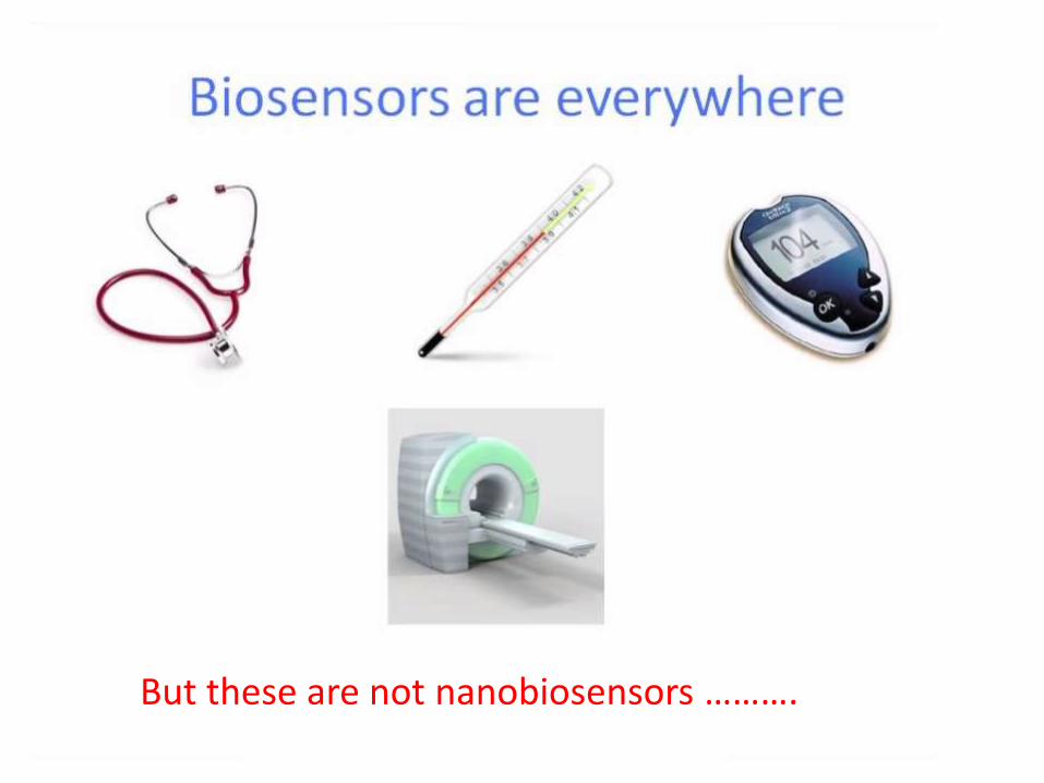
But these are not nanobiosensors ……….
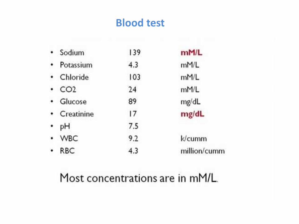
Blood test
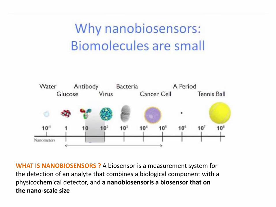
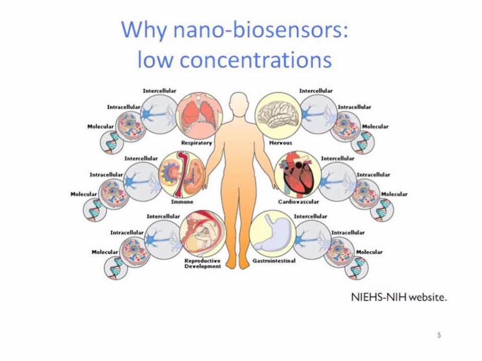
WHAT IS NANOBIOSENSORS ? A biosensor is a measurement system for the detection of an analyte that combines a biological component with a physicochemical detector, and a nanobiosensoris a biosensor that on the nano-scale size
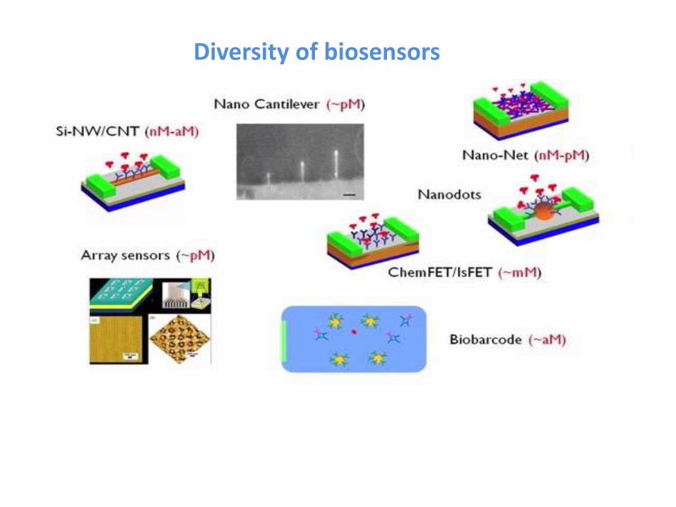
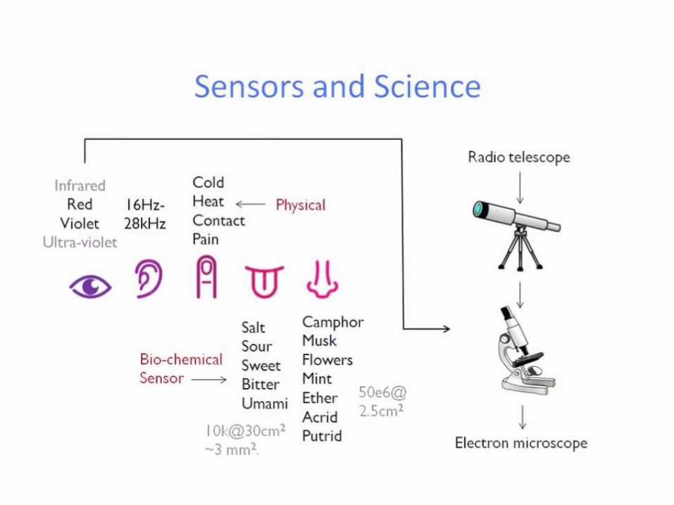
Diversity of biosensors
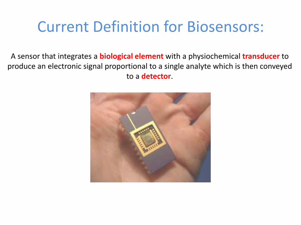
Current Definition for Biosensors:
A sensor that integrates a biological element with a physiochemical transducer to produce an electronic signal proportional to a single analyte which is then conveyed
to a detector.

Components of a Biosensor
Detector
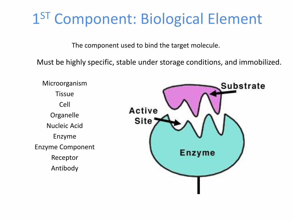
1ST Component: Biological Element
Microorganism
Tissue
Cell
Organelle
Nucleic Acid
Enzyme
Enzyme Component
Receptor
Antibody
The component used to bind the target molecule.
Must be highly specific, stable under storage conditions, and immobilized.
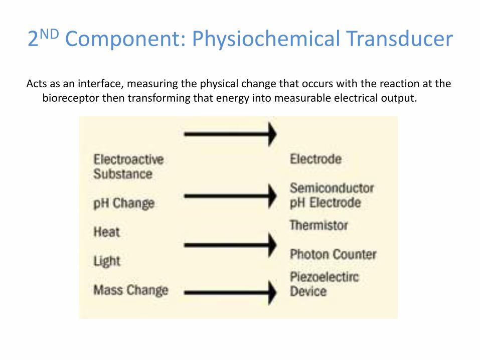
2ND Component: Physiochemical Transducer
Acts as an interface, measuring the physical change that occurs with the reaction at the bioreceptor then transforming that energy into measurable electrical output.
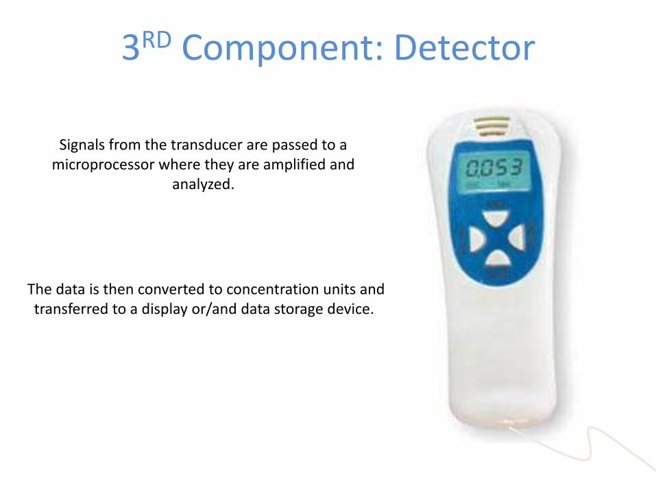
3RD Component: Detector
Signals from the transducer are passed to a microprocessor where they are amplified and
analyzed.
The data is then converted to concentration units and transferred to a display or/and data storage device.
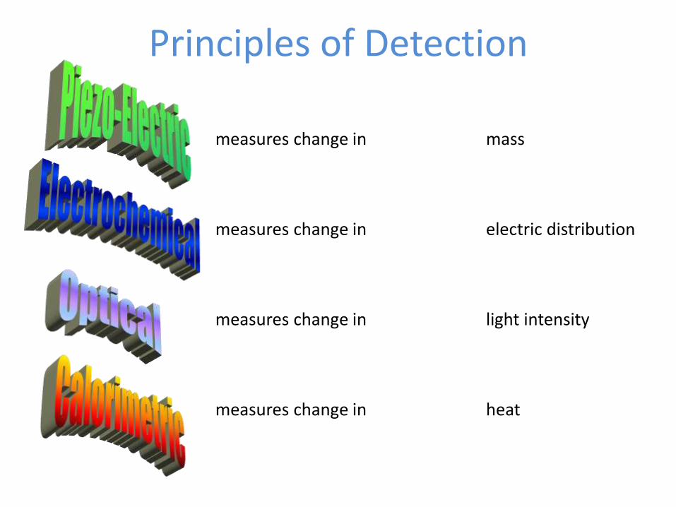
Principles of Detection
measures change in mass
measures change in electric distribution
measures change in light intensity
measures change in heat
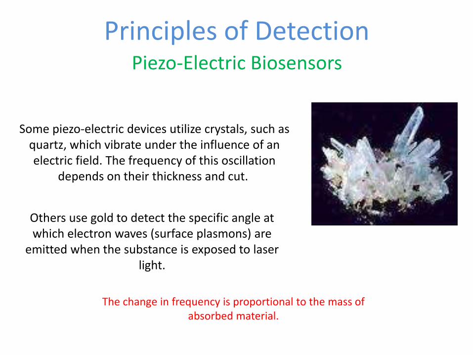
Principles of DetectionPiezo-Electric Biosensors
The change in frequency is proportional to the mass of absorbed material.
Some piezo-electric devices utilize crystals, such as quartz, which vibrate under the influence of an electric field. The frequency of this oscillation
depends on their thickness and cut.
Others use gold to detect the specific angle at which electron waves (surface plasmons) are
emitted when the substance is exposed to laser light.
Principles of DetectionElectrochemical Biosensors
•Amperometric for applied current: Movement of e- in redox reactions detected
when a potential is applied between two electrodes.
•Potentiometric for voltage: Change in distribution of charge is detected using
ion-selective electrodes, such as pH-meters.
•Conductimetric for impedance
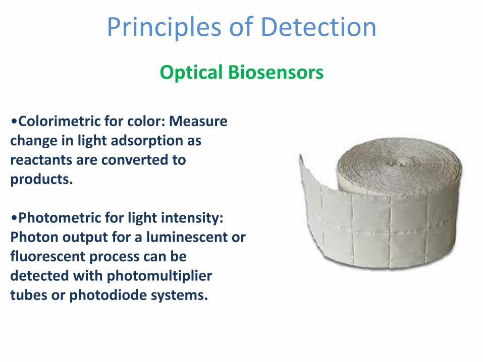
Principles of Detection
Optical Biosensors
•Colorimetric for color: Measure change in light adsorption as reactants are converted to products.
•Photometric for light intensity: Photon output for a luminescent or fluorescent process can be detected with photomultiplier tubes or photodiode systems.

Principles of DetectionCalorimetric Biosensors
If the enzyme catalyzed reaction is exothermic, two thermistors may be used to measure the difference in resistance between reactant and product and, hence, the analyte concentration.
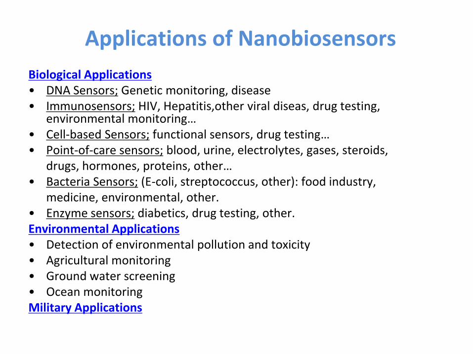
Applications of Nanobiosensors
Biological Applications• DNA Sensors; Genetic monitoring, disease• Immunosensors; HIV, Hepatitis,other viral diseas, drug testing,
environmental monitoring…• Cell-based Sensors; functional sensors, drug testing…• Point-of-care sensors; blood, urine, electrolytes, gases, steroids,
drugs, hormones, proteins, other…• Bacteria Sensors; (E-coli, streptococcus, other): food industry,
medicine, environmental, other.• Enzyme sensors; diabetics, drug testing, other.Environmental Applications• Detection of environmental pollution and toxicity• Agricultural monitoring• Ground water screening • Ocean monitoringMilitary Applications
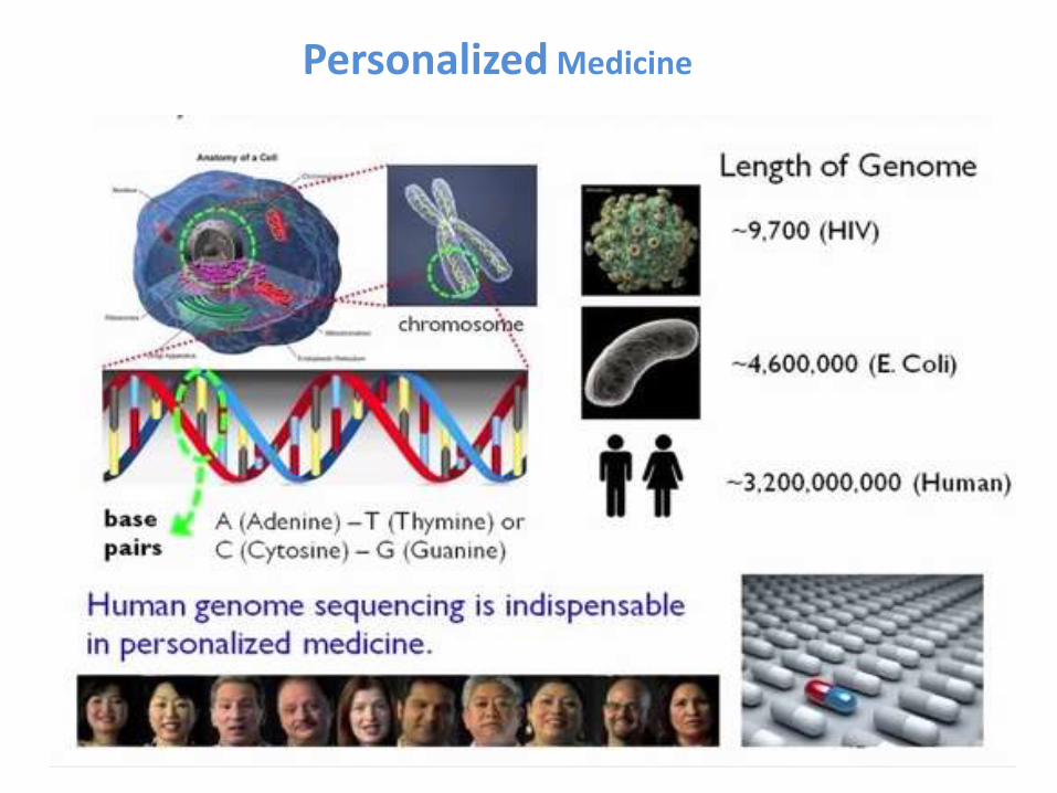
Personalized Medicine
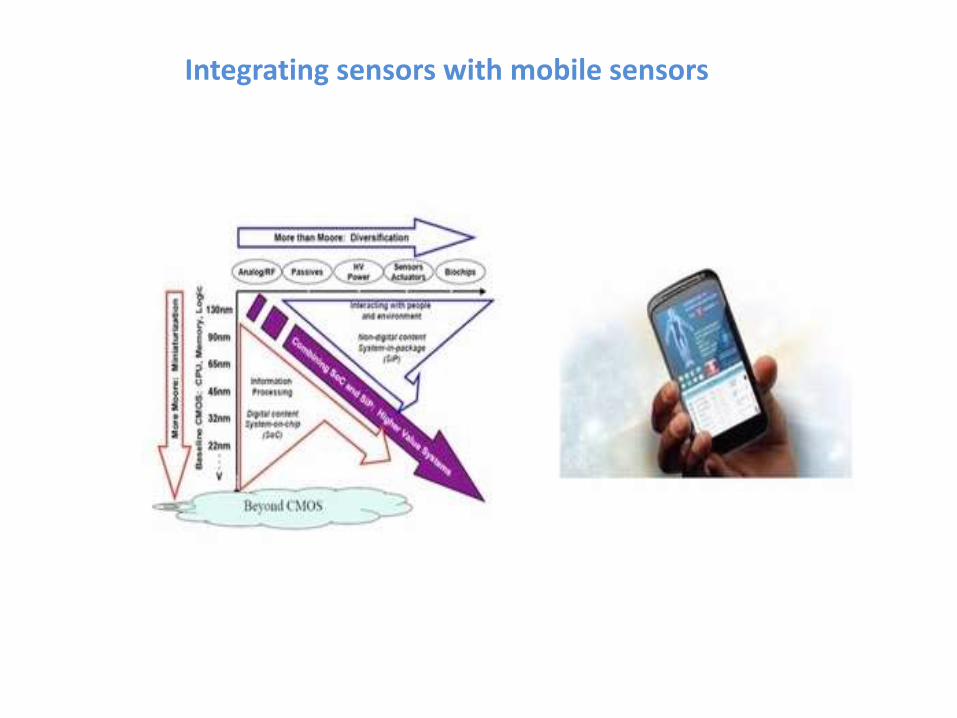
Integrating sensors with mobile sensors
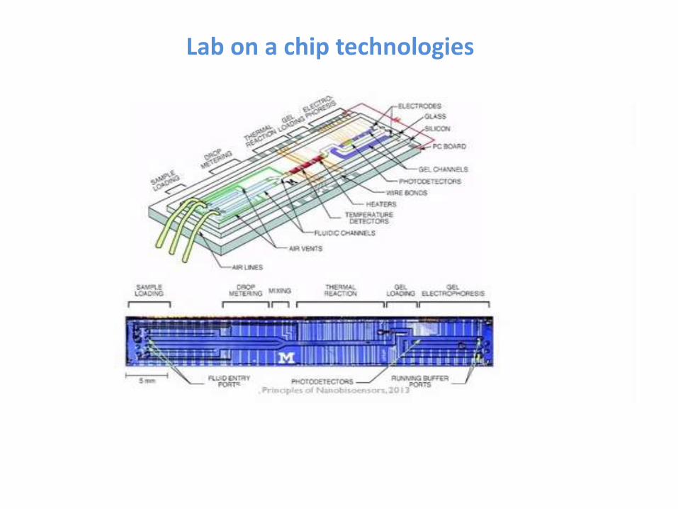
Lab on a chip technologies
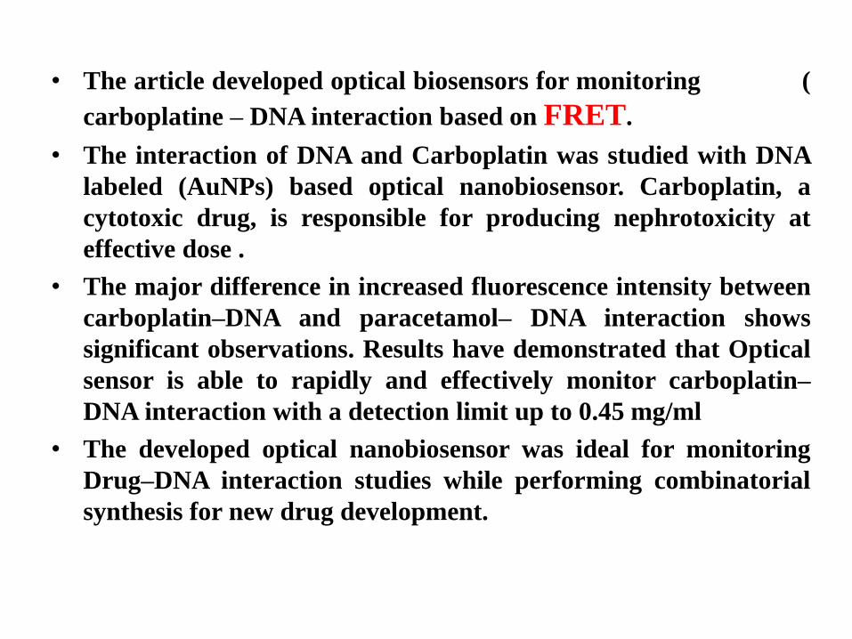
• The article developed optical biosensors for monitoring (
carboplatine – DNA interaction based on FRET.
• The interaction of DNA and Carboplatin was studied with DNA
labeled (AuNPs) based optical nanobiosensor. Carboplatin, a
cytotoxic drug, is responsible for producing nephrotoxicity at
effective dose .
• The major difference in increased fluorescence intensity between
carboplatin–DNA and paracetamol– DNA interaction shows
significant observations. Results have demonstrated that Optical
sensor is able to rapidly and effectively monitor carboplatin–
DNA interaction with a detection limit up to 0.45 mg/ml
• The developed optical nanobiosensor was ideal for monitoring
Drug–DNA interaction studies while performing combinatorial
synthesis for new drug development.
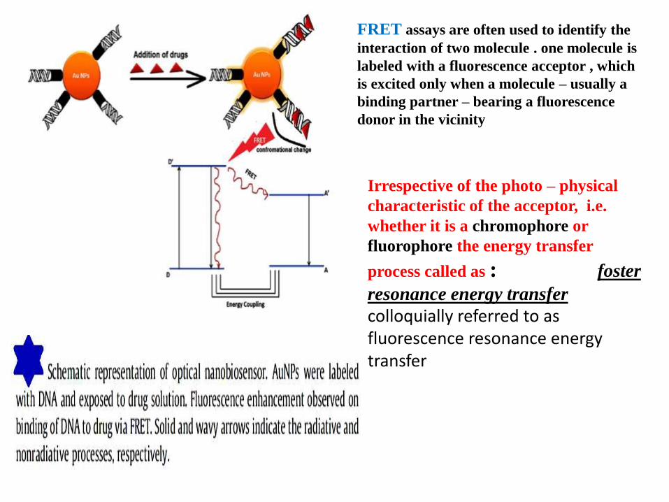
FRET assays are often used to identify the
interaction of two molecule . one molecule is
labeled with a fluorescence acceptor , which
is excited only when a molecule – usually a
binding partner – bearing a fluorescence
donor in the vicinity
Irrespective of the photo – physical
characteristic of the acceptor, i.e.
whether it is a chromophore or
fluorophore the energy transfer
process called as : foster
resonance energy transfer
colloquially referred to as fluorescence resonance energy transfer
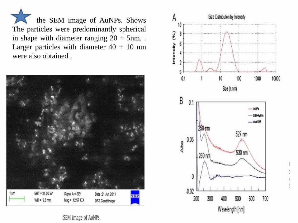
the SEM image of AuNPs. Shows
The particles were predominantly spherical
in shape with diameter ranging 20 + 5nm. .
Larger particles with diameter 40 + 10 nm
were also obtained .

the average fluorescence intensity of the carboplatin–DNA and paracetamol–
DNA interaction obtained by collecting data from three independent mea-
surements at the same conditions. The results showed that the fluorescence
intensity of carboplatin–DNA interaction was enhanced along with the increase
of carboplatin concentration. While, the fluorescence intensity of paracetamol–
DNA interaction increased slightly, this suggested that a very weak interaction is
occurring
The comparisons of analytical performances for
determining carboplatin–DNA and
paracetamol–DNA interaction
• the facile fabrication of a self-reporting, highly sensitive
and selective optical urea nanobiosensor using chitosan-g-
polypyrrole (CHIT-g-PPy) nanomicelles as a sensing
platform. Urease was immobilized on the spherical
micellar surface to create an ultrasensitive self-reporting
nanobiosystem for urea.
• This promising approach provides a novel methodology for
self-reporting bio-assembly over nanostructure polymer
micelles and furnishes the basis for fabrication of sensitive
and efficient optical nanobiosensors.
• Owing to the selective hydrolysis of the urea, the proposed
nanoreactor could be used in the development of artificial
kidneys.
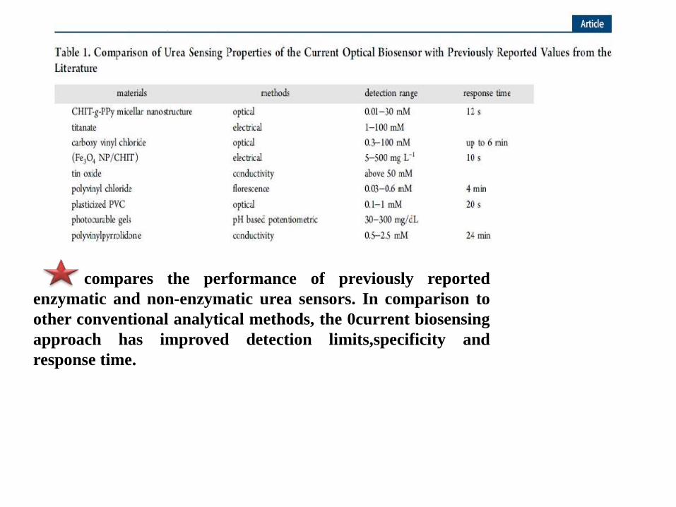
compares the performance of previously reported
enzymatic and non-enzymatic urea sensors. In comparison to
other conventional analytical methods, the 0current biosensing
approach has improved detection limits,specificity and
response time.
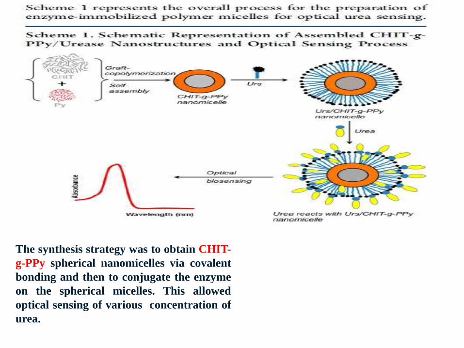
The synthesis strategy was to obtain CHIT-
g-PPy spherical nanomicelles via covalent
bonding and then to conjugate the enzyme
on the spherical micelles. This allowed
optical sensing of various concentration of
urea.
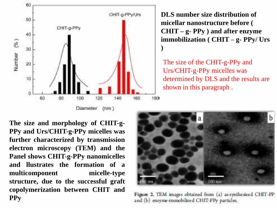
The size of the CHIT-g-PPy and
Urs/CHIT-g-PPy micelles was
determined by DLS and the results are
shown in this paragraph .
DLS number size distribution of
micellar nanostructure before (
CHIT – g- PPy ) and after enzyme
immobilization ( CHIT – g- PPy/ Urs
)
The size and morphology of CHIT-g-
PPy and Urs/CHIT-g-PPy micelles was
further characterized by transmission
electron microscopy (TEM) and the
Panel shows CHIT-g-PPy nanomicelles
and llustrates the formation of a
multicomponent micelle-type
structure, due to the successful graft
copolymerization between CHIT and
PPy
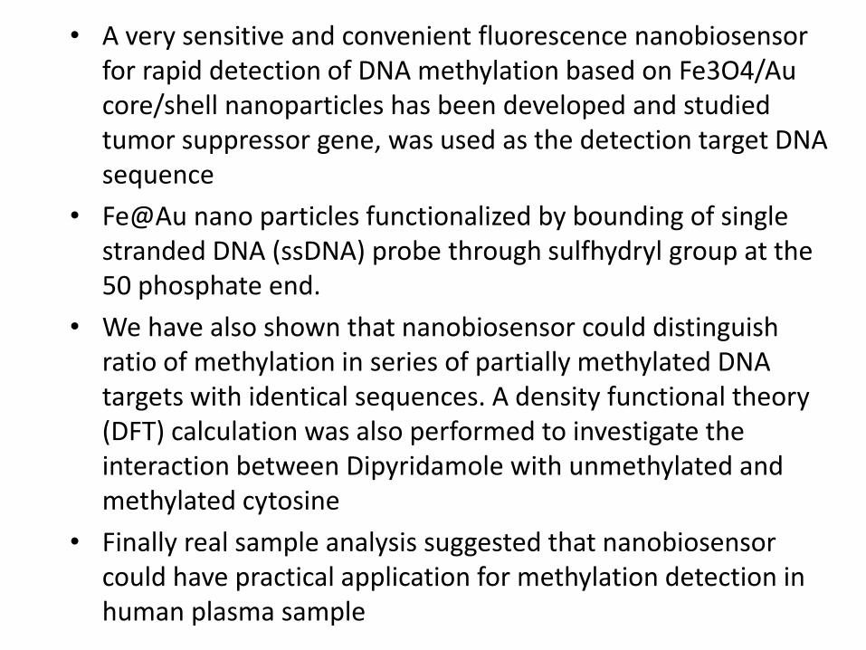
• A very sensitive and convenient fluorescence nanobiosensor for rapid detection of DNA methylation based on Fe3O4/Au core/shell nanoparticles has been developed and studied tumor suppressor gene, was used as the detection target DNA sequence
• Fe@Au nano particles functionalized by bounding of single stranded DNA (ssDNA) probe through sulfhydryl group at the 50 phosphate end.
• We have also shown that nanobiosensor could distinguish ratio of methylation in series of partially methylated DNA targets with identical sequences. A density functional theory (DFT) calculation was also performed to investigate the interaction between Dipyridamole with unmethylated and methylated cytosine
• Finally real sample analysis suggested that nanobiosensor could have practical application for methylation detection in human plasma sample
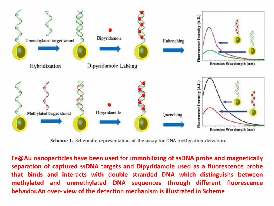
Fe@Au nanoparticles have been used for immobilizing of ssDNA probe and magneticallyseparation of captured ssDNA targets and Dipyridamole used as a fluorescence probethat binds and interacts with double stranded DNA which distinguishs betweenmethylated and unmethylated DNA sequences through different fluorescencebehavior.An over- view of the detection mechanism is illustrated in Scheme

Electron microscopy analysis The shape and size of the nanoparticles were determined by SEM images of the nanoparticles after and before the shell formation which is shown in Fig. 1a and b.The nanoparticles appeared nearly spherical and had an average diameter of about 25 nm and 30nm for Fe3O4 and Fe@Au one srespectively. The increase in the diameter of the some nanoparticles could be not only due to gold coating,but also to the aggregation of several Fe3O4 nanoparticles coated by the same gold shell
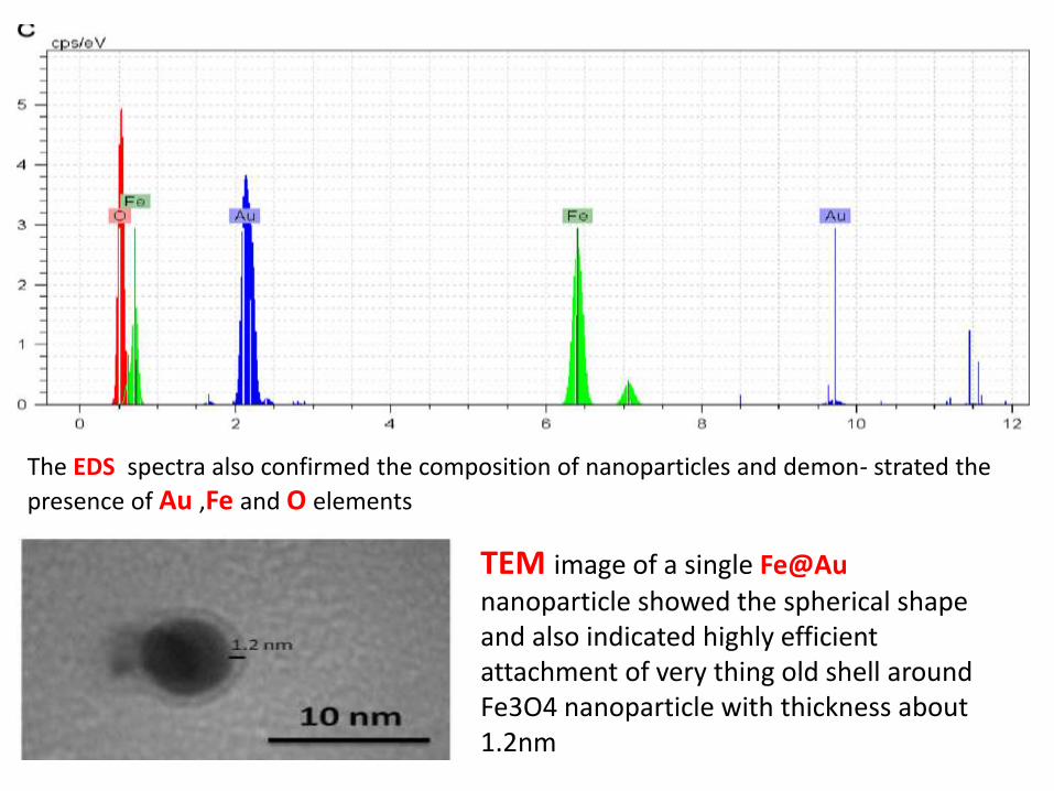
The EDS spectra also confirmed the composition of nanoparticles and demon- strated the
presence of Au ,Fe and O elements
TEM image of a single Fe@Au
nanoparticle showed the spherical shape and also indicated highly efficient attachment of very thing old shell around Fe3O4 nanoparticle with thickness about 1.2nm
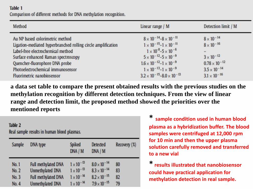
a data set table to compare the present obtained results with the previous studies on the
methylation recognition by different detection techniques. From the view of linear
range and detection limit, the proposed method showed the priorities over the
mentioned reports
* sample condition used in human blood
plasma as a hybridization buffer. The blood samples were centrifuged at 12,000 rpm for 10 min and then the upper plasma solution carefully removed and transferred to a new vial
* results illustrated that nanobiosensor
could have practical application for methylation detection in real sample.