name date period chapter 3 cell structure section 1
TRANSCRIPT
Name _______________________________________________ Date _________ Period _____ Chapter 3 Cell Structure
Section 1: Looking at Cells Objectives
•Describe how scientists measure the length of objects. •Relate magnification and resolution in the use of microscopes. •Analyze how light microscopes function. •Compare light microscopes with electron microscopes. •Describe the scanning tunneling microscope.
Cells Under the Microscope Measuring Cell Structures
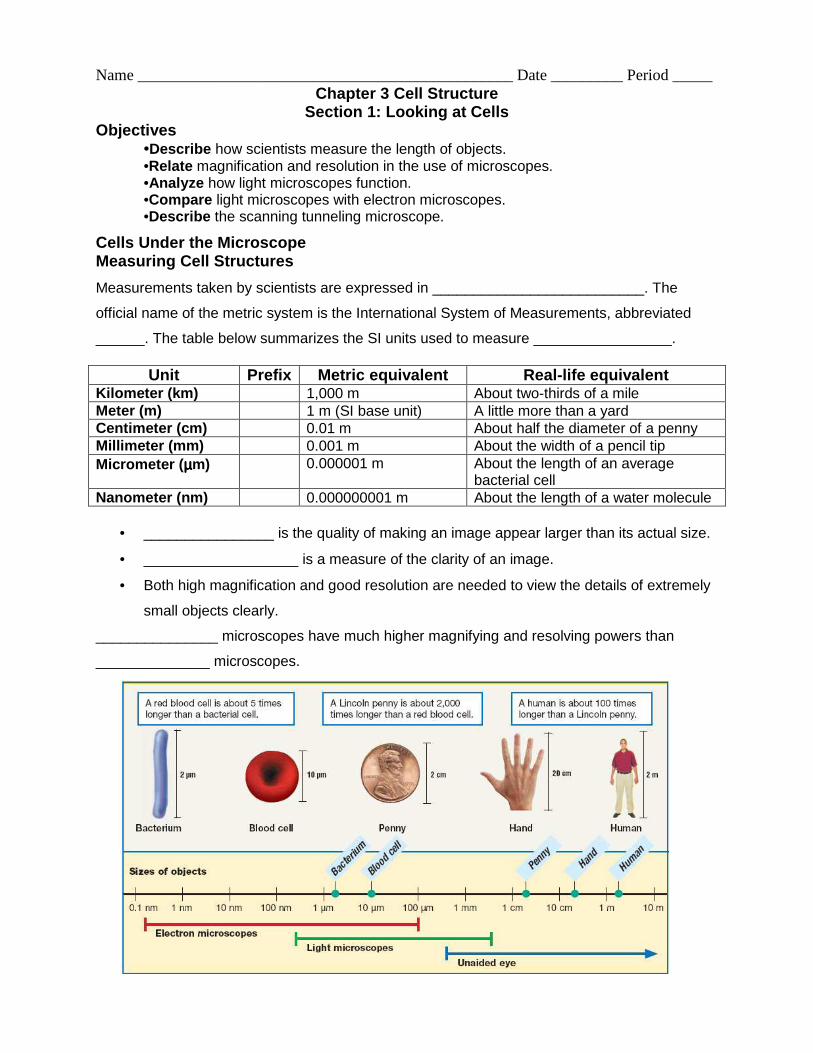
Measurements taken by scientists are expressed in __________________________. The
official name of the metric system is the International System of Measurements, abbreviated
______. The table below summarizes the SI units used to measure _________________.
• ________________ is the quality of making an image appear larger than its actual size.
• ___________________ is a measure of the clarity of an image.
• Both high magnification and good resolution are needed to view the details of extremely
small objects clearly.
_______________ microscopes have much higher magnifying and resolving powers than
______________ microscopes.
Unit Prefix Metric equivalent Real-life equivalent Kilometer (km) 1,000 m About two-thirds of a mile Meter (m) 1 m (SI base unit) A little more than a yard Centimeter (cm) 0.01 m About half the diameter of a penny Millimeter (mm) 0.001 m About the width of a pencil tip Micrometer ( µµµµm) 0.000001 m About the length of an average
bacterial cell Nanometer (nm) 0.000000001 m About the length of a water molecule
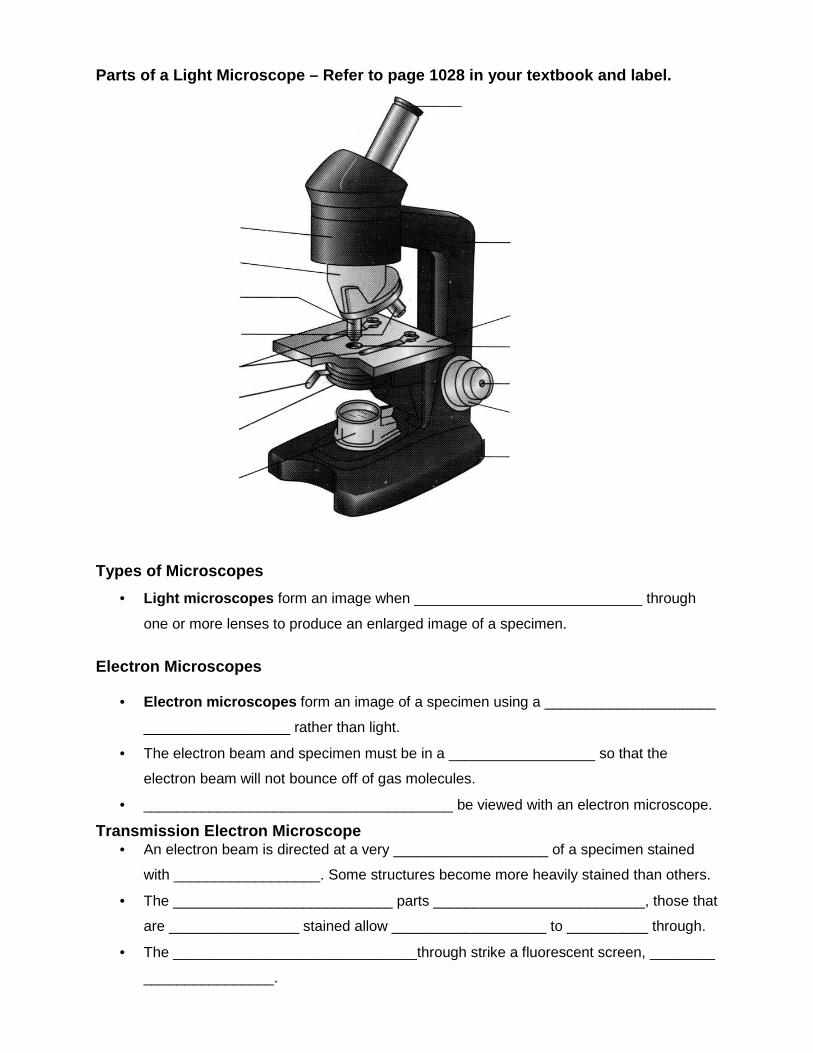
Parts of a Light Microscope – Refer to page 1028 in your textbook and label. Types of Microscopes
• Light microscopes form an image when ____________________________ through
one or more lenses to produce an enlarged image of a specimen.
Electron Microscopes
• Electron microscopes form an image of a specimen using a _____________________
__________________ rather than light.
• The electron beam and specimen must be in a __________________ so that the
electron beam will not bounce off of gas molecules.
• ______________________________________ be viewed with an electron microscope.
Transmission Electron Microscope • An electron beam is directed at a very ___________________ of a specimen stained
with __________________. Some structures become more heavily stained than others.
• The ___________________________ parts __________________________, those that
are ________________ stained allow ___________________ to __________ through.
• The ______________________________through strike a fluorescent screen, ________
________________.
Scanning Electron Microscope • An electron beam is focused on a specimen coated with a very ___________________
___________________.
• The electrons that _______________________ the specimen _____________________
on a fluorescent screen.
• The image shows __________________________ details of the surface of a specimen.
Scanning Tunneling Microscope
• A ______________________________ measures ______________________________
caused by electrons that leak, or tunnel, from the surface of the object being viewed.
• A computer tracks the movement of the probe _______________________________ of
the object.
• The image shows ________________________________ of the surface of a specimen.
• ______________________________ and objects as small as atoms can be viewed.
Section 2: Cell Features Objectives
• List the three parts of the cell theory. • Determine why cells must be relatively small. • Compare the structure of prokaryotic cells with that of eukaryotic cells. • Describe the structure of cell membranes.
The Cell Theory The Cell Theory has three parts:
1. All living things are made of ________________________________.
2. Cells are the ________________________________________________ in organisms.
3. All cells _______________________________________.
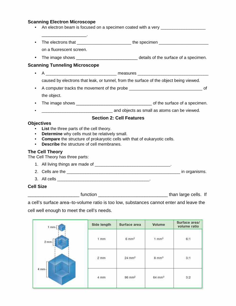
Cell Size
____________________ function ___________________________ than large cells. If
a cell’s surface area–to-volume ratio is too low, substances cannot enter and leave the
cell well enough to meet the cell’s needs.
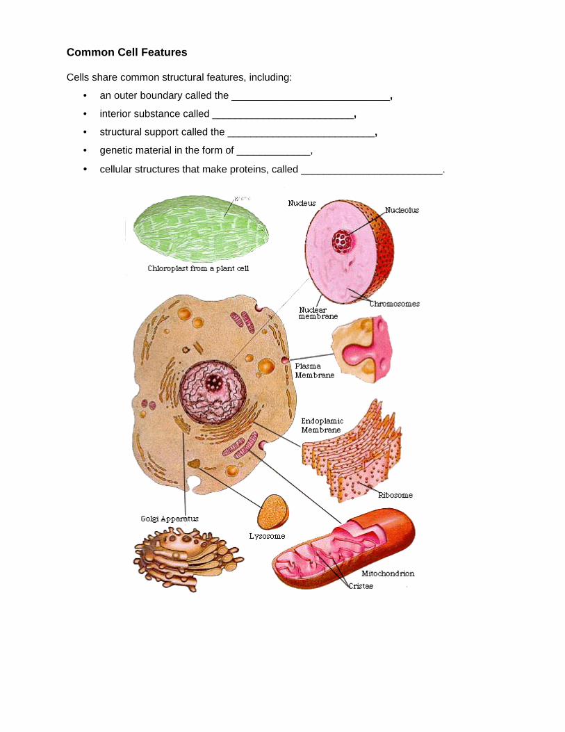
Common Cell Features Cells share common structural features, including:
• an outer boundary called the ____________________________,
• interior substance called _________________________,
• structural support called the __________________________,
• genetic material in the form of _____________,
• cellular structures that make proteins, called _________________________.
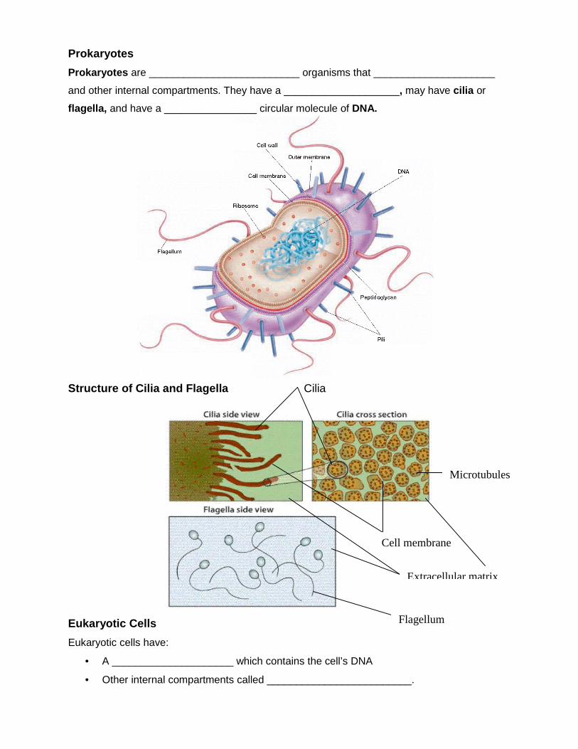
Prokaryotes
Prokaryotes are __________________________ organisms that _____________________
and other internal compartments. They have a ____________________, may have cilia or
flagella, and have a ________________ circular molecule of DNA.
Structure of Cilia and Flagella Cilia
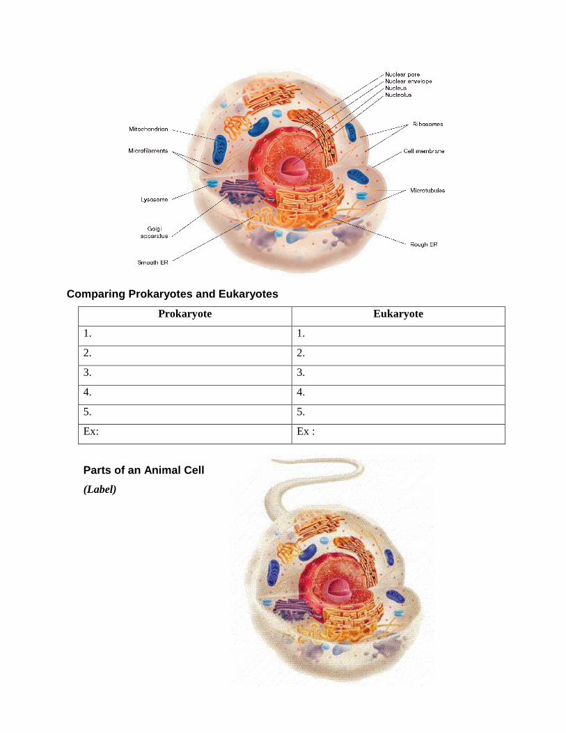
Eukaryotic Cells
Eukaryotic cells have:
• A _____________________ which contains the cell’s DNA
• Other internal compartments called _________________________.
Cell membrane
Extracellular matrix
Flagellum
Microtubules
Comparing Prokaryotes and Eukaryotes
Prokaryote Eukaryote
1. 1.
2. 2.
3. 3.
4. 4.
5. 5.
Ex: Ex :
Parts of an Animal Cell
(Label)
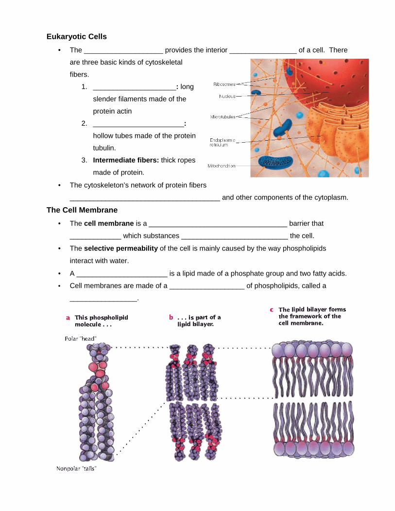
Eukaryotic Cells
• The ____________________ provides the interior _________________ of a cell. There
are three basic kinds of cytoskeletal
fibers.
1. _____________________: long
slender filaments made of the
protein actin
2. _______________________:
hollow tubes made of the protein
tubulin.
3. Intermediate fibers: thick ropes
made of protein.
• The cytoskeleton’s network of protein fibers
______________________________________ and other components of the cytoplasm.
The Cell Membrane
• The cell membrane is a ___________________________________ barrier that
_____________ which substances ___________________________ the cell.
• The selective permeability of the cell is mainly caused by the way phospholipids
interact with water.
• A _______________________ is a lipid made of a phosphate group and two fatty acids.
• Cell membranes are made of a ___________________ of phospholipids, called a
_________________.
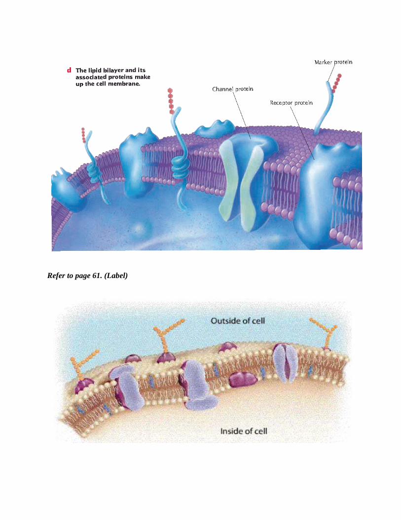
Refer to page 61. (Label)