nadph and glutathione redox link tca cycle activity to … · 4 1 the impact of metabolic activity...
TRANSCRIPT
1
1
2
3
4
5
6
NADPH and glutathione redox link TCA cycle activity to endoplasmic 7 reticulum stress 8
9
Erica R. Gansemer1, Kyle S. McCommis8, Michael Martino8, Abdul Qaadir 10 King-McAlpin2, Matthew J. Potthoff2,4,5,6, Brian N. Finck8, Eric B. Taylor3,4,5,6,7 11
and D. Thomas Rutkowski1,5,6,7* 12 13 1Department of Anatomy and Cell Biology, 2Department of Pharmacology, 3Department 14 of Biochemistry, 4Obesity Research Initiative, 5Abboud Cardiovascular Research 15 Center, 6Fraternal Order of Eagles Diabetes Research Center, 7Holden Comprehensive 16 Cancer Center, Carver College of Medicine, University of Iowa 17
8Center for Human Nutrition, Department of Medicine, Washington University School of 18 Medicine in Saint Louis 19
20 *corresponding author: [email protected] 21 22
.CC-BY-NC-ND 4.0 International licenseis made available under aThe copyright holder for this preprint (which was not peer-reviewed) is the author/funder. It. https://doi.org/10.1101/822775doi: bioRxiv preprint
2
Abstract 1
2
Endoplasmic reticulum (ER) stress is associated with dysregulated metabolism, but little is 3
known about how the ER responds to metabolic activity. Here, working primarily in mouse 4
hepatocytes, we show that decreasing the availability of substrate for the TCA cycle diminished 5
NADPH production and attenuated ER stress in a manner that depended on glutathione 6
oxidation. ER stress was also alleviated by impairing either TCA-dependent NADPH production 7
or Glutathione Reductase. Conversely, stimulating TCA activity favored NADPH production, 8
glutathione reduction, and ER stress. Validating these findings, we show that deletion of the 9
mitochondrial pyruvate carrier, which is known to decrease TCA cycle activity and protect the 10
liver from diet-induced injury, also diminished NADPH, elevated glutathione oxidation, and 11
alleviated ER stress. These results provide independent genetic evidence that mitochondrial 12
oxidative metabolism is linked to ER homeostasis. Our results demonstrate a novel pathway of 13
communication between mitochondria and the ER, through relay of redox metabolites. 14
15
16
.CC-BY-NC-ND 4.0 International licenseis made available under aThe copyright holder for this preprint (which was not peer-reviewed) is the author/funder. It. https://doi.org/10.1101/822775doi: bioRxiv preprint
3
Introduction 1
2
As the gateway to the secretory pathway, the ER must properly synthesize and fold nascent 3
secretory and membrane proteins. Disruption of this process, known as ER stress, is associated 4
with many disease states, including obesity and its comorbidities (Mohan, R, Brown, Ayyappan, 5
& G, 2019). Thus, it is important to understand how ER homeostasis is perturbed, particularly by 6
metabolic disruption. The ER and the mitochondrial network, although not connected to each 7
other by secretory pathway traffic, are intertwined both physically and functionally. The ER 8
makes close physical contacts with mitochondria to facilitate the exchange of metabolites 9
(Raffaello, Mammucari, Gherardi, & Rizzuto, 2016; Vance, 2014; Yoboue, Sitia, & Simmen, 10
2018). The ER also communicates with mitochondria via signaling from the unfolded protein 11
response (UPR), which is activated by ER stress and signals through the three ER-resident 12
stress sensors IRE1, PERK, and ATF6 (Walter & Ron, 2011). The UPR regulates mitochondrial 13
activity at several levels, including enhancing mitochondrial protein quality control, augmenting 14
ER-mitochondrial interactions and calcium signaling, and contributing to mitochondrial 15
depolarization and initiation of apoptosis (Y. Fan & Simmen, 2019; Gutierrez & Simmen, 2018; 16
Rainbolt, Saunders, & Wiseman, 2014). While the pathways through which ER stress and the 17
UPR regulate mitochondrial function are becoming clearer, the converse—how mitochondrial 18
function impacts ER homeostasis—is less understood. 19
In the liver, the UPR suppresses a large number of genes involved in metabolic 20
processes (Rutkowski et al., 2008). One of the processes suppressed is fatty acid b-oxidation 21
(DeZwaan-McCabe et al., 2017), which takes place in mitochondria. The role of the UPR is to 22
restore ER function during stress, suggesting that suppressing b-oxidation in the mitochondria is 23
functionally beneficial to achieving ER homeostasis. However, it is not clear how metabolic 24
activity within the mitochondria might be conveyed to the ER. 25
.CC-BY-NC-ND 4.0 International licenseis made available under aThe copyright holder for this preprint (which was not peer-reviewed) is the author/funder. It. https://doi.org/10.1101/822775doi: bioRxiv preprint
4
The impact of metabolic activity on ER homeostasis is most evident form the association 1
of ER stress with obesity—particularly in highly metabolically active tissues such as liver, 2
pancreas, and adipose (Cnop, Foufelle, & Velloso, 2012). Lipotoxicity (i.e., damage caused by 3
the inappropriate accumulation of lipids in non-adipose tissue), inflammation, and oxidative 4
stress have all been shown to contribute to obesity-associated ER stress (Fu, Watkins, & 5
Hotamisligil, 2012; Salvado, Palomer, Barroso, & Vazquez-Carrera, 2015). Yet, independent of 6
diet content, feeding after a fast is sufficient to elicit ER stress in the liver (Gomez & Rutkowski, 7
2016; Oyadomari, Harding, Zhang, Oyadomari, & Ron, 2008; Pfaffenbach et al., 2010) and to 8
alter the extent of physical contacts between the ER and mitochondria (Theurey et al., 2016). 9
These findings suggest that ER homeostasis is acutely and intrinsically connected with 10
metabolism even apart from the problems brought on by obesity. However, the biochemical 11
pathways linking catabolism to ER function are not known. 12
The tricarboxylic acid (TCA) cycle is the central hub of metabolism, participating in both 13
catabolism and anabolism. Acetyl-CoA enters the cycle after either oxidative breakdown of lipids 14
within the mitochondria or after conversion of pyruvate, generated from glycolysis in the cytosol, 15
by the mitochondrial pyruvate dehydrogenase complex. Canonically, TCA activity yields NADH 16
and FADH2 for the electron transport chain. However, the TCA cycle also provides precursors 17
for biosynthetic pathways (Owen, Kalhan, & Hanson, 2002). In addition, the cycle can produce 18
NADPH in addition to NADH, due to the activity of isozymes that reside either in the 19
mitochondria (isocitrate dehydrogenase 2/IDH2) or cytosol (IDH1 or malic enzyme/ME1) 20
(Rydstrom, 2006). Therefore, activity of the TCA cycle is likely to influence cellular processes by 21
mechanisms beyond just the production of ATP from the electron transport chain. 22
NADPH is used as a cofactor by glutathione reductase (GR) to reduce oxidized 23
glutathione (GSSGà2GSH), and likewise by thioredoxin reductase to reduce oxidized 24
thioredoxin. Both of these molecules contribute to defense against oxidative stress (Sies, 25
Berndt, & Jones, 2017), and both have connections to ER protein biogenesis. Thioredoxin has 26
.CC-BY-NC-ND 4.0 International licenseis made available under aThe copyright holder for this preprint (which was not peer-reviewed) is the author/funder. It. https://doi.org/10.1101/822775doi: bioRxiv preprint
5
been shown to be a source of electrons for reduction and isomerization of disulfide bonds of ER 1
client proteins (Poet et al., 2017). The oxidized form of glutathione (GSSG), which predominates 2
in the ER lumen compared to the cytosol, was formerly thought to reoxidize protein disulfide 3
isomerase (PDI) to promote ER disulfide bond formation. However, since the discovery of 4
alternative pathways for PDI reoxidation (Frand & Kaiser, 1999; Tu, Ho-Schleyer, Travers, & 5
Weissman, 2000; Zito, Melo, et al., 2010), the role of glutathione in the ER is now much less 6
clear (Delaunay-Moisan, Ponsero, & Toledano, 2017; Tsunoda et al., 2014). Whether elevated 7
GSSG might be beneficial to ER function under some cellular conditions but detrimental in 8
others is also unknown. 9
Despite the centrality of the TCA cycle to cellular function and hints that its activity might 10
be tied to ER stress (Mogilenko et al., 2019; Xin et al., 2018), its involvement in ER homeostasis 11
has not been investigated. Here, we used primary hepatocytes and other metabolically active 12
cell types to investigate the relationship between metabolic activity and ER stress. We show that 13
TCA cycle activity links lipid and carbohydrate catabolism to ER homeostasis through 14
production of NADPH and redox regulation of glutathione. Our findings delineate a novel 15
mechanism of communication from mitochondria to the ER, reveal an unexpected protective 16
role for GSSG, and provide a plausible pathway by which ER homeostasis is linked to metabolic 17
activity. 18
19
.CC-BY-NC-ND 4.0 International licenseis made available under aThe copyright holder for this preprint (which was not peer-reviewed) is the author/funder. It. https://doi.org/10.1101/822775doi: bioRxiv preprint
6
Results 1
2
Inhibition of b-oxidation alleviates ER stress in metabolically active cell types 3
We have previously shown in vivo and in hepatoma cells in vitro that inhibiting b-oxidation either 4
pharmacologically or genetically diminishes ER stress signaling (Tyra, Spitz, & Rutkowski, 5
2012). In order to identify the pathway by which b-oxidation and ER homeostasis are linked, we 6
first asked whether etomoxir, which blocks b-oxidation by inhibiting the CPT1-dependent 7
transport of fatty acyl-CoAs into the mitochondria for oxidation (Weis, Cowan, Brown, Foster, & 8
McGarry, 1994; Yao et al., 2018), could diminish ER stress signaling in primary hepatocytes in 9
vitro as it does in the liver in vivo. Treatment of primary hepatocytes with the ER stressor 10
tunicamycin (TM) upregulated UPR-responsive mRNAs, whereas etomoxir cotreatment 11
suppressed this effect (Figure 1A). Dampened ER stress signaling was also evident from 12
diminishment of the splicing of the IRE1a nuclease target Xbp1 (Figure 1B) and of the 13
upregulation of the stress-regulated factor CHOP (Figure 1C). Because these events are 14
differentially regulated by the three limbs of the UPR, our data suggest that etomoxir diminishes 15
signaling from all three UPR pathways. This result is consistent with our previous findings, in 16
which ER stress signaling in hepatoma cells was diminished by etomoxir, by inhibition of b-17
oxidation with a separate agent, and by knockdown of CPT1 (Tyra et al., 2012). 18
We next determined if the apparent protective effects of etomoxir were limited to one cell 19
type or to one ER stressor. Using the selection of UPR-regulated mRNAs in Figure 1A as a 20
broad indicator of ER stress signaling, we found that etomoxir also diminished UPR activation in 21
C2C12 myoblasts (Figure 1D), and immortalized (data not shown) and primary (Figure 1E) 22
brown adipocytes. Hepatocytes, myocytes, and brown adipocytes are characterized by 23
particularly high lipid metabolic activity (Frayn, Arner, & Yki-Jarvinen, 2006). In contrast, 24
etomoxir had no significant effect on the expression of UPR target genes in primary mouse 25
.CC-BY-NC-ND 4.0 International licenseis made available under aThe copyright holder for this preprint (which was not peer-reviewed) is the author/funder. It. https://doi.org/10.1101/822775doi: bioRxiv preprint
7
embryonic fibroblasts (Figure S1). In primary hepatocytes subjected to ER stress by palmitate 1
loading, etomoxir likewise diminished ER stress signaling (Figure 1G). (As expected, the 2
unsaturated fatty acid oleate did not cause ER stress.) Palmitate is thought to elicit ER not by 3
disrupting protein folding per se but by altering ER membrane fluidity, activating the UPR stress 4
sensors through their transmembrane domains (Volmer, van der Ploeg, & Ron, 2013). Thus, 5
etomoxir diminishes ER stress induced by stressors that act by divergent mechanisms. 6
The effects of etomoxir on ER stress signaling could be due to improvement of ER 7
homeostasis in some way, or to simple inhibition of UPR signaling. We did not observe any 8
robust differences in the efficiency with which ER client proteins were secreted in hepatocytes 9
treated with etomoxir compared to those that were not treated (data not shown). However, as a 10
separate assessment of ER homeostasis, we examined ER ultrastructure in hepatocytes treated 11
with TM, with or without etomoxir. Consistent with previous reports (Finnie, 2001; Rutkowski et 12
al., 2006), TM elicited marked ER dilation accompanied by a loss of lamellar structure and 13
overall disorganization, although some areas of grossly normal ER were present and were 14
predominantly juxtaposed near mitochondria. In contrast, these disruptions were largely 15
prevented by cotreatment with etomoxir (Figure 2A) as confirmed by two independent, blinded 16
scorers (Figure 2B). Although etomoxir diminished ER stress signaling, it did not block it 17
entirely. Higher doses of TM elicited UPR activation in etomoxir-treated cells to an extent 18
comparable to that observed with lower doses in non-etomoxir-treated cells (Figure 2C). 19
Therefore, the UPR in etomoxir-treated cells remained competent for signaling. Together, these 20
results suggest that inhibiting b-oxidation alleviates ER stress. 21
To provide a direct test of whether etomoxir could alleviate ER stress caused by the 22
accumulation of misfolded protein, we examined its ability to reverse ER stress associated with 23
overexpression of the null Hong Kong (NHK) protein. NHK is encoded by a nonsense mutation 24
of a1-antitrpysin that results in a truncated product which is retained in the ER and undergoes 25
.CC-BY-NC-ND 4.0 International licenseis made available under aThe copyright holder for this preprint (which was not peer-reviewed) is the author/funder. It. https://doi.org/10.1101/822775doi: bioRxiv preprint
8
ER-associated degradation, resulting in ER stress in diverse cell types (Nagasawa, Higashi, 1
Hosokawa, Kaufman, & Nagata, 2007; Ordonez et al., 2013; Sifers, Brashears-Macatee, Kidd, 2
Muensch, & Woo, 1988; J. Wu et al., 2007). We transduced primary hepatocytes with a 3
recombinant adenovirus expressing NHK under doxycycline (Dox)-inducible control (Ad-TetOn-4
NHK). As a control, cells were transduced with adenovirus constitutively overexpressing GFP 5
instead (Ad-GFP). No ER stress was observed in Ad-TetOn-NHK-transduced cells in the 6
absence of Dox (not shown). As expected, Dox treatment elicited ER stress in cells transduced 7
with Ad-TetOn-NHK, but not in cells expressing Ad-GFP. The stress induced by NHK was 8
markedly diminished by etomoxir (Figure 3A). Overexpression of NHK disrupted ER structure, 9
resulting in the appearance of structurally amorphous ER puncta that were diminished by 10
etomoxir treatment (Figure 3B). This finding was again substantiated by blinded analysis of EM 11
images (Figure 3C). In principle, etomoxir treatment could alleviate NHK-dependent ER stress 12
by reducing protein synthesis. However, we found no evidence that global translation rates were 13
altered by etomoxir (Figure S2). Consistent with this observation, we found that, 4 hours after 14
NHK induction, steady-state expression of NHK did not differ in cells treated or not with etomoxir 15
(Figure 3D). However, at later time points, we observed diminished steady state expression of 16
NHK in etomoxir-treated cells, and this distinction was lost when degradation of NHK was 17
blocked by the proteasomal inhibitor MG-132 (Figure 3D). While the mechanisms by which 18
etomoxir enhances ERAD remain under investigation, these results provide a separate line of 19
evidence that inhibiting b-oxidation improves ER homeostasis, perhaps by affecting misfolded 20
protein clearance. 21
22
Inhibiting b-oxidation protects ER function through glutathione redox 23
We and others have previously shown that etomoxir raises the cellular ratio of oxidized (GSSG) 24
to reduced (GSH) glutathione (Merrill et al., 2002; Pike, Smift, Croteau, Ferrick, & Wu, 2010; 25
.CC-BY-NC-ND 4.0 International licenseis made available under aThe copyright holder for this preprint (which was not peer-reviewed) is the author/funder. It. https://doi.org/10.1101/822775doi: bioRxiv preprint
9
Tyra et al., 2012). Given the association of GSSG with the oxidative protein folding environment 1
in the ER, we examined whether etomoxir could make the ER more oxidizing to proteins in the 2
lumen. This was done by monitoring the oxidation of albumin, an endogenous hepatocyte ER 3
client protein that forms 17 disulfide bonds (Peters & Davidson, 1982). We tested the resistance 4
of albumin to reduction by treating cells with DTT, lysing them, and then modifying free 5
sulfhydryls with PEG-maleimide, which retards migration upon SDS-PAGE (Winther & Thorpe, 6
2014). We found that treatment with DTT led to almost complete reduction of albumin in 7
otherwise untreated cells, whereas etomoxir caused a significant increase in the preponderance 8
of albumin that remained oxidized (Figure 4A). These results suggest that glutathione redox 9
might mediate the protective effects of etomoxir. 10
To first determine if glutathione redox plays a role in the protective effects of etomoxir, 11
we used 2-AAPA to inhibit glutathione reductase (Seefeldt et al., 2009; Zhao et al., 2009), an 12
enzyme with both mitochondrial and cytosolic activity that couples NADPH oxidation to GSSG 13
reduction. We predicted that this treatment would phenocopy the protective effects of etomoxir. 14
As expected, treatment with 2-AAPA diminished cellular GSH levels and increased GSSG, thus 15
significantly elevating the GSSG/GSH ratio, similar to the effects of etomoxir (Figure 4B). Also 16
similarly to etomoxir, 2-AAPA treatment alleviated ER stress, as determined by attenuated Xbp1 17
splicing (Figure 4C) and diminished upregulation of UPR target genes (Figure 4D). Therefore, 18
inhibiting glutathione reduction phenocopies the protective effects of etomoxir. 19
We next tested the converse prediction: that promoting glutathione reduction would 20
cause ER stress. We treated hepatocytes with auranofin, which inhibits redox-active 21
selenoproteins, including thioredoxin reductases and, at the dose used here, glutathione 22
peroxidases (Chaudiere & Tappel, 1984; Roberts & Shaw, 1998; Scarbrough et al., 2012). In 23
the liver, glutathione peroxidase 1 is two orders of magnitude more abundant than any 24
thioredoxin reductase (Lai, Kolippakkam, & Beretta, 2008). Therefore, presumably due to its 25
effects on glutathione peroxidase, auranofin treatment elevated cellular GSH levels and 26
.CC-BY-NC-ND 4.0 International licenseis made available under aThe copyright holder for this preprint (which was not peer-reviewed) is the author/funder. It. https://doi.org/10.1101/822775doi: bioRxiv preprint
10
diminished GSSG, leading to a decrease in the GSSG/GSH ratio (Figure 5A). The relative 1
resistance of albumin to reduction by DTT upon treatment with auranofin suggested that the ER 2
environment was more reducing, whereas treatment with 2-AAPA made it more oxidizing 3
(Figure 5B). Auranofin treatment alone was sufficient to induce a modest level of ER stress, 4
seen in upregulation of UPR target genes in the absence of any other ER stress stimulus 5
(Figure 5C). More importantly, auranofin completely blocked the ability of etomoxir to alleviate 6
ER stress (Figure 5D). This epistatic relationship suggests that the protective effect of etomoxir 7
requires glutathione oxidation. 8
9
TCA cycle activity links oxidative metabolism to ER homeostasis 10
Although glutathione reductase uses NADPH to reduce glutathione, b-oxidation yields NADH 11
and FADH2 but not NADPH. However, b-oxidation yields acetyl-CoA, which enters the TCA 12
cycle by condensation with oxaloacetate for further oxidation. We therefore speculated that 13
etomoxir might diminish production of NADPH from the TCA cycle, thereby resulting in elevated 14
GSSG. In this model, it is not b-oxidation per se that is linked to ER homeostasis, but rather 15
TCA cycle activity and the resultant production of NADPH. This model first predicted that 16
etomoxir would diminish levels of both acetyl-CoA and NADPH—predictions which we then 17
confirmed (Figure 6A, B). The model also predicted that impairing TCA-dependent NADPH 18
production would elevate the GSSG/GSH ratio and alleviate ER stress, similarly to etomoxir 19
treatment. Three TCA cycle isozymes generate NADPH: mitochondrial isocitrate 20
dehydrogenase 2 (IDH2) and cytosolic IDH1 and malic enzyme (ME1). (The NADPH-producing 21
mitochondrial ME3 is not expressed in the mouse liver). Using siRNAs, we specifically knocked 22
down mRNA expression of each of these (Figure 6C). Knockdown of each gene diminished 23
GSH levels, and knockdown of Idh2 and Me1 also increased GSSG levels, thus elevating the 24
.CC-BY-NC-ND 4.0 International licenseis made available under aThe copyright holder for this preprint (which was not peer-reviewed) is the author/funder. It. https://doi.org/10.1101/822775doi: bioRxiv preprint
11
GSSG/GSH ratio (Figure 6D). Further, ER stress was diminished by knockdown of each of the 1
three genes (Figure 6E). 2
The Pyruvate Dehydrogenase complex oxidizes pyruvate produced by glycolysis to 3
acetyl-CoA, and is a major control point of TCA cycle activity for carbohydrate metabolism 4
(Gray, Tompkins, & Taylor, 2014). Pyruvate Dehydrogenase Kinases (PDKs) phosphorylate and 5
inactivate this enzyme complex. To determine the effects of stimulating TCA cycle activity, we 6
treated hepatocytes with dichloroacetate, which stimulates substrate entry into the TCA cycle by 7
inhibiting PDKs (Constantin-Teodosiu, Simpson, & Greenhaff, 1999; C. Y. Wu et al., 2018). 8
Dichloroacetate treatment substantially elevated the NADPH/NADP+ ratio (Figure 7A). In 9
addition, dichloroaceetate treatment alone caused ER stress to an extent that was nearly as 10
robust as the bona fide ER stressor TM, as seen by upregulation of UPR target genes (Figure 11
7B) and splicing of Xbp1 (Figure 7C). Therefore, these data provide direct evidence that 12
mitochondrial oxidative catabolic activity causes ER stress. 13
14
Ablation of the Mitochondrial Pyruvate Carrier alleviates ER stress 15
Finally, we wished to test whether ER stress could be alleviated by non-pharmacological 16
manipulation of TCA cycle activity. The Mitochondrial Pyruvate Carrier (MPC) is composed of 17
two subunits, MPC1 and MPC2, and mediates mitochondrial import of pyruvate (Bricker et al., 18
2012; Herzig et al., 2012). Loss of either subunit eliminates MPC activity, and liver-specific 19
ablation of the MPC diminishes liver damage and inflammation in mice on obesogenic diets 20
(Gray et al., 2015; McCommis et al., 2015; McCommis et al., 2017). In the absence of MPC 21
activity, import of pyruvate into the mitochondria is greatly diminished, meaning that less acetyl-22
CoA can be produced by pyruvate dehydrogenase and therefore that TCA cycle flux is 23
diminished (Gray et al., 2015; Rauckhorst et al., 2017). Therefore, ablating the MPC should 24
mimic the effects of etomoxir treatment since both ultimately result in diminished acetyl-CoA 25
availability for oxidation in the TCA cycle. To test this prediction, we first examined the effects of 26
.CC-BY-NC-ND 4.0 International licenseis made available under aThe copyright holder for this preprint (which was not peer-reviewed) is the author/funder. It. https://doi.org/10.1101/822775doi: bioRxiv preprint
12
deleting MPC1 by CRISPR in C2C12 myocytes. We found that loss of MPC1 diminished the 1
NADPH/NADP+ ratio and elevated the GSSG/GSH ratio in these cells (Figure 8A, B). 2
Diminished UPR activation at both the protein (Figure 8C) and mRNA (Figure 8D) levels 3
indicated that MPC1-deficient cells were also remarkably resistant to ER stress induced by TM 4
or by the ER calcium-disrupting agent thapsigargin (TG). These findings were confirmed in 5
primary hepatocytes taken from mice lacking MPC2 in the liver (Mpc2LKO), when compared to 6
cells from mice with an intact allele (Mpc2fl/fl). In cells lacking MPC2, both the upregulation of 7
UPR target genes (Figure 8E) and Xbp1 mRNA splicing (Figure 8F, G) were attenuated. These 8
findings provide direct genetic evidence that ER homeostasis is responsive to the availability of 9
TCA cycle substrates. 10
11
.CC-BY-NC-ND 4.0 International licenseis made available under aThe copyright holder for this preprint (which was not peer-reviewed) is the author/funder. It. https://doi.org/10.1101/822775doi: bioRxiv preprint
13
Discussion 1
2
Our results elucidate a pathway by which the ER senses metabolic activity (Figure 9). We 3
propose that NADPH and GSSG convey TCA cycle status: decreasing the availability of acetyl-4
CoA—either from lipid or carbohydrate catabolism—dampens NADPH production and disfavors 5
glutathione reduction, with the increased GSSG ultimately rendering the ER lumen more 6
oxidizing and protecting ER homeostasis. Conversely, enhancing acetyl-CoA availability 7
ultimately favors GSH over GSSG and causes ER stress. That these relationships were seen in 8
hepatocytes, myocytes, and brown adipocytes, but were conspicuously absent in fibroblasts, 9
hints that oxidative metabolism might represent an intrinsic challenge to ER functionality and 10
might explain why feeding itself appears to be an ER stressor. Although there are many 11
potential reasons that it might benefit the cell to tie ER protein processing to TCA cycle status—12
for example amino acid availability, ATP supply, lipid availability, etc.—the one we currently 13
favor is the balancing of the cellular redox budget. Both the oxidation of nutrients in the 14
mitochondria and the oxidation of nascent proteins in the ER generate toxic reactive oxygen 15
species (ROS): in the mitochondria by electron leakage during oxidative phosphorylation 16
(Murphy, 2009), and in the ER by catalysis of disulfide bond formation by ER oxidoreductase 17
(ERO1) (Higa & Chevet, 2012). That inhibiting b-oxidation renders the ER more oxidizing 18
suggests that the cell might be able to safely oxidize nutrients or nascent proteins, but not both 19
simultaneously. As with the elucidation of any new pathway, our findings raise many further 20
questions about how the pathway is initiated and regulated, and how it ultimately impacts 21
cellular and organismal physiology. 22
That increased TCA activity elicits ER stress does not necessarily portend cellular 23
dysfunction. It is possible that ER stress induced by enhanced metabolic activity might “prime” 24
the ER by eliciting prophylactic ER stress. Brief exposures to stress are known to precondition 25
cells against subsequent stresses (Rutkowski & Kaufman, 2007). Metabolic fluxes are, at least 26
.CC-BY-NC-ND 4.0 International licenseis made available under aThe copyright holder for this preprint (which was not peer-reviewed) is the author/funder. It. https://doi.org/10.1101/822775doi: bioRxiv preprint
14
outside the context of overnutrition and obesity, transient. Insulin action on the liver during 1
feeding is known to promote both oxidative catabolism through PDH dephosphorylation (Moule 2
& Denton, 1997; Wieland, Patzelt, & Loffler, 1972) and protein biogenesis through mTOR 3
activation (Howell & Manning, 2011). UPR activation induced by enhanced TCA flux could 4
enhance the functionality of the organelle in anticipation of the increase in protein synthesis—5
including synthesis of ER client proteins—that follows in the post-absorptive state. 6
While our data suggest that production of NADPH by the TCA cycle is a key event in our 7
proposed pathway, it is not yet clear how NADPH production corresponds to actual activity of 8
the cycle—given that metabolites can enter and exit the cycle at multiple points depending on 9
nutritional conditions—nor how the cellular compartmentalization of NADPH production 10
contributes to the transmission of the NADPH status to the ER. The NADPH/NADP+ ratio is 11
regulated by nutritional state, exercise, diet, and circadian rhythms (Bradshaw, 2019; Ying, 12
2008). Only a portion of this NADPH is generated by the TCA cycle; other contributors include 13
the pentose phosphate pathway of glucose metabolism and one-carbon folate metabolism, both 14
of which are cytosolic. The fractional contribution of these pathways to total NADPH pools 15
varies by cell type (J. Fan et al., 2014). In addition, we found here that inhibition of either the 16
mitochondrial (IDH2) or cytosolic (IDH1 and ME1) NADPH-producing enzymes diminishes ER 17
stress, and we have found previously that inhibition of the pentose phosphate pathway does 18
likewise (Tyra et al., 2012). These findings imply that inhibiting NADPH production from any 19
source protects ER function. How, then, are the cytosolic sources of NADPH linked to 20
glutathione redox, since glutathione reductase contains a mitochondrial targeting signal? 21
(Cytosolic glutathione reductase activity has been reported, possibly arising from translation 22
initiation at a downstream start codon or inefficient mitochondrial targeting; (Kelner & Montoya, 23
2000)) Nicotinamide nucleotides are not thought to be competent for trans-mitochondrial 24
transport (Lewis et al., 2014). One possibility is that diminishment of NADPH also inhibits 25
thioredoxin reductase in the cytosol, and that cytosolic glutathione becomes more oxidized 26
.CC-BY-NC-ND 4.0 International licenseis made available under aThe copyright holder for this preprint (which was not peer-reviewed) is the author/funder. It. https://doi.org/10.1101/822775doi: bioRxiv preprint
15
because it must compensate in the reduction of reactive oxygen species for the shortage of 1
reduced thioredoxin. An alternate possibility is that cytosolic NADPH manipulation ultimately 2
affects matrix NADPH status through metabolite shuttles, which are used to convey reducing 3
equivalents across the mitochondrial membranes by coupling mitochondrial redox reactions with 4
their cytosolic counterparts (for example, using the transport of citrate to couple IDH2 and IDH1) 5
(Taylor, 2017). A next step will be to determine how TCA activity affects compartmentalization of 6
NADPH arising from various sources. 7
Our results are surprising also in their implication that oxidized glutathione has a 8
beneficial role in ER function. It is well-established that reducing agents elicit ER stress and 9
activate the UPR, which speaks to the importance of the ER oxidative environment in the 10
protein folding process. However, reducing equivalents are needed as well in order to activate 11
ERO1 (Kim, Sideris, Sevier, & Kaiser, 2012) and to reduce disulfide isomerases so that they 12
can catalyze reduction of improper disulfide bonds (Schwaller, Wilkinson, & Gilbert, 2003). 13
Disruption of this capacity impairs the secretory pathway transport of model proteins with non-14
sequential disulfide bonds that must undergo such isomerization to avoid being trapped in non-15
native conformations (Poet et al., 2017). How oxidation and reduction are balanced within the 16
ER lumen is not well-understood, and that understanding is confounded by the observations 17
that neither ablation of both ERO1 isoforms (Zito, Chin, Blais, Harding, & Ron, 2010) nor 18
apparent depletion of total ER glutathione (Tsunoda et al., 2014) appreciably impacts ER 19
protein oxidation capacity or stress sensitivity except for specialized substrates. It could be that 20
ER oxidative capacity and the relative need for oxidized glutathione varies by cell type. 21
An important question for future work is how elevated GSSG in the mitochondria and/or 22
cytosol is transmitted to the ER. Does a change in the total cellular GSSG/GSH ratio ultimately 23
change that ratio in the ER, or does GSSG exert its effects on the ER environment indirectly? 24
To date, no mechanism for import of GSSG into the ER has been described. One possibility is 25
that diminishing the cytosolic level of GSH suppresses its import into the ER, thereby elevating 26
.CC-BY-NC-ND 4.0 International licenseis made available under aThe copyright holder for this preprint (which was not peer-reviewed) is the author/funder. It. https://doi.org/10.1101/822775doi: bioRxiv preprint
16
the ER GSSG/GSH ratio as well. However, given that the ratio of GSH to GSSG in the cytosol is 1
approximately 100:1, it seems unlikely that cytosolic GSH is limiting in this way. Alternatively, 2
perhaps sites of ER-mitochondrial contact facilitate spatially restricted exchange of glutathione 3
as they do for other metabolites such as calcium and reactive oxygen species (Joseph, Booth, 4
Young, & Hajnoczky, 2019). Ultimately, determining how elevated GSSG outside the ER leads 5
to a more oxidizing ER lumen will require compartment-specific monitoring and manipulation of 6
the glutathione redox state. 7
Our results also raise the question of what aspect(s) of ER functional capacity are 8
altered by TCA cycle activity and glutathione redox. Activation of the UPR generally serves as a 9
readout for ER stress, but it is not clear what diminished ER stress implies in terms of an actual 10
accumulation of unfolded proteins, particularly because some stressors, such as palmitate 11
loading, activate the UPR apparently independent of the protein folding process (Volmer et al., 12
2013). There are relatively few approaches for disentangling truly dysfunctional protein 13
processing from the criteria by which the UPR perceives ER stress, which are still not well 14
understood. For example, one might expect a dysfunctional ER to secrete proteins more slowly; 15
on the other hand, perhaps enhanced ER retention time and chaperone association of client 16
proteins facilitates proper protein folding under stressful conditions. In any case, we have yet to 17
find any robust differences in nascent ER protein trafficking, processing, or aggregation in cells 18
in which TCA activity is manipulated other than apparently enhanced clearance of NHK (Figure 19
4D). It remains to be determined whether this effect extends to other substrates and cell types. 20
One intriguing—but speculative—possibility is that GSSG might paradoxically protect the ER by 21
exacerbating protein misfolding, if doing so renders misfolded client proteins more readily 22
recognized and either refolded or cleared rather than futilely engaged with the ER quality control 23
machinery. Whatever the case, we infer that inhibiting TCA activity actually protects ER 24
homeostasis in part because of its effects on ER ultrastructure. In principle, a hyperoxidizing ER 25
might simply blunt the UPR rather than improve ER function, since activation of at least the UPR 26
.CC-BY-NC-ND 4.0 International licenseis made available under aThe copyright holder for this preprint (which was not peer-reviewed) is the author/funder. It. https://doi.org/10.1101/822775doi: bioRxiv preprint
17
sensors ATF6 and IRE1 can be inhibited by oxidation (Nadanaka, Okada, Yoshida, & Mori, 1
2007; J. M. Wang et al., 2018). However, there is no evidence that enforcing reduction of these 2
sensors is sufficient for their activation, meaning there would then be no reason to expect DCA 3
to cause ER stress on its own. Moving forward, the redox status and activity of BiP will be of 4
particular interest, given its roles in protein folding, ERAD, protein translocation, and UPR 5
signaling (Pobre, Poet, & Hendershot, 2019), and the fact that oxidative modifications can alter 6
the nature of its associations with nascent proteins (J. Wang, Pareja, Kaiser, & Sevier, 2014) 7
and with the translocation channel (Ponsero et al., 2017). Perhaps manipulations of TCA activity 8
will provide a new tool for understanding the relationships between ER oxidation, ER stress, and 9
UPR activation. 10
Although our findings extend to other cell types beyond hepatocytes, they seem 11
particularly relevant to liver disease. Obesity is the leading cause of non-alcoholic fatty liver 12
disease (NAFLD) which, along with its downstream consequences—steatohepatitis, cirrhosis, 13
and liver cancer—is the most common liver disease in the world (Araujo, Rosso, Bedogni, 14
Tiribelli, & Bellentani, 2018). ER stress and dysregulation of the UPR are associated with 15
NAFLD/NASH in humans (Gonzalez-Rodriguez et al., 2014; Lake et al., 2014; Lebeaupin et al., 16
2015; Lebeaupin et al., 2018), and in mice on NASH-promoting diets(Charlton et al., 2011; 17
Rahman et al., 2007). ER stress might promote NASH by aggravating diet-induced 18
steatosis(Lee, Scapa, Cohen, & Glimcher, 2008; Oyadomari et al., 2008; Rutkowski et al., 19
2008), and ER stress has also been shown to directly activate inflammatory signaling 20
cascades(Lebeaupin et al., 2018; Özcan et al., 2004; Willy, Young, Stevens, Masuoka, & Wek, 21
2015) and to promote hepatocyte cell death(Iracheta-Vellve et al., 2016; Olivares & Henkel, 22
2015). NAFLD is associated with increased TCA flux in the liver (Rauckhorst et al., 2017; 23
Satapati et al., 2012; Sunny, Parks, Browning, & Burgess, 2011), raising the question of 24
whether NAFLD progression is accelerated by ER stress arising from this increased flux, and 25
whether diminished hepatic TCA flux in mice lacking MPC activity protects the liver at least in 26
.CC-BY-NC-ND 4.0 International licenseis made available under aThe copyright holder for this preprint (which was not peer-reviewed) is the author/funder. It. https://doi.org/10.1101/822775doi: bioRxiv preprint
18
part by alleviating or preventing ER stress. On one hand, oxidative stress is recognized to 1
contribute to NAFLD progression, and glutathione is known to protect against NAFLD 2
progression (Liu, Baker, Baker, & Zhu, 2015), although most of those studies have examined 3
glutathione synthesis rather than glutathione redox. Conversely, our observation of elevated 4
GSSG in cells lacking MPC1 (Figure 8B) suggests that, at a minimum, GSSG is not 5
incompatible with diminished sensitivity to NAFLD. Whether TCA-dependent NADPH production 6
contributes to NAFLD is not known. IDH1 and IDH2 function in vivo has mostly been examined 7
in the context of transforming mutations in gliomas that change IDH activity. Both IDH1 and 8
IDH2 are widely expressed, and to our knowledge no liver-specific deletion of either has been 9
created. Thus, the role of the axis identified here in NAFLD has not been directly tested. 10
In conclusion, we have identified a novel NADPH- and glutathione-dependent pathway 11
through which TCA cycle activity impacts ER homeostasis. We expect that this pathway will be 12
relevant to the physiology and pathophysiology of liver, muscle, adipose, and other highly 13
metabolically active cell types. 14
.CC-BY-NC-ND 4.0 International licenseis made available under aThe copyright holder for this preprint (which was not peer-reviewed) is the author/funder. It. https://doi.org/10.1101/822775doi: bioRxiv preprint
19
Materials and Methods 1
2
Cell culture and drug treatments 3
Primary hepatocytes were isolated from mice of both sexes between 6-12 weeks of age. Mice 4
were anesthetized with isoflurane for the duration of the isolation. The liver was perfused 5
through the portal vein with freshly prepared Perfusion Medium followed by digestion with Liver 6
Digest Medium. Media formulas were as follows: Liver Perfusion Medium: HBSS, no calcium, no 7
magnesium, no phenol red (Life Technologies, Carlsbad, CA), 0.5 mM EDTA, 0.5 mM EGTA, 25 8
mM HEPES, and penicillin-streptomycin (10,000 U/mL); Liver Digest Medium: HBSS, calcium, 9
magnesium, no phenol red, 25 mM HEPES, penicillin-streptomycin (10,000 U/mL), 3.6 mg 10
Trypsin Inhibitor (Sigma, St. Louis, MO), 25 mg Collagenase Type IV (210 U/mg) (Worthington 11
Biochemical Corp., Lakewood, NJ). Flow rates were 4 ml/min for 5 min for perfusion, and 8 min 12
for digestion. The liver was quickly excised, placed in cold Wash Medium (DMEM, 10 mM 13
HEPES, 5% FBS, 100 μg/mL penicillin-streptomycin), dispersed by tearing Glisson’s capsule, 14
and filtered through a sterile 70 μm cell strainer. Hepatocyte suspensions were centrifuged at 15
500 rpm for 3 min and resuspended in 30 mL of Wash Medium with 35% Percoll. Cells were 16
centrifuged for 5 min at 1000 rpm, followed by resuspension in Wash Medium for a final wash 17
with centrifugation for 3 min at 500 rpm. Viable hepatocytes were resuspended in Hepatocyte 18
Medium (William’s E, 5% FBS, 10 nM insulin, 100 nM dexamethasone, 100 nM triiodothyronine, 19
and 100 μg/mL penicillin-streptomycin), or, for Mpcfl/fl and liver-specific knockout (Mpc2LKO) 20
hepatocytes, in high glucose DMEM, 10% FBS, penicillin-streptomycin, and 0.5 µg/ml 21
amphotericin B, and plated on collagen-coated tissue culture plates. Media was changed 4 h 22
after plating to remove any non-adherent cells. MPC2-deficient hepatocytes were isolated from 23
Mpc2fl/fl animals bred into the Albumin-CRE line. MPC1-deficient C2C12 cells were generated 24
by CRISPR and cultured as described (Oonthonpan, Rauckhorst, Gray, Boutron, & Taylor, 25
.CC-BY-NC-ND 4.0 International licenseis made available under aThe copyright holder for this preprint (which was not peer-reviewed) is the author/funder. It. https://doi.org/10.1101/822775doi: bioRxiv preprint
20
2019). gRNA sequences were 5′-GCGCTCCTACCGGTGCCCGA-3′ and 5′-1
GCCAACGGCACGGCCATGGC-3′. 2
Primary (pBAT) and immortalized (iBAT) brown adipocytes were isolated and cultured as 3
described (Markan et al., 2014). Primary mouse embryonic fibroblasts were isolated and 4
cultured as described (Scheuner et al., 2001). Drug treatments used the times and 5
concentrations indicated in the figure legends. TM, auranofin, and DCA were from Millipore 6
Sigma; etomoxir, 2-AAPA, and MG-132 from Cayman Chemical; stocks of each of these were 7
stored at -20°C in DMSO. PA and OA (Millipore Sigma) were diluted stepwise in DMSO to 200 8
mM, and then to 100 mM in 10% fatty acid-free BSA (Millipore Sigma), followed by incubation at 9
40°C for 90 min with gentle agitation. Doxycycline (Millipore Sigma) stocks were stored at -20°C 10
in water. 11
12
Adenovirus experiments 13
A cDNA encoding the NHK allele of a1-antitrpysin was cloned into an adenoviral shuttle vector 14
downstream of a TRE-Tight promoter, and the shuttle was recombined with a backbone 15
expressing rtTA under the RSV promoter. Virus was purified by the University of Iowa Viral 16
Vector Core. Primary hepatocytes were infected with Ad-TetOn-NHK or Ad-CMV-eGFP at a 17
multiplicity of infection of 1:1. Infection began when media was changed 4 h after cells were 18
plated. Cells were incubated with adenovirus for at least 12 h prior to addition of doxycycline or 19
other treatments. NHK expression was induced with 500 ng/mL doxycycline in fresh media at 20
the time of treatment. 21
22
dsiRNA knockdown experiments 23
Primary hepatocytes were cultured overnight prior to transfection. Hepatocytes were transfected 24
with 2.75 µM dsiRNA (Integrated DNA Technologies, Coralville, IA) in nuclease-free duplex 25
.CC-BY-NC-ND 4.0 International licenseis made available under aThe copyright holder for this preprint (which was not peer-reviewed) is the author/funder. It. https://doi.org/10.1101/822775doi: bioRxiv preprint
21
buffer using the Viromer® Blue transfection kit (Origene) following the manufacturer’s protocol. 1
A non-targeting dsiRNA was used as a control and two dsiRNAs for each gene of interest (Idh1, 2
Idh2, and Me1) were used. Hepatocytes were transfected with dsiRNA for 24 h before 3
experimental treatments. Targeting sequences were as follows: Idh1: 4
GUACAACCAGGAUAAGUCAAUUGAA, GUUGAAGAAUUCAAGUUGAAACAAA; Idh2: 5
AUUUAUAUUGCUCUGGAAUCACATG, AUCUUUGACAAGCACUAUAAGACTG; Me1: 6
GCCAUUGUUCAAAAGAUAAAACCAA, ACCUUUCUAUCAGAUAUUAAAAUAT; non-targeting 7
control: CGUUAAUCGCGUAUAAUACGCGUAT 8
9
Biochemical Assays 10
Levels of total (GSx), oxidized (GSSG), and reduced (GSH) glutathione were measured using a 11
Glutathione Fluorometric Assay Kit (Biovision, Milpitas, CA) following the manufacturer’s 12
protocol. NADPH levels were measured using an NADP/NADPH Colorimetric Quantification Kit, 13
and acetyl-CoA levels using the PicoProbe™ Acetyl-CoA Fluorometric Assay Kit (Biovision) 14
following the manufacturer’s protocols, with the addition of 6N perchloric acid to precipitate 15
proteins. 16
17
RNA and Protein Analyses 18
Protein lysates were processed for immunoblot as described (Rutkowski et al. 2006). Primary 19
antibodies were: CHOP (Santa Cruz sc-7351 or Proteintech 15204-1-AP), BiP (BD Biosciences 20
610978), PeIF2a (Invitrogen 44-728G), MPC1 (Cell Signaling Technology 14462), calnexin 21
(loading control; Enzo ADI-SPA-865), actin (loading control; MP Biomedicals 691001), a1-22
antitrypsin (Dako A0012). The oxidative state of the ER was measured by incorporation of PEG-23
maleimide (mm(PEG)24) (ThermoFisher) as described (Tyra et al., 2012). Samples were run on 24
Tris-tricine or Tris-HCl SDS-PAGE gels and transferred to 0.45 µm Immobilon-P Polyvinylidene 25
.CC-BY-NC-ND 4.0 International licenseis made available under aThe copyright holder for this preprint (which was not peer-reviewed) is the author/funder. It. https://doi.org/10.1101/822775doi: bioRxiv preprint
22
Fluoride (PVDF) (Millipore) for Western blotting using ECL Prime substrate (GE Healthcare). 1
qRT-PCR, including primer validation by standard curve and melt curve analysis, was as 2
described (Rutkowski et al., 2006). Briefly, RNA was isolated following the standard Trizol 3
protocol and RNA oncentrations were obtained using the Qubit RNA Broad Range kit. 4
Concentrations were normalized, and cDNA was synthesized using 400 ng RNA with 5
PrimeScript RT Master Mix (Takara). PCR reactions were performed using TB Green Premix Ex 6
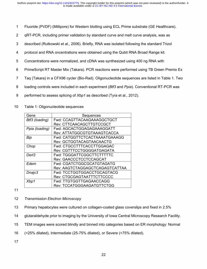
Taq (Takara) in a CFX96 cycler (Bio-Rad). Oligonucleotide sequences are listed in Table 1. Two 7
loading controls were included in each experiment (Btf3 and Ppia). Conventional RT-PCR was 8
performed to assess splicing of Xbp1 as described (Tyra et al., 2012). 9
Table 1: Oligonucleotide sequences 10
Gene Sequences Btf3 (loading) Fwd: CCAGTTACAAGAAAGGCTGCT
Rev: CTTCAACAGCTTGTCCGCT Ppia (loading) Fwd: AGCACTGGAGAGAAAGGATT
Rev: ATTATGGCGTGTAAAGTCACCA Bip Fwd: CATGGTTCTCACTAAAATGAAAGG
Rev: GCTGGTACAGTAACAACTG Chop Fwd: CTGCCTTTCACCTTGGAGAC
Rev: CGTTTCCTGGGGATGAGATA Derl3 Fwd: TGGGATTCGGCTTCTTTTTC
Rev: GAACCCTCCTCCAGCAT Edem Fwd: CGATCTGGCGCATGTAGATG
Rev: AAGTCTAGGAGCTCAGAGTCATTAA Dnajc3 Fwd: TCCTGGTGGACCTGCAGTACG
Rev: CTGCGAGTAATTTCTTCCCC Xbp1 Fwd: TTGTGGTTGAGAACCAGG
Rev: TCCATGGGAAGATGTTCTGG 11
Transmission Electron Microscopy 12
Primary hepatocytes were cultured on collagen-coated glass coverslips and fixed in 2.5% 13
glutaraldehyde prior to imaging by the University of Iowa Central Microscopy Research Facility. 14
TEM images were scored blindly and binned into categories based on ER morphology: Normal 15
(<25% dilated), Intermediate (25-75% dilated), or Severe (>75% dilated). 16
17
.CC-BY-NC-ND 4.0 International licenseis made available under aThe copyright holder for this preprint (which was not peer-reviewed) is the author/funder. It. https://doi.org/10.1101/822775doi: bioRxiv preprint
23
Statistical Analyses 1
Continuous variables were reported as the mean ± standard deviation and were analyzed using 2
the two-tailed Student’s t-test with Benjamini-Hochberg post-hoc correction for multiple 3
comparisons. For qRT-PCR, significance was calculated prior to transformation of Ct values out 4
of the log phase. A post-correction alpha of 0.05 was used to determine statistical significance. 5
6
Acknowledgments and Funding 7
The authors would like to thank the University of Iowa Viral Vector Core and Central Microscopy 8
Research Facility for technical assistance. Funding sources were as follows: DTR: 9
R01GM115424 (NIH); ERG: T32GM067795 (NIH); MJP: R01DK106104 (NIH); BNF: DK104735 10
(NIH); EBT: DK104998 (NIH); and Central Microscopy Research Facility: 1 S10 RR018998 11
(NIH) (for JEOL JEM-1230 Transmission Electron Microscope) 12
13
Author Contributions 14
ERG and DTR conceived and designed the experiments. ERG, KSM, MM, and AQK-M 15
performed the experiments. ERG, KSM, MJP, BNF, EBT, and DTR interpreted the data. ERG 16
and DTR wrote the manuscript. All authors edited and approved the manuscript. 17
18
Competing Interests 19
BNF is a shareholder and member of the Scientific Advisory Board for Cirius Therapeutics. KSM 20
received research support from Cirius Therapeutics between 2017-2019. EBT receives research 21
grant funding from MPC-related research administered through the University of Iowa from 22
Cirius Therapeutics and Poxel SA. 23
24
25
.CC-BY-NC-ND 4.0 International licenseis made available under aThe copyright holder for this preprint (which was not peer-reviewed) is the author/funder. It. https://doi.org/10.1101/822775doi: bioRxiv preprint
24
References: 1
Araujo, A. R., Rosso, N., Bedogni, G., Tiribelli, C., & Bellentani, S. (2018). Global epidemiology 2 of non-alcoholic fatty liver disease/non-alcoholic steatohepatitis: What we need in the 3 future. Liver Int, 38 Suppl 1, 47-51. doi:10.1111/liv.13643 4
Bradshaw, P. C. (2019). Cytoplasmic and Mitochondrial NADPH-Coupled Redox Systems in the 5 Regulation of Aging. Nutrients, 11(3). doi:10.3390/nu11030504 6
Bricker, D. K., Taylor, E. B., Schell, J. C., Orsak, T., Boutron, A., Chen, Y. C., . . . Rutter, J. 7 (2012). A mitochondrial pyruvate carrier required for pyruvate uptake in yeast, 8 Drosophila, and humans. Science, 337(6090), 96-100. doi:10.1126/science.1218099 9
Charlton, M., Krishnan, A., Viker, K., Sanderson, S., Cazanave, S., McConico, A., . . . Gores, G. 10 (2011). Fast food diet mouse: novel small animal model of NASH with ballooning, 11 progressive fibrosis, and high physiological fidelity to the human condition. Am J Physiol 12 Gastrointest Liver Physiol, 301(5), G825-834. doi:10.1152/ajpgi.00145.2011 13
Chaudiere, J., & Tappel, A. L. (1984). Interaction of gold(I) with the active site of selenium-14 glutathione peroxidase. J Inorg Biochem, 20(4), 313-325. doi:10.1016/0162-15 0134(84)85030-8 16
Cnop, M., Foufelle, F., & Velloso, L. A. (2012). Endoplasmic reticulum stress, obesity and 17 diabetes. Trends Mol Med, 18(1), 59-68. doi:10.1016/j.molmed.2011.07.010 18
Constantin-Teodosiu, D., Simpson, E. J., & Greenhaff, P. L. (1999). The importance of pyruvate 19 availability to PDC activation and anaplerosis in human skeletal muscle. Am J Physiol, 20 276(3), E472-478. doi:10.1152/ajpendo.1999.276.3.E472 21
Delaunay-Moisan, A., Ponsero, A., & Toledano, M. B. (2017). Reexamining the Function of 22 Glutathione in Oxidative Protein Folding and Secretion. Antioxid Redox Signal, 27(15), 23 1178-1199. doi:10.1089/ars.2017.7148 24
DeZwaan-McCabe, D., Sheldon, R. D., Gorecki, M. C., Guo, D. F., Gansemer, E. R., Kaufman, 25 R. J., . . . Rutkowski, D. T. (2017). ER Stress Inhibits Liver Fatty Acid Oxidation while 26 Unmitigated Stress Leads to Anorexia-Induced Lipolysis and Both Liver and Kidney 27 Steatosis. Cell Rep, 19(9), 1794-1806. doi:10.1016/j.celrep.2017.05.020 28
Fan, J., Ye, J., Kamphorst, J. J., Shlomi, T., Thompson, C. B., & Rabinowitz, J. D. (2014). 29 Quantitative flux analysis reveals folate-dependent NADPH production. Nature, 30 510(7504), 298-302. doi:10.1038/nature13236 31
Fan, Y., & Simmen, T. (2019). Mechanistic Connections between Endoplasmic Reticulum (ER) 32 Redox Control and Mitochondrial Metabolism. Cells, 8(9). doi:10.3390/cells8091071 33
Finnie, J. W. (2001). Effect of tunicamycin on hepatocytes in vitro. J Comp Pathol, 125(4), 318-34 321. doi:10.1053/jcpa.2001.0510 35
Frand, A. R., & Kaiser, C. A. (1999). Ero1p oxidizes protein disulfide isomerase in a pathway for 36 disulfide bond formation in the endoplasmic reticulum. Mol Cell, 4(4), 469-477. Retrieved 37 from http://www.ncbi.nlm.nih.gov/pubmed/10549279 38
Frayn, K. N., Arner, P., & Yki-Jarvinen, H. (2006). Fatty acid metabolism in adipose tissue, 39 muscle and liver in health and disease. Essays Biochem, 42, 89-103. 40 doi:10.1042/bse0420089 41
Fu, S., Watkins, S. M., & Hotamisligil, G. S. (2012). The role of endoplasmic reticulum in hepatic 42 lipid homeostasis and stress signaling. Cell Metab, 15(5), 623-634. 43 doi:10.1016/j.cmet.2012.03.007 44
Gomez, J. A., & Rutkowski, D. T. (2016). Experimental reconstitution of chronic ER stress in the 45 liver reveals feedback suppression of BiP mRNA expression. Elife, 5. 46 doi:10.7554/eLife.20390 47
Gonzalez-Rodriguez, A., Mayoral, R., Agra, N., Valdecantos, M. P., Pardo, V., Miquilena-Colina, 48 M. E., . . . Valverde, A. M. (2014). Impaired autophagic flux is associated with increased 49
.CC-BY-NC-ND 4.0 International licenseis made available under aThe copyright holder for this preprint (which was not peer-reviewed) is the author/funder. It. https://doi.org/10.1101/822775doi: bioRxiv preprint
25
endoplasmic reticulum stress during the development of NAFLD. Cell Death Dis, 5, 1 e1179. doi:10.1038/cddis.2014.162 2
Gray, L. R., Sultana, M. R., Rauckhorst, A. J., Oonthonpan, L., Tompkins, S. C., Sharma, A., . . . 3 Taylor, E. B. (2015). Hepatic Mitochondrial Pyruvate Carrier 1 Is Required for Efficient 4 Regulation of Gluconeogenesis and Whole-Body Glucose Homeostasis. Cell Metab, 5 22(4), 669-681. doi:10.1016/j.cmet.2015.07.027 6
Gray, L. R., Tompkins, S. C., & Taylor, E. B. (2014). Regulation of pyruvate metabolism and 7 human disease. Cell Mol Life Sci, 71(14), 2577-2604. doi:10.1007/s00018-013-1539-2 8
Gutierrez, T., & Simmen, T. (2018). Endoplasmic reticulum chaperones tweak the mitochondrial 9 calcium rheostat to control metabolism and cell death. Cell Calcium, 70, 64-75. 10 doi:10.1016/j.ceca.2017.05.015 11
Herzig, S., Raemy, E., Montessuit, S., Veuthey, J. L., Zamboni, N., Westermann, B., . . . 12 Martinou, J. C. (2012). Identification and functional expression of the mitochondrial 13 pyruvate carrier. Science, 337(6090), 93-96. doi:10.1126/science.1218530 14
Higa, A., & Chevet, E. (2012). Redox signaling loops in the unfolded protein response. Cell 15 Signal, 24(8), 1548-1555. doi:10.1016/j.cellsig.2012.03.011 16
Howell, J. J., & Manning, B. D. (2011). mTOR couples cellular nutrient sensing to organismal 17 metabolic homeostasis. Trends Endocrinol Metab, 22(3), 94-102. 18 doi:10.1016/j.tem.2010.12.003 19
Iracheta-Vellve, A., Petrasek, J., Gyongyosi, B., Satishchandran, A., Lowe, P., Kodys, K., . . . 20 Szabo, G. (2016). Endoplasmic Reticulum Stress-induced Hepatocellular Death 21 Pathways Mediate Liver Injury and Fibrosis via Stimulator of Interferon Genes. J Biol 22 Chem, 291(52), 26794-26805. doi:10.1074/jbc.M116.736991 23
Joseph, S. K., Booth, D. M., Young, M. P., & Hajnoczky, G. (2019). Redox regulation of ER and 24 mitochondrial Ca(2+) signaling in cell survival and death. Cell Calcium, 79, 89-97. 25 doi:10.1016/j.ceca.2019.02.006 26
Kelner, M. J., & Montoya, M. A. (2000). Structural organization of the human glutathione 27 reductase gene: determination of correct cDNA sequence and identification of a 28 mitochondrial leader sequence. Biochem Biophys Res Commun, 269(2), 366-368. 29 doi:10.1006/bbrc.2000.2267 30
Kim, S., Sideris, D. P., Sevier, C. S., & Kaiser, C. A. (2012). Balanced Ero1 activation and 31 inactivation establishes ER redox homeostasis. J Cell Biol, 196(6), 713-725. 32 doi:10.1083/jcb.201110090 33
Lai, K. K., Kolippakkam, D., & Beretta, L. (2008). Comprehensive and quantitative proteome 34 profiling of the mouse liver and plasma. Hepatology, 47(3), 1043-1051. 35 doi:10.1002/hep.22123 36
Lake, A. D., Novak, P., Hardwick, R. N., Flores-Keown, B., Zhao, F., Klimecki, W. T., & 37 Cherrington, N. J. (2014). The adaptive endoplasmic reticulum stress response to 38 lipotoxicity in progressive human nonalcoholic fatty liver disease. Toxicol Sci, 137(1), 26-39 35. doi:10.1093/toxsci/kft230 40
Lebeaupin, C., Proics, E., de Bieville, C. H., Rousseau, D., Bonnafous, S., Patouraux, S., . . . 41 Bailly-Maitre, B. (2015). ER stress induces NLRP3 inflammasome activation and 42 hepatocyte death. Cell Death Dis, 6, e1879. doi:10.1038/cddis.2015.248 43
Lebeaupin, C., Vallee, D., Rousseau, D., Patouraux, S., Bonnafous, S., Adam, G., . . . Bailly-44 Maitre, B. (2018). Bax inhibitor-1 protects from nonalcoholic steatohepatitis by limiting 45 inositol-requiring enzyme 1 alpha signaling in mice. Hepatology, 68(2), 515-532. 46 doi:10.1002/hep.29847 47
Lee, A. H., Scapa, E. F., Cohen, D. E., & Glimcher, L. H. (2008). Regulation of hepatic 48 lipogenesis by the transcription factor XBP1. Science, 320(5882), 1492-1496. 49 doi:10.1126/science.1158042 50
.CC-BY-NC-ND 4.0 International licenseis made available under aThe copyright holder for this preprint (which was not peer-reviewed) is the author/funder. It. https://doi.org/10.1101/822775doi: bioRxiv preprint
26
Lewis, C. A., Parker, S. J., Fiske, B. P., McCloskey, D., Gui, D. Y., Green, C. R., . . . Metallo, C. 1 M. (2014). Tracing compartmentalized NADPH metabolism in the cytosol and 2 mitochondria of mammalian cells. Mol Cell, 55(2), 253-263. 3 doi:10.1016/j.molcel.2014.05.008 4
Liu, W., Baker, S. S., Baker, R. D., & Zhu, L. (2015). Antioxidant Mechanisms in Nonalcoholic 5 Fatty Liver Disease. Curr Drug Targets, 16(12), 1301-1314. 6 doi:10.2174/1389450116666150427155342 7
Markan, K. R., Naber, M. C., Ameka, M. K., Anderegg, M. D., Mangelsdorf, D. J., Kliewer, S. A., 8 . . . Potthoff, M. J. (2014). Circulating FGF21 is liver derived and enhances glucose 9 uptake during refeeding and overfeeding. Diabetes, 63(12), 4057-4063. 10 doi:10.2337/db14-0595 11
McCommis, K. S., Chen, Z., Fu, X., McDonald, W. G., Colca, J. R., Kletzien, R. F., . . . Finck, B. 12 N. (2015). Loss of Mitochondrial Pyruvate Carrier 2 in the Liver Leads to Defects in 13 Gluconeogenesis and Compensation via Pyruvate-Alanine Cycling. Cell Metab, 22(4), 14 682-694. doi:10.1016/j.cmet.2015.07.028 15
McCommis, K. S., Hodges, W. T., Brunt, E. M., Nalbantoglu, I., McDonald, W. G., Holley, C., . . . 16 Finck, B. N. (2017). Targeting the mitochondrial pyruvate carrier attenuates fibrosis in a 17 mouse model of nonalcoholic steatohepatitis. Hepatology, 65(5), 1543-1556. 18 doi:10.1002/hep.29025 19
Merrill, C. L., Ni, H., Yoon, L. W., Tirmenstein, M. A., Narayanan, P., Benavides, G. R., . . . 20 Morgan, K. T. (2002). Etomoxir-induced oxidative stress in HepG2 cells detected by 21 differential gene expression is confirmed biochemically. Toxicol Sci, 68(1), 93-101. 22 Retrieved from 23 http://www.ncbi.nlm.nih.gov/entrez/query.fcgi?cmd=Retrieve&db=PubMed&dopt=Citation24 &list_uids=12075114 25
Mogilenko, D. A., Haas, J. T., L'Homme, L., Fleury, S., Quemener, S., Levavasseur, M., . . . 26 Dombrowicz, D. (2019). Metabolic and Innate Immune Cues Merge into a Specific 27 Inflammatory Response via the UPR. Cell, 178(1), 263. doi:10.1016/j.cell.2019.06.017 28
Mohan, S., R, P. R. M., Brown, L., Ayyappan, P., & G, R. K. (2019). Endoplasmic reticulum 29 stress: A master regulator of metabolic syndrome. Eur J Pharmacol, 860, 172553. 30 doi:10.1016/j.ejphar.2019.172553 31
Moule, S. K., & Denton, R. M. (1997). Multiple signaling pathways involved in the metabolic 32 effects of insulin. Am J Cardiol, 80(3A), 41A-49A. doi:10.1016/s0002-9149(97)00457-8 33
Murphy, M. P. (2009). How mitochondria produce reactive oxygen species. Biochem J, 417(1), 34 1-13. doi:10.1042/BJ20081386 35
Nadanaka, S., Okada, T., Yoshida, H., & Mori, K. (2007). Role of disulfide bridges formed in the 36 luminal domain of ATF6 in sensing endoplasmic reticulum stress. Mol Cell Biol, 27(3), 37 1027-1043. doi:MCB.00408-06 [pii]10.1128/MCB.00408-06 38
Nagasawa, K., Higashi, T., Hosokawa, N., Kaufman, R. J., & Nagata, K. (2007). Simultaneous 39 induction of the four subunits of the TRAP complex by ER stress accelerates ER 40 degradation. EMBO Rep, 8(5), 483-489. Retrieved from 41 http://www.ncbi.nlm.nih.gov/entrez/query.fcgi?cmd=Retrieve&db=PubMed&dopt=Citation42 &list_uids=17380188 43
Olivares, S., & Henkel, A. S. (2015). Hepatic Xbp1 Gene Deletion Promotes Endoplasmic 44 Reticulum Stress-induced Liver Injury and Apoptosis. J Biol Chem, 290(50), 30142-45 30151. doi:10.1074/jbc.M115.676239 46
Oonthonpan, L., Rauckhorst, A. J., Gray, L. R., Boutron, A. C., & Taylor, E. B. (2019). Two 47 human patient mitochondrial pyruvate carrier mutations reveal distinct molecular 48 mechanisms of dysfunction. JCI Insight, 5. doi:10.1172/jci.insight.126132 49
.CC-BY-NC-ND 4.0 International licenseis made available under aThe copyright holder for this preprint (which was not peer-reviewed) is the author/funder. It. https://doi.org/10.1101/822775doi: bioRxiv preprint
27
Ordonez, A., Snapp, E. L., Tan, L., Miranda, E., Marciniak, S. J., & Lomas, D. A. (2013). 1 Endoplasmic reticulum polymers impair luminal protein mobility and sensitize to cellular 2 stress in alpha1-antitrypsin deficiency. Hepatology, 57(5), 2049-2060. 3 doi:10.1002/hep.26173 4
Owen, O. E., Kalhan, S. C., & Hanson, R. W. (2002). The key role of anaplerosis and 5 cataplerosis for citric acid cycle function. J Biol Chem, 277(34), 30409-30412. 6 doi:10.1074/jbc.R200006200 7
Oyadomari, S., Harding, H. P., Zhang, Y., Oyadomari, M., & Ron, D. (2008). Dephosphorylation 8 of translation initiation factor 2alpha enhances glucose tolerance and attenuates 9 hepatosteatosis in mice. Cell Metab, 7(6), 520-532. 10
Özcan, U., Cao, Q., Yilmaz, E., Lee, A. H., Iwakoshi, N. N., Ozdelen, E., . . . Hotamisligil, G. S. 11 (2004). Endoplasmic reticulum stress links obesity, insulin action, and type 2 diabetes. 12 Science, 306(5695), 457-461. Retrieved from 13 http://www.ncbi.nlm.nih.gov/entrez/query.fcgi?cmd=Retrieve&db=PubMed&dopt=Citation14 &list_uids=15486293 15
Peters, T., Jr., & Davidson, L. K. (1982). The biosynthesis of rat serum albumin. In vivo studies 16 on the formation of the disulfide bonds. J Biol Chem, 257(15), 8847-8853. Retrieved 17 from https://www.ncbi.nlm.nih.gov/pubmed/7096338 18
Pfaffenbach, K. T., Nivala, A. M., Reese, L., Ellis, F., Wang, D., Wei, Y., & Pagliassotti, M. J. 19 (2010). Rapamycin Inhibits Postprandial-Mediated X-Box-Binding Protein-1 Splicing in 20 Rat Liver. J Nutr, 140(5), 879-884. doi:jn.109.119883 [pii]10.3945/jn.109.119883 21
Pike, L. S., Smift, A. L., Croteau, N. J., Ferrick, D. A., & Wu, M. (2010). Inhibition of fatty acid 22 oxidation by etomoxir impairs NADPH production and increases reactive oxygen species 23 resulting in ATP depletion and cell death in human glioblastoma cells. Biochim Biophys 24 Acta, 1807(6), 726-734. Retrieved from 25 http://www.ncbi.nlm.nih.gov/entrez/query.fcgi?cmd=Retrieve&db=PubMed&dopt=Citation26 &list_uids=21692241 27
Pobre, K. F. R., Poet, G. J., & Hendershot, L. M. (2019). The endoplasmic reticulum (ER) 28 chaperone BiP is a master regulator of ER functions: Getting by with a little help from 29 ERdj friends. J Biol Chem, 294(6), 2098-2108. doi:10.1074/jbc.REV118.002804 30
Poet, G. J., Oka, O. B., van Lith, M., Cao, Z., Robinson, P. J., Pringle, M. A., . . . Bulleid, N. J. 31 (2017). Cytosolic thioredoxin reductase 1 is required for correct disulfide formation in the 32 ER. Embo J, 36(5), 693-702. doi:10.15252/embj.201695336 33
Ponsero, A. J., Igbaria, A., Darch, M. A., Miled, S., Outten, C. E., Winther, J. R., . . . Toledano, 34 M. B. (2017). Endoplasmic Reticulum Transport of Glutathione by Sec61 Is Regulated by 35 Ero1 and Bip. Mol Cell, 67(6), 962-973 e965. doi:10.1016/j.molcel.2017.08.012 36
Raffaello, A., Mammucari, C., Gherardi, G., & Rizzuto, R. (2016). Calcium at the Center of Cell 37 Signaling: Interplay between Endoplasmic Reticulum, Mitochondria, and Lysosomes. 38 Trends Biochem Sci, 41(12), 1035-1049. doi:10.1016/j.tibs.2016.09.001 39
Rahman, S. M., Schroeder-Gloeckler, J. M., Janssen, R. C., Jiang, H., Qadri, I., Maclean, K. N., 40 & Friedman, J. E. (2007). CCAAT/enhancing binding protein beta deletion in mice 41 attenuates inflammation, endoplasmic reticulum stress, and lipid accumulation in diet-42 induced nonalcoholic steatohepatitis. Hepatology, 45(5), 1108-1117. 43 doi:10.1002/hep.21614 44
Rainbolt, T. K., Saunders, J. M., & Wiseman, R. L. (2014). Stress-responsive regulation of 45 mitochondria through the ER unfolded protein response. Trends Endocrinol Metab, 46 25(10), 528-537. doi:10.1016/j.tem.2014.06.007 47
Rauckhorst, A. J., Gray, L. R., Sheldon, R. D., Fu, X., Pewa, A. D., Feddersen, C. R., . . . 48 Taylor, E. B. (2017). The mitochondrial pyruvate carrier mediates high fat diet-induced 49
.CC-BY-NC-ND 4.0 International licenseis made available under aThe copyright holder for this preprint (which was not peer-reviewed) is the author/funder. It. https://doi.org/10.1101/822775doi: bioRxiv preprint
28
increases in hepatic TCA cycle capacity. Mol Metab, 6(11), 1468-1479. 1 doi:10.1016/j.molmet.2017.09.002 2
Roberts, J. R., & Shaw, C. F., 3rd. (1998). Inhibition of erythrocyte selenium-glutathione 3 peroxidase by auranofin analogues and metabolites. Biochem Pharmacol, 55(8), 1291-4 1299. doi:10.1016/s0006-2952(97)00634-5 5
Rutkowski, D. T., Arnold, S. M., Miller, C. N., Wu, J., Li, J., Gunnison, K. M., . . . Kaufman, R. J. 6 (2006). Adaptation to ER stress is mediated by differential stabilities of pro-survival and 7 pro-apoptotic mRNAs and proteins. PLoS Biol, 4(11), e374. doi:06-PLBI-RA-0091R3 [pii] 8
10.1371/journal.pbio.0040374 9 Rutkowski, D. T., & Kaufman, R. J. (2007). That which does not kill me makes me stronger: 10
adapting to chronic ER stress. Trends Biochem Sci, 32(10), 469-476. doi:S0968-11 0004(07)00213-7 [pii]10.1016/j.tibs.2007.09.003 12
Rutkowski, D. T., Wu, J., Back, S. H., Callaghan, M. U., Ferris, S. P., Iqbal, J., . . . Kaufman, R. 13 J. (2008). UPR pathways combine to prevent hepatic steatosis caused by ER stress-14 mediated suppression of transcriptional master regulators. Dev Cell, 15(6), 829-840. 15 doi:S1534-5807(08)00475-9 [pii]10.1016/j.devcel.2008.10.015 16
Rydstrom, J. (2006). Mitochondrial NADPH, transhydrogenase and disease. Biochim Biophys 17 Acta, 1757(5-6), 721-726. doi:10.1016/j.bbabio.2006.03.010 18
Salvado, L., Palomer, X., Barroso, E., & Vazquez-Carrera, M. (2015). Targeting endoplasmic 19 reticulum stress in insulin resistance. Trends Endocrinol Metab, 26(8), 438-448. 20 doi:10.1016/j.tem.2015.05.007 21
Satapati, S., Sunny, N. E., Kucejova, B., Fu, X., He, T. T., Mendez-Lucas, A., . . . Burgess, S. C. 22 (2012). Elevated TCA cycle function in the pathology of diet-induced hepatic insulin 23 resistance and fatty liver. J Lipid Res, 53(6), 1080-1092. doi:10.1194/jlr.M023382 24
Scarbrough, P. M., Mapuskar, K. A., Mattson, D. M., Gius, D., Watson, W. H., & Spitz, D. R. 25 (2012). Simultaneous inhibition of glutathione- and thioredoxin-dependent metabolism is 26 necessary to potentiate 17AAG-induced cancer cell killing via oxidative stress. Free 27 Radic Biol Med, 52(2), 436-443. doi:10.1016/j.freeradbiomed.2011.10.493 28
Scheuner, D., Song, B., McEwen, E., Liu, C., Laybutt, R., Gillespie, P., . . . Kaufman, R. J. 29 (2001). Translational control is required for the unfolded protein response and in vivo 30 glucose homeostasis. Mol Cell, 7(6), 1165-1176. Retrieved from 31 http://www.ncbi.nlm.nih.gov/entrez/query.fcgi?cmd=Retrieve&db=PubMed&dopt=Citation32 &list_uids=11430820 33
Schwaller, M., Wilkinson, B., & Gilbert, H. F. (2003). Reduction-reoxidation cycles contribute to 34 catalysis of disulfide isomerization by protein-disulfide isomerase. J Biol Chem, 278(9), 35 7154-7159. doi:10.1074/jbc.M211036200 36
Seefeldt, T., Zhao, Y., Chen, W., Raza, A. S., Carlson, L., Herman, J., . . . Guan, X. (2009). 37 Characterization of a novel dithiocarbamate glutathione reductase inhibitor and its use 38 as a tool to modulate intracellular glutathione. J Biol Chem, 284(5), 2729-2737. 39 doi:10.1074/jbc.M802683200 40
Sies, H., Berndt, C., & Jones, D. P. (2017). Oxidative Stress. Annu Rev Biochem, 86, 715-748. 41 doi:10.1146/annurev-biochem-061516-045037 42
Sifers, R. N., Brashears-Macatee, S., Kidd, V. J., Muensch, H., & Woo, S. L. (1988). A 43 frameshift mutation results in a truncated alpha 1-antitrypsin that is retained within the 44 rough endoplasmic reticulum. J Biol Chem, 263(15), 7330-7335. 45
Sunny, N. E., Parks, E. J., Browning, J. D., & Burgess, S. C. (2011). Excessive hepatic 46 mitochondrial TCA cycle and gluconeogenesis in humans with nonalcoholic fatty liver 47 disease. Cell Metab, 14(6), 804-810. doi:10.1016/j.cmet.2011.11.004 48
.CC-BY-NC-ND 4.0 International licenseis made available under aThe copyright holder for this preprint (which was not peer-reviewed) is the author/funder. It. https://doi.org/10.1101/822775doi: bioRxiv preprint
29
Taylor, E. B. (2017). Functional Properties of the Mitochondrial Carrier System. Trends Cell Biol, 1 27(9), 633-644. doi:10.1016/j.tcb.2017.04.004 2
Theurey, P., Tubbs, E., Vial, G., Jacquemetton, J., Bendridi, N., Chauvin, M. A., . . . Rieusset, J. 3 (2016). Mitochondria-associated endoplasmic reticulum membranes allow adaptation of 4 mitochondrial metabolism to glucose availability in the liver. J Mol Cell Biol, 8(2), 129-5 143. doi:10.1093/jmcb/mjw004 6
Tsunoda, S., Avezov, E., Zyryanova, A., Konno, T., Mendes-Silva, L., Pinho Melo, E., . . . Ron, 7 D. (2014). Intact protein folding in the glutathione-depleted endoplasmic reticulum 8 implicates alternative protein thiol reductants. Elife, 3, e03421. doi:10.7554/eLife.03421 9
Tu, B. P., Ho-Schleyer, S. C., Travers, K. J., & Weissman, J. S. (2000). Biochemical basis of 10 oxidative protein folding in the endoplasmic reticulum. Science, 290(5496), 1571-1574. 11 Retrieved from http://www.ncbi.nlm.nih.gov/pubmed/11090354 12
Tyra, H. M., Spitz, D. R., & Rutkowski, D. T. (2012). Inhibition of fatty acid oxidation enhances 13 oxidative protein folding and protects hepatocytes from endoplasmic reticulum stress. 14 Mol Biol Cell, 23(5), 811-819. doi:mbc.E11-12-1011 [pii]10.1091/mbc.E11-12-1011 15
Vance, J. E. (2014). MAM (mitochondria-associated membranes) in mammalian cells: lipids and 16 beyond. Biochim Biophys Acta, 1841(4), 595-609. doi:10.1016/j.bbalip.2013.11.014 17
Volmer, R., van der Ploeg, K., & Ron, D. (2013). Membrane lipid saturation activates 18 endoplasmic reticulum unfolded protein response transducers through their 19 transmembrane domains. Proc Natl Acad Sci U S A, 110(12), 4628-4633. 20 doi:10.1073/pnas.1217611110 21
Walter, P., & Ron, D. (2011). The unfolded protein response: from stress pathway to 22 homeostatic regulation. Science, 334(6059), 1081-1086. doi:334/6059/1081 23 [pii]10.1126/science.1209038 24
Wang, J., Pareja, K. A., Kaiser, C. A., & Sevier, C. S. (2014). Redox signaling via the molecular 25 chaperone BiP protects cells against endoplasmic reticulum-derived oxidative stress. 26 Elife, 3, e03496. doi:10.7554/eLife.03496 27
Wang, J. M., Qiu, Y., Yang, Z., Kim, H., Qian, Q., Sun, Q., . . . Zhang, K. (2018). IRE1alpha 28 prevents hepatic steatosis by processing and promoting the degradation of select 29 microRNAs. Sci Signal, 11(530). doi:10.1126/scisignal.aao4617 30
Weis, B. C., Cowan, A. T., Brown, N., Foster, D. W., & McGarry, J. D. (1994). Use of a selective 31 inhibitor of liver carnitine palmitoyltransferase I (CPT I) allows quantification of its 32 contribution to total CPT I activity in rat heart. Evidence that the dominant cardiac CPT I 33 isoform is identical to the skeletal muscle enzyme. J Biol Chem, 269(42), 26443-26448. 34 Retrieved from 35 http://www.ncbi.nlm.nih.gov/entrez/query.fcgi?cmd=Retrieve&db=PubMed&dopt=Citation36 &list_uids=7929365 37
Wieland, O. H., Patzelt, C., & Loffler, G. (1972). Active and inactive forms of pyruvate 38 dehydrogenase in rat liver. Effect of starvation and refeeding and of insulin treatment on 39 pyruvate-dehydrogenase interconversion. Eur J Biochem, 26(3), 426-433. 40 doi:10.1111/j.1432-1033.1972.tb01783.x 41
Willy, J. A., Young, S. K., Stevens, J. L., Masuoka, H. C., & Wek, R. C. (2015). CHOP links 42 endoplasmic reticulum stress to NF-kappaB activation in the pathogenesis of 43 nonalcoholic steatohepatitis. Mol Biol Cell, 26(12), 2190-2204. doi:10.1091/mbc.E15-01-44 0036 45
Winther, J. R., & Thorpe, C. (2014). Quantification of thiols and disulfides. Biochim Biophys 46 Acta, 1840(2), 838-846. doi:10.1016/j.bbagen.2013.03.031 47
Wu, C. Y., Satapati, S., Gui, W., Wynn, R. M., Sharma, G., Lou, M., . . . Merritt, M. E. (2018). A 48 novel inhibitor of pyruvate dehydrogenase kinase stimulates myocardial carbohydrate 49
.CC-BY-NC-ND 4.0 International licenseis made available under aThe copyright holder for this preprint (which was not peer-reviewed) is the author/funder. It. https://doi.org/10.1101/822775doi: bioRxiv preprint
30
oxidation in diet-induced obesity. J Biol Chem, 293(25), 9604-9613. 1 doi:10.1074/jbc.RA118.002838 2
Wu, J., Rutkowski, D. T., Dubois, M., Swathirajan, J., Saunders, T., Wang, J., . . . Kaufman, R. 3 J. (2007). ATF6alpha optimizes long-term endoplasmic reticulum function to protect cells 4 from chronic stress. Dev Cell, 13(3), 351-364. doi:S1534-5807(07)00266-3 5 [pii]10.1016/j.devcel.2007.07.005 6
Xin, Y., Dominguez Gutierrez, G., Okamoto, H., Kim, J., Lee, A. H., Adler, C., . . . Gromada, J. 7 (2018). Pseudotime Ordering of Single Human beta-Cells Reveals States of Insulin 8 Production and Unfolded Protein Response. Diabetes, 67(9), 1783-1794. 9 doi:10.2337/db18-0365 10
Yao, C. H., Liu, G. Y., Wang, R., Moon, S. H., Gross, R. W., & Patti, G. J. (2018). Identifying off-11 target effects of etomoxir reveals that carnitine palmitoyltransferase I is essential for 12 cancer cell proliferation independent of beta-oxidation. PLoS Biol, 16(3), e2003782. 13 doi:10.1371/journal.pbio.2003782 14
Ying, W. (2008). NAD+/NADH and NADP+/NADPH in cellular functions and cell death: 15 regulation and biological consequences. Antioxid Redox Signal, 10(2), 179-206. 16 doi:10.1089/ars.2007.1672 17
Yoboue, E. D., Sitia, R., & Simmen, T. (2018). Redox crosstalk at endoplasmic reticulum (ER) 18 membrane contact sites (MCS) uses toxic waste to deliver messages. Cell Death Dis, 19 9(3), 331. doi:10.1038/s41419-017-0033-4 20
Zhao, Y., Seefeldt, T., Chen, W., Wang, X., Matthees, D., Hu, Y., & Guan, X. (2009). Effects of 21 glutathione reductase inhibition on cellular thiol redox state and related systems. Arch 22 Biochem Biophys, 485(1), 56-62. doi:S0003-9861(09)00064-2 23 [pii]10.1016/j.abb.2009.03.001 24
Zito, E., Chin, K. T., Blais, J., Harding, H. P., & Ron, D. (2010). ERO1-beta, a pancreas-specific 25 disulfide oxidase, promotes insulin biogenesis and glucose homeostasis. J Cell Biol, 26 188(6), 821-832. doi:10.1083/jcb.200911086 27
Zito, E., Melo, E. P., Yang, Y., Wahlander, A., Neubert, T. A., & Ron, D. (2010). Oxidative 28 protein folding by an endoplasmic reticulum-localized peroxiredoxin. Mol Cell, 40(5), 29 787-797. doi:10.1016/j.molcel.2010.11.010 30
31
32
.CC-BY-NC-ND 4.0 International licenseis made available under aThe copyright holder for this preprint (which was not peer-reviewed) is the author/funder. It. https://doi.org/10.1101/822775doi: bioRxiv preprint
31
Figure Legends 1
2
Figure 1: Inhibiting b-oxidation attenuates UPR activity in metabolically active cells and 3
protects against different ER stressors. (A-C) Primary hepatocytes were treated with vehicle 4
or 250 ng/mL tunicamycin (TM) in the presence or absence of 25 µg/mL etomoxir (ET) for 8 h. 5
Activation of the UPR was assessed by qRT-PCR of a sampling of UPR target genes (A), 6
splicing of Xbp1 by conventional RT-PCR (B), and CHOP protein expression by immunoblot (C). 7
In (B) and (C), and elsewhere, each lane represents an independently treated well. Loading 8
control for immunoblot was calnexin, which does not change in response to TM or ET. (D, E) 9
Same as except using C2C12 myoblasts (D) or primary brown adipocytes (E). (F) mRNA 10
expression of UPR markers was assessed by qRT-PCR in primary hepatocytes treated with 200 11
µM palmitate (PA) or oleate (OA) and 25 µg/mL ET for 8 h. For this and all subsequent figures, 12
error bars represent means ± S.D.M. *, p<0.05, **, p<0.01, ***, p<0.001 by two-tailed t-test and 13
Benjamini-Hochberg adjustment for multiple comparisons. p-values between 0.05 and 0.1 are 14
given as values. NS, p>0.1. 15
16
Figure 2: Inhibiting b-oxidation improves ER ultrastructure. (A) Primary hepatocytes were 17
treated with vehicle, 250 ng/mL TM, 25 µg/mL ET, or TM and ET for 8 h and fixed in 2.5% 18
glutaraldehyde. Fixed cells were imaged by transmission electron microscopy. Inset panels are 19
indicated. Blue arrowheads represent areas of structurally normal ER, while orange arrowhead 20
represent dysmorphic ER. (B) Images were scored blindly and binned based on the percentage 21
of “dilated” ER in the image. Normal (blue): <25%, Moderate (gray): 25-75%, and Severe 22
(orange): >75%. n = 9-10 cell images per group. (C) Primary hepatocytes were treated with 25 23
µg/ml ET in the presence of 0.5, 2, or 10 µg/ml TM for 8 h, and expression of Bip and Chop was 24
detected by qRT-PCR. 25
.CC-BY-NC-ND 4.0 International licenseis made available under aThe copyright holder for this preprint (which was not peer-reviewed) is the author/funder. It. https://doi.org/10.1101/822775doi: bioRxiv preprint
32
1
Figure 3: Inhibiting b-oxidation attenuates ER stress induced by overexpression of a 2
constitutively misfolded protein. (A) Primary hepatocytes were infected with Ad-CMV-eGFP 3
(control) or Ad-TetOn-NHK at an MOI of 1:1. NHK expression was induced with 500 ng/mL 4
doxycycline (Dox) in the presence or absence of 25 µg/mL ET. mRNA expression of UPR 5
markers was measured by qRT-PCR. (B) Primary hepatocytes infected with Ad-TetOn-NHK and 6
treated with Dox and/or ET for 8 h were fixed in 2.5% glutaraldehyde for TEM. Orange 7
arrowheads in insets denote dysmorphic ER. (C) Images were scored as described in Figure 2; 8
n = 10 cell images per group. (D) Primary hepatocytes infected with Ad-TetOn-NHK were 9
treated with Dox and ET as in (A) for 4 or 8 h in the presence or absence of 5 µM MG-132. NHK 10
expression was analyzed by immunoblot. Loading control was calnexin. 11
12
Figure 4: Inhibiting Glutathione Reductase attenuates ER stress. (A) Primary hepatocytes 13
were treated with 25 µg/mL ET for 4 h followed by addition of 10 mM DTT for 0, 30, or 60 min. 14
Protein lysates were treated with 4 µM mm(PEG)24 prior to SDS-PAGE and immunoblotting to 15
detect endogenous albumin. The percentage of oxidized albumin is given below each group, 16
with statistical comparison between ET-treated and non-treated cells. (B) Primary hepatocytes 17
were treated with 25 µg/ml ET or 25 µM 2-AAPA for 8 h. Levels of total (GSxtot), reduced (GSH), 18
and oxidized (GSSG) glutathione, and the ratio of GSSG to GSH were measured 19
fluorometrically and expressed relative to untreated cells. (C) Splicing of Xbp1 mRNA was 20
measured by conventional RT-PCR, and (D) mRNA expression of UPR markers was measured 21
by qRT-PCR in cells treated for 8 h with TM and/or 2-AAPA. 22
23
Figure 5: Glutathione oxidation is required for inhibition of b-oxidation to alleviate ER 24
stress. (A) Levels of total, reduced, and oxidized glutathione were measured in primary 25
.CC-BY-NC-ND 4.0 International licenseis made available under aThe copyright holder for this preprint (which was not peer-reviewed) is the author/funder. It. https://doi.org/10.1101/822775doi: bioRxiv preprint
33
hepatocytes treated with 10 µM auranofin (AuF) for 8 h. Glutathione was quantified as in Figure 1
4. (B) Hepatocytes were treated with 2-AAPA or AuF for 4 h prior to DTT and mm(PEG)24 as in 2
Figure 4. (C) mRNA expression of UPR markers after treatment with AuF for 8h was quantified. 3
(D) Hepatocytes were treated with TM, ET, and AuF as indicated. mRNA expression of UPR 4
markers was measured by qRT-PCR. 5
6
Figure 6: Diminishing TCA-dependent production of NADPH alleviates ER stress. (A) 7
Acetyl-CoA levels or (B) the NADPH/NADP+ ratio were measured in primary hepatocytes 8
treated with ET for 8 h and is expressed relative to untreated cells. (C) Idh1, Idh2, and Me1 9
were knocked down with dsiRNA prior to treatment with TM for 8 h. A non-targeting dsiRNA was 10
used as a control. Relative mRNA expression of Idh1, Idh2, and Me1 was measured by qRT-11
PCR. (D) Glutathione was quantified in cells with knocked down Idh1, Idh2, or Me1 (first oligo 12
pair from Figure 6C in each case). (E) Expression of UPR target genes was assessed in cells 13
from (C, D). 14
15
Figure 7: Stimulating TCA substrate entry causes ER stress. (A-C) Primary hepatocytes 16
were treated with 4 µM dichloroacetate (DCA) or TM for 8 h. The NADPH/NADP+ ratio (A), UPR 17
target gene expression (C), or Xbp1 mRNA splicing (B) were analyzed as in previous figures. 18
19
Figure 8: Attenuation of TCA flux by deletion of the Mitochondrial Pyruvate Carrier 20
alleviates ER stress. (A, B) The NADPH/NADP+ ratio (A), and the GSSG/GSH ratio (B) 21
measured as above in parental wild-type and Mpc1-/- C2C12 myoblasts. (C) Expression of BiP, 22
CHOP, phosphorylated eIF2α, and MPC1 was analyzed by immunoblot in wild-type and Mpc1-/- 23
C2C12 myocytes treated with 100 ng/mL TM, 1 µg/mL TM, or 100 nM thapsigargin (TG). 24
Loading control was actin. (D) Relative mRNA expression of UPR markers from cells treated 25
similarly to (C) was quantified by qRT-PCR. (E) Primary hepatocytes from Mpc2fl/fl and Mpc2LKO 26
.CC-BY-NC-ND 4.0 International licenseis made available under aThe copyright holder for this preprint (which was not peer-reviewed) is the author/funder. It. https://doi.org/10.1101/822775doi: bioRxiv preprint
34
mice were treated with 250 ng/mL TM, and gene expression was measured by qRT-PCR. The 1
Mpc2-/- untreated and w.t. TM-treated were reversed by cut/paste to keep ordering consistent 2
with the rest of the data in the paper but the image was otherwise unmodified. (F) Splicing of 3
Xbp1 in TM-treated hepatocytes from Mpc2fl/fl or Mpc2LKO mice was assessed by conventional 4
RT-PCR. (G) Xbp1 splicing was quantified from 8 independent samples (4 from cells taken from 5
a male mouse, 4 from cells taken from a female mouse). 6
7
Figure 9: Proposed model describing the relationship between TCA cycle activity and ER 8
homeostasis. See Discussion for details. 9
.CC-BY-NC-ND 4.0 International licenseis made available under aThe copyright holder for this preprint (which was not peer-reviewed) is the author/funder. It. https://doi.org/10.1101/822775doi: bioRxiv preprint
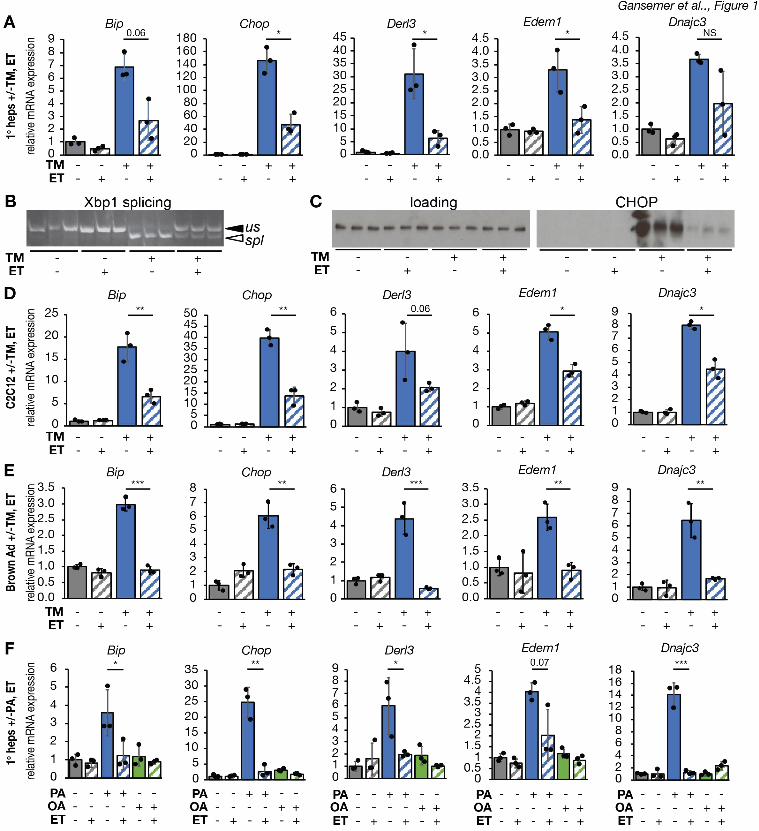
.CC-BY-NC-ND 4.0 International licenseis made available under aThe copyright holder for this preprint (which was not peer-reviewed) is the author/funder. It. https://doi.org/10.1101/822775doi: bioRxiv preprint
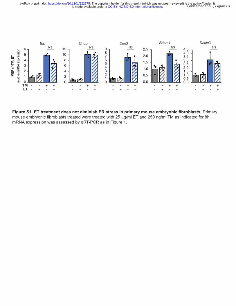
Gansemer et al.., Figure S1
Figure S1. ET treatment does not diminish ER stress in primary mouse embryonic fibroblasts. Primary mouse embryonic fibroblasts treated were treated with 25 μg/ml ET and 250 ng/ml TM as indicated for 8h. mRNA expression was assessed by qRT-PCR as in Figure 1.
0123456
02468
1012
123456789
0.0
0.5
1.0
1.5
2.0
2.5
0.51.01.52.02.53.03.54.04.5
relat
ive m
RNA
expr
essio
nM
EF +
/-TM
, ET
Bip Chop Derl3 Edem1 Dnajc3NSNS NS NS NS
-- +++- +-
TMET
-- +++- +-
-- +++- +-
-- +++- +-
-- +++- +-
0 0.0
.CC-BY-NC-ND 4.0 International licenseis made available under aThe copyright holder for this preprint (which was not peer-reviewed) is the author/funder. It. https://doi.org/10.1101/822775doi: bioRxiv preprint
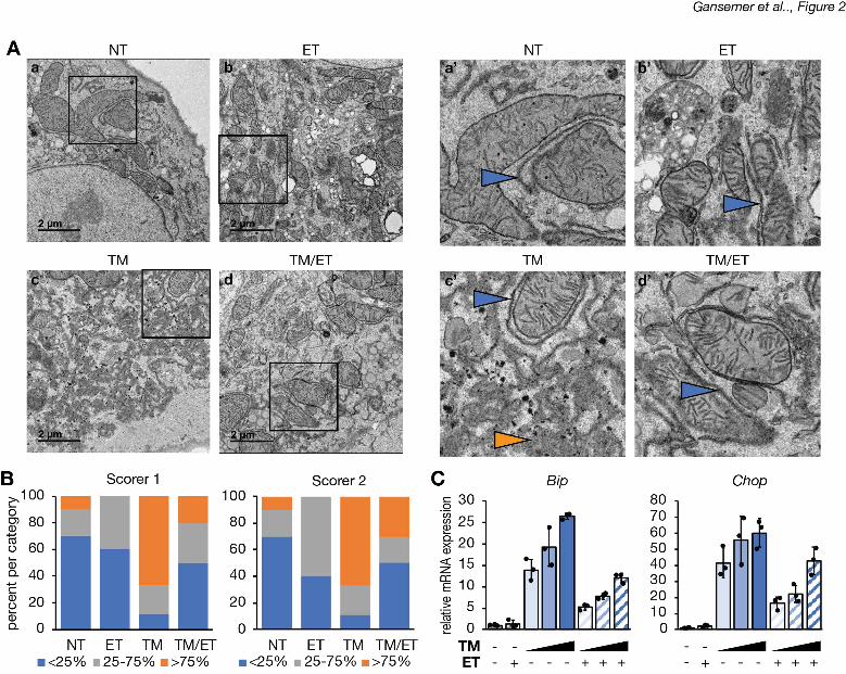
.CC-BY-NC-ND 4.0 International licenseis made available under aThe copyright holder for this preprint (which was not peer-reviewed) is the author/funder. It. https://doi.org/10.1101/822775doi: bioRxiv preprint
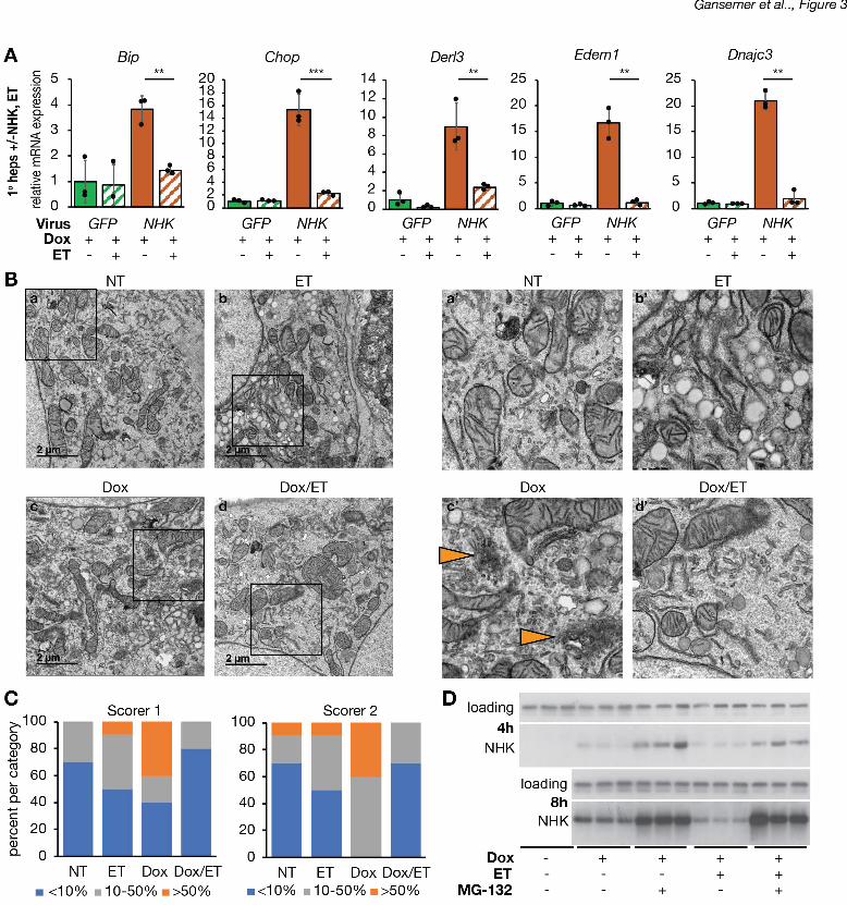
.CC-BY-NC-ND 4.0 International licenseis made available under aThe copyright holder for this preprint (which was not peer-reviewed) is the author/funder. It. https://doi.org/10.1101/822775doi: bioRxiv preprint
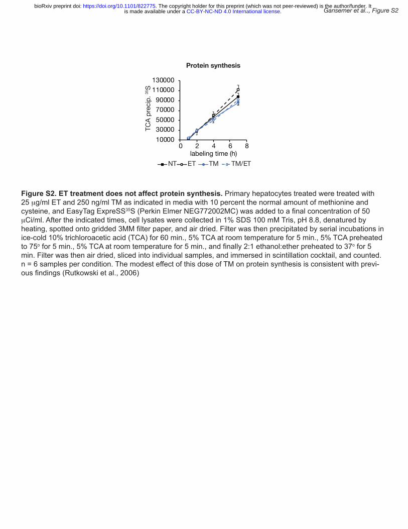
1000030000500007000090000
110000130000
0 2 4 6 8
Protein synthesis
NT TMET TM/ET
TCA
prec
ip. 35
Slabeling time (h)
Gansemer et al.., Figure S2
Figure S2. ET treatment does not affect protein synthesis. Primary hepatocytes treated were treated with 25 μg/ml ET and 250 ng/ml TM as indicated in media with 10 percent the normal amount of methionine and cysteine, and EasyTag ExpreSS35S (Perkin Elmer NEG772002MC) was added to a final concentration of 50 μCi/ml. After the indicated times, cell lysates were collected in 1% SDS 100 mM Tris, pH 8.8, denatured by heating, spotted onto gridded 3MM filter paper, and air dried. Filter was then precipitated by serial incubations in ice-cold 10% trichloroacetic acid (TCA) for 60 min., 5% TCA at room temperature for 5 min., 5% TCA preheated to 75o for 5 min., 5% TCA at room temperature for 5 min., and finally 2:1 ethanol:ether preheated to 37o for 5 min. Filter was then air dried, sliced into individual samples, and immersed in scintillation cocktail, and counted. n = 6 samples per condition. The modest effect of this dose of TM on protein synthesis is consistent with previ-ous findings (Rutkowski et al., 2006)
.CC-BY-NC-ND 4.0 International licenseis made available under aThe copyright holder for this preprint (which was not peer-reviewed) is the author/funder. It. https://doi.org/10.1101/822775doi: bioRxiv preprint
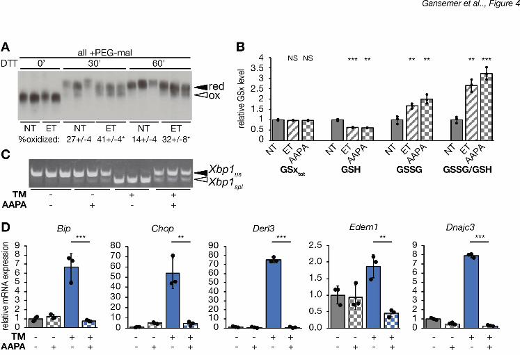
.CC-BY-NC-ND 4.0 International licenseis made available under aThe copyright holder for this preprint (which was not peer-reviewed) is the author/funder. It. https://doi.org/10.1101/822775doi: bioRxiv preprint
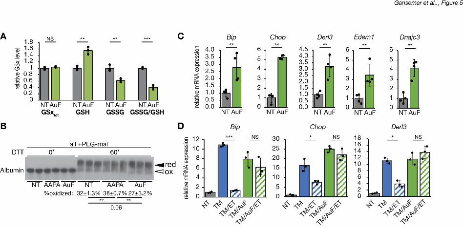
.CC-BY-NC-ND 4.0 International licenseis made available under aThe copyright holder for this preprint (which was not peer-reviewed) is the author/funder. It. https://doi.org/10.1101/822775doi: bioRxiv preprint
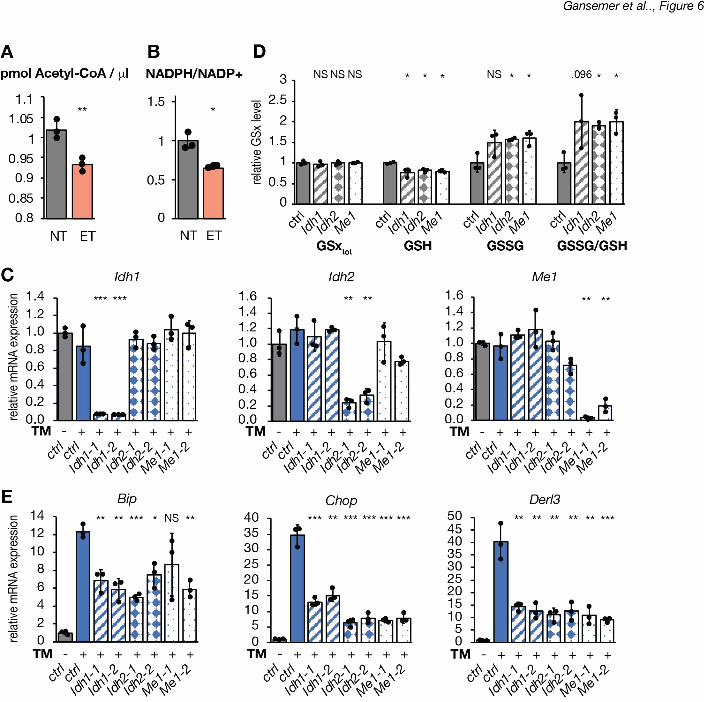
.CC-BY-NC-ND 4.0 International licenseis made available under aThe copyright holder for this preprint (which was not peer-reviewed) is the author/funder. It. https://doi.org/10.1101/822775doi: bioRxiv preprint
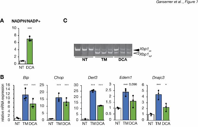
.CC-BY-NC-ND 4.0 International licenseis made available under aThe copyright holder for this preprint (which was not peer-reviewed) is the author/funder. It. https://doi.org/10.1101/822775doi: bioRxiv preprint
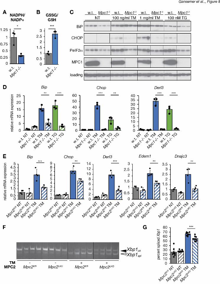
.CC-BY-NC-ND 4.0 International licenseis made available under aThe copyright holder for this preprint (which was not peer-reviewed) is the author/funder. It. https://doi.org/10.1101/822775doi: bioRxiv preprint
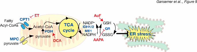
.CC-BY-NC-ND 4.0 International licenseis made available under aThe copyright holder for this preprint (which was not peer-reviewed) is the author/funder. It. https://doi.org/10.1101/822775doi: bioRxiv preprint