myocardium segmentation improvement with anisotropic ...myocardium segmentation improvement with...
TRANSCRIPT

Myocardium Segmentation Improvement with Anisotropic AnomalousDiffusion Filter Applied to Cardiac Magnetic Resonance Imaging
Antonio Carlos da S. Senra Filho1, Gustavo C. Barizon1, Luiz O. Murta Junior1
1 University of Sao Paulo, Ribeirao Preto, Brazil
Abstract
Cardiologic magnetic resonance imaging (MRI) has re-cently been improved by faster acquisition and higher res-olution hardware. Commercially available MRI equipmentis able to capture contrast agents with the needed timeand space definition to map myocardial viability. MRI my-ocardial imaging has an emerging role in cardiology stud-ies, and it has experienced a crescent relevance in clini-cal investigations. Although MRI has potential for clini-cal investigation and application, an efficient digital filteris needed in order to allow robust myocardial segmenta-tion. This paper proposes anisotropic anomalous diffu-sion (AAD) filtering to reduce noise levels while preserv-ing myocardial traits. The proposed AAD filter followsthe porous media equation consistent with inhomogenouscomplex media, and thus appropriate to model biologicalsystems. In this study, the porous media equation togetherwith gradient driven diffusion has been applied to digitalimage smoothing. Eleven MRI T1 weighted cardiology im-ages were used hereby to evaluate both the AAD and clas-sical Gaussian filter in a segmentation pipeline. in orderto study the filtering application in a automatic segmen-tation algorithm (Geodesic Active Contour). The myocar-dial area, i.e. epicardic and endocardic border, was de-lineated with both the AAD and Gaussian filter. We cal-culated the root mean square error, when compared to themanual traces, to measure automatic segmentation quality.The AAD filter show a significant segmentation accuracyenhancement (p < 0.001), while no significant differencewas found between the AAD filtered and manually seg-mented images. The findings suggest that AAD filtered im-age segmentations have similar reliability to manual seg-mentation.
1. Introduction
The advances in medical imaging hardware lead to in-creased spatial and temporal resolution. Following the ad-vance in instrumentation , the image processing filteringis growing in many diagnostic image modalities and has
a crucial importance in studies such as oncology [1], braindiseases [2] and cardiology applications [3]. Recently, car-diac MRI imaging protocol has been highly improved dueto faster imaging acquisition and higher spatial resolution[4]. Therefore, there is a emergent necessity for efficientmethods of image processing in this special type of image.
The anomalous diffusion theory, described by the gen-eralized Fokker-Planck equations [5], are partial differen-tial equation that have been applied in recent years to sev-eral applications in Physics, Economics and other areasof knowledge [5]. In particular, the porous media equa-tion[5, 6] has been applied to digital images smoothing[7] with the formulation of anisotropic anomalous diffu-sion (AAD) filtering obtaining promising results especiallywith medical imaging denoising. Previous studies suggestthat the anomalous distribution paradigm is more suitablefor the study of diffusion behavior in media with high com-plexity features [5, 8].
There are several imaging techniques in MRI that couldprovide a crucial relevance to biomedical image diagnos-tic. A very powerful MRI application is in cardiologystudies that has grown in recent years with faster imageacquisition and better voxel resolution [4]. However, af-ter the image acquisition, some diagnostic measurementsare performed with manual tracing. One of those manualtask is the miocardial segmentation, i.e. endocardium andepicardium, that is an important evaluation for tissue di-agnostic. Some automatic approaches have been proposedfor area calculations, but with limited success [4].
Our study intends to apply the AAD filter to the cardi-ology MRI protocol to improve automatic miocardic areameasurement. We evaluated the use of AAD filter as asmoothing step previous to segmentation that could pro-vide a better accuracy for this type of measurement. In thisevaluation we verified the ability of AAD filter in reducingboth the time necessary to perform the manual method andthe human error intrinsic to the manual process.
1.1. The anomalous filtering method
The anomalous diffusion filters are defined from theporous media equation [5, 6], to which we can assign
ISSN 2325-8861 Computing in Cardiology 2014; 41:929-932.929

the anisotropic anomalous diffusion method (AAD). TheAAD filler formulation is shown in Equation (1), where itis in the discrete forms.
.
Iφ,t+1 = Iφ,t +[Dq(−→r , t).∇2I
(2−q)φ,t
](1)
Where I(−→r , t) represents the image pixel intensity atposition−→r and time t. TheDq(
−→r , t) parameter representsthe generalized diffusion coefficient that is a fusion be-tween the generalized diffusion coefficient, Equation (2),and the stop edge function [9], as seen in Equation (3).
Dq =
12 .α
23−q .
(√(1−q)π .
Γ(1+ 11−q )
Γ( 32 + 1
1−q )
) 2−2q3−q
q < 1
12 .α q = 1
12 .α
23−q .
(√(q−1)π .
Γ( 1q−1 )
Γ( 1q−1−
12 )
) 2−2q3−q
1 < q < 2
(2)
Dq(−→r , t) = Dq.exp(−|∇I(−→r , t)|2/κ2) (3)
Where α = (2− q)(3− q). For q 6= 1, Equation (2), theDq parameter represents the generalized diffusion coeffi-cient consistent with q-Gaussian probability distributions[5]. From the q parameter settings of the generalized dif-fusion coefficient, anomalous filters are defined and canbe applied in digital image. For q > 1 we obtain a longrange probability distribution, with infinite support, andfor q < 1 we obtain a probability distributions of finitesupport, which are bounded by certain cutoffs [5]. Eachdistribution has its characteristic noise attenuation, and forq > 1 are reported in some studies as more efficient forsome MRI medical imaging modalities [7, 10].
Two key parameters for adjusting the AAD filters are:the q parameter and the generalized diffusion coefficient(Dq) parameter. For the q parameter, we can use the val-ues of 0 < q < 2, due to algorithm stability problemswhen it is applied to digital images [10]. The q parameteris responsible for determining the probability distributionwhich is used throughout the filtering process. For q = 1we return to the normal distribution, which returns to theclassical filtering paradigm, and q 6= 1 uses the q-Gaussiandistributions [5]. As for the generalized diffusion coeffi-cient, Dq , we use the Equation (2) to define the local max-imum intensity. Equation (2) informs the chosen probabil-ity distribution and it has the same definition as found forthe Perona and Malik filter [9] when q = 1.
2. Materials and Methods
The time t and the generalized diffusion coefficient Dq
parameters were fixed to t = 5 and Dq = 1, respec-tively. These values were chosen to generate a q-Gaussian
probability distribution with standard deviation suitablefor smoothing with minimal distortion in the images [10].The anomalous parameter, called the q value, was set upto q = 1.3, which demonstrate a good filtering perfor-mance when it is applied in T1 weighted magnetic reso-nance imaging [10].
We used T1-weighted MRI images in this study in a 3Tesla (3T) MRI tomograph, echo planar image (EPI) ac-quisition technique, TE/TR 1.64 ms/3.67 s, flip angle of45o, spatial resolution of 1.41×1.41 mm per pixel, and 10mm spacing between slices. Only the slice that has the di-astolic position was used for the endocardic and epicardicregion. The image orientation was set to short axis planes.No contrast agents were used here. Eleven images, fromdifferent patients, were selected for this study.
The miocardial area, i.e. epicardial and endocardial tis-sue borders, was used as a quality measurement to infer thesegmentation quality. The miocardial area was calculatedafter the image segmentation performed by the geodesicactive contour algorithm [11].The myocardium segmenta-tion was done with ITK and VTK image processing tools.The ITK (Insight Toolkit) is a multi platform that providesdevelopers with a comprehensive set of software tools foranalysis, segmentation and image registration. The VTKconsists of a library that provides a set of tools for scientificvisualization and has also been used for 2D and 3D imagevisualization. The Figure 1 shows the major componentsinvolved in the application of the Geodesic Active Contourto a segmentation task. The manual segmentations weremade, with inter subjects, for statistical comparison withthe automatic segmentation. All the manual segmentationmeasurements were calculated using the original image,i.e. no filter was used for the area manual estimation. Theroot mean square error (RMSE) was used to study the areameasurement variation between the automatic segmenta-tion and the manual segmentation.
3. Results and Discussion
Following the segmentation procedures, we obtain themyocardium area that are show in Table 1. It can be seenthat the AAD spatial filter application promotes better ac-curacy in border location, both in internal and external re-gion of the myocardium. When the area values were ob-tained by segmenting the original image (without filter ap-plication), we noted that there are some segmentation er-rors, as seen in Figure 2. The pixel intensity fluctuationrepresenting the noise and the natural tissue contrast re-sults in uncertainty about border location that implies insegmentation error. Even with the high spatial resolutionand signal noise ratio provided by the T1 weighted image,the non use of a noise suavization process would bring er-ror to the area measurement.
For comparison reasons, we performed manual area seg-
930

Figure 1. Diagram for the Geodesic Active Contour applied to a miocardial segmentation task. Input image for segmenta-tion can be chosen as a filtered image, with AAD or Gaussian filters, or raw image. The image resulting from this processis the micardial segmented image as shown in Figure 2.
Figure 2. Miocardic segmentation in the short axis of an MRI image with the AAD filter, Gaussian filter and the originalimage. All the results are show in a row, where it has the same order: filtered image, miocardic segmentation, areasegmented and a miocardic segmented amplification, respectively. A) Gaussian filtering, B) Original image without anyfiltering application, and C) Anisotropic anomalous diffusion filtering. See the white arrows that are pointing to a betterlocal region segmentation.
mentation and measurements which obtained the value ofamanual = 1172.42 ± 146.64. The RMSE values areshown in Table 1 and also the area values obtained withthe application of spatial filters, which had better area es-timation, in comparison with the manual approach. It isobserved that the AAD filter is statistically equivalent tothe manual approach (p < 0.001) that is not seen with theother two methods, with and without filter application.
The filtering performance obtained with the Gaussian
filter got similar results in comparison with the real esti-mated value, obtained manually. However, the spatial fil-tering approach show less precision, given by the standarddeviation, where it demonstrates low filtration efficiencydue to the intense blurring on the entire image. The Figure2 illustrates the case of the Gaussian filter, which stronglydistorts the structures contained in the image. On the con-trary, the AAD filter obtained a better edge preservationbetween the image structures and also decreased the noise
931
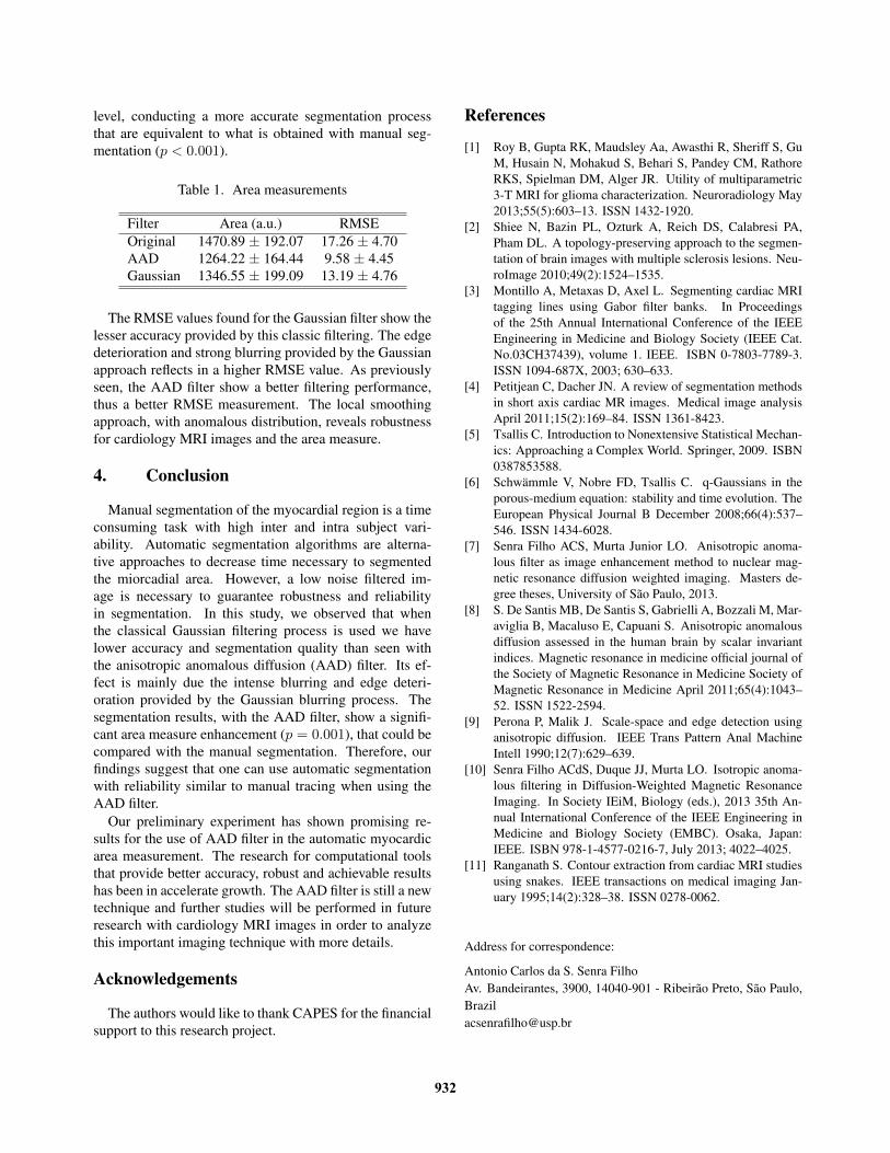
level, conducting a more accurate segmentation processthat are equivalent to what is obtained with manual seg-mentation (p < 0.001).
Table 1. Area measurements
Filter Area (a.u.) RMSEOriginal 1470.89 ± 192.07 17.26 ± 4.70AAD 1264.22 ± 164.44 9.58 ± 4.45Gaussian 1346.55 ± 199.09 13.19 ± 4.76
The RMSE values found for the Gaussian filter show thelesser accuracy provided by this classic filtering. The edgedeterioration and strong blurring provided by the Gaussianapproach reflects in a higher RMSE value. As previouslyseen, the AAD filter show a better filtering performance,thus a better RMSE measurement. The local smoothingapproach, with anomalous distribution, reveals robustnessfor cardiology MRI images and the area measure.
4. Conclusion
Manual segmentation of the myocardial region is a timeconsuming task with high inter and intra subject vari-ability. Automatic segmentation algorithms are alterna-tive approaches to decrease time necessary to segmentedthe miorcadial area. However, a low noise filtered im-age is necessary to guarantee robustness and reliabilityin segmentation. In this study, we observed that whenthe classical Gaussian filtering process is used we havelower accuracy and segmentation quality than seen withthe anisotropic anomalous diffusion (AAD) filter. Its ef-fect is mainly due the intense blurring and edge deteri-oration provided by the Gaussian blurring process. Thesegmentation results, with the AAD filter, show a signifi-cant area measure enhancement (p = 0.001), that could becompared with the manual segmentation. Therefore, ourfindings suggest that one can use automatic segmentationwith reliability similar to manual tracing when using theAAD filter.
Our preliminary experiment has shown promising re-sults for the use of AAD filter in the automatic myocardicarea measurement. The research for computational toolsthat provide better accuracy, robust and achievable resultshas been in accelerate growth. The AAD filter is still a newtechnique and further studies will be performed in futureresearch with cardiology MRI images in order to analyzethis important imaging technique with more details.
Acknowledgements
The authors would like to thank CAPES for the financialsupport to this research project.
References
[1] Roy B, Gupta RK, Maudsley Aa, Awasthi R, Sheriff S, GuM, Husain N, Mohakud S, Behari S, Pandey CM, RathoreRKS, Spielman DM, Alger JR. Utility of multiparametric3-T MRI for glioma characterization. Neuroradiology May2013;55(5):603–13. ISSN 1432-1920.
[2] Shiee N, Bazin PL, Ozturk A, Reich DS, Calabresi PA,Pham DL. A topology-preserving approach to the segmen-tation of brain images with multiple sclerosis lesions. Neu-roImage 2010;49(2):1524–1535.
[3] Montillo A, Metaxas D, Axel L. Segmenting cardiac MRItagging lines using Gabor filter banks. In Proceedingsof the 25th Annual International Conference of the IEEEEngineering in Medicine and Biology Society (IEEE Cat.No.03CH37439), volume 1. IEEE. ISBN 0-7803-7789-3.ISSN 1094-687X, 2003; 630–633.
[4] Petitjean C, Dacher JN. A review of segmentation methodsin short axis cardiac MR images. Medical image analysisApril 2011;15(2):169–84. ISSN 1361-8423.
[5] Tsallis C. Introduction to Nonextensive Statistical Mechan-ics: Approaching a Complex World. Springer, 2009. ISBN0387853588.
[6] Schwammle V, Nobre FD, Tsallis C. q-Gaussians in theporous-medium equation: stability and time evolution. TheEuropean Physical Journal B December 2008;66(4):537–546. ISSN 1434-6028.
[7] Senra Filho ACS, Murta Junior LO. Anisotropic anoma-lous filter as image enhancement method to nuclear mag-netic resonance diffusion weighted imaging. Masters de-gree theses, University of Sao Paulo, 2013.
[8] S. De Santis MB, De Santis S, Gabrielli A, Bozzali M, Mar-aviglia B, Macaluso E, Capuani S. Anisotropic anomalousdiffusion assessed in the human brain by scalar invariantindices. Magnetic resonance in medicine official journal ofthe Society of Magnetic Resonance in Medicine Society ofMagnetic Resonance in Medicine April 2011;65(4):1043–52. ISSN 1522-2594.
[9] Perona P, Malik J. Scale-space and edge detection usinganisotropic diffusion. IEEE Trans Pattern Anal MachineIntell 1990;12(7):629–639.
[10] Senra Filho ACdS, Duque JJ, Murta LO. Isotropic anoma-lous filtering in Diffusion-Weighted Magnetic ResonanceImaging. In Society IEiM, Biology (eds.), 2013 35th An-nual International Conference of the IEEE Engineering inMedicine and Biology Society (EMBC). Osaka, Japan:IEEE. ISBN 978-1-4577-0216-7, July 2013; 4022–4025.
[11] Ranganath S. Contour extraction from cardiac MRI studiesusing snakes. IEEE transactions on medical imaging Jan-uary 1995;14(2):328–38. ISSN 0278-0062.
Address for correspondence:
Antonio Carlos da S. Senra FilhoAv. Bandeirantes, 3900, 14040-901 - Ribeirao Preto, Sao Paulo,[email protected]
932