“myo = muscle” the muscular system. muscular system there are 650 skeletal musclesthere are 650...
TRANSCRIPT

““myo = musclemyo = muscle””
The Muscular SystemThe Muscular System

Muscular SystemMuscular System• There are There are 650 skeletal muscles650 skeletal muscles
• They They make up 40-50% of our body weightmake up 40-50% of our body weight
• Muscles play a crucial role in communicationMuscles play a crucial role in communication
• The smallest muscle in the body is the The smallest muscle in the body is the stapedius and controls the stapes (stirrup bone stapedius and controls the stapes (stirrup bone in the ear)in the ear)
• The largest muscle in the body???The largest muscle in the body???– Gluteus maximus!Gluteus maximus!

Important functions Movement – both voluntary & involuntary
Maintaining posture
Supporting soft tissues within body cavities
Stabilizing joints
Guarding entrances & exits of the body
Maintaining body temperature

CharacteristicsCharacteristics
• Excitability (irritability)Excitability (irritability)
• Contractility (shrink)Contractility (shrink)
• Extensibility (stretch)Extensibility (stretch)
• Elasticity (recoil)Elasticity (recoil)

5
• Skeletal muscle– Striated, has stripes– Voluntary, multiple nuclei– Attached to bones
• Cardiac muscle– Striated, has stripes– Involuntary– Responsible for heart contractions
• Smooth muscleSmooth muscle::– non-striated, no stripes non-striated, no stripes – found in the digestive tract, walls of blood vessels, found in the digestive tract, walls of blood vessels,
reproductive system reproductive system – controlled by the autonomic nervous systemcontrolled by the autonomic nervous system
Types of Muscles

6
Types of Muscles

Anatomy of MusclesAnatomy of Muscles• Muscle cells are called
muscle fibers• Nerve and blood
supply• Motor Unit
– Muscle fiber and neuron– ~150 fiber per neuron
• Neuromuscular Junction– Communicate at
synapse– Ach (acetylcholine) –
neurotransmitter in muscle contraction

Levels of Organization of a Muscle
MuscleMuscle Fascicles Fascicles Muscle Fibers (cells) Muscle Fibers (cells) MyofibrilsMyofibrils Thick/Thin Thick/Thin filamentsfilaments

Connective Tissue Connective Tissue ComponentsComponents
• Tendons– Muscle to bone
• Aponeuroses– Broad connection
to bone (flat sheet between abs and dorsal muscles)
Layers of connective tissue called fascia group muscles fibers together and give them strength

Connective Tissue Connective Tissue ComponentsComponents
• Endomysium surrounds individual muscle fiber.
• Perimysium holds muscle cells together to form a fascicle.
• Epimysium surrounds groups of fasicles.

SacromereSacromere• Sacromere is the individual contractile unit of the Sacromere is the individual contractile unit of the
muscle cellmuscle cellConsists of:*Sarcolemma*Nuclei*Sarcoplasmic reticulum*Transverse tubules*Mitochondria

MyofibrilsMyofibrils• Myofibrils: protein strands inside of muscle Myofibrils: protein strands inside of muscle
cell cell – ThinThin filaments: actin, troponin, filaments: actin, troponin,
tropomyosintropomyosin– ThickThick filaments: myosin filaments: myosin– Z line: boundary between sarcomeresZ line: boundary between sarcomeres

13
2 Types of Myosin Filament
• Red (Type I) fibers (slow twitch)– Fatigue resistant endurance– use aerobic metabolism
• White (Type II) fibers (fast twitch) – participate in brief, powerful
movements – use anaerobic metabolism
= slow twitch
= fast twitch

14
The Effects of Exercise on Muscle
• Muscles respond to damage by producing more actin and myosin filaments. This increases muscle size, but not number.
• Disuse of muscles reverses this process, as in space travel.
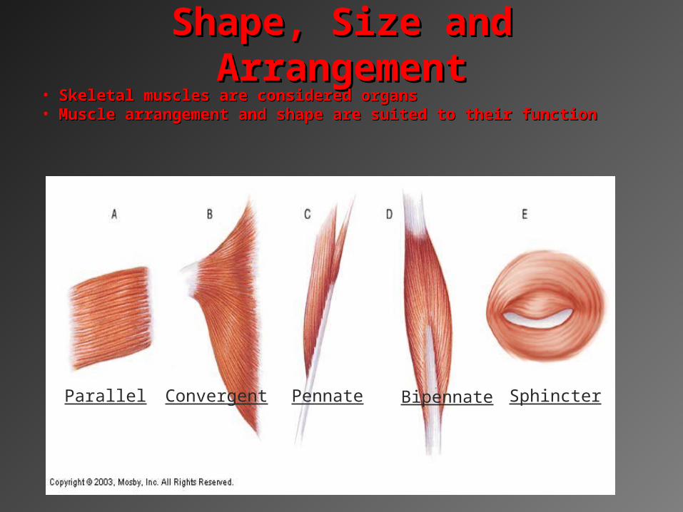
Shape, Size and Shape, Size and ArrangementArrangement
• Skeletal muscles are considered organsSkeletal muscles are considered organs• Muscle arrangement and shape are suited to their functionMuscle arrangement and shape are suited to their function
Parallel Convergent Pennate Bipennate Sphincter

• During contraction one bones usually stays During contraction one bones usually stays stationarystationary
• Skeletal muscles can only pull, not pushSkeletal muscles can only pull, not push• OriginOrigin is point of attachment that doesn’t move is point of attachment that doesn’t move• InsertionInsertion is point that does move with contraction is point that does move with contraction
Origin and Insertion Origin and Insertion PointsPoints

Muscle ActionsMuscle ActionsAgonist (Ex: biceps brachii)Agonist (Ex: biceps brachii)
– Also called the prime moverAlso called the prime mover– Contracts to produce movementContracts to produce movement
Antagonist (Ex: triceps brachii)Antagonist (Ex: triceps brachii)– Performs opposing action of Performs opposing action of
agonistagonist– Relaxes during contractionRelaxes during contraction
Synergists (Ex: bracioradialis)Synergists (Ex: bracioradialis)– Contract at same time of Contract at same time of
agonistagonist– Makes prime movers action Makes prime movers action
more efficientmore efficient
Fixator musclesFixator muscles– Stabilize jointsStabilize joints– Help maintain posture during Help maintain posture during
movementmovement

Lever SystemsLever Systems
First class leversFirst class levers– Work like seesawsWork like seesaws
Second class leversSecond class levers– Work like wheelbarrowWork like wheelbarrow
Third class leversThird class levers– Work like shovelWork like shovel– Most common type in bodyMost common type in body

How Muscles are NamedHow Muscles are Named
LocationLocation– Brachialis(arm) & gluteus(buttocks)Brachialis(arm) & gluteus(buttocks)
ActionAction– Adductor brevisAdductor brevis
Direction of fibersDirection of fibers– Rectus abdominisRectus abdominis
ShapeShape– DeltoidDeltoid (triangular) (triangular)
Number of heads/divisionsNumber of heads/divisions– Biceps, triceps, quadricepsBiceps, triceps, quadriceps
Origin and Insertion Origin and Insertion – SternocleidomastoidSternocleidomastoid (sternum, clavicle, mastoid) (sternum, clavicle, mastoid)
Relative Size of muscleRelative Size of muscle– Gluteus maximus, medius and minimusGluteus maximus, medius and minimus

Quick CheckQuick Check1.1. Connective tissue that covers Connective tissue that covers
individual muscle fibers?individual muscle fibers?
2. Connective tissue that covers and 2. Connective tissue that covers and connects fascicles?connects fascicles?
3. Connective tissue that covers and 3. Connective tissue that covers and connects groups of fascicles to connects groups of fascicles to form a whole muscle?form a whole muscle?
4. What connects muscle to bone.4. What connects muscle to bone.

Quick CheckQuick Check
5.5.Name three fiber arrangements?Name three fiber arrangements?
6.6. What is the point of attachment that does What is the point of attachment that does not move with contraction?not move with contraction?
7.7. What point does move with attachment?What point does move with attachment?
8. Muscle action that contracts to performs 8. Muscle action that contracts to performs a movement?a movement?
9. What is the most common type of lever 9. What is the most common type of lever system found in the body?system found in the body?

Thick and Thin Filaments
A. Thin Filament
Actin
Troponin
Tropomyosin
B. Thick Filament
Myosin

23
Muscle Fiber ContractionMuscle Fiber Contraction
#Sarcomere Shortening

24
Muscle Fiber Contraction
• In the resting muscle, troponin prevents interactions between actin and myosin.
• The arrival of an action potential (the first step the first step (response to stimulus) in the chain of events leading to (response to stimulus) in the chain of events leading to contraction)contraction)triggers an internal release of calcium.
• Calcium binds with troponin and allows actin and myosin to interact.
• The myosin filaments slide past the actin filaments, shortening the sarcomeres and contracting the fiber.
• Calcium is taken up again into internal organelles, troponin prevents actin and myosin interaction, and the fiber relaxes. Sliding Filament Theory

25Copyright (c) Allyn & Bacon 2004
Muscle Fiber Contraction
When stored Ca is released in the myofibril, it deactivates troponin, myosin interacts with actin by paddling.ATP then releases the myosin from the acitn.
Role of ATP
26
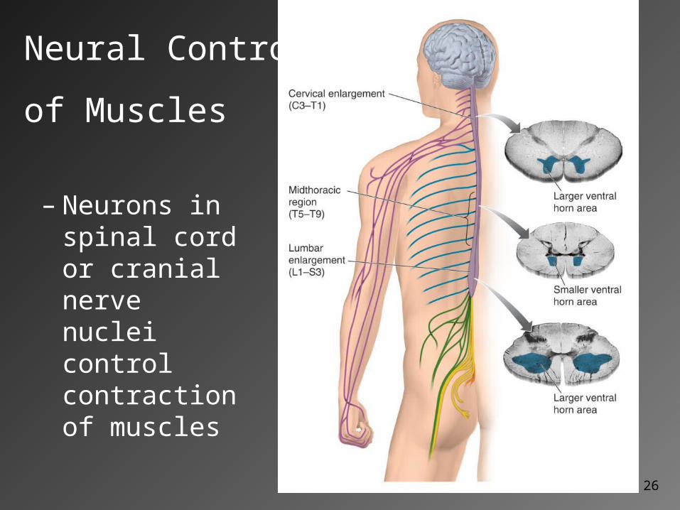
– Neurons in spinal cord or cranial nerve nuclei control contraction of muscles
Neural Control
of Muscles

Action Potential
1. Threshold is met.
2. Brief hyperpolarization.
Action Potential

28
The Control of Muscle Contractions
• A single action potential may be sufficient to produce contraction (twitch contraction)
• Varying amounts of force may be supplied by:– varying the firing rate of alpha motor neurons.
– recruitment (activating more motor units as more load is placed on a muscle).

Twitch Contraction
• Includes:– 1. Latent Period– 2. Contractile Phase– 3. Relaxation Phase

Wave Summation
• Muscle Twitch

31
Control of Alpha Motor Neurons
• Alpha motor neurons receive input from:– Muscle spindles
– Golgi tendon organs
– Brainstem and motor cortex neurons
– Spinal interneurons

Muscle Disorders
• Muscle Atrophy: shrinkage of muscles due to lack of use.
• Muscle Spasms: uncontrolled tetanic contractions of a muscle group
• Muscle Cramp: painful muscle spasm usually due to ion and water imbalance.
• Rigor Mortis: no ATP so myosin head doesn’t release from actin. Role of ATP

Muscle Disorders
• Muscular Dystrophy: lack protein dystrophin which holds muscle fibers together. – Death usually occurs ~21 from
cardiac/respiratory muscle degeneration
• Myasthenia Graves: immune system attacks at neuromuscular junction nerve can’t stimulate muscle contraction– Death usually results from respiratory distress